Skip to content

High-quality research proteins.

  • 1-800-805-6531

  • Mon - Sat 8.00-18.00, Sun - Closed

  • info@prospecbio.com

  • PO Box 6591 East Brunswick NJ 08816

  • Products
    • Cytokines
    • Growth factors
    • Chemokines
    • Cd antigens
    • Neurotrophins
    • Hormones
    • Enzymes
    • Viral antigens
    • Recombinant proteins
    • Natural proteins
    • Monoclonal antibodies
    • Polyclonal antibodies
    • Thermal Packaging
  • About
    • Company
    • Founder
    • Contribution
  • Customers
    • Eastern Asia
    • Eastern Europe
    • North America
    • South America
    • South East Asia
    • Southern Asia
    • Western Europe
  • Ordering
    • Ordering Method
    • Ordering - Credit Card
    • Ordering - PO
    • Shipping Fees
    • FAQs
  • Services
    • DNA Cloning
    • Protein Expression
    • Monoclonal Antibody
    • Fermentation
    • Technology License
  • Contact us
Log in

Country/region

  • USD $ | Albania
  • USD $ | Andorra
  • USD $ | Argentina
  • USD $ | Armenia
  • USD $ | Australia
  • USD $ | Austria
  • USD $ | Azerbaijan
  • USD $ | Belarus
  • USD $ | Belgium
  • USD $ | Bolivia
  • USD $ | Brazil
  • USD $ | Bulgaria
  • USD $ | Cambodia
  • USD $ | Canada
  • USD $ | Chile
  • USD $ | China
  • USD $ | Colombia
  • USD $ | Costa Rica
  • USD $ | Croatia
  • USD $ | Cyprus
  • USD $ | Czechia
  • USD $ | Denmark
  • USD $ | Estonia
  • USD $ | Finland
  • USD $ | France
  • USD $ | Georgia
  • USD $ | Germany
  • USD $ | Greece
  • USD $ | Hong Kong SAR
  • USD $ | Hungary
  • USD $ | Iceland
  • USD $ | India
  • USD $ | Ireland
  • USD $ | Israel
  • USD $ | Italy
  • USD $ | Japan
  • USD $ | Kazakhstan
  • USD $ | Latvia
  • USD $ | Liechtenstein
  • USD $ | Lithuania
  • USD $ | Madagascar
  • USD $ | Malta
  • USD $ | Mexico
  • USD $ | Moldova
  • USD $ | Monaco
  • USD $ | Netherlands
  • USD $ | New Zealand
  • USD $ | North Macedonia
  • USD $ | Norway
  • USD $ | Panama
  • USD $ | Paraguay
  • USD $ | Peru
  • USD $ | Philippines
  • USD $ | Poland
  • USD $ | Portugal
  • USD $ | Romania
  • USD $ | Singapore
  • USD $ | Slovakia
  • USD $ | Slovenia
  • USD $ | South Africa
  • USD $ | South Korea
  • USD $ | Spain
  • USD $ | Sri Lanka
  • USD $ | Sweden
  • USD $ | Switzerland
  • USD $ | Taiwan
  • USD $ | Thailand
  • USD $ | Türkiye
  • USD $ | Ukraine
  • USD $ | United Kingdom
  • USD $ | United States
  • USD $ | Uruguay
  • USD $ | Vatican City
  • USD $ | Venezuela
  • USD $ | Vietnam
  • Facebook
  • Instagram
logoProSpec - Recombinant Protein Manufacturer for academic and R&D industry
Become a distributor
Account Log in
Wishlist
Cart Cart(0)
  • Products
    +
    • Cytokines
    • Growth factors
    • Chemokines
    • Cd antigens
    • Neurotrophins
    • Hormones
    • Enzymes
    • Viral antigens
    • Recombinant proteins
    • Natural proteins
    • Monoclonal antibodies
    • Polyclonal antibodies
    • Thermal Packaging
  • About
    +
    • Company
    • Founder
    • Contribution
  • Customers
    +
    • Eastern Asia
    • Eastern Europe
    • North America
    • South America
    • South East Asia
    • Southern Asia
    • Western Europe
  • Ordering
    +
    • Ordering Method
    • Ordering - Credit Card
    • Ordering - PO
    • Shipping Fees
    • FAQs
  • Services
    +
    • DNA Cloning
    • Protein Expression
    • Monoclonal Antibody
    • Fermentation
    • Technology License
  • Contact us

Item added to your cart

View cart
View All
  • Cytokines
  • Growth factors
  • Chemokines
  • Cd antigens
  • Neurotrophins
  • Hormones
  • Enzymes
  • Viral antigens
  • Recombinant proteins
  • Natural proteins
  • Monoclonal antibodies
  • Polyclonal antibodies
  • Thermal Packaging
  • Tumor Necrosis Factor

    Tumor Necrosis Factor

    View All
  • 4-1BB

    4-1BB

    View All
  • Adiponectin

    Adiponectin

    View All
  • AIF1

    AIF1

    View All
  • Other Cytokines

    Other Cytokines

    View All
  • AITRL

    AITRL

    View All
  • Angiopoietin

    Angiopoietin

    View All
  • Apolipoprotein

    Apolipoprotein

    View All
  • B-Cell Activating Factor

    B-Cell Activating Factor

    View All
  • Beta Defensin

    Beta Defensin

    View All
  • Bone Morphogenetic Protein

    Bone Morphogenetic Protein

    View All
  • B type Natriuretic Peptide

    B type Natriuretic Peptide

    View All
  • BST

    BST

    View All
  • Betacellulin

    Betacellulin

    View All
  • Cardiotrophin

    Cardiotrophin

    View All
  • Activin

    Activin

    View All
  • Fibroblast Growth Factor

    Fibroblast Growth Factor

    View All
  • Other Growth Factors

    Other Growth Factors

    View All
  • CSF

    CSF

    View All
  • CTGF

    CTGF

    View All
  • IGFBP

    IGFBP

    View All
  • VEGF Protein

    VEGF Protein

    View All
  • EGF

    EGF

    View All
  • Epigen

    Epigen

    View All
  • Erythropoietin

    Erythropoietin

    View All
  • Insulin-Like Growth Factor

    Insulin-Like Growth Factor

    View All
  • Growth Hormone

    Growth Hormone

    View All
  • HDGF

    HDGF

    View All
  • Hepatocyte Growth Factor

    Hepatocyte Growth Factor

    View All
  • Insulin

    Insulin

    View All
  • BCA-1/ BLC (CXCL13)

    BCA-1/ BLC (CXCL13)

    View All
  • BRAK (CXCL14)

    BRAK (CXCL14)

    View All
  • C-10 (CCL6)

    C-10 (CCL6)

    View All
  • Other Chemokines

    Other Chemokines

    View All
  • MEC (CCL28)

    MEC (CCL28)

    View All
  • LD78-beta (CCL3L1)

    LD78-beta (CCL3L1)

    View All
  • CTACK (CCL27)

    CTACK (CCL27)

    View All
  • CXCL16

    CXCL16

    View All
  • CXCL17

    CXCL17

    View All
  • Platelet Factor-4 (CXCL4)

    Platelet Factor-4 (CXCL4)

    View All
  • ENA-78 (CXCL5)

    ENA-78 (CXCL5)

    View All
  • Eotaxin (CCL11,24,26)

    Eotaxin (CCL11,24,26)

    View All
  • Exodus-2 (CCL21)

    Exodus-2 (CCL21)

    View All
  • Fractalkine (CX3CL1)

    Fractalkine (CX3CL1)

    View All
  • CXCL6

    CXCL6

    View All
  • Other CD Antigens

    Other CD Antigens

    View All
  • CD14

    CD14

    View All
  • CD164

    CD164

    View All
  • CD1

    CD1

    View All
  • CD2

    CD2

    View All
  • CD200

    CD200

    View All
  • CD207

    CD207

    View All
  • CD226

    CD226

    View All
  • CD23

    CD23

    View All
  • CD244

    CD244

    View All
  • CD247

    CD247

    View All
  • CD27

    CD27

    View All
  • CD274

    CD274

    View All
  • CD300

    CD300

    View All
  • CD33

    CD33

    View All
  • Other Neurotrophins

    Other Neurotrophins

    View All
  • Beta-NGF

    Beta-NGF

    View All
  • BDNF

    BDNF

    View All
  • CDNF

    CDNF

    View All
  • CNTF

    CNTF

    View All
  • GDNF

    GDNF

    View All
  • Glia Maturation Factor

    Glia Maturation Factor

    View All
  • MANF

    MANF

    View All
  • Midkine

    Midkine

    View All
  • Neuroglobin

    Neuroglobin

    View All
  • Neurotrophic factors

    Neurotrophic factors

    View All
  • Neuregulin

    Neuregulin

    View All
  • Neuropilin

    Neuropilin

    View All
  • Pigment Epithelium-Derived Factor

    Pigment Epithelium-Derived Factor

    View All
  • Pleiotrophin

    Pleiotrophin

    View All
  • Peptide Hormones

    Peptide Hormones

    View All
  • Other Hormones

    Other Hormones

    View All
  • HCG

    HCG

    View All
  • Endothelin

    Endothelin

    View All
  • Exendin

    Exendin

    View All
  • FSH

    FSH

    View All
  • Glucagon

    Glucagon

    View All
  • GHRP

    GHRP

    View All
  • GLP

    GLP

    View All
  • Thyrostimulin

    Thyrostimulin

    View All
  • Inhibin A

    Inhibin A

    View All
  • LHRH

    LHRH

    View All
  • Melanotan

    Melanotan

    View All
  • Procalcitonin

    Procalcitonin

    View All
  • PTH

    PTH

    View All
  • Peroxiredoxin

    Peroxiredoxin

    View All
  • Peptidase

    Peptidase

    View All
  • Synthetase

    Synthetase

    View All
  • Transferase

    Transferase

    View All
  • Hydrolase

    Hydrolase

    View All
  • Dehydrogenase

    Dehydrogenase

    View All
  • ACE2

    ACE2

    View All
  • Esterase

    Esterase

    View All
  • Protein Kinases

    Protein Kinases

    View All
  • Other Enzymes

    Other Enzymes

    View All
  • Phosphatase

    Phosphatase

    View All
  • Deaminase

    Deaminase

    View All
  • Oxygenase

    Oxygenase

    View All
  • Adenylate Kinase

    Adenylate Kinase

    View All
  • Lyase

    Lyase

    View All
  • Chagas

    Chagas

    View All
  • SARS

    SARS

    View All
  • Influenza

    Influenza

    View All
  • ASFV

    ASFV

    View All
  • Astrovirus

    Astrovirus

    View All
  • Borrelia

    Borrelia

    View All
  • Helicobacter Pylori

    Helicobacter Pylori

    View All
  • Chikungunya

    Chikungunya

    View All
  • Chlamydia

    Chlamydia

    View All
  • Cytomegalo

    Cytomegalo

    View All
  • Dengue

    Dengue

    View All
  • EBV

    EBV

    View All
  • Ebola

    Ebola

    View All
  • Feline Leukemia Virus

    Feline Leukemia Virus

    View All
  • Hepatitis A

    Hepatitis A

    View All
  • Other

    Other

    View All
  • Actin

    Actin

    View All
  • ADAM

    ADAM

    View All
  • Complement Component

    Complement Component

    View All
  • Ag85

    Ag85

    View All
  • Eukaryotic Translation Initiation Factor

    Eukaryotic Translation Initiation Factor

    View All
  • Anterior Gradient Protein

    Anterior Gradient Protein

    View All
  • Heat Shock Protein

    Heat Shock Protein

    View All
  • Alpha-2-HS-Glycoprotein

    Alpha-2-HS-Glycoprotein

    View All
  • Hemoglobin

    Hemoglobin

    View All
  • Allergy

    Allergy

    View All
  • Anaplasma

    Anaplasma

    View All
  • Angiogenin

    Angiogenin

    View All
  • Ankyrin Repeat Domain

    Ankyrin Repeat Domain

    View All
  • Annexin

    Annexin

    View All
  • Other Natural Proteins

    Other Natural Proteins

    View All
  • Aprotinin

    Aprotinin

    View All
  • Transferrin

    Transferrin

    View All
  • Avidin

    Avidin

    View All
  • Anti Coagulation Factors

    Anti Coagulation Factors

    View All
  • Natural Albumin

    Natural Albumin

    View All
  • Natural Coagulation Factors

    Natural Coagulation Factors

    View All
  • Fibronectin

    Fibronectin

    View All
  • Thrombin

    Thrombin

    View All
  • Anti Human Enzyme

    Anti Human Enzyme

    View All
  • Other Monoclonal Antibodies

    Other Monoclonal Antibodies

    View All
  • Anti Human Cytokine

    Anti Human Cytokine

    View All
  • Anti Human Heat Shock Protein

    Anti Human Heat Shock Protein

    View All
  • Anti Mouse Lymphocyte

    Anti Mouse Lymphocyte

    View All
  • Anti Human Chemokine

    Anti Human Chemokine

    View All
  • Anti Human Lymphocyte

    Anti Human Lymphocyte

    View All
  • Anti Viral Monoclonal

    Anti Viral Monoclonal

    View All
  • Anti Mouse Cytokine

    Anti Mouse Cytokine

    View All
  • Anti-GST Monoclonal

    Anti-GST Monoclonal

    View All
  • Other Polyclonal Antibodies

    Other Polyclonal Antibodies

    View All
  • Anti Viral

    Anti Viral

    View All
  • Anti-GST Monoclonal

    Anti-GST Monoclonal

    View All
Back

Search results

1000 results found for “Bartonella H.”

Name

Description

Product #

Price

Quantity

Shipping Method

  • View Data Sheet

    Name :

    HEV ORF2 (633-659 a.a.)

    Description:

    Hepatitis E Virus ORF2 (633-659 a.a.) Recombinant

    Product # :

    HEV-271

    Price :

    Quantity :

    Shipping Method :

    Ice Icon

    Shipped with Ice Packs

    Add To Cart

    More Info

    • description
    • formulation
    • purity
    • More Info

    Description

    The E.coli derived HEV protein genotype-2 is fused with beta-galactosidase at the N-Terminus and contains the HEV immunodominant ORF2 633-659 a.a.

    Formulation

    20mM Tris-HCl pH-8, 8M urea and 10mM B-ME.

    Purity

    HEV ORF2 Protein is >95% pure as determined by 10% PAGE (coomassie staining).

    More Info

    • Introduction

      Hepatitis E virus (HEV), the major etiologic agent of enterically transmitted non-A, non-B hepatitis worldwide, is a spherical, non-enveloped, single stranded RNA virus that is approximately 32 to 34 nm in diameter. HEV belongs to a genus of HEV-like viruses (unassigned genus). HEV has a single-stranded polyadenylated RNA genome of approximately 8 kb. Based on its physicochemical properties it is presumed to be a calici-like virus.

    • Stability

      HEV ORF2 protein although stable at 4°C for 1 week, should be stored below -18°C. Please prevent freeze thaw cycles.

    • Applications

      HEV ORF2 antigen is suitable for ELISA and Western blots, excellent antigen for detection of HEV with minimal specificity problems.

    • Specificity

      Immunoreactive with sera HEV-infected individuals.

    • Purification Method

      HEV ORF2 protein was purified by proprietary chromatographic technique.

    ProSpec's products are furnished for LABORATORY RESEARCH USE ONLY. The product may not be used as drugs, agricultural or pesticidal products, food additives or household chemicals.

    Hev Orf2 633 659
  • View Data Sheet

    Name :

    Dengue Polyvalent ELISA

    Description:

    Polyvalent Dengue Antigen-I for ELISA Recombinant

    Product # :

    DEN-027

    Price :

    Quantity :

    Shipping Method :

    Ice Icon

    Shipped with Ice Packs

    Add To Cart

    More Info

    • description
    • source
    • formulation
    • purity
    • More Info

    Description

    Polyvalent dengue antigens are 18kDa proteins containing subtype 1,2,3 and 4 and each of them has equal amount protein. They are a new group of antigens specifically designed for ELISA assay. All the recombinant peptides are fused with a 6xHis Tag. Polyvalent dengue is purified by proprietary chromatographic technique.

    Source

    Escherichia Coli.

    Formulation

    Phosphate buffered saline, pH-9.4 and 25mM K₂CO₃.

    Purity

    Protein is >95% pure as determined by 10% PAGE (coomassie staining).

    More Info

    • Introduction

      Caused by one of four closely related virus serotypes of the genus Flavivirus, family Flaviviridae, each serotype is sufficiently different that there is no cross-protection and epidemics caused by multiple serotypes (hyperendemicity) can occur. In cell culture experiments and mice Morpholino antisense oligos have shown specific activity against Dengue virus.

    • Physical Appearance

      Sterile filtered colorless solution.

    • Stability

      Polyvalent dengue Recombinant although stable at 4°C for 1 week, should be stored below -18°C. Please prevent freeze thaw cycles.

    ProSpec's products are furnished for LABORATORY RESEARCH USE ONLY. The product may not be used as drugs, agricultural or pesticidal products, food additives or household chemicals.

    Dengue Polyvalent Elisa
  • View Data Sheet

    Name :

    HSV-2 VP13/14

    Description:

    Herpes Simplex Virus-2 VP13/14 Recombinant

    Product # :

    HSV-227

    Price :

    Quantity :

    Shipping Method :

    Ice Icon

    Shipped with Ice Packs

    Add To Cart

    More Info

    • description
    • source
    • formulation
    • purity
    • More Info

    Description

    The E.Coli derived HSV-2 VP13/14 recombinant protein, 1-280 amino acids, is fused to a Six histidine tag at C-terminus and has a MW of 31kDa (pI 6.77).

    Source

    Escherichia Coli.

    Formulation

    10 mM Phosphate buffer pH 7.6; 75 mM NaCl.

    Purity

    Protein is >90% pure as determined by SDS PAGE.

    More Info

    • Introduction

      Entry of HSV into the host cell involves interactions of several viral glycoproteins with cell surface receptors. The virus particle is covered by an envelope which, when bound to specific receptors on the cell surface, will fuse with the cell membrane and create an opening, or pore, through which the virus enters the host cell. The sequential stages of HSV entry are analagous to those of other viruses. At first, complementary receptors on the virus and cell surface bring the two membranes into proximity. In an intermediate state, the two membranes begin to merge, forming a hemifusion state. Finally, a stable entry pore is formed through which the viral envelope contents are introduced to the host cell.

    • Physical Appearance

      Sterile Filtered clear solution.

    • Stability

      HSV-2 VP13/14 although stable at 4°C for 1 week, should be stored below -18°C. Please prevent freeze thaw cycles.

    • Applications

      ELISA, WB, Flow-Through.

    ProSpec's products are furnished for LABORATORY RESEARCH USE ONLY. The product may not be used as drugs, agricultural or pesticidal products, food additives or household chemicals.

    Hsv 2 Vp13 14
  • View Data Sheet

    Name :

    HTLV-1 gp21

    Description:

    HTLV-1 gp21 Recombinant

    Product # :

    HIV-141

    Price :

    Quantity :

    Shipping Method :

    Ice Icon

    Shipped with Ice Packs

    Add To Cart

    More Info

    • description
    • formulation
    • purity
    • More Info

    Description

    The E.Coli derived recombinant protein contains the gp21 immunodominant region, 1-152 amino acids, the MW is 17.9 kDa. Fused with a His tag at C-Terminus.

    Formulation

    1mg/ml solution in PBS

    Purity

    Protein is > 90% pure as determined by SDS-PAGE.

    More Info

    • Introduction

      Human T-lymphotropic virus (HTLV) is a human, single-stranded RNA retrovirus that causes T-cell leukemia and T-cell lymphoma. The virus activates a subset of T-helper cellscalled Th1cells. The result is a proliferation of Th1 cells and overproduction of Th1 related cytokines (mainly IFN-gamma and TNF-alpha). Feedback mechanisms of these cytokines cause a suppression of the Th2 lymphocytes and a reduction of Th2 cytokine production (mainly IL-4, IL-5, IL-10 and IL-13). The end result is a reduction in the ability of the infected host to mount an adequate immune response to invading organisms that require a predominantly Th2 dependant response (these include parasitic infections and production of mucosal and humoral antibodies).

    • Stability

      HTLV-1 gp21 although stable at 4°C for 1 week, should be stored below -18°C. Please prevent freeze thaw cycles.

    • Purification Method

      HTLV-1 gp21 was purified by proprietary chromatographic technique.

    ProSpec's products are furnished for LABORATORY RESEARCH USE ONLY. The product may not be used as drugs, agricultural or pesticidal products, food additives or household chemicals.

    Htlv 1 Gp21
  • View Data Sheet

    Name :

    OmpA S.Enteritidis

    Description:

    Salmonella Enteritidis Outer Membrane Protein-A Recombinant

    Outer Membrane Protein-A, OmpA.

    Product # :

    PRO-1918

    Price :

    Quantity :

    Shipping Method :

    Ice Icon

    Shipped with Ice Packs

    Add To Cart

    More Info

    • description
    • source
    • formulation
    • purity
    • More Info

    Description

    The Recombinant Salmonella Enteritidis Outer Membrane Protein A, E.Coli derived, 330 amino acids, contains the ompA immunodominant regions. The protein is fused to a His tag at C-terminal and purified by standard chromatography techniques.

    Source

    Escherichia Coli.

    Formulation

    PBS and 25MmM Arginine.

    Purity

    Protein is >95% pure as determined by 10% PAGE (coomassie staining).

    More Info

    • Introduction

      The OmpA protein is one of the main outer-membrane proteins of a large array of Gram-negative bacteria such as A.salmonicida, Shigella dysenteriae and E.coli.OmpA’s major physiological functions include maintenance of the structural integrity and morphology of the cells and porin activity, as well as a role in conjugation and bacteriophage binding.Achromogenic atypical Aeromonas salmonicida is the causative agent of goldfish ulcer disease.Virulence of this bacterium is associated with the production of a paracrystalline outer membrane A-layer protein.The species specific structural gene for the monomeric form of A-protein was cloned into a pET-3d plasmid in order to express and produce a recombinant form of the protein in E.coli BL21(DE3). The induced protein was isolated from inclusion bodies by a simple solubilization-renaturation procedure and purified by ion exchange chromatography on Q-Sepharose to over 95% pure monomeric protein.Recombinant A-protein was compared by biochemical, immunological and molecular methods with the A-protein isolated from atypical A.salmonicida bacterial cells by the glycine and the membrane extraction methods.

    • Synonyms

      Outer Membrane Protein-A, OmpA.

    • Physical Appearance

      Sterile Filtered clear solution.

    • Stability

      OmpA S.Enteritidis Recombinant although stable at 4°C for 1 week, should be stored below -18°C. Please prevent freeze thaw cycles.

    • Applications

      Immunoassay. Outer membrane protein A (ompA) of S. Enteritidis is a protein directly exposing to outsides of this organism, In hens, the production of antibodies against outer membrane protein A (ompA) during the infection has been demonstrated by inoculating both the complete bacterium and expressed protein produced from ompA DNA vaccine. Vaccination by ompA protein to hens is a poteintail tool to control S. enteritidis contaminated eggs into market, and prevent human foodborne disease from eggs.

    ProSpec's products are furnished for LABORATORY RESEARCH USE ONLY. The product may not be used as drugs, agricultural or pesticidal products, food additives or household chemicals.

    Ompa Senteritidis
  • View Data Sheet

    Name :

    CHIKV E2

    Description:

    Chikungunya E2 Recombinant

    Product # :

    CHI-003

    Price :

    Quantity :

    Shipping Method :

    Ice Icon

    Shipped with Ice Packs

    Add To Cart

    More Info

    • description
    • source
    • formulation
    • purity
    • More Info

    Description

    Recombinant Chikungunya E2 produced in E.coli having a molecular weight of 38kDa (migrates at 38-40kDa on 10% SDS-PAGE).

    Source

    Escherichia Coli.

    Formulation

    Sterile Filtered solution containing PBS and 25mM K2CO3.

    Purity

    Protein is >95% pure as determined by SDS-PAGE.

    More Info

    • Introduction

      Chikungunya is an infection caused by the chikungunya virus which is passed to humans by two species of mosquito of the genus Aedes: A. albopictus and A. aegypti. Animal reservoirs of the virus include monkeys, birds, cattle, and rodents. The features of the disease are a sudden onset of fever 2-4 days after exposure. The fever typically lasts 2-7 days, while the associated joint pains usually last weeks or months but sometimes years. The mortality rate is a little less than 1 in 1,000. The disease has occurred in outbreaks in Asia, Europe and the Americas since 2004. CHIKV is a single-stranded positive-sense RNA genome, 11,800 nts long which encodes 2 open reading frames. The nucleocapsid is tightly enveloped by a host-derived lipid bilayer (envelope) supporting the virus-encoded envelope proteins. 80 glycoprotein spikes are C- terminally anchored within the viral envelope. The structural polyprotein is translated from a viral sub genomic mRNA, while as the 5 structural proteins (capsid, E3, E2, 6K, E1) are translated as a single polyprotein, from which capsid (C) is cleaved off to encapsidate. The envelope polyprotein precursor E3-E2-6K-E1 is translocated to the endoplasmatic reticulum. Polyprotein is processed by host signalases, resulting in E3, E2 & E1 forming viral hetero-trimeric spikes. The viral spikes majorly contains E2 and E1 facilitate cell receptor recognition, cell entry thru pH-dependent endocytosis and support viral budding.

    • Physical Appearance

      Sterile filtered colorless solution.

    • Stability

      CHIKV E2 although stable at 4°C for 1 week, should be stored below -18°C. Please prevent freeze thaw cycles.

    • Applications

      Rapid test and Immunoassay.

    • Purification Method

      Purified by proprietary chromatographic technique.

    ProSpec's products are furnished for LABORATORY RESEARCH USE ONLY. The product may not be used as drugs, agricultural or pesticidal products, food additives or household chemicals.

    Chikv E2
  • View Data Sheet

    Name :

    UCHL1 (1-126) Human

    Description:

    Ubiquitin Carboxyl-Terminal Hydrolase L1 (1-126) Human Recombinant

    PGP 9.5, UCHL1, PGP9.5, PARK5.

    Product # :

    PRO-2823

    Price :

    Quantity :

    Shipping Method :

    Room Temp Icon

    Shipped at Room temp

    Add To Cart

    More Info

    • description
    • source
    • formulation
    • purity
    • More Info

    Description

    The UCHL1 Human is created as a recombinant protein with a 4kda N-terminal fusion of His Tag. The UCHL1 His-Tagged Fusion Protein, produced in E. coli, is a 19kDa protein containing 126 amino acid residues of the UCHL1 Human, 1-126 amino acids.

    Source

    Escherichia Coli.

    Formulation

    Each mg was lyophilized with 1xPBS, 0.4% SDS and 4mM DTT.

    Purity

    Greater than 90% as determined by SDS-PAGE.

    More Info

    • Synonyms

      PGP 9.5, UCHL1, PGP9.5, PARK5.

    • Physical Appearance

      Filtered White lyophilized (freeze-dried) powder.

    • Stability

      Store lyophilized UCHL1 at -20°C. Aliquot the product after reconstitution to avoid repeated freezing/thawing cycles. Reconstituted protein can be stored at 4°C for a limited period of time; it does not show any change after two weeks at 4°C.

    • Solubility

      It is recommended to add deionized water to prepare a working stock solution of approximately 0.5mg/ml and let the lyophilized pellet dissolve completely. Product is not sterile! Please filter the product by an appropriate sterile filter before using it on cell culture.

    • Background

      Human Ubiquitin Carboxyl-Terminal Hydrolase L1 (UCH-L1) is an important enzyme involved in the ubiquitin-proteasome system, which regulates protein degradation and turnover in cells.

      UCHL1 Function:

      UCHL1 mainly removes ubiquitin molecules from proteins, thereby recycling ubiquitin and regulating protein stability. This action is important for controlling various cellular processes and maintaining cellular homeostasis and.

      UCHL1 Structure

      UCHL1 is characterized by a catalytic domain which facilitates its hydrolase activity, allowing it to cleave ubiquitin from substrates.

      UCHL1 Role in Neurodegeneration

      UCHL1 has been implicated in neurodegenerative diseases, such as Parkinson’s and Alzheimer's disease. Its expression levels and activity can affect neuronal survival and function.

      UCH-L1 is also studied in the context of cancer. Altered levels of UCHL1 may affect on tumour progression and response to therapy.

      UCHL1 Biomarker Potential

      UCHL1 is being investigated as a potential biomarker for diseases, especially neurodegenerative disorders due to its involvement in various pathologies.

      UCHL1 Research

      Ongoing studies focus on understanding its role in various cellular contexts, its regulation, and its potential as a therapeutic target.

      In conclusion, UCHL1 is a very important component of cellular regulation, with implications in health and disease.

    ProSpec's products are furnished for LABORATORY RESEARCH USE ONLY. The product may not be used as drugs, agricultural or pesticidal products, food additives or household chemicals.

    Uchl1 Protein
  • View Data Sheet

    Name :

    HB-Pre-sAg Antibody

    Description:

    Hepatitis-B Pre Surface , Mouse antibody

    Product # :

    ANT-774

    Price :

    Quantity :

    Shipping Method :

    Ice Icon

    Shipped with Ice Packs

    Add To Cart

    More Info

    • formulation
    • More Info

    Formulation

    PBS and 0.01% NaAzide.

    More Info

    • Introduction

      HBsAg is the surface antigen of the Hepatitis-B-Virus (HBV). The capsid of a virus has different surface proteins from the rest of the virus. The antigen is a protein that binds specifically on one of these surface proteins. It is commonly referred to as the Australian Antigen.

    • Physical Appearance

      Sterile Filtered solution.

    • Immunogen

      HBsAg purified from human sera pool

    • Ig Subclass

      Mouse IgG1.

    • Clone

      PS26VN

    • Specificity

      HBsAg pre-S antigen

    • Shipping Conditions

      Antibody is shipped as in liquid form with ice packs.

    • Type

      Mouse antibody Monoclonal.

    • Storage Procedures

      For periods up to 1 month store at 4°C, for longer periods of time, store at -20°C. Prevent freeze thaw cycles.

    ProSpec's products are furnished for LABORATORY RESEARCH USE ONLY. The product may not be used as drugs, agricultural or pesticidal products, food additives or household chemicals.

    Hepatitis B Pre Surface Antibody
  • View Data Sheet

    Name :

    HAV VP3

    Description:

    Hepatitis A Virus VP3 Recombinant

    Product # :

    HAV-226

    Price :

    Quantity :

    Shipping Method :

    Ice Icon

    Shipped with Ice Packs

    Add To Cart

    More Info

    • description
    • source
    • formulation
    • purity
    • More Info

    Description

    The E.coli derived a recombinant protein contains the VP3 immunodominant regions, having 245 amino acids and a Mw of 25-28kDa on SDS-PAGE. HAV VP3 protein is fused to a 6xHis--tag at C-terminus & purified by proprietary chromatographic techniques.

    Source

    Escherichia Coli.

    Formulation

    PBS and 50mM Arginine

    Purity

    HAV VP3 protein is >95% pure as determined by 12% PAGE (coomassie staining).

    More Info

    • Introduction

      Forty-two antigenic domains were identified across the hepatitis A virus (HAV) polyprotein by using a set of 237 overlapping 20-mer synthetic peptides spanning the entire HAV polyprotein. Nineteen antigenic domains were found within the structural proteins, and 22 were found within the nonstructural proteins, with 1 domain spanning the junction of VP1 and P2A proteins. Five of these domains were considered immunodominant, as judged by both the breadth and the strength of their immunoreactivity. One domain is located within the VP2 protein at position 57-90 aa. A second domain, located at position 767-842 aa, contains the C-terminal part of the VP1 protein and the entire P2A protein. A third domain, located at position 1403-1456 aa, comprises the C-terminal part of the P2C protein and the N-terminal half of the P3A protein. The fourth domain, located at position 1500-1519 aa, includes almost the entire P3B, and the last domain, located at position 1719-1764 aa, contains the C-terminal region of the P3C protein and the N-terminal region of the P3D protein. Four of the five most immunoreactive domains are derived from small HAV proteins and/or encompass protein cleavage sites separating different HAV proteins.

    • Stability

      HAV VP3 Protein although stable at 4°C for 1 week, should be stored below -18°C. Please prevent freeze thaw cycles.

    ProSpec's products are furnished for LABORATORY RESEARCH USE ONLY. The product may not be used as drugs, agricultural or pesticidal products, food additives or household chemicals.

    Hepatitis A Virus Vp3
  • View Data Sheet

    Name :

    Influenza-A Paired Antibody

    Description:

    Mouse Anti Influenza-A Paired

    Product # :

    ANT-651

    Price :

    Quantity :

    Shipping Method :

    Ice Icon

    Shipped with Ice Packs

    Add To Cart

    More Info

    • description
    • formulation
    • purity
    • More Info

    Description

    Influenza-A gold conjugation antibody and Influenza-A capture antibody are used to develop rapid test for Influenza-A rapid test. Please note that when ordering for example: 100µg antibody we ship 50µg from each of the antibodies (100µg in total).

    Formulation

    * Influenza-A gold conjugation antibody (7.4mg/1ml) in PBS, pH 7.4.
    * Influenza-A capture antibody (6.4mg/1ml) in PBS, pH 7.4.

    Purity

    Greater than 90%.

    More Info

    • Physical Appearance

      2 vials of sterile Filtered clear colorless solution.

    • Stability

      Store at 4°C if entire vial will be used within 2-4 weeks. Store, frozen at -20°C for longer periods of time.

    • Applications

      Lateral flow immunoassay.

    • Type

      Mouse antibody Monoclonal.

    • Purification Method

      Purified monoclonal IgG by protein A chromatography.

    ProSpec's products are furnished for LABORATORY RESEARCH USE ONLY. The product may not be used as drugs, agricultural or pesticidal products, food additives or household chemicals.

    Influenza A Paired Antibody
  • View Data Sheet

    Name :

    HIV-1 gp41, HRP

    Description:

    HIV-1 gp41 Recombinant, HRP labeled

    Product # :

    HIV-118

    Price :

    Quantity :

    Shipping Method :

    Ice Icon

    Shipped with Ice Packs

    Add To Cart

    More Info

    • description
    • source
    • formulation
    • purity
    • More Info

    Description

    HIV-1 gp41 HRP labeledis a non-glycosylated polypeptide chain, containing 288 amino acids (466-753 a.a.) and having a Mw of 32kDa. HIV1 gp41 HRP labeled is fused to an 114kDa beta-galactosidase tag at N-terminus having a total Mw of 146kDa.

    Source

    Escherichia Coli.

    Formulation

    (1mg/ml) 8M urea, 20mM Tris-HCl pH-8.0 and 10mM b-mercaptoethanol.

    Purity

    Greater than 95.0% as determined by SDS-PAGE.

    More Info

    • Introduction

      Human immunodeficiency virus (HIV) is a retrovirusthat can lead to a condition in which the immune systembegins to fail, leading to opportunistic infections. HIV primarily infects vital cells in the humanimmune systemsuch as helper T cells(specifically CD4+ T cells), macrophagesand dendritic cells. HIV infection leads to low levels of CD4+ T cells through three main mechanisms: firstly, direct viral killing of infected cells; secondly, increased rates of apoptosisin infected cells; and thirdly, killing of infected CD4+ T cells by CD8 cytotoxic lymphocytesthat recognize infected cells. When CD4+ T cell numbers decline below a critical level, cell-mediated immunityis lost, and the body becomes progressively more susceptible to opportunistic infections. HIV was classified as a member of the genus Lentivirus, part of the family of Retroviridae. Lentiviruses have many common morphologies and biological properties. Many species are infected by lentiviruses, which are characteristically responsible for long-duration illnesses with a long incubation period. Lentiviruses are transmitted as single-stranded, positive-sense, enveloped RNA viruses. Upon entry of the target cell, the viral RNA genomeis converted to double-stranded DNAby a virally encoded reverse transcriptasethat is present in the virus particle. This viral DNA is then integrated into the cellular DNA by a virally encoded integraseso that the genome can be transcribed. Once the virus has infected the cell, two pathways are possible: either the virus becomes latentand the infected cell continues to function, or the virus becomes active and replicates, and a large number of virus particles are liberated that can then infect other cells.

    • Physical Appearance

      Sterile filtered colorless clear solution.

    • Stability

      HIV-1 horseradish peroxidase although stable at 4°C for 1 week, should be stored below -18°C. Please prevent freeze thaw cycles.

    • Specificity

      Reacts strongly with human HIV positive serum.

    ProSpec's products are furnished for LABORATORY RESEARCH USE ONLY. The product may not be used as drugs, agricultural or pesticidal products, food additives or household chemicals.

    Hiv 1 Gp41 Hrp
  • View Data Sheet

    Name :

    HIV-2 gp32, HRP

    Description:

    HIV-2 gp32 Recombinant, HRP Labeled

    Product # :

    HIV-137

    Price :

    Quantity :

    Shipping Method :

    Ice Icon

    Shipped with Ice Packs

    Add To Cart

    More Info

    • description
    • source
    • formulation
    • purity
    • More Info

    Description

    HIV-2 gp32 HRP Labeled recombinant- contains the full-length sequence of HIV-2 envelope immunodominant regions gp32 having a Mw of 32kDa and fused to a beta-galactosidase at N-terminus.

    Source

    Escherichia Coli.

    Formulation

    0.01M Na2CO3, 10mM EDTA, 14mM beta-ME and 0.02% Sarcosyl.

    Purity

    Greater than 95.0% as determined by SDS-PAGE.

    More Info

    • Introduction

      HIV-1 and HIV-2 appear to package their RNA differently. HIV-1 binds to any appropriate RNA whereas HIV-2 preferentially binds to mRNA which creates the Gag protein itself. This means that HIV-1 is better able to mutate. HIV-2 is transmitted in the same ways as HIV-1: Through exposure to bodily fluids such as blood, semen, tears and vaginal fluids.
      Immunodeficiency develops more slowly with HIV-2.
      HIV-2 is less infectious in the early stages of the virus than with HIV-1.
      The infectiousness of HIV-2 increases as the virus progresses.
      Major differences include reduced pathogenicity of HIV-2 relative to HIV-1, enhanced immune control of HIV-2 infection and often some degree of CD4-independence. Despite considerable sequence and phenotypic differences between HIV-1 and 2 envelopes, structurally they are quite similar. Both membrane-anchored proteins eventually form the 6-helix bundles from the N-terminal and C-terminal regions of the ectodomain, which is common to many viral and cellular fusion proteins and which seems to drive fusion.
      HIV-1 gp41 helical regions can form more stable 6-helix bundles than
      HIV-2 gp41 helical regions however HIV-2 fusion occurs at a lower threshold temperature (25°C), does not require Ca2+ in the medium, is insensitive to treatment of target cells with cytochalasin B, and is not affected by target membrane glycosphingolipid composition.

    • Physical Appearance

      Sterile filtered colorless clear solution.

    • Stability

      HIV-2 gp-32 although stable at room temperature for 3 weeks, should be stored at 4°C.

    ProSpec's products are furnished for LABORATORY RESEARCH USE ONLY. The product may not be used as drugs, agricultural or pesticidal products, food additives or household chemicals.

    Hiv 2 Gp 32 Hrp
  • View Data Sheet

    Name :

    hchA E.Coli

    Description:

    Chaperone Protein hchA E.Coli Recombinant

    Chaperone protein hchA, EcHsp31, Hsp31, hchA, yedU, yzzC, b1967, JW1950.

    Product # :

    HSP-043

    Price :

    Quantity :

    Shipping Method :

    Ice Icon

    Shipped with Ice Packs

    Add To Cart

    More Info

    • description
    • source
    • formulation
    • purity
    • More Info

    Description

    hchA produced in E.Coli is a single, non-glycosylated polypeptide chain containing 303 amino acids (1-283 a.a.) and having a molecular mass of 33.3kDa.hchA is fused to a 20 amino acid His-tag at N-terminus & purified by proprietary chromatographic techniques.

    Source

    Escherichia Coli.

    Formulation

    The hchA contains (1mg/ml) 20mM Tris-HCl buffer (pH8.0), 20% glycerol 0.1M NaCl and 1mM DTT.

    Purity

    Greater than 95.0% as determined by SDS-PAGE.

    More Info

    • Introduction

      Escherichia coli Hsp31 (HchA) is a homodimeric member of the ThiI/DJ-1/PfpI superfamily which combines molecular chaperone and aminopeptidase activities. HchA uses temperature-induced exposure of structured hydrophobic domains to capture and stabilize early unfolding protein intermediates under severe thermal stress.

    • Synonyms

      Chaperone protein hchA, EcHsp31, Hsp31, hchA, yedU, yzzC, b1967, JW1950.

    • Physical Appearance

      Sterile filtered colorless solution.

    • Stability

      hchA E.Coli Recombinant although stable at 4°C for 1 week, should be stored below -18°C. Please prevent freeze thaw cycles.

    • Amino Acid Sequence

      MGSSHHHHHH SSGLVPRGSH MTVQTSKNPQ VDIAEDNAFF PSEYSLSQYT SPVSDLDGVD YPKPYRGKHK ILVIAADERY LPTDNGKLFS TGNHPIETLL PLYHLHAAGF EFEVATISGL MTKFEYWAMP HKDEKVMPFF EQHKSLFRNP KKLADVVASL NADSEYAAIF VPGGHGALIG LPESQDVAAA LQWAIKNDRF VISLCHGPAA FLALRHGDNP LNGYSICAFP DAADKQTPEI GYMPGHLTWY FGEELKKMGM NIINDDITGR VHKDRKLLTG DSPFAANALG KLAAQEMLAA YAG.

    ProSpec's products are furnished for LABORATORY RESEARCH USE ONLY. The product may not be used as drugs, agricultural or pesticidal products, food additives or household chemicals.

    Hcha Ecoli
  • View Data Sheet

    Name :

    HDHD1 Human

    Description:

    Haloacid Dehalogenase-Like Hydrolase Domain Containing 1 Human Recombinant

    Pseudouridine-5'-monophosphatase, 5'-PsiMPase, Haloacid dehalogenase-like hydrolase domain-containing protein 1, Haloacid dehalogenase-like hydrolase domain-containing protein 1A, Protein GS1, HDHD1, DXF68S1E, FAM16AX, GS1, HDHD1A.

    Product # :

    ENZ-028

    Price :

    Quantity :

    Shipping Method :

    Ice Icon

    Shipped with Ice Packs

    Add To Cart

    More Info

    • description
    • source
    • formulation
    • purity
    • More Info

    Description

    HDHD1 Human Recombinant fused with a 20 amino acid His tag at N-terminus produced in E.Coli is a single, non-glycosylated, polypeptide chain containing 248 amino acids (1-228 a.a.) and having a molecular mass of 27.4kDa. The HDHD1 is purified by proprietary chromatographic techniques.

    Source

    Escherichia Coli.

    Formulation

    The HDHD1 solution (1mg/ml) contains 20mM Tris-HCl buffer (pH8.0), 20% glycerol,
    0.1M NaCl and 1mM DTT.

    Purity

    Greater than 95.0% as determined by SDS-PAGE.

    More Info

    • Introduction

      Haloacid dehalogenase-like hydrolase domain-containing protein 1A (HDHD1A) is a member of the adiponutrin family. HDHD1A is interesting because it is an X-linked gene that escapes X-inactivation. HDHD1A is particularly important in the perception of human X chromosome structural organization as well as the mechanism of X-inactivation.

    • Synonyms

      Pseudouridine-5'-monophosphatase, 5'-PsiMPase, Haloacid dehalogenase-like hydrolase domain-containing protein 1, Haloacid dehalogenase-like hydrolase domain-containing protein 1A, Protein GS1, HDHD1, DXF68S1E, FAM16AX, GS1, HDHD1A.

    • Physical Appearance

      Sterile Filtered colorless solution.

    • Stability

      Store at 4°C if entire vial will be used within 2-4 weeks. Store, frozen at -20°C for longer periods of time. For long term storage it is recommended to add a carrier protein (0.1% HSA or BSA).Avoid multiple freeze-thaw cycles.

    • Amino Acid Sequence

      MGSSHHHHHH SSGLVPRGSH MAAPPQPVTH LIFDMDGLLL DTERLYSVVF QEICNRYDKK YSWDVKSLVM GKKALEAAQI IIDVLQLPMS KEELVEESQT KLKEVFPTAA LMPGAEKLII HLRKHGIPFA LATSSGSASF DMKTSRHKEF FSLFSHIVLG DDPEVQHGKP DPDIFLACAK RFSPPPAMEK CLVFEDAPNG VEAALAAGMQ VVMVPDGNLS RDLTTKATLV LNSLQDFQPE LFGLPSYE.

    ProSpec's products are furnished for LABORATORY RESEARCH USE ONLY. The product may not be used as drugs, agricultural or pesticidal products, food additives or household chemicals.

    Hdhd1 Human
  • View Data Sheet

    Name :

    DHFR Mouse

    Description:

    Dihydrofolate Reductase Mouse Recombinant

    Dihydrofolate reductase, DHFR, DHFRP1, AA607882, AI662710, AW555094, 8430436I03Rik.

    Product # :

    ENZ-069

    Price :

    Quantity :

    Shipping Method :

    Ice Icon

    Shipped with Ice Packs

    Add To Cart

    More Info

    • description
    • source
    • formulation
    • purity
    • biological activity
    • More Info

    Description

    DHFR Mouse Recombinant fused with a 20 amino acid His tag at N-terminus produced in E.Coli is a single, non-glycosylated, polypeptide chain containing 207 amino acids (1-187 a.a.) and having a molecular mass of 23.8kDa. The DHFR is purified by proprietary chromatographic techniques.

    Source

    Escherichia Coli.

    Formulation

    The DHFR solution (1 mg/ml) contains 20mM Tris-HCl buffer(pH 8.0), 10% glycerol, 2mM DTT and 0.1M NaCl.

    Purity

    Greater than 95.0% as determined by SDS-PAGE.

    Biological Activity

    Specific activity is > 0.2 units/mg, in which one unit will convert 1.0 umole of 7,8 –dihydrofloate and beta-NADPH to 5,6,7,8-tetrahydrofloate and beta-NADP per min at pH 6.5 at 25C.

    More Info

    • Introduction

      Dihydrofolate reductase (DHFR) is an enzyme that reduces dihydrofolic acid to tetrahydrofolic acid, with NADPH as electron donor that can be converted to the kinds of tetrahydrofolate cofactors applied in 1-carbon transfer chemistry. DHFR converts dihydrofolate into tetrahydrofolate, which is a methyl group shuttle required for the de novo synthesis of purines, thymidylic acid, and specific amino acids. Even though the functional DHFR gene is mapped to chromosome 5, numerous intronless processed pseudogenes or dihydrofolate reductase-like genes are identified on separate chromosomes. DHFR deficiency is associated with megaloblastic anemia. DHFR knockdown plays a role in the anticancer activity of 2-hydroxyoleic acid. DHFR gene insertion/deletion polymorphism is linked to variation in serum and red blood cell folate concentrations in women.

    • Synonyms

      Dihydrofolate reductase, DHFR, DHFRP1, AA607882, AI662710, AW555094, 8430436I03Rik.

    • Physical Appearance

      Sterile Filtered colorless solution.

    • Stability

      Store at 4°C if entire vial will be used within 2-4 weeks. Store, frozen at -20°C for longer periods of time. For long term storage it is recommended to add a carrier protein (0.1% HSA or BSA).Avoid multiple freeze-thaw cycles.

    • Amino Acid Sequence

      MGSSHHHHHH SSGLVPRGSH MVRPLNCIVA VSQNMGIGKN GDLPWPPLRN EFKYFQRMTT TSSVEGKQNL VIMGRKTWFS IPEKNRPLKD RINIVLSREL KEPPRGAHFL AKSLDDALRL IEQPELASKV DMVWIVGGSS VYQEAMNQPG HLRLFVTRIM QEFESDTFFP EIDLGKYKLL PEYPGVLSEV QEEKGIKYKF EVYEKKD.

    ProSpec's products are furnished for LABORATORY RESEARCH USE ONLY. The product may not be used as drugs, agricultural or pesticidal products, food additives or household chemicals.

    Dhfr Mouse
  • View Data Sheet

    Name :

    ST6GAL1 Human

    Description:

    ST6 Beta-Galactosamide Alpha-2,6-Sialyltranferase 1 Human Recombinant

    ST6 Beta-Galactosamide Alpha-2,6-Sialyltranferase 1, ST6Gal I, SIAT1, CMP-N-Acetylneuraminate-Beta-Galactosamide-Alpha-2,6-Sialyltransferase 1, B-Cell Antigen CD75, Alpha 2,6-ST 1, EC 2.4.99.1, ST6GalI, CMP-N-Acetylneuraminate Beta-Galactosamide Alpha-2,6-Sialyltransferase, Sialyltransferase 1 (Beta-Galactoside Alpha-2,6-Sialyltransferase), Sialyltransferase 1 (Beta-Galactoside Alpha-2,6-Sialytransferase),Beta-Galactoside Alpha-2,6-Sialyltransferase 1, Sialyltransferase 1, ST6N, ST6GAL1.

    Product # :

    ENZ-893

    Price :

    Quantity :

    Shipping Method :

    Ice Icon

    Shipped with Ice Packs

    Add To Cart

    More Info

    • description
    • source
    • formulation
    • purity
    • More Info

    Description

    ST6GAL1 Human Recombinant produced in E.Coli is a single, non-glycosylated polypeptide chain containing 403 amino acids (27-406 a.a) and having a molecular mass of 46kDa.ST6GAL1 is fused to a 23 amino acid His-tag at N-terminus & purified by proprietary chromatographic techniques.

    Source

    Escherichia Coli.

    Formulation

    ST6GAL1 protein solution (1mg/ml) containing 20mM Tris-HCl (pH 8.0) and 10% glycerol.

    Purity

    Greater than 90.0% as determined by SDS-PAGE.

    More Info

    • Introduction

      ST6GAL1 also known as ST6 Beta-Galactosamide Alpha-2,6-Sialyltranferase 1, is part of the glycosyltransferase family 29. ST6GAL1 is a type II membrane protein which catalyzes the transfer of sialic acid from CMP-sialic acid to galactose-containing substrates. Furthermore, ST6GAL1 is normally found in the Golgi however it can be proteolytically processed to a soluble form, ST6GAL1 is also involved in the generation of the cell-surface carbohydrate determinants as well differentiation antigens HB-6, CD75, and CD76.

    • Synonyms

      ST6 Beta-Galactosamide Alpha-2,6-Sialyltranferase 1, ST6Gal I, SIAT1, CMP-N-Acetylneuraminate-Beta-Galactosamide-Alpha-2,6-Sialyltransferase 1, B-Cell Antigen CD75, Alpha 2,6-ST 1, EC 2.4.99.1, ST6GalI, CMP-N-Acetylneuraminate Beta-Galactosamide Alpha-2,6-Sialyltransferase, Sialyltransferase 1 (Beta-Galactoside Alpha-2,6-Sialyltransferase), Sialyltransferase 1 (Beta-Galactoside Alpha-2,6-Sialytransferase),Beta-Galactoside Alpha-2,6-Sialyltransferase 1, Sialyltransferase 1, ST6N, ST6GAL1.

    • Physical Appearance

      Sterile Filtered colorless solution.

    • Stability

      Store at 4°C if entire vial will be used within 2-4 weeks. Store, frozen at -20°C for longer periods of time. For long term storage it is recommended to add a carrier protein (0.1% HSA or BSA).Avoid multiple freeze-thaw cycles.

    • Amino Acid Sequence

      MGSSHHHHHH SSGLVPRGSH MGSKEKKKGS YYDSFKLQTK EFQVLKSLGK LAMGSDSQSV SSSSTQDPHR GRQTLGSLRG LAKAKPEASF QVWNKDSSSK NLIPRLQKIW KNYLSMNKYK VSYKGPGPGI KFSAEALRCH LRDHVNVSMV EVTDFPFNTS EWEGYLPKES IRTKAGPWGR CAVVSSAGSL KSSQLGREID DHDAVLRFNG APTANFQQDV GTKTTIRLMN SQLVTTEKRF LKDSLYNEGI LIVWDPSVYH SDIPKWYQNP DYNFFNNYKT YRKLHPNQPF YILKPQMPWE LWDILQEISP EEIQPNPPSS GMLGIIIMMT LCDQVDIYEF LPSKRKTDVC YYYQKFFDSA CTMGAYHPLL YEKNLVKHLN QGTDEDIYLL GKATLPGFRT IHC

    ProSpec's products are furnished for LABORATORY RESEARCH USE ONLY. The product may not be used as drugs, agricultural or pesticidal products, food additives or household chemicals.

    St6Gal1 Human
  • View Data Sheet

    Name :

    TFF2 Human, His

    Description:

    Trefoil Factor-2 Human Recombinant, His Tag

    TFF-2, Spasmolytic polypeptide, Spasmolysin, SML1, Trefoil factor 2, SP, TFF2.

    Product # :

    CYT-611

    Price :

    Quantity :

    Shipping Method :

    Room Temp Icon

    Shipped at Room temp

    Add To Cart

    More Info

    • description
    • source
    • formulation
    • purity
    • More Info

    Description

    TFF-2 Human Recombinant produced in E.Coli is a single, non-glycosylated, polypeptide chain containing 116 amino acids (24-129) which includes a 10 amino acid His Tag fused at N-terminus and having a total molecular mass of 13.2 kDa. TFF2 Human Recombinant is purified by proprietary chromatographic techniques.

    Source

    Escherichia Coli.

    Formulation

    The TFF2 protein was lyophilized from 0.4μm filtered solution at a concentration of 0.5mg/ml containing 20mM Tris pH-7.5, and 20mM NaCl.

    Purity

    Greater than 95.0% as determined by SDS-PAGE.

    More Info

    • Introduction

      Proteins of the TFF family are characterized by obtaining a minimum of 1 copy of the trefoil motif, a 40-amino acid domain that contains 3 conserved disulfides. Trefoil Factors are stable secretory proteins expressed in gastrointestinal mucosa which protect the mucosa from insults, stabilize the mucus layer and affect healing of the epithelium.TFF2 inhibits gastric acid motility & secretion. TFF2 stabilizes glycoproteins in the mucus gel through interactions with carbohydrate side chains.

    • Synonyms

      TFF-2, Spasmolytic polypeptide, Spasmolysin, SML1, Trefoil factor 2, SP, TFF2.

    • Physical Appearance

      Filtered White lyophilized (freeze-dried) powder.

    • Stability

      Lyophilized TFF2 although stable at room temperature for 3 weeks, should be stored desiccated below -18°C. Upon reconstitution TFF2 should be stored at 4°C between 2-7 days and for future use below -18°C.For long term storage it is recommended to add a carrier protein (0.1% HSA or BSA).Please prevent freeze-thaw cycles.

    • Solubility

      It is recommended to add deionized water to prepare a working stock solution of approximately 0.5mg/ml and let the lyophilized pellet dissolve completely. Product is not sterile! Please filter the product by an appropriate sterile filter before using it in the cell culture.

    • Amino Acid Sequence

      MKHHHHHHAS EKPSPCQCSR LSPHNRTNCG FPGITSDQCF DNGCCFDSSV TGVPWCFHPL PKQESDQCVM EVSDRRNCGY PGISPEECAS RKCCFSNFIF EVPWCFFPKSVEDCHY.

    ProSpec's products are furnished for LABORATORY RESEARCH USE ONLY. The product may not be used as drugs, agricultural or pesticidal products, food additives or household chemicals.

    Tff2 Human His
  • View Data Sheet

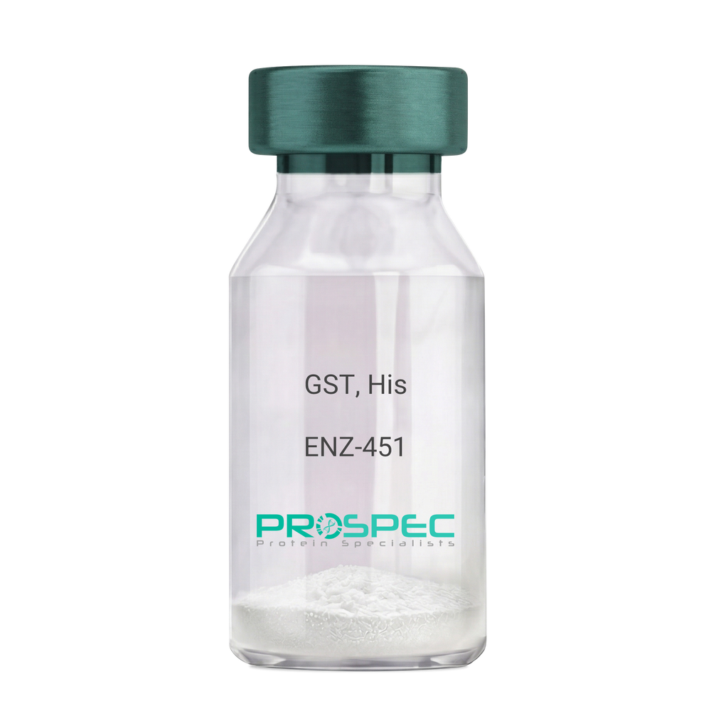
    Name :

    GST, His

    Description:

    Glutathione S-Transferase Recombinant, His Tag

    Glutathione S-Transferase, GST, Glutathione S-transferase class-mu 28 kDa isozyme, GST 28, EC 2.5.1.18, Sj28GST, Sj28 antigen.

    Product # :

    ENZ-451

    Price :

    Quantity :

    Shipping Method :

    Ice Icon

    Shipped with Ice Packs

    Add To Cart

    More Info

    • description
    • source
    • formulation
    • purity
    • biological activity
    • More Info

    Description

    Recombinant Schistosoma japonicum GST full length protein contains a total of 244 amino acids (1-218 a.a.) expressed in E.coli, having a molecular mass of 28.3kDa. The GST protein is fused to a 20 amino acids His-Tag at N-terminus. The GST protein is purified by proprietary chromatographic techniques.

    Source

    Escherichia Coli.

    Formulation

    GST is supplied in PBS pH 7.4 & 10% glycerol.

    Purity

    Greater than 90% as determined by SDS-PAGE.

    Biological Activity

    >10 units/mg, & is defined as the amount of enzyme that conjugate 1.0 u mole of 1-chloro-2,4-dinitrobenzene (CDNB) with reduced glutathione per minute at pH-6.5 at 25C.

    More Info

    • Introduction

      Antioxidant enzyme Glutathione S- Transferase (GST) is thought to do the primary cellular defense mechanism against reactive oxygen species. GST reduces lipid hydroperoxides through its Se-independent glutathione peroxidase activity. The enzyme also detoxifies lipid peroxidation end products such as 4-hydroxynonenal (4-HNE).
      The soluble GST is a 26 kDa protein which occurs as a dimer in all aerobic organisms. Each monomer has two domains, one that binds GSH and is an /-structure similar to thioredoxin and the other, all helical, that binds the hydrophobic substrate. The GST -fusion protein expression system is a widely used recombinant protein expression system that allows a peptide or a regulatory protein domain to be expressed as a fusion to the C-terminus of Schistosoma japonicum GST. Fusion proteins also possess GST -enzymatic activity and can undergo dimerization similar to in vivo. The fusion protein can be purified via GST -affinity column chromatography. In most cases, the desired peptides or domains are removed from GST by applying a specific protease that recognizes and cleaves the linker between the protein domain and GST. The technique has been widely used to generate different kinds of proteins for crystallization, molecular immunology studies, the production of vaccines and studies involving protein-protein and protein-DNA interactions.

    • Synonyms

      Glutathione S-Transferase, GST, Glutathione S-transferase class-mu 28 kDa isozyme, GST 28, EC 2.5.1.18, Sj28GST, Sj28 antigen.

    • Physical Appearance

      Sterile Filtered clear solution.

    • Stability

      Store at 4°C if entire vial will be used within 2-4 weeks. Store, frozen at -20°C for longer periods of time. For long term storage it is recommended to add a carrier protein (0.1% HSA or BSA).Avoid multiple freeze-thaw cycles.

    • Amino Acid Sequence

      MGSSHHHHHH SSGLVPRGSH MSPILGYWKI KGLVQPTRLL LEYLEEKYEE HLYERDEGDK WRNKKFELGL EFPNLPYYID GDVKLTQSMAIIRYIADKHN MLGGCPKERA EISMLEGAVL DIRYGVSRIA YSKDFETLKV DFLSKLPEML KMFEDRLCHK TYLNGDHVTH PDFMLYDALDVVLYMDPMCL DAFPKLVCFK KRIEAIPQID KYLKSSKYIA WPLQGWQATF GGGDHPPKSD LVPR.

    ProSpec's products are furnished for LABORATORY RESEARCH USE ONLY. The product may not be used as drugs, agricultural or pesticidal products, food additives or household chemicals.

    Glutathione S Transferase His
  • View Data Sheet

    Name :

    YWHAH Human

    Description:

    Tyr-3/Trp-5 Monooxygenase Activation Protein ETA Human Recombinant

    14-3-3 ETA, YWHAH, YWHA1, Protein AS1, Tyr-3/Trp-5 Monooxygenase Activation Protein ETA.

    Product # :

    PKA-087

    Price :

    Quantity :

    Shipping Method :

    Ice Icon

    Shipped with Ice Packs

    Add To Cart

    More Info

    • description
    • source
    • formulation
    • purity
    • More Info

    Description

    YWHAH Human Recombinant produced in E.Coli is a single, non-glycosylated, polypeptide chain containing 246 amino acids and having a molecular mass of 28.2kDa. The YWHAH is purified by proprietary chromatographic techniques.

    Source

    Escherichia Coli.

    Formulation

    The YWHAH protein solution (1mg/ml) containing Phosphate Buffered Saline (pH7.4) 10% glycerol.

    Purity

    Greater than 90.0% as determined by SDS-PAGE.

    More Info

    • Introduction

      YWHAH belongs to the 14-3-3 family of proteins which mediate signal transduction by binding to phosphoserine-containing proteins. 14-3-3 ETA is found in plants and mammals, and there is 99% identity to the mouse, rat and bovine orthologs. YWHAH gene contains a 7 base pair repeat sequence in its 5' UTR, and changes in the number of this repeat has been associated with early-onset schizophrenia.
      14-3-3 eta is specific to the site of joint inflammation.
      14-3-3 proteins are colocalized with Lewy bodies in Parkinson disease, though there is no specific staining for the 14-3-3 eta subunit.
      There are 3 different isoforms types of 14-3-3: Beta, Gamma and ETA that are DAL-1/Protein 4.1B-binding proteins.

    • Synonyms

      14-3-3 ETA, YWHAH, YWHA1, Protein AS1, Tyr-3/Trp-5 Monooxygenase Activation Protein ETA.

    • Physical Appearance

      Sterile filtered colorless solution.

    • Stability

      Store at 4°C if entire vial will be used within 2-4 weeks. Store, frozen at -20°C for longer periods of time. For long term storage it is recommended to add a carrier protein (0.1% HSA or BSA).Avoid multiple freeze-thaw cycles.

    • Amino Acid Sequence

      MGDREQLLQR ARLAEQAERY DDMASAMKAV TELNEPLSNE DRNLLSVAYK NVVGARRSSW RVISSIEQKT MADGNEKKLE KVKAYREKIE KELETVCNDV LSLLDKFLIK NCNDFQYESK VFYLKMKGDY YRYLAEVASG EKKNSVVEAS EAAYKEAFEI SKEQMQPTHP IRLGLALNFS VFYYEIQNAP EQACLLAKQA FDDAIAELDT LNEDSYKDST LIMQLLRDNL TLWTSDQQDE EAGEGN.

    ProSpec's products are furnished for LABORATORY RESEARCH USE ONLY. The product may not be used as drugs, agricultural or pesticidal products, food additives or household chemicals.

    Human Ywhah
  • View Data Sheet

    Name :

    Streptolysin-O

    Description:

    Streptolysin-O Streptococcus Pyogenes Recombinant

    Streptolysin O, Thiol-activated cytolysin, slo.

    Product # :

    PRO-2302

    Price :

    Quantity :

    Shipping Method :

    Room Temp Icon

    Shipped at Room temp

    Add To Cart

    More Info

    • description
    • source
    • formulation
    • purity
    • More Info

    Description

    Recombinant Streptococcus Pyogenes Streptolysin-O produced in E.coli is a single, non-glycosylated, polypeptide chain containing 538 amino acids and having a molecular mass of 60.1kDa.The Streptolysin-O is purified by proprietary chromatographic techniques.

    Source

    Escherichia Coli.

    Formulation

    Streptolysin-O protein was lyophilized from a 0.2µm filtered concentrated solution in PBS, pH 7.4.

    Purity

    Greater than 97.0% as determined by
    (a) Analysis by RP-HPLC.
    (b) Analysis by SDS-PAGE.

    More Info

    • Introduction

      Streptolysin-O is a sulfhydryl-activated toxin which causes cytolysis by forming pores in cholesterol containing host membranes. After binding to target membranes, the Streptolysin-O protein undergoes a major conformation change, leading to its insertion in the host membrane and creation of an oligomeric pore complex. Cholesterol may be needed for binding to host membranes, membrane insertion and pore formation. Streptolysin-O can be reversibly inactivated by oxidation.

    • Synonyms

      Streptolysin O, Thiol-activated cytolysin, slo.

    • Physical Appearance

      Sterile Filtered white lyophilized powder.

    • Stability

      Lyophilized Streptolysin-O although stable at room temperature for 3 weeks, should be stored desiccated below -18°C. Upon reconstitution Streptolysin-O should be stored at 4°C between 2-7 days and for future use below -18°C. For long term storage it is recommended to add a carrier protein (0.1% HSA or BSA). Please prevent freeze-thaw cycles.

    • Solubility

      It is recommended to reconstitute the lyophilized Streptolysin-O in sterile 18M-cm H2O not less than 100µg/ml, which can then be further diluted to other aqueous solutions.

    • Amino Acid Sequence

      NKQNTASTET TTTNEQPKPE SSELTTEKAG QKTDDMLNSN DMIKLAPKEM PLESAEKEEK KSEDKKKSEE DHTEEINDKI YSLNYNELEV LAKNGETIEN FVPKEGVKKA DKFIVIERKK KNINTTPVDI SIIDSVTDRT YPAALQLANK GFTENKPDAV VTKRNPQKIH IDLPGMGDKA TVEVNDPTYA NVSTAIDNLV NQWHDNYSGG NTLPARTQYT ESMVYSKSQI EAALNVNSKI LDGTLGIDFK SISKGEKKVM IAAYKQIFYT VSANLPNNPA DVFDKSVTFK ELQRKGVSNE APPLFVSNVA YGRTVFVKLE TSSKSNDVEA AFSAALKGTD VKTNGKYSDI LENSSFTAVV LGGDAAEHNK VVTKDFDVIR NVIKDNATFS RKNPAYPISY TSVFLKNNKI AGVNNRTEYV ETTSTEYTSG KINLSHQGAY VAQYEILWDE INYDDKGKEV ITKRRWDNNW YSKTSPFSTV IPLGANSRNI RIMARECTGL AWEWWRKVID ERDVKLSKEI NVNISGSTLS PYGSITYK.

    ProSpec's products are furnished for LABORATORY RESEARCH USE ONLY. The product may not be used as drugs, agricultural or pesticidal products, food additives or household chemicals.

    Streptolysin O
  • View Data Sheet

    Name :

    Enterokinase Bovine His

    Description:

    Enteropeptidase/ Enterokinase Bovine Recombinant His Tag

    Enteropeptidase, EC 3.4.21.9, Enterokinase, Serine protease 7, ENTK, MGC133046.

    Product # :

    ENZ-655

    Price :

    Quantity :

    Shipping Method :

    Ice Icon

    Shipped with Ice Packs

    Add To Cart

    More Info

    • description
    • source
    • formulation
    • More Info

    Description

    Enterokinase Bovine Recombinant produced in E.Coli is a single, non-glycosylated, polypeptide chain containing 241 amino acids with a 6 × His at C-terminus and having a molecular mass of 28.0kDa.The Enterokinase Bovine is purified by proprietary chromatographic techniques.

    Source

    Escherichia Coli.

    Formulation

    Bovine EK is supplied in 50mM Tris-HCl, pH 8.0, 0.5M NaCl and 50% glycerol.

    More Info

    • Introduction

      Enteropeptidase or enterokinase is an enzymeinvolved in human digestion. It is produced by cells in the duodenum wall, and is secreted from duodenum's glands, the crypts of Lieberk?hn, whenever ingested food enters the duodenum from the stomach. Enteropeptidase has the critical job of turning trypsinogen(a zymogen) to trypsin, indirectly activating a number of pancreaticdigestive enzymes.
      Enteropeptidase is a serine proteaseenzyme(EC3.4.21.9). Enteropeptidase is a part of the Chymotrypsin-clan of serine proteases, and is structurally similar to these proteins.

    • Synonyms

      Enteropeptidase, EC 3.4.21.9, Enterokinase, Serine protease 7, ENTK, MGC133046.

    • Physical Appearance

      Sterile liquid solution.

    • Stability

      One year when stored at -20°C. Please avoid freeze-thaw cycles.

    • Unit Definition

      One unit is defined as the amount of enzyme needed to cleave 50ug of fusion protein in 16 hours to 95% completion at 25°C in a buffer containing 25mM Tris-HCl, pH 7.6, 50mM NaCl, and 2mM CaCl2.

    ProSpec's products are furnished for LABORATORY RESEARCH USE ONLY. The product may not be used as drugs, agricultural or pesticidal products, food additives or household chemicals.

    Enterokinase Bovine His
  • View Data Sheet

    Name :

    SF20 Mouse, His

    Description:

    MYDGF Mouse Recombinant, His Tag

    D17Wsu104e, Il25, Ly6elg, MYDGF, Interleukin-25, IL-25, Stromal cell-derived growth factor SF20.

    Product # :

    CYT-1040

    Price :

    Quantity :

    Shipping Method :

    Ice Icon

    Shipped with Ice Packs

    Add To Cart

    More Info

    • description
    • source
    • formulation
    • purity
    • More Info
    • sds-page

    Description

    MYDGF Mouse Recombinant produced in E.Coli is a single, non-glycosylated polypeptide chain containing 165 amino acids (25-166 a.a) and having a molecular mass of 18.1kDa. MYDGF is fused to a 23 amino acid His-tag at N-terminus & purified by proprietary chromatographic techniques.

    Source

    Escherichia Coli.

    Formulation

    MYDGF protein solution (0.5mg/ml) containing 20mM Tris-HCl (pH8.0), 0.1M NaCl and 10% glycerol.

    Purity

    Greater than 95.0% as determined by SDS-PAGE.

    sds-page

    SF20 Mouse sds-page - Product image 1

    More Info

    • Introduction

      Myeloid-derived growth factor (Mydgf) is a paracrine-acting protein and a bone marrow-derived monocyte which stimulates cardiac myocyte survival and adaptive angiogenesis for cardiac protection and repair after myocardial infarction. Mydgf induces endothelial cell proliferation through a MAPK1/3-, STAT3- and CCND1-mediated signaling lane. When comparing wild-type mice to mice with a Mydgf-deficiency, the later develop larger infarct scars and more acute contractile dysfunction.

    • Synonyms

      D17Wsu104e, Il25, Ly6elg, MYDGF, Interleukin-25, IL-25, Stromal cell-derived growth factor SF20.

    • Physical Appearance

      Sterile Filtered clear solution.

    • Stability

      Store at 4°C if entire vial will be used within 2-4 weeks. Store, frozen at -20°C for longer periods of time. For long term storage it is recommended to add a carrier protein (0.1% HSA or BSA).Avoid multiple freeze-thaw cycles.

    • Amino Acid Sequence

      MGSSHHHHHH SSGLVPRGSH MGSVSEPTTV PFDVRPGGVV HSFSQDVGPG NKFTCTFTYA SQGGTNEQWQ MSLGTSEDSQ HFTCTIWRPQ GKSYLYFTQF KAELRGAEIE YAMAYSKAAF ERESDVPLKS EEFEVTKTAV SHRPGAFKAE LSKLVIVAKA ARSEL.

    ProSpec's products are furnished for LABORATORY RESEARCH USE ONLY. The product may not be used as drugs, agricultural or pesticidal products, food additives or household chemicals.

    Mydgf Mouse
  • View Data Sheet

    Name :

    GDNF Human

    Description:

    Glial-Derived Neurotrophic Factor Human Recombinant

    ATF1, ATF2, HFB1-GDNF, GDNF.

    Product # :

    CYT-305

    Price :

    Quantity :

    Shipping Method :

    Room Temp Icon

    Shipped at Room temp

    Add To Cart

    More Info

    • description
    • source
    • formulation
    • purity
    • biological activity
    • More Info
    • sds-page

    Description

    Glial derived Neurotrophic Factor Human Recombinant produced in E.Coli is a non-glycosylated disulfide-linked homodimer containing 2 x 135 amino acids and having a total molecular mass of approximately 30kDa. GDNF is purified by proprietary chromatographic techniques.

    Source

    Escherichia Coli.

    Formulation

    GDNF was lyophilized from a 0.2µm filtered concentrated solution in 1×PBS, pH 7.4 and 5% Trehalose.

    Purity

    Greater than 95.0% as determined by analysis by SDS-PAGE.

    Biological Activity

    The ED50 was determined by the proliferation of rat C6 cells is < 0.1 ng/ml, corresponding to a specific activity of > 1.0x10⁷ units/mg.

    sds-page

    GDNF sds-page - Product image 1

    More Info

    • Introduction

      GDNF promotes the survival and differentiation of minergic neurons in culture, and is able to prevent apoptosis of motor neurons induced by axotomy. The encoded protein is processed to a mature secreted form that exists as a homodimer. The mature form of the protein is a ligand for the product of the RET (rearranged during transfection) protooncogene. In addition to the transcript encoding GDNF, two additional alternative transcripts encoding distinct proteins, referred to as astrocyte-derived trophic factors, have also been described. Mutations in this gene may be associated with Hirschsprung disease.
      GDNF enhances survival and morphological differentiation of minergic neurons and increases their high-affinity uptake.

    • Synonyms

      ATF1, ATF2, HFB1-GDNF, GDNF.

    • Physical Appearance

      Sterile Filtered White lyophilized (freeze-dried) powder.

    • Stability

      Lyophilized Glial-derived Neurotrophic Factor although stable at room temperature for 3 weeks, should be stored desiccated below -18°C. Upon reconstitution GDNF should be stored at 4°C between 2-7 days and for future use below -18°C.For long term storage it is recommended to add a carrier protein (0.1% HSA or BSA).Please prevent freeze-thaw cycles.

    • Solubility

      It is recommended to reconstitute the lyophilized Glial Derived Neurotrophic Factor in sterile 18MΩ-cm H2O not less than 100µg/ml, which can then be further diluted to other aqueous solutions.

    • Amino Acid Sequence

      SPDKQMAVLP RRERNRQAAA ANPENSRGKG RRGQRGKNRG CVLTAIHLNV TDLGLGYETK EELIFRYCSG SCDAAETTYD KILKNLSRNR RLVSDKVGQA CCRPIAFDDD LSFLDDNLVY HILRKHSAKR CGCI.

    • Background

      What is the molecular weight/Mw of GDNF HUMAN Protein?
      GDNF HUMAN Protein has a total Mw of 30kDa.

      What is the source or expression system of GDNF HUMAN Protein?
      Escherichia Coli.

      What is the Purity of GDNF HUMAN Protein?
      GDNF HUMAN Protein is >95% pure as determined by SDS-PAGE.

      What is the Biological Activity of GDNF HUMAN Protein?
      The ED50 was determined by the proliferation of rat C6 cells is < 0.1 ng/ml, corresponding to a specific activity of > 1.0x10⁷ units/mg.

      What is the amino acid sequence of GDNF HUMAN Protein?
      SPDKQMAVLP RRERNRQAAA ANPENSRGKG RRGQRGKNRG CVLTAIHLNV TDLGLGYETK EELIFRYCSG SCDAAETTYD KILKNLSRNR RLVSDKVGQA CCRPIAFDDD LSFLDDNLVY HILRKHSAKR CGCI.
      What applications can GDNF HUMAN Protein be used in?
      GDNF HUMAN Protein can probably be used in western blot, ELISA and Lateral Flow.

      What is the endotoxin level for GDNF HUMAN Protein?
      The endotoxin level is minimal, GDNF HUMAN Protein was purified using conventional chromatography techniques.


    ProSpec's products are furnished for LABORATORY RESEARCH USE ONLY. The product may not be used as drugs, agricultural or pesticidal products, food additives or household chemicals.

    Gdnf Human
  • View Data Sheet

    Name :

    T.gondii ROP4

    Description:

    Toxoplasma Gondii ROP4 (RH2) Mosaic Recombinant

    Product # :

    TOX-266

    Price :

    Quantity :

    Shipping Method :

    Ice Icon

    Shipped with Ice Packs

    Add To Cart

    More Info

    • description
    • source
    • formulation
    • purity
    • More Info

    Description

    The E.coli derived recombinant artificial mosaic protein contains the ROP4 (RH2) immunodominant regions, the protein is fused to Glutathione transferase.

    Source

    Escherichia Coli.

    Formulation

    25mM Tris-HCl pH 8.0, 5mM Glutathione, 1.5M urea and 50% glycerol.

    Purity

    Protein is >95% pure as determined by 10% PAGE (coomassie staining).

    More Info

    • Introduction

      The life cycle of Toxoplasma gondii has two phases. The coccidia like takes place only in members of the Felidae family which makes these animals the parasite's primary host. The asexual part of the life cycle can take place in any warm-blooded animal, like other mammals(including felines) and birds. T. gondii constructing daughter scaffolds within the mother cell. In the intermediate hosts (including felines), the parasite invades cells, forming intracellular so-called parasitophorous vacuoles containing bradyzoites, the slowly replicating form of the parasit. Vacuoles form tissue cysts mainly within the muscles and brain. Since they are within cells, the host's immune system does not detect these cysts. Resistance to antibiotics varies, but the cysts are very difficult to eradicate entirely. Within these vacuoles T. gondii propagates by a series of binary fissions until the infected cell eventually bursts and tachyzoites are released. Tachyzoites are the motile, asexually reproducing form of the parasite. Unlike the bradyzoites, the free tachyzoites are usually efficiently cleared by the host's immune response, although some manage to infect cells and form bradyzoites, thus maintaining the infection.

    • Stability

      Toxoplasma protein although stable at 4°C for 1 week, should be stored below -18°C. Please prevent freeze thaw cycles.

    • Applications

      Toxoplasma antigen is suitable for ELISA and Western blots, excellent antigen for detection of Toxoplasma gondii- with minimal specificity problems.

    • Specificity

      Immunoreactive with sera of Toxoplasma gondii-infected individuals.

    • Purification Method

      Toxoplasma protein was purified by proprietary chromatographic technique.

    ProSpec's products are furnished for LABORATORY RESEARCH USE ONLY. The product may not be used as drugs, agricultural or pesticidal products, food additives or household chemicals.

    Tgondii Rop4
  • 1
  • …
  • 15
  • 16
  • 17
  • 18
  • 19
  • …
  • 42
Your browser does not support the video tag.

Products

  • Cytokines
  • Growth Factors
  • Chemokines
  • CD Antigens
  • Neurotrophins
  • Hormones
  • Enzymes
  • Viral Antigens
  • Recombinant Proteins
  • Natural Proteins
  • Monoclonal Antibodies
  • Polyclonal Antibodies
  • Thermal Packaging

About

  • Company
  • Founder
  • Contribution
  • Contact Us
  • My Account

Ordering

  • Ordering Method
  • Ordering - Credit Card
  • Ordering PO
  • Shipping Fees
  • FAQ's

Legal

  • Terms & Conditions
  • Privacy
  • Site Map
  • info@prospecbio.com
  • Facebook
  • Instagram
  • Linkedin Profile

© 2026 ProSpec-Tany TechnoGene Ltd. All Rights Reserved.

  • Choosing a selection results in a full page refresh.
  • Opens in a new window.