Search results
1000 results found for “BATF”
Name
Description
Product #
Price
Quantity
Shipping Method
- View Data Sheet
Name :
IL17F MouseDescription:
Interleukin-17F Mouse Recombinant
Cytokine ML-1, IL-17F, Interleukin-17F precursor, IL17F, ML1, ML-1.
Product # :
CYT-642Price :
Quantity :
Shipping Method :
Shipped at Room temp
More Info
- description
- source
- formulation
- purity
- More Info
Description
IL17F Mouse Recombinant produced in E.Coli is a homodimeric, non-glycosylated polypeptide chain containing a total of 266 amino acids and having a molecular mass of 29.8 kDa. The Mouse IL-17F is purified by proprietary chromatographic techniques.
Source
Escherichia Coli.
Formulation
IL17F was lyophilized from a concentrated (1mg/ml) solution containing no additives.
Purity
Greater than 97.0% as determined by(a) Analysis by RP-HPLC.
(b) Analysis by SDS-PAGE.More Info
-
Introduction
IL-17F having an accession number of Q96PD4 is a cytokine that shares sequence similarity with IL17. IL-17F is expressed by activated T cells, and has been shown to stimulate the production of several other cytokines, including IL6, IL8, and CSF2/GM-CSF. IL-17F inhibits the angiogenesis of endothelial cells and induce endothelial cells to produce IL2, TGFB1/TGFB, and monocyte chemoattractant protein-1. IL-17F induces stromal cells to produce proinflammatory and hematopoietic cytokines. Intestinal IL17F gene expression is increased in active CD.
IL-17A & IL-17F alleles influence the susceptibility to and pathophysiological features of ulcerative colitis independently. IL-17F and MIF gene polymorphisms are significantly associated with the development of functional dyspepsia.
The initiation of IL-17F/IL-17R signaling pathway requires the receptor ubiquitination by TRAF6. IL-17F induces expression of IFN-gamma-inducible protein 10 (IP-10) by activating Raf1-mitogen-activated protein kinase 1/2-extracellular-regulated kinase 1/2-p90 ribosomal S6 kinase-cyclic AMP response element-binding protein signaling pathway. -
Synonyms
Cytokine ML-1, IL-17F, Interleukin-17F precursor, IL17F, ML1, ML-1.
-
Physical Appearance
Sterile Filtered White lyophilized (freeze-dried) powder.
-
Stability
Lyophilized Murine IL17F although stable at room temperature for 3 weeks, should be stored desiccated below -18°C. Upon reconstitution IL17F should be stored at 4°C between 2-7 days and for future use below -18°C.For long term storage it is recommended to add a carrier protein (0.1% HSA or BSA).Please prevent freeze-thaw cycles.
-
Solubility
It is recommended to reconstitute the lyophilized Mouse IL17F in sterile 18MΩ-cm H2O not less than 100µg/ml, which can then be further diluted to other aqueous solutions.
-
Amino Acid Sequence
RKNPKAGVPALQKAGNCPPLEDNTVRVDIRIFNQNQGISVPREFQNRSSSP
WDYNITRDPHRFPSEIAEAQCRHSGCINAQGQEDSTMNSVAIQQEILVLRR
EPQGCSNSFRLEKMLLKVGCTCVKPIVHQAA.
ProSpec's products are furnished for LABORATORY RESEARCH USE ONLY. The product may not be used as drugs, agricultural or pesticidal products, food additives or household chemicals.
- View Data Sheet
Name :
TNFR (22-211) HumanDescription:
Tumor Necrosis Factor Receptor (22-211 a.a.) Human Recombinant
CD120a, FPF, MS5, p55, p55-R, p60, TBP1, TNF-R, TNF-R-I, TNF-R55, TNFAR, TNFR1, TNFR1-d2,TNFR55, TNFR60, Tumor necrosis factor receptor superfamily member 1A, Tumor necrosis factor receptor superfamily member 1A.
Product # :
CYT-851Price :
Quantity :
Shipping Method :
Shipped with Ice Packs
More Info
- description
- source
- formulation
- purity
- More Info
Description
TNFR Human Recombinant produced in E.Coli is a single, non-glycosylated polypeptide chain containing 213 amino acids (22-211 a.a) and having a molecular mass of 23.6kDa.TNFR is fused to a 23 amino acid His-tag at N-terminus & purified by proprietary chromatographic techniques.
Source
Escherichia Coli.
Formulation
TNFR protein solution (1mg/ml) containing 20mM Tirs-HCl (pH8.0) and 10% glycerol.
Purity
Greater than 85% as determined by SDS-PAGE.
More Info
-
Introduction
TNFR1 belongs to the TNF-receptor superfamily. TNFR1 is a receptor for TNFSF2/TNF-alpha and homotrimeric TNFSF1/lymphotoxin-alpha.
There are 2 types of soluble TNF receptors: sTNFR-I and sTNFR-II, which act to neutralize the biological activities of TNF alpha and TNF beta. The levels of these soluble receptors seem to increase as a result of shedding of the extracellular domains of the membrane bound receptors. TNF-a, TNFR1 and TNFR2 have roles in cellular differentiation. TNFR1 and TNFR2 function in cell type-specific renal injury.
TNFR1 is capable of signaling both cell survival and apoptosis. TNFR1-induced apoptosis requires 2 sequential signaling complexes. TNFR1 is capable of activating NF-kappaB, mediate apoptosis, and function as a regulator of inflammation. Oxidative stress promotes TNFR1 and TNFR2 self-interaction, ligand-independent and enhanced ligand-dependent TNF signaling. TNFR1 contributes to the induction of non-cytocidal TNF effects including anti-viral state and activation of the acid sphingomyelinase. Human TNFR1 has a major region which controls cell surface expression. High levels of soluble TNF receptors are found in the amniotic fluid of pregnant women.
Germline mutations of the extracellular domains of TNFR1 are linked to the autosomal dominant periodic fever syndrome. The impaired receptor clearance is believed to be a mechanism of the disease. Familial hibernian fever (FHF) is caused by defects in TNFRSF1A gene. -
Synonyms
CD120a, FPF, MS5, p55, p55-R, p60, TBP1, TNF-R, TNF-R-I, TNF-R55, TNFAR, TNFR1, TNFR1-d2,TNFR55, TNFR60, Tumor necrosis factor receptor superfamily member 1A, Tumor necrosis factor receptor superfamily member 1A.
-
Physical Appearance
Sterile filtered colorless solution.
-
Stability
Store at 4°C if entire vial will be used within 2-4 weeks. Store, frozen at -20°C for longer periods of time. For long term storage it is recommended to add a carrier protein (0.1% HSA or BSA).Avoid multiple freeze-thaw cycles.
-
Amino Acid Sequence
MGSSHHHHHH SSGLVPRGSH MGSIYPSGVI GLVPHLGDRE KRDSVCPQGK YIHPQNNSIC CTKCHKGTYL YNDCPGPGQD TDCRECESGS FTASENHLRH CLSCSKCRKE MGQVEISSCT VDRDTVCGCR KNQYRHYWSE NLFQCFNCSL CLNGTVHLSC QEKQNTVCTC HAGFFLRENE CVSCSNCKKS LECTKLCLPQ IENVKGTEDS GTT.
ProSpec's products are furnished for LABORATORY RESEARCH USE ONLY. The product may not be used as drugs, agricultural or pesticidal products, food additives or household chemicals.
- View Data Sheet
Name :
GDF15 MouseDescription:
Growth and Differentiation factor 15 Mouse Recombinant
Growth/differentiation factor 15, GDF-15.
Product # :
CYT-857Price :
Quantity :
Shipping Method :
Shipped with Ice Packs
More Info
- description
- source
- formulation
- purity
- More Info
- sds-page
Description
GDF15 Mouse Recombinant produced in E.Coli is a single, non-glycosylated polypeptide chain containing 138 amino acids (189-303 a.a) and having a molecular mass of 14.9kDa. GDF15 is fused to a 23 amino acid His-tag at N-terminus & purified by proprietary chromatographic techniques.
Source
Escherichia Coli.
Formulation
GDF15 protein solution (1.0mg/ml) containing 20mM Phosphate buffer (pH 8.0) and 10% glycerol.
Purity
Greater than 90.0% as determined by SDS-PAGE.
sds-page
More Info
-
Introduction
GDF15 is part of the TGF-Beta superfamily which is involved in regulating inflammatory and apoptotic pathways in injured tissues and throughout disease processes. GDF15 is most abundant in the liver. Its expression in liver can be considerably up-regulated in during injury of organs such as liver, kidney, heart and lung. GDF-15 promotes proliferation or growth arrest and differentiation due to differences in cellular differentiation. GDF15 prevents apoptosis in cerebellar granule neurons by activating Akt and inhibiting endogenously active ERK. GDF15 is a novel autocrine/endocrine factor that antagonizes the hypertrophic response and loss of ventricular performance.
-
Synonyms
Growth/differentiation factor 15, GDF-15.
-
Physical Appearance
Sterile Filtered colorless solution.
-
Stability
Store at 4°C if entire vial will be used within 2-4 weeks. Store, frozen at -20°C for longer periods of time. For long term storage it is recommended to add a carrier protein (0.1% HSA or BSA).Avoid multiple freeze-thaw cycles.
-
Amino Acid Sequence
MGSSHHHHHH SSGLVPRGSH MGSSAHAHPR DSCPLGPGRC CHLETVQATL EDLGWSDWVL SPRQLQLSMC VGECPHLYRS ANTHAQIKAR LHGLQPDKVP APCCVPSSYT PVVLMHRTDS GVSLQTYDDL VARGCHCA.
-
Background
What is the molecular weight/Mw of GDF15 MOUSE Protein?
GDF15 MOUSE Protein has a total Mw of 14.9kDa.
What is the source or expression system of GDF15 MOUSE Protein?
Escherichia Coli.
What is the Purity of GDF15 MOUSE Protein?
GDF15 MOUSE Protein is >90% pure as determined by SDS-PAGE.
What is the Biological Activity of GDF15 MOUSE Protein?
The biological functionality of GDF15 MOUSE Protein will be determined in the future.
What is the amino acid sequence of GDF15 MOUSE Protein?
MGSSHHHHHH SSGLVPRGSH MGSSAHAHPR DSCPLGPGRC CHLETVQATL EDLGWSDWVL SPRQLQLSMC VGECPHLYRS ANTHAQIKAR LHGLQPDKVP APCCVPSSYT PVVLMHRTDS GVSLQTYDDL VARGCHCA.
What applications can GDF15 MOUSE Protein be used in?
GDF15 MOUSE Protein can probably be used in western blot, ELISA and Lateral Flow.
What is the endotoxin level for GDF15 MOUSE Protein?
The endotoxin level is minimal, GDF15 MOUSE Protein was purified using conventional chromatography techniques.
ProSpec's products are furnished for LABORATORY RESEARCH USE ONLY. The product may not be used as drugs, agricultural or pesticidal products, food additives or household chemicals.
- View Data Sheet
Name :
PDGF-BB EquineDescription:
Platelet Derived Growth Factor-BB Equine Recombinant
Glioma-derived growth factor, GDGF, Osteosarcoma-derived Growth Factor, ODGF, SIS, SSV, PDGF2, c-sis, FLJ12858, PDGF-BB, PDGF B-chain, Platelet-derived growth factor beta polypeptide, Becaplermin.
Product # :
CYT-1199Price :
Quantity :
Shipping Method :
Shipped at Room temp
More Info
- description
- source
- formulation
- purity
- biological activity
- More Info
Description
PDGF-BB Equine Recombinant produced in E.Coli is a single, non-glycosylated, polypeptide dimer chain containing 2 x 110 amino acids and having a total molecular mass of 24.8kDa.The PDGF-BB is purified by proprietary chromatographic techniques.
Source
Escherichia Coli.
Formulation
Lyophilized from a sterile (0.2µ) filtered solution containing 10 mM sodium phosphate, pH 7.5.
Purity
Greater than 95.0% as determined by SDS-PAGE.
Biological Activity
The ED50 as determined by 3T3 proliferation is ≤ 30 ng/mL, corresponding to a specific activity of ≥ 3.3 x 10^4 units/mg.
More Info
-
Introduction
PDGF-BB is a member of the platelet-derived growth factor family. The four members of this family are mitogenic factors for cells of mesenchymal origin and are characterized by a motif of eight cysteines. This gene product can exist either as a homodimer (PDGF-BB) or as a heterodimer with the platelet-derived growth factor alpha polypeptide (PDGF-AB), where the dimers are connected by disulfide bonds. Mutations in this gene are associated with meningioma. Reciprocal translocations between chromosomes 22 and 7, at sites where this gene and that for COL1A1 are located, are associated with a particular type of skin tumor called dermatofibrosarcoma protuberans resulting from unregulated expression of growth factor. Two splice variants have been identified for this gene.
-
Synonyms
Glioma-derived growth factor, GDGF, Osteosarcoma-derived Growth Factor, ODGF, SIS, SSV, PDGF2, c-sis, FLJ12858, PDGF-BB, PDGF B-chain, Platelet-derived growth factor beta polypeptide, Becaplermin.
-
Physical Appearance
Sterile Filtered White lyophilized (freeze-dried) powder.
-
Stability
Lyophilized PDGF-BB although stable at room temperature for 3 weeks, should be stored desiccated below -18°C. Upon reconstitution PDGF-BB Human should be stored at 4°C between 2-7 days and for future use below -18°C.For long term storage it is recommended to add a carrier protein (0.1% HSA or BSA).Please prevent freeze-thaw cycles.
-
Solubility
It is recommended to reconstitute the lyophilized PDGF-BB in sterile water at a concentration of 0.1 mg/ml, which can then be further diluted to other aqueous solutions.
-
Amino Acid Sequence
MSLGSLAVAE PAMIAECKTR TEVFEISRRL IDRTNANFLV WPPCVEVQRC SGCCNNRHVQ CRPTQVQLRP VQVRKIEIVR KKPTFKKATV TLEDHLACKC ETVGAARPVT
ProSpec's products are furnished for LABORATORY RESEARCH USE ONLY. The product may not be used as drugs, agricultural or pesticidal products, food additives or household chemicals.
- View Data Sheet
Name :
TGFB3 Human, CHODescription:
Transforming Growth Factor-Beta 3 Human Recombinant, CHO
Transforming Growth Factor-beta3, TGFB3, ARVD, FLJ16571, TGF-beta3.
Product # :
CYT-1259Price :
Quantity :
Shipping Method :
Shipped at Room temp
More Info
- description
- source
- formulation
- purity
- biological activity
- More Info
Description
TGFB3 Human Recombinant produced in CHO cells is a glycosylated, polypeptide homodimer chain containing 2x112 amino acids and having a total molecular mass of 25.0kDa. The TGFB3 is purified by proprietary chromatographic techniques.
Source
CHO cells.
Formulation
TGFB3 was lyophilized from a concentrated solution containing 35% (v/v) Acetonitrile and 0.1% (v/v) TFA.
Purity
Greater than 97.0% as determined by: (a) Analysis by HPLC. (b) Analysis by SDS-PAGE.
Biological Activity
The ED50 as determined by its ability to inhibit the mouse IL-4-dependent proliferation of mouse HT-2 cells is less than 0.05 ng/ml, corresponding to a specific activity of ≥ 2.0 × 10⁷ units/mg.
More Info
-
Synonyms
Transforming Growth Factor-beta3, TGFB3, ARVD, FLJ16571, TGF-beta3.
-
Physical Appearance
Sterile Filtered White lyophilized (freeze-dried) powder.
-
Stability
Lyophilized Transforming Growth Factor-Beta 3 Recombinant although stable at room temperature for 3 weeks, should be stored desiccated below -18°C. Upon reconstitution TGFB3 should be stored at 4°C between 2-7 days and for future use below -18°C. For long term storage it is recommended to add a carrier protein (0.1% HSA or BSA). Please prevent freeze-thaw cycles.
-
Solubility
It is recommended to reconstitute the lyophilized TGFB3 in sterile 4mM HCl to a concentration of 0.1-1.0 mg/mL. Stock solutions should be apportioned into working aliquots and stored at ≤-20°C which can then be further diluted to other aqueous solutions.
-
Amino Acid Sequence
ALDTNYCFRN LEENCCVRPL YIDFRQDLGW KWVHEPKGYY ANFCSGPCPY LRSADTTHST VLGLYNTLNP EASASPCCVP QDLEPLTILY YVGRTPKVEQ LSNMVVKSCK CS.
-
Background
Transforming growth factor betas (TGF Betas) mediate many cell-cell interactions that occur during embryonic development. 3 TGF Betas have been identified in mammals: TGF Beta 1, TGF Beta 2 and TGF Beta 3. each are synthesized as precursor proteins that are very similar in that each is cleaved to yield a 112 amino acid polypeptide that remains associated with the latent portion of the molecule.
ProSpec's products are furnished for LABORATORY RESEARCH USE ONLY. The product may not be used as drugs, agricultural or pesticidal products, food additives or household chemicals.
- View Data Sheet
Name :
TNFSF8 Human, Sf9Description:
CD30 Ligand Human Recombinant, Sf9
Tumor Necrosis Factor Superfamily Member 8, Tumor Necrosis Factor (Ligand) Superfamily, Member 8, CD153 Antigen, CD30 Ligand, CD30LG, CD30-L, CD30L, Tumor Necrosis Factor (Ligand) Superfamily Member 8, Tumor Necrosis Factor Ligand 3A, CD30 Antigen Ligand, TNLG3A, CD153, Tumor necrosis factor ligand superfamily member 8, TNFSF8, CD30 ligand.
Product # :
CYT-954Price :
Quantity :
Shipping Method :
Shipped with Ice Packs
More Info
- description
- source
- formulation
- purity
- More Info
Description
TNFSF8 produced in Sf9 Baculovirus cells is a single, glycosylated polypeptide chain containing 181 amino acids (63-234a.a.) and having a molecular mass of 20.7kDa (Molecular size on SDS-PAGE will appear at approximately 18-40kDa). TNFSF8 is expressed with a 6 amino acid His tag at C-Terminus and purified by proprietary chromatographic techniques.
Source
Sf9, Baculovirus cells.
Formulation
TNFSF8 protein solution (0.5mg/ml) contains Phosphate Buffered Saline (pH 7.4) and 10% glycerol.
Purity
Greater than 85% as determined by SDS-PAGE.
More Info
-
Introduction
CD30 Ligand (TNFSF8) is a cytokine which is a member of the tumor necrosis factor (TNF) ligand family. The TNFSF8 cytokine is a ligand for TNFRSF8/CD30, which is a cell surface antigen and a marker for Hodgkin lymphoma and related hematologic malignancies. The employment of the TNFSF8 cytokine expressed on B cell surface has an inhibitory role in modulating Ig class switch. TNFSF8 enhances cell proliferation of some lymphoma cell lines, while inducing cell death and reducing cell proliferation of other lymphoma cell lines. The pleiotropic biological activities of the TNFSF8 cytokine on different CD30+ lymphoma cell lines has a pathophysiologic role in Hodgkin's and some non-Hodgkin's lymphomas.
-
Synonyms
Tumor Necrosis Factor Superfamily Member 8, Tumor Necrosis Factor (Ligand) Superfamily, Member 8, CD153 Antigen, CD30 Ligand, CD30LG, CD30-L, CD30L, Tumor Necrosis Factor (Ligand) Superfamily Member 8, Tumor Necrosis Factor Ligand 3A, CD30 Antigen Ligand, TNLG3A, CD153, Tumor necrosis factor ligand superfamily member 8, TNFSF8, CD30 ligand.
-
Physical Appearance
Sterile filtered colorless solution.
-
Stability
Store at 4°C if entire vial will be used within 2-4 weeks. Store, frozen at -20°C for longer periods of time. For long term storage it is recommended to add a carrier protein (0.1% HSA or BSA).Avoid multiple freeze-thaw cycles.
-
Amino Acid Sequence
ADPQRTDSIP NSPDNVPLKG GNCSEDLLCI LKRAPFKKSW AYLQVAKHLN KTKLSWNKDG ILHGVRYQDG NLVIQFPGLY FIICQLQFLV QCPNNSVDLK LELLINKHIK KQALVTVCES GMQTKHVYQN LSQFLLDYLQ VNTTISVNVD TFQYIDTSTF PLENVLSIFL YSNSDHHHHH H.
ProSpec's products are furnished for LABORATORY RESEARCH USE ONLY. The product may not be used as drugs, agricultural or pesticidal products, food additives or household chemicals.
- View Data Sheet
Name :
Recombinant TNF-a AntibodyDescription:
Recombinant Anti Human Tumor Necrosis Factor-Alpha
Product # :
ANT-599Price :
Quantity :
Shipping Method :
Shipped with Ice Packs
More Info
- description
- source
- formulation
- purity
- biological activity
- More Info
Description
Recombinant TNF-a Antibody is a recombinant human IgG1 monoclonal antibody specific for human tumor necrosis factor (TNF). Recombinant TNF-a Antibody is produced by recombinant DNA technology in a Chinese Hamster Ovary mammalian cell expression system in a serum-free medium and has a molecular weight of approximately 148 kDa.
Source
CHO.
Formulation
The Recombinant TNF-a Antibody 53mg/ml solution contains 6.16 mg/ml of sodium chloride, 0.86 mg/ml of monobasic sodium phosphate dihydrate, 1.53 mg/ml of dibasic sodium phosphate dihydrate, 0.3 mg/ml of sodium citrate, 1.30 mg/ml of citric acidmonohydrate, 12 mg/ml of mannitol, 1mg/ml of polysorbate 80, pH-5.
Purity
Greater than 98.0% as determined by:
(a) Analysis by SEC-HPLC.
(b) Analysis by SDS-PAGE.Biological Activity
The EC50 as determined by L929 cell proliferation assay for neutralization reaction between TNF-a Antibody and TNFA, Perform a comparison of a dilution series of the Sample solution with a dilution series of the Standard solution, measured potency was found to be 1.2 X 10⁴EU/mg.More Info
-
Introduction
Tumor necrosis factor is a cytokine involved in systemic inflammation and is a member of a group of cytokines that all stimulate the acute phase reaction. TNF is mainly secreted by macrophages.
TNF causes apoptotic cell death, cellular proliferation, differentiation, inflammation, tumorigenesisand viral replication, TNF is also involved in lipid metabolism, and coagulation. TNF's primary role is in the regulation of immune cells.
Dysregulation and, in particular, overproduction of TNF have been implicated in a variety of human diseases- autoimmune diseases, insulin resistance, and cancer. -
Physical Appearance
Clear and colorless solution.
-
Stability
Recombinant TNF-a Antibody should be stored between 2-8°C and should be protected from light. DO NOT FREEZE. DO NOT SHAKE.
-
Amino Acid Sequence
LIGHT CHAIN
DIQMTQSPSSLSASVGDRVTITCRASQGIRNYLAWYQQKPGKAPKLLIYAASTLQSGVPSRFSGSGSGTDF
TLTISSLQPEDVATYYCQRYNRAPYTFGQGTKVEIKRTVAAPSVFIFPPSDEQLKSGTASVVCLLNNFYPR
EAKVQWKVDNALQSGNSQESVTEQDSKDSTYSLSSTLTLSKADYEKHKVYACEVTHQGLSSPVTKSFNRGEC
HEAVY CHAIN
EVQLVESGGGLVQPGRSLRLSCAASGFTFDDYAMHWVRQAPGKGLEWVSAITWNSGHIDYADSVEGRFTISR
DNAKNSLYLQMNSLRAEDTAVYYCAKVSYLSTASSLDYWGQGTLVTVSSASTKGPSVFPLAPSSKSTSGGTA
ALGCLVKDYFPEPVTVSWNSGALTSGVHTFPAVLQSSGLYSLSSVVTVPSSSLGTQTYICNVNHKPSNTKVD
KKVEPKSCDKTHTCPPCPAPELLGGPSVFLFPPKPKDTLMISRTPEVTCVVVDVSHEDPEVKFNWYVDGVEV
HNAKTKPREEQYNSTYRVVSVLTVLHQDWLNGKEYKCKVSNKALPAPIEKTISKAKGQPREPQVYTLPPSRD
ELTKNQVSLTCLVKGFYPSDIAVEWESNGQPENNYKTTPPVLDSDGSFFLYSKLTVDKSRWQQGNVFSCSVM
HEALHNHYTQKSLSLSPGK.
ProSpec's products are furnished for LABORATORY RESEARCH USE ONLY. The product may not be used as drugs, agricultural or pesticidal products, food additives or household chemicals.
- View Data Sheet
Name :
CFD HumanDescription:
Complement Factor D Human
Complement factor D, Adipsin, C3 convertase activator, Properdin factor D, CFD, DF, PFD.
Product # :
PRO-2699Price :
Quantity :
Shipping Method :
Shipped with Ice Packs
More Info
- description
- source
- formulation
- purity
- More Info
Description
Human Complement Factor D produced in Human plasma is glycosylated polypeptide chain having a total molecular mass of 24kDa.
Source
Human Plasma.
Formulation
CFD protein solution contains 10mM Sodium phosphate and 145mM NaCl, pH 7.3.
Purity
Greater than 95.0% as determined by SDS-PAGE.
More Info
-
Introduction
CFD is an important component of the alternative pathway of complement activation. CFD cleaves and activates factor B when it binds C3b or a C3b-like protein such as C3 or CVF. CFD is a serine protease that exists as a mature protease, but it exhibits a highly restricted specificity and it appears to be substrate activated. CFD cleaves factor B bound to C3b leading to the release of the Ba fragment and leaving the Bb fragment bound to C3b. The C3b,Bb complex is called a C3 or C5 convertase because it converts these proteins to their active forms by cleaving off the small peptides C3a and C5a, respectively.
-
Synonyms
Complement factor D, Adipsin, C3 convertase activator, Properdin factor D, CFD, DF, PFD.
-
Physical Appearance
Sterile filtered solution.
-
Stability
CFD Human is stable at 4°C if entire vial will be used within 2-4 weeks.Store, frozen below -20°C for longer periods of time.For long term storage it is recommended to add a carrier protein (0.1% HSA or BSA).Avoid multiple freeze-thaw cycles.
-
Human Virus Test
Plasma from each donor has been tested and found negative for antibody to HIV-1, HIV-2, HCV and HBSAG.
ProSpec's products are furnished for LABORATORY RESEARCH USE ONLY. The product may not be used as drugs, agricultural or pesticidal products, food additives or household chemicals.
- View Data Sheet
Name :
CBFB HUmanDescription:
Core Binding Factor Beta Human Recombinant
PEBP2B, polyomavirus enhancer binding protein b, PEA2, CBF-beta.
Product # :
PRO-534Price :
Quantity :
Shipping Method :
Shipped with Ice Packs
More Info
- description
- source
- formulation
- purity
- More Info
Description
CBFB Recombinant Human produced in E.Coli is a single, non-glycosylated polypeptide chain containing 182 amino acids (1-202 a.a.) and having a molecular mass of 23.6 kDa. The CBFB is fused to 20 amino acid His-Tag at N-terminus and purified by proprietary chromatographic techniques.
Source
Escherichia Coli.
Formulation
0.5mg/ml solution containing 20mM MES pH-6, 0.1mM PMSF & 10% glycerol.
Purity
Greater than 85.0% as determined by SDS-PAGE.
More Info
-
Introduction
CBFB beta subunit is a heterodimeric core-binding transcription factor that is part of the PEBP2/CBF transcription factor family which controls a host of genes particulary to hematopoiesis and osteogenesis. CBFB is a non-DNA binding regulatory subunit which increases DNA binding by alpha subunit.
-
Synonyms
PEBP2B, polyomavirus enhancer binding protein b, PEA2, CBF-beta.
-
Physical Appearance
Sterile filtered colorless solution.
-
Stability
CBFB Human although stable at 4C for 1 week, should be stored below -18C. Please prevent freeze thaw cycles.
-
Amino Acid Sequence
MGSSHHHHHH SSGLVPRGSH MPRVVPDQRS KFENEEFFRK LSRECEIKYT GFRDRPHEER QARFQNACRD GRSEIAFVAT GTNLSLQFFP
ASWQGEQRQT PSREYVDLER EAGKVYLKAP MILNGVCVIW KGWIDLQRLD GMGCLEFDEE RAQQEDALAQ QAFEEARRRT REFEDRDRSH
REEMEVRVSQ LLAVTGKKTT RP.
ProSpec's products are furnished for LABORATORY RESEARCH USE ONLY. The product may not be used as drugs, agricultural or pesticidal products, food additives or household chemicals.
- View Data Sheet
Name :
VEGF HumanDescription:
Vascular Endothelial Growth Factor Human Recombinant
Vascular endothelial growth factor A, VEGF-A, Vascular permeability factor, VPF, VEGF, MGC70609.
Product # :
CYT-241Price :
Quantity :
Shipping Method :
Shipped at Room temp
More Info
- description
- source
- formulation
- purity
- biological activity
- More Info
Description
Vascular Endothelial Growth Factor Human Recombinant produced in E.Coli is a double, non-glycosylated, polypeptide chain containing 165 amino acids and having a molecular mass of 38.2kDa.The VEGF is purified by proprietary chromatographic techniques.
Source
Escherichia Coli.
Formulation
The VEGF protein was lyophilized from a concentrated (1mg/ml) solution with no additives.
Purity
Greater than 95.0% as determined by:
(a) Analysis by RP-HPLC.
(b) Analysis by SDS-PAGE.Biological Activity
Determined by the dose-dependent stimulation of the proliferation of human umbilical vein endothelial cells (HUVEC) using a concentration range of 3.7-5.6 ng/ml, corresponding to a Specific Activity of 178,570-270,270IU/mg.More Info
-
Introduction
Vascular endothelial growth factor (VEGF) is an important signaling protein involved in vessel formation As its name implies, VEGF activity has been mostly studied on cells of the vascular endothelium, although it does have effects on a number of other cell types (e.g. stimulation monocyte/macrophage migration, neurons, cancer cells, kidney epithelial cells ). VEGF mediates increased vascular permeability, induces vasculogenesis and endothelial cell production, promotes cell migration, and inhibits apoptosis. In vitro, VEGF has been shown to stimulate endothelial cell mitogenesis and cell migration. VEGF is also a vasodilator and increases microvascular permeability and was originally referred to as vascular permeability factor. VEGF is located in normal cartilage though only osteoarthritic cartilage expresses the VEGF receptors, NP1, VEGFR1 and VEGFR2. The VEGF level in the culture media from OA chondrocytes was more than 3 folds higher than in media from normal chondrocytes
-
Synonyms
Vascular endothelial growth factor A, VEGF-A, Vascular permeability factor, VPF, VEGF, MGC70609.
-
Physical Appearance
Sterile Filtered White lyophilized (freeze-dried) powder.
-
Stability
Lyophilized VEGF although stable at room temperature for 3 weeks, should be stored desiccated below -18°C. Upon reconstitution VEGF should be stored at 4°C between 2-7 days and for future use below -18°C.For long term storage it is recommended to add a carrier protein (0.1% HSA or BSA).Please prevent freeze-thaw cycles.
-
Solubility
It is recommended to reconstitute the lyophilized VEGF in sterile 18M-cm H2O not less than 100µg/ml, which can then be further diluted to other aqueous solutions.
-
Amino Acid Sequence
APMAEGGGQN HHEVVKFMDV YQRSYCHPIE TLVDIFQEYP DEIEYIFKPS CVPLMRCGGC CNDEGLECVP TEESNITMQI MRIKPHQGQH IGEMSFLQHN KCECRPKKDR ARQENPCGPC SERRKHLFVQ DPQTCKCSCK NTDSRCKARQ LELNERTCRC DKPRR
-
Protein content
VEGF protein quantitation was carried out by two independent methods:1. UV spectroscopy at 280 nm using the absorbency value of 0.2875 as the extinction coefficient for a 0.1% (1mg/ml) solution. This value is calculated by the PC GENE computer analysis program of protein sequences (IntelliGenetics). 2. Analysis by RP-HPLC, using a calibrated solution of VEGF as a Reference Standard.
ProSpec's products are furnished for LABORATORY RESEARCH USE ONLY. The product may not be used as drugs, agricultural or pesticidal products, food additives or household chemicals.
- View Data Sheet
Name :
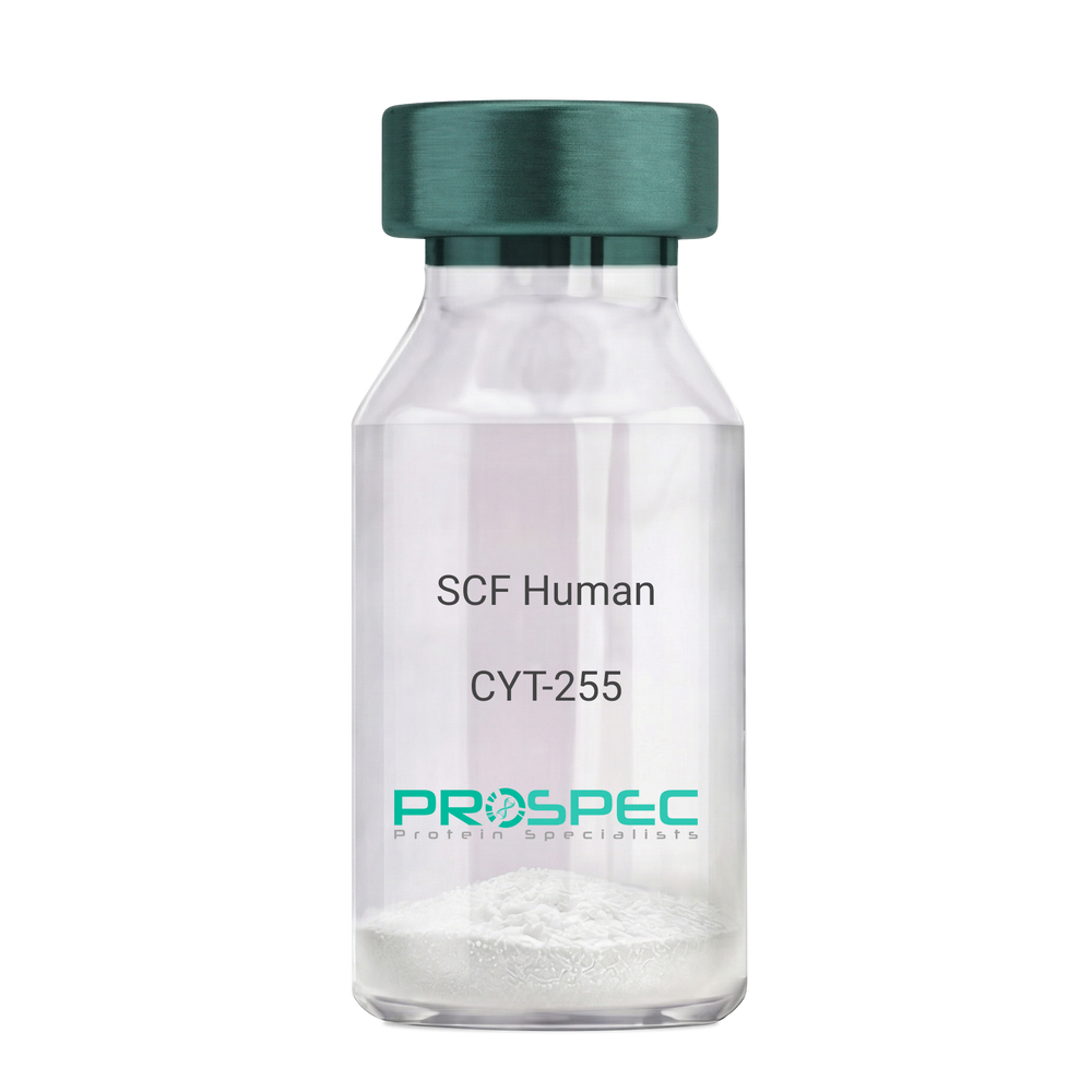
SCF HumanDescription:
Stem Cell Factor Human Recombinant
Kit ligand Precursor, C-kit ligand, SCF, Mast cell growth factor, MGF, SF, KL-1, Kitl, DKFZp686F2250.
Product # :
CYT-255Price :
Quantity :
Shipping Method :
Shipped at Room temp
More Info
- description
- source
- formulation
- purity
- biological activity
- More Info
Description
Stem Cell Factor Human Recombinant produced in E.Coli is a single, non-glycosylated polypeptide chain containing 165 amino acids and having a molecular mass of 18409 Dalton. The SCF is purified by proprietary chromatographic techniques.
Source
Escherichia Coli.
Formulation
Lyophilized from a concentrated (1mg/ml) solution in water containing 0.02% NaHCO3.
Purity
Greater than 98.0% as determined by:
(a) Analysis by SEC-HPLC.
(b) Analysis by SDS-PAGE.Biological Activity
The ED50 as determined by the dose-dependant stimulation of Human TF-1 cells is < 2 ng/ml, corresponding to a Specific Activity of 500,000 IU/mg.More Info
-
Introduction
Stem cell factor / KIT ligand (SCF) is a cytokine which binds CD117(c-Kit). SCF is also known as "steel factor" or "c-kit ligand". SCF exists in two forms, cell surface bound SCF and soluble (or free) SCF. Soluble SCF is produced by the cleavage of surface bound SCF by metalloproteases. SCF is a growth factor important for the survival, proliferation, and differentiation of hematopoietic stem cells and other hematopoietic progenitor cells. One of its roles is to change the BFU-E (burst-forming unit-erythroid) cells, which are the earliest erythrocyte precursors in the erythrocytic series, into the CFU-E (colony-forming unit-erythroid).
-
Synonyms
Kit ligand Precursor, C-kit ligand, SCF, Mast cell growth factor, MGF, SF, KL-1, Kitl, DKFZp686F2250.
-
Physical Appearance
Sterile Filtered White lyophilized (freeze-dried) powder.
-
Stability
Lyophilized KIT ligand although stable at room temperature for 3 weeks, should be stored desiccated below -18°C. Upon reconstitution SCF should be stored at 4°C between 2-7 days and for future use below -18°C.For long term storage it is recommended to add a carrier protein (0.1% HSA or BSA).Please prevent freeze-thaw cycles.
-
Solubility
It is recommended to reconstitute the lyophilized Stem Cell Factor in sterile 18MΩ-cm H2O not less than 100µg/ml, which can then be further diluted to other aqueous solutions.
-
Amino Acid Sequence
The sequence of the first five N-terminal amino acids was determined and was found to be Met-Glu-Gly-Ile-Cys.
-
Protein content
Protein quantitation was carried out by two independent methods1. UV spectroscopy at 280 nm using the absorbency value of 0.52 as the extinction coefficient for a 0.1% (1mg/ml) solution. This value is calculated by the PC GENE computer analysis program of protein sequences (IntelliGenetics). 2. Analysis by RP-HPLC, using a standard solution of Stem Cell Factor as a Reference Standard.
ProSpec's products are furnished for LABORATORY RESEARCH USE ONLY. The product may not be used as drugs, agricultural or pesticidal products, food additives or household chemicals.
- View Data Sheet
Name :
TGFB3 (24-412 a.a.) HumanDescription:
Transforming Growth Factor-Beta 3 (24-412 a.a.) Human Recombinant
Transforming Growth Factor, Beta 3, Prepro-Transforming Growth Factor Beta-3, TGF-Beta-3, ARVD1, RNHF, Arrhythmogenic Right Ventricular Dysplasia 1, Transforming Growth Factor Beta-3, TGF-Beta3, ARVD, Transforming growth factor beta-3, TGF-beta-3.
Product # :
CYT-886Price :
Quantity :
Shipping Method :
Shipped with Ice Packs
More Info
- description
- source
- formulation
- purity
- More Info
Description
TGFB3 Human Recombinant produced in E.Coli is a single, non-glycosylated polypeptide chain containing 412 amino acids (24-412 a.a) and having a molecular mass of 47.2kDa. TGFB3 is fused to a 23 amino acid His-tag at N-terminus & purified by proprietary chromatographic techniques.
Source
Escherichia Coli.
Formulation
TGFB3 protein solution (0.5mg/ml) containing 20mM Tris-HCl (pH8.0) and 10% glycerol.
Purity
Greater than 85.0% as determined by SDS-PAGE.
More Info
-
Introduction
Transforming growth factor betas (TGF Betas) mediate many cell-cell interactions that occur during embryonic development. Three TGF Betas have been identified in mammals. TGF Beta 1, TGF Beta 2 and TGF Beta 3 are each synthesized as precursor proteins that are very similar in that each is cleaved to yield a 112 amino acid polypeptide that remains associated with the latent portion of the molecule.
-
Synonyms
Transforming Growth Factor, Beta 3, Prepro-Transforming Growth Factor Beta-3, TGF-Beta-3, ARVD1, RNHF, Arrhythmogenic Right Ventricular Dysplasia 1, Transforming Growth Factor Beta-3, TGF-Beta3, ARVD, Transforming growth factor beta-3, TGF-beta-3.
-
Physical Appearance
Sterile Filtered colorless solution.
-
Stability
Store at 4°C if entire vial will be used within 2-4 weeks. Store, frozen at -20°C for longer periods of time. For long term storage it is recommended to add a carrier protein (0.1% HSA or BSA).Avoid multiple freeze-thaw cycles.
-
Amino Acid Sequence
MGSSHHHHHH SSGLVPRGSH MGSLSTCTTL DFGHIKKKRV EAIRGQILSK LRLTSPPEPT VMTHVPYQVL ALYNSTRELL EEMHGEREEG CTQENTESEY YAKEIHKFDM IQGLAEHNEL AVCPKGITSK VFRFNVSSVE KNRTNLFRAE FRVLRVPNPS SKRNEQRIEL FQILRPDEHI AKQRYIGGKN LPTRGTAEWL SFDVTDTVRE WLLRRESNLG LEISIHCPCH TFQPNGDILE NIHEVMEIKF KGVDNEDDHG RGDLGRLKKQ KDHHNPHLIL MMIPPHRLDN PGQGGQRKKR ALDTNYCFRN LEENCCVRPL YIDFRQDLGW KWVHEPKGYY ANFCSGPCPY LRSADTTHST VLGLYNTLNP EASASPCCVP QDLEPLTILY YVGRTPKVEQ LSNMVVKSCK CS.
ProSpec's products are furnished for LABORATORY RESEARCH USE ONLY. The product may not be used as drugs, agricultural or pesticidal products, food additives or household chemicals.
- View Data Sheet
Name :
JAZF1 HumanDescription:
JAZF zinc finger 1 Human Recombinant
Juxtaposed with another zinc finger protein 1 , TAK1-interacting protein 27, Zinc finger protein 802, JAZF1, TIP27, ZNF802, JAZF zinc finger 1, Juxtaposed With Another Zinc Finger Gene 1.
Product # :
PRO-1444Price :
Quantity :
Shipping Method :
Shipped with Ice Packs
More Info
- description
- source
- formulation
- purity
- More Info
Description
JAZF1 Human Recombinant produced in E.Coli is a single, non-glycosylated polypeptide chain containing 266 amino acids (1-243 a.a) and having a molecular mass of 29.5kDa.JAZF1 is fused to a 23 amino acid His-tag at N-terminus & purified by proprietary chromatographic techniques.
Source
Escherichia Coli.
Formulation
JAZF1 protein solution (1mg/ml) containing 20mM Tris-HCl buffer (pH 8.0), 0.4M UREA and 10% glycerol.
Purity
Greater than 85.0% as determined by SDS-PAGE.
More Info
-
Introduction
JAZF zinc finger 1 (JAZF1) is a nuclear protein with 3 C2H2-type zinc fingers, which functions as a transcriptional repressor. Chromosomal anomalies involving the JAZF1 are linked with endometrial stromal tumors. Alternatively spliced variants which encode different protein isoforms have been described; though, not all variants have been fully characterized.
-
Synonyms
Juxtaposed with another zinc finger protein 1 , TAK1-interacting protein 27, Zinc finger protein 802, JAZF1, TIP27, ZNF802, JAZF zinc finger 1, Juxtaposed With Another Zinc Finger Gene 1.
-
Physical Appearance
Sterile Filtered colorless solution.
-
Stability
Store at 4°C if entire vial will be used within 2-4 weeks. Store, frozen at -20°C for longer periods of time. For long term storage it is recommended to add a carrier protein (0.1% HSA or BSA).Avoid multiple freeze-thaw cycles.
-
Amino Acid Sequence
MGSSHHHHHH SSGLVPRGSH MGSMTGIAAA SFFSNTCRFG GCGLHFPTLA DLIEHIEDNH IDTDPRVLEK QELQQPTYVA LSYINRFMTD AARREQESLK KKIQPKLSLT LSSSVSRGNV STPPRHSSGS LTPPVTPPIT PSSSFRSSTP TGSEYDEEEV DYEESDSDES WTTESAISSE AILSSMCMNG GEEKPFACPV PGCKKRYKNV NGIKYHAKNG HRTQIRVRKP FKCRCGKSYK TAQGLRHHTI NFHPPVSAEI IRKMQQ
ProSpec's products are furnished for LABORATORY RESEARCH USE ONLY. The product may not be used as drugs, agricultural or pesticidal products, food additives or household chemicals.
- View Data Sheet
Name :
KLF12 HumanDescription:
Kruppel-Like Factor 12 Human Recombinant
AP-2rep, AP2REP, HSPC122, Krueppel-like factor 12, Transcriptional repressor AP-2rep.
Product # :
PRO-2090Price :
Quantity :
Shipping Method :
Shipped with Ice Packs
More Info
- description
- source
- formulation
- purity
- More Info
Description
KLF12 Human Recombinant produced in E.Coli is a single, non-glycosylated polypeptide chain containing 425 amino acids (1-402 a.a) and having a molecular mass of 46.6kDa.KLF12 is fused to a 23 amino acid His-tag at N-terminus & purified by proprietary chromatographic techniques.
Source
Escherichia Coli.
Formulation
KLF12 protein solution (0.5mg/ml) contains 20mM Tris-HCl buffer, (pH8.0) ,10% glycerol and 0.4M Urea.
Purity
Greater than 85.0% as determined by SDS-PAGE.
More Info
-
Introduction
Krueppel-like factor 12 (KLF12) belongs to the Kruppel-like zinc finger protein family and can repress expression of the AP-2 alpha gene by binding to a particular site in the AP-2 alpha gene promoter. AP-2 alpha (Activator protein-2 alpha) is a developmentally-regulated transcription factor and vital regulator of gene expression during vertebrate development and carcinogenesis. KLF12 gene repression entails binding with a corepressor, CtBP1.
-
Synonyms
AP-2rep, AP2REP, HSPC122, Krueppel-like factor 12, Transcriptional repressor AP-2rep.
-
Physical Appearance
Sterile Filtered clear solution.
-
Stability
Store at 4°C if entire vial will be used within 2-4 weeks. Store, frozen at -20°C for longer periods of time. For long term storage it is recommended to add a carrier protein (0.1% HSA or BSA).Avoid multiple freeze-thaw cycles.
-
Amino Acid Sequence
MGSSHHHHHH SSGLVPRGSH MGSMNIHMKR KTIKNINTFE NRMLMLDGMP AVRVKTELLE SEQGSPNVHN YPDMEAVPLL LNNVKGEPPE DSLSVDHFQT QTEPVDLSIN KARTSPTAVS SSPVSMTASA SSPSSTSTSS SSSSRLASSP TVITSVSSAS SSSTVLTPGP LVASASGVGG QQFLHIIHPV PPSSPMNLQS NKLSHVHRIP VVVQSVPVVY TAVRSPGNVN NTIVVPLLED GRGHGKAQMD PRGLSPRQSK SDSDDDDLPN VTLDSVNETG STALSIARAV QEVHPSPVSR VRGNRMNNQK FPCSISPFSI ESTRRQRRSE SPDSRKRRIH RCDFEGCNKV YTKSSHLKAH RRTHTGEKPY KCTWEGCTWK FARSDELTRH YRKHTGVKPF KCADCDRSFS RSDHLALHRR RHMLV.
ProSpec's products are furnished for LABORATORY RESEARCH USE ONLY. The product may not be used as drugs, agricultural or pesticidal products, food additives or household chemicals.
- View Data Sheet
Name :
RAC2 HumanDescription:
RAC2 Human Recombinant
p21-Rac2, Small G protein, GX, RAC2, HSPC022, EN-7.
Product # :
PRO-619Price :
Quantity :
Shipping Method :
Shipped with Ice Packs
More Info
- description
- source
- formulation
- purity
- More Info
Description
Recombinant RAC2 produced in E.Coli is a single, non-glycosylated polypeptide chain containing 419 amino acids and having a molecular mass of 47 kDa. RAC2 is fused to GST and purified by conventional chromatography techniques.
Source
Escherichia Coli.
Formulation
The RAC2 protein solution (1mg/ml) contains 1x PBS pH-7.4.
Purity
Greater than 90.0% as determined by SDS-PAGE.
More Info
-
Introduction
RAC2 is a small signaling G protein/GTPase which is part of the RAS superfamily of small GTP-binding proteins which regulate cell growth, actin cytoskeletal organization, cell proliferation, survival, cell cycle progression, gene transcription rgulation and the activation of protein kinases. RAC2 cycles between an active gtp-bound and inactive gdp-bound state. In active state, RAC2 binds to a variety of effector proteins to regulate cellular responses, such as secretory processes, phagocytose of apoptotic cells and epithelial cell polarization. RAC2 is involved in the regulation of the nadph oxidase.
-
Synonyms
p21-Rac2, Small G protein, GX, RAC2, HSPC022, EN-7.
-
Physical Appearance
Sterile filtered colorless solution.
-
Stability
Store at 4°C if entire vial will be used within 2-4 weeks. Store, frozen at -20°C for longer periods of time. For long term storage it is recommended to add a carrier protein (0.1% HSA or BSA). Avoid multiple freeze-thaw cycles.
-
Amino Acid Sequence
MSPILGYWKI KGLVQPTRLL LEYLEEKYEE HLYERDEGDK WRNKKFELGL EFPNLPYYID GDVKLTQSMA IIRYIADKHN MLGGCPKERA EISMLEGAVL DIRYGVSRIA YSKDFETLKV DFLSKLPEML KMFEDRLCHK TYLNGDHVTH PDFMLYDALD VVLYMDPMCL DAFPKLVCFK KRIEAIPQID KYLKSSKYIA WPLQGWQATF GGGDHPPKSD LVPRGSHMQA IKCVVVGDGA VGKTCLLISY TTNAFPGEYI PTVFDNYSAN VMVDSKPVNL GLWDTAGQED YDRLRPLSYP QTDVFLICFS LVSPASYENV RAKWFPEVRH HCPSTPIILV GTKLDLRDDK DTIEKLKEKK
LAPITYPQGL ALAKEIDSVK YLECSALTQR GLKTVFDEAI RAV
LCPQPTR QQKRACSLL.
ProSpec's products are furnished for LABORATORY RESEARCH USE ONLY. The product may not be used as drugs, agricultural or pesticidal products, food additives or household chemicals.
- View Data Sheet
Name :
TNFRSF10C AntibodyDescription:
TRAIL Receptor-3, Mouse Anti Human
Tumor Necrosis Factor Receptor Superfamily, Member 10c, Decoy Without An , Intracellular Domain, TRAILR3, DCR1, TRID, TNF-Related Apoptosis-Inducing Ligand Receptor, Antagonist Decoy Receptor For TRAIL/Apo-2L, Decoy TRAIL Receptor Without Death Domain, Lymphocyte Inhibitor Of TRAIL, Decoy Receptor,TRAIL-R, LIT,Tumor Necrosis Factor Receptor Superfamily Member 10C, TRAIL Receptor Without an Intracellular Domain, Cytotoxic TRAIL Receptor, TRAIL Receptor, CD263 Antigen, DCR1-TNFR, CD263.
Product # :
ANT-741Price :
Quantity :
Shipping Method :
Shipped with Ice Packs
More Info
- formulation
- More Info
Formulation
1mg/ml containing PBS, pH-7.4, 10% Glycerol and 0.02% Sodium Azide.
More Info
-
Introduction
TRAIL Receptor-3, also known as TNFRSF10C belongs to theTNF-receptor super family. TNFRSF10C contains an extracellular TRAIL-binding domain as well as a transmembranedomain, however no cytoplasmic death domain. Furthermore, TNFRSF10C is not capable of inducing apoptosis, and is considered toact as an antagonistic receptor which protects cells from TRAIL-induced apoptosis.
-
Synonyms
Tumor Necrosis Factor Receptor Superfamily, Member 10c, Decoy Without An , Intracellular Domain, TRAILR3, DCR1, TRID, TNF-Related Apoptosis-Inducing Ligand Receptor, Antagonist Decoy Receptor For TRAIL/Apo-2L, Decoy TRAIL Receptor Without Death Domain, Lymphocyte Inhibitor Of TRAIL, Decoy Receptor,TRAIL-R, LIT,Tumor Necrosis Factor Receptor Superfamily Member 10C, TRAIL Receptor Without an Intracellular Domain, Cytotoxic TRAIL Receptor, TRAIL Receptor, CD263 Antigen, DCR1-TNFR, CD263.
-
Physical Appearance
Sterile filtered colorless solution.
-
Immunogen
Anti-human TNFRSF10C mAb, is derived from hybridization of mouse SP2/0 myeloma cells with spleen cells from BALB/c mice immunized with Recombinant human TNFRSF10C amino acids 26-236 purified from E. coli.
-
Ig Subclass
Mouse IgG1 heavy chain and κ light chain.
-
Clone
PAT1E12AT.
-
Applications
The antibody has been tested by ELISA, Western blot analysis, Flow cytometry and ICC/IF to assure specificity and reactivity. Since application varies, however, each investigation should be titrated by the reagent to obtain optimal results.
-
Type
Mouse Anti Human Monoclonal.
-
Storage Procedures
For periods up to 1 month store at 4°C, for longer periods of time, store at -20°C. Prevent freeze thaw cycles.
-
Purification Method
TNFRSF10C antibody was purified from mouse ascitic fluids by protein-A affinity chromatography.
ProSpec's products are furnished for LABORATORY RESEARCH USE ONLY. The product may not be used as drugs, agricultural or pesticidal products, food additives or household chemicals.
- View Data Sheet
Name :
BMP 4 HumanDescription:
Bone Morphogenetic Protein-4 Human Recombinant
BMP4, ZYME, BMP2B, BMP2B1.
Product # :
CYT-361Price :
Quantity :
Shipping Method :
Shipped at Room temp
More Info
- description
- source
- formulation
- purity
- More Info
Description
Bone Morphogenetic Protein-4 Human Recombinant produced in E.Coli is a monomeric, non-glycosylated, Polypeptide chain containing 116 amino acids and having a molecular mass of 13kDa. The BMP-4 is purified by proprietary chromatographic techniques.
Source
Escherichia Coli.
Formulation
BMP-4 was lyophilized from a 0.2µm filtered concentrated (1mg/ml) solution in 20mM Na2CO3 buffer, pH 9.0.
Purity
Greater than 95.0% as determined by:
(a) Analysis by RP-HPLC.
(b) Analysis by SDS-PAGE.More Info
-
Introduction
The protein encoded by this gene is a member of the bone morphogenetic protein family which is part of the transforming growth factor-beta superfamily. The superfamily includes large families of growth and differentiation factors. Bone morphogenetic proteins were originally identified by an ability of demineralized bone extract to induce endochondral osteogenesis in vivo in an extraskeletal site. This particular family member plays an important role in the onset of endochondral bone formation in humans, and a reduction in expression has been associated with a variety of bone diseases, including the heritable disorder Fibrodysplasia Ossificans Progressiva. Alternative splicing in the 5' untranslated region of this gene has been described and three variants are described, all encoding an identical protein.
-
Synonyms
BMP4, ZYME, BMP2B, BMP2B1.
-
Physical Appearance
Sterile Filtered White lyophilized (freeze-dried) powder.
-
Stability
Lyophilized Bone Morphogenetic Protein-4 although stable at room temperature for 3 weeks, should be stored desiccated below -18°C. Upon reconstitution BMP4 should be stored at 4°C between 2-7 days and for future use below -18°C.For long term storage it is recommended to add a carrier protein (0.1% HSA or BSA).Please prevent freeze-thaw cycles.
-
Solubility
It is recommended to reconstitute the lyophilized Bone Morphogenetic Protein-4 in sterile 18M-cm H2O not less than 100µg/ml, which can then be further diluted to other aqueous solutions.
-
Amino Acid Sequence
SPKHHSQRAR KKNKNCRRHS LYVDFSDVGW NDWIVAPPGY QAFYCHGDCP FPLADHLNST NHAIVQTLVN SVNSSIPKAC CVPTELSAIS MLYLDEYDKV VLKNYQEMVV EGCGCR.
-
Background
What You Should Know About Bone Morphogenetic Protein-4 (BMP-4) Human Recombinant
As part of the transforming growth factor-beta (TGF-β) superfamily, Bone morphogenetic protein-4 (BMP-4) participates in multiple developmental processes, from embryogenesis to bone and cartilage formation.
Since this signaling protein is involved in many physiological processes, its laboratory-produced version has been studied for different medical applications. Additionally, a reduction in BMP-4 expression has been associated with multiple diseases, leading to further research into its potential therapeutic benefits.
Are you interested in learning more about Bone Morphogenetic Protein-4 (BMP-4) human recombinant? Read on to find more information!
How Does Bone Morphogenetic Protein-4 (BMP-4) Work?
Bone Morphogenetic Protein-4 (BMP-4) regulates microRNAs miR-494 and miR-126-5p expression, controlling endothelial cells' involvement and function in angiogenesis. As such, it has diverse effects on cell growth, differentiation, and survival.
The Role of BMP-4
This protein emits signals that promote the formation of different tissues and organs, including the bones and cartilage, kidneys, teeth, and the neural tube. In other words, it's essential for the development of the heart, skeleton, and central nervous system.
However, the role of BMP-4 goes beyond these processes. It participates in different physiological activities, such as:
- Embryonic development
- Wound healing
- Bone remodeling
- Immune response modulation
- Tissue repair
- Cardiac development and function
What Is Bone Morphogenetic Protein-4 (BMP-4) Human Recombinant?
To replicate the effects of the BMP4 found in humans and explore its possible therapeutic applications, many laboratories have started producing this protein in Chinese hamster ovary (CHO) cells.
As mentioned, decreased BMP-4 expression has been associated with different diseases, including bone disorders, fibrosis, and cancer, which can cause other conditions, such as organ dysfunction.
More research is needed, but BMP-4 human recombinant (rhBMP4) produced in CHO has the potential to address these diseases and could be used for other medical applications. These are some examples:
- Cancer therapy
- Development of engineered tissues and organs
- Bone regeneration for the treatment of osteoporosis and nonunion fractures
- Bone growth and fusion in spinal fusion surgeries (the U.S. Food and Drug Administration approved some bone morphogenetic proteins for these procedures)
- Promotion of tissue repair and regeneration
Final Thoughts BMP-4
Although BMP-4 human recombinant produced in CHO offers potential benefits, several challenges remain, including possible side effects, as high doses can cause inflammation, bone overgrowth, and other issues.
However, the long-term effects of rhBMP4 are still under investigation. Further research will provide solutions to address these challenges and allow experts to explore this laboratory-produced protein's power in different medical fields.
What is the molecular weight/Mw of BMP4 Protein?
BMP4 Protein has a total Mw of 13kDa.
What is the source or expression system of BMP4 Protein?
Escherichia Coli.
What is the Purity of BMP4 Protein?
BMP4 Protein is >95% pure as determined by SDS-PAGE.
What is the Biological Activity of BMP4 Protein?
The biological functionality of BMP4 Protein will be determined in the future.
What is the amino acid sequence of BMP4 Protein?
SPKHHSQRAR KKNKNCRRHS LYVDFSDVGW NDWIVAPPGY QAFYCHGDCP FPLADHLNST NHAIVQTLVN SVNSSIPKAC CVPTELSAIS MLYLDEYDKV VLKNYQEMVV EGCGCR.
What applications can BMP4 Protein be used in?
BMP4 Protein can probably be used in western blot, ELISA and Lateral Flow.
What is the endotoxin level for BMP4 Protein?
The endotoxin level is minimal, BMP4 Protein was purified using conventional chromatography techniques.
ProSpec's products are furnished for LABORATORY RESEARCH USE ONLY. The product may not be used as drugs, agricultural or pesticidal products, food additives or household chemicals.
- View Data Sheet
Name :
AIF1 HumanDescription:
Allograft Inflammatory Factor 1 Human Recombinant
AIF-1, Allograft inflammatory factor 1, Em:AF129756.17, G1, IBA1, Ionized calcium-binding adapter molecule 1, IRT-1, Protein G1, AIF1.
Product # :
CYT-697Price :
Quantity :
Shipping Method :
Shipped with Ice Packs
More Info
- description
- source
- formulation
- purity
- More Info
- sds-page
Description
AIF1 produced in E.Coli is a single, non-glycosylated polypeptide chain containing 167 amino acids (1-147a.a.) and having a molecular mass of 18.9kDa. AIF1 is fused to a 20 amino acid His-tag at N-terminus & purified by proprietary chromatographic techniques.
Source
E. Coli.
Formulation
The AIF1 protein solution (0.5mg/1ml) is formulated in 20mM Tris-HCl buffer (pH8.0)
2mM DTT, 200mM NaCl and 20% glycerol.Purity
Greater than 95% as determined by SDS PAGE.
sds-page
More Info
-
Introduction
Human AIF1 protein shares 98% homology/identity with that of rat. AIF1 is expressed in macrophages and neutrophils. The expression of AIF1 transcripts is upregulated by IFN-g in rat macrophages. AIF1 is expressed selectively in human macrophage-like cell lines, and in a subset of CD68(+) macrophages in the interstitial and perivascular spaces of human heart allografts. In quiescent cultured human vascular smooth muscle cells synthesis of AIF1 is induced by IFN-g, IL1b, and conditioned medium of T-cells. Overexpression of AIF1 in human VSMCs results in enhanced growth of these cells. AIF1 is expressed during apoptosis rat mammary gland and ventral prostate tissues. Allograft Inflammatory Factor 1 is expressed by several tumor-associated activated macrophages and microglial cells in rat and human gliomas. There is an evident relationship of AIF1-expressing activated macrophages and microglial cells with tumor malignancy in humans.
-
Synonyms
AIF-1, Allograft inflammatory factor 1, Em:AF129756.17, G1, IBA1, Ionized calcium-binding adapter molecule 1, IRT-1, Protein G1, AIF1.
-
Stability
Store AIF1 at 4°C if entire vial will be used within 2-4 weeks. Store, frozen at -20°C for longer periods of time.For long term storage it is recommended to add a carrier protein (0.1% HSA or BSA).Avoid multiple freeze-thaw cycles.
-
Amino Acid Sequence
MGSSHHHHHH SSGLVPRGSH MSQTRDLQGG KAFGLLKAQQ EERLDEINKQ FLDDPKYSSD EDLPSKLEGF KEKYMEFDLN GNGDIDIMSL KRMLEKLGVP KTHLELKKLI GEVSSGSGET FSYPDFLRMM LGKRSAILKM ILMYEEKARE KEKPTGPPAK KAISELP.
-
Background
Allograft Inflammatory Factor 1 Human Recombinant: Uncovering its Role in Immune Responses and Therapeutic Prospects
1. Abstract
This paper explores the Allograft Inflammatory Factor 1 Human Recombinant (AIF-1), a cytoplasmic, IFN-gamma-inducible calcium-binding protein involved in inflammation and immunity. We review the structure, biological roles, and involvement of AIF-1 in disease pathology. The therapeutic potential of AIF-1 in immune-related disorders is also explored.
2. Introduction
AIF-1, also known as IBA1, plays an important role in immune responses. It is associated with various immune cells, particularly macrophages, and has been implicated in numerous inflammatory and immune-related diseases. Understanding the function of AIF-1 could aid the development of novel therapeutic strategies.
3. Structure and Signaling of AIF-1
AIF-1 is a small 17 kDa protein with an EF-hand calcium-binding motif. Although the precise mechanism by which AIF-1 exerts its functions is not entirely clear, it is known to regulate the activation, migration, and proliferation of macrophages, key cells involved in immune responses.
4. Biological Functions of AIF-1
AIF-1 has been shown to play key roles in macrophage activation and function, which are central to inflammation and immunity. It is also implicated in cell survival, proliferation, and differentiation.
5. AIF-1 in Disease Pathology
AIF-1 has been associated with a range of inflammatory and immune-related diseases, including rheumatoid arthritis, atherosclerosis, and multiple sclerosis. It is also implicated in several cancers, further underscoring its broad physiological and pathological relevance.
6. Therapeutic Potential of AIF-1
Given its pivotal role in immune responses, AIF-1 presents an intriguing target for therapeutic interventions in immune-related diseases. Modulating the activity of AIF-1 could potentially alleviate pathological inflammation and autoimmunity.
7. Conclusion and Future Perspectives
Our knowledge of AIF-1 and its functions has significantly improved in recent years, but much remains to be discovered. Further research into AIF-1's exact molecular mechanisms and roles in disease will undoubtedly contribute to the development of novel therapeutic strategies.
What is the molecular weight/Mw of AIF1 Protein?
AIF1 Protein has a total Mw of 18.9kDa.
What is the source or expression system of AIF1 Protein?
Escherichia Coli.
What is the Purity of AIF1 Protein?
AIF1 Protein is >95% pure as determined by SDS-PAGE.
What is the Biological Activity of AIF1 Protein?
The biological functionality of AIF1 Protein will be determined in the future.
What is the amino acid sequence of AIF1 Protein?
MGSSHHHHHH SSGLVPRGSH MSQTRDLQGG KAFGLLKAQQ EERLDEINKQ FLDDPKYSSD EDLPSKLEGF KEKYMEFDLN GNGDIDIMSL KRMLEKLGVP KTHLELKKLI GEVSSGSGET FSYPDFLRMM LGKRSAILKM ILMYEEKARE KEKPTGPPAK KAISELP.
What applications can AIF1 Protein be used in?
AIF1 Protein can probably be used in western blot, ELISA and Lateral Flow.
What is the endotoxin level for AIF1 Protein?
The endotoxin level is minimal, AIF1 Protein was purified using conventional chromatography techniques.
ProSpec's products are furnished for LABORATORY RESEARCH USE ONLY. The product may not be used as drugs, agricultural or pesticidal products, food additives or household chemicals.
- View Data Sheet
Name :
C1QTNF3 HumanDescription:
Complement C1q Tumor Necrosis Factor-Related Protein 3 Human Recombinant
Complement C1q tumor necrosis factor-related protein 3, Secretory protein CORS26, C1QTNF3, CTRP3, Cors, Corcs, CORS26, FLJ37576, Cartducin.
Product # :
PRO-653Price :
Quantity :
Shipping Method :
Shipped at Room temp
More Info
- description
- source
- formulation
- purity
- More Info
Description
C1QTNF3 Human Recombinant produced in E.Coli is a single, non-glycosylated, Polypeptide chain containing 234 amino acids and having a molecular mass of 25.4 kDa. The protein contains an extra 10 aa His tag at N-terminus. The C1QTNF3 amino acid sequence is identical to UniProtKB/Swiss-Prot entry Q9BXJ4 amino acids 23–246. The C1QTNF3 is purified by proprietary chromatographic techniques.
Source
Escherichia Coli.
Formulation
Human C1QTNF3 was filtered (0.4µm) and lyophilized in 0.5 mg/ml in 0.05M Acetate buffer pH4.
Purity
The purity of C1QTNF3 is greater than 95% as determined by SDS PAGE.
More Info
-
Introduction
C1QTNF3 also called Cartducin is a novel angiogenic factor in the formation of neointima following angioplasty. C1QTNF3 a paralog of Acrp30 (adiponectin). C1QTNF3 is a secretory protein produced by chondrogenic precursors & proliferating chondrocytes, and belongs to a novel C1q family of proteins. Cartducin promotes the growth of mesenchymal chondroprogenitor cells & chondrosarcoma-derived chondrocytic cells in vitro. Cartducin stimulates mesenchymal chondroprogenitor cell proliferation through extracellular signal-regulated kinase and phosphatidylinositol 3-kinase/Akt pathways. C1QTNF3 promotes proliferation & the migration of endothelial cells.
-
Synonyms
Complement C1q tumor necrosis factor-related protein 3, Secretory protein CORS26, C1QTNF3, CTRP3, Cors, Corcs, CORS26, FLJ37576, Cartducin.
-
Stability
Store lyophilized C1QTNF3 at -20°C. Aliquot the product after reconstitution to avoid repeated freezing/thawing cycles. Reconstituted C1QTNF3 can be stored at 4°C for a limited period of time; it does not show any change after two weeks at 4°C.
-
Solubility
It is recommended to add 0.1M Acetate buffer pH4 to prepare a working stock solution of approximately 0.5 mg/ml and let the lyophilized pellet dissolve completely. For conversion into higher pH value, we recommend intensive dilution by relevant buffer to a concentration of 10µg/ml. In higher concentrations the solubility of the protein is limited. Product is not sterile! Please filter the product by an appropriate sterile filter before using it in the cell culture.
-
Amino Acid Sequence
MKHHHHHHAS QDEYMESPQT GGLPPDCSKC CHGDYSFRGY QGPPGPPGPP GIPGNHGNNG NNGATGHEGA KGEKGDKGDL GPRGERGQHG PKGEKGYPGI PPELQIAFMA SLATHFSNQN SGIIFSSVET NIGNFFDVMT GRFGAPVSGV YFFTFSMMKH EDVEEVYVYL MHNGNTVFSM YSYEMKGKSD TSSNHAVLKL AKGDEVWLRM GNGALHGDHQ RFSTFAGFLLFETK.
ProSpec's products are furnished for LABORATORY RESEARCH USE ONLY. The product may not be used as drugs, agricultural or pesticidal products, food additives or household chemicals.
- View Data Sheet
Name :
GDF6 HumanDescription:
Bone Morphogenetic protein-13 Human Recombinant
Growth Differentiation Factor 6, Growth/Differentiation Factor 16, Bone Morphogenetic Protein 13, BMP-13, BMP13, GDF-6, Klippel-Feil Malformation, Segmentation Syndrome 1, Klip-Feil Malformation, Klippel-Feil Syndrome, MCOPCB6, SCDO4, CDMP2, LCA17, MCOP4, GDF16, KFS1, KFSL, SGM1, KFM, KFS, GDF6.
Product # :
CYT-938Price :
Quantity :
Shipping Method :
Shipped at Room temp
More Info
- description
- source
- formulation
- purity
- biological activity
- More Info
Description
BMP13 Human Recombinant produced in E.coli is a non-glycosylated disulfide linked homodimer containing 2 chains of 120 amino acids and having a molecular mass of 27.1kDa.The BMP-13 is purified by proprietary chromatographic techniques.
Source
Escherichia Coli.
Formulation
BMP-13 protein was lyophilized from a 0.2µm filtered concentrated solution in 30% Acetonitrile and 0.1% TFA.
Purity
Greater than 95.0% as determined by: (a) Analysis by HPLC. (b) Analysis by SDS-PAGE.
Biological Activity
The ED50 as determined by inducing alkaline phosphatase production of murine ATDC5 cells is less than 2.0µg/ml, corresponding to a specific activity of > 500IU/mg.More Info
-
Introduction
Growth/differentiation factors (GDF1-GDF15) belong to the BMP family of TGF-beta superfamily proteins. These factors are produced as inactive preproproteins which are subsequently cleaved and assembled into active secreted homodimers. BMP13 is a growth factor which controls proliferation and cellular differentiation in the retina and bone formation. BMP13 has a central role in regulating apoptosis during retinal development. GDF proteins are vital during embryonic development, particularly in the skeletal, nervous, and muscular systems. BMP13 gene mutations result in colobomata, which are congenital abnormalities in ocular development, and in Klippel-Feil syndrome (KFS), which is a congenital disorder of spinal segmentation.
-
Synonyms
Growth Differentiation Factor 6, Growth/Differentiation Factor 16, Bone Morphogenetic Protein 13, BMP-13, BMP13, GDF-6, Klippel-Feil Malformation, Segmentation Syndrome 1, Klip-Feil Malformation, Klippel-Feil Syndrome, MCOPCB6, SCDO4, CDMP2, LCA17, MCOP4, GDF16, KFS1, KFSL, SGM1, KFM, KFS, GDF6.
-
Physical Appearance
Sterile Filtered White lyophilized (freeze-dried) powder.
-
Stability
Lyophilized BMP13 although stable at room temperature for 3 weeks, should be stored desiccated below -18°C. Upon reconstitution BMP-13 should be stored at 4°C between 2-7 days and for future use below -18°C. For long term storage it is recommended to add a carrier protein (0.1% HSA or BSA). Please prevent freeze-thaw cycles.
-
Solubility
It is recommended to reconstitute the lyophilized BMP13 in sterile 18M-cm H2O not less than 100µg/ml, which can then be further diluted to other aqueous solutions.
-
Amino Acid Sequence
TAFASRHGKR HGKKSRLRCS KKPLHVNFKE LGWDDWIIAP LEYEAYHCEG VCDFPLRSHL EPTNHAIIQT LMNSMDPGST PPSCCVPTKL TPISILYIDA GNNVVYKQYE DMVVESCGCR.
-
Background
Bone Morphogenetic Protein-13 Human Recombinant: Unraveling its Potential in Tissue Engineering and Regenerative Medicine
Abstract:
Bone Morphogenetic Protein-13 (BMP-13) human recombinant is a pivotal member of the bone morphogenetic protein family, known for its crucial role in tissue development, regeneration, and repair. This research paper aims to provide a comprehensive analysis of BMP-13, including its characteristics, signaling pathways, and potential therapeutic applications. Furthermore, innovative methodologies for the production and optimization of BMP-13 human recombinant are proposed, shedding light on its future implications in the field of tissue engineering and regenerative medicine.
Introduction:
Tissue engineering and regenerative medicine hold great promise in addressing tissue repair and regeneration challenges. BMP-13, a prominent member of the BMP family, plays a vital role in orchestrating cellular responses during tissue development and healing. This paper explores the distinctive features of BMP-13 and presents novel approaches for the production and optimization of BMP-13 human recombinant, aiming to unlock its therapeutic potential in various regenerative contexts.
Characteristics and Signaling Pathways:
BMP-13 is a secreted growth factor belonging to the transforming growth factor-beta (TGF-β) superfamily. It exerts its biological effects by binding to specific cell surface receptors, initiating intricate intracellular signaling cascades. BMP-13 signaling pathways, including Smad-dependent and Smad-independent pathways, regulate critical processes such as cell differentiation, proliferation, and extracellular matrix synthesis, influencing tissue development and repair.
Production of BMP-13 Human Recombinant:
Efficient production methodologies are crucial for harnessing the therapeutic potential of BMP-13 human recombinant. Various recombinant protein expression systems, such as mammalian cells or baculovirus-insect cell systems, have been utilized for the production of functional BMP-13. Optimization strategies, including codon optimization, signal peptide engineering, and protein folding enhancement, have been employed to improve the yield and bioactivity of BMP-13 recombinant protein.
Potential Therapeutic Applications:
BMP-13 human recombinant holds immense promise in the field of tissue engineering and regenerative medicine. Its involvement in cartilage formation, osteogenesis, and tissue repair makes it a potential candidate for the treatment of musculoskeletal disorders, joint injuries, and cartilage defects. Furthermore, the ability of BMP-13 to modulate cell behavior and tissue remodeling indicates its wider therapeutic applications in diverse regenerative processes.
Conclusion:
BMP-13 human recombinant emerges as a crucial regulator in tissue engineering and regenerative medicine, offering significant potential for tissue repair and regeneration. Optimizing production methodologies and further unraveling its signaling mechanisms will undoubtedly enhance its therapeutic applications. Given its involvement in cartilage and bone formation, as well as tissue repair, BMP-13 human recombinant represents a valuable tool for promoting tissue regeneration and addressing the unmet clinical needs in regenerative medicine.
What is the molecular weight/Mw of GDF6 Protein?
GDF6 Protein has a total Mw of 27.1kDa.
What is the source or expression system of GDF6 Protein?
Escherichia Coli.
What is the Purity of GDF6 Protein?
GDF6 Protein is >95% pure as determined by SDS-PAGE.
What is the Biological Activity of GDF6 Protein?
The ED50 as determined by inducing alkaline phosphatase production of murine ATDC5 cells is less than 2.0µg/ml, corresponding to a specific activity of > 500IU/mg.
What is the amino acid sequence of GDF6 Protein?
TAFASRHGKR HGKKSRLRCS KKPLHVNFKE LGWDDWIIAP LEYEAYHCEG VCDFPLRSHL EPTNHAIIQT LMNSMDPGST PPSCCVPTKL TPISILYIDA GNNVVYKQYE DMVVESCGCR.
What applications can GDF6 Protein be used in?
GDF6 Protein can probably be used in western blot, ELISA and Lateral Flow.
What is the endotoxin level for GDF6 Protein?
The endotoxin level is minimal, GDF6 Protein was purified using conventional chromatography techniques.
ProSpec's products are furnished for LABORATORY RESEARCH USE ONLY. The product may not be used as drugs, agricultural or pesticidal products, food additives or household chemicals.
- View Data Sheet
Name :
BMP3 HumanDescription:
Bone Morphogenetic protein-3 Human Recombinant
Bone Morphogenetic Protein 3, Osteogenin, Bone Morphogenetic Protein 3 (Osteogenic), Bone Morphogenetic Protein 3A, BMP-3A, BMP-3, Bone Morphogenetic Protein-3, BMP3A, BMP3.
Product # :
CYT-937Price :
Quantity :
Shipping Method :
Shipped at Room temp
More Info
- description
- source
- formulation
- purity
- biological activity
- More Info
Description
BMP3 Human Recombinant produced in E.coli is a non-glycosylated disulfide linked homodimer containing 2 chains of 110 amino acids and having a molecular mass of 24.8kDa.The BMP-3 is purified by proprietary chromatographic techniques.
Source
Escherichia Coli.
Formulation
BMP-3 protein was lyophilized from a 0.2µm filtered concentrated solution in 30% Acetonitrile and 0.1% TFA.
Purity
Greater than 95.0% as determined by: (a) Analysis by HPLC. (b) Analysis by SDS-PAGE.
Biological Activity
The ED50 as determined by its ability to inhibit BMP-2-induced activity in murine MC3T3- E1 cells.More Info
-
Introduction
Bone Morphogenetic Protein 3 (BMP3) is one of the BMPs, some of which are members of the TGF-beta superfamily (BMP2-7). There are more than 13 BMPs, which are involved in inducing cartilage and bone formation, embryogenesis and morphogenesis of various tissues and organs. In addition, BMPs regulate the growth, differentiation, chemotaxis, and apoptosis of various cell types. Akin to most other TGF-beta family proteins, BMPs are extremely conserved across animal species. At the amino acid sequence level, mature human and rat BMP3 are 98% identical.
-
Synonyms
Bone Morphogenetic Protein 3, Osteogenin, Bone Morphogenetic Protein 3 (Osteogenic), Bone Morphogenetic Protein 3A, BMP-3A, BMP-3, Bone Morphogenetic Protein-3, BMP3A, BMP3.
-
Physical Appearance
Sterile Filtered White lyophilized (freeze-dried) powder.
-
Stability
Lyophilized BMP3 although stable at room temperature for 3 weeks, should be stored desiccated below -18°C. Upon reconstitution BMP-3 should be stored at 4°C between 2-7 days and for future use below -18°C. For long term storage it is recommended to add a carrier protein (0.1% HSA or BSA). Please prevent freeze-thaw cycles.
-
Solubility
It is recommended to reconstitute the lyophilized BMP3 in sterile 4mM HCl not less than 100µg/ml, which can then be further diluted to other aqueous solutions.
-
Amino Acid Sequence
QWIEPRNCAR RYLKVDFADI GWSEWIISPK SFDAYYCSGA CQFPMPKSLK PSNHATIQSI VRAVGVVPGI PEPCCVPEKM SSLSILFFDE NKNVVLKVYP NMTVESCACR.
-
Background
Bone Morphogenetic Protein-3 Human Recombinant: Unveiling the Potential of a Key Regulator in Tissue Regeneration
Abstract:
Bone Morphogenetic Protein-3 (BMP-3) human recombinant is a critical member of the bone morphogenetic protein family, known for its role in tissue development, repair, and regeneration. This research paper provides a comprehensive analysis of BMP-3, including its characteristics, signaling pathways, and potential therapeutic applications. Additionally, innovative methodologies for the production and optimization of BMP-3 human recombinant are proposed, shedding light on its future implications in the field of regenerative medicine.
Introduction:
Tissue regeneration is a complex biological process requiring precise molecular cues. BMP-3, a crucial member of the BMP family, plays a significant role in tissue development and regeneration. This paper explores the unique features of BMP-3 and presents novel approaches for its production and optimization, aiming to unlock its therapeutic potential in various regenerative contexts.
Characteristics and Signaling Pathways:
BMP-3 is a secreted protein that binds to cell surface receptors, initiating intracellular signaling cascades. It influences cell differentiation, proliferation, and extracellular matrix synthesis through both Smad-dependent and Smad-independent signaling pathways. BMP-3 signaling regulates critical processes involved in tissue regeneration, including chondrogenesis and osteogenesis.
Production of BMP-3 Human Recombinant:
Efficient production methodologies are essential for harnessing the therapeutic potential of BMP-3 human recombinant. Recombinant protein expression systems, such as Escherichia coli or mammalian cells, have been utilized to produce functional BMP-3. Optimization strategies, including codon optimization, signal peptide engineering, and protein folding optimization, have been employed to enhance the yield and activity of BMP-3 recombinant protein.
Potential Therapeutic Applications:
BMP-3 human recombinant holds significant promise in the field of regenerative medicine. It plays a crucial role in bone and cartilage regeneration, making it a potential candidate for the treatment of skeletal disorders and tissue injuries. Additionally, BMP-3 signaling influences tissue remodeling and wound healing, suggesting its broader therapeutic applications in other regenerative processes.
Conclusion:
BMP-3 human recombinant represents a key regulator in tissue regeneration, with immense potential in regenerative medicine. Optimizing production methodologies and further unraveling its signaling mechanisms will enhance its therapeutic applications. With its implications in bone and cartilage regeneration and its role in tissue remodeling, BMP-3 human recombinant emerges as a promising tool for promoting tissue repair and regeneration.
What is the molecular weight/Mw of BMP3 Protein?
BMP3 Protein has a total Mw of 24.8kDa.
What is the source or expression system of BMP3 Protein?
Escherichia Coli.
What is the Purity of BMP3 Protein?
BMP3 Protein is >95% pure as determined by SDS-PAGE.
What is the Biological Activity of BMP3 Protein?
The ED50 as determined by its ability to inhibit BMP-2-induced activity in murine MC3T3- E1 cells.
What is the amino acid sequence of BMP3 Protein?
QWIEPRNCAR RYLKVDFADI GWSEWIISPK SFDAYYCSGA CQFPMPKSLK PSNHATIQSI VRAVGVVPGI PEPCCVPEKM SSLSILFFDE NKNVVLKVYP NMTVESCACR.
What applications can BMP3 Protein be used in?
BMP3 Protein can probably be used in western blot, ELISA and Lateral Flow.
What is the endotoxin level for BMP3 Protein?
The endotoxin level is minimal, BMP3 Protein was purified using conventional chromatography techniques.
ProSpec's products are furnished for LABORATORY RESEARCH USE ONLY. The product may not be used as drugs, agricultural or pesticidal products, food additives or household chemicals.
- View Data Sheet
Name :
GATC HumanDescription:
Glutamyl-TRNA Amidotransferase, Subunit C Human Recombinant
Glutamyl-TRNA(Gln) Amidotransferase Subunit C, Glu-AdT Subunit C, 15E1.2, Glutamyl-TRNA(Gln) Amidotransferase Subunit C Homolog (Bacterial), Glutamyl-TRNA(Gln) Amidotransferase Subunit C Mitochondrial, Glutamyl-TRNA(Gln) Amidotransferase Subunit C Homolog, EC 6.3.5, Protein 15E1.2.
Product # :
ENZ-722Price :
Quantity :
Shipping Method :
Shipped with Ice Packs
More Info
- description
- source
- formulation
- purity
- More Info
Description
GATC Human Recombinant produced in E.Coli is a single, non-glycosylated polypeptide chain containing 159 amino acids (1-136 a.a) and having a molecular mass of 17.5kDa (Molecular size on SDS-PAGE will appear higher).GATC is fused to a 23 amino acid His-tag at N-terminus & purified by proprietary chromatographic techniques.
Source
Escherichia Coli.
Formulation
GATC protein solution (0.25mg/ml) containing 20mM Tris-HCl buffer (pH 8.0), 0.15M NaCl, 30% glycerol and 1mM DTT.
Purity
Greater than 85.0% as determined by SDS-PAGE.
More Info
-
Introduction
Glutamyl-TRNA Amidotransferase, Subunit C also known as GATC allows the formation of properly charged Gln-tRNA(Gln) through the transamidation of misacylated Glu-tRNA(Gln) in the mitochondria. The reaction occurs in the attendance of glutamine and ATP through an activated gamma-phospho-Glu-tRNA(Gln). In addition, GATC is subunit of the heterotrimeric GatCAB amidotransferase (AdT) complex, composed of A (QRSL1), B (PET112) and C (GATC) subunits.
-
Synonyms
Glutamyl-TRNA(Gln) Amidotransferase Subunit C, Glu-AdT Subunit C, 15E1.2, Glutamyl-TRNA(Gln) Amidotransferase Subunit C Homolog (Bacterial), Glutamyl-TRNA(Gln) Amidotransferase Subunit C Mitochondrial, Glutamyl-TRNA(Gln) Amidotransferase Subunit C Homolog, EC 6.3.5, Protein 15E1.2.
-
Physical Appearance
Sterile Filtered clear solution.
-
Stability
Store at 4°C if entire vial will be used within 2-4 weeks. Store, frozen at -20°C for longer periods of time. For long term storage it is recommended to add a carrier protein (0.1% HSA or BSA).Avoid multiple freeze-thaw cycles.
-
Amino Acid Sequence
MGSSHHHHHH SSGLVPRGSH MGSMWSRLVW LGLRAPLGGR QGFTSKADPQ GSGRITAAVI EHLERLALVD FGSREAVARL EKAIAFADRL RAVDTDGVEP MESVLEDRCL YLRSDNVVEG NCADELLQNS HRVVEEYFVA PPGNISLPKL DEQEPFPHS
ProSpec's products are furnished for LABORATORY RESEARCH USE ONLY. The product may not be used as drugs, agricultural or pesticidal products, food additives or household chemicals.
- View Data Sheet
Name :
TAX1BP3 HumanDescription:
Tax1 Binding Protein 3 Human Recombinant
Tax1 (human T-cell leukemia virus type I) binding protein 3, TIP-1, Glutaminase-interacting protein 3, Tax interaction protein 1.
Product # :
PRO-981Price :
Quantity :
Shipping Method :
Shipped with Ice Packs
More Info
- description
- source
- formulation
- purity
- More Info
Description
TAX1BP3 Human Recombinant produced in E.Coli is a single, non-glycosylated polypeptide chain containing 144 amino acids (1-124 a.a.) and having a molecular mass of 15.8kDa.TAX1BP3 is fused to a 20 amino acid His-tag at N-terminus & purified by proprietary chromatographic techniques.
Source
E.coli.
Formulation
TAX1BP3 protein solution (1mg/ml) containing 20mM Tris-HCl buffer (pH8.0), 1mM DTT, 0.1M NaCl and 20% glycerol.
Purity
Greater than 95% as determined by SDS-PAGE.
More Info
-
Introduction
TAX1BP3 holds one PDZ domain, which uses for interactions with other proteins like the interaction with the C-terminus of Rhotekin that facilitates Rho-mediated activation of the FOS serum response element. In addition, PDZ domain interacts with beta-catenin to inhibit the transcriptional activity of beta-catenin. TAX1BP3 exists in several cell lines, such as HeLa, and is expressed in small quantities in peripheral blood leukocytes.
-
Synonyms
Tax1 (human T-cell leukemia virus type I) binding protein 3, TIP-1, Glutaminase-interacting protein 3, Tax interaction protein 1.
-
Physical Appearance
Sterile Filtered colorless solution.
-
Stability
Store at 4°C if entire vial will be used within 2-4 weeks. Store, frozen at -20°C for longer periods of time. For long term storage it is recommended to add a carrier protein (0.1% HSA or BSA).Avoid multiple freeze-thaw cycles.
-
Amino Acid Sequence
MGSSHHHHHH SSGLVPRGSH MSYIPGQPVT AVVQRVEIHK LRQGENLILG FSIGGGIDQD PSQNPFSEDK TDKGIYVTRV SEGGPAEIAG LQIGDKIMQV NGWDMTMVTH DQARKRLTKR SEEVVRLLVT RQSLQKAVQQ SMLS.
ProSpec's products are furnished for LABORATORY RESEARCH USE ONLY. The product may not be used as drugs, agricultural or pesticidal products, food additives or household chemicals.
- View Data Sheet
Name :
IGF1 PAT6F8AT AntibodyDescription:
Insulin-Like Growth Factor-1 Clone PAT6F8AT, Mouse Anti Human
Somatomedin C, IGF-I, IGFI, IGF1, IGF-IA, Mechano growth factor, MGF.
Product # :
ANT-538Price :
Quantity :
Shipping Method :
Shipped with Ice Packs
More Info
- formulation
- More Info
Formulation
1mg/ml containing PBS, pH-7.4, 10% Glycerol and 0.02% Sodium Azide.
More Info
-
Introduction
The somatomedins, or insulin-like growth factors (IGFs), comprise a family of peptides that play important roles in mammalian growth and development. IGF1 mediates many of the growth-promoting effects of growth hormone (GH; MIM 139250). Early studies showed that growth hormone did not directly stimulate the incorporation of sulfate into cartilage, but rather acted through a serum factor, termed 'sulfation factor,' which later became known as 'somatomedin' (Daughaday et al., 1972). Three main somatomedins have been characterized: somatomedin C (IGF1), somatomedin A (IGF2; MIM 147470), and somatomedin B (MIM 193190) (Rotwein, 1986; Rosenfeld, 2003).
-
Synonyms
Somatomedin C, IGF-I, IGFI, IGF1, IGF-IA, Mechano growth factor, MGF.
-
Physical Appearance
Sterile filtered colorless solution.
-
Immunogen
Anti-human IGF1 mAb, clone PAT6F8AT, is derived from hybridization of mouse F0 myeloma cells with spleen cells from BALB/c mice immunized with a recombinant human IGF1 protein 49-118 amino acids purified from E. coli.
-
Ig Subclass
Mouse IgG2b heavy chain and k light chain.
-
Clone
PAT6F8AT.
-
Applications
HSPA5 antibody has been tested by ELISA, Western blot analysis to assure specificity and reactivity. Since application varies, however, each investigation should be titrated by the reagent to obtain optimal results. Recommended starting dilution is 1:1000.
-
Type
Mouse Anti Human Monoclonal.
-
Storage Procedures
For periods up to 1 month store at 4°C, for longer periods of time, store at -20°C. Prevent freeze thaw cycles.
-
Purification Method
IGF1 antibody was purified from mouse ascitic fluids by protein-A affinity chromatography.
ProSpec's products are furnished for LABORATORY RESEARCH USE ONLY. The product may not be used as drugs, agricultural or pesticidal products, food additives or household chemicals.