Search results
1000 results found for “Hepatitis A”
Name
Description
Product #
Price
Quantity
Shipping Method
- View Data Sheet
Name :
AASDHPPT HumanDescription:
Aminoadipate-Semialdehyde Dehydrogenase-Phosphopantetheinyl Transferase Human Recombinant
L-aminoadipate-semialdehyde dehydrogenase-phosphopantetheinyl transferase, 4'-phosphopantetheinyl transferase, Alpha-aminoadipic semialdehyde dehydrogenase-phosphopantetheinyl transferase, AASD-PPT, LYS5 ortholog, AASDHPPT, LYS2, LYS5, CGI-80, DKFZp566E2346.
Product # :
ENZ-008Price :
Quantity :
Shipping Method :
Shipped with Ice Packs
More Info
- description
- source
- formulation
- purity
- More Info
Description
AASDHPPT Human Recombinant fused with a 21 amino acid His tag at N-terminus produced in E.Coli is a single, non-glycosylated, polypeptide chain containing 316 amino acids (14-309 a.a.) and having a molecular mass of 36.4kDa. The AASDHPPT is purified by proprietary chromatographic techniques.
Source
Escherichia Coli.
Formulation
The AASDHPPT solution (1mg/ml) contains 20mM Tris-HCl Buffer (pH 8.0), 1mM DTT and 10% Glycerol.
Purity
Greater than 95.0% as determined by SDS-PAGE.
More Info
-
Introduction
AASDHPPT is a member of the P-Pant transferase superfamily. AASDHPPT catalyzes the post-translational modification of target proteins by phosphopantetheine and can transfer the 4'-phosphopantetheine moiety from coenzyme A to a serine residue of a broad range of acceptors, such as the acyl carrier domain of FASN (in vitro). AASDHPPT is similar to Saccharomyces cerevisiae LYS5, which is required for the activation of the alpha-aminoadipate dehydrogenase in the biosynthetic pathway of lysine. AASDHPPT is found in the heart, skeletal muscle, placenta, testis, brain, pancreas, liver and kidney. It’s been suggested that defects in the human AASDHPPT gene result in pipecolic acidemia.
-
Synonyms
L-aminoadipate-semialdehyde dehydrogenase-phosphopantetheinyl transferase, 4'-phosphopantetheinyl transferase, Alpha-aminoadipic semialdehyde dehydrogenase-phosphopantetheinyl transferase, AASD-PPT, LYS5 ortholog, AASDHPPT, LYS2, LYS5, CGI-80, DKFZp566E2346.
-
Physical Appearance
Sterile Filtered colorless solution.
-
Stability
Store at 4°C if entire vial will be used within 2-4 weeks. Store, frozen at -20°C for longer periods of time. For long term storage it is recommended to add a carrier protein (0.1% HSA or BSA).Avoid multiple freeze-thaw cycles.
-
Amino Acid Sequence
MGSSHHHHHH SSGLVPRGSH MEGVRWAFSC GTWLPSRAEW LLAVRSIQPE EKERIGQFVF ARDAKAAMAG RLMIRKLVAE KLNIPWNHIR LQRTAKGKPV LAKDSSNPYP NFNFNISHQG DYAVLAAEPE LQVGIDIMKT SFPGRGSIPE FFHIMKRKFT NKEWETIRSF KDEWTQLDMF YRNWALKESF IKAIGVGLGF ELQRLEFDLS PLNLDIGQVY KETRLFLDGE EEKEWAFEES KIDEHHFVAV ALRKPDGSRH QDVPSQDDSK PTQRQFTILN FNDLMSSAVP MTPEDPSFWD CFCFTEEIPI RNGTKS.
ProSpec's products are furnished for LABORATORY RESEARCH USE ONLY. The product may not be used as drugs, agricultural or pesticidal products, food additives or household chemicals.
- View Data Sheet
Name :
HEXA HumanDescription:
Hexosaminidase A Human Recombinant
TSD, hexosaminidase A, Beta-hexosaminidase subunit alpha, Beta-N-acetylhexosaminidase subunit alpha, Hexosaminidase subunit A, N-acetyl-beta-glucosaminidase subunit alpha.
Product # :
ENZ-683Price :
Quantity :
Shipping Method :
Shipped with Ice Packs
More Info
- description
- source
- formulation
- purity
- More Info
Description
HEXA Human Recombinant produced in E.Coli is a single, non-glycosylated polypeptide chain containing 464 amino acids (89-529 a.a) and having a molecular mass of 52.9 kDa.HEXA is fused to a 23 amino acid His-tag at N-terminus.
Source
Escherichia Coli.
Formulation
HEXA protein solution (1mg/ml) contains 20mM Tris-HCl buffer (pH 8.0), 0.4M UREA and 10% glycerol.
Purity
Greater than 85.0% as determined by SDS-PAGE.
More Info
-
Introduction
HEXA is the alpha subunit of the lysosomal enzyme beta-hexosaminidase which, combined with the cofactor GM2 activator protein, catalyzes the degradation of the ganglioside GM2, and other molecules having N-acetyl hexosamines terminus. The two subunits composing Beta-hexosaminidase, alpha and beta, belong to the glycosyl hydrolases family and are encoded by distinct genes. Alpha subunit gene mutations can cause Tay-Sachs disease (GM2-gangliosidosis type I).
-
Synonyms
TSD, hexosaminidase A, Beta-hexosaminidase subunit alpha, Beta-N-acetylhexosaminidase subunit alpha, Hexosaminidase subunit A, N-acetyl-beta-glucosaminidase subunit alpha.
-
Physical Appearance
Sterile Filtered clear solution.
-
Stability
Store at 4°C if entire vial will be used within 2-4 weeks. Store, frozen at -20°C for longer periods of time. For long term storage it is recommended to add a carrier protein (0.1% HSA or BSA).Avoid multiple freeze-thaw cycles.
-
Amino Acid Sequence
MGSSHHHHHH SSGLVPRGSH MGSTLEKNVL VVSVVTPGCN QLPTLESVEN YTLTINDDQC LLLSETVWGA LRGLETFSQL VWKSAEGTFF INKTEIEDFP RFPHRGLLLD TSRHYLPLSS ILDTLDVMAY NKLNVFHWHL VDDPSFPYES FTFPELMRKG SYNPVTHIYT AQDVKEVIEY ARLRGIRVLA EFDTPGHTLS WGPGIPGLLT PCYSGSEPSG TFGPVNPSLN NTYEFMSTFF LEVSSVFPDF YLHLGGDEVD FTCWKSNPEI QDFMRKKGFG EDFKQLESFY IQTLLDIVSS YGKGYVVWQE VFDNKVKIQP DTIIQVWRED IPVNYMKELE LVTKAGFRAL LSAPWYLNRI SYGPDWKDFY VVEPLAFEGT PEQKALVIGG EACMWGEYVD NTNLVPRLWP RAGAVAERLW SNKLTSDLTF AYERLSHFRC ELLRRGVQAQ PLNVGFCEQE FEQT.
ProSpec's products are furnished for LABORATORY RESEARCH USE ONLY. The product may not be used as drugs, agricultural or pesticidal products, food additives or household chemicals.
- View Data Sheet
Name :
AHSGDescription:
Alpha-2-HS-Glycoprotein Human
Alpha-2-HS-glycoprotein, Fetuin-A, Alpha-2-Z-globulin, Ba-alpha-2-glycoprotein, AHSG, FETUA, AHS, A2HS, HSGA, PRO2743.
Product # :
PRO-418Price :
Quantity :
Shipping Method :
Shipped at Room temp
More Info
- description
- source
- formulation
- purity
- More Info
Description
The human Alpha-2-HS-Glycoprotein containing 349 a.a was purified from the human plasma. The total molecular mass is 37.3kDa (calculated without glycosylation).
Source
Purified from the human plasma.
Formulation
Filtered (0.4µm) and lyophilized from 0.5 mg/ml in 20Mm Tris buffer and 200mM NaCl, pH 8.0.
Purity
Greater than 95% as determined by SDS-PAGE.
More Info
-
Introduction
Fetuin is a liver-produced negative acute phase protein composed of two subunits, the A and B chains.
Fetuin homologs have been identified in several species including rat, sheep, pig, rabbit, guinea pig, cattle, mouse and human. Multiple physiological roles for these homologs have been suggested, including ability to bind to hydroxyapatite crystals and to specifically inhibit the tyrosine kinase (TK) activity of the insulin receptor (IR).
Fetuin-A (alpha2-Heremans-Schmid glycoprotein; AHSG) is an important circulating inhibitor of calcification in vivo, and is downregulated during the acute-phase response.
Sera from patients on long-term dialysis with low AHSG concentrations showed impaired ex-vivo capacity to inhibit CaxPO4 precipitation.
Fetuin may influence the resolution of inflammation by modulating the phagocytosis of apoptotic cells by macrophages.
ASHG blocks TGF-beta-dependent signaling in osteoblastic cells, and mice lacking ASHG display growth plate defects, increased bone formation with age, and enhanced cytokine-dependent osteogenesis. -
Synonyms
Alpha-2-HS-glycoprotein, Fetuin-A, Alpha-2-Z-globulin, Ba-alpha-2-glycoprotein, AHSG, FETUA, AHS, A2HS, HSGA, PRO2743.
-
Physical Appearance
Filtered White lyophilized (freeze-dried) powder.
-
Stability
Store the lyophilized protein at -20°C. Aliquot the product after reconstitution to avoid repeated freezing/thawing cycles. Reconstituted protein can be stored at 4°C for a limited period of time; it does not show any change after two weeks at 4°C.
-
Solubility
It is recommended to add deionized water to prepare a working stock solution of approximately 0.5mg/ml and let the lyophilized pellet dissolve completely. Product is not sterile! Please filter the product by an appropriate sterile filter before using it in the cell culture.
ProSpec's products are furnished for LABORATORY RESEARCH USE ONLY. The product may not be used as drugs, agricultural or pesticidal products, food additives or household chemicals.
- View Data Sheet
Name :
AKR1D1 Human, HisDescription:
Aldo-Keto Reductase Family 1 Member D1 Human Recombinant, His Tag
3-oxo-5-beta-steroid 4-dehydrogenase, Aldo-keto reductase family 1 member D1, Delta(4)-3-ketosteroid 5-beta-reductase, Delta(4)-3-oxosteroid 5-beta-reductase, KR1D1, SRD5B1, CBAS2, 3o5bred.
Product # :
ENZ-098Price :
Quantity :
Shipping Method :
Shipped with Ice Packs
More Info
- description
- source
- formulation
- purity
- More Info
Description
AKR1D1 Human Recombinant fused with a 20 amino acid His tag at N-terminus produced in E.Coli is a single, non-glycosylated, polypeptide chain containing 346 amino acids (1-326 a.a.) and having a molecular mass of 39.5kDa. The AKR1D1 is purified by proprietary chromatographic techniques.
Source
Escherichia Coli.
Formulation
The AKR1D1 solution (0.5 mg/ml) contains 20mM Tris-HCl buffer (pH 8.0), 1mM DTT, 20% glycerol and 100mM NaCl.
Purity
Greater than 90.0% as determined by SDS-PAGE.
More Info
-
Introduction
Aldo-keto reductase family 1 member D1 (AKR1D1) belongs to the AKR superfamily. The AKR family proteins are soluble NADPH oxidoreductases, which have vital roles in the metabolism of drugs, carcinogens and reactive aldehydes. AKR1D1 is also responsible for the catalysis of the 5-beta-reduction of bile acid intermediates and steroid hormones that carry a delta (4)-3-1 structure. AKR1D1 is highly expressed in the liver, colon and testis. Deficiency of the AKR1D1 enzyme may contribute to hepatic dysfunction.
-
Synonyms
3-oxo-5-beta-steroid 4-dehydrogenase, Aldo-keto reductase family 1 member D1, Delta(4)-3-ketosteroid 5-beta-reductase, Delta(4)-3-oxosteroid 5-beta-reductase, KR1D1, SRD5B1, CBAS2, 3o5bred.
-
Physical Appearance
Sterile Filtered colorless solution.
-
Stability
Store at 4°C if entire vial will be used within 2-4 weeks. Store, frozen at -20°C for longer periods of time. For long term storage it is recommended to add a carrier protein (0.1% HSA or BSA).Avoid multiple freeze-thaw cycles.
-
Amino Acid Sequence
MGSSHHHHHH SSGLVPRGSH MDLSAASHRI PLSDGNSIPI IGLGTYSEPK STPKGACATS VKVAIDTGYR HIDGAYIYQN EHEVGEAIRE KIAEGKVRRE DIFYCGKLWA TNHVPEMVRP TLERTLRVLQ LDYVDLYIIE VPMAFKPGDE IYPRDENGKW LYHKSNLCAT WEAMEACKDA GLVKSLGVSN FNRRQLELIL NKPGLKHKPV SNQVECHPYF TQPKLLKFCQ QHDIVITAYS PLGTSRNPIW VNVSSPPLLK DALLNSLGKR YNKTAAQIVL RFNIQRGVVV IPKSFNLERI KENFQIFDFS LTEEEMKDIE ALNKNVRFVE LLMWRDHPEY PFHDEY.
ProSpec's products are furnished for LABORATORY RESEARCH USE ONLY. The product may not be used as drugs, agricultural or pesticidal products, food additives or household chemicals.
- View Data Sheet
Name :
IFN Beta 1a HumanDescription:
IFN-Beta 1a Human Recombinant
Leukocyte IFN, B cell IFN, Type I IFN, IFNB1, IFB, IFF, IFNB, IFN-b 1a ,MGC96956.
Product # :
CYT-236Price :
Quantity :
Shipping Method :
Shipped at Room temp
More Info
- description
- source
- formulation
- purity
- biological activity
- More Info
Description
IFN-beta 1a Human Recombinant produced in CHO (Chinese Hamster Ovarian) cells is a single, glycosylated polypeptide chain containing 166 amino acids and having a molecular mass of 22500 Dalton.IFN-beta1a is purified by proprietary chromatographic techniques.
Source
CHO (Chinese Hamster Ovarian) cells.
Formulation
Lyophilized from a solution containing 91 mg Human Albumin, and 620 mg mannitol and 50mM Acetate acid pH 3.8.
Purity
Greater than 99.0% as determined by:
(a) Analysis by RP-HPLC.
(b) Analysis by SDS-PAGE.Biological Activity
The specific activity as determined in a viral resistance assay (human "Wish" cell line and VSV virus or the monkey VERO cell line with EMCV virus) was found to be 270 x 106 IU/mg.More Info
-
Introduction
IFN-beta has antiviral, antibacterial and anticancer activities.
-
Synonyms
Leukocyte IFN, B cell IFN, Type I IFN, IFNB1, IFB, IFF, IFNB, IFN-b 1a ,MGC96956.
-
Stability
Lyophilized IFN-beta 1a although stable at room temperature for 3 weeks, should be stored desiccated below -18°C. Upon reconstitution IFN-Beta1a should be stored at 4°C between 2-7 days and for future use below -18°C.Please prevent freeze-thaw cycles.
-
Solubility
It is recommended to reconstitute the lyophilized IFN beta 1a in sterile 18MΩ-cm H2O not less than 100µg/ml, which can then be further diluted to other aqueous solutions.
-
Background
What is the molecular weight/Mw of IFN BETA 1A HUMAN Protein?
IFN BETA 1A HUMAN Protein has a total Mw of 22.5kDa.
What is the source or expression system of IFN BETA 1A HUMAN Protein?
CHO (Chinese Hamster Ovarian) cells.
What is the Purity of IFN BETA 1A HUMAN Protein?
IFN BETA 1A HUMAN Protein is >99% pure as determined by SDS-PAGE.
What is the Biological Activity of IFN BETA 1A HUMAN Protein?
The specific activity as determined in a viral resistance assay (human "Wish" cell line and VSV virus or the monkey VERO cell line with EMCV virus) was found to be 270 x 106 IU/mg.
What is the amino acid sequence of IFN BETA 1A HUMAN Protein?
IFN BETA 1A HUMAN Protein is composed from 166 amino acids.
What applications can IFN BETA 1A HUMAN Protein be used in?
IFN BETA 1A HUMAN Protein can probably be used in western blot, ELISA and Lateral Flow.
What is the endotoxin level for IFN BETA 1A HUMAN Protein?
The endotoxin level is minimal, IFN BETA 1A HUMAN Protein was purified using conventional chromatography techniques.
ProSpec's products are furnished for LABORATORY RESEARCH USE ONLY. The product may not be used as drugs, agricultural or pesticidal products, food additives or household chemicals.
- View Data Sheet
Name :
PDGF AA RatDescription:
Platelet-Derived Growth Factor AA Rat Recombinant
Platelet-derived growth factor subunit A, PDGF subunit A, PDGF-1, Platelet-derived growth factor A chain, Platelet-derived growth factor alpha polypeptide, Pdgfa, Rpa1.
Product # :
CYT-776Price :
Quantity :
Shipping Method :
Shipped at Room temp
More Info
- description
- source
- formulation
- purity
- biological activity
- More Info
Description
Platelet-derived Growth Factor AA Human Recombinant is a disulfide-linked homodimer Consists of two A chains containing 111 amino acids each and having a total molecular mass of 25.3KDa. PDGF-AA is purified by proprietary chromatographic techniques.
Source
Escherichia Coli.
Formulation
PDGF-AA was lyophilized from a 0.2µm filtered concentrated solution in PBS, pH7.0.
Purity
Greater than 97.0% as determined by:
(a) Analysis by RP-HPLC.
(b) Analysis by SDS-PAGE.Biological Activity
The ED50, as determined by a cell proliferation assay using murine Balb/c 3T3 cells is less than 5.0 ng/ml, corresponding to a specific activity of > 2.0 × 10⁵ IU/mg.More Info
-
Introduction
PDGF-AA, PDGF-BB and PDGF-AB, are potent mitogens for a variety of cell types including smooth muscle cells, connective tissue cells, bone and cartilage cells, and some blood cells. The PDGF is stored in platelet alpha-granules and released upon platelet activation. The PDGF is involved in a number of biological processes, including hyperplasia, chemotaxis, embryonic neuron development, and respiratory tubule epithelial cell development. Two distinct signaling receptors used by PDGF
have been identified and named PDGFR-alpha and PDGFR-beta. PDGFR-alpha is high-affinity receptor for each of the three PDGF forms. On the other hand, PDGFR-beta interacts with only PDGF-BB and PDGF-AB. -
Synonyms
Platelet-derived growth factor subunit A, PDGF subunit A, PDGF-1, Platelet-derived growth factor A chain, Platelet-derived growth factor alpha polypeptide, Pdgfa, Rpa1.
-
Physical Appearance
Sterile Filtered White lyophilized (freeze-dried) powder.
-
Stability
Lyophilized Platelet-derived Growth Factor AA although stable at room temperature for 3 weeks, should be stored desiccated below -18°C. Upon reconstitution PDGF-AA should be stored at 4°C between 2-7 days and for future use below -18°C. For long term storage it is recommended to add a carrier protein (0.1% HSA or BSA). Please prevent freeze-thaw cycles.
-
Solubility
It is recommended to reconstitute the lyophilized Platelet-derived Growth Factor-AA in Sterile 4mM HCl to a concentration of 0.1-1.0 mg/ml. Further dilutions should be made in appropriate buffered solutions.
-
Amino Acid Sequence
MSIEEAIPAV CKTRTVIYEI PRSQVDPTSA NFLIWPPCVE VKRCTGCCNT SSVKCQPSRV HHRSVKVAKV EYVRKKPKLK EVQVRLEEHL ECACATSNLN PDHREEETDV R.
ProSpec's products are furnished for LABORATORY RESEARCH USE ONLY. The product may not be used as drugs, agricultural or pesticidal products, food additives or household chemicals.
- View Data Sheet
Name :
SAA EquineDescription:
Serum Amyloid A (APO-SAA) Equine Recombinant
Product # :
CYT-1222Price :
Quantity :
Shipping Method :
Shipped at Room temp
More Info
- description
- source
- formulation
- purity
- More Info
Description
SAA Equine produced in E.coli is a single, non-glycosylated polypeptide chain (1-110 a.a) containing 120 a.a and having a molecular mass of 13,580 Dalton. SAA s fused with a 10 amino acids affinity tag at N-Terminus and purified by proprietary chromatographic techniques.
Source
Escherichia Coli.
Formulation
SAA was lyophilized from 0.01M HCl (pH 2.0).
Purity
Greater than 95.0% as determined by SDS-PAGE.
More Info
-
Physical Appearance
Sterile Filtered White lyophilized (freeze-dried) powder.
-
Stability
Lyophilized SAA although stable at room temperature for 3 weeks, should be stored desiccated below -18°C. Upon reconstitution Serum Amyloid A (APO-SAA) should be stored at 4°C between 2-7 days and for future use below -18°C. For long term storage it is recommended to add a carrier protein (0.1% HSA or BSA). Please prevent freeze-thaw cycles.
-
Solubility
It is recommended to reconstitute the lyophilized SAA in 0.01M HCl (pH 2.0) not less than 100µg/ml, which can then be further diluted to other aqueous solutions.
-
Background
Elevated SAA levels serve as sensitive markers for various inflammatory disorders, including infections, autoimmune diseases, and cancer. SAA assays, often utilizing recombinant SAA proteins, are invaluable in clinical diagnostics, aiding in disease monitoring and treatment evaluation. Moreover, the targeted modulation of SAA activities presents novel therapeutic strategies, offering potential interventions in conditions characterized by excessive inflammation.
SAA Recombinant Protein, as a representative of the intricate SAA family, embodies the complexities of inflammation, immune regulation, and lipid metabolism. Its versatile roles in health and disease underscore its significance in biology and medicine. By unraveling the molecular intricacies of SAA, scientists pave the way for innovative diagnostic methods, therapeutic interventions, and a deeper understanding of the intricate interplay between inflammation and various physiological processes. This research not only illuminates the multifaceted nature of SAA but also holds the promise of transformative advancements in both clinical practice and scientific discovery.
ProSpec's products are furnished for LABORATORY RESEARCH USE ONLY. The product may not be used as drugs, agricultural or pesticidal products, food additives or household chemicals.
- View Data Sheet
Name :
SARS Nucleocapsid, 1-49 a.a.Description:
SARS-Associated Coronavirus Nuclecapsid (1-49 a.a.), Recombinant
Product # :
SARS-005Price :
Quantity :
Shipping Method :
Shipped with Ice Packs
More Info
- description
- source
- formulation
- purity
- More Info
Description
The E.coli derived recombinant protein contains the Nucleocapsid protein 1-49 amino acids immunodominant regions.
Source
Escherichia Coli.
Formulation
SARS Nucleocaspid protein solution is supplied in PBS.
Purity
Protein is >90% pure as determined SDS-PAGE.
More Info
-
Introduction
The SARS Nucleocaspid is a protein that has a crucial structural importance, by making the virions associates with the RNA genome and therefore create a nucleocapsid that has the specific helical form. There has been found 3 different domain structures to the nucleocapsid antigen. This protein contains 3 outer structural proteins, the membrane (M), envelope (E), and spike (S) proteins.
-
Physical Appearance
Sterile Filtered clear solution.
-
Stability
Protein is shipped on ice packs. Upon arrival, Store at -20°C.
-
Specificity
Immunoreactive with sera of SARS-infected individuals.
-
Purification Method
Purified by proprietary chromatographic technique.
ProSpec's products are furnished for LABORATORY RESEARCH USE ONLY. The product may not be used as drugs, agricultural or pesticidal products, food additives or household chemicals.
- View Data Sheet
Name :
SPA 33.4kDaDescription:
Staphylococcal Protein-A 33.4kDa Recombinant
Immunoglobulin G-binding protein A, IgG-binding protein A, Staphylococcal protein A, SPA.
Product # :
PRO-1921Price :
Quantity :
Shipping Method :
Shipped at Room temp
More Info
- description
- source
- formulation
- purity
- More Info
Description
SPA Recombinant produced in E.Coli is a homodimeric non-glycosylated polypeptide chains comprised of 5 IgG-binding domains E-D-A-B-C aligned in series containing 296 amino acids and having a molecular mass of 33.4kDa containing little or no carbohydrate. Cell wall binding region, cell membrane binding region and albumin binding region were removed to ensure the highest specific IgG binding.
Source
Escherichia Coli.
Formulation
SPA protein was lyophilized with no additives.
Purity
Greater than 97.0% as determined by: (a) Analysis by HPLC.(b) Analysis by SDS-PAGE.
More Info
-
Introduction
Protein A is a cell wall component produced by several strains of Staphylococcus aureus. The recombinant Protein A is genetically engineered protein and holds 5 IgG-binding regions of protein A. Recombinant Protein A functions basically the same as native Protein A and is perfect for purification of polyclonal or monoclonal IgG antibodies. Protein A binds to human IgG1, IgG2 and IgG4, mouse IgG2a, IgG2b and IgG3 and rat IgG2c. Protein A also binds to total IgG from rabbit, pig, dog, cat, and guinea pig.
-
Synonyms
Immunoglobulin G-binding protein A, IgG-binding protein A, Staphylococcal protein A, SPA.
-
Physical Appearance
Sterile Filtered White lyophilized (freeze-dried) powder.
-
Stability
Lyophilized SPA although stable at room temperature for 3 weeks, should be stored desiccated below -18°C. Upon reconstitution SPA should be stored at 4°C between 2-7 days and for future use below -18°C. For long term storage it is recommended to add a carrier protein (0.1% HSA or BSA). Please prevent freeze-thaw cycles.
-
Solubility
It is recommended to reconstitute the lyophilized SPA in sterile 18MΩ-cm H2O not less than 0.1mg/ml, which can then be further diluted to other aqueous solutions.
-
Amino Acid Sequence
NAAQHDEAQQ NAFYQVLNMP NLNADQRNGF IQSLKDDPSQ SANVLGEAQK LNDSQAPKAD AQQNNFNKDQ QSAFYEILNM PNLNEAQRNG FIQSLKDDPS QSTNVLGEAK KLNESQAPKA DNNFNKEQQN AFYEILNMPN LNEEQRNGFI QSLKDDPSQS ANLLSEAKKL NESQAPKADN KFNKEQQNAF YEILHLPNLN EEQRNGFIQS LKDDPSQSAN LLAEAKKLND AQAPKADNKF NKEQQNAFYE ILHLPNLTEE QRNGFIQSLK DDPSVSKEIL AEAKKLNDAQ APKEED.
ProSpec's products are furnished for LABORATORY RESEARCH USE ONLY. The product may not be used as drugs, agricultural or pesticidal products, food additives or household chemicals.
- View Data Sheet
Name :
MS4A1 HumanDescription:
Membrane Spanning 4-Domains A1 Human Recombinant
MS4A1, B1, Bp35, CD20, CVID5, LEU-16, MS4A2, S7, B-lymphocyte surface antigen B1, Leukocyte surface antigen Leu-16, Membrane-spanning 4-domains subfamily A member 1.
Product # :
PRO-2515Price :
Quantity :
Shipping Method :
Shipped with Ice Packs
More Info
- description
- source
- formulation
- purity
- More Info
Description
MS4A1 Human Recombinant produced in Sf9 Baculovirus cells is a single, glycosylated polypeptide chain containing 97 amino acids (210-297a.a.) and having a molecular mass of 11.1kDa (Molecular size on SDS-PAGE will appear at approximately 18-28kDa).MS4A1 is fused with a 9 amino acids His tag at C-Terminus and purified by proprietary chromatographic techniques.
Source
Sf9, Insect cells.
Formulation
MS4A1 protein solution (0. 5mg/ml) contains Phosphate buffered saline (pH7.4) and 10% glycerol.
Purity
Greater than 90% as determined by SDS-PAGE.
More Info
-
Introduction
MS4A1 is an activated-glycosylated phosphoprotein expressed on the surface of all B-cells. The expression begins at the pro-B phase and until maturity the concentration increases in order to allow optimal B-cell immune response, specifically against T-independent antigens. MS4A1 takes part in the microenvironmental interactions of B cells and also in the regulation of B-cell activation and proliferation. MS4A1 also plays a role as a calcium channel in the cell membrane.
-
Synonyms
MS4A1, B1, Bp35, CD20, CVID5, LEU-16, MS4A2, S7, B-lymphocyte surface antigen B1, Leukocyte surface antigen Leu-16, Membrane-spanning 4-domains subfamily A member 1.
-
Physical Appearance
Sterile Filtered colorless solution.
-
Stability
Store at 4°C if entire vial will be used within 2-4 weeks. Store, frozen at -20°C for longer periods of time. For long term storage it is recommended to add a carrier protein (0.1% HSA or BSA).Avoid multiple freeze-thaw cycles.
-
Amino Acid Sequence
ADPGIVENEW KRTCSRPKSN IVLLSAEEKK EQTIEIKEEV VGLTETSSQP KNEEDIEIIP IQEEEEEETE TNFPEPPQDQ ESSPIENDSS PHHHHHH.
ProSpec's products are furnished for LABORATORY RESEARCH USE ONLY. The product may not be used as drugs, agricultural or pesticidal products, food additives or household chemicals.
- View Data Sheet
Name :
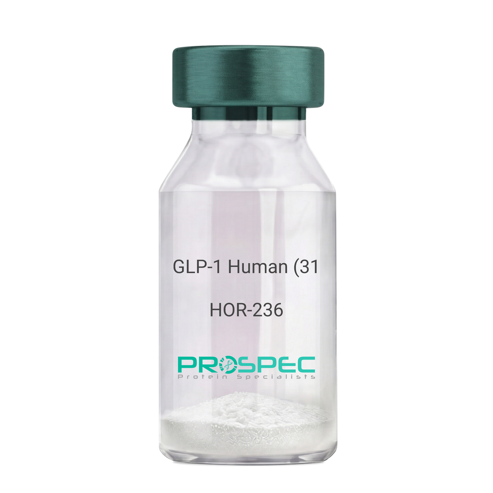
GLP-1 Human (31 a.a.)Description:
Glucagon Like Peptide-1 (31 a.a.) Human Recombinant
GLP1, GLP2, GRPP.
Product # :
HOR-236Price :
Quantity :
Shipping Method :
Shipped at Room temp
More Info
- description
- source
- formulation
- purity
- More Info
Description
Glucagon Like Peptide-1 Human Recombinant produced in E.Coli is a single, non-glycosylated, polypeptide chain containing 31 amino acids and having a molecular mass of 3.3 kDa. The GLP-1 is purified by proprietary chromatographic techniques.
Source
Escherichia Coli.
Formulation
The Recombinant GLP-1 concentrated solution (1mg/ml) was lyophilized after extensive dialyses against 0.1mg sodium phosphate monobasic & 1.6mg sodium phosphate dibasic.
Purity
Greater than 98.0% as determined by:
(a) Analysis by RP-HPLC.
(b) Analysis by SDS-PAGE.More Info
-
Introduction
Glucagon-like peptide-1 (GLP-1) is derived from the transcription product of the proglucagon gene. The major source of GLP-1 in the body is the intestinal L cell that secretes GLP-1 as a guthormone. The biologically active forms of GLP-1 are: GLP-1-(7-37) and GLP-1-(7-36)NH2.
GLP-1 secretion by L cells is dependent on the presence of nutrients in the lumen of the small intestine. The secretagogues (agents that causes or stimulates secretion) of this hormone include major nutrients like carbohydrate, proteinand lipid. Once in the circulation, GLP-1 has a half life of less than 2 minutes, due to rapid degradation by the enzyme dipeptidyl peptidase-4.
GLP-1 possesses several physiological properties that make it a subject of intensive investigation as a potential treatment of diabetes mellitus. The known physiological functions of GLP-1 include: Increases insulin secretion from the pancreas in a glucose-dependent manner, decreases glucagon secretion from the pancreas, increases beta cells mass and insulin gene expression, inhibits acid secretion and gastric emptying in the stomach, decreases food intake by increasing satiety. -
Synonyms
GLP1, GLP2, GRPP.
-
Physical Appearance
Sterile Filtered White lyophilized (freeze-dried) powder.
-
Stability
Lyophilized Glucagon Like Peptide-1 although stable at room temperature for 3 weeks, should be stored desiccated below -18°C. Upon reconstitution GLP-1 should be stored at 4°C between 2-7 days and for future use below -18°C. For long term storage it is recommended to add a carrier protein (0.1% HSA or BSA).Please prevent freeze-thaw cycles.
-
Solubility
It is recommended to reconstitute the lyophilized Glucagon Like Peptide-1 in sterile 18MΩ-cm H2O not less than 100µg/ml, which can then be further diluted to other aqueous solutions.
-
Amino Acid Sequence
HAEGTFTSDV SSYLEGQAAK EFIAWLVKGR G.
ProSpec's products are furnished for LABORATORY RESEARCH USE ONLY. The product may not be used as drugs, agricultural or pesticidal products, food additives or household chemicals.
- View Data Sheet
Name :
XAGE1A HumanDescription:
X Antigen Family, Member 1A Human Recombinant
X antigen family member 1, XAGE-1, Cancer/testis antigen 12.1, CT12.1, G antigen family D member 2, XAGE1A, GAGED2, XAGE1, CTP9, CT12.1C.
Product # :
PRO-1256Price :
Quantity :
Shipping Method :
Shipped with Ice Packs
More Info
- description
- source
- formulation
- purity
- More Info
Description
XAGE1A Human Recombinant produced in E.Coli is a single, non-glycosylated polypeptide chain containing 104 amino acids (1-81 a.a) and having a molecular mass of 11.5kDa.XAGE1A is fused to a 23 amino acid His-tag at N-terminus & purified by proprietary chromatographic techniques.
Source
Escherichia Coli.
Formulation
XAGE1A protein solution (0.25mg/ml) containing 20mM Tris-HCl buffer (pH 8.0), 0.2M NaCl, 20% glycerol and 2mM DTT.
Purity
Greater than 85.0% as determined by SDS-PAGE.
More Info
-
Introduction
G antigen family D member 2 (XAGE1A) belongs to the XAGE subfamily, which is a member of the GAGE family. The GAGE genes are expressed various tumors and in a number of fetal and reproductive tissues. XAGE1A contains a nuclear localization signal and shares a sequence similarity with other GAGE/PAGE proteins. XAGE1A is strongly expressed in Ewing's sarcoma, alveolar rhabdomyosarcoma and normal testis. Due to the expression pattern and the sequence similarity, the XAGE1A protein is also a member of a family of CT (cancer-testis) antigens.
-
Synonyms
X antigen family member 1, XAGE-1, Cancer/testis antigen 12.1, CT12.1, G antigen family D member 2, XAGE1A, GAGED2, XAGE1, CTP9, CT12.1C.
-
Physical Appearance
Sterile Filtered colorless solution.
-
Stability
Store at 4°C if entire vial will be used within 2-4 weeks. Store, frozen at -20°C for longer periods of time. For long term storage it is recommended to add a carrier protein (0.1% HSA or BSA).Avoid multiple freeze-thaw cycles.
-
Amino Acid Sequence
MGSSHHHHHH SSGLVPRGSH MGSMESPKKK NQQLKVGILH LGSRQKKIRI QLRSQCATWK VICKSCISQT PGINLDLGSG VKVKIIPKEE HCKMPEAGEE QPQV.
ProSpec's products are furnished for LABORATORY RESEARCH USE ONLY. The product may not be used as drugs, agricultural or pesticidal products, food additives or household chemicals.
- View Data Sheet
Name :
OmpA S.EnteritidisDescription:
Salmonella Enteritidis Outer Membrane Protein-A Recombinant
Outer Membrane Protein-A, OmpA.
Product # :
PRO-1918Price :
Quantity :
Shipping Method :
Shipped with Ice Packs
More Info
- description
- source
- formulation
- purity
- More Info
Description
The Recombinant Salmonella Enteritidis Outer Membrane Protein A, E.Coli derived, 330 amino acids, contains the ompA immunodominant regions. The protein is fused to a His tag at C-terminal and purified by standard chromatography techniques.
Source
Escherichia Coli.
Formulation
PBS and 25MmM Arginine.
Purity
Protein is >95% pure as determined by 10% PAGE (coomassie staining).
More Info
-
Introduction
The OmpA protein is one of the main outer-membrane proteins of a large array of Gram-negative bacteria such as A.salmonicida, Shigella dysenteriae and E.coli.OmpA’s major physiological functions include maintenance of the structural integrity and morphology of the cells and porin activity, as well as a role in conjugation and bacteriophage binding.Achromogenic atypical Aeromonas salmonicida is the causative agent of goldfish ulcer disease.Virulence of this bacterium is associated with the production of a paracrystalline outer membrane A-layer protein.The species specific structural gene for the monomeric form of A-protein was cloned into a pET-3d plasmid in order to express and produce a recombinant form of the protein in E.coli BL21(DE3). The induced protein was isolated from inclusion bodies by a simple solubilization-renaturation procedure and purified by ion exchange chromatography on Q-Sepharose to over 95% pure monomeric protein.Recombinant A-protein was compared by biochemical, immunological and molecular methods with the A-protein isolated from atypical A.salmonicida bacterial cells by the glycine and the membrane extraction methods.
-
Synonyms
Outer Membrane Protein-A, OmpA.
-
Physical Appearance
Sterile Filtered clear solution.
-
Stability
OmpA S.Enteritidis Recombinant although stable at 4°C for 1 week, should be stored below -18°C. Please prevent freeze thaw cycles.
-
Applications
Immunoassay. Outer membrane protein A (ompA) of S. Enteritidis is a protein directly exposing to outsides of this organism, In hens, the production of antibodies against outer membrane protein A (ompA) during the infection has been demonstrated by inoculating both the complete bacterium and expressed protein produced from ompA DNA vaccine. Vaccination by ompA protein to hens is a poteintail tool to control S. enteritidis contaminated eggs into market, and prevent human foodborne disease from eggs.
ProSpec's products are furnished for LABORATORY RESEARCH USE ONLY. The product may not be used as drugs, agricultural or pesticidal products, food additives or household chemicals.
- View Data Sheet
Name :
MAT2A HumanDescription:
Methionine Adenosyltransferase II Alpha Human Recombinant
MATA2, MATII, SAMS2, MAT-2A, S-adenosylmethionine synthetase isoform type-2, AdoMet synthetase 2, Methionine adenosyltransferase 2, Methionine adenosyltransferase II, MAT2A, AMS2.
Product # :
ENZ-320Price :
Quantity :
Shipping Method :
Shipped with Ice Packs
More Info
- description
- source
- formulation
- purity
- More Info
Description
MAT2A Human Recombinant fused with His tag (20 a.a.) at N-terminus produced in E.Coli is a single, non-glycosylated, polypeptide chain containing 415 amino acids and having a molecular mass of 45.8 kDa.The MAT2A is purified by proprietary chromatographic techniques.
Source
Escherichia Coli.
Formulation
The MAT2A solution (1mg/ml) contains 20mM Tris pH-8 & 10% glycerol.
Purity
Greater than 95.0% as determined by SDS-PAGE.
More Info
-
Introduction
MAT2A is an important enzyme in cellular metabolism and catalyzes the formation of S-adenosylmethionine (SAMe) from L-methionine and ATP. MAT2A is expressed in extrahepatic tissues. In liver, MAT2A expression associates with growth, dedifferentiation, and cancer. NF-kappa B and AP-1 are necessary for basal MAT2A expression in HepG2 cells and mediate the increase in MAT2A expression in response to TNF-alpha. Up-regulation of MAT2A provides growth improvement and s-adenosylmethionine and methylthioadenosine thus can block mitogenic signaling in colon cancer cells. Lower expression of both MAT2A and MAT2beta and interfere with leptin signaling in liver cancer cells.
-
Synonyms
MATA2, MATII, SAMS2, MAT-2A, S-adenosylmethionine synthetase isoform type-2, AdoMet synthetase 2, Methionine adenosyltransferase 2, Methionine adenosyltransferase II, MAT2A, AMS2.
-
Physical Appearance
Sterile Filtered colorless solution.
-
Stability
Store at 4°C if entire vial will be used within 2-4 weeks. Store, frozen at -20°C for longer periods of time. For long term storage it is recommended to add a carrier protein (0.1% HSA or BSA).Please prevent freeze-thaw cycles.
-
Amino Acid Sequence
MGSSHHHHHH SSGLVPRGSH MNGQLNGFHE AFIEEGTFLF TSESVGEGHP DKICDQISDA VLDAHLQQDP DAKVACETVA KTGMILLAGE ITSRAAVDYQ KVVREAVKHI GYDDSSKGFD YKTCNVLVAL EQQSPDIAQG VHLDRNEEDI GAGDQGLMFG YATDETEECM PLTIVLAHKL NAKLAELRRN GTLPWLRPDS KTQVTVQYMQ DRGAVLPIRV HTIVISVQHD EEVCLDEMRD ALKEKVIKAV VPAKYLDEDT IYHLQPSGRF VIGGPQGDAG LTGRKIIVDT YGGWGAHGGG AFSGKDYTKV DRSAAYAARW VAKSLVKGGL CRRVLVQVSY AIGVSHPLSI SIFHYGTSQK SERELLEIVK KNFDLRPGVI VRDLDLKKPI YQRTAAYGHF GRDSFPWEVP KKLKY.
ProSpec's products are furnished for LABORATORY RESEARCH USE ONLY. The product may not be used as drugs, agricultural or pesticidal products, food additives or household chemicals.
- View Data Sheet
Name :
HERV-KDescription:
Endogenous retrovirus K Envelope Human Recombinant
Product # :
HER-001Price :
Quantity :
Shipping Method :
Shipped with Ice Packs
More Info
- description
- source
- formulation
- purity
- More Info
Description
The E.Coli derived HERV-K recombinant truncated protein is fused to a Six histidine tag at C-terminus and has a MW of 51.5kDa (pI 9.06).
Source
Escherichia Coli.
Formulation
10mM Tris-HCl, pH 7.2.
Purity
Protein is >90% pure as determined by SDS PAGE.
More Info
-
Introduction
HERVK is related with several tumors such as passenger virus, and anti-Retroviral therapy against amyotrophic lateral sclerosis/ALS has exhibited to decrease the symptoms of amyotrophic lateral sclerosis/ALS. High counts of viral sequences exist in the human genome but stay silent. Though, in pathological conditions, these viruses are produced. Human Endogenous Retrovirus-K, is produced in neurons of a subpopulation of patients with amyotrophic lateral sclerosis/ALS, a progressive neurodegenerative disease. The envelope protein of this HERV results in degeneration of neurons, and transgenic animals producing this protein advance an ALS-like syndrome triggered by nucleolar dysfunction in motor neurons. Reactivation of the HERVK is controlled by the transcription factor TDP-43. Therefore, therapeutic tactics against this virus alter the course of the disease.
-
Physical Appearance
Sterile Filtered clear solution.
-
Stability
HERV-K although stable at 4°C for 1 week, should be stored below -18°C. Please prevent freeze thaw cycles.
-
Amino Acid Sequence
MWTVPSFTND SYQVYNVFST NSFQLLTVKR TPHEAWRVPL TTKTNKTKGL PDCPKKPTNG PFIVTSILWD NCNAPKAVVL QTLAMGIVID WAPKGHYWQD CSSKNTLCSE FIYSLDYIEH GWQSYTMRQR VSPYPFKWMD TGIAPPRPKI IHPFFTPEHP ELWKLAAALS GIKIWNTTYQ LLRTKTKTPT FNITLISEWV IPIRSCVKPP YMLLVGNIIM MPDAQTIECH NCKLFTCIDA TFNPTTSILL VRAREGVWIP VSLHRPWESS PSIHIVNEVL KDILKRTKRF IFTLIAVLAG LLAVTATAAT AGVAIRSSVQ TAHYVEACQK NSSRLWNSQA QIDQKLANQI NDLRQSVTWL GDRVMNLQHR MQLQCDWNTS DYCITPYAYN QDQHSWENVS RHLKAWDDNL TLDISQLKEQIFEASQAHLS TVPGSHIFEG ITKQLPDFNP FKWLKPVRGS LLLLALLILV CLCCLLLVCRCL.
-
Applications
Detection of Human Endogenious Retrovirus K in individuals is untested.
ProSpec's products are furnished for LABORATORY RESEARCH USE ONLY. The product may not be used as drugs, agricultural or pesticidal products, food additives or household chemicals.
- View Data Sheet
Name :
GPT1 AntibodyDescription:
Anti Human Glutamate Pyruvate Transaminase 1
Product # :
ANT-778Price :
Quantity :
Shipping Method :
Shipped with Ice Packs
More Info
- description
- formulation
- purity
- More Info
Description
Monoclonal antibody to glutamate-pyruvate-transaminase-1
Formulation
PBS, sodium chloride and 0.095 % NaN3 as a preservative.
Purity
Protein is >95% pure.
More Info
-
Physical Appearance
Sterile Filtered clear solution.
-
Stability
GPT1 Antibody although stable at 4°C for 1 week, should be stored below -18°C. Please prevent freeze thaw cycles.
-
Applications
Immunoassay.
-
Background
Monoclonal antibodies to Glutamate-Pyruvate Transaminase 1 (GPT/ALT1) are engineered proteins designed to bind GPT1, a key
metabolic enzyme, for applications such as Western Blotting, ELISA, and immunofluorescence. These antibodies (often Mouse IgG1) are needed to recognize liver injury/ ALT1 metabolic activity. -
Type
Mouse antibody Monoclonal.
-
Purification Method
Purified monoclonal IgG1 by protein A chromatography.
ProSpec's products are furnished for LABORATORY RESEARCH USE ONLY. The product may not be used as drugs, agricultural or pesticidal products, food additives or household chemicals.
- View Data Sheet
Name :
HIV-1 p24 CoreDescription:
HIV-1 p24 Core Recombinant
Product # :
HIV-103Price :
Quantity :
Shipping Method :
Shipped with Ice Packs
More Info
- description
- source
- formulation
- purity
- More Info
Description
HIV-1 p24 Core is a 24kDa non-glycosylated polypeptide chain, containing the full length sequence of the HIV-1 p24.
Source
Escherichia Coli.
Formulation
50mM Tris, pH 8.5 and 8M urea.
Purity
Greater than 95.0% as determined by HPLC analysis and SDS-PAGE.
More Info
-
Introduction
Human immunodeficiency virus (HIV) is a retrovirusthat can lead to a condition in which the immune systembegins to fail, leading to opportunistic infections. HIV primarily infects vital cells in the humanimmune systemsuch as helper T cells(specifically CD4+ T cells), macrophagesand dendritic cells. HIV infection leads to low levels of CD4+ T cells through three main mechanisms: firstly, direct viral killing of infected cells; secondly, increased rates of apoptosisin infected cells; and thirdly, killing of infected CD4+ T cells by CD8 cytotoxic lymphocytesthat recognize infected cells. When CD4+ T cell numbers decline below a critical level, cell-mediated immunityis lost, and the body becomes progressively more susceptible to opportunistic infections. HIV was classified as a member of the genus Lentivirus, part of the family of Retroviridae. Lentiviruses have many common morphologies and biological properties. Many species are infected by lentiviruses, which are characteristically responsible for long-duration illnesses with a long incubation period. Lentiviruses are transmitted as single-stranded, positive-sense, enveloped RNA viruses. Upon entry of the target cell, the viral RNA genomeis converted to double-stranded DNAby a virally encoded reverse transcriptasethat is present in the virus particle. This viral DNA is then integrated into the cellular DNA by a virally encoded integraseso that the genome can be transcribed. Once the virus has infected the cell, two pathways are possible: either the virus becomes latentand the infected cell continues to function, or the virus becomes active and replicates, and a large number of virus particles are liberated that can then infect other cells.
-
Physical Appearance
Sterile filtered colorless clear solution.
-
Stability
HIV-1 p24 core although stable at 4°C for 1 week, should be stored below -18°C. Please prevent freeze thaw cycles.
-
Specificity
Immunoreactive with all sera of HIV-1 infected individuals.
ProSpec's products are furnished for LABORATORY RESEARCH USE ONLY. The product may not be used as drugs, agricultural or pesticidal products, food additives or household chemicals.
- View Data Sheet
Name :
CMV Pp65, 561 a.a.Description:
Cytomegalo Virus Pp65(UL83), 561 a.a.Recombinant
Product # :
CMV-218Price :
Quantity :
Shipping Method :
Shipped with Ice Packs
More Info
- description
- source
- formulation
- purity
- More Info
Description
The E.Coli derived 62.8 kDa recombinant protein contains the CMV Pp65 (UL83) immunodominant regions, having 561 amino acids.
Source
Escherichia Coli.
Formulation
25mM Tris-Hcl pH 8, 8M Urea, 5mM bMe.
Purity
CMV Pp65 protein is >95% pure as determined by SDS-PAGE.
More Info
-
Introduction
CMV is part of the Betaherpesvirinae subfamily of Herpesviridae; including herpes simplex virustypes 1 and 2, varicella-zoster virus, and Epstein-Barrvirus. The herpesviruses has the common ability to stay latentover long periods of time. CMV has the largest genome of the herpes viruses, ranging from 230-240 kilobase pairs. CMV is a double-stranded linear DNA virus with 162 hexagonal protein capsomeres surrounded by a lipid membrane. Human CMV is composed of unique and inverted repeats that include the existence of 4 genome isomers caused by inversion of L-S genome components (class E). Replication may be divided into immediate early, delayed early, and late gene expression based on time of synthesis after infection. The DNA is replicated by rolling circles. In vitro, CMV replicates in human fibroblasts.
-
Stability
CMV Pp65 protein although stable at 4°C for 1 week, should be stored below -18°C. Please prevent freeze thaw cycles.
-
Applications
CMV Pp65 antigen is suitable for ELISA and Western blots, excellent antigen for detection of CMV with minimal specificity problems.
-
Specificity
Immunoreactive with sera of CMV-infected individuals.
-
Purification Method
CMV Pp65 was purified by proprietary chromatographic technique.
ProSpec's products are furnished for LABORATORY RESEARCH USE ONLY. The product may not be used as drugs, agricultural or pesticidal products, food additives or household chemicals.
- View Data Sheet
Name :
Cys-Protein-A/G/LDescription:
Cys-Protein A/G/L Recombinant
Product # :
PRO-1935Price :
Quantity :
Shipping Method :
Shipped at Room temp
More Info
- description
- source
- formulation
- purity
- More Info
Description
Recombinant Protein-A/G/L produced in E.Coli is a single non-glycosylated polypeptide chain fused with a Cys at N-terminus. Protein- A/G/L is comprised of 5 IgG-binding regions of Protein A (E-D-A-B-C), 2 of protein G (C1-C3) and 5 of Protein L (B1-B2-B3-B4-B5) containing 806 amino acids in total and having a molecular mass of 89.3kDa. Cell wall binding region, cell membrane binding region and albumin binding region have been eliminated from the recombinant Protein- A/G/L to guarantee the maximum specific IgG binding.
Source
Escherichia Coli.
Formulation
Protein- A/G/L was lyophilized without any additives.
Purity
Greater than 95.0% as determined by:
(a) Analysis by SEC-HPLC.
(b) Analysis by SDS-PAGE.More Info
-
Introduction
The recombinant Protein A/G/L is a genetically engineered protein which combines the IgG binding profiles of all Protein A, Protein G and Protein L. Protein A/G/L is a gene fusion product. Recombinant fusion protein A/G/L is comprised of 5 Ig-binding regions of protein L (B1-B2-B3-B4-B5), 5 IgG binding domains from Protein A (E-D-A-B-C) and 2 Ig-binding region of protein G (C1-C3). The recombinant Protein A/G/L is ideal for purification of monoclonal or polyclonal IgG antibodies. Protein A/G/L binds to IgG from humans, mice, rats, cows, goats, sheep, rabbits, guinea pigs, pigs, dogs and cats.
-
Physical Appearance
Sterile Filtered White lyophilized (freeze-dried) powder.
-
Stability
Lyophilized Protein-A/G/L although stable at room temperature for 3 weeks, should be stored desiccated below -18°C. Upon reconstitution Protein-A/G/L should be stored at 4°C between 2-7 days and for future use below -18°C. For long term storage it is recommended to add a carrier protein (0.1% HSA or BSA). Please prevent freeze-thaw cycles.
-
Solubility
It is recommended to reconstitute the lyophilized Protein-A/G/L in sterile 18M-cm H2O not less than 0.1mg/ml, which can then be further diluted to other aqueous solutions.
-
Amino Acid Sequence
CNAAQHDEAQ QNAFYQVLNM PNLNADQRNG FIQSLKDDPS QSANVLGEAQ KLNDSQAPKA DAQQNNFNKD QQSAFYEILN MPNLNEAQRN GFIQSLKDDP SQSTNVLGEA KKLNESQAPK ADNNFNKEQQ NAFYEILNMP NLNEEQRNGF IQSLKDDPSQ SANLLSEAKK LNESQAPKAD NKFNKEQQNA FYEILHLPNL NEEQRNGFIQ SLKDDPSQSA NLLAEAKKLN DAQAPKADNK FNKEQQNAFY EILHLPNLTE EQRNGFIQSL KDDPSVSKEI LAEAKKLNDA QAPKEEDSLE GSGSGTYKLI LNGKTLKGET TTEAVDAATA EKVFKQYAND NGVDGEWTYD DATKTFTVTE KPEVIDASEL TPAVTTYKLV INGKTLKGET TTKAVDAETA EKAFKQYAND NGVDGVWTYD DATKTFTVTE EPRARPGSGS GKEETPETPE TDSEEEVTIK ANLIFANGST QTAEFKGTFE KATSEAYAYA DTLKKDNGEY TVDVADKGYT LNIKFAGKEK TPEEPKEEVT IKANLIYADG KTQTAEFKGT FEEATAEAYR YADALKKDNG EYTVDVADKG YTLNIKFAGK EKTPEEPKEE VTIKANLIYA DGKTQTAEFK GTFEEATAEA YRYADLLAKE NGKYTVDVAD KGYTLNIKFA GKEKTPEEPK EEVTIKANLI YADGKTQTAE FKGTFAEATA EAYRYADLLA KENGKYTADL EDGGYTINIR FAGKKVDEKP EEKEQVTIKE NIYFEDGTVQ TATFKGTFAE ATAEAYRYAD LLSKEHGKYT ADLEDGGYTI NIRFAG.
ProSpec's products are furnished for LABORATORY RESEARCH USE ONLY. The product may not be used as drugs, agricultural or pesticidal products, food additives or household chemicals.
- View Data Sheet
Name :
HIV-1 p24, HisDescription:
HIV-1 p24 Recombinant, His Tag
Product # :
HIV-159Price :
Quantity :
Shipping Method :
Shipped with Ice Packs
More Info
- description
- source
- formulation
- purity
- More Info
Description
HIV-1 p24 His Tag Recombinant produced in E.Coli is a single, non-glycosylated polypeptide chain containing 188 amino acids (155-321 a.a.) and having a molecular mass of 21.2 kDa. The HIV-1 p24 is fused to a 21 amino acid His Tag and purified by conventional chromatography.
Source
Escherichia Coli.
Formulation
The HIV-1 p24 protein solution contains 20mM Tris-HCl pH-8, 0.1mM PMSF, 0.1M NaCl and 10% glycerol.
Purity
Greater than 80.0% as determined by SDS-PAGE.
More Info
-
Introduction
The HIV1 p24 His Tag protein performs complex orchestrated tasks during the assembly, budding maturation and infection stages of the viral replication cycle. Throughout viral assembly, the proteins form membrane associations and self-associations that ultimately result in budding of an immature virion from the infected cell. HIV-1 P24 gag, the key capsid protein of the HIV-1 virion, is used in clinical trials as one of the components of the HIV-1 vaccine because of the great extent of sequence homology between different isolates.
-
Physical Appearance
Sterile Filtered colorless solution.
-
Stability
Store at 4°C if entire vial will be used within 2-4 weeks. Store, frozen at -20°C for longer periods of time. For long term storage it is recommended to add a carrier protein (0.1% HSA or BSA).Avoid multiple freeze-thaw cycles.
-
Amino Acid Sequence
MGSSHHHHHH SSGLVPRGSH MWVKVVEEKA FSPEVIPMFS ALSEGATPQD LNTMLNTVGG HQAAMQMLKE TINEEAAEWD RLHPVHAGPI APGQMREPRG SDIAGTTSTL QEQIGWMTHN PPIPVGEIYK RWIILGLNKI VRMYSPTSIL DIRQGPKEPF RDYVDRFYKT LRAEQASQEV
KNWMTETL.
ProSpec's products are furnished for LABORATORY RESEARCH USE ONLY. The product may not be used as drugs, agricultural or pesticidal products, food additives or household chemicals.
- View Data Sheet
Name :
AngiotensinDescription:
Angiotensin
Angiotensinogen, Serpin A8, ANHU, SERPINA8.
Product # :
ENZ-283Price :
Quantity :
Shipping Method :
Shipped at Room temp
More Info
- description
- source
- purity
- More Info
Description
Angiotensin contains a total of 8 amino acids having a molecular weight of 1031.2 Dalton and a molecular formula of C49H70N14O11.
Source
Synthetic.
Purity
Greater than 98.0% as determined by RP-HPLC.
More Info
-
Introduction
Angiotensin is an oligopeptide in the blood that causes vasoconstriction, increased blood pressure, and release of aldosterone from the adrenal cortex. It is a powerful dipsogen. It is derived from the precursor molecule angiotensinogen, a serum globulin produced in the liver. It plays an important role in the renin-angiotensin system.
The protein encoded by this gene, pre-angiotensinogen or angiotensinogen precursor, is expressed in the liver and is cleaved by the enzyme renin in response to lowered blood pressure. The resulting product, angiotensin I is then cleaved by angiotensin converting enzyme (ACE) to generate the physiologically active enzyme angiotensin II. The protein is involved in maintaining blood pressure and in the pathogenesis of essential hypertension and preeclampsia. -
Synonyms
Angiotensinogen, Serpin A8, ANHU, SERPINA8.
-
Physical Appearance
Sterile Filtered white lyophilized powder.
-
Stability
Lyophilized Angiotensin although stable at room temperature for 3 weeks, should be stored desiccated below -18°C. Upon reconstitution Serpin A8 should be stored at 4°C between 2-7 days and for future use below -18°C.Please prevent freeze-thaw cycles.
-
Solubility
It is recommended to reconstitute the lyophilized Angiotensin in sterile 18MΩ-cm H2O not less than 100 µg/ml or more than 10 mg/ml solutions.
-
Amino Acid Sequence
Asn-Arg-Val-Tyr-Val-His-Pro-Phe-OH.
ProSpec's products are furnished for LABORATORY RESEARCH USE ONLY. The product may not be used as drugs, agricultural or pesticidal products, food additives or household chemicals.
- View Data Sheet
Name :
Encephalitis Japanese 12kdaDescription:
Japanese Encephalitis Virus 12kda Recombinant
Product # :
TBE-288Price :
Quantity :
Shipping Method :
Shipped with Ice Packs
More Info
- description
- source
- formulation
- purity
- More Info
Description
Recombinant Japanese Encephalitis Virus produced in E. coli contains 110 amino acids and having a Mw of 12kDa. Encephalitis Japanese is fused to a His tag at its N-terminus and purified by proprietary chromatographic technique.
Source
Escherichia Coli.
Formulation
Encephalitis Japanese solution contains PBS & 25mM K2CO3.
Purity
Protein is >95% pure as determined by 10% PAGE (coomassie staining).
More Info
-
Physical Appearance
Sterile Filtered clear solution.
-
Stability
Store at 4°C if entire vial will be used within 2-4 weeks. Store, frozen at -20°C for longer periods of time. For long term storage it is recommended to add a carrier protein (0.1% HSA or BSA). Avoid multiple freeze-thaw cycles.
-
Applications
ELISA.
-
Background
Recombinant JEV is designed to elicit a strong immune response, making it a valuable tool for vaccine development. Studies have shown that rJEV can induce robust neutralizing antibodies and cellular immune responses in animal models, providing protection against wild-type JEV infection. Pathogenicity and Virulence Factors: rJEV is used to dissect the roles of various viral proteins in disease pathogenesis. By mutating specific genes, researchers can identify virulence factors and understand how the virus interacts with host cells and evades the immune system.
ProSpec's products are furnished for LABORATORY RESEARCH USE ONLY. The product may not be used as drugs, agricultural or pesticidal products, food additives or household chemicals.
- View Data Sheet
Name :
ABO HumanDescription:
ABO Blood Group Human Recombinant
Histo-blood group ABO system transferase, Fucosylglycoprotein 3-alpha-galactosyltransferase, Fucosylglycoprotein alpha-N-acetylgalactosaminyltransferase, Glycoprotein-fucosylgalactoside alpha-N-acetylgalactosaminyltransferase, Glycoprotein-fucosylgalactoside alpha-galactosyltransferase, Histo-blood group A transferase, A transferase, Histo-blood group B transferase, B transferase, NAGAT, ABO, GTB, A3GALNT, A3GALT1.
Product # :
ENZ-167Price :
Quantity :
Shipping Method :
Shipped with Ice Packs
More Info
- description
- source
- formulation
- purity
- More Info
Description
ABO Human Recombinant produced in E.Coli is a single, non-glycosylated polypeptide chain containing 322 amino acids (54-354 a.a) and having a molecular mass of 37.4kDa.ABO is fused to a 21 amino acid His-tag at N-terminus & purified by proprietary chromatographic techniques.
Source
Escherichia Coli.
Formulation
ABO protein solution (0.5mg/ml) containing 20mM Tris-HCl buffer (pH 8.0), 2mM DTT, 20% glycerol and 200mM NaCl.
Purity
Greater than 85.0% as determined by SDS-PAGE.
More Info
-
Introduction
NAGAT (ABO) is a member of the glycosyltransferase 6 family. The ABO protein is the basis of the ABO blood group system and related to the first discovered blood group system, ABO. The allele that is present in an individual determines the blood group. The histo-blood group ABO is comprised of 3 carbohydrate antigens: A, B, and H. A, B, and AB individuals express a glycosyltransferase activity which converts the H antigen to the A antigen (by addition of UDP-GalNAc) or to the B antigen (by addition of UDP-Gal), whereas O individuals are deficient of such activity.
-
Synonyms
Histo-blood group ABO system transferase, Fucosylglycoprotein 3-alpha-galactosyltransferase, Fucosylglycoprotein alpha-N-acetylgalactosaminyltransferase, Glycoprotein-fucosylgalactoside alpha-N-acetylgalactosaminyltransferase, Glycoprotein-fucosylgalactoside alpha-galactosyltransferase, Histo-blood group A transferase, A transferase, Histo-blood group B transferase, B transferase, NAGAT, ABO, GTB, A3GALNT, A3GALT1.
-
Physical Appearance
Sterile Filtered colorless solution.
-
Stability
Store at 4°C if entire vial will be used within 2-4 weeks. Store, frozen at -20°C for longer periods of time. For long term storage it is recommended to add a carrier protein (0.1% HSA or BSA).Avoid multiple freeze-thaw cycles.
-
Amino Acid Sequence
MGSSHHHHHH SSGLVPRGSH MAVREPDHLQ RVSLPRMVYP QPKVLTPCRK DVLVVTPWLA PIVWEGTFNI DILNEQFRLQ NTTIGLTVFA IKKYVAFLKL FLETAEKHFM VGHRVHYYVF TDQPAAVPRV TLGTGRQLSV LEVRAYKRWQ DVSMRRMEMI SDFCERRFLS EVDYLVCVDV DMEFRDHVGV EILTPLFGTL HPGFYGSSRE AFTYERRPQS QAYIPKDEGD FYYLGGFFGG SVQEVQRLTR ACHQAMMVDQ ANGIEAVWHD ESHLNKYLLR HKPTKVLSPE YLWDQQLLGW PAVLRKLRFT AVPKNHQAVR NP.
ProSpec's products are furnished for LABORATORY RESEARCH USE ONLY. The product may not be used as drugs, agricultural or pesticidal products, food additives or household chemicals.
- View Data Sheet
Name :
AK2 HumanDescription:
Adenylate Kinase 2 Human Recombinant
ADK2, AK-2, Adenylate kinase isoenzyme 2 mitochondrial, ATP-AMP transphosphorylase 2, adenylate kinase 2.
Product # :
PKA-260Price :
Quantity :
Shipping Method :
Shipped with Ice Packs
More Info
- description
- source
- formulation
- purity
- biological activity
- More Info
Description
AK2 Human Recombinant produced in E.Coli is a single, non-glycosylated, polypeptide chain containing 259 amino acids and having a molecular mass of 28.6 kDa. AK2 is fused to 20 a.a. His-Tag at N-terminus and purified by proprietary chromatographic techniques.
Source
Escherichia Coli.
Formulation
AK2 solution containing 20mM Tris pH-7.5, 5mM DTT and 20% glycerol.
Purity
Greater than 95.0% as determined by SDS-PAGE.
Biological Activity
Specific activity: > 1.5 units/ml. One unit will convert 2.0 umoles of ADP to ATP + AMP per minute at pH 7.5 at 25C.More Info
-
Introduction
Adenylate kinases play a role in regulating the adenine nucleotide composition within a cell by catalyzing the reversible transfer of phosphate groups among adenine nucleotides. There are 3 types of adenylate kinase isozymes, AK1, AK2, and AK3 in vertebrates. Expression of these isozymes are tissue-specific and developmentally regulated. AK2 is localized in the mitochondrial intermembrane space and is involved in apoptosis. AK2 is mutated in individuals with reticular dysgenesis.
-
Synonyms
ADK2, AK-2, Adenylate kinase isoenzyme 2 mitochondrial, ATP-AMP transphosphorylase 2, adenylate kinase 2.
-
Physical Appearance
Sterile filtered colorless solution.
-
Stability
AK2 Human Recombinant although stable at 4°C for 1 week, should be stored below -18°C. Please prevent freeze thaw cycles.
-
Amino Acid Sequence
MGSSHHHHHH SSGLVPRGSH MAPSVPAAEP EYPKGIRAVL LGPPGAGKGTQAPRLAENFC VCHLATGDML RAMVASGSEL GKKLKATMDA GKLVSDEMVV ELIEKNLETP LCKNGFLLDG FPRTVRQAEM LDDLMEKRKE KLDSVIEFSIPDSLLIRRIT GRLIHPKSGR SYHEEFNPPK EPMKDDITGE PLIRRSDDNE KALKIRLQAY HTQTTPLIEY YRKRGIHSAI DASQTPDVVF ASILAAFSKA TCKDLVMFI.
ProSpec's products are furnished for LABORATORY RESEARCH USE ONLY. The product may not be used as drugs, agricultural or pesticidal products, food additives or household chemicals.