Search results
1000 results found for “other neurotrophins”
Name
Description
Product #
Price
Quantity
Shipping Method
- View Data Sheet
Name :
F11R HumanDescription:
F11 Receptor Human Recombinant
Junctional adhesion molecule A, JAM-A, Junctional adhesion molecule 1, JAM-1, Platelet F11 receptor, Platelet adhesion molecule 1, PAM-1, CD321, F11R, JAM1, JCAM, JAM, KAT, JAMA.
Product # :
PRO-1112Price :
Quantity :
Shipping Method :
Shipped with Ice Packs
More Info
- description
- source
- formulation
- purity
- More Info
Description
F11R Human Recombinant produced in E.Coli is a single, non-glycosylated polypeptide chain containing 238 amino acids (26-238 a.a) and having a molecular mass of 25.8kDa.F11R is fused to a 25 amino acid His-tag at N-terminus & purified by proprietary chromatographic techniques.
Source
Escherichia Coli.
Formulation
F11R protein solution (1mg/ml) containing 20mM Tris-HCl buffer (pH8.0), 10% glycerol, 0.15M NaCl and 1mM DTT.
Purity
Greater than 90.0% as determined by SDS-PAGE.
More Info
-
Introduction
F11R (CD321) is a member of the immunoglobulin superfamily. F11R has a role in epithelial tight junction formation. Tight junctions exemplify one type of cell-to-cell adhesion in epithelial or endothelial cell sheets, establishing continuous seals around cells and acting as a physical barrier to thwart solutes and water from passing easily through the paracellular space. F11R protein can function as a receptor for reovirus, or as a ligand for the integrin LFA1, involved in leukocyte transmigration, or a platelet receptor.
-
Synonyms
Junctional adhesion molecule A, JAM-A, Junctional adhesion molecule 1, JAM-1, Platelet F11 receptor, Platelet adhesion molecule 1, PAM-1, CD321, F11R, JAM1, JCAM, JAM, KAT, JAMA.
-
Physical Appearance
Sterile Filtered colorless solution.
-
Stability
Store at 4°C if entire vial will be used within 2-4 weeks. Store, frozen at -20°C for longer periods of time. For long term storage it is recommended to add a carrier protein (0.1% HSA or BSA).Avoid multiple freeze-thaw cycles.
-
Amino Acid Sequence
MGSSHHHHHH SSGLVPRGSH MGSHMLGSVT VHSSEPEVRI PENNPVKLSC AYSGFSSPRV EWKFDQGDTT RLVCYNNKIT ASYEDRVTFL PTGITFKSVT REDTGTYTCM VSEEGGNSYG EVKVKLIVLV PPSKPTVNIP SSATIGNRAV LTCSEQDGSP PSEYTWFKDG IVMPTNPKST
RAFSNSSYVL NPTTGELVFD PLSASDTGEY SCEARNGYGT PMTSNAVRME AVERNVGV.
ProSpec's products are furnished for LABORATORY RESEARCH USE ONLY. The product may not be used as drugs, agricultural or pesticidal products, food additives or household chemicals.
- View Data Sheet
Name :
EDAR HumanDescription:
Ectodysplasin A Receptor Human Recombinant
Ectodysplasin A Receptor, DL, Ectodysplasin 1, Anhidrotic Receptor, Anhidrotic Ectodysplasin Receptor 1, Ectodermal Dysplasia Receptor, Ectodysplasin-A Receptor, Downless Homolog, EDA-A1 Receptor, ECTD10A, ECTD10B, EDA3, HRM1, ED3, Tumor Necrosis Factor Receptor Superfamily Member EDAR, Downless, Mouse, Homolog Of, EDA-A1R, EDA1R, ED1R, ED5, Tumor necrosis factor receptor superfamily member EDAR.
Product # :
PRO-2092Price :
Quantity :
Shipping Method :
Shipped with Ice Packs
More Info
- description
- source
- formulation
- purity
- More Info
Description
EDAR Human Recombinant produced in E.Coli is a single, non-glycosylated polypeptide chain containing 445 amino acids (27-448 a.a) and having a molecular mass of 48.2kDa. EDAR is fused to a 23 amino acid His-tag at N-terminus & purified by proprietary chromatographic techniques.
Source
Escherichia Coli.
Formulation
EDAR protein solution (0.25mg/ml) containing 20mM Tris-HCl (pH 8.0) and 10% glycerol.
Purity
Greater than 85.0% as determined by SDS-PAGE.
More Info
-
Introduction
Ectodysplasin A Receptor, also known as EDAR belongs to the tumor necrosis factor receptor family. EDAR is a receptor for the soluble ligand ectodysplasin A, and is capable of activating the nuclear factor-kappaB, JNK, as well as caspase-independent cell death pathways. EDAR is necessary for the development of hair, teeth, and other ectodermal derivatives. Furthermore, mutations in EDAR resulted in autosomal dominant and recessive forms of hypohidrotic ectodermal dysplasia.
-
Synonyms
Ectodysplasin A Receptor, DL, Ectodysplasin 1, Anhidrotic Receptor, Anhidrotic Ectodysplasin Receptor 1, Ectodermal Dysplasia Receptor, Ectodysplasin-A Receptor, Downless Homolog, EDA-A1 Receptor, ECTD10A, ECTD10B, EDA3, HRM1, ED3, Tumor Necrosis Factor Receptor Superfamily Member EDAR, Downless, Mouse, Homolog Of, EDA-A1R, EDA1R, ED1R, ED5, Tumor necrosis factor receptor superfamily member EDAR.
-
Physical Appearance
Sterile Filtered clear solution.
-
Stability
Store at 4°C if entire vial will be used within 2-4 weeks. Store, frozen at -20°C for longer periods of time.For long term storage it is recommended to add a carrier protein (0.1% HSA or BSA).Please avoid freeze thaw cycles.
-
Amino Acid Sequence
MGSSHHHHHH SSGLVPRGSH MGSEYSNCGE NEYYNQTTGL CQECPPCGPG EEPYLSCGYG TKDEDYGCVP CPAEKFSKGG YQICRRHKDC EGFFRATVLT PGDMENDAEC GPCLPGYYML ENRPRNIYGM VCYSCLLAPP NTKECVGATS GASANFPGTS GSSTLSPFQH AHKELSGQGH LATALIIAMS TIFIMAIAIV LIIMFYILKT KPSAPACCTS HPGKSVEAQV SKDEEKKEAP DNVVMFSEKD EFEKLTATPA KPTKSENDAS SENEQLLSRS VDSDEEPAPD KQGSPELCLL SLVHLAREKS ATSNKSAGIQ SRRKKILDVY ANVCGVVEGL SPTELPFDCL EKTSRMLSST YNSEKAVVKT WRHLAESFGL KRDEIGGMTD GMQLFDRIST AGYSIPELLT KLVQIERLDA VESLCADILE WAGVVPPASQ PHAAS.
ProSpec's products are furnished for LABORATORY RESEARCH USE ONLY. The product may not be used as drugs, agricultural or pesticidal products, food additives or household chemicals.
- View Data Sheet
Name :
NFU1 HumanDescription:
NFU1 Human Recombinant
CGI-33, HIRIP, HIRIP5, MMDS1, Nfu, NifU, NIFUC, NFU1 iron-sulfur cluster scaffold homolog, mitochondrial, HIRA-interacting protein 5.
Product # :
PRO-2129Price :
Quantity :
Shipping Method :
Shipped with Ice Packs
More Info
- description
- source
- formulation
- purity
- More Info
Description
NFU1 Human Recombinant produced in E.Coli is a single, non-glycosylated polypeptide chain containing 268 amino acids (10-254 a.a) and having a molecular mass of 29.9kDa.NFU1 is fused to a 23 amino acid His-tag at N-terminus & purified by proprietary chromatographic techniques.
Source
Escherichia Coli.
Formulation
NFU1 protein solution (0.5mg/ml) containing Phosphate buffer saline (pH 7.4), 20% glycerol and 1mM DTT.
Purity
Greater than 90.0% as determined by SDS-PAGE.
More Info
-
Introduction
NFU1 is a protein which is localized to mitochondria and plays a vital role in iron-sulfur cluster biogenesis. The NFU1 protein constructs and transfers 4Fe-4S clusters to target apoproteins including succinate dehydrogenase and lipoic acid synthase. NFU1 gene mutations cause multiple mitochondrial dysfunctions syndrome-1, and pseudogenes of the NFU1 gene are located on the short arms of chromosomes 1 and 3.
-
Synonyms
CGI-33, HIRIP, HIRIP5, MMDS1, Nfu, NifU, NIFUC, NFU1 iron-sulfur cluster scaffold homolog, mitochondrial, HIRA-interacting protein 5.
-
Physical Appearance
Sterile Filtered clear solution.
-
Stability
Store at 4°C if entire vial will be used within 2-4 weeks. Store, frozen at -20°C for longer periods of time. For long term storage it is recommended to add a carrier protein (0.1% HSA or BSA).Avoid multiple freeze-thaw cycles.
-
Amino Acid Sequence
MGSSHHHHHH SSGLVPRGSH MGSGAAAVAA GLRRRFCHML KNPYTIKKQP LHQFVQRPLF PLPAAFYHPV RYMFIQTQDT PNPNSLKFIP GKPVLETRTM DFPTPAAAFR SPLARQLFRI EGVKSVFFGP DFITVTKENE ELDWNLLKPD IYATIMDFFA SGLPLVTEET PSGEAGSEED DEVVAMIKEL LDTRIRPTVQ EDGGDVIYKG FEDGIVQLKL QGSCTSCPSS IITLKNGIQN MLQFYIPEVE GVEQVMDDES DEKEANSP.
ProSpec's products are furnished for LABORATORY RESEARCH USE ONLY. The product may not be used as drugs, agricultural or pesticidal products, food additives or household chemicals.
- View Data Sheet
Name :
UNC119B HumanDescription:
UNC-119 Homolog B Human Recombinant
Unc-119 homolog B (C. elegans), POC7B, POC7 centriolar protein homolog B, MGC5139.
Product # :
PRO-1034Price :
Quantity :
Shipping Method :
Shipped with Ice Packs
More Info
- description
- source
- formulation
- purity
- More Info
Description
UNC119B Human Recombinant produced in E.coli is a single, non-glycosylated polypeptide chain containing 271 amino acids (1-251) and having a molecular mass of 30.3kDa.UNC119B is fused to a 20 amino acid His-tag at N-terminus & purified by proprietary chromatographic techniques.
Source
E.coli.
Formulation
The UNC119B solution (0.5mg/ml) contains 20mM Tris-HCl buffer (pH 8.0), 100mM NaCl, 1mM DTT and 10% glycerol.
Purity
Greater than 90% as determined by SDS-PAGE.
More Info
-
Introduction
UCN119B is a member of the PDE6D/unc-119 family. UCN119B has a large number of photoreceptors of the retina. Additionaly, UCN119B has a strong homology with the C. elegans unc119 and is able to functionally complement the C. elegans unc119 mutation gene.
-
Synonyms
Unc-119 homolog B (C. elegans), POC7B, POC7 centriolar protein homolog B, MGC5139.
-
Physical Appearance
Sterile Filtered colorless solution.
-
Stability
Store at 4°C if entire vial will be used within 2-4 weeks. Store, frozen at -20°C for longer periods of time. For long term storage it is recommended to add a carrier protein (0.1% HSA or BSA).Avoid multiple freeze-thaw cycles.
-
Amino Acid Sequence
MGSSHHHHHH SSGLVPRGSH MSGSNPKAAA AASAAGPGGL VAGKEEKKKA GGGVLNRLKA RRQAPHHAAD DGVGAAVTEQ ELLALDTIRP EHVLRLSRVT ENYLCKPEDN IYSIDFTRFK IRDLETGTVL FEIAKPCVSD QEEDEEEGGG DVDISAGRFV RYQFTPAFLR LRTVGATVEF TVGDKPVSNF RMIERHYFRE HLLKNFDFDF GFCIPSSRNT CEHIYEFPQL SEDVIRLMIE NPYETRSDSF YFVDNKLIMH NKADYAYNGG Q
ProSpec's products are furnished for LABORATORY RESEARCH USE ONLY. The product may not be used as drugs, agricultural or pesticidal products, food additives or household chemicals.
- View Data Sheet
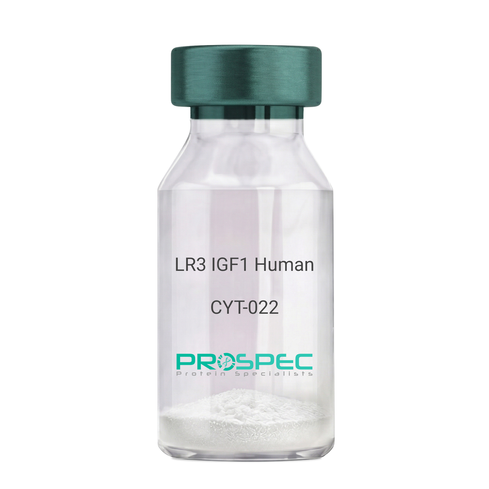
Name :
IGF1 LR3 HumanDescription:
LR3 Insulin Like Growth Factor-1 Human Recombinant
R3 IGF1, R3 IGF-1, R3IGF1, R3IGF-1, LONG IGF1, LONG IGF-1, LONG R3 IGF1, LONG R3IGF1, LONG R3 IGF-1, LONG R3IGF-1.
Product # :
CYT-022Price :
Quantity :
Shipping Method :
Shipped at Room temp
More Info
- description
- source
- formulation
- purity
- biological activity
- More Info
- sds-page
Description
The LR3 is a long-term analog of human IGF-1, specifically designed and manufactured for mammalian cell culture to support large-scale manufacturing of recombinant biopharmaceuticals. Recombinant Human LR3 Insulin Like Growth Factor-1 produced in E.Coli is a single, non-glycosylated, polypeptide chain containing 83 amino acids and having a molecular mass of 9.1kDa.
Source
Escherichia Coli.
Formulation
Lyophilized from a 0.2µm filtered concentrated solution in 20mM PB, pH 7.2.
Purity
Greater than 97.0% as determined by SDS-PAGE.
Biological Activity
The ED50 as determined by the stimulation of protein synthesis in L6 myoblasts is less than 10ng/ml, corresponding to a specific activity of 100,000units/mg.sds-page
More Info
-
Introduction
IGF-1 (Insulin-like growth factor-1) is a major hormonal mediator of statural growth. Under regular circumstances, GH (growth hormone) binds to its receptor in the liver, and other tissues, and stimulates the synthesis/secretion of IGF-1. In target tissues, the Type 1 IGF receptor, that is homologous to the insulin receptor, is activated by IGF-1, leading to intracellular signaling which stimulates multiple processes leading to statural growth. IGF-1 metabolic actions are partly directed at stimulating the uptake of glucose, fatty acids, and amino acids so that metabolism supports growing tissues.
-
Synonyms
R3 IGF1, R3 IGF-1, R3IGF1, R3IGF-1, LONG IGF1, LONG IGF-1, LONG R3 IGF1, LONG R3IGF1, LONG R3 IGF-1, LONG R3IGF-1.
-
Physical Appearance
Sterile Filtered White lyophilized (freeze-dried) powder.
-
Stability
Lyophilized LR3 IGF1 although stable at room temperature for 3 weeks, should be stored desiccated below -18°C. Upon reconstitution the LR3 IGF1 should be stored at 4°C between 2-7 days and for future use below -18°C.For long term storage it is recommended to add a carrier protein (0.1% HSA or BSA).Please prevent freeze-thaw cycles.
-
Solubility
It is recommended to reconstitute the lyophilized LR3 IGF1 in sterile 18M-cm H2O at a concentration of 100µg/ml, which can then be further diluted to other aqueous solutions.
-
Amino Acid Sequence
MFPAMPLSSLFVNGPRTLCGAELVDALQFVCGDRGFYFNKPTGYGSSSRRAPQTGIV DECCFRSCDLRRLEMYCAPLKPAKSA.
-
Background
What is the molecular weight/Mw of IGF1 LR3 HUMAN Protein?
IGF1 LR3 HUMAN Protein has a total Mw of 9.1kDa.
What is the source or expression system of IGF1 LR3 HUMAN Protein?
Escherichia Coli.
What is the Purity of IGF1 LR3 HUMAN Protein?
IGF1 LR3 HUMAN Protein is >98% pure as determined by SDS-PAGE.
What is the Biological Activity of IGF1 LR3 HUMAN Protein?
The ED50 as determined by the stimulation of protein synthesis in L6 myoblasts is less than 10ng/ml, corresponding to a specific activity of 100,000units/mg.
What is the amino acid sequence of IGF1 LR3 HUMAN Protein?
MFPAMPLSSLFVNGPRTLCGAELVDALQFVCGDRGFYFNKPTGYGSSSRRAPQTGIV DECCFRSCDLRRLEMYCAPLKPAKSA.
What applications can IGF1 LR3 HUMAN Protein be used in?
IGF1 LR3 HUMAN Protein can probably be used in western blot, ELISA and Lateral Flow.
What is the endotoxin level for IGF1 LR3 HUMAN Protein?
The endotoxin level is minimal, IGF1 LR3 HUMAN Protein was purified using conventional chromatography techniques.
ProSpec's products are furnished for LABORATORY RESEARCH USE ONLY. The product may not be used as drugs, agricultural or pesticidal products, food additives or household chemicals.
- View Data Sheet
Name :
STIM1 HumanDescription:
Stromal Interaction Molecule 1 Human Recombinant
Stromal interaction molecule 1, GOK, D11S4896E, STIM-1, STIM1.
Product # :
PRO-620Price :
Quantity :
Shipping Method :
Shipped with Ice Packs
More Info
- description
- source
- formulation
- purity
- More Info
Description
Recombinant STIM1 produced in E.Coli is a single, non-glycosylated polypeptide chain (aa 23-213) containing a total of 343 amino acids and having a molecular mass of 38kDa. STIM1 is fused to a 152 aa Calmodulin tag at N-terminus and purified by conventional chromatography techniques.
Source
Escherichia Coli.
Formulation
The STIM1 protein solution (1mg/ml) contains 20mM Tris-HCl pH-7.5.
Purity
Greater than 90.0% as determined by SDS-PAGE.
More Info
-
Introduction
STIM1 is a type-1 transmembrane protein that is necessary for store-operated Ca(2+) entry, a process of extracellular Ca(2+) influx in response to the depletion of Ca(2+) stores in the endoplasmic reticulum (ER). STIM1 localizes predominantly to the ER; upon Ca(2+) release from the ER, STIM1 translocates to the ER-plasma membrane junctions and activates Ca(2+) channels. STIM1 is an adhesion molecule involved in early hematopoiesis by mediating attachment to stromal cells. STIM1 controls the survival and/or proliferation of b-cell precursors. STIM1 gene is one of several genes located in the imprinted gene domain of 11p15.5, a significant tumor-suppressor gene region. Alterations in this region have been associated with the Beckwith-Wiedemann syndrome, Wilms tumor, rhabdomyosarcoma, adrenocrotical carcinoma, and lung, ovarian, and breast cancer. STIM1 is involved in malignancies as well as early hematopoiesis, by mediating attachment to stromal cells. STIM1 is oriented in a head-to-tail configuration with the ribonucleotide reductase 1 gene (RRM1), with the 3' end of this gene situated 1.6 kb from the 5' end of the RRM1 gene.
-
Synonyms
Stromal interaction molecule 1, GOK, D11S4896E, STIM-1, STIM1.
-
Physical Appearance
Sterile filtered colorless solution.
-
Stability
Store at 4°C if entire vial will be used within 2-4 weeks. Store, frozen at -20°C for longer periods of time. For long term storage it is recommended to add a carrier protein (0.1% HSA or BSA). Avoid multiple freeze-thaw cycles.
-
Amino Acid Sequence
MADQLTEEQI AEFKEAFSLF DKDGDGTITT KELGTVMRSL GQNPTEAELQ DMINEVDADG NGTIDFPEFL TMMARKMKDT DSEEEIREAF RVFDKDGNGY ISAAELRHVM TNLGEKLTDE EVDEMIREAD IDGDGQVNYE EFVQMMTAKG SMLSHSHSEK ATGTSSGANS EESTAAEFCR IDKPLCHSED EKLSFEAVRN IHKLMDDDAN GDVDVEESDE FLREDLNYHD PTVKHSTFHG EDKLISVEDL WKAWKSSEVY NWTVDEVVQW LITYVELPQY EETFRKLQLS GHAMPRLAVT NTTMTGTVLK MTDRSHRQKL QLKALDTVLF GPPLLTRHNH LKD.
ProSpec's products are furnished for LABORATORY RESEARCH USE ONLY. The product may not be used as drugs, agricultural or pesticidal products, food additives or household chemicals.
- View Data Sheet
Name :
TNF a MouseDescription:
Tumor Necrosis Factor-Alpha Mouse Recombinant
TNF-alpha, Tumor necrosis factor ligand superfamily member 2, TNF-a, Cachectin, DIF, TNFA, TNFSF2.
Product # :
CYT-252Price :
Quantity :
Shipping Method :
Shipped at Room temp
More Info
- description
- source
- formulation
- purity
- biological activity
- More Info
Description
Tumor Necrosis Factor-a Mouse Recombinant produced in E. coli is a single, non glycosylated, polypeptide chain containing 157 amino acids and having a molecular mass of 17301.32 Dalton. The TNF-alpha is purified by standard chromatographic techniques.
Source
Escherichia Coli.
Formulation
Lyophilized from a 0.2µm filtered concentrated solution in PBS, pH 7.2.
Purity
Greater than 97.0% as determined by:
(a) Analysis by RP-HPLC.
(c) Analysis by SDS-PAGE.Biological Activity
The ED50 as determined by the cytolysis of murine L929 cells in the presence of Actinomycin D is < 0.1ng/ml, corresponding to a Specific Activity of 10,000,000 Units/mg.More Info
-
Introduction
Tumor necrosis factor is a cytokine involved in systemic inflammation and is a member of a group of cytokines that all stimulate the acute phase reaction. TNF is mainly secreted by macrophages.
TNF causes apoptotic cell death, cellular proliferation, differentiation, inflammation, tumorigenesis and viral replication, TNF is also involved in lipid metabolism, and coagulation. TNF's primary role is in the regulation of immune cells.
Dysregulation and, in particular, overproduction of TNF have been implicated in a variety of human diseases- autoimmune diseases, insulin resistance, and cancer. -
Synonyms
TNF-alpha, Tumor necrosis factor ligand superfamily member 2, TNF-a, Cachectin, DIF, TNFA, TNFSF2.
-
Physical Appearance
Sterile Filtered White lyophilized (freeze-dried) powder.
-
Stability
Lyophilized Tumor Necrosis Factor-a although stable at room temperature for 3 weeks, should be stored desiccated below -18°C. Upon reconstitution TNF-a should be stored at 4°C between 2-7 days and for future use below -18°C.For long term storage it is recommended to add a carrier protein (0.1% HSA or BSA).Please prevent freeze-thaw cycles.
-
Solubility
It is recommended to reconstitute the lyophilized Tumor Necrosis Factor-alpha in sterile 18M-cm H2O not less than 100µg/ml, which can then be further diluted to other aqueous solutions.
-
Amino Acid Sequence
MLRSSSQNSS DKPVAHVVAN HQVEEQLEWL SQRANALLAN GMDLKDNQLV VPADGLYLVY SQVLFKGQGC PDYVLLTHTV SRFAISYQEK VNLLSAVKSP CPKDTPEGAE LKPWYEPIYL GGVFQLEKGD QLSAEVNLPK YLDFAESGQV YFGVIAL
-
Background
Tumor Necrosis Factor-alpha (TNF-α) is a pro-inflammatory cytokine that plays a critical role in the regulation of immune responses, inflammation, and cell survival. It is primarily produced by activated macrophages, but can also be secreted by other immune cells, such as T cells, natural killer cells, and mast cells. TNF-α is involved in a wide range of physiological and pathological processes, including the defense against infections, the development of autoimmune diseases, and the progression of cancer.
TNF-α exerts its effects by binding to two distinct cell surface receptors, TNF receptor 1 (TNFR1) and TNF receptor 2 (TNFR2). Upon binding to its receptors, TNF-α activates multiple signaling pathways, including the nuclear factor-kappa B (NF-κB) pathway, the mitogen-activated protein kinase (MAPK) pathway, and the apoptotic pathway. These signaling pathways regulate various cellular processes, such as inflammation, cell proliferation, differentiation, and apoptosis.
In the context of infections, TNF-α plays a crucial role in the body's defense against pathogens. It promotes the recruitment and activation of immune cells, enhances the production of other pro-inflammatory cytokines, and stimulates the expression of adhesion molecules on endothelial cells, facilitating the migration of immune cells to the site of infection. TNF-α also helps to induce fever, which is an important component of the body's immune response to infections.
However, excessive or prolonged production of TNF-α can contribute to the development of chronic inflammatory diseases, such as rheumatoid arthritis, inflammatory bowel disease, and psoriasis. In these conditions, elevated levels of TNF-α promote the infiltration of immune cells into the affected tissues, leading to tissue damage and the perpetuation of inflammation. The central role of TNF-α in the pathogenesis of these diseases has led to the development of anti-TNF-α therapies, which have revolutionized the treatment of chronic inflammatory diseases. These therapies include monoclonal antibodies, such as infliximab and adalimumab, and soluble TNF receptor fusion proteins, such as etanercept. Anti-TNF-α therapies have been shown to be effective in reducing inflammation, improving symptoms, and slowing disease progression in patients with chronic inflammatory diseases.
In the context of cancer, TNF-α has complex and context-dependent effects on tumor development and progression. On one hand, TNF-α can promote anti-tumor immunity by activating immune cells and stimulating the production of other pro-inflammatory cytokines. On the other hand, chronic inflammation driven by TNF-α can promote tumor growth, angiogenesis, and metastasis. Therefore, the role of TNF-α in cancer is still an area of active research, and the development of TNF-α-targeted therapies for cancer remains a challenge.
In conclusion, TNF-α is a pro-inflammatory cytokine that plays a critical role in the regulation of immune responses, inflammation, and cell survival. Its involvement in various physiological and pathological processes has made it an important target for the development of therapies for chronic inflammatory diseases and cancer. Anti-TNF-α therapies have revolutionized the treatment of chronic inflammatory diseases, but the complex role of TNF-α in cancer remains an area of ongoing research. Understanding the precise mechanisms by which TNF-α contributes to disease pathogenesis will be crucial for the development of more effective and targeted therapies.
-
Protein content
Protein quantitation was carried out by two independent methods1. UV spectroscopy at 280 nm using the absorbency value of 1.24 as the extinction coefficient for a 0.1% (1mg/ml) solution. This value is calculated by the PC GENE computer analysis program of protein sequences (IntelliGenetics). 2. Analysis by RP-HPLC, using a calibrated solution of TNF-a as a Reference Standard.
ProSpec's products are furnished for LABORATORY RESEARCH USE ONLY. The product may not be used as drugs, agricultural or pesticidal products, food additives or household chemicals.
- View Data Sheet
Name :
FCGRT MouseDescription:
Fc Fragment Of IgG Receptor And Transporter Mouse Recombinant
IgG receptor FcRn large subunit p51, FcRn, IgG Fc fragment receptor transporter alpha chain, Neonatal Fc receptor, Fcgrt, Fcrn.
Product # :
PRO-2376Price :
Quantity :
Shipping Method :
Shipped with Ice Packs
More Info
- description
- source
- formulation
- purity
- More Info
Description
FCGRT Mouse Recombinant produced in Sf9 Baculovirus cells is a single, glycosylated polypeptide chain containing 285 amino acids (22-297 a.a) and having a molecular mass of 32.1kDa (Molecular size on SDS-PAGE will appear at approximately 40-57kDa).FCGRT is fused to a 6 amino acid His-tag at C-terminus & purified by proprietary chromatographic techniques.
Source
Sf9, Baculovirus cells.
Formulation
FCGRT protein solution (0.5mg/ml) contains Phosphate buffered saline (pH7.4) and 10% glycerol.
Purity
Greater than 85.0% as determined by SDS-PAGE.
More Info
-
Introduction
Fc Fragment Of IgG Receptor And Transporter also known as FCGRT is a transmembrane glycoprotein with structural homology to MHC class 1 proteins. FCGRT is widely expressed in endothelial and epithelial cells and takes a vital part in IgG homeostasis. Moreover, FCGRT is expressed in neutrophils in addition myeloid antigen presenting cells. FCGRT can enhance IgG-meditated phagocytosis as well as antigen presentation by heses cells, however it promotes the degradation of opsonizing IgG rather than returning it to the circulation.
-
Synonyms
IgG receptor FcRn large subunit p51, FcRn, IgG Fc fragment receptor transporter alpha chain, Neonatal Fc receptor, Fcgrt, Fcrn.
-
Physical Appearance
Sterile filtered colorless solution.
-
Stability
Store at 4°C if entire vial will be used within 2-4 weeks. Store, frozen at -20°C for longer periods of time. For long term storage it is recommended to add a carrier protein (0.1% HSA or BSA).Avoid multiple freeze-thaw cycles.
-
Amino Acid Sequence
ADPSETRPPL MYHLTAVSNP STGLPSFWAT GWLGPQQYLT YNSLRQEADP CGAWMWENQV SWYWEKETTD LKSKEQLFLE ALKTLEKILN GTYTLQGLLG CELASDNSSV PTAVFALNGE EFMKFNPRIG NWTGEWPETE IVANLWMKQP DAARKESEFL LNSCPERLLG HLERGRRNLE WKEPPSMRLK ARPGNSGSSV LTCAAFSFYP PELKFRFLRN GLASGSGNCS TGPNGDGSFH AWSLLEVKRG DEHHYQCQVE HEGLAQPLTV DLDSSARSSH HHHHH.
ProSpec's products are furnished for LABORATORY RESEARCH USE ONLY. The product may not be used as drugs, agricultural or pesticidal products, food additives or household chemicals.
- View Data Sheet
Name :
GMFB AntibodyDescription:
Glia Maturation Factor Beta, Mouse Anti Human
Glia maturation factor beta, GMFB, GMF-B, GMF-beta, GMF.
Product # :
ANT-680Price :
Quantity :
Shipping Method :
Shipped with Ice Packs
More Info
- formulation
- More Info
Formulation
1mg/ml containing PBS, pH-7.4, 10% Glycerol and 0.02% Sodium Azide.
More Info
-
Introduction
Glia Maturation Factor-Beta (GMF-Beta) is a 17 kDa protein nerve gorwth factor identified as a growth and differentiation factor in the vertebrate brain.
Glia Maturation Factor-Beta stimulates differentiation of normal neurons as well as glial cells. GMFB inhibits the proliferation of the N-18 neuroblastoma line and the C6 glioma line while promoting their phenotypic expression.
GMF-beta inhances the phenotypic expression of glia & neurons thus inhibits the proliferation of their respective tumors when added to cell culture. Although astrocytes produce GMF-b and stores it inside the cells, they don’t secrete the GMF-B into the cultured medium. Cell- surface GMFb acts on the target cells at close range when cells are in direct contact. GMF-Beta is produced by thymic epithelial cells and plays an important role in T cell development in favor of CD4+ T cells.
GMF-Beta is a brain-specific protein which belongs to the actin-binding proteins (ADF) family. GMF-beta appears to play a role in the differentiation, maintenance, and regeneration of the nervous system. It also supports the progression of certain auto-immune diseases, possibly through its ability to induce the production and secretion of various pro-inflammatory cytokines. -
Synonyms
Glia maturation factor beta, GMFB, GMF-B, GMF-beta, GMF.
-
Physical Appearance
Sterile filtered colorless solution.
-
Immunogen
Anti-human GMFB mAb, is derived from hybridization of mouse F0 myeloma cells with spleen cells from BALB/c mice immunized with recombinant human GMFB amino acids 1-142 purified from E. coli.
-
Ig Subclass
Mouse IgG2a heavy chain and Kappa light chain.
-
Clone
PAT44D8AT.
-
Applications
GMFB antibody has been tested by ELISA, Western blot and ICC/IF to assure specificity and reactivity. Since application varies, however, each investigation should be titrated by the reagent to obtain optimal results.
-
Type
Mouse Anti Human Monoclonal.
-
Storage Procedures
For periods up to 1 month store at 4°C, for longer periods of time, store at -20°C. Prevent freeze thaw cycles.
-
Purification Method
GMFB antibody was purified from mouse ascitic fluids by protein-A affinity chromatography.
ProSpec's products are furnished for LABORATORY RESEARCH USE ONLY. The product may not be used as drugs, agricultural or pesticidal products, food additives or household chemicals.
- View Data Sheet
Name :
PARK7 MouseDescription:
Parkinson Disease Protein 7 Mouse Recombinant
Protein deglycase DJ-1, Parkinson disease protein 7 homolog.
Product # :
PRO-2226Price :
Quantity :
Shipping Method :
Shipped with Ice Packs
More Info
- description
- source
- formulation
- purity
- More Info
Description
PARK7 Mouse Recombinant produced in E.Coli is a single, non-glycosylated polypeptide chain containing 212 amino acids (1-189 a.a) and having a molecular mass of 22.4kDa. PARK7 is fused to a 23 amino acid His-tag at N-terminus & purified by proprietary chromatographic techniques.
Source
Escherichia Coli.
Formulation
PARK7 protein solution (0.5mg/ml) containing Phosphate Buffered Saline (pH 7.4), 20% glycerol and 1mM DTT.
Purity
Greater than 90.0% as determined by SDS-PAGE.
More Info
-
Introduction
The PARK7 is a ubiquitously expressed protein involved in various cellular processes including spermatogenesis and fertilization, cancer, RNA-binding, androgen-receptor signaling and oxidative stress. Mutations in the PARK7 are the cause of autosomal recessive early-onset Parkinson’s disease 7 (Park7).
-
Synonyms
Protein deglycase DJ-1, Parkinson disease protein 7 homolog.
-
Physical Appearance
Sterile Filtered colorless solution.
-
Stability
Store at 4°C if entire vial will be used within 2-4 weeks. Store, frozen at -20°C for longer periods of time. For long term storage it is recommended to add a carrier protein (0.1% HSA or BSA).Avoid multiple freeze-thaw cycles.
-
Amino Acid Sequence
MGSSHHHHHH SSGLVPRGSH MGSMASKRAL VILAKGAEEM ETVIPVDVMR RAGIKVTVAG LAGKDPVQCS RDVMICPDTS LEDAKTQGPY DVVVLPGGNL GAQNLSESPM VKEILKEQES RKGLIAAICA GPTALLAHEV GFGCKVTTHP LAKDKMMNGS HYSYSESRVE KDGLILTSRG PGTSFEFALA IVEALVGKDM ANQVKAPLVL KD.
ProSpec's products are furnished for LABORATORY RESEARCH USE ONLY. The product may not be used as drugs, agricultural or pesticidal products, food additives or household chemicals.
- View Data Sheet
Name :
TNFRSF8 MouseDescription:
CD30 Ligand Receptor Mouse Recombinant
Tumor Necrosis Factor Receptor Superfamily, Member 8, Lymphocyte Activation Antigen CD30, CD30L Receptor, Ki-1 Antigen, D1S166E, CD30, Cytokine Receptor CD30, CD30 Antigen, Ki-1
Product # :
CYT-1165Price :
Quantity :
Shipping Method :
Shipped with Ice Packs
More Info
- description
- source
- formulation
- purity
- More Info
Description
TNFRSF8 Mouse Recombinant produced in Baculovirus is a single glycosylated polypeptide chain containing 479 amino acids (19-258 aa) and having a molecular mass of 52.2kDa.TNFRSF8 is fused to a 239 amino acid hIgG-His tag at C-terminus and purified by proprietary chromatographic techniques.
Source
Sf9, Baculovirus cells.
Formulation
TNFRSF8 protein (1mg/ml) contains 10% glycerol and Phosphate-Buffered Saline (pH 7.4).
Purity
Greater than 90.0% as determined by SDS-PAGE.
More Info
-
Introduction
Tumor necrosis factor receptor superfamily member 8 (TNFRSF8) is a receptor for TNFSF8/CD30L. TNFRSF8 has a role in the regulation of cellular growth and transformation of activated lymphoblasts. In addition, the TNFRSF8 protein regulates gene expression via activation of NF-kappa-B. TNFRSF8 being a regulator of apoptosis, induces cell death or proliferation, depending on the cell type.
-
Synonyms
Tumor Necrosis Factor Receptor Superfamily, Member 8, Lymphocyte Activation Antigen CD30, CD30L Receptor, Ki-1 Antigen, D1S166E, CD30, Cytokine Receptor CD30, CD30 Antigen, Ki-1
-
Physical Appearance
Sterile Filtered colorless solution.
-
Stability
Store at 4°C if entire vial will be used within 2-4 weeks. Store, frozen at -20°C for longer periods of time. For long term storage it is recommended to add a carrier protein (0.1% HSA or BSA).Avoid multiple freeze-thaw cycles.
-
Amino Acid Sequence
FPTDRPLKTT CAGDLSHYPG EAARNCCYQC PSGLSPTQPC PRGPAHCRKQ CAPDYYVNED GKCTACVTCL PGLVEKAPCS GNSPRICECQ PGMHCCTPAV NSCARCKLHC SGEEVVKSPG TAKKDTICEL PSSGSGPNCS NPGDRKTLTS HATPQAMPTL ESPANDSARS LLPMRVTNLV QEDATELVKV PESSSSKARE PSPDPGNAEK NMTLELPSPG TLPDISTSEN SKEPASTAST LEPKSCDKTH TCPPCPAPEL LGGPSVFLFP PKPKDTLMIS RTPEVTCVVV DVSHEDPEVK FNWYVDGVEV HNAKTKPREE QYNSTYRVVS VLTVLHQDWL NGKEYKCKVS NKALPAPIEK TISKAKGQPR EPQVYTLPPS RDELTKNQVS LTCLVKGFYP SDIAVEWESN GQPENNYKTT PPVLDSDGSF FLYSKLTVDK SRWQQGNVFS CSVMHEALHN HYTQKSLSLS PGKHHHHHH
ProSpec's products are furnished for LABORATORY RESEARCH USE ONLY. The product may not be used as drugs, agricultural or pesticidal products, food additives or household chemicals.
- View Data Sheet
Name :
ARF5 HumanDescription:
ADP-Ribosylation Factor 5 Human Recombinant
ADP-ribosylation factor 5, ARF5.
Product # :
PRO-245Price :
Quantity :
Shipping Method :
Shipped with Ice Packs
More Info
- description
- source
- formulation
- purity
- More Info
Description
ARF5 produced in E.Coli is a single, non-glycosylated polypeptide chain containing 200 amino acids (1-180 a.a) and having a molecular mass of 22.6kDa.ARF5 is fused to a 20 amino acid His-tag at N-terminus & purified by proprietary chromatographic techniques.
Source
Escherichia Coli.
Formulation
ARF5 protein solution (1mg/ml) containing 20mM Tris-HCl buffer (pH 8.0), 1mM DTT, 20% glycerol and 0.1M NaCl.
Purity
Greater than 90.0% as determined by SDS-PAGE.
More Info
-
Introduction
ADP-ribosylation factor 5 (ARF5) is a small guanine nucleotide-binding protein which enhances the enzymatic activities of cholera toxin. ARF-dependent regulatory mechanisms include the coordination of spectrin interactions with golgi membranes and the connection of actin to the golgi via rho family-dependent G-protein localization and WASP/Arp2/3 complexes. ARF5 is involved in vesicular transport and functioning via phospholipase D activation.
-
Synonyms
ADP-ribosylation factor 5, ARF5.
-
Physical Appearance
Sterile filtered colorless solution.
-
Stability
Store at 4°C if entire vial will be used within 2-4 weeks. Store, frozen at -20°C for longer periods of time. For long term storage it is recommended to add a carrier protein (0.1% HSA or BSA).Avoid multiple freeze-thaw cycles.
-
Amino Acid Sequence
MGSSHHHHHH SSGLVPRGSH MGLTVSALFS RIFGKKQMRI LMVGLDAAGK TTILYKLKLG EIVTTIPTIG FNVETVEYKN ICFTVWDVGG QDKIRPLWRH YFQNTQGLIF VVDSNDRERV QESADELQKM LQEDELRDAV LLVFANKQDM PNAMPVSELT DKLGLQHLRS RTWYVQATCA
TQGTGLYDGL DWLSHELSKR.
ProSpec's products are furnished for LABORATORY RESEARCH USE ONLY. The product may not be used as drugs, agricultural or pesticidal products, food additives or household chemicals.
- View Data Sheet
Name :
PNOC HumanDescription:
Prepronociceptin Human Recombinant
Prepronociceptin, Nocistatin, OFQ, Pre-Pro-N/OFQ, Pronociceptin, Orphanin FQ, Nociceptin, PpN/OFQ, N/OFQ, PPNOC.
Product # :
PRO-1442Price :
Quantity :
Shipping Method :
Shipped with Ice Packs
More Info
- description
- source
- formulation
- purity
- More Info
Description
PNOC Human Recombinant produced in E.coli is a single, non-glycosylated polypeptide chain containing 180 amino acids (20-176) and having a molecular mass of 20.6kDa. PNOC is fused to a 23 amino acid His-tag at N-terminus.
Source
Escherichia Coli.
Formulation
PNOC protein solution (1mg/ml) containing 20mM Tris-HCl buffer (pH 8.0), 0.4M urea and 10% glycerol.
Purity
Greater than 90.0% as determined by SDS-PAGE.
More Info
-
Introduction
Prepronociceptin (PNOC) is the ligand of the opioid receptor-like receptor (OPRL1). PNOC functions as a transmitter in the brain by modulating nociceptive and locomotor behavior. The PNOC protein may also be involved in neuronal differentiation and development.
-
Synonyms
Prepronociceptin, Nocistatin, OFQ, Pre-Pro-N/OFQ, Pronociceptin, Orphanin FQ, Nociceptin, PpN/OFQ, N/OFQ, PPNOC.
-
Physical Appearance
Sterile Filtered colorless solution.
-
Stability
Store at 4°C if entire vial will be used within 2-4 weeks. Store, frozen at -20°C for longer periods of time. For long term storage it is recommended to add a carrier protein (0.1% HSA or BSA).Avoid multiple freeze-thaw cycles.
-
Amino Acid Sequence
MGSSHHHHHH SSGLVPRGSH MGSSCQRDCL TCQEKLHPAL DSFDLEVCIL ECEEKVFPSP LWTPCTKVMA RSSWQLSPAA PEHVAAALYQ PRASEMQHLR RMPRVRSLFQ EQEEPEPGME EAGEMEQKQL QKRFGGFTGA RKSARKLANQ KRFSEFMRQY LVLSMQSSQR RRTLHQNGNV.
ProSpec's products are furnished for LABORATORY RESEARCH USE ONLY. The product may not be used as drugs, agricultural or pesticidal products, food additives or household chemicals.
- View Data Sheet
Name :
CRCP HumanDescription:
CGRP Receptor Component Human Recombinant
CGRP receptor component protein, CGRP-RCP, RCP, RCP9, Calcitonin gene-related peptide-receptor component protein, RNA polymerase III subunit C9, DNA-directed RNA polymerase III subunit RPC9, HsC17, MGC111194.
Product # :
PRO-919Price :
Quantity :
Shipping Method :
Shipped with Ice Packs
More Info
- description
- source
- formulation
- purity
- More Info
Description
CRCP Human Recombinant produced in E.coli is a single, non-glycosylated polypeptide chain containing 168 amino acids (1-148) and having a molecular mass of 19.0 kDa.The CRCP is fused to a 20 amino acid His-Tag at N-terminus and purified by proprietary chromatographic techniques.
Source
E.coli.
Formulation
The CRCP solution (0.5mg/ml) contains 20mM Tris-HCl buffer (pH 8.0), 0.1M NaCl, 1mM DTT and 20% glycerol.
Purity
Greater than 95% as determined by SDS-PAGE.
More Info
-
Introduction
CRCP is a ubiquitous coupling protein for the calcitonin gene-related peptide and adrenomedullin receptors. CRCP controls ligand sensitivity in several tissues and has DNA-directed RNA polymerase activity, catalytic activity and calcitonin receptor activity.
-
Synonyms
CGRP receptor component protein, CGRP-RCP, RCP, RCP9, Calcitonin gene-related peptide-receptor component protein, RNA polymerase III subunit C9, DNA-directed RNA polymerase III subunit RPC9, HsC17, MGC111194.
-
Physical Appearance
Sterile Filtered colorless solution.
-
Stability
Store at 4°C if entire vial will be used within 2-4 weeks. Store, frozen at -20°C for longer periods of time. For long term storage it is recommended to add a carrier protein (0.1% HSA or BSA).Avoid multiple freeze-thaw cycles.
-
Amino Acid Sequence
MGSSHHHHHH SSGLVPRGSH MEVKDANSAL LSNYEVFQLL TDLKEQRKES GKNKHSSGQQ NLNTITYETL KYISKTPCRH QSPEIVREFL TALKSHKLTK AEKLQLLNHR PVTAVEIQLM VEESEERLTE EQIEALLHTV TSILPAEPEA EQKKNTNSNV AMDEEDPA
ProSpec's products are furnished for LABORATORY RESEARCH USE ONLY. The product may not be used as drugs, agricultural or pesticidal products, food additives or household chemicals.
- View Data Sheet
Name :
VEGF Rat (120a.a.), YeastDescription:
Vascular Endothelial Growth Factor (120a.a.) Rat Recombinant, Yeast
Vascular endothelial growth factor A, VEGF-A, Vascular permeability factor, VPF, VEGF, MGC70609.
Product # :
CYT-1127Price :
Quantity :
Shipping Method :
Shipped at Room temp
More Info
- description
- source
- formulation
- purity
- biological activity
- More Info
Description
Vascular Endothelial Growth Factor(120a.a.) Rat Recombinant produced in yeast is a disulfide-linked homodimer consisting of 2x121 amino acid polypeptide chains, having a molecular mass of approximately 18.5kDa each.VEGF is purified by proprietary chromatographic techniques.
Source
Saccharomyces cerevisiae
Formulation
Lyophilized from a 0.2μm filtered concentrated solution in PBS, pH 7.4.
Purity
Greater than 90.0% as determined by:
(a) Analysis by RP-HPLC.
(b) Analysis by SDS-PAGE.Biological Activity
The ED50 was measured in a cell proliferation assay using HUVEC human umbilical vein endothelial cells and was found to be 2‑10 ng/ml.
More Info
-
Introduction
Vascular endothelial growth factor is an important signaling protein involved in both angiogenesis and vasculogenesis.VEGF activity has been mostly studied on cells of the vascular endothelium, although it does have effects on a number of other cell types (e.g. stimulation monocyte/macrophage migration, neurons, cancer cells, kidney epithelial cells ).VEGF mediates increased vascular permeability, induces angiogenesis, vasculogenesis and endothelial cell growth, promotes cell migration, and inhibits apoptosis. In vitro, VEGF has been shown to stimulate endothelial cell mitogenesis and cell migration. VEGF is also a vasodilator and increases microvascular permeability and was originally referred to as vascular permeability factor.
Elevated levels of VEGF is linked to POEMS syndrome, also known as Crow-Fukase syndrome. Mutations in VEGF have been associated with proliferative and nonproliferative diabetic retinopathy. -
Synonyms
Vascular endothelial growth factor A, VEGF-A, Vascular permeability factor, VPF, VEGF, MGC70609.
-
Physical Appearance
Sterile Filtered White lyophilized (freeze-dried) powder.
-
Stability
Lyophilized VEGF although stable at room temperature for 3 weeks, should be stored desiccated below -18°C. Upon reconstitution Vascular Endothelial Growth Factor should be stored at 4°C between 2-7 days and for future use below -18°C.Please prevent freeze-thaw cycles.
-
Solubility
It is recommended to reconstitute the lyophilized Vascular Endothelial Growth Factor in sterile 18MΩ-cm H2O not less than 100µg/ml, which can then be further diluted to other aqueous solutions.
-
Amino Acid Sequence
MAPTTEGEQK AHEVVKFMDV YQRSYCRPIE TLVDIFQEYP DEIEYIFKPS CVPLMRCAGC CNDEALECVP TSESNVTMQI MRIKPHQSQH IGEMSFLQHS RCECRPKKDR TKPEKCDKPR R.
ProSpec's products are furnished for LABORATORY RESEARCH USE ONLY. The product may not be used as drugs, agricultural or pesticidal products, food additives or household chemicals.
- View Data Sheet
Name :
SYT5 HumanDescription:
Synaptotagmin V Human Recombinant
Synaptotagmin V, Synaptotagmin 5, synaptotagmin-5, sytV, SytV.
Product # :
PRO-1738Price :
Quantity :
Shipping Method :
Shipped with Ice Packs
More Info
- description
- source
- formulation
- purity
- More Info
Description
SYT5 Human Recombinant produced in E.Coli is a single, non-glycosylated polypeptide chain containing 301 amino acids (109-386aa) and having a molecular mass of 33.6kDa.SYT5 is fused to a 23 amino acid His-tag at N-terminus & purified by proprietary chromatographic techniques.
Source
Escherichia Coli.
Formulation
SYT5 protein solution (0.25mg/ml) containing 20mM Tris-HCl(pH 8.0) containing 40% glycerol, 0.2M NaCl and 2mM DTT.
Purity
Greater than 90.0% as determined by SDS-PAGE.
More Info
-
Introduction
Synaptotagmin V, (SYT5) is a member of synaptotagmin family, which is a family of type III membrane proteins characterized by cytoplasmic repeats related to protein kinase C regulatory (C2) domains that are considered to bind calcium. Synaptotagmins function as negative regulators of vesicle fusion, allowing fusion in the attendance of calcium, and as calcium receptors or sensor molecules. Among the diseases associated with SYT5 are labyrinthitis, and thyroiditis.
-
Synonyms
Synaptotagmin V, Synaptotagmin 5, synaptotagmin-5, sytV, SytV.
-
Physical Appearance
Sterile Filtered clear solution.
-
Stability
Store at 4°C if entire vial will be used within 2-4 weeks. Store, frozen at -20°C for longer periods of time. For long term storage it is recommended to add a carrier protein (0.1% HSA or BSA).Avoid multiple freeze-thaw cycles.
-
Amino Acid Sequence
MGSSHHHHHH SSGLVPRGSH MGSLGRLQYS LDYDFQSGQL LVGILQAMGL AALDLGGSSD PYVRVYLLPD KRRRYETKVH RQTLNPHFGE TFAFKVPYVE LGGRVLVMAV YDFDRFSRND AIGEVRVPMS SVDLGRPVQA WRELQAAPRE EQEKLGDICF SLRYVPTAGK LTVIVLEAKN LKKMDVGGLS DPYVKVHLLQ GGKKVRKKKT TIKKNTLNPY YNEAFSFEVP CDQVQKVQVE LTVLDYDKLG KNEAIGRVAV GAAAGGAGLR HWADMLANPR RPIAQWHSLR PPDRVRLLPA P
ProSpec's products are furnished for LABORATORY RESEARCH USE ONLY. The product may not be used as drugs, agricultural or pesticidal products, food additives or household chemicals.
- View Data Sheet
Name :
EGFPDescription:
Enhanced Green Fluorescent Protein Recombinant
Green fluorescent protein, GFP.
Product # :
PRO-1606Price :
Quantity :
Shipping Method :
Shipped at Room temp
More Info
- description
- source
- formulation
- purity
- More Info
Description
Recombinant EGFP produced in E.coli cells is a single non-glycosylated protein containing 239 amino acid chain and having a molecular mass of 26.9kDa. EGFP is purified by proprietary chromatographic techniques.
Source
Escherichia Coli.
Formulation
The EGFP was lyophilized from a 0.2µm filtered concentrated solution in PBS pH 7.4.
Purity
Greater than 95.0% as determined by:
(a) Analysis by RP-HPLC.
(b) Analysis by SDS-PAGE.More Info
-
Introduction
GFP, also known as Green Fluorescent Protein, is a protein produced by the jellyfish (Aequorea Victoria) that produces bioluminescence in the green zone of the noticeable spectrum. Green Fluorescent Protein is a useful and ubiquitous instrument for producing chimeric proteins, where it functions as a fluorescent protein tag. GFP is expressed in most known cell types and is used as a noninvasive fluorescent marker in living cells and organisms. Green Fluorescent Protein permits a broad range of applications where it has functioned as a cell lineage tracer, reporter of gene expression, or as a measure of protein-protein interactions. Enhanced GFP (eGFP) has F64L and S65T mutations, which make GFP show increased fluorescence and fold more efficiently under 370.
-
Synonyms
Green fluorescent protein, GFP.
-
Physical Appearance
Sterile Filtered White lyophilized (freeze-dried) powder.
-
Stability
Lyophilized EGFP although stable at room temperature for 3 weeks, should be stored desiccated below -180C. Upon reconstitution EGFP should be stored at 40C between 2-7 days and for future use below -180C.For long term storage it is recommended to add a carrier protein (0.1% HSA or BSA).Please prevent freeze-thaw cycles.
-
Solubility
It is recommended to reconstitute the lyophilized EGFP in sterile distilled H2O not less than 100µg/ml, which can then be further diluted to other aqueous solutions.
-
Amino Acid Sequence
MVSKGEELFT GVVPILVELD GDVNGHKFSV SGEGEGDATY GKLTLKFICT TGKLPVPWPT LVTTLTYGVQ CFSRYPDHMK QHDFFKSAMP EGYVQERTIF FKDDGNYKTR AEVKFEGDTL VNRIELKGID FKEDGNILGH KLEYNYNSHN VYIMADKQKN GIKVNFKIRH NIEDGSVQLA DHYQQNTPIG DGPVLLPDNH YLSTQSALSK DPNEKRDHMV LLEFVTAAGI TLGMDELYK
ProSpec's products are furnished for LABORATORY RESEARCH USE ONLY. The product may not be used as drugs, agricultural or pesticidal products, food additives or household chemicals.
- View Data Sheet
Name :
SRSF1 Human, Sf9Description:
Serine/arginine-Rich Splicing Factor 1 Human Recombinant, Sf9
SRSF1, ASF, SF2, SF2p33, SFRS1, Splicing Factor, Arginine/Serine-Rich, 30-KD, A, Alternate Splicing Factor, SRp30a, ASF-1, Serine And Arginine Rich Splicing Factor 1, Pre-MRNA-Splicing Factor SF2, P33 Subunit, Splicing Factor, Arginine/Serine-Rich 1, Serine/Arginine-Rich Splicing Factor 1, Alternative-Splicing Factor 1, SR Splicing Factor 1, Splicing Factor 2.
Product # :
PRO-2417Price :
Quantity :
Shipping Method :
Shipped with Ice Packs
More Info
- description
- source
- formulation
- purity
- More Info
Description
SRSF1 Human Recombinant produced in Sf9 Baculovirus cells is a single, glycosylated polypeptide chain containing 254 amino acids (1-248) and having a molecular mass of 28.5kDa. SRSF1 is fused to 6 amino acid His-Tag at C-terminus and purified by proprietary chromatographic techniques.
Source
Sf9, Baculovirus cells.
Formulation
SRSF1 protein solution (0.25mg/ml) containing 20mM Tris-HCl buffer (pH 8.0), 100mM KCl, 1mM DTT, 0.2mM EDTA and 40% Glycerol.
Purity
Greater than 95.0% as determined by SDS-PAGE.
More Info
-
Introduction
Serine/arginine-rich splicing factor 1 (SFRS1) belongs to the arginine/serine-rich splicing factor protein family, and functions in both constitutive and alternative pre-mRNA splicing. SFRS1 binds to pre-mRNA transcripts and components of the spliceosome, and can either initiate or inhibit splicing depending on the position of the pre-mRNA binding site. The ability of SFRS1 to activate splicing is controlled by phosphorylation and interactions with other splicing factor associated proteins.
-
Synonyms
SRSF1, ASF, SF2, SF2p33, SFRS1, Splicing Factor, Arginine/Serine-Rich, 30-KD, A, Alternate Splicing Factor, SRp30a, ASF-1, Serine And Arginine Rich Splicing Factor 1, Pre-MRNA-Splicing Factor SF2, P33 Subunit, Splicing Factor, Arginine/Serine-Rich 1, Serine/Arginine-Rich Splicing Factor 1, Alternative-Splicing Factor 1, SR Splicing Factor 1, Splicing Factor 2.
-
Physical Appearance
Sterile Filtered colorless solution.
-
Stability
Store at 4°C if entire vial will be used within 2-4 weeks. Store, frozen at -20°C for longer periods of time. For long term storage it is recommended to add a carrier protein (0.1% HSA or BSA).Avoid multiple freeze-thaw cycles.
-
Amino Acid Sequence
MSGGGVIRGP AGNNDCRIYV GNLPPDIRTK DIEDVFYKYG AIRDIDLKNR RGGPPFAFVE FEDPRDAEDA VYGRDGYDYD GYRLRVEFPR SGRGTGRGGG GGGGGGAPRG RYGPPSRRSE NRVVVSGLPP SGSWQDLKDH MREAGDVCYA DVYRDGTGVV EFVRKEDMTY AVRKLDNTKF RSHEGETAYI RVKVDGPRSP SYGRSRSRSR SRSRSRSRSN SRSRSYSPRR SRGSPRYSPR HSRSRSRTHH HHHH.
ProSpec's products are furnished for LABORATORY RESEARCH USE ONLY. The product may not be used as drugs, agricultural or pesticidal products, food additives or household chemicals.
- View Data Sheet
Name :
NOV HumanDescription:
Nephroblastoma Overexpressed Human Recombinant
Protein NOV homolog, NovH, CCN family member 3, nsulin-like growth factor-binding protein 9, IBP-9, IGF-binding protein 9, IGFBP-9, Nephroblastoma-overexpressed gene protein homolog, NOV, CCN3, IGFBP9, NOVH.
Product # :
CYT-805Price :
Quantity :
Shipping Method :
Shipped at Room temp
More Info
- description
- source
- formulation
- purity
- biological activity
- More Info
Description
Nephroblastoma Overexpressed Human Recombinant produced in E.Coli is a single, non-glycosylated, polypeptide chain containing 331 amino acids and having a molecular mass of 36.2 kDa. The NOV is purified by proprietary chromatographic techniques.
Source
Escherichia Coli.
Formulation
Lyophilized from a 0.2µm filtered concentrated solution in 20mM Tris-HCl, pH 8.6 and 150 mM NaCl.
Purity
Greater than 95.0% as determined by:(a) Analysis by RP-HPLC.(b) Analysis by SDS-PAGE.
Biological Activity
Determined by a cell proliferation assay using murine Balb/c 3T3 cells is less than 1.0 µg/ml, corresponding to a specific activity of > 1000 IU/mg. range of 10.0 -50.0 ng/ml, corresponding to a specific activity of 20,000-100,000units/mg.More Info
-
Introduction
Nephroblastoma Overexpressed (NOV) which is encoded by the NOV gene is a part of the CCN (CTGF/CYR61/NOV) family. NOV takes part in reducing tumorgenicity and proliferation of certain cancer cell lines. NOV interacts with numerous proteins and is involved in both internal and external cell signaling. NOV is expressed in particular tumors, including Wilm’s tumor and most nephroblastomas and is also exerts proangiogenic activities.
-
Synonyms
Protein NOV homolog, NovH, CCN family member 3, nsulin-like growth factor-binding protein 9, IBP-9, IGF-binding protein 9, IGFBP-9, Nephroblastoma-overexpressed gene protein homolog, NOV, CCN3, IGFBP9, NOVH.
-
Physical Appearance
Sterile Filtered White lyophilized (freeze-dried) powder.
-
Stability
Lyophilized NOV although stable at room temperature for 3 weeks, should be stored desiccated below -18°C. Upon reconstitution NOV should be stored at 4°C between 2-7 days and for future use below -18°C. For long term storage it is recommended to add a carrier protein (0.1% HSA or BSA). Please prevent freeze-thaw cycles.
-
Solubility
It is recommended to reconstitute the lyophilized NOV in sterile 18M-cm H2O not less than 100µg/ml, which can then be further diluted to other aqueous solutions.
-
Amino Acid Sequence
MQVAATQRCP PQCPGRCPAT PPTCAPGVRA VLDGCSCCLV CARQRGESCS DLEPCDESSG LYCDRSADPS NQTGICTAVE GDNCVFDGVI YRSGEKFQPS CKFQCTCRDG QIGCVPRCQL DVLLPEPNCP APRKVEVPGE CCEKWICGPD EEDSLGGLTL AAYRPEATLG VEVSDSSVNC IEQTTEWTAC SKSCGMGFST RVTNRNRQCE MLKQTRLCMV RPCEQEPEQP TDKKGKKCLR TKKSLKAIHL QFKNCTSLHT YKPRFCGVCS DGRCCTPHNT KTIQAEFQCS PGQIVKKPVM VIGTCTCHTN CPKNNEAFLQ ELELKTTRGK M.
ProSpec's products are furnished for LABORATORY RESEARCH USE ONLY. The product may not be used as drugs, agricultural or pesticidal products, food additives or household chemicals.
- View Data Sheet
Name :
PTHrP N15 HumanDescription:
Parathyroid Hormone Related Protein N15 Labeled Human Recombinant
Parathyroid Hormone 2, PTH2, TIPF39, Tuberoinfundibular 39 Residue Protein.
Product # :
HOR-005Price :
Quantity :
Shipping Method :
Shipped at Room temp
More Info
- description
- source
- formulation
- purity
- More Info
Description
PTHrP N15 Human Recombinant produced in E.Coli is a single, non-glycosylated, polypeptide chain containing 86 amino acids, having an MW of 10033 Da labeled by the stable isotope N15.The PTHrP is purified by proprietary chromatographic techniques.
Source
Escherichia Coli.
Formulation
PthRp N15 protein was lyophilized from a 0.2µm filtered concentrated solution in 1xPBS,
pH 7.4.Purity
Greater than 95.0% as determined by:
(a) Analysis by RP-HPLC.
(b) Analysis by SDS-PAGE.More Info
-
Introduction
PTHrP is a powerful and discriminating agonist of PTH2R which takes part in adenyl cyclase activation and intracellular calcium levels elevation. PTHrP encourages protein kinase C beta activation, recruitment of beta-arrestin and PTH2R internalization. Additionally, PTHrP inhibits cell proliferation through its contribution to PTH2R activation, activates nociceptors and nociceptive circuits and acts as a neuropeptide in spermatogenesis.
-
Synonyms
Parathyroid Hormone 2, PTH2, TIPF39, Tuberoinfundibular 39 Residue Protein.
-
Physical Appearance
Sterile Filtered White lyophilized (freeze-dried) powder.
-
Stability
Lyophilized PTHrP N15 although stable at room temperature for 3 weeks, should be stored desiccated below -18C. Upon reconstitution PTHrP N15 should be stored at 4C between 2-7 days and for future use below -18C.For long term storage it is recommended to add a carrier protein (0.1% HSA or BSA).Please prevent freeze-thaw cycles.
-
Solubility
It is recommended to be briefly centrifuged prior to opening to bring the contents to the bottom. Reconstitute in 10mM HAc to a concentration of 0.1-1.0 mg/mL. Further dilutions should be made in appropriate buffered solutions.
-
Amino Acid Sequence
AVSEHQLLHD KGKSIQDLRR RFFLHHLIAE IHTAEIRATS EVSPNSKPSP NTKNHPVRFG SDDEGRYLTQ ETNKVETYKE QPLKTP.
ProSpec's products are furnished for LABORATORY RESEARCH USE ONLY. The product may not be used as drugs, agricultural or pesticidal products, food additives or household chemicals.
- View Data Sheet
Name :
TGFB1 Human RecombinantDescription:
Transforming Growth Factor-Beta 1 Human Recombinant
Transforming growth factor beta-1, TGF-beta-1, CED, DPD1, TGFB, TGF-b 1, LAP, TGFB1.
Product # :
CYT-716Price :
Quantity :
Shipping Method :
Shipped at Room temp
More Info
- description
- source
- formulation
- purity
- biological activity
- More Info
Description
TGFB1 Human Recombinant produced in CHO cells is a glycosylated homodimeric polypeptide chain containing 2 x 112 amino acids and having a total molecular mass of 25.6kDa. The TGFB1 is purified by proprietary chromatographic techniques.
Source
CHO cells.
Formulation
Lyophilized from a sterile filtered solution containing 0.1 % trifluoroacetic acid (TFA) And trehalose (1:20 protein to Trehalose ratio).
Purity
Greater than 95.0% as determined by SDS-PAGE.
Biological Activity
The ED50 as determined by the dose-dependent inhibition of IL-4-induced proliferation of HT-2 cells is 0.142ng/ml, corresponding to a specific activity of 7.4x10⁶units/mg.
More Info
-
Introduction
Transforming growth factor betas (TGFBetas) mediate many cell-cell interactions that occur during embryonic development. Three TGFBetas have been identified in mammals. TGFBeta1, TGFBeta2 and TGFBeta3 are each synthesized as precursor proteins that are very similar in that each is cleaved to yield a 112 amino acid polypeptide that remains associated with the latent portion of the molecule.
-
Synonyms
Transforming growth factor beta-1, TGF-beta-1, CED, DPD1, TGFB, TGF-b 1, LAP, TGFB1.
-
Physical Appearance
Sterile Filtered White lyophilized (freeze-dried) powder.
-
Stability
Lyophilized TGFB1 although stable at room temperature for 3 weeks, should be stored desiccated below -18°C. Upon reconstitution TGFB1 Human should be stored at 4°C between 2-7 days and for future use below -18°C. For long term storage it is recommended to add a carrier protein (0.1% HSA or BSA). Please prevent freeze-thaw cycles.
-
Solubility
It is recommended to reconstitute the lyophilized TGFB1 in sterile 10mM HCl at a concentration of 0.1 mg/ml, which can then be further diluted to other aqueous solutions.
-
Amino Acid Sequence
ALDTNYCFSS TEKNCCVRQL YIDFRKDLGW KWIHEPKGYH ANFCLGPCPY IWSLDTQYSK VLALYNQHNP GASAAPCCVP QALEPLPIVY YVGRKPKVEQ LSNMIVRSCK CS.
-
Background
Title: Transforming Growth Factor-Beta 1 Human Recombinant: A Promising Tool for Biomedical Research
Abstract:
Transforming Growth Factor-Beta 1 (TGF-β1) is a crucial cytokine involved in diverse cellular processes. This research paper provides an in-depth analysis of human recombinant TGF-β1, focusing on its production, purification, and applications in biomedical research. The paper discusses the significance of TGF-β1 in tissue engineering, regenerative medicine, and immunology. Furthermore, it elucidates the potential therapeutic implications of recombinant TGF-β1 in various diseases and highlights ongoing research in the field. The information presented in this paper aims to enhance the understanding of TGF-β1 and its utility as a research tool in biomedical sciences.Introduction:
Transforming Growth Factor-Beta 1 (TGF-β1) is a multifunctional cytokine that regulates cellular processes such as cell growth, differentiation, and immune modulation. Human recombinant TGF-β1 is synthesized using genetic engineering techniques, enabling the production of large quantities of biologically active protein for research purposes.Production and Purification:
Recombinant TGF-β1 is typically produced in expression systems such as bacteria, yeast, or mammalian cells. The protein is then purified using various chromatographic techniques to obtain a highly pure and active form. Quality control measures ensure the biological activity and integrity of the recombinant protein.Biomedical Applications:
Human recombinant TGF-β1 has found broad applications in biomedical research. In tissue engineering and regenerative medicine, it plays a critical role in promoting cell proliferation, extracellular matrix production, and tissue repair. TGF-β1 is also involved in immune modulation, influencing immune cell differentiation and function. Recombinant TGF-β1 is a valuable tool for studying these processes and developing therapeutic interventions.Therapeutic Implications:
The dysregulation of TGF-β1 signaling is associated with various diseases, including fibrosis, cancer, and autoimmune disorders. Recombinant TGF-β1 offers potential therapeutic applications through its ability to modulate cellular responses. Ongoing research aims to develop targeted therapies that specifically regulate TGF-β1 signaling for the treatment of these conditions.Conclusion:
Human recombinant TGF-β1 holds immense potential as a research tool in biomedical sciences. Its production, purification, and applications in tissue engineering, regenerative medicine, and immunology contribute to advancing our understanding of cellular processes and disease mechanisms. With ongoing research, recombinant TGF-β1 may pave the way for novel therapeutic strategies in various medical fields.
ProSpec's products are furnished for LABORATORY RESEARCH USE ONLY. The product may not be used as drugs, agricultural or pesticidal products, food additives or household chemicals.
- View Data Sheet
Name :
BATF3 HumanDescription:
Basic Leucine Zipper Transcription Factor ATF-Like 3 Human Recombinant
JDP1, JUNDM1, SNFT, Basic leucine zipper transcriptional factor ATF-like 3, B-ATF-3, 21 kDa small nuclear factor isolated from T-cells, Jun dimerization protein p21SNFT, BATF3.
Product # :
PRO-1871Price :
Quantity :
Shipping Method :
Shipped with Ice Packs
More Info
- description
- source
- formulation
- purity
- More Info
Description
BATF3 Human Recombinant produced in E.Coli is a single, non-glycosylated polypeptide chain containing 150 amino acids (1-127 a.a) and having a molecular mass of 16.9kDa. BATF3 is fused to a 23 amino acid His-tag at N-terminus & purified by proprietary chromatographic techniques.
Source
Escherichia Coli.
Formulation
BATF3 protein solution (0.25mg/ml) containing 20mM Tris-HCl buffer (pH 8.0), 0.2M NaCl and 30% glycerol.
Purity
Greater than 90.0% as determined by SDS-PAGE.
More Info
-
Introduction
Basic Leucine Zipper Transcription Factor ATF-Like 3 (BATF3) which is localizes to the nucleus, contains 1 bZIP domain. BATF3 functions as a negative regulator of AP-1-mediated transcription when interacting with c-Jun, particularly by heterodimerizing with c-Jun and binding to DNA response elements. BATF3 also takes part in repression of interleukin-2.
-
Synonyms
JDP1, JUNDM1, SNFT, Basic leucine zipper transcriptional factor ATF-like 3, B-ATF-3, 21 kDa small nuclear factor isolated from T-cells, Jun dimerization protein p21SNFT, BATF3.
-
Physical Appearance
Sterile Filtered colorless solution.
-
Stability
Store at 4°C if entire vial will be used within 2-4 weeks. Store, frozen at -20°C for longer periods of time. For long term storage it is recommended to add a carrier protein (0.1% HSA or BSA).Avoid multiple freeze-thaw cycles.
-
Amino Acid Sequence
MGSSHHHHHH SSGLVPRGSH MGSMSQGLPA AGSVLQRSVA APGNQPQPQP QQQSPEDDDR KVRRREKNRV AAQRSRKKQT QKADKLHEEY ESLEQENTML RREIGKLTEE LKHLTEALKE HEKMCPLLLC PMNFVPVPPR PDPVAGCLPR.
ProSpec's products are furnished for LABORATORY RESEARCH USE ONLY. The product may not be used as drugs, agricultural or pesticidal products, food additives or household chemicals.
- View Data Sheet
Name :
BMP3 HumanDescription:
Bone Morphogenetic protein-3 Human Recombinant
Bone Morphogenetic Protein 3, Osteogenin, Bone Morphogenetic Protein 3 (Osteogenic), Bone Morphogenetic Protein 3A, BMP-3A, BMP-3, Bone Morphogenetic Protein-3, BMP3A, BMP3.
Product # :
CYT-937Price :
Quantity :
Shipping Method :
Shipped at Room temp
More Info
- description
- source
- formulation
- purity
- biological activity
- More Info
Description
BMP3 Human Recombinant produced in E.coli is a non-glycosylated disulfide linked homodimer containing 2 chains of 110 amino acids and having a molecular mass of 24.8kDa.The BMP-3 is purified by proprietary chromatographic techniques.
Source
Escherichia Coli.
Formulation
BMP-3 protein was lyophilized from a 0.2µm filtered concentrated solution in 30% Acetonitrile and 0.1% TFA.
Purity
Greater than 95.0% as determined by: (a) Analysis by HPLC. (b) Analysis by SDS-PAGE.
Biological Activity
The ED50 as determined by its ability to inhibit BMP-2-induced activity in murine MC3T3- E1 cells.More Info
-
Introduction
Bone Morphogenetic Protein 3 (BMP3) is one of the BMPs, some of which are members of the TGF-beta superfamily (BMP2-7). There are more than 13 BMPs, which are involved in inducing cartilage and bone formation, embryogenesis and morphogenesis of various tissues and organs. In addition, BMPs regulate the growth, differentiation, chemotaxis, and apoptosis of various cell types. Akin to most other TGF-beta family proteins, BMPs are extremely conserved across animal species. At the amino acid sequence level, mature human and rat BMP3 are 98% identical.
-
Synonyms
Bone Morphogenetic Protein 3, Osteogenin, Bone Morphogenetic Protein 3 (Osteogenic), Bone Morphogenetic Protein 3A, BMP-3A, BMP-3, Bone Morphogenetic Protein-3, BMP3A, BMP3.
-
Physical Appearance
Sterile Filtered White lyophilized (freeze-dried) powder.
-
Stability
Lyophilized BMP3 although stable at room temperature for 3 weeks, should be stored desiccated below -18°C. Upon reconstitution BMP-3 should be stored at 4°C between 2-7 days and for future use below -18°C. For long term storage it is recommended to add a carrier protein (0.1% HSA or BSA). Please prevent freeze-thaw cycles.
-
Solubility
It is recommended to reconstitute the lyophilized BMP3 in sterile 4mM HCl not less than 100µg/ml, which can then be further diluted to other aqueous solutions.
-
Amino Acid Sequence
QWIEPRNCAR RYLKVDFADI GWSEWIISPK SFDAYYCSGA CQFPMPKSLK PSNHATIQSI VRAVGVVPGI PEPCCVPEKM SSLSILFFDE NKNVVLKVYP NMTVESCACR.
-
Background
Bone Morphogenetic Protein-3 Human Recombinant: Unveiling the Potential of a Key Regulator in Tissue Regeneration
Abstract:
Bone Morphogenetic Protein-3 (BMP-3) human recombinant is a critical member of the bone morphogenetic protein family, known for its role in tissue development, repair, and regeneration. This research paper provides a comprehensive analysis of BMP-3, including its characteristics, signaling pathways, and potential therapeutic applications. Additionally, innovative methodologies for the production and optimization of BMP-3 human recombinant are proposed, shedding light on its future implications in the field of regenerative medicine.
Introduction:
Tissue regeneration is a complex biological process requiring precise molecular cues. BMP-3, a crucial member of the BMP family, plays a significant role in tissue development and regeneration. This paper explores the unique features of BMP-3 and presents novel approaches for its production and optimization, aiming to unlock its therapeutic potential in various regenerative contexts.
Characteristics and Signaling Pathways:
BMP-3 is a secreted protein that binds to cell surface receptors, initiating intracellular signaling cascades. It influences cell differentiation, proliferation, and extracellular matrix synthesis through both Smad-dependent and Smad-independent signaling pathways. BMP-3 signaling regulates critical processes involved in tissue regeneration, including chondrogenesis and osteogenesis.
Production of BMP-3 Human Recombinant:
Efficient production methodologies are essential for harnessing the therapeutic potential of BMP-3 human recombinant. Recombinant protein expression systems, such as Escherichia coli or mammalian cells, have been utilized to produce functional BMP-3. Optimization strategies, including codon optimization, signal peptide engineering, and protein folding optimization, have been employed to enhance the yield and activity of BMP-3 recombinant protein.
Potential Therapeutic Applications:
BMP-3 human recombinant holds significant promise in the field of regenerative medicine. It plays a crucial role in bone and cartilage regeneration, making it a potential candidate for the treatment of skeletal disorders and tissue injuries. Additionally, BMP-3 signaling influences tissue remodeling and wound healing, suggesting its broader therapeutic applications in other regenerative processes.
Conclusion:
BMP-3 human recombinant represents a key regulator in tissue regeneration, with immense potential in regenerative medicine. Optimizing production methodologies and further unraveling its signaling mechanisms will enhance its therapeutic applications. With its implications in bone and cartilage regeneration and its role in tissue remodeling, BMP-3 human recombinant emerges as a promising tool for promoting tissue repair and regeneration.
What is the molecular weight/Mw of BMP3 Protein?
BMP3 Protein has a total Mw of 24.8kDa.
What is the source or expression system of BMP3 Protein?
Escherichia Coli.
What is the Purity of BMP3 Protein?
BMP3 Protein is >95% pure as determined by SDS-PAGE.
What is the Biological Activity of BMP3 Protein?
The ED50 as determined by its ability to inhibit BMP-2-induced activity in murine MC3T3- E1 cells.
What is the amino acid sequence of BMP3 Protein?
QWIEPRNCAR RYLKVDFADI GWSEWIISPK SFDAYYCSGA CQFPMPKSLK PSNHATIQSI VRAVGVVPGI PEPCCVPEKM SSLSILFFDE NKNVVLKVYP NMTVESCACR.
What applications can BMP3 Protein be used in?
BMP3 Protein can probably be used in western blot, ELISA and Lateral Flow.
What is the endotoxin level for BMP3 Protein?
The endotoxin level is minimal, BMP3 Protein was purified using conventional chromatography techniques.
ProSpec's products are furnished for LABORATORY RESEARCH USE ONLY. The product may not be used as drugs, agricultural or pesticidal products, food additives or household chemicals.
- View Data Sheet
Name :
SNTA1 HumanDescription:
Syntrophin, Alpha 1 Human Recombinant
Alpha-1-syntrophin, 59 kDa dystrophin-associated protein A1 acidic component 1, Pro-TGF-alpha cytoplasmic domain-interacting protein 1, TACIP1, Syntrophin-1, SNTA1, SNT1, LQT12, dJ1187J4.5.
Product # :
PRO-1016Price :
Quantity :
Shipping Method :
Shipped with Ice Packs
More Info
- description
- source
- formulation
- purity
- More Info
Description
SNTA1 Human Recombinant produced in E.Coli is a single, non-glycosylated polypeptide chain containing 528 amino acids (1-505 a.a.) and having a molecular mass of 56.3kDa. SNTA1 is fused to a 23 amino acid His-tag at N-terminus & purified by proprietary chromatographic techniques.
Source
E.coli.
Formulation
SNTA1 protein solution (1mg/ml) containing 20mM Tris-HCl buffer (pH 8.0), 10% glycerol, 1mM DTT and 0.15M NaCl.
Purity
Greater than 80% as determined by SDS-PAGE.
More Info
-
Introduction
SNTA1 is a member of the syntrophin gene family. SNTA1 is a peripheral membrane protein found linked with dystrophin and dystrophin-related proteins. Dystrophin is a large, rod-like cytoskeletal protein located at the inner surface of muscle fibers. Dystrophin is absent in Duchenne Muscular Dystrophy patients, however it is present in reduced amounts in Becker Muscular Dystrophy patients. Syntrophins are cytoplasmic peripheral membrane scaffold proteins and components of the dystrophin-associated protein complex. The N-terminal PDZ domain of SNTA1 interacts with the C-terminus of the pore-forming alpha subunit (SCN5A) of the cardiac sodium channel Nav1.5. In addition, SNTA1 associates cardiac sodium channels with the nitric oxide synthase-PMCA4b (plasma membrane Ca-ATPase subtype 4b) complex in cardiomyocytes. The SNTA1 gene is a predisposition locus for Long-QT syndrome (LQT) - an inherited disorder associated with sudden cardiac death from arrhythmia - and sudden infant death syndrome (SIDS). SNTA1 also associates with dystrophin and dystrophin-related proteins at the neuromuscular junction and modifies intracellular calcium ion levels in muscle tissue.
-
Synonyms
Alpha-1-syntrophin, 59 kDa dystrophin-associated protein A1 acidic component 1, Pro-TGF-alpha cytoplasmic domain-interacting protein 1, TACIP1, Syntrophin-1, SNTA1, SNT1, LQT12, dJ1187J4.5.
-
Physical Appearance
Sterile Filtered colorless solution.
-
Stability
Store at 4°C if entire vial will be used within 2-4 weeks. Store, frozen at -20°C for longer periods of time. For long term storage it is recommended to add a carrier protein (0.1% HSA or BSA).Avoid multiple freeze-thaw cycles.
-
Amino Acid Sequence
MGSSHHHHHH SSGLVPRGSH MGSMASGRRA PRTGLLELRA GAGSGAGGER WQRVLLSLAE DVLTVSPADG DPGPEPGAPR EQEPAQLNGA AEPGAGPPQL PEALLLQRRR VTVRKADAGG LGISIKGGRE NKMPILISKI FKGLAADQTE ALFVGDAILS VNGEDLSSAT HDEAVQVLKK TGKEVVLEVK YMKDVSPYFK NSTGGTSVGW DSPPASPLQR QPSSPGPTPR NFSEAKHMSL KMAYVSKRCT PNDPEPRYLE ICSADGQDTL FLRAKDEASA RSWATAIQAQ VNTLTPRVKD ELQALLAATS TAGSQDIKQI GWLTEQLPSG GTAPTLALLT EKELLLYLSL PETREALSRP ARTAPLIATR LVHSGPSKGS VPYDAELSFA LRTGTRHGVD THLFSVESPQ ELAAWTRQLV DGCHRAAEGV QEVSTACTWN GRPCSLSVHI DKGFTLWAAE PGAARAVLLR QPFEKLQMSS DDGASLLFLD FGGAEGEIQL DLHSCPKTIV FIIHSFLSAK VTRLGLLA.
ProSpec's products are furnished for LABORATORY RESEARCH USE ONLY. The product may not be used as drugs, agricultural or pesticidal products, food additives or household chemicals.