Search results
327 results found for “fragile histidine triad”
Name
Description
Product #
Price
Quantity
Shipping Method
- View Data Sheet
Name :
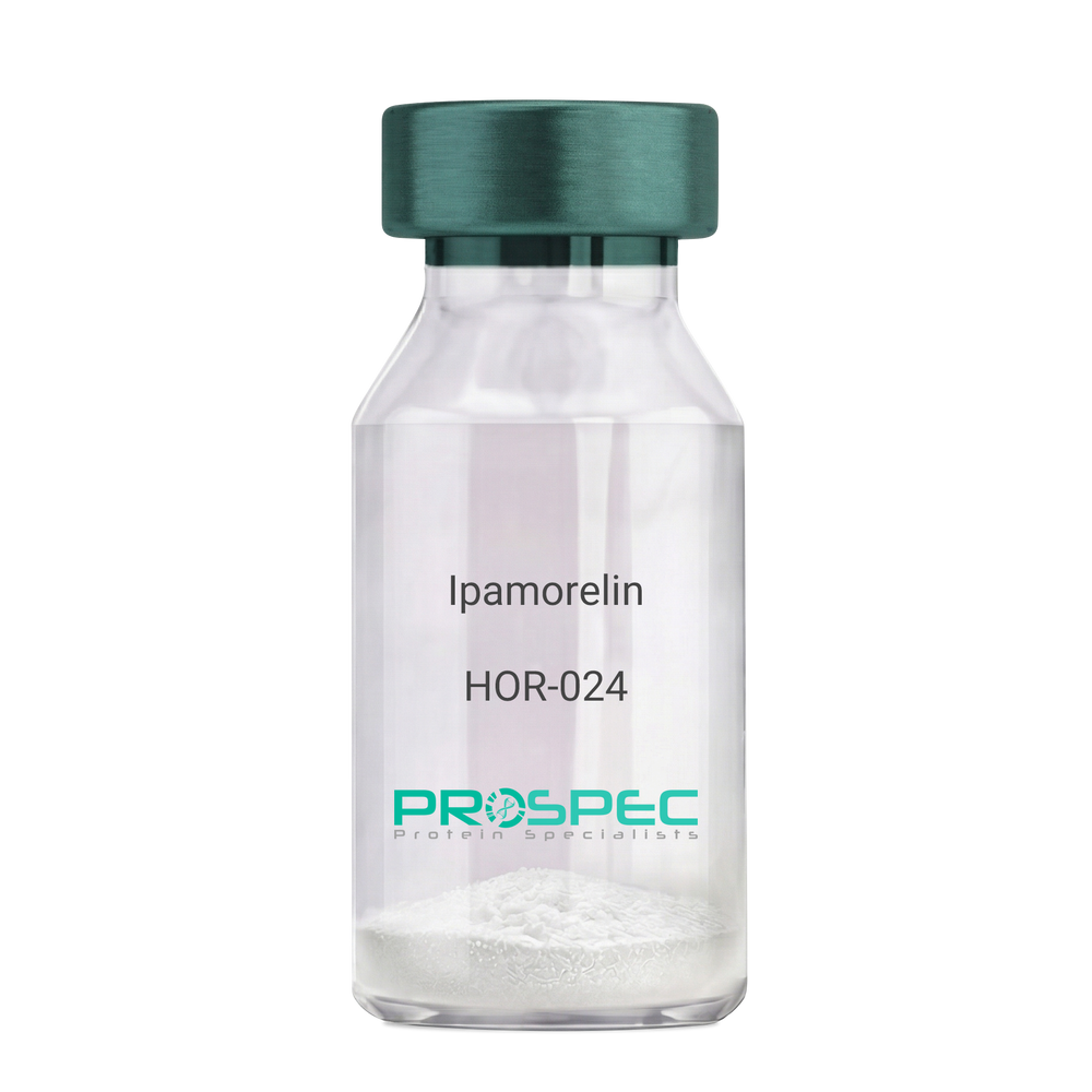
IpamorelinDescription:
Ipamorelin
Ipamorelin
Product # :
HOR-024Price :
Quantity :
Shipping Method :
Shipped at Room temp
More Info
- description
- formulation
- purity
- More Info
Description
Ipamorelin Synthetic is a single, non-glycosylated polypeptide chain containing 4 amino acids, having a molecular mass of 711.85 Dalton and a Molecular formula of C38H49N9O5.
Formulation
The protein was lyophilized with no additives.
Purity
Greater than 97.0% as determined by analysis by RP-HPLC.
More Info
-
Introduction
Ipamorelin is a peptide selective agonist of the ghrelin/growth hormone secretagogue receptor and a growth hormone secretagogue. Ipamorelin is a pentapeptide that was derived from GHRP1. Ipamorelin significantly increases plasma growth hormone levels in both animals and humans. Like pralmorelin and GHRP-6, ipamorelin does not affect prolactin, FSH, LH or TSH levels. However, unlike GHRP2 and GHRP6, but as growth hormone-releasing hormone (GHRH), ipamorelin does not stimulate the secretion of adrenocorticotropic hormone (ACTH) or cortisol, and is highly selective for inducing the secretion only of GH.
-
Synonyms
Ipamorelin
-
Physical Appearance
Sterile Filtered White lyophilized (freeze-dried) powder.
-
Stability
Lyophilized Ipamorelin although stable at room temperature for 3 weeks, should be stored desiccated below -18°C. Upon reconstitution Ipamorelin should be stored at 4°C between 2-7 days and for future use below -18°C. For long term storage it is recommended to add a carrier protein (0.1% HSA or BSA). Please prevent freeze-thaw cycles.
-
Solubility
It is recommended to reconstitute the lyophilized Ipamorelin in sterile 18MΩ-cm H2O not less than 100 µg/ml, which can then be further diluted to other aqueous solutions.
-
Amino Acid Sequence
Aib-His-D-2-Nal-D-Phe-Lys-NH2.
ProSpec's products are furnished for LABORATORY RESEARCH USE ONLY. The product may not be used as drugs, agricultural or pesticidal products, food additives or household chemicals.
- View Data Sheet
Name :
KIR2DL3 AntibodyDescription:
Killer Cell Immunoglobulin-Like Receptor 2 Domains Long Cytoplasmic Tail 3, Mouse Anti Human
Killer cell immunoglobulin-like receptor 2DL3, MHC class I NK cell receptor, Natural killer-associated transcript 2, NKAT-2, NKAT2a, NKAT2b, p58 natural killer cell receptor clone CL-6, p58 NK receptor, p58.2 MHC class-I-specific NK receptor, Killer inhibitory receptor cl 2-3, KIR-023GB, CD158 antigen-like family member B2, CD158b2 antigen, KIR2DL3, CD158B2, KIRCL23, NKAT2, p58, NKAT, GL183, CD158b, KIR-K7b, KIR-K7c, MGC129943.
Product # :
ANT-300Price :
Quantity :
Shipping Method :
Shipped with Ice Packs
More Info
- formulation
- More Info
Formulation
1mg/ml containing PBS, pH-7.4, & 0.1% Sodium Azide.
More Info
-
Introduction
Killer-cell immunoglobulin-like receptors (KIRs), are a family of cell surface glycoproteins found on Natural Killer (NK) Cells, which are important cells of the immune system. They control the killing function of these cells by interacting with MHC class I molecules, which are expressed on all cell types. This interaction allows them to identify virally infected cells or tumor cells that have a distinctive low level of Class I MHC on their surface. The majority of KIRs are inhibitory, which means that their recognition of MHC suppresses the cytotoxic activity of their NK cell. Only a limited number of KIRs have the capacity to activate cells. The KIR genes are found in a cluster on chromosome 19q13.4 within the 1 Mb leukocyte receptor complex (LRC). KIR molecules are extremely polymorphic, meaning their gene sequences differ significantly between individuals, so that different individuals have different arrays/repertoires of KIR genes. The KIR proteins are categorized by the number of extracellular immunoglobulin domains (2D or 3D) and by whether they have a long (L) or short (S) cytoplasmic domain. KIR proteins with the long cytoplasmic domain transduce inhibitory signals upon ligand binding via an immune tyrosine-based inhibitory motif (ITIM). Whereas KIR proteins with the short cytoplasmic domain lack the ITIM motif and instead associate with the TYRO protein tyrosine kinase binding protein to transduce activating signals. KIR2DL3 is an inhibitory Killer Cell Ig-like Receptor (KIR, previously called p58 KIR, cl-6, NKAT2 or KIR-K7), which recognizes class I MHC molecules (HLA-Cw1, -Cw3, -Cw7, and Cw8). KIR2DL3 inhibits the activity of NK cells thus preventing cell lysis.
-
Synonyms
Killer cell immunoglobulin-like receptor 2DL3, MHC class I NK cell receptor, Natural killer-associated transcript 2, NKAT-2, NKAT2a, NKAT2b, p58 natural killer cell receptor clone CL-6, p58 NK receptor, p58.2 MHC class-I-specific NK receptor, Killer inhibitory receptor cl 2-3, KIR-023GB, CD158 antigen-like family member B2, CD158b2 antigen, KIR2DL3, CD158B2, KIRCL23, NKAT2, p58, NKAT, GL183, CD158b, KIR-K7b, KIR-K7c, MGC129943.
-
Physical Appearance
Sterile Filtered colorless solution.
-
Immunogen
Anti-human KIR2DL3 mAb, is derived from hybridization of mouse P3-x63-Ag8.653 myeloma cells with spleen cells from BALB/c mice immunized with recombinant human KIR2DL3 amino acids 19-161 purified from E. coli.
-
Ig Subclass
Mouse IgG2a heavy chains and κ light chain.
-
Clone
P190IIC311AT.
-
Applications
KIR2DL3 antibody has been tested by ELISA and Western blot immunofluorescent staining with flow cytometric analysis to assure specificity. Since application varies, however, each investigation should be titrated by the reagent to obtain optimal results. Applications include flow cytometry (5-10g/1×1000000), ELISA (1:1,000 when tested against the immunized protein) and western blot analysis (1μg/ml).
-
Type
Mouse Anti Human Monoclonal.
-
Storage Procedures
For periods up to 1 month store at 4°C, for longer periods of time, store at -20°C. Prevent freeze thaw cycles.
-
Purification Method
KIR2DL3 antibody was purified from mouse ascitic fluids by protein-G affinity chromatography.
ProSpec's products are furnished for LABORATORY RESEARCH USE ONLY. The product may not be used as drugs, agricultural or pesticidal products, food additives or household chemicals.
- View Data Sheet
Name :
NDUFS2 HumanDescription:
Histidine NADH Dehydrogenase Fe-S Protein 2 Human Recombinant
CI-49 , NADH dehydrogenase [ubiquinone] iron-sulfur protein 2, mitochondrial, Complex I-49kD, CI-49kD, NADH-ubiquinone oxidoreductase 49 kDa subunit, NDUFS2.
Product # :
ENZ-737Price :
Quantity :
Shipping Method :
Shipped with Ice Packs
More Info
- description
- source
- formulation
- purity
- More Info
Description
NDUFS2 Human Recombinant produced in E.coli is a single, non-glycosylated polypeptide chain containing 410 amino acids (77-463a.a) and having a molecular mass of 46.5kDa. NDUFS2 is fused to a 23 amino acid His-tag at N-terminus & purified by proprietary chromatographic techniques.
Source
Escherichia Coli.
Formulation
The NDUFS2 solution (0.25mg/ml) contains 20mM Tris-HCl buffer (pH 8.0), 0.4M Urea and 10% glycerol.
Purity
Greater than 80% as determined by SDS-PAGE.
More Info
-
Introduction
NDUFS2 is a core subunit of the mitochondrial membrane respiratory chain NADH dehydrogenase (Complex I) which is a part of the minimal assembly required for catalysis. Complex I takes part in the transfer of electrons from NADH to the respiratory chain. Histidine NADH Dehydrogenase Fe-S Protein 2 (NDUFS2) is required for catalytic activity. Imperfections in NDUFS2 are the source of complex I mitochondrial respiratory chain deficiency, which is characterized by many symptoms including liver failure, cardiomyopathy and neurodegeneration.
-
Synonyms
CI-49 , NADH dehydrogenase [ubiquinone] iron-sulfur protein 2, mitochondrial, Complex I-49kD, CI-49kD, NADH-ubiquinone oxidoreductase 49 kDa subunit, NDUFS2.
-
Physical Appearance
Sterile Filtered clear solution.
-
Stability
Store at 4°C if entire vial will be used within 2-4 weeks. Store, frozen at -20°C for longer periods of time. For long term storage it is recommended to add a carrier protein (0.1% HSA or BSA).Avoid multiple freeze-thaw cycles.
-
Amino Acid Sequence
MGSSHHHHHH SSGLVPRGSH MGSVKNITLN FGPQHPAAHG VLRLVMELSG EMVRKCDPHI GLLHRGTEKL IEYKTYLQAL PYFDRLDYVS MMCNEQAYSL AVEKLLNIRP PPRAQWIRVL FGEITRLLNH IMAVTTHALD LGAMTPFFWL FEEREKMFEF YERVSGARMH AAYIRPGGVH QDLPLGLMDD IYQFSKNFSL RLDELEELLT NNRIWRNRTI DIGVVTAEEA LNYGFSGVML RGSGIQWDLR KTQPYDVYDQ VEFDVPVGSR GDCYDRYLCR VEEMRQSLRI IAQCLNKMPP GEIKVDDAKV SPPKRAEMKT SMESLIHHFK LYTEGYQVPP GATYTAIEAP KGEFGVYLVS DGSSRPYRCK IKAPGFAHLA GLDKMSKGHM LADVVAIIGT QDIVFGEVDR.
ProSpec's products are furnished for LABORATORY RESEARCH USE ONLY. The product may not be used as drugs, agricultural or pesticidal products, food additives or household chemicals.
- View Data Sheet
Name :
Tamm HorsfallDescription:
Recombinant Human Tamm Horsfall Glycoprotein
Tamm-Horsfall urinary glycoprotein, THP, FJHN, HNFJ, THGP, MCKD2, ADMCKD2, UMOD, Uromodulin.
Product # :
ENZ-1206Price :
Quantity :
Shipping Method :
Shipped at Room temp
More Info
- description
- source
- formulation
- purity
- More Info
- SDS-PAGE
Description
Uromodulin Human Recombinant protein produced from HEK Cells, is a polypeptide chain containing 595 amino acids ( 25-613 a.a. ) and having a total Mw of 65 kDa.
Source
HEK293
Formulation
The UMOD protein was lyophilized from 0.4μm filtered solution containing 50mM NaCl, 0.02M TRIS, pH 7.5.
Purity
Greater than 90.0% as determined by SDS-PAGE analysis.
SDS-PAGE
More Info
-
Introduction
Uromodulin (Tamm–Horsfall protein) is produced mainly by cells in the kidney’s thick ascending limb and is the most abundant protein in normal urine.
Uromodulin takes part in salt and water regulation, helps prevent urinary tract infections and kidney stones, and influences inflammation and immune activity in the kidney.
Reduced Uromodulin levels is associated with chronic kidney disease.
UMOD mutations causes unproper protein folding and thus transported incorrectly, resulting in its accumulation inside kidney tubular cells that damages the tubules and cause kidney disease (ADTKD-UMOD), associated with high uric acid, gout, and progressive kidney failure.
-
Synonyms
Tamm-Horsfall urinary glycoprotein, THP, FJHN, HNFJ, THGP, MCKD2, ADMCKD2, UMOD, Uromodulin.
-
Physical Appearance
Filtered White lyophilized (freeze-dried) powder.
-
Stability
Lyophilized UMOD although stable at room temperature for 3 weeks, should be stored at -18C. Upon reconstitution UMOD should be stored at 4C between 2-7 days and for future use below -18C.
For long term storage it is recommended to add a carrier protein (0.1% HSA or BSA).
Please prevent freeze-thaw cycles.
-
Solubility
Add deionized water to prepare a working stock solution of approximately 0.5mg/ml and let the lyophilized pellet dissolve completely. Product is not sterile! Please filter the product by an appropriate sterile filter before using it in the cell culture.
-
Amino Acid Sequence
DTSEARWCSE CHSNATCTED EAVTTCTCQE GFTGDGLTCV DLDECAIPGA HNCSANSSCV NTPGSFSCVC PEGFRLSPGL GCTDVDECAE PGLSHCHALA TCVNVVGSYL CVCPAGYRGD GWHCECSPGS CGPGLDCVPE GDALVCADPC QAHRTLDEYW RSTEYGEGYA CDTDLRGWYR FVGQGGARMA ETCVPVLRCN TAAPMWLNGT HPSSDEGIVS RKACAHWSGH CCLWDASVQV KACAGGYYVY NLTAPPECHL AYCTDPSSVE GTCEECSIDE DCKSNNGRWH CQCKQDFNIT DISLLEHRLE CGANDMKVSL GKCQLKSLGF DKVFMYLSDS RCSGFNDRDN RDWVSVVTPA RDGPCGTVLT RNETHATYSN TLYLADEIII RDLNIKINFA CSYPLDMKVS LKTALQPMVS ALNIRVGGTG MFTVRMALFQ TPSYTQPYQG SSVTLSTEAF LYVGTMLDGG DLSRFALLMT NCYATPSSNA TDPLKYFIIQ DRCPHTRDST IQVVENGESS QGRFSVQMFR FAGNYDLVYL HCEVYLCDTM NEKCKPTCSG TRFRSGSVID QSRVLNLGPI TRKGVQATVH HHHHH
-
Background
Uromodulin is the most abundant protein in normal urine. Its secretion in urine follows proteolytic cleavage of the ectodomain of its glycosyl phosphatidylinosital-anchored counterpart that is situated on the luminal cell surface of the loop of Henle. Uromodulin plays a role as a constitutive inhibitor of calcium crystallization in renal fluids. Secretion of uromodulin in urine provides protection against urinary tract infections caused by uropathogenic bacteria. Defects in Uromodulin expression are associated with the autosomal dominant renal disorders medullary cystic kidney disease-2 (MCKD2) and familial juvenile hyperuricemic nephropathy (FJHN). These disorders are characterized by juvenile onset of hyperuricemia, gout, and progressive renal failure. While several transcript variants may exist for this gene, the full-length natures of only two have been described to date. UMOD is involved in regulating the circulating activity of cytokines as it binds to il-1, il-2 and tnf with high affinity.
What is the molecular weight/Mw of UMOD Protein?
UMOD Protein has a total Mw of 65kDa.
What is the source or expression system of UMOD Protein?
HEK293.
What is the Purity of UMOD Protein?
UMOD Protein is >90% pure as determined by SDS-PAGE.
What is the Biological Activity of UMOD Protein?
The biological functionality of UMOD Protein will be determined in the future.
What is the amino acid sequence of UMOD Protein?
UMOD Protein is composed from 595 amino acids.
What applications can UMOD Protein be used in?
UMOD Protein can probably be used in western blot, ELISA and Lateral Flow.
What is the endotoxin level for UMOD Protein?
The endotoxin level is minimal, UMOD Protein was purified using conventional chromatography techniques.
ProSpec's products are furnished for LABORATORY RESEARCH USE ONLY. The product may not be used as drugs, agricultural or pesticidal products, food additives or household chemicals.
- View Data Sheet
Name :
FLAG PAT3G6AT antibodyDescription:
FLAG peptide Clone PAT3G6AT, Mouse Anti Human
Product # :
ANT-736Price :
Quantity :
Shipping Method :
Shipped with Ice Packs
More Info
- formulation
- More Info
Formulation
1mg/ml containing PBS, pH-7.4, 10% Glycerol and 0.02% Sodium Azide.
More Info
-
Introduction
A fusion tag called FLAG consists of eight amino acids (AspTyrLysAspAspAspAspLys) including an enterokinase-cleavage site. FLAG is specifically designed for immunoaffinity chromatography. It allows elution under non-denaturing conditions. Several antibodies against this peptide have been developed. One antibody denoted as M1 binds the peptide in the presence of bivalent metal cations preferably Ca(+). Elution is effected by chelating agents. Another strategy is competitive elution with excess of free Flag Peptide. The FLAG peptide purifies and detects recombinant fusion proteins. FLAG peptide is useful in Western blotting, immunocytochemistry, immunoprecipitation, flow cytometry, protein purification, and in the study of protein-protein interactions, cell ultrastructure, and protein localization. Flag peptide is a hydrophilic tag which significantly improves the detection and purification of recombinant fusion proteins.
-
Physical Appearance
Sterile filtered colorless solution.
-
Immunogen
Anti-human FLAG mAb, is derived from hybridization of mouse F0 myeloma cells with spleen cells from BALB/c mice immunized with a Synthetic peptide (DYKDDDDKC)-KLH.
-
Ig Subclass
Mouse IgG1 heavy chain and k light chain.
-
Clone
PAT3G6AT.
-
Applications
The antibody has been tested by ELISA, Western blot analysis, Flow cytometry and ICC/IF to assure specificity and reactivity. Since application varies, however, each investigation should be titrated by the reagent to obtain optimal results.
-
Type
Mouse Anti Human Monoclonal.
-
Storage Procedures
For periods up to 1 month store at 4°C, for longer periods of time, store at -20°C. Prevent freeze thaw cycles.
-
Purification Method
FLAG antibody was purified from mouse ascitic fluids by protein-A affinity chromatography.
ProSpec's products are furnished for LABORATORY RESEARCH USE ONLY. The product may not be used as drugs, agricultural or pesticidal products, food additives or household chemicals.
- View Data Sheet
Name :
CHI3L2 AntibodyDescription:
Chitinase 3-Like 2, Mouse Anti Human
Chitinase 3-Like 2, Chondrocyte Protein 39, YKL-39, YKL39, Chitinase-3-Like Protein 2, CHIL2, CHI3L2.
Product # :
ANT-703Price :
Quantity :
Shipping Method :
Shipped with Ice Packs
More Info
- formulation
- More Info
Formulation
1mg/ml containing PBS, pH-7.4, 10% Glycerol and 0.02% Sodium Azide.
More Info
-
Introduction
Chitinase 3-Like 2 (CHI3L2) is similar to bacterial chitinases but lacks chitinase activity. CHI3L2 protein is secreted and is involved in cartilage biogenesis. CHI3L2 is a lectin, which binds chitooligosaccharides and other glycans with high affinity, but not heparin.
-
Synonyms
Chitinase 3-Like 2, Chondrocyte Protein 39, YKL-39, YKL39, Chitinase-3-Like Protein 2, CHIL2, CHI3L2.
-
Physical Appearance
Sterile filtered colorless solution.
-
Immunogen
Anti-human CHI3L2 mAb, is derived from hybridization of mouse F0 myeloma cells with spleen cells from BALB/c mice immunized with recombinant human CHI3L2 amino acids 27-390 purified from E. coli.
-
Ig Subclass
Mouse IgG2b heavy chain and κ light chain.
-
Clone
PAT2B5AT.
-
Applications
CHI3L2 antibody has been tested by ELISA and Western blot analysis to assure specificity and reactivity. Since application varies, however, each investigation should be titrated by the reagent to obtain optimal results.
-
Type
Mouse Anti Human Monoclonal.
-
Storage Procedures
For periods up to 1 month store at 4°C, for longer periods of time, store at -20°C. Prevent freeze thaw cycles.
-
Purification Method
CHI3L2 antibody was purified from mouse ascitic fluids by protein-A affinity chromatography.
ProSpec's products are furnished for LABORATORY RESEARCH USE ONLY. The product may not be used as drugs, agricultural or pesticidal products, food additives or household chemicals.
- View Data Sheet
Name :
NDUFS6 HumanDescription:
Histidine NADH Dehydrogenase Fe-S Protein 6 Human Recombinant
NADH Dehydrogenase (Ubiquinone) Fe-S Protein 6 13kDa (NADH-Coenzyme Q Reductase), Complex I Mitochondrial Respiratory Chain 13-KD Subunit, NADH Dehydrogenase [Ubiquinone] Iron-Sulfur Protein 6 Mitochondrial, NADH: Ubiquinone Oxidoreductase NDUFS6 Subunit, NADH-Ubiquinone Oxidoreductase 13 KDa-A Subunit, Complex I-13kD-A, CI13KDA.
Product # :
ENZ-725Price :
Quantity :
Shipping Method :
Shipped with Ice Packs
More Info
- description
- source
- formulation
- purity
- More Info
Description
NADH Dehydrogenase (Ubiquinone) Fe-S Protein 6 13kDa (NADH-Coenzyme Q Reductase), Complex I Mitochondrial Respiratory Chain 13-KD Subunit, NADH Dehydrogenase [Ubiquinone] Iron-Sulfur Protein 6 Mitochondrial, NADH: Ubiquinone Oxidoreductase NDUFS6 Subunit, NADH-Ubiquinone Oxidoreductase 13 KDa-A Subunit, Complex I-13kD-A, CI13KDA.
Source
Escherichia Coli.
Formulation
The NDUFS6 solution (0.25mg/ml) contains 20mM Tris-HCl buffer (pH 8.0), 0.15M NaCl, 1mM DTT and 30% glycerol.
Purity
Greater than 90% as determined by SDS-PAGE.
More Info
-
Introduction
NADH Dehydrogenase Fe-S Protein 6 (NDUFS6) is a subunit of the NADH: ubiquinone oxidoreductase (complex I), which is the 1st enzyme complex in the electron transport chain of the mitochondria. This complex operates in the transfer of electrons from NADH to the respiratory chain. NDUFS6 is one of seven subunits in the iron-sulfur protein segment. DNA alterations in NDUFS6 are the cause for mitochondrial complex I deficiency, a disease which results in an extensive assortment of clinical disorders, like adult-onset neurodegenerative disorders and neonatal disease.
-
Synonyms
NADH Dehydrogenase (Ubiquinone) Fe-S Protein 6 13kDa (NADH-Coenzyme Q Reductase), Complex I Mitochondrial Respiratory Chain 13-KD Subunit, NADH Dehydrogenase [Ubiquinone] Iron-Sulfur Protein 6 Mitochondrial, NADH: Ubiquinone Oxidoreductase NDUFS6 Subunit, NADH-Ubiquinone Oxidoreductase 13 KDa-A Subunit, Complex I-13kD-A, CI13KDA.
-
Physical Appearance
Sterile Filtered clear solution.
-
Stability
Store at 4°C if entire vial will be used within 2-4 weeks. Store, frozen at -20°C for longer periods of time. For long term storage it is recommended to add a carrier protein (0.1% HSA or BSA).Avoid multiple freeze-thaw cycles.
-
Amino Acid Sequence
MGSSHHHHHH SSGLVPRGSH MGSFGVRVSP TGEKVTHTGQ VYDDKDYRRI RFVGRQKEVN ENFAIDLIAE QPVSEVETRV IACDGGGGAL GHPKVYINLD KETKTGTCGY CGLQFRQHHH
ProSpec's products are furnished for LABORATORY RESEARCH USE ONLY. The product may not be used as drugs, agricultural or pesticidal products, food additives or household chemicals.
- View Data Sheet
Name :
SIRPA HumanDescription:
Signal-Regulatory Protein Alpha Human Recombinant
Signal-Regulatory Protein Alpha, SHPS1, CD172 Antigen-Like Family Member A, Inhibitory Receptor SHPS-1, Macrophage Fusion Receptor, PTPNS1, SIRP, P84, BIT, MFR, Brain-Immunoglobulin-Like Molecule With Tyrosine-Based Activation Motifs, Brain Ig-Like Molecule With Tyrosine-Based Activation Motifs, Protein Tyrosine Phosphatase, Non-Receptor Type Substrate 1, Tyrosine-Protein Phosphatase Non-Receptor Type Substrate 1, Tyrosine Phosphatase SHP Substrate 1, Signal-Regulatory Protein Alpha-1 , ignal-Regulatory Protein Alpha-2, Signal-Regulatory Protein Alpha-3, SHP Substrate 1, CD172a Antigen, Myd-1 Antigen, MyD-1 Antigen, Sirp-Alpha-1, Sirp-Alpha-2, Sirp-Alpha-3, CD172A, SHPS-1, MYD-1, MYD1, Tyrosine-protein phosphatase non-receptor type substrate 1.
Product # :
PRO-2080Price :
Quantity :
Shipping Method :
Shipped with Ice Packs
More Info
- description
- source
- formulation
- purity
- More Info
Description
SIRPA Human Recombinant produced in E.Coli is a single, non-glycosylated polypeptide chain containing 370 amino acids (27-373 a.a) and having a molecular mass of 40.4kDa. SIRPA is fused to a 23 amino acid His-tag at N-terminus & purified by proprietary chromatographic techniques.
Source
Escherichia Coli.
Formulation
SIRPA protein solution (0.5mg/ml) containing PBS buffer (pH 7.4), 10% glycerol and 1mM DTT.
Purity
Greater than 90.0% as determined by SDS-PAGE.
More Info
-
Introduction
Signal-Regulatory Protein Alpha, SIRPA belongs to the signal-regulatory-protein (SIRP) family, as well as theimmunoglobulin super family. The members of the SIRP family are receptor-type transmembrane glycoproteins which areinvolved in the negative regulation of receptor tyrosine kinase-coupled signaling processes. SIRPA can bephosphorylated by tyrosine kinases. The phospho-tyrosine residues of this PTP have been shown to recruit SH2domain containing tyrosine phosphatases (PTP), and perform as substrates of PTPs. SIRPA take part insignal transduction mediated by a variety of growth factor receptors. CD47 has been shown to be a ligand for SIRPA.
-
Synonyms
Signal-Regulatory Protein Alpha, SHPS1, CD172 Antigen-Like Family Member A, Inhibitory Receptor SHPS-1, Macrophage Fusion Receptor, PTPNS1, SIRP, P84, BIT, MFR, Brain-Immunoglobulin-Like Molecule With Tyrosine-Based Activation Motifs, Brain Ig-Like Molecule With Tyrosine-Based Activation Motifs, Protein Tyrosine Phosphatase, Non-Receptor Type Substrate 1, Tyrosine-Protein Phosphatase Non-Receptor Type Substrate 1, Tyrosine Phosphatase SHP Substrate 1, Signal-Regulatory Protein Alpha-1 , ignal-Regulatory Protein Alpha-2, Signal-Regulatory Protein Alpha-3, SHP Substrate 1, CD172a Antigen, Myd-1 Antigen, MyD-1 Antigen, Sirp-Alpha-1, Sirp-Alpha-2, Sirp-Alpha-3, CD172A, SHPS-1, MYD-1, MYD1, Tyrosine-protein phosphatase non-receptor type substrate 1.
-
Physical Appearance
Sterile Filtered colorless solution.
-
Stability
Store at 4°C if entire vial will be used within 2-4 weeks. Store, frozen at -20°C for longer periods of time. For long term storage it is recommended to add a carrier protein (0.1% HSA or BSA).Avoid multiple freeze-thaw cycles.
-
Amino Acid Sequence
MGSSHHHHHH SSGLVPRGSH MGSGVAGEEE LQVIQPDKSV LVAAGETATL RCTATSLIPV GPIQWFRGAG PGRELIYNQK EGHFPRVTTV SDLTKRNNMD FSIRIGNITP ADAGTYYCVK FRKGSPDDVE FKSGAGTELS VRAKPSAPVV SGPAARATPQ HTVSFTCESH GFSPRDITLK WFKNGNELSD FQTNVDPVGE SVSYSIHSTA KVVLTREDVH SQVICEVAHV TLQGDPLRGT ANLSETIRVP PTLEVTQQPV RAENQVNVTC QVRKFYPQRL QLTWLENGNV SRTETASTVT ENKDGTYNWM SWLLVNVSAH RDDVKLTCQV EHDGQPAVSK SHDLKVSAHP KEQGSNTAAE NTGSNERNIY.
ProSpec's products are furnished for LABORATORY RESEARCH USE ONLY. The product may not be used as drugs, agricultural or pesticidal products, food additives or household chemicals.
- View Data Sheet
Name :
IL 2 Human, YeastDescription:
Interleukin-2 Human Recombinant, Yeast
Interleukin-2, T-cell growth factor (TCGF), Interleukin-2, Lymphokine, IL-2.
Product # :
CYT-797Price :
Quantity :
Shipping Method :
Shipped at Room temp
More Info
- description
- source
- formulation
- purity
- biological activity
- More Info
Description
Interleukin-2 Human Recombinant produced in yeast is a single, glycosylated polypeptide chain containing 134 amino acids and having a molecular mass of 14 kDa. The IL-2 is purified by proprietary chromatographic techniques.
Source
Pichia pastoris.
Formulation
The protein was lyophilized from a 0.2µm filtered solution in 20mM sodium phosphate buffer pH 7.0 in absence of any carrier protein.
Purity
Greater than 98% as determined by SDS-PAGE.
Biological Activity
The biological activity is determined by the dose dependent proliferation of mouse CTLL–2 cells. Optimal concentration for individual application should be determined by a dose response assay. ED50 range = 0.08–0.5ng/mlMore Info
-
Introduction
IL2 is a secreted cytokine that is important for the proliferation of T and B lymphocytes. The receptor of this cytokine is a heterotrimeric protein complex whose gamma chain is also shared by interleukin 4 (IL4) and interleukin 7 (IL7). The expression of this gene in mature thymocytes is monoallelic, which represents an unusual regulatory mode for controlling the precise expression of a single gene. The targeted disruption of a similar gene in mice leads to ulcerative colitis-like disease, which suggests an essential role of this gene in the immune response to antigenic stimuli.
-
Synonyms
Interleukin-2, T-cell growth factor (TCGF), Interleukin-2, Lymphokine, IL-2.
-
Physical Appearance
Sterile Filtered White lyophilized (freeze-dried) powder.
-
Stability
Lyophilized Interleukin-2 although stable at room temperature for 3 weeks, should be stored desiccated below -18C. Upon reconstitution Interleukin-2 should be stored at 4C between 2-7 days and for future use below -18C. Please prevent freeze-thaw cycles.
-
Solubility
It is recommended to reconstitute the lyophilized Interleukin-2 in sterile 18M-cm H2O not less than 100µg/ml, which can then be further diluted to other aqueous solutions.
-
Amino Acid Sequence
A P T S S S T K K T Q L Q L E H L L L D L Q M I L N G I N N Y K N P K L T R M L T F K F Y M P K K A T E L K H L Q C L E E E L K P L E E V L N L A Q S K N F H L R P R D L I S N I N V I V L E L K G S E T T F M C E Y A D E T A T I V E F L N R W I T F C Q S I I S T L T.
ProSpec's products are furnished for LABORATORY RESEARCH USE ONLY. The product may not be used as drugs, agricultural or pesticidal products, food additives or household chemicals.
- View Data Sheet
Name :
T.pallidum TmpA partialDescription:
Treponema pallidum TmpA (partial) Recombinant
Product # :
TRP-245Price :
Quantity :
Shipping Method :
Shipped with Ice Packs
More Info
- description
- source
- formulation
- purity
- More Info
Description
The E.Coli derived 68kDa recombinant protein contains the Trp. Pallidum TmpA immunodominant regions, 23-41 amino acids and 288-325 amino acids.
Source
Escherichia Coli.
Formulation
10mM Tris-HCl pH8.0, 20mM DDT, 1mM EDTA and 8M urea.
Purity
Protein is >95% pure as determined by 10% PAGE (coomassie staining).
More Info
-
Introduction
Treponema pallidum is a gram-negative spirochaete bacterium and is considered to be metabolically crippled. There are at least four known subspecies: T. pallidum pallidum, T. pallidum pertenue, T. pallidum carateum and T. pallidum endemicum. The helical structure of T. pallidum pallidum allows it to move in a corkscrew motion through viscous mediums such as mucus. Treponema pallidum sub sp. pallidum has one of the smallest bacterial genomes at 1.14 million base pairs (Mb) and has limited metabolic capabilities, reflecting its adaptation through genome reduction to the rich environment of mammalian tissue.
-
Stability
T.pallidum TmpA partial although stable at 4°C for 1 week, should be stored below -18°C. Please prevent freeze thaw cycles.
-
Applications
This protein binds to murine anti-TmpA protein monoclonal antibodies and Treponema pallidum converted human serum polyclonal antibodies in ELISA and Western Elisa. Dot Blots and Lateral Flow immunochromatographic diagnostics test.
-
Specificity
Immunoreactive with sera of Trp. Pallidum infected individuals.
-
Purification Method
Purified by proprietary chromatographic technique.
ProSpec's products are furnished for LABORATORY RESEARCH USE ONLY. The product may not be used as drugs, agricultural or pesticidal products, food additives or household chemicals.
- View Data Sheet
Name :
Resistin Rat, HisDescription:
Resistin Rat Recombinant, His Tag
Cysteine-rich secreted protein FIZZ3, Adipose tissue-specific secretory factor, ADSF, C/EBP-epsilon-regulated myeloid-specific secreted cysteine-rich protein, Cysteine-rich secreted protein A12-alpha-like 2, RSTN, XCP1, RETN1, MGC126603, MGC126609.
Product # :
CYT-458Price :
Quantity :
Shipping Method :
Shipped at Room temp
More Info
- description
- source
- formulation
- purity
- More Info
Description
Resistin Rat Recombinant is manufactured with N-terminal fusion of His tag. Resistin Rat Recombinant His-Tagged Fusion Protein is an 11.9 kDa protein containing 94 amino acid residues of the Resistin Rat and 16 additional amino acid residues – His Tag (underlined).
Source
Escherichia Coli.
Formulation
Filtered (0.4µm) and lyophilized from 0.5mg/ml in 20mM Tris pH 8.0.
Purity
Greater than 95% as determined by SDS-PAGE.
More Info
-
Introduction
Resistin, a product of the RSTN gene, is a peptide hormone belonging to the class of cysteine-rich secreted proteins (monomeric peptide contains 11 cysteine residues) referred to as the RELM family, and is also described as ADSF (Adipose Tissue-Specific Secretory Factor) or FIZZ3 (Found in Inflammatory Zone 3). Mouse resistin is expressed as a 114 amino acid prepeptide; its hydrofobic Nterminal 20 amino acid signal peptide is cleaved before its secretion. Mouse resistin circulates in blood as a homodimeric protein consisting of two 94 amino acid polypeptides, which are disulfide-linked via Cys26.
Resistin may be an important link between obesity. Mouse resistin, specifically produced and secreted by adipocyte, acts on skeletal muscle myocytes, hepatocytes and adipocytes themselves so that it reduces their sensitivity. Steppan et al. have suggested that resistin suppressed the ability to stimulate glucose uptake. They have also suggested that resistin was present at elevated levels in blood of obese mice, and was down regulated by fasting and by antidiabetic drugs. Way et al., on the other hand, have found that resistin expression is severely suppressed in obesity.
Other studies have shown that mouse resistin increases during the differentiation of adipocytes, but it also seems to inhibit adipogenesis. In contrast, the human adipogenic differentiation is likely to be associated with a down regulation of resistin gene expression. -
Synonyms
Cysteine-rich secreted protein FIZZ3, Adipose tissue-specific secretory factor, ADSF, C/EBP-epsilon-regulated myeloid-specific secreted cysteine-rich protein, Cysteine-rich secreted protein A12-alpha-like 2, RSTN, XCP1, RETN1, MGC126603, MGC126609.
-
Physical Appearance
Filtered White lyophilized (freeze-dried) powder.
-
Stability
Store lyophilized protein at -20°C. Aliquot the product after reconstitution to avoid repeated freezing/thawing cycles. Reconstituted protein can be stored at 4°C for a limited period of time; it does not show any change after two weeks at 4°C.
-
Solubility
It is recommended to add deionized water to prepare a working stock solution of approximately 0.5mg/ml and let the lyophilized pellet dissolve completely. Product is not sterile! Please filter the product by an appropriate sterile filter before using it on cell culture.
-
Amino Acid Sequence
MRGSHHHHHH GMASHMPSMS LCPMDEAISK KINQDFSSLL PAAMKNTVLH CWSVSSRGRL ASCPEGTTVT SCSCGSGCGS WDVREDTMCH CQCGSIDWTA ARCCTLRVGS.
ProSpec's products are furnished for LABORATORY RESEARCH USE ONLY. The product may not be used as drugs, agricultural or pesticidal products, food additives or household chemicals.
- View Data Sheet
Name :
Resistin Human, HisDescription:
Resistin Human Recombinant, His Tag
Cysteine-rich secreted protein FIZZ3, Adipose tissue-specific secretory factor, ADSF, C/EBP-epsilon-regulated myeloid-specific secreted cysteine-rich protein, Cysteine-rich secreted protein A12-alpha-like 2, RSTN, XCP1, RETN1, MGC126603, MGC126609.
Product # :
CYT-256Price :
Quantity :
Shipping Method :
Shipped with Ice Packs
More Info
- description
- source
- formulation
- purity
- More Info
Description
Resistin Human Recombinant produced in E.Coli is a single, non-glycosylated, Polypeptide chain containing a 92 amino acids fragment (17-108) of the mature Human Resistin, having a total molecular mass of 14.23kDa and fused with a 4.5kDa amino-terminal hexahistidine tag. The Resistin is purified by proprietary chromatographic techniques.
Source
Escherichia Coli.
Formulation
Resistin protein solution is supplied in 20mM Tris-HCl pH 8.0, 5mM EDTA and 50% glycerol.
Purity
Greater than 95% as determined by SDS-PAGE.
More Info
-
Introduction
Resistin, a product of the RSTN gene, is a peptide hormone belonging to the class of cysteine-rich secreted proteins which is termed the RELM family, and is also described as ADSF (Adipose Tissue- Specific Secretory Factor) and FIZZ3 (Found in Inflammatory Zone). Human resistin contains 108 amino acids as a prepeptide, and its hydrofobic signal peptide is cleaved before its secretion. Resistin circulates in human blood as a dimeric protein consisting of two 92 amino acid polypeptides, which are disulfide-linked via Cys26.
Resistin may be an important link between obesity and insulin resistance. Mouse resistin, specifically produced and secreted by adipocyte, acts on skeletal muscle myocytes, hepatocytes and adipocytes themselves so that it reduces their sensitivity to insulin. Steppan et al. have suggested that resistin suppresses the ability of insulin to stimulate glucose uptake. They have also suggested that resistin is present at elevated levels in blood of obese mice, and is down regulated by fasting and antidiabetic drugs. Way et al., on the other hand, have found that resistin expression is severly suppressed in obesity and is stimulated by several antidiabetic drugs.
Other studies have shown that mouse resistin increases during the differentiation of adipocytes, but it also seems to inhibit adipogenesis. In contrast, the human adipogenic differentiation is likely to be associated with a down regulation of resistin gene expression. -
Synonyms
Cysteine-rich secreted protein FIZZ3, Adipose tissue-specific secretory factor, ADSF, C/EBP-epsilon-regulated myeloid-specific secreted cysteine-rich protein, Cysteine-rich secreted protein A12-alpha-like 2, RSTN, XCP1, RETN1, MGC126603, MGC126609.
-
Physical Appearance
Sterile filtered colorless solution.
-
Stability
Store at 4°C if entire vial will be used within 2-4 weeks. Store, frozen at -20°C for longer periods of time.Please avoid freeze thaw cycles.
ProSpec's products are furnished for LABORATORY RESEARCH USE ONLY. The product may not be used as drugs, agricultural or pesticidal products, food additives or household chemicals.
- View Data Sheet
Name :
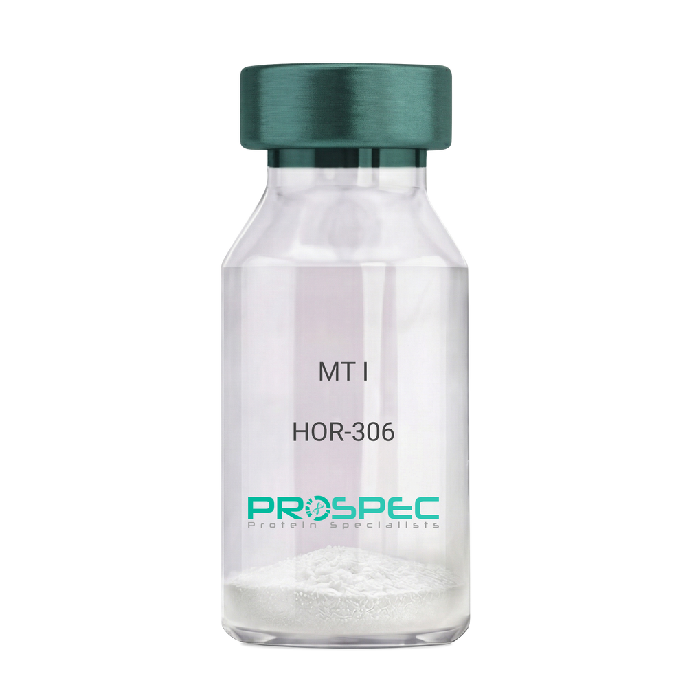
MT IDescription:
Melanotan-I
Melanotan-I, MT-I, Melanotan-1.
Product # :
HOR-306Price :
Quantity :
Shipping Method :
Shipped at Room temp
More Info
- description
- formulation
- purity
- More Info
Description
Melanotan-I has the amino acid sequence of Ser-Tyr-Ser-Nle-Glu-His-D-Phe-Arg-Trp-Gly-Lys-Pro-Val and a molecular weight of 1647.4 Dalton.
Formulation
The protein (1mg/ml) was lyophilized with no additives.
Purity
Greater than 98.0% as determined by RP-HPLC.
More Info
-
Synonyms
Melanotan-I, MT-I, Melanotan-1.
-
Physical Appearance
Sterile Filtered off-White lyophilized (freeze-dried) powder.
-
Stability
Lyophilized Melanotan-I although stable at room temperature for 3 weeks, should be stored desiccated below -18°C. Upon reconstitution MT-I should be stored at 4°C between 2-7 days and for future use below -18°C.For long term storage it is recommended to add a carrier protein (0.1% HSA or BSA).Please prevent freeze-thaw cycles.
-
Solubility
It is recommended to reconstitute the lyophilized Melanotan-I in sterile 1% acetic acid not less than 100 µg/ml, which can then be further diluted to other aqueous solutions.
ProSpec's products are furnished for LABORATORY RESEARCH USE ONLY. The product may not be used as drugs, agricultural or pesticidal products, food additives or household chemicals.
- View Data Sheet
Name :
HK2 AntibodyDescription:
Hexokinase-2, Mouse Anti Human
Hexokinase-2, EC 2.7.1.1, HK2, Hexokinase type II, HK II, Muscle form hexokinase, HXK2, DKFZp686M1669.
Product # :
ANT-378Price :
Quantity :
Shipping Method :
Shipped with Ice Packs
More Info
- formulation
- More Info
Formulation
1mg/ml containing PBS, pH-7.4, & 0.1% Sodium Azide.
More Info
-
Introduction
Hexokinases phosphorylate glucose to produce glucose-6-phosphate, thus committing glucose to the glycolytic pathway. Hexokinase 2 is the predominant form found in skeletal muscle. It localizes to the outer membrane of mitochondria. Expression of this gene is insulin-responsive, and studies in rat suggest that it is involved in the increased rate of glycolysis seen in rapidly growing cancer cells.
-
Synonyms
Hexokinase-2, EC 2.7.1.1, HK2, Hexokinase type II, HK II, Muscle form hexokinase, HXK2, DKFZp686M1669.
-
Immunogen
Anti-human Hexokinase-2 mAb is derived from hybridization of mouse F0 myeloma cells with spleen cells from BALB/c mice immunized with recombinant human Hexokinase-2 amino acids 1-917 purified from E. coli.
-
Ig Subclass
Mouse IgG1 heavy chain and κ light chain.
-
Clone
P1A7AT.
-
Applications
Hexokinase-2 antibody has been tested by ELISA and Western blot analysis to assure specificity and reactivity. Since application varies, however, each investigation should be titrated by the reagent to obtain optimal results. Recommended dilution range for Western blot analysis is 1:1,000 ~ 3,000. Recommended starting dilution is 1:2,000.
-
Type
Mouse Anti Human Monoclonal.
-
Storage Procedures
For periods up to 1 month store at 4°C, for longer periods of time, store at -20°C. Prevent freeze thaw cycles.
-
Purification Method
Hexokinase-2 antibody was purified from mouse ascitic fluids by protein-G affinity chromatography.
ProSpec's products are furnished for LABORATORY RESEARCH USE ONLY. The product may not be used as drugs, agricultural or pesticidal products, food additives or household chemicals.
- View Data Sheet
Name :
MG HumanDescription:
Menopausal Gonadotropin Human
Product # :
HOR-251Price :
Quantity :
Shipping Method :
Shipped at Room temp
More Info
- description
- source
- formulation
- biological activity
- More Info
Description
Menopausal Gonadotropin Human is produced from a sterile preparation of placental glucoprotein urine of post-menopausal women.The MG is purified by proprietary chromatographic techniques.
Source
Urine of post-menopausal women.
Formulation
The Human MG was lyophilized from a concentrated (1mg/ml) solution with no additives.
Biological Activity
100 IU/mg of FSH and 100 IU/mg LH.More Info
-
Physical Appearance
Sterile Filtered White lyophilized (freeze-dried) powder.
-
Stability
Lyophilized Menopausal Gonadotropin Human although stable at room temperature for 3 weeks, should be stored desiccated below -18°C. Upon reconstitution MG should be stored at 4°C between 2-7 days and for future use below -18°C.For long term storage it is recommended to add a carrier protein (0.1% HSA or BSA).Please prevent freeze-thaw cycles.
-
Solubility
It is recommended to reconstitute the lyophilized MG in sterile 18MΩ-cm H2O not less than 100 µg/ml, which can then be further diluted to other aqueous solutions.
-
Contaminants
Free of: HbsAg, Hepatitis B surface antigen and antibodies to HIV, Hepatitis C and HIV.
ProSpec's products are furnished for LABORATORY RESEARCH USE ONLY. The product may not be used as drugs, agricultural or pesticidal products, food additives or household chemicals.