Search results
327 results found for “fragile histidine triad”
Name
Description
Product #
Price
Quantity
Shipping Method
- View Data Sheet
Name :
HCV NS3 Genotype-1bDescription:
Hepatitis C Virus NS3 Genotype-1b Recombinant
Product # :
HCV-204Price :
Quantity :
Shipping Method :
Shipped with Ice Packs
More Info
- description
- formulation
- purity
- More Info
Description
The E.coli derived 26.2 kDa recombinant protein contains the HCV NS3 c33c immunodominant regions. The 252 amino acid protein is fused to 6xHis tag.
Formulation
50mM NaPO4 pH 8.3 and 10mM DTT.
Purity
HCV NS3 Genotype-1b protein is >95% pure as determined by 10% PAGE (coomassie staining).
More Info
-
Introduction
HCV is a small 50nm, enveloped, single-stranded, positive sense RNAvirus in the family Flaviviridae.
HCV has a high rate of replication with approximately one trillion particles produced each day in an infected individual. Due to lack of proofreading by the HCV RNA polymerase, the HCV has an exceptionally high mutation rate, a factor that may help it elude the host's immune response. Hepatitis C virus is classified into six genotypes(1-6) with several subtypes within each genotype. The preponderance and distribution of HCV genotypes varies globally. Genotype is clinically important in determining potential response to interferon-based therapy and the required duration of such therapy. Genotypes 1 and 4 are less responsive to interferon-based treatment than are the other genotypes (2, 3, 5 and 6). -
Stability
HCV NS3 Genotype-1b although stable at 4°C for 1 week, should be stored below -18°C. Please prevent freeze thaw cycles.
-
Amino Acid Sequence
Starts: MRDSDSQTFQ
Ends: DSVIDCNTCVT. -
Applications
HCV NS3 Genotype-1b antigen is suitable for ELISA and Western blots, excellent antigen for detection of HCV with minimal specificity problems.
-
Specificity
Immunoreactive with sera of HCV-infected individuals.
-
Purification Method
Purified by proprietary chromatographic technique.
ProSpec's products are furnished for LABORATORY RESEARCH USE ONLY. The product may not be used as drugs, agricultural or pesticidal products, food additives or household chemicals.
- View Data Sheet
Name :
T.pallidum p41Description:
Treponema pallidum p41 Recombinant
Product # :
TRP-242Price :
Quantity :
Shipping Method :
Shipped with Ice Packs
More Info
- description
- source
- formulation
- purity
- More Info
Description
The E.coli derived recombinant protein contains the outer membrane T.Pallidum p41 immunodominant regions. The protein is fused with a Beta-galactosidase tag.
Source
Escherichia Coli.
Formulation
10mM Tris-HCl pH8.0, 1mM EDTA, 1mMDTT and 8M urea.
Purity
Treponema pallidum protein is >90% pure as determined by 10% PAGE (coomassie staining).
More Info
-
Introduction
Treponema pallidum is a gram-negative spirochaete bacterium and is considered to be metabolically crippled. There are at least four known subspecies: T. pallidum pallidum, T. pallidum pertenue, T. pallidum carateum and T. pallidum endemicum. The helical structure of T. pallidum pallidum allows it to move in a corkscrew motion through viscous mediums such as mucus. Treponema pallidum sub sp. pallidum has one of the smallest bacterial genomes at 1.14 million base pairs (Mb) and has limited metabolic capabilities, reflecting its adaptation through genome reduction to the rich environment of mammalian tissue.
-
Stability
T.pallidum p41 although stable at 4°C for 1 week, should be stored below -18°C. Please prevent freeze thaw cycles.
-
Applications
Treponema pallidum is suitable for ELISA and Western blots, excellent antigen for detection of T. Pallidum with minimal specificity problems.
-
Specificity
Immunoreactive with sera of T.Pallidum infected individuals.
-
Purification Method
Treponema pallidum protein was purified by proprietary chromatographic technique.
ProSpec's products are furnished for LABORATORY RESEARCH USE ONLY. The product may not be used as drugs, agricultural or pesticidal products, food additives or household chemicals.
- View Data Sheet
Name :
GPD2 HumanDescription:
Glycerol-3-Phosphate Dehydrogenase 2 Human Recombinant
Glycerol-3-phosphate dehydrogenase mitochondrial, glycerol-3-phosphate dehydrogenase 2 (mitochondrial), GPDH-M, GPD-M, mtGPD, GPD2, GDH2, GPDM, mGPDH.
Product # :
ENZ-437Price :
Quantity :
Shipping Method :
Shipped with Ice Packs
More Info
- description
- source
- formulation
- purity
- More Info
Description
GPD2 Human Recombinant produced in E.Coli is single, a non-glycosylated, Polypeptide chain containing 558 amino acids fragment (43-600) corresponding to the GlpA domain fragment of the mature protein, having a total molecular mass of 66.26kDa and fused with a 4.5kDa amino-terminal hexahistidine tag. The GPD2 is purified by proprietary chromatographic techniques.
Source
Escherichia Coli.
Formulation
GPD2 protein solution is supplied in 20mM Tris-HCl pH 8, 1mM EDTA and 50% glycerol.
Purity
Greater than 95.0% as determined by SDS-PAGE.
More Info
-
Introduction
GPD2 (Mitochondrial glycerol-3-phosphate dehydrogenase) is a Ca2+-sensitive, FAD-binding protein, which is located on the outer surface of the inner mitochondrial membrane. Two isoforms have been identified for mGPD: Isoform 1 is comprised of 727 a.a. residues, while isoform 2 lacks 126 a.a. residues of the N-terminus. GPD2 catalyses the oxidation of glycerol-3-phosphate to DHAP (dihydroxyacetone phosphate) with associated reduction of the enzyme-bound FAD. GPD2 is a testis-specific promoter of mitochondrial GPDH. GPD2 along with a cytosolic NAD-linked GPD forms the glycerol phosphate shuttle that uses the interconversion of G-3-P and DHAP to transfer reducing equivalents into mitochondria, which results in the reoxidation of NADH produced during glycolysis.
GPD2 deficiency contributes to the impairment of glucose-stimulated INS discharge in a number of animal models of non-INS dependent diabetes mellitus. GPD2 up-regulation as a result of a highly glycolytic environment contributes to the general increase in ROS generation and may lead to the progression of prostate cancer. -
Synonyms
Glycerol-3-phosphate dehydrogenase mitochondrial, glycerol-3-phosphate dehydrogenase 2 (mitochondrial), GPDH-M, GPD-M, mtGPD, GPD2, GDH2, GPDM, mGPDH.
-
Physical Appearance
Sterile Filtered clear solution.
-
Stability
Store at 4°C if entire vial will be used within 2-4 weeks. Store, frozen at -20°C for longer periods of time. Please avoid freeze thaw cycles.
ProSpec's products are furnished for LABORATORY RESEARCH USE ONLY. The product may not be used as drugs, agricultural or pesticidal products, food additives or household chemicals.
- View Data Sheet
Name :
HOMER3 HumanDescription:
Homer Homolog-3 Human Recombinant
Homer homolog 3 (Drosophila)1 2 , HOMER-31 2 , homer protein homolog 32 , Homer neuronal immediate early gene 32, Homer-33, VESL32.
Product # :
PRO-1108Price :
Quantity :
Shipping Method :
Shipped with Ice Packs
More Info
- description
- source
- formulation
- purity
- More Info
Description
HOMER3 Human Recombinant produced in E. coli is a single polypeptide chain containing 386 amino acids (1-361) and having a molecular mass of 42.5kDa.HOMER3 is fused to a 24 amino acid His-tag at N-terminus & purified by proprietary chromatographic techniques.
Source
E.coli.
Formulation
The HOMER3 solution (1mg/1ml) contains 20mM Tris-HCl buffer (pH 8.0), 150mM NaCl, 1mM DTT and 30% glycerol.
Purity
Greater than 90% as determined by SDS-PAGE.
More Info
-
Introduction
HOMER3 belongs to the HOMER family of postsynaptic density scaffolding proteins which have an analogous domain structure consisting of an N-terminal Enabled/vasodilator-stimulated phosphoprotein homology 1 domain that facilitates protein-protein relations, and two leucine zipper and a carboxy-terminal coiled-coil domain motifs which take part in self-oligomerization.
-
Synonyms
Homer homolog 3 (Drosophila)1 2 , HOMER-31 2 , homer protein homolog 32 , Homer neuronal immediate early gene 32, Homer-33, VESL32.
-
Physical Appearance
Sterile Filtered colorless solution.
-
Stability
Store at 4°C if entire vial will be used within 2-4 weeks. Store, frozen at -20°C for longer periods of time. For long term storage it is recommended to add a carrier protein (0.1% HSA or BSA).Avoid multiple freeze-thaw cycles.
-
Amino Acid Sequence
MGSSHHHHHH SSGLVPRGSH MGSHMMSTAR EQPIFSTRAH VFQIDPATKR NWIPAGKHAL TVSYFYDATR NVYRIISIGG AKAIINSTVT PNMTFTKTSQ KFGQWADSRA NTVYGLGFAS EQHLTQFAEK FQEVKEAARL AREKSQDGGE LTSPALGLAS HQVPPSPLVS ANGPGEEKLF RSQSADAPGP TERERLKKML SEGSVGEVQW EAEFFALQDS NNKLAGALRE ANAAAAQWRQ QLEAQRAEAE RLRQRVAELE AQAASEVTPT GEKEGLGQGQ SLEQLEALVQ TKDQEIQTLK SQTGGPREAL EAAEREETQQ KVQDLETRNA ELEHQLRAME RSLEEARAER ERARAEVGRA AQLLDVSLFE LSELREGLAR LAEAAP
ProSpec's products are furnished for LABORATORY RESEARCH USE ONLY. The product may not be used as drugs, agricultural or pesticidal products, food additives or household chemicals.
- View Data Sheet
Name :
FGF 1 Mouse, HisDescription:
Fibroblast Growth Factor-acidic Mouse Recombinant, His Tag
HBGF-1, ECGF-beta, FIBP, FGFIBP, FIBP-1, ECGF, ECGFA, GLIO703, FGF1, FGF-a.
Product # :
CYT-072Price :
Quantity :
Shipping Method :
Shipped with Ice Packs
More Info
- description
- source
- formulation
- purity
- More Info
- sds-page
Description
FGF-1 Mouse Recombinant produced in E.Coli is a single, non-glycosylated polypeptide chain containing 161 amino acids (16-155 a.a) and having a molecular mass of 18kDa (Molecular weight on SDS-PAGE will appear higher).FGF-1 is fused to a 21 amino acid His-tag at N-terminus & purified by proprietary chromatographic techniques.
Source
Escherichia Coli.
Formulation
FGF-1 protein solution (1mg/ml) containing 20mM Tris-HCl buffer (pH 8.0), 1mM DTT, 30% glycerol and 0.1M NaCl.
Purity
Greater than 90.0% as determined by SDS-PAGE.
sds-page
More Info
-
Introduction
Acidic fibroblast growth factor is a member of the fibroblast growth factor (FGF) family. FGF family members possess broad mitogenic and cell survival activities, and are involved in a variety of biological processes, including embryonic development, cell growth, morphogenesis, tissue repair, tumor growth and invasion. This protein functions as a modifier of endothelial cell migration and proliferation, as well as an angiogenic factor. It acts as a mitogen for a variety of mesoderm- and neuroectoderm-derived cells in vitro, thus is thought to be involved in organogenesis. Three alternatively spliced variants encoding different isoforms have been described. The heparin-binding growth factors are angiogenic agents in vivo and are potent mitogens for a variety of cell types in vitro. There are differences in the tissue distribution and concentration of these 2 growth factors.
-
Synonyms
HBGF-1, ECGF-beta, FIBP, FGFIBP, FIBP-1, ECGF, ECGFA, GLIO703, FGF1, FGF-a.
-
Physical Appearance
Sterile Filtered colorless clear solution.
-
Stability
Store at 4°C if entire vial will be used within 2-4 weeks.Store, frozen at -20°C for longer periods of time. For long term storage it is recommended to add a carrier protein (0.1% HSA or BSA).Avoid multiple freeze-thaw cycles.
-
Amino Acid Sequence
MGSSHHHHHH SSGLVPRGSH MFNLPLGNYK KPKLLYCSNG GHFLRILPDG TVDGTRDRSD QHIQLQLSAE SAGEVYIKGT ETGQYLAMDT EGLLYGSQTP NEECLFLERL EENHYNTYTS KKHAEKNWFV GLKKNGSCKR GPRTHYGQKA ILFLPLPVSS D.
-
Background
What is the molecular weight/Mw of FGF 1 Protein?
FGF 1 Protein has a total Mw of 18kDa.
What is the source or expression system of FGF 1 Protein?
Escherichia Coli.
What is the Purity of FGF 1 Protein?
FGF 1 Protein is >90% pure as determined by SDS-PAGE.
What is the Biological Activity of FGF 1 Protein?
The biological functionality of FGF 1 Protein will be determined in the future.
What is the amino acid sequence of FGF 1 Protein?
MGSSHHHHHH SSGLVPRGSH MFNLPLGNYK KPKLLYCSNG GHFLRILPDG TVDGTRDRSD QHIQLQLSAE SAGEVYIKGT ETGQYLAMDT EGLLYGSQTP NEECLFLERL EENHYNTYTS KKHAEKNWFV GLKKNGSCKR GPRTHYGQKA ILFLPLPVSS D.
What applications can FGF 1 Protein be used in?
FGF 1 Protein can probably be used in western blot, ELISA and Lateral Flow.
What is the endotoxin level for FGF 1 Protein?
The endotoxin level is minimal, FGF 1 Protein was purified using conventional chromatography techniques.
ProSpec's products are furnished for LABORATORY RESEARCH USE ONLY. The product may not be used as drugs, agricultural or pesticidal products, food additives or household chemicals.
- View Data Sheet
Name :
IL 1 alpha Rat, HisDescription:
Interleukin-1 alpha Rat Recombinant, His Tag
Interleukin-1 alpha, IL-1 alpha.
Product # :
CYT-913Price :
Quantity :
Shipping Method :
Shipped with Ice Packs
More Info
- description
- source
- formulation
- purity
- biological activity
- More Info
Description
IL 1 alpha Rat Recombinant produced in E.Coli is a single, non-glycosylated polypeptide chain containing 179 amino acids (115-270 a.a) and having a molecular mass of 20.2kDa. IL 1 alpha is fused to a 23 amino acid His-tag at N-terminus & purified by proprietary chromatographic techniques.
Source
Escherichia Coli.
Formulation
IL 1 alpha protein solution (1mg/ml) containing Phosphate Buffered Saline (pH 7.4) and 10% glycerol.
Purity
Greater than 95.0% as determined by SDS-PAGE.
Biological Activity
The biological activity was Rat IL1 alpha was measured in a cell proliferation assay using D10.G4.1 mouse helper T cell. The ED50 for this effect is less or equal to 20 pg/ml.
More Info
-
Introduction
IL-1 alpha is produced by activated macrophages, stimulates thymocyte proliferation by inducing il-2 release, b-cell maturation and proliferation, and fibroblast growth factor activity. IL1A proteins are involved in the inflammatory response, being identified as endogenous pyrogens, and are reported to stimulate the release of prostaglandin and collagenase from synovial cells.
-
Synonyms
Interleukin-1 alpha, IL-1 alpha.
-
Physical Appearance
Sterile Filtered colorless solution.
-
Stability
Store at 4°C if entire vial will be used within 2-4 weeks. Store, frozen at -20°C for longer periods of time. For long term storage it is recommended to add a carrier protein (0.1% HSA or BSA). Avoid multiple freeze-thaw cycles.
-
Amino Acid Sequence
MGSSHHHHHH SSGLVPRGSH MGSSAPHSFQ NNLRYKLIRI VKQEFIMNDS LNQNIYVDMD RIHLKAASLN DLQLEVKFDM YAYSSGGDDS KYPVTLKVSN TQLFVSAQGE DKPVLLKEIP ETPKLITGSE TDLIFFWEKI NSKNYFTSAA FPELLIATKE QSQVHLARGL PSMIDFQIS.
ProSpec's products are furnished for LABORATORY RESEARCH USE ONLY. The product may not be used as drugs, agricultural or pesticidal products, food additives or household chemicals.
- View Data Sheet
Name :
EBI3 MacaqueDescription:
Epstein Barr Virus Induced 3 Macaque Recombinant
IL-27B, IL27B, IL 27-B, EBI-3, Interleukin-27 beta, IL-27 subunit beta, Epstein-Barr virus-induced gene 3 protein homolog, EBI3.
Product # :
CYT-1036Price :
Quantity :
Shipping Method :
Shipped at Room temp
More Info
- description
- source
- formulation
- purity
- More Info
Description
EBI3 Macaque Recombinant produced in E.Coli is a single, non-glycosylated polypeptide chain containing 210 amino acids and having a molecular mass of 23.4kDa. The EBI3 is purified by proprietary chromatographic techniques.
Source
Escherichia Coli.
Formulation
Lyophilized from a sterile filtered solution containing 0.1 % trifluoroacetic acid (TFA).
Purity
Greater than 95.0% as determined by SDS-PAGE.
More Info
-
Introduction
EBI3 has an induced expression in B lymphocytes in reaction to Epstein-Barr virus infection. EBI3 encodes a secreted glycoprotein belonging to the hematopoietin receptor family, and heterodimerizes with a 28 kDa protein to form iIL-27. EBI3 drives rapid clonal expansion of naive cd4(+) t-cells. EBI3 strongly synergizes with IL-12 to activate IFN-gamma production of naive cd4(+) t-cells. EBI3 mediates its biologic effects through the cytokine receptor wsx-1/tccr.
-
Synonyms
IL-27B, IL27B, IL 27-B, EBI-3, Interleukin-27 beta, IL-27 subunit beta, Epstein-Barr virus-induced gene 3 protein homolog, EBI3.
-
Physical Appearance
Sterile Filtered White lyophilized (freeze-dried) powder.
-
Stability
Lyophilized EBI3 although stable at room temperature for 3 weeks, should be stored desiccated below -18°C. Upon reconstitution EBI3 Macaque should be stored at 4°C between 2-7 days and for future use below -18°C. For long term storage it is recommended to add a carrier protein (0.1% HSA or BSA). Please prevent freeze-thaw cycles.
-
Solubility
It is recommended to reconstitute the lyophilized EBI3 in sterile 18MΩ-cm H2O at a concentration of 100µg/ml, which can then be further diluted to other aqueous solutions.
-
Amino Acid Sequence
MRKGPPAALT LPRVQCRAPR YPIAVDCSWT LPPAPNSTSP VSFIATYRFG MAARGHSWPC LQQTPASTSC TIADVRLFSM APYVLNVTAV HPWGSSSSFV PFIAEHIIKP DPPEGVRLSP LAERQLQVQW EPPRSWPFPE IFSLKYWIRY KRQGAARFHQ VGPIEATSFI LRAVRPRARY CVQVAAQDLT DYGELSDWSL PATTPMSPGK.
-
Background
What is the molecular weight/Mw of EBI3 Protein?
EBI3 Protein has a total Mw of 23.4kDa.
What is the source or expression system of EBI3 Protein?
Escherichia Coli.
What is the Purity of EBI3 Protein?
EBI3 Protein is >90% pure as determined by SDS-PAGE.
What is the Biological Activity of EBI3 Protein?
The biological functionality of EBI3 Protein will be determined in the future.
What is the amino acid sequence of EBI3 Protein?
MRKGPPAALT LPRVQCRAPR YPIAVDCSWT LPPAPNSTSP VSFIATYRFG MAARGHSWPC LQQTPASTSC TIADVRLFSM APYVLNVTAV HPWGSSSSFV PFIAEHIIKP DPPEGVRLSP LAERQLQVQW EPPRSWPFPE IFSLKYWIRY KRQGAARFHQ VGPIEATSFI LRAVRPRARY CVQVAAQDLT DYGELSDWSL PATTPMSPGK.
What applications can EBI3 Protein be used in?
EBI3 Protein can probably be used in western blot, ELISA and Lateral Flow.
What is the endotoxin level for EBI3 Protein?
The endotoxin level is minimal, EBI3 Protein was purified using conventional chromatography techniques.
ProSpec's products are furnished for LABORATORY RESEARCH USE ONLY. The product may not be used as drugs, agricultural or pesticidal products, food additives or household chemicals.
- View Data Sheet
Name :
H3N2 BrisbaneDescription:
H3N2 Influenza-A Virus Brisbane 10/07
Product # :
IHA-024Price :
Quantity :
Shipping Method :
Shipped with Ice Packs
More Info
- description
- formulation
- purity
- More Info
Description
Allantoic fluid of 10 days old embryonated eggs, inoculated with influenza A virus, strain A/Brisbane/10/07. The Influenza Virus was purified by Ultracentrifugation with 10-40 % sucrose gradient.
Formulation
The H3N2 A/Brisbane/10/07 solution contains STE, 0.1% sodium azide (NaN3) and 0.005% thimerosal.
Purity
Greater than 90.0% as determined byAnalysis by SDS-PAGE.
More Info
-
Introduction
H3N2 is a subtype of the influenza A virus. Its name derives from the forms of the two kinds of proteinson the surface of its coat, hemagglutinin(H) and neuraminidase(N). H3N2 exchanges genes for internal proteins with other influenza subtypes. H3N2 has tended to dominate in prevalence over H1N1, H1N2, and influenza B. H3N2 strain descended from H2N2by antigenic shift, in which genes from multiple subtypes re-assorted to form a new virus. Both the H2N2and H3N2 strains contained genesfrom avian influenzaviruses.
-
Physical Appearance
Sterile Filtered whitish (milky) solution.
-
Stability
A/Brisbane/10/07although stable 4°C for 4 weeks, should be stored below -18°C. Please prevent freeze-thaw cycles.
-
Inactivation
Thimerosal and beta propiolactone treatmentThis product has been treated in a manner consistent with methods of inactivation. Generally accepted good laboratory practices appropriate to microbiological/viral safe handling practices and techniques are required at work.
-
Immunological Activity
Tested with anti-influenza A monoclonal antibodies in ELISA.Serological studies of influenza A virus, immunogen for antibody production.
ProSpec's products are furnished for LABORATORY RESEARCH USE ONLY. The product may not be used as drugs, agricultural or pesticidal products, food additives or household chemicals.
- View Data Sheet
Name :
TNF a Rat, HisDescription:
Tumor Necrosis Factor-alpha Rat Recombinant, His Tag
Tumor Necrosis Factor-alpha, TNF a His, Cachectin, TNF-alpha, Tumor necrosis factor ligand superfamily member 2, TNF-a, N-terminal fragment, NTF, Intracellular domain 1, Intracellular domain 2, ICD2, C-domain 1, C-domain 2, Tumor necrosis factor, soluble form, Tnfa, Tnfsf2, RATTNF, Tnfa.
Product # :
CYT-900Price :
Quantity :
Shipping Method :
Shipped with Ice Packs
More Info
- description
- source
- formulation
- purity
- More Info
Description
TNF a Rat Recombinant produced in E.Coli is a single, non-glycosylated polypeptide chain containing 181 amino acids (80-235a.a.) and having a molecular mass of 19.9kDa.TNF a is fused to a 25 amino acid His-tag at N-terminus & purified by proprietary chromatographic techniques.
Source
Escherichia Coli.
Formulation
TNF a protein solution (1mg/ml) containing Phosphate Buffer Saline (pH7.4), 10% glycerol and 1mM DTT.
Purity
Greater than 90.0% as determined by analysis by SDS-PAGE.
More Info
-
Introduction
Tumor necrosis factor is a cytokine involved in systemic inflammation and is a member of a group of cytokines that all stimulate the acute phase reaction. TNF is mainly secreted by macrophages.
TNF causes apoptotic cell death, cellular proliferation, differentiation, inflammation, tumorigenesis and viral replication, TNF is also involved in lipid metabolism, and coagulation. TNF's primary role is in the regulation of immune cells.
Dysregulation and, in particular, overproduction of TNF have been implicated in a variety of human diseases- autoimmune diseases, insulin resistance, and cancer. -
Synonyms
Tumor Necrosis Factor-alpha, TNF a His, Cachectin, TNF-alpha, Tumor necrosis factor ligand superfamily member 2, TNF-a, N-terminal fragment, NTF, Intracellular domain 1, Intracellular domain 2, ICD2, C-domain 1, C-domain 2, Tumor necrosis factor, soluble form, Tnfa, Tnfsf2, RATTNF, Tnfa.
-
Physical Appearance
Sterile Filtered clear solution.
-
Stability
Store at 4°C if entire vial will be used within 2-4 weeks. Store, frozen at -20°C for longer periods of time. For long term storage it is recommended to add a carrier protein (0.1% HSA or BSA).Avoid multiple freeze-thaw cycles.
-
Amino Acid Sequence
MGSSHHHHHH SSGLVPRGSH MGSHMLRSSS QNSSDKPVAH VVANHQAEEQ LEWLSQRANA LLANGMDLKD NQLVVPADGL YLIYSQVLFK GQGCPDYVLL THTVSRFAIS YQEKVSLLSA IKSPCPKDTP EGAELKPWYE PMYLGGVFQL EKGDLLSAEV NLPKYLDITE SGQVYFGVIA L.
ProSpec's products are furnished for LABORATORY RESEARCH USE ONLY. The product may not be used as drugs, agricultural or pesticidal products, food additives or household chemicals.
- View Data Sheet
Name :
DDAVPDescription:
Desmopressin
Product # :
HOR-270Price :
Quantity :
Shipping Method :
Shipped at Room temp
More Info
- description
- formulation
- purity
- More Info
Description
Desmopressin also called ADH (Anti-Diuretic Hormone) has a molecular formula of C46H64N14O12S2 , Mpr-Tyr-Phe-Gln-Asn-Cys-Pro-D-Arg-Gly-NH2 having a Mw of 1069.23 Dalton.
Formulation
The Desmopressin peptide was lyophilized with no additives.
Purity
Greater than 98.0% as determined by:
(a) Analysis by RP-HPLC.
(b) Analysis by SDS-PAGE.More Info
-
Introduction
Desmopressin is the first vasopressin analog with a very high and very specific antidiuretic effect, has been widely used for different therapeutic purposes and is believed to be partly responsible for the formation of memories learning and memory processes. Desmopressin increases urine concentration and decreases urine production. Desmopressin is used to prevent and control excessive thirst, urination, and dehydration.
-
Physical Appearance
Sterile Filtered White lyophilized (freeze-dried) powder.
-
Stability
Lyophilized Desmopressin although stable at room temperature for 3 weeks, should be stored desiccated below -18°C. Upon reconstitution DDAVP should be stored at 4°C between 2-7 days and for future use below -18°C. For long term storage it is recommended to add a carrier protein (0.1% HSA or BSA).Please prevent freeze-thaw cycles.
-
Solubility
It is recommended to reconstitute the lyophilized DDAVP in sterile 18MΩ-cm H2O not less than 100 µg/ml, which can then be further diluted to other aqueous solutions.
ProSpec's products are furnished for LABORATORY RESEARCH USE ONLY. The product may not be used as drugs, agricultural or pesticidal products, food additives or household chemicals.
- View Data Sheet
Name :
IL 33 Rat, HisDescription:
Interleukin-33 Rat Recombinant, His Tag
Interleukin-33, IL-33.
Product # :
CYT-906Price :
Quantity :
Shipping Method :
Shipped with Ice Packs
More Info
- description
- source
- formulation
- purity
- More Info
Description
IL 33 Rat Recombinant produced in E.Coli is a single, non-glycosylated polypeptide chain containing 179 amino acids (109-264 a.a) and having a molecular mass of 19.8kDa. IL 33 is fused to a 23 amino acid His-tag at N-terminus & purified by proprietary chromatographic techniques.
Source
Escherichia Coli.
Formulation
IL 33 protein solution (0.5mg/ml) containing Phosphate Buffered Saline (pH7.4) and 10% glycerol.
Purity
Greater than 90.0% as determined by SDS-PAGE.
More Info
-
Introduction
nterleukin 33 (IL-33) is a 32kDa proinflammatory cytokine that may also regulate gene transcription in producer cells. IL-33 is structurally related to IL-1, which induces helper T cells to produce type 2 cytokines and acts through the receptor IL1RL-1 (IL1 receptor-like-1), which is known also as ST2. Binding of IL-33 to this receptor activates NF-kappa-B and MAP kinases and induces in vitro Th2 cells to produce cytokines. In vivo, IL-33 induces expression of IL-4, IL-5, IL-13 and leads to severe pathological changes in mucosal organs and in vitro, it can be divided to N-terminal fragment of 12kDa and C-terminal fragment of 18kDa by cleavage of caspase-1.
-
Synonyms
Interleukin-33, IL-33.
-
Physical Appearance
Sterile Filtered colorless solution.
-
Stability
Store at 4°C if entire vial will be used within 2-4 weeks. Store, frozen at -20°C for longer periods of time. For long term storage it is recommended to add a carrier protein (0.1% HSA or BSA). Avoid multiple freeze-thaw cycles.
-
Amino Acid Sequence
MGSSHHHHHH SSGLVPRGSH MGSSIQGTSL LTESCALSTY NDQSVSFVLE NGCYVINVED CGKNQEKDKV LLRYYESSFP AQSGDGVDGK KLMVNMSPIK DTDIWLNAND KDYSVELQKG DVSPPDQAFF VLHKKSSDFV SFECKNLPGT YIGVKDNQLA LVEENDESCN NIMFKLSKM.
ProSpec's products are furnished for LABORATORY RESEARCH USE ONLY. The product may not be used as drugs, agricultural or pesticidal products, food additives or household chemicals.
- View Data Sheet
Name :
FLT1 D3 HumanDescription:
Vascular Endothelial Growth Factor Receptor-1 D3 Human Recombinant
FLT-1, FLT1, Tyrosine-protein kinase receptor FLT, Flt-1, Tyrosine-protein kinase FRT, Fms-like tyrosine kinase 1, VEGFR-1.
Product # :
PKA-234Price :
Quantity :
Shipping Method :
Shipped at Room temp
More Info
- description
- source
- formulation
- purity
- biological activity
- More Info
Description
FLT1 D1-3 Human Recombinant produced in baculovirus is monomeric, glycosylated, polypeptide containing 327 amino acids and having a molecular mass of 45 kDa. The soluble receptor protein contains only the first 3 extracellular domains, which contain all the information necessary for binding of VEGF.The FLT1 is purified by proprietary chromatographic techniques.
Source
Insect Cells.
Formulation
FLT1 D1-3 was lyophilized from a concentrated (1mg/ml) sterile solution containing 1xPBS.
Purity
Greater than 90.0% as determined by SDS-PAGE.
Biological Activity
The activity of FLT1D1-3 was determined by its ability to inhibit the VEGF-165-induced proliferation of HUVE cells.
More Info
-
Introduction
Endothelial cells express three different vascular endothelial growth factor (VEGF) receptors, belonging to the family of receptor tyrosine kinases (RTKs). They are named VEGFR-1 (Flt-1), VEGFR-2 (KDR/Flk-1), VEGFR-3 (Flt-4). Their expression is almost exclusively restricted to endothelial cells, but VEGFR-1 can also be found on monocytes, dendritic cells and on trophoblast cells. The flt-1 gene was first described in 1990. The receptor contains seven immunoglobulin-like extracellular domains, a single transmembrane region and an intracellular splited tyrosine kinase domain. Compared to VEGFR-2 the Flt-1 receptor has a higher affinity for VEGF but a weaker signaling activity. VEGFR-1 thus leads not to proliferation of endothelial cells, but mediates signals for differentiation. Interestingly a naturally occuring soluble variant of VEGFR-1 (sVEGFR-1) was found in HUVE supernatants in 1996, which is generated by alternative splicing of the flt-1 mRNA. The biological functions of sVEGFR-1 still are not clear, but it seems to be an endogenous regulator of angiogenesis, binding VEGF with the same affinity as the full-length receptor.
-
Synonyms
FLT-1, FLT1, Tyrosine-protein kinase receptor FLT, Flt-1, Tyrosine-protein kinase FRT, Fms-like tyrosine kinase 1, VEGFR-1.
-
Physical Appearance
Sterile Filtered White lyophilized (freeze-dried) powder.
-
Stability
Lyophilized FLT-1 although stable at room temperature for 3 weeks, should be stored desiccated below -18°C. Upon reconstitution FLT1 should be stored at 4°C between 2-7 days and for future use below -18°C.For long term storage it is recommended to add a carrier protein (0.1% HSA or BSA).Please prevent freeze-thaw cycles.
-
Solubility
It is recommended to reconstitute the lyophilized FLT1 D3 in sterile water not less than 100 µg/ml, which can then be further diluted to other aqueous solutions.
-
Amino Acid Sequence
SKLKDPELSLKGTQHIMQAGQTLHLQCRGEAAHKWSLPEMVSKESERLSI TKSACGRNGKQFCSTLTLNTAQANHTGFYSCKYLAVPTSKKKETESAIYI FISDTGRPFVEMYSEIPEIIHMTEGRELVIPCRVTSPNITVTLKKFPLDT LIPDGKRIIWDSRKGFIISNATYKEIGLLTCEATVNGHLYKTNYLTHRQT NTIIDVQISTPRPVKLLRGHTLVLNCTATTPLNTRVQMTWSYPDEKNKRA SVRRRIDQSNSHANIFYSVLTIDKMQNKDKGLYTCRVRSGPSFKSVNTSV HIYDKAFITVKHRKQQVLETVAGKRSY.
ProSpec's products are furnished for LABORATORY RESEARCH USE ONLY. The product may not be used as drugs, agricultural or pesticidal products, food additives or household chemicals.
- View Data Sheet
Name :
FGFR3 HumanDescription:
Fibroblast Growth Factor Receptor 3 Fc Chimera Human Recombinant
Achondroplasia, Thanatophoric Dwarfism, CD333, ACH, CEK2, JTK4, HSFGFR3EX, FGFR3.
Product # :
PKA-232Price :
Quantity :
Shipping Method :
Shipped at Room temp
More Info
- description
- source
- formulation
- purity
- More Info
Description
Soluble FGFR3 Human recombinant fused using a Xa cleavage site with the Fc part of human IgG1 produced in Fc Chimera is a heterodimeric, glycosylated, Polypeptide chain containing 593aa and having a molecular mass of 170kDa.
Source
Insect Cells.
Formulation
FGFR3 was lyophilized from a concentrated (1 mg/ml) sterile solution containing PBS.
Purity
Greater than 90.0% as determined by:(a) Analysis by silver stain.(b) Analysis by SDS-PAGE.
More Info
-
Introduction
Fibroblast Growth Factors (FGFs) comprise a family of at least eighteen structurally realted proteins that are involved in a multitude of physiological and pathological cellular processes, including cell growth, differentation, angiogenesis, wound healing and tumorgenesis. The biological activities of the FGFs are mediated by a family if type I transmembrane tyrosine kinases which undergo dimerization and autophosphorylation after ligand binding. Four distinct genes encoding closely related FGF receptors, FGFR-1to -4 are known. Multiple forms of FGFR-1 to -3 are generated by alternative splicing of the mRNAs. A frequent splicing event involving FGFR-1 and -2 results in receptors containing all three Ig domains, referred to as the alpha isoform, or only IgII and IgIII, referred to as the ? isoform. Only the alpha isoform has been identified for FGFR-3 and FGFR-4. Additional splicing events for FGFR-1 to -3, involving the C-terminal half of the IgIII domain encoded by two mutually exclusive alternative exons, generate FGF receptors with alternative IgIII domains (IIIb and IIIc). A IIIa isoform which is a secreted FGF binding protein containing only the N-terminal half of the IgIII domain plus some intron sequences has also been reported for FGFR-1. Mutations in FGFR-1 to -3 have been found in patients with birth defects involving craniosynostosis.
-
Synonyms
Achondroplasia, Thanatophoric Dwarfism, CD333, ACH, CEK2, JTK4, HSFGFR3EX, FGFR3.
-
Physical Appearance
Sterile Filtered White lyophilized (freeze-dried) powder.
-
Stability
Lyophilized FGFR3 although stable at room temperature for 3 weeks, should be stored desiccated below -18oC. Upon reconstitution FGFR3 should be stored at 4oC between 2-7 days and for future use below -18oC.For long term storage it is recommended to add a carrier protein (0.1% HSA or BSA).Please prevent freeze-thaw cycles.
-
Solubility
It is recommended to reconstitute the lyophilized FGFR-3 in sterile PBS not less than 50µg/ml, which can then be further diluted to other aqueous solutions.
-
Amino Acid Sequence
ESLGTEQRVV GRAAEVPGPE PGQQEQLVFG SGDAVELSCP PPGGGPMGPT VWVKDGTGLV PSERVLVGPQ RLQVLNASHE DSGAYSCRQR LTQRVLCHFS VRVTDAPSSG DDEDGEDEAE DTGVDTGAPY WTRPERMDKK LLAVPAANTV RFRCPAAGNP TPSISWLKNG REFRGEHRIG GIKLRHQQWS LVMESVVPSD RGNYTCVVEN KFGSIRQTYT LDVLERSPHR PILQAGLPAN QTAVLGSDVE FHCKVYSDAQ PHIQWLKHVE VNGSKVGPDG TPYVTVLKTA GANTTDKELE VLSLHNVTFE DAGEYTCLAG NSIGFSHHSA WLVVLPAEEE LVEADEAGDP RRASIEGRGD PEEPKSCDKT HTCPPCPAPE LLGGPSVFLF PPKPKDTLMI SRTPEVTCVV VDVSHEDPEV KFNWYVDGVE VHNAKTKPRE EQYNSTYRVV SVTVLHQDWL NGKEYKCKVS NKALPAPIEK TISKAKGQPR EPQVYTLPPS RDELTKNQVS LTCLVKGFYP SDIAVEWESN GQPENNYKTT PPVLDSDGSF FLYSKLTVDK SRWQQGNVFS CSVMHEALHN HYTQKSLSLS PGK.
ProSpec's products are furnished for LABORATORY RESEARCH USE ONLY. The product may not be used as drugs, agricultural or pesticidal products, food additives or household chemicals.
- View Data Sheet
Name :
QTRTD1 HumanDescription:
Queuine TRNA-Ribosyltransferase Domain Containing 1 Human Recombinant
Queuine TRNA-Ribosyltransferase Domain Containing 1, EC 2.4.2.29, Queuine TRNA-Ribosyltransferase Domain-Containing Protein 1, QTRTD1.
Product # :
ENZ-833Price :
Quantity :
Shipping Method :
Shipped with Ice Packs
More Info
- description
- source
- formulation
- purity
- More Info
Description
QTRTD1 Human Recombinant produced in E.Coli is a single, non-glycosylated polypeptide chain containing 438 amino acids (1-415 a.a) and having a molecular mass of 49.1kDa.QTRTD1 is fused to a 23 amino acid His-tag at N-terminus & purified by proprietary chromatographic techniques.
Source
Escherichia Coli.
Formulation
QTRTD1 protein solution (0.25mg/ml) containing Phosphate buffered saline (pH7.4) 30% glycerol, 2mM DTT, 1mM EDTA and 0.1mM PMSF.
Purity
Greater than 95.0% as determined by SDS-PAGE.
More Info
-
Introduction
Queuine TRNA-Ribosyltransferase Domain Containing 1, also known as QTRTD1 act together with QTRT1 to form an active queuine tRNA-ribosyltransferase. QTRTD1 exchanges queuine for the guanine at the wobble position of tRNAs along with GUN anticodons (tRNA-Asp,-Asn,-His and -Tyr), in that way forming the hypermodified nucleoside queuosine.
-
Synonyms
Queuine TRNA-Ribosyltransferase Domain Containing 1, EC 2.4.2.29, Queuine TRNA-Ribosyltransferase Domain-Containing Protein 1, QTRTD1.
-
Physical Appearance
Sterile Filtered colorless solution.
-
Stability
Store at 4°C if entire vial will be used within 2-4 weeks. Store, frozen at -20°C for longer periods of time. For long term storage it is recommended to add a carrier protein (0.1% HSA or BSA).Avoid multiple freeze-thaw cycles.
-
Amino Acid Sequence
MGSSHHHHHH SSGLVPRGSH MGSMKLSLTK VVNGCRLGKI KNLGKTGDHT MDIPGCLLYT KTGSAPHLTH HTLHNIHGVP AMAQLTLSSL AEHHEVLTEY KEGVGKFIGM PESLLYCSLH DPVSPCPAGY VTNKSVSVWS VAGRVEMTVS KFMAIQKALQ PDWFQCLSDG EVSCKEATSI KRVRKSVDRS LLFLDNCLRL QEESEVLQKS VIIGVIEGGD VMEERLRSAR ETAKRPVGGF LLDGFQGNPT TLEARLRLLS SVTAELPEDK PRLISGVSRP DEVLECIERG VDLFESFFPY QVTERGCALT FSFDYQPNPE ETLLQQNGTQ EEIKCMDQIK KIETTGCNQE ITSFEINLKE KKYQEDFNPL VRGCSCYCCK NHTRAYIHHL LVTNELLAGV LLMMHNFEHY FGFFHYIREA LKSDKLAQLK ELIHRQAS.
ProSpec's products are furnished for LABORATORY RESEARCH USE ONLY. The product may not be used as drugs, agricultural or pesticidal products, food additives or household chemicals.
- View Data Sheet
Name :
CD3G HumanDescription:
CD3G Human Recombinant
T-cell surface glycoprotein CD3 gamma chain, CD3-GAMMA, T3G, CD3G.
Product # :
PRO-1392Price :
Quantity :
Shipping Method :
Shipped with Ice Packs
More Info
- description
- source
- formulation
- purity
- More Info
Description
CD3G Human Recombinant produced in E.Coli is a single, non-glycosylated polypeptide chain containing 117 amino acids (23-116a.a.) and having a molecular mass of 13.1kDa. CD3G is fused to a 23 amino acid His-tag at N-terminus & purified by proprietary chromatographic techniques.
Source
E.coli.
Formulation
CD3G protein solution (1mg/ml) containing 20mM Tris-HCl buffer (pH 8.0), 0.4M urea and 10% glycerol.
Purity
Greater than 90% as determined by SDS-PAGE.
More Info
-
Introduction
CD3-gamma polypeptide is encoded by CD3 Gamma (CD3G) which together with CD3-epsilon, -delta and -zeta, and the T-cell receptor alpha/beta and gamma/delta heterodimers, configures the T-cell receptor-CD3 complex. The T-cell receptor-CD3 complex takes part in coupling antigen recognition to numerous intracellular signal-transduction pathways. The genes encoding the epsilon, gamma and delta polypeptides are located in the same cluster on chromosome 11. Deficiencies in this gene are associated with T cell immunodeficiency.
-
Synonyms
T-cell surface glycoprotein CD3 gamma chain, CD3-GAMMA, T3G, CD3G.
-
Physical Appearance
Sterile Filtered colorless solution.
-
Stability
Store at 4°C if entire vial will be used within 2-4 weeks. Store, frozen at -20°C for longer periods of time. For long term storage it is recommended to add a carrier protein (0.1% HSA or BSA).Avoid multiple freeze-thaw cycles.
-
Amino Acid Sequence
MGSSHHHHHH SSGLVPRGSH MGSQSIKGNH LVKVYDYQED GSVLLTCDAE AKNITWFKDG KMIGFLTEDK KKWNLGSNAK DPRGMYQCKG SQNKSKPLQV YYRMCQNCIE LNAATIS.
ProSpec's products are furnished for LABORATORY RESEARCH USE ONLY. The product may not be used as drugs, agricultural or pesticidal products, food additives or household chemicals.
- View Data Sheet
Name :
H3N2 ShandongDescription:
H3N2 Influenza-A Virus Shandong/9/93
Product # :
IHA-004Price :
Quantity :
Shipping Method :
Shipped with Ice Packs
More Info
- description
- formulation
- purity
- More Info
Description
Allantoic fluid of 10 days old embryonated eggs, inoculated with influenza A virus, strain A/Shandong/9/93. The Influenza Virus was purified by Ultracentrifugation with 10-40 % sucrose gradient.
Formulation
The H3N2A/ Shandong/9/93 solution (1.0mg/ml) contains STE, 0.1% sodium azide (NaN3) and 0.005% thimerosal.
Stir thoroughly prior to its use.
Purity
Greater than 90.0%.
More Info
-
Introduction
H3N2 is a subtype of the influenza A virus. Its name derives from the forms of the two kinds of proteinson the surface of its coat, hemagglutinin(H) and neuraminidase(N). H3N2 exchanges genes for internal proteins with other influenza subtypes. H3N2 has tended to dominate in prevalence over H1N1, H1N2, and influenza B. H3N2 strain descended from H2N2by antigenic shift, in which genes from multiple subtypes re-assorted to form a new virus. Both the H2N2and H3N2 strains contained genesfrom avian influenzaviruses.
-
Physical Appearance
Opaque suspension.
-
Stability
A/Shandong/9/93 although stable 4°C for 4 weeks, should be stored below -18°C. Please prevent freeze-thaw cycles.
-
Inactivation
Thimerosal and beta propiolactone treatmentThis product has been treated in a manner consistent with methods of inactivation. Generally accepted good laboratory practices appropriate to microbiological/viral safe handling practices and techniques are required at work.
-
Immunological Activity
Serological studies of influenza A virus, immunogen for antibody production.Tested with anti-influenza A monoclonal antibodies in ELISA.
ProSpec's products are furnished for LABORATORY RESEARCH USE ONLY. The product may not be used as drugs, agricultural or pesticidal products, food additives or household chemicals.
- View Data Sheet
Name :
HMGCS1 HumanDescription:
3-Hydroxy-3-Methylglutaryl-CoA Synthase 1 Human Recombinant
3-Hydroxy-3-Methylglutaryl-CoA Synthase 1 (Soluble), 3-Hydroxy-3-Methylglutaryl-Coenzyme A Synthase 1 (Soluble), 3-Hydroxy-3-Methylglutaryl Coenzyme A (HMG-CoA) Synthase, EC 2.3.3.10, HMGCS, Hydroxymethylglutaryl-CoA Synthase, Cytoplasmic, 3-Hydroxy-3-Methylglutaryl Coenzyme A Synthase, HMG-CoA Synthase.
Product # :
ENZ-870Price :
Quantity :
Shipping Method :
Shipped with Ice Packs
More Info
- description
- source
- formulation
- purity
- More Info
Description
HMGCS1 Human Recombinant produced in E.Coli is a single, non-glycosylated polypeptide chain containing 543 amino acids (1-520 a.a) and having a molecular mass of 59.7kDa.HMGCS1 is fused to a 23 amino acid His-tag at N-terminus & purified by proprietary chromatographic techniques.
Source
Escherichia Coli.
Formulation
HMGCS1 protein solution (0.5mg/ml) containing Phosphate buffered saline (pH7.4), 10% glycerol and 1mM DTT.
Purity
Greater than 80.0% as determined by SDS-PAGE.
More Info
-
Introduction
3-Hydroxy-3-Methylglutaryl-CoA Synthase 1, also known as HMGCS1 is a member of the Belongs to the HMG-CoA synthase family. HMGCS1 condenses acetyl-CoA with acetoacetyl-CoA to form HMG-CoA, which is the substrate for HMG-CoA reductase.
-
Synonyms
3-Hydroxy-3-Methylglutaryl-CoA Synthase 1 (Soluble), 3-Hydroxy-3-Methylglutaryl-Coenzyme A Synthase 1 (Soluble), 3-Hydroxy-3-Methylglutaryl Coenzyme A (HMG-CoA) Synthase, EC 2.3.3.10, HMGCS, Hydroxymethylglutaryl-CoA Synthase, Cytoplasmic, 3-Hydroxy-3-Methylglutaryl Coenzyme A Synthase, HMG-CoA Synthase.
-
Physical Appearance
Sterile Filtered clear solution.
-
Stability
Store at 4°C if entire vial will be used within 2-4 weeks. Store, frozen at -20°C for longer periods of time. For long term storage it is recommended to add a carrier protein (0.1% HSA or BSA).Avoid multiple freeze-thaw cycles.
-
Amino Acid Sequence
MGSSHHHHHH SSGLVPRGSH MGSMPGSLPL NAEACWPKDV GIVALEIYFP SQYVDQAELE KYDGVDAGKY TIGLGQAKMG FCTDREDINS LCMTVVQNLM ERNNLSYDCI GRLEVGTETI IDKSKSVKTN LMQLFEESGN TDIEGIDTTN ACYGGTAAVF NAVNWIESSS WDGRYALVVA GDIAVYATGN ARPTGGVGAV ALLIGPNAPL IFERGLRGTH MQHAYDFYKP DMLSEYPIVD GKLSIQCYLS ALDRCYSVYC KKIHAQWQKE GNDKDFTLND FGFMIFHSPY CKLVQKSLAR MLLNDFLNDQ NRDKNSIYSG LEAFGDVKLE DTYFDRDVEK AFMKASSELF SQKTKASLLV SNQNGNMYTS SVYGSLASVL AQYSPQQLAG KRIGVFSYGS GLAATLYSLK VTQDATPGSA LDKITASLCD LKSRLDSRTG VAPDVFAENM KLREDTHHLV NYIPQGSIDS LFEGTWYLVR VDEKHRRTYA RRPTPNDDTL DEGVGLVHSN IATEHIPSPA KKVPRLPATA AEPEAAVISN GEH
ProSpec's products are furnished for LABORATORY RESEARCH USE ONLY. The product may not be used as drugs, agricultural or pesticidal products, food additives or household chemicals.
- View Data Sheet
Name :
B3GNT2 HumanDescription:
Beta-1,3-N-Acetylglucosaminyltransferase 2 Human Recombinant
B3GNT2, B3GN-T2, B3GNT, B3GNT-2, B3GNT1, BETA3GNT, BGnT-2, BGNT2, N-acetyllactosaminide beta-1,3-N-acetylglucosaminyltransferase 2, Beta-1,3-N-acetylglucosaminyltransferase 1, BGnT-1, Beta-1,3-Gn-T1, Beta3Gn-T1, Beta-1,3-galactosyltransferase 7, Beta-1,3-GalTase 7, Beta3Gal-T7, Beta3GalT7, b3Gal-T7, Beta-3-Gx-T7, beta-GlcNAc beta-1,3-galactosyltransferase 7, betaGal beta-1,3-N-acetylglucosaminyltransferase 2, BGnT-2, Beta-1,3-Gn-T2, Beta-1,3-N-acetylglucosaminyltransferase 2, Beta3Gn-T2, beta-N-acetylglucosamine beta-1,3-galactosyltransferase 7.
Product # :
ENZ-973Price :
Quantity :
Shipping Method :
Shipped with Ice Packs
More Info
- description
- source
- formulation
- purity
- More Info
Description
B3GNT2 produced in Sf9 Baculovirus cells is a single, glycosylated polypeptide chain containing 375 amino acids (29-397a.a.) and having a molecular mass of 43.5kDa (Molecular size on SDS-PAGE will appear at approximately 40-57kDa). B3GNT2 is expressed with a 6 amino acid His tag at C-Terminus and purified by proprietary chromatographic techniques.
Source
Sf9, Baculovirus cells.
Formulation
B3GNT2 protein solution (0.5mg/ml) contains Phosphate Buffered Saline (pH 7.4) and 10% glycerol.
Purity
Greater than 90.0% as determined by SDS-PAGE.
More Info
-
Introduction
Beta-1,3-N-Acetylglucosaminyltransferase 2 (B3GNT2) is a part of the beta-1,3-N-acetylglucosaminyltransferase family which takes part in the synthesis of poly-N-acetyllactosamine. B3GNT2 is a type II transmembrane protein which prefers the substrate of lacto-N-neotetraose. B3GNT2 catalyzes the initiation and elongation of poly-N-acetyllactosamine chains and comprises the main polylactosamine synthase.
-
Synonyms
B3GNT2, B3GN-T2, B3GNT, B3GNT-2, B3GNT1, BETA3GNT, BGnT-2, BGNT2, N-acetyllactosaminide beta-1,3-N-acetylglucosaminyltransferase 2, Beta-1,3-N-acetylglucosaminyltransferase 1, BGnT-1, Beta-1,3-Gn-T1, Beta3Gn-T1, Beta-1,3-galactosyltransferase 7, Beta-1,3-GalTase 7, Beta3Gal-T7, Beta3GalT7, b3Gal-T7, Beta-3-Gx-T7, beta-GlcNAc beta-1,3-galactosyltransferase 7, betaGal beta-1,3-N-acetylglucosaminyltransferase 2, BGnT-2, Beta-1,3-Gn-T2, Beta-1,3-N-acetylglucosaminyltransferase 2, Beta3Gn-T2, beta-N-acetylglucosamine beta-1,3-galactosyltransferase 7.
-
Physical Appearance
Sterile Filtered colorless solution.
-
Stability
Store at 4°C if entire vial will be used within 2-4 weeks. Store, frozen at -20°C for longer periods of time. For long term storage it is recommended to add a carrier protein (0.1% HSA or BSA). Avoid multiple freeze-thaw cycles.
-
Amino Acid Sequence
ADPKSSSQEK NGKGEVIIPK EKFWKISTPP EAYWNREQEK LNRQYNPILS MLTNQTGEAG RLSNISHLNY CEPDLRVTSV VTGFNNLPDR FKDFLLYLRC RNYSLLIDQP DKCAKKPFLL LAIKSLTPHF ARRQAIRESW GQESNAGNQT VVRVFLLGQT PPEDNHPDLS DMLKFESEKH QDILMWNYRD TFFNLSLKEV LFLRWVSTSC PDTEFVFKGD DDVFVNTHHI LNYLNSLSKT KAKDLFIGDV IHNAGPHRDK KLKYYIPEVV YSGLYPPYAG GGGFLYSGHL ALRLYHITDQ VHLYPIDDVY TGMCLQKLGL VPEKHKGFRT FDIEEKNKNN ICSYVDLMLV HSRKPQEMID IWSQLQSAHL KCHHHHHH.
ProSpec's products are furnished for LABORATORY RESEARCH USE ONLY. The product may not be used as drugs, agricultural or pesticidal products, food additives or household chemicals.
- View Data Sheet
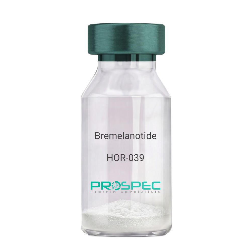
Name :
BremelanotideDescription:
Bremelanotide
PT-141, Rekynda, bremelanotide acetate.
Product # :
HOR-039Price :
Quantity :
Shipping Method :
Shipped at Room temp
More Info
- description
- formulation
- purity
- More Info
Description
Bremelanotide Synthetic is a single, non-glycosylated polypeptide chain containing 6 amino acids, having a molecular mass of 1025.16 Dalton and a Molecular formula of C50H68N40O10.
Formulation
The protein was lyophilized with no additives.
Purity
Greater than 97.0% as determined by analysis by RP-HPLC.
More Info
-
Synonyms
PT-141, Rekynda, bremelanotide acetate.
-
Physical Appearance
Sterile Filtered White lyophilized (freeze-dried) powder.
-
Stability
Lyophilized Bremelanotide although stable at room temperature for 3 weeks, should be stored desiccated below -18°C. Upon reconstitution Bremelanotide should be stored at 4°C between 2-7 days and for future use below -18°C. For long term storage it is recommended to add a carrier protein (0.1% HSA or BSA). Please prevent freeze-thaw cycles.
-
Solubility
It is recommended to reconstitute the lyophilized Bremelanotide in sterile 18MΩ-cm H2O not less than 100 µg/ml, which can then be further diluted to other aqueous solutions.
-
Amino Acid Sequence
Ac-Nle-cyclo(-Asp-His-D-Phe-Arg-Trp-Lys)-OH.
-
Background
Bremelanotide (PT-141), a synthetic heptapeptide, is a novel therapeutic agent primarily utilized for the treatment of sexual dysfunction. This paper provides an exhaustive review of Bremelanotide, detailing its biochemical structure, mechanism of action, therapeutic applications, and future prospects in clinical medicine.
Bremelanotide (PT-141), derived from Melanotan II, is a synthetic peptide developed for its role in treating sexual dysfunction in both men and women (Clayton et al., 2016). This paper presents a comprehensive examination of Bremelanotide, its biological properties, and potential therapeutic uses.
Bremelanotide acts as a non-selective agonist of the melanocortin receptors, primarily MC3R and MC4R, present in the central nervous system. The peptide initiates its action in the hypothalamus, leading to downstream effects on sexual desire and arousal (King et al., 2007).
The US Food and Drug Administration approved Bremelanotide for the treatment of premenopausal women with hypoactive sexual desire disorder (HSDD) in 2019. In clinical trials, it demonstrated efficacy in enhancing sexual desire and reducing distress associated with HSDD (Kingsberg et al., 2019).
While the current focus of Bremelanotide is on treating sexual dysfunction, its future potential may extend to other areas due to its unique mechanism of action. Further research is needed to explore the full therapeutic potential of this intriguing peptide. In conclusion, Bremelanotide represents a significant advance in sexual dysfunction therapeutics, with potential implications extending beyond this realm.
ProSpec's products are furnished for LABORATORY RESEARCH USE ONLY. The product may not be used as drugs, agricultural or pesticidal products, food additives or household chemicals.
- View Data Sheet
Name :
NME2 RatDescription:
Non-Metastatic Cells 2 Rat Recombinant
Nucleoside diphosphate kinase B, NDK B, NDP kinase B, Histidine protein kinase NDKB, P18.
Product # :
PKA-082Price :
Quantity :
Shipping Method :
Shipped with Ice Packs
More Info
- description
- source
- formulation
- purity
- More Info
Description
NME2 Rat Recombinant produced in E.Coli is a single, non-glycosylated polypeptide chain containing 175 amino acids (1-152 a.a) and having a molecular mass of 19.7kDa. NME2 is fused to a 23 amino acid His-tag at N-terminus & purified by proprietary chromatographic techniques.
Source
Escherichia Coli.
Formulation
NME2 protein solution (1mg/ml) containing Phosphate Buffered Saline pH 7.4 and 10% glycerol.
Purity
Greater than 90.0% as determined by SDS-PAGE.
More Info
-
Introduction
NME2 takes an important part in the synthesis of nucleoside triphosphates other than ATP. NME2 negatively controls Rho activity by interacting with AKAP13/LBC. NME2 acts as a transcriptional activator of the MYC gene. NME2 binds DNA non-specifically. NME2 is a heterodimeric enzyme functioning as a nucleoside diphosphate kinase. NME1 and NME2 contain 152 amino acids, A and B polypeptide chains of the NM23 enzyme, respectively. NME2 is identical to the beta subunit of human erythrocyte NDP kinase. NDP kinases participate in the synthesis of nucleoside triphosphates, and NM23 is involved in the regulation of signal transduction by complexing with G proteins, causing activation/inactivation of developmental pathways.
-
Synonyms
Nucleoside diphosphate kinase B, NDK B, NDP kinase B, Histidine protein kinase NDKB, P18.
-
Physical Appearance
Sterile Filtered clear solution.
-
Stability
Store at 4°C if entire vial will be used within 2-4 weeks. Store, frozen at -20°C for longer periods of time. For long term storage it is recommended to add a carrier protein (0.1% HSA or BSA).Avoid multiple freeze-thaw cycles.
-
Amino Acid Sequence
MGSSHHHHHH SSGLVPRGSH MGSMANLERT FIAIKPDGVQ RGLVGEIIKR FEQKGFRLVA MKFLRASEEH LKQHYIDLKD RPFFPGLVKY MNSGPVVAMV WEGLNVVKTG RVMLGETNPA DSKPGTIRGD FCIQVGRNII HGSDSVESAE KEIGLWFKPE ELIDYKSCAH DWVYE.
ProSpec's products are furnished for LABORATORY RESEARCH USE ONLY. The product may not be used as drugs, agricultural or pesticidal products, food additives or household chemicals.
- View Data Sheet
Name :
KLK3 AntibodyDescription:
Kallikrein-3, Mouse Anti Human
Prostate-specific antigen, PSA, Gamma-seminoprotein, Seminin, Kallikrein-3, P-30 antigen, Semenogelase, KLK3, APS, hK3, KLK2A1.
Product # :
ANT-088Price :
Quantity :
Shipping Method :
Shipped with Ice Packs
More Info
- formulation
- More Info
Formulation
1mg/ml containing PBS, pH-7.4, & 0.1% Sodium Azide.
More Info
-
Introduction
Kallikrein-3 (KLK3) belongs to the kallikrein-related peptidase family. Kallikreins are a subgroup of serine proteases having various physiological functions. Numerous kallikreins are involved in carcinogenesis and some may be prospective cancer and other disease biomarkers. Kallikrein-3 is 1 of the 15 kallikrein subfamily members located in a cluster on chromosome 19 and is a protease present in seminal plasma. KLK3 hydrolyzes semenogelin-1 consequently leading to the liquefaction of the seminal coagulum. KLK3 is assumed to act normally in the liquefaction of seminal coagulum, probably by hydrolysis of the high molecular mass seminal vesicle protein. Serum level of the KLK3 protein, called PSA in the clinical setting, is beneficial in the diagnosis and monitoring of prostatic carcinoma.
-
Synonyms
Prostate-specific antigen, PSA, Gamma-seminoprotein, Seminin, Kallikrein-3, P-30 antigen, Semenogelase, KLK3, APS, hK3, KLK2A1.
-
Immunogen
Anti-human KLK3 mAb, is derived from hybridization of mouse F0 myeloma cells with spleen cells from BALB/c mice immunized with a recombinant human KLK3 amino acids 25-261 purified from E. coli.
-
Ig Subclass
Mouse IgG1 heavy chain and ? light chain.
-
Clone
P1D10AT.
-
Applications
KLK3 antibody has been tested by ELISA, Western blot analysis to assure specificity and reactivity. Since application varies, however, each investigation should be titrated by the reagent to obtain optimal results. Recommended dilution range for Western blot analysis is 1:500 ~ 1:5000.
-
Type
Mouse Anti Human Monoclonal.
-
Storage Procedures
For periods up to 1 month store at 4°C, for longer periods of time, store at -20°C. Prevent freeze thaw cycles.
-
Purification Method
KLK3 antibody was purified from mouse ascitic fluids by protein-G affinity chromatography.
ProSpec's products are furnished for LABORATORY RESEARCH USE ONLY. The product may not be used as drugs, agricultural or pesticidal products, food additives or household chemicals.
- View Data Sheet
Name :
DUSP3 AntibodyDescription:
Dual Specificity Phosphatase 3, Mouse Anti Human
Dual specificity phosphatase 3, VHR, Vaccinia virus phosphatase VH1-related, Dual specificity protein phosphatase VHR, Vaccinia H1-related phosphatase, serine/threonine specific protein phosphatase, DUSP3.
Product # :
ANT-594Price :
Quantity :
Shipping Method :
Shipped with Ice Packs
More Info
- formulation
- More Info
Formulation
1mg/ml containing PBS, pH-7.4, 10% Glycerol and 0.02% Sodium Azide.
More Info
-
Introduction
DUSP3 belongs to the dual specificity protein phosphatase subfamily which inactivates their target kinases by dephosphorylating both the phosphoserine/threonine and phosphotyrosine residues. DUSP3 negatively regulate members of the mitogen-activated protein kinase superfamily that are related to cellular proliferation and differentiation. DUSP3 is expressed in both breast and ovarian tissues and displays activity both for tyrosine-protein phosphate and serine-protein phosphate, but exhibits a strong preference toward phosphotyrosines, specifically dephosphorylates and inactivates ERK1 and ERK2.
-
Synonyms
Dual specificity phosphatase 3, VHR, Vaccinia virus phosphatase VH1-related, Dual specificity protein phosphatase VHR, Vaccinia H1-related phosphatase, serine/threonine specific protein phosphatase, DUSP3.
-
Physical Appearance
Sterile filtered colorless solution.
-
Immunogen
Anti-human DUSP3 mAb, is derived from hybridization of mouse F0 myeloma cells with spleen cells from BALB/c mice immunized with recombinant human DUSP3 amino acids 1-185 purified from E. coli.
-
Ig Subclass
Mouse IgG2b heavy chain and k light chain.
-
Clone
PAT9E6AT.
-
Applications
DUSP3 antibody has been tested by ELISA, Western blot analysis to assure specificity and reactivity. Since application varies, however, each investigation should be titrated by the reagent to obtain optimal results. Recommended starting dilution for Western blot analysis is 1:3000.
-
Type
Mouse Anti Human Monoclonal.
-
Storage Procedures
For periods up to 1 month store at 4°C, for longer periods of time, store at -20°C. Prevent freeze thaw cycles.
-
Purification Method
DUSP3 antibody was purified from mouse ascitic fluids by protein-A affinity chromatography.
ProSpec's products are furnished for LABORATORY RESEARCH USE ONLY. The product may not be used as drugs, agricultural or pesticidal products, food additives or household chemicals.
- View Data Sheet
Name :
TRAIL Human (114-281 a.a.)Description:
TRAIL/APO 2 Ligand (114-281 a.a.) Human Recombinant
Tumor necrosis factor ligand superfamily member 10, TNF-related apoptosis-inducing ligand, Protein TRAIL, Apo-2 ligand, Apo-2L, CD253 antigen, TL2, APO2L, TNFSF10.
Product # :
CYT-546Price :
Quantity :
Shipping Method :
Shipped with Ice Packs
More Info
- description
- source
- formulation
- purity
- More Info
Description
Soluble TNF-related apoptosis-inducing ligand Human Recombinant produced in E.Coli is a single, non-glycosylated polypeptide chain containing 169 amino acids (114-281) and having a molecular mass of 19.6 kDa. The sTRAIL is purified by proprietary chromatographic techniques.
Source
Escherichia Coli.
Formulation
1mg/ml in 20mM Tris-HCl pH-7.5, 300mM NaCl, 0.1mM DTT & 10% glycerol.
Purity
Greater than 95.0% as determined by SDS-PAGE.
More Info
-
Introduction
TNF-related apoptosis-inducing ligand (TRAIL) is a ligand molecule which induces apoptosis. It is a type II transmembrane protein with homology to other members of the tumor necrosis factor family.
In humans, the gene that encodes for TRAIL is located at chromosome 3q26.
TRAIL binds to the death receptors, DR4 and DR5. The process of apoptosis is caspase-8-dependent. This protein preferentially induces apoptosis in transformed and tumor cells, but does not appear to kill normal cells although it is expressed at a significant level in most normal tissues. -
Synonyms
Tumor necrosis factor ligand superfamily member 10, TNF-related apoptosis-inducing ligand, Protein TRAIL, Apo-2 ligand, Apo-2L, CD253 antigen, TL2, APO2L, TNFSF10.
-
Physical Appearance
Sterile Filtered colorless liquid.
-
Stability
Store at 4°C if entire vial will be used within 2-4 weeks. Store, frozen at -20°C for longer periods of time. For long term storage it is recommended to add a carrier protein (0.1% HSA or BSA).Avoid multiple freeze-thaw cycles.
-
Amino Acid Sequence
MVRERGPQRV AAHITGTRGR SNTLSSPNSK NEKALGRKIN SWESSRSGHS FLSNLHLRNGELVIHEKGFY YIYSQTYFRF QEEIKENTKN DKQMVQYIYK YTSYPDPILL MKSARNSCWSKDAEYGLYSI YQGGIFELKE NDRIFVSVTN EHLIDMDHEA SFFGAFLVG.
ProSpec's products are furnished for LABORATORY RESEARCH USE ONLY. The product may not be used as drugs, agricultural or pesticidal products, food additives or household chemicals.
- View Data Sheet
Name :
RetatrutideDescription:
Retatrutide
Product # :
HOR-058Price :
Quantity :
Shipping Method :
Shipped at Room temp
More Info
- description
- formulation
- purity
- More Info
Description
Retatrutide is a synthetic single, non-glycosylated polypeptide chain containing 39 amino acids, having a molecular mass of 4731 Dalton and a Molecular formula of C221H342N46O68.
Formulation
The protein was lyophilized with no additives.
Purity
Greater than 97.0% as determined by analysis by RP-HPLC.
More Info
-
Physical Appearance
Sterile Filtered White lyophilized (freeze-dried) powder.
-
Stability
Lyophilized Retatrutide although stable at room temperature for 3 weeks, should be stored desiccated below -18°C. Upon reconstitution Retatrutide should be stored at 4°C between 2-7 days and for future use below -18°C.
For long term storage it is recommended to add a carrier protein (0.1% HSA or BSA).
Please prevent freeze-thaw cycles.
-
Solubility
It is recommended to reconstitute the lyophilized Retatrutide in sterile 18MΩ-cm H2O not less than 100 µg/ml, which can then be further diluted to other aqueous solutions.
-
Amino Acid Sequence
H-Tyr-Aib-Gln-Gly-Thr-Phe-Thr-SerAsp-Tyr-Ser-Ile-α-Me-Leu-Leu-Asp-Lys-{diacid-C20-gamma-Glu- (AEEA)-Lys}-Ala-Gln-Aib-Ala-Phe-Ile-Glu-Tyr-Leu-Leu-Glu-Gly-GlyPro-Ser-Ser-Gly-Ala-Pro-Pro-Pro-Ser-NH2.
-
Background
Retatrutide is atriple agonist targeting the GIP, GLP-1, and glucagon receptors which offers a distinct metabolic profile that goes beyond previous dual agonists like Tirzepatide. Retatrutide takes a very important part in metabolic pharmacotherapy, achieving high weight loss of nearly 30% in Phase 3 trials. Retatrutide’s triple-receptor agonism—mainly the inclusion of the glucagon receptor—provides a superior metabolic advantage in clearing liver fat and increasing thermogenesis compared to dual agonists.
ProSpec's products are furnished for LABORATORY RESEARCH USE ONLY. The product may not be used as drugs, agricultural or pesticidal products, food additives or household chemicals.