Search results
493 results found for “Lymphotactin (XCL1)”
Name
Description
Product #
Price
Quantity
Shipping Method
- View Data Sheet
Name :
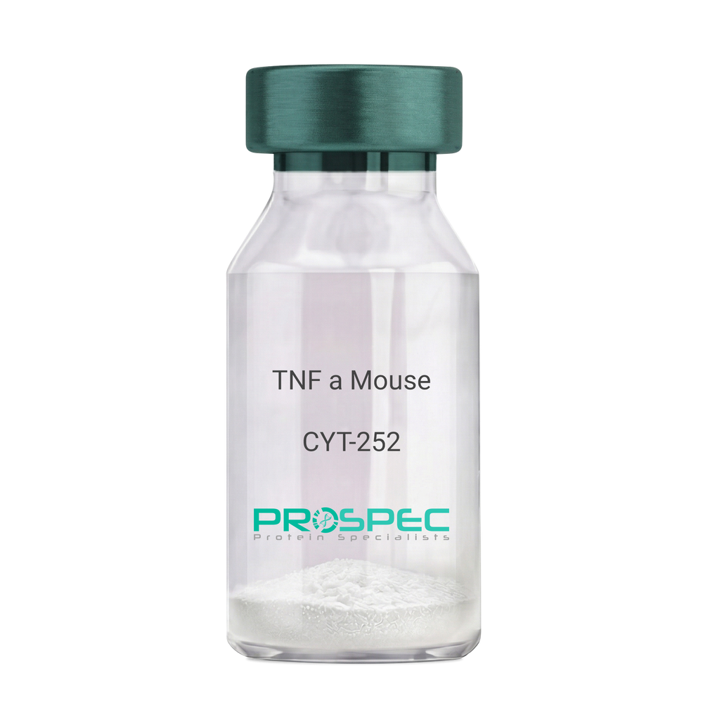
TNF a MouseDescription:
Tumor Necrosis Factor-Alpha Mouse Recombinant
TNF-alpha, Tumor necrosis factor ligand superfamily member 2, TNF-a, Cachectin, DIF, TNFA, TNFSF2.
Product # :
CYT-252Price :
Quantity :
Shipping Method :
Shipped at Room temp
More Info
- description
- source
- formulation
- purity
- biological activity
- More Info
- Citations
Description
Tumor Necrosis Factor-a Mouse Recombinant produced in E. coli is a single, non glycosylated, polypeptide chain containing 157 amino acids and having a molecular mass of 17301.32 Dalton. The TNF-alpha is purified by standard chromatographic techniques.
Source
Escherichia Coli.
Formulation
Lyophilized from a 0.2µm filtered concentrated solution in PBS, pH 7.2.
Purity
Greater than 97.0% as determined by:
(a) Analysis by RP-HPLC.
(c) Analysis by SDS-PAGE.Biological Activity
The ED50 as determined by the cytolysis of murine L929 cells in the presence of Actinomycin D is < 0.1ng/ml, corresponding to a Specific Activity of 10,000,000 Units/mg.Citations
More Info
-
Introduction
Tumor necrosis factor is a cytokine involved in systemic inflammation and is a member of a group of cytokines that all stimulate the acute phase reaction. TNF is mainly secreted by macrophages.
TNF causes apoptotic cell death, cellular proliferation, differentiation, inflammation, tumorigenesis and viral replication, TNF is also involved in lipid metabolism, and coagulation. TNF's primary role is in the regulation of immune cells.
Dysregulation and, in particular, overproduction of TNF have been implicated in a variety of human diseases- autoimmune diseases, insulin resistance, and cancer. -
Synonyms
TNF-alpha, Tumor necrosis factor ligand superfamily member 2, TNF-a, Cachectin, DIF, TNFA, TNFSF2.
-
Physical Appearance
Sterile Filtered White lyophilized (freeze-dried) powder.
-
Stability
Lyophilized Tumor Necrosis Factor-a although stable at room temperature for 3 weeks, should be stored desiccated below -18°C. Upon reconstitution TNF-a should be stored at 4°C between 2-7 days and for future use below -18°C.For long term storage it is recommended to add a carrier protein (0.1% HSA or BSA).Please prevent freeze-thaw cycles.
-
Solubility
It is recommended to reconstitute the lyophilized Tumor Necrosis Factor-alpha in sterile 18M-cm H2O not less than 100µg/ml, which can then be further diluted to other aqueous solutions.
-
Amino Acid Sequence
MLRSSSQNSS DKPVAHVVAN HQVEEQLEWL SQRANALLAN GMDLKDNQLV VPADGLYLVY SQVLFKGQGC PDYVLLTHTV SRFAISYQEK VNLLSAVKSP CPKDTPEGAE LKPWYEPIYL GGVFQLEKGD QLSAEVNLPK YLDFAESGQV YFGVIAL
-
Background
Tumor Necrosis Factor-alpha (TNF-α) is a pro-inflammatory cytokine that plays a critical role in the regulation of immune responses, inflammation, and cell survival. It is primarily produced by activated macrophages, but can also be secreted by other immune cells, such as T cells, natural killer cells, and mast cells. TNF-α is involved in a wide range of physiological and pathological processes, including the defense against infections, the development of autoimmune diseases, and the progression of cancer.
TNF-α exerts its effects by binding to two distinct cell surface receptors, TNF receptor 1 (TNFR1) and TNF receptor 2 (TNFR2). Upon binding to its receptors, TNF-α activates multiple signaling pathways, including the nuclear factor-kappa B (NF-κB) pathway, the mitogen-activated protein kinase (MAPK) pathway, and the apoptotic pathway. These signaling pathways regulate various cellular processes, such as inflammation, cell proliferation, differentiation, and apoptosis.
In the context of infections, TNF-α plays a crucial role in the body's defense against pathogens. It promotes the recruitment and activation of immune cells, enhances the production of other pro-inflammatory cytokines, and stimulates the expression of adhesion molecules on endothelial cells, facilitating the migration of immune cells to the site of infection. TNF-α also helps to induce fever, which is an important component of the body's immune response to infections.
However, excessive or prolonged production of TNF-α can contribute to the development of chronic inflammatory diseases, such as rheumatoid arthritis, inflammatory bowel disease, and psoriasis. In these conditions, elevated levels of TNF-α promote the infiltration of immune cells into the affected tissues, leading to tissue damage and the perpetuation of inflammation. The central role of TNF-α in the pathogenesis of these diseases has led to the development of anti-TNF-α therapies, which have revolutionized the treatment of chronic inflammatory diseases. These therapies include monoclonal antibodies, such as infliximab and adalimumab, and soluble TNF receptor fusion proteins, such as etanercept. Anti-TNF-α therapies have been shown to be effective in reducing inflammation, improving symptoms, and slowing disease progression in patients with chronic inflammatory diseases.
In the context of cancer, TNF-α has complex and context-dependent effects on tumor development and progression. On one hand, TNF-α can promote anti-tumor immunity by activating immune cells and stimulating the production of other pro-inflammatory cytokines. On the other hand, chronic inflammation driven by TNF-α can promote tumor growth, angiogenesis, and metastasis. Therefore, the role of TNF-α in cancer is still an area of active research, and the development of TNF-α-targeted therapies for cancer remains a challenge.
In conclusion, TNF-α is a pro-inflammatory cytokine that plays a critical role in the regulation of immune responses, inflammation, and cell survival. Its involvement in various physiological and pathological processes has made it an important target for the development of therapies for chronic inflammatory diseases and cancer. Anti-TNF-α therapies have revolutionized the treatment of chronic inflammatory diseases, but the complex role of TNF-α in cancer remains an area of ongoing research. Understanding the precise mechanisms by which TNF-α contributes to disease pathogenesis will be crucial for the development of more effective and targeted therapies.
-
Protein content
Protein quantitation was carried out by two independent methods1. UV spectroscopy at 280 nm using the absorbency value of 1.24 as the extinction coefficient for a 0.1% (1mg/ml) solution. This value is calculated by the PC GENE computer analysis program of protein sequences (IntelliGenetics). 2. Analysis by RP-HPLC, using a calibrated solution of TNF-a as a Reference Standard.
ProSpec's products are furnished for LABORATORY RESEARCH USE ONLY. The product may not be used as drugs, agricultural or pesticidal products, food additives or household chemicals.
- View Data Sheet
Name :
SARS MERS Spike S1Description:
Mouse Anti SARS MERS Spike S1
Product # :
ANT-766Price :
Quantity :
Shipping Method :
Shipped with Ice Packs
More Info
- description
- formulation
- purity
- More Info
- Citations
Description
SARS MERS Spike-S1 antibody is specific for the S1 domain of the spike protein of the MERS virus.
Formulation
1mg/ml in PBS and 0.1% NaN3.
Purity
Greater than 90% as determined by SDS-PAGE gel.
Citations
More Info
-
Introduction
SARS Coronavirus is an enveloped virus containing three outer structural proteins, namely the membrane (M), envelope (E), and spike (S) proteins. Spike (S)-glycoprotein of the virus interacts with a cellular receptor and mediates membrane fusion to allow viral entry into susceptible target cells. Accordingly, S-protein plays an important role in virus infection cycle and is the primary target of neutralizing antibodies.It has recently been shown that SARS is caused by a human coronavirus. Human coronaviruses are the major cause of upper respiratory tract illness in humans, such as the common cold. Coronaviruses are positive-stranded RNA viruses, featuring the largest viral RNA genomes known to date (27-31 kb). The first step in coronavirus infection is binding of the viral spike protein, a 139-kDa protein, to certain receptors on host cells. The spike protein is the main surface antigen of the coronavirus. The most prominent protein in the culture supernatants infected with SARS virus is a 46 kDa nucleocapsid protein. This suggests that the nucleocapsid protein is a major immunogen that may be useful for early diagnostics.
-
Physical Appearance
Sterile Filtered liquid formulation.
-
Stability
Store at 4°C, stable for 2 weeks. For long-term storage, store at -20°C.
-
Applications
ELISA.
-
Isotype
IgG1.
-
Type
Mouse antibody Monoclonal.
-
Purification Method
Protein A affinity purified.
ProSpec's products are furnished for LABORATORY RESEARCH USE ONLY. The product may not be used as drugs, agricultural or pesticidal products, food additives or household chemicals.
- View Data Sheet
Name :
1F8 ChagasDescription:
1F8 Chagas Recombinant
Product # :
CCH-002Price :
Quantity :
Shipping Method :
Shipped at Room temp
More Info
- description
- source
- formulation
- purity
- More Info
- Citations
Description
Recombinant Chagas 1F8 protein (Epitope of the flagellar calcium-binding protein FCABP of Trypanosoma cruzi) produced in E.Coli, is a polypeptide chain having a total of 234 a.a (includes his tag). The protein is fused to a 6-His tag having a total Mw of 25.8kDa and a theoretical pI of 5.39.
Source
Escherichia Coli.
Formulation
1F8 Chagas antigen was lyophilized from 20mM Hepes, 300mM NaCl, pH-8, and varying 284mM imidazole.
Purity
Protein is >90% pure as determined by SDS PAGE.
Citations
More Info
-
Introduction
Flagellar calcium-binding protein (Protein FcaBP) is a protein which may be involved in swift motility of Trypanosoma, having an imperative role in flagellar structure and calcium metabolism, as it exhibits 4 EF-hand type (helix-loop-helix) structural domains, of which 3 may contain calcium-binding sites. Calcium has a vital role in the motility of Trypanosoma. The subcellular localization of Protein FcaBP is in the flagella of T. cruzi.
-
Stability
Store lyophilized 1F8 Chagas antigen at 2-8°C. After reconstitution, store at -20°C. Prevent freeze/thaw cycles.
-
Solubility
It is recommended to reconstitute the lyophilized 1F8 Chagas protein in sterile distilled pyrogen free water at 100µg/ml, which can then be further diluted to other aqueous solutions.
-
Applications
Reacts with sera from infected patients, Useful for immunoassays.
ProSpec's products are furnished for LABORATORY RESEARCH USE ONLY. The product may not be used as drugs, agricultural or pesticidal products, food additives or household chemicals.
- View Data Sheet
Name :
FTCD HumanDescription:
Formiminotransferase Cyclodeaminase Human Recombinant
Formiminotransferase-cyclodeaminase, FTCD, LCHC1, LC-1.
Product # :
ENZ-302Price :
Quantity :
Shipping Method :
Shipped with Ice Packs
More Info
- description
- source
- formulation
- purity
- More Info
- Citations
Description
Formiminotransferase Cyclodeaminase Human Recombinant (also called liver cytosol type 1) produced in SF9, is a glycosylated, polypeptide chain having a molecular mass of 59,749 Dalton.The FTCD is expressed with a -6xHis tag and purified by proprietary chromatographic techniques.
Source
Sf9 insect cells.
Formulation
FTCD is supplied in 16mM HEPES buffer pH-7.6, 240mM sodium chloride, and 20% glycerol.
Purity
Greater than 95% as determined by SDS-PAGE.
Citations
More Info
-
Introduction
Formiminotransferase cyclodeaminase is an enzyme which catalyzes the conversion of formiminoglutamateand tetrahydrofolateinto formiminotetrahydrofolateand glutamate.
Formiminotransferase cyclodeaminase, a bifunctional enzyme of tetrahydrofolate synthesis, is the target antigen of anti-LC1 (liver cytosol antigen type 1) autoantibodies. Presence of LC1 autoantibodies is a marker for type 2 autoimmune hepatitis (for which anti-LKM 1/cytochrome P450 2D6 autoantibodies are a further marker).
Serum LC1 autoantibody concentrations appear to fluctuate in parallel with aminotransferase levels, a particularly intriguing observation that suggests a possible role of LC1 autoreactivity in the pathogenic mechanism leading to hepatocyte injury. -
Synonyms
Formiminotransferase-cyclodeaminase, FTCD, LCHC1, LC-1.
-
Stability
Store at 4°C if entire vial will be used within 2-4 weeks. Store, frozen at -20°Cfor longer periods of time. Avoid multiple freeze-thaw cycles.
-
Immunological Functions
1. Binds IgG-type human auto-antibodies.2. Standard ELISA test (checker-board analysis of positive/negative sera panels, immuno-dot test).
-
coating concentration
0.35-0.7 µg/ml (depending on the type of ELISA plate and coating buffer).Suitable for biotinylation and iodination.
ProSpec's products are furnished for LABORATORY RESEARCH USE ONLY. The product may not be used as drugs, agricultural or pesticidal products, food additives or household chemicals.
- View Data Sheet
Name :
CoV-2-S1Description:
Coronavirus 2019 Spike Glycoprotein-S1, Recombinant
Product # :
SARS-011Price :
Quantity :
Shipping Method :
Shipped with Ice Packs
More Info
- description
- source
- formulation
- purity
- More Info
- Citations
Description
The HEK293 derived recombinant protein contains the Novel Coronavirus 2019-nCoV Spike Glycoprotein S1, Wuhan-Hu-1 strain, amino acids 1-674 fused to Fc tag at C-terminal.
Source
HEK293
Formulation
nCoV-S1 protein solution is supplied in DPBS pH 7.4.DPBS pH 7.4.
Purity
Protein is >85% pure as determined SDS-PAGE.
Citations
More Info
-
Introduction
A human infecting coronavirus (viral pneumonia) called 2019 novel coronavirus, 2019-nCoV was found in the fish market at the city of Wuhan, Hubei province of China on December 2019.
The 2019-nCoV shares an 87% identity to the 2 bat-derived severe acute respiratory syndrome 2018 SARS-CoV-2 located in Zhoushan of eastern China. 2019-nCoV has an analogous receptor-BD-structure to that of 2018 SARS-CoV, even though there is a.a. diversity so thus the 2019-nCoV might bind to ACE2 receptor protein (angiotensin-converting enzyme 2) in humans.
While bats are possibly the host of 2019-nCoV, researchers suspect that animal from the ocean sold at the seafood market was an intermediate host. RSCU analysis proposes that the 2019-nCoV is a recombinant within the viral spike glycoprotein between the bat coronavirus and an unknown coronavirus.
-
Physical Appearance
Sterile Filtered clear solution.
-
Stability
Protein is shipped on ice packs. Upon arrival, Store at -20°C.
-
Purification Method
Purified by Protein-G chromatographic technique.
ProSpec's products are furnished for LABORATORY RESEARCH USE ONLY. The product may not be used as drugs, agricultural or pesticidal products, food additives or household chemicals.
- View Data Sheet
Name :
RB1 HumanDescription:
Retinoblastoma Associated Protein Human Recombinant
RB, OSRC, RB-1, RB1, p105-Rb, OSTEOSARCOMA, RETINOBLASTOMA-RELATED,PP110, Retinoblastoma-associated protein.
Product # :
PRO-584Price :
Quantity :
Shipping Method :
Shipped at Room temp
More Info
- description
- source
- formulation
- purity
- More Info
- Citations
Description
Retinoblastoma Human Recombinant fused with 6X His tag produced in E.Coli is a single, non-glycosylated, polypeptide chain containing 146 amino acids and having a molecular mass of 16.5 kDa.The Retinoblastoma is purified by proprietary chromatographic techniques.
Source
Escherichia Coli.
Formulation
The RB1 was lyophilized from 1xPBS pH-7.4.
Purity
Greater than 95.0% as determined by:
(a) Analysis by RP-HPLC.
(b) Analysis by SDS-PAGE.Citations
More Info
-
Introduction
Retinoblastoma (RB) is an embryonic malignant neoplasm of retinal origin. It almost always presents in early childhood and is often bilateral. Spontaneous regression ('cure') occurs in some cases. Retinoblastoma acts as a regulator of other genes and forms a complex with adenovirus e1a and with sv40 large t antigen. Retinoblastoma acts as a tumor suppressor and modulats functionally certain cellular proteins with which t and e1a compete for pocket binding. Retinoblastoma is potent inhibitor of e2f-mediated trans-activation, recruits and targets histone methyltransferase suv39h1 leading to epigenetic transcriptional repression, inhibits the intrinsic kinase activity of taf1.
-
Synonyms
RB, OSRC, RB-1, RB1, p105-Rb, OSTEOSARCOMA, RETINOBLASTOMA-RELATED,PP110, Retinoblastoma-associated protein.
-
Physical Appearance
Sterile Filtered White lyophilized (freeze-dried) powder.
-
Stability
Lyophilized Retinoblastoma although stable at room temperature for 3 weeks, should be stored desiccated below -18°C. Upon reconstitution Retinoblastoma should be stored at 4°C between 2-7 days and for future use below -18°C.For long term storage it is recommended to add a carrier protein (0.1% HSA or BSA).Please prevent freeze-thaw cycles.
-
Solubility
It is recommended to reconstitute the lyophilized Retinoblastoma in sterile 18MΩ-cm H2O not less than 100 µg/ml, which can then be further diluted to other aqueous solutions.
-
Amino Acid Sequence
MASFPSSPLRIPGGNIYISPLKSPYKISEGLPTPTKMTPRSRILVSIGESFG
TSEKFQKINQMVCNSDRVLKRSAEGSNPPKPLKKLRFDIEGSDEADGSK
HLPGESKFQQKLAEMTSTRTRMQKQKMNDSMDTSNKEEKHHHHHH.
ProSpec's products are furnished for LABORATORY RESEARCH USE ONLY. The product may not be used as drugs, agricultural or pesticidal products, food additives or household chemicals.
- View Data Sheet
Name :
IL 31 HumanDescription:
Interleukin-31 Human Recombinant
Interleukin 31, IL31, IL-31.
Product # :
CYT-625Price :
Quantity :
Shipping Method :
Shipped at Room temp
More Info
- description
- source
- formulation
- purity
- biological activity
- More Info
- Citations
Description
IL-31 human recombinant produced in E.Coli is a single, non-glycosylated, polypeptide chain containing 141 amino acids (24-164 a.a.) and having a molecular mass of 15.8 kDa.
Source
Escherichia Coli.
Formulation
The IL-31 (1mg/ml) was lyophilized with 20mM Phosphate buffer, 150mM NaCl pH-7.4.
Purity
Greater than 95.0% as determined by(a) Analysis by SEC-HPLC.
(b) Analysis by SDS-PAGE.Biological Activity
The ED50 was determined by its ability to activate STAT following receptor ligand interaction and found to be <5ng/ml, corresponding to a specific activity of 200,000 units.Citations
More Info
-
Introduction
IL-31 produced by activated Th2-type T cells, cooperates with a heterodimeric receptor consisting of IL-31 Receptor Anatagonist and Onconstatin-M Receptor that is continuesly expressed on epithelial cells and keratinocytes. IL-31 plays a role in the promotion of allergic skin disorders and in regulating other allergic diseases, such as asthma. IL-31 is involved in the itching sensation and endorses the scratching behavior in NC/Nga mice with atopic dermatitis. IL-31 expression is connectd with CLA(+) T cells and contributes to the development of atopic dermatitis-induced skin inflammation and pruritus. IL-31 is a powerful inducer of proinflammatory mediators in human colonic SEMFs. IL-31 takes part as a proinflammatory cytokine derived from Th2 cells.
Serum IL-31 level is higher in patients with atopic dermatitis. IL-31 is involved in a broad range of immune- & non-immune cells & possesses potential pleiotropic physiological functions, including regulating hematopoiesis & immune response, causing inflammatory bowel disease, airway hypersensitivity & dermatitis. -
Synonyms
Interleukin 31, IL31, IL-31.
-
Physical Appearance
Sterile Filtered White lyophilized (freeze-dried) powder.
-
Stability
Lyophilized IL-31 Recombinant although stable at room temperature for 3 weeks, should be stored desiccated below -18°C. Upon reconstitution IL-31 should be stored at 4°C between 2-7 days and for future use below -18°C.Please prevent freeze-thaw cycles.
-
Solubility
It is recommended to reconstitute the lyophilized IL-31 in sterile 18MΩ-cm H2O not less than 100µg/ml, which can then be further diluted to other aqueous solutions.
-
Amino Acid Sequence
SHTLPVRLLR PSDDVQKIVE ELQSLSKMLL KDVEEEKGVL VSQNYTLPCL SPDAQPPNNI HSPAIRAYLK TIRQLDNKSV IDEIIEHLDK LIFQDAPETN ISVPTDTHEC KRFILTISQQ FSECMDLALK SLTSGAQQAT T.
ProSpec's products are furnished for LABORATORY RESEARCH USE ONLY. The product may not be used as drugs, agricultural or pesticidal products, food additives or household chemicals.
- View Data Sheet
Name :
C1QTNF6 HumanDescription:
Complement C1q Tumor Necrosis Factor-Related Protein 6 Human Recombinant
CTRP6, C1QTNF6, ZACRP6, Complement C1q tumor necrosis factor-related protein 6.
Product # :
PRO-655Price :
Quantity :
Shipping Method :
Shipped with Ice Packs
More Info
- description
- source
- formulation
- purity
- More Info
- Citations
Description
C1QTNF6 Human Recombinant produced in E.Coli is a single, non-glycosylated, Polypeptide chain containing 242 amino acids and having a molecular mass of 27 kDa. The protein contains an extra His tag at N-terminus. The C1QTNF6 amino acid sequence is identical to UniProtKB/Swiss-Prot entry Q9BXI9 amino acids 29–259.The C1QTNF6 is purified by proprietary chromatographic techniques.
Source
Escherichia Coli.
Formulation
0.23mg/ml in 30mM Acetate Buffer pH-4.
Purity
Greater than 95% as determined by SDS PAGE.
Citations
More Info
-
Introduction
C1QTNF6 is a collagen type-IV specific for basement membranes. C1QTNF6 is vital for embryonic development.
-
Synonyms
CTRP6, C1QTNF6, ZACRP6, Complement C1q tumor necrosis factor-related protein 6.
-
Physical Appearance
Sterile Filtered clear solution.
-
Stability
Store at 4°C if entire vial will be used within 2-4 weeks. Store, frozen at -20°C for longer periods of time. Avoid multiple freeze-thaw cycles.
-
Amino Acid Sequence
MKHHHHHHAS TFDRAVASGC QRCCDSEDPL DPAHVSSASS SGRPHALPEI RPYINITILK GDKGDPGPMG LPGYMGREGP QGEPGPQGSK GDKGEMGSPG APCQKRFFAF SVGRKTALHS GEDFQTLLFE RVFVNLDGCF DMATGQFAAP LRGIYFFSLN VHSWNYKETY VHIMHNQKEA VILYAQPSER SIMQSQSVML DLAYGDRVWV RLFKRQRENA IYSNDFDTYI TFSGHLIKAEDD.
ProSpec's products are furnished for LABORATORY RESEARCH USE ONLY. The product may not be used as drugs, agricultural or pesticidal products, food additives or household chemicals.
- View Data Sheet
Name :
IL36B HumanDescription:
Interleukin-36 Beta Human Recombinant
Interleukin 36 beta, interleukin 1 family member 8 (eta), Interleukin-1 homolog 2, IL1F8 (Canonical product IL-1F8a), IL-1F8 (FIL1-eta), Interleukin-1 Superfamily e, IL1H2, MGC126880, MGC126882.
Product # :
CYT-159Price :
Quantity :
Shipping Method :
Shipped at Room temp
More Info
- description
- source
- formulation
- purity
- biological activity
- More Info
- Citations
Description
IL36B Human Recombinant produced in E.Coli is a single, non-glycosylated, polypeptide chain containing 157 amino acids and having a molecular mass of 17.7kDa.The IL36B is purified by proprietary chromatographic techniques.
Source
Escherichia Coli.
Formulation
Lyophilized from a 0.2µm filtered concentrated solution in 1×PBS, pH 7.4.
Purity
Greater than 95.0% as determined by SDS-PAGE.
Biological Activity
Measured by its binding ability in a functional ELISA to bind recombinant human IL-1 Rrp2 Fc Chimera.Citations
More Info
-
Introduction
Human IL-36b belongs to the IL-1 family that includes IL-1b, IL-1a, IL-1ra, IL-18, IL-36ra (IL1F5), IL-36b (IL1F8), IL-36g (IL1F9), IL-37 (IL1F7) and IL-38 (IL-1F10). The IL-1 family members display a 12 b-strand, b-trefoil configuration, and are thought to have ascended from a mutual ancestral gene. IL-36 beta is known to be actively secreted. Cells expressing IL-36 beta include resting and activated monocytes and B cells. The receptor for IL-36 beta is a blend of IL-1 Rrp2 and IL-1 RAcP. Recombinant IL-36 beta stimulates processes involving NF-kB and MAPK in an IL-1 Rrp2-dependent manner.
-
Synonyms
Interleukin 36 beta, interleukin 1 family member 8 (eta), Interleukin-1 homolog 2, IL1F8 (Canonical product IL-1F8a), IL-1F8 (FIL1-eta), Interleukin-1 Superfamily e, IL1H2, MGC126880, MGC126882.
-
Physical Appearance
Sterile Filtered White lyophilized (freeze-dried) powder.
-
Stability
Lyophilized IL36B Human although stable at room temperature for 3 weeks, should be stored desiccated below -18°C. Upon reconstitution IL36B should be stored at 4°C between 2-7 days and for future use below -18°C.Please prevent freeze-thaw cycles.
-
Solubility
It is recommended to reconstitute the lyophilized IL36B in sterile 18M-cm H2O not less than 100µg/ml, which can then be further diluted to other aqueous solutions.
-
Amino Acid Sequence
MNPQREAAPK SYAIRDSRQM VWVLSGNSLI AAPLSRSIKP VTLHLIACRD TEFSDKEKGN MVYLGIKGKD LCLFCAEIQG KPTLQLKEKN IMDLYVEKKA QKPFLFFHNK EGSTSVFQSV SYPGWFIATS TTSGQPIFLT KERGITNNTN FYLDSVE
ProSpec's products are furnished for LABORATORY RESEARCH USE ONLY. The product may not be used as drugs, agricultural or pesticidal products, food additives or household chemicals.
- View Data Sheet
Name :
TNFRSF17 HumanDescription:
B-Cell Maturation Antigen Human Recombinant
BCMA, CD269, Tumor Necrosis Factor Receptor Superfamily Member 17, BCM, TNFRSF17, B-cell maturation protein, CD269 antigen.
Product # :
CYT-598Price :
Quantity :
Shipping Method :
Shipped at Room temp
More Info
- description
- source
- formulation
- purity
- More Info
- Citations
Description
TNFRSF17 Human Recombinant produced in E.Coli is a single, non-glycosylated, polypeptide chain containing 50 amino acids and having a molecular mass of 5.3 kDa. The TNFRSF17 is purified by standard chromatographic techniques.
Source
Escherichia Coli.
Formulation
1mg of TNFRSF17 Human contain 20mM sodium phosphate buffer, pH-7.4, and 130mM NaCl.
Purity
Greater than 98.0% as determined by:
(a) Analysis by RP-HPLC.
(b) Analysis by SDS-PAGE.Citations
More Info
-
Introduction
TNFRSF17 is a receptor for tnfsf13b/blys/baff and tnfsf13/april. TNFRSF17 promotes b-cell survival and plays a role in the regulation of humoral immunity. TNFRSF17 activates nf-kappa-b and jnk. TNFRSF17 is a member of the TNF-receptor superfamily. TNFRSF17 is expressed in mature B lymphocytes, and is invloved in B cell development and autoimmune response. TNFRSF17 specifically binds to the tumor TNFSF13B/TALL-1/BAFF, which causes NF-kappaB and MAPK8/JNK activation. TNFRSF17 binds to a variety of TRAF family members, and therefore transduces signals for cell survival and proliferation. TNFRSF17 is a type III membrane protein having 1 extracellular cysteine rich domain. Within the TNFRSF, it shares the highest homology with TACI. BCMA and TACI have both been shown to bind to APRIL and BAFF, members of the TNF ligand superfamily. BCMA expression has been found in immune organs. TNFRSF17 appears to be localized to the Golgi compartment. The binding of BCMA to APRIL or BAFF has been shown to stimulate IgM production in peripheral blood B cells and increase the survival of cultured B cells.
-
Synonyms
BCMA, CD269, Tumor Necrosis Factor Receptor Superfamily Member 17, BCM, TNFRSF17, B-cell maturation protein, CD269 antigen.
-
Physical Appearance
Sterile Filtered White lyophilized (freeze-dried) powder.
-
Stability
Lyophilized TNFRSF17 although stable at room temperature for 3 weeks, should be stored desiccated below -18°C. Upon reconstitution TNFRSF17 should be stored at 4°C between 2-7 days and for future use below -18°C.For long term storage it is recommended to add a carrier protein (0.1% HSA or BSA).Please prevent freeze-thaw cycles.
-
Solubility
It is recommended to reconstitute the lyophilized TNFRSF17 in sterile 18MΩ-cm H2O not less than 100µg/ml, which can then be further diluted to other aqueous solutions.
-
Amino Acid Sequence
AGQCSQNEYF DSLLHACIPC QLRCSSNTPP LTCQRYCNAS VTNSVKGTNA.
ProSpec's products are furnished for LABORATORY RESEARCH USE ONLY. The product may not be used as drugs, agricultural or pesticidal products, food additives or household chemicals.
- View Data Sheet
Name :
CD81 HumanDescription:
CD81 Human Recombinant
CD81 Molecule, CD81 Antigen (Target Of Antiproliferative Antibody 1), 26 KDa Cell Surface Protein TAPA-1, Tetraspanin-28, CD81 Antigen, Tspan-28, TSPAN28, TAPA1, Target Of The Antiproliferative Antibody 1, CVID6, S5.7, 26 kDa cell surface protein TAPA-1, Target of the antiproliferative antibody 1, Tetraspanin-28, Tspan-28.
Product # :
PRO-2472Price :
Quantity :
Shipping Method :
Shipped with Ice Packs
More Info
- description
- source
- formulation
- purity
- More Info
- Citations
Description
CD81 produced in Sf9 Baculovirus cells is a single, glycosylated polypeptide chain containing 331 amino acids (113-201a.a.) and having a molecular mass of 37.0kDa. (Molecular size on SDS-PAGE will appear at approximately 40-57kDa).CD81 is expressed with a 242 amino acid hIgG-His-tag at C-Terminus and purified by proprietary chromatographic techniques.
Source
Sf9, Baculovirus cells.
Formulation
CD81 protein solution (0.5mg/ml) contains Phosphate Buffered Saline (pH 7.4) and 10% glycerol.
Purity
Greater than 95.0% as determined by SDS-PAGE.
Citations
More Info
-
Introduction
CD81 antigen isoform 1, also known as CD81, is part of the tetraspanin family. CD81 interacts directly with immunoglobulin superfamily member 8 (IGSF8, CD316) and CD36. In B cells, CD81 is part of a complex with CD21, CD19 and Leu13. Furthermore, this complex decreases the threshold for B cell activation through B cell receptors by linking Ag specific recognition and CD21 mediator recognition. While on T cells CD81 connects with CD4 and CD8 and provides a costimulatory signal with CD3.
-
Synonyms
CD81 Molecule, CD81 Antigen (Target Of Antiproliferative Antibody 1), 26 KDa Cell Surface Protein TAPA-1, Tetraspanin-28, CD81 Antigen, Tspan-28, TSPAN28, TAPA1, Target Of The Antiproliferative Antibody 1, CVID6, S5.7, 26 kDa cell surface protein TAPA-1, Target of the antiproliferative antibody 1, Tetraspanin-28, Tspan-28.
-
Physical Appearance
Sterile filtered colorless solution.
-
Stability
Store at 4°C if entire vial will be used within 2-4 weeks.Store, frozen at -20°C for longer periods of time. For long term storage it is recommended to add a carrier protein (0.1% HSA or BSA).Avoid multiple freeze-thaw cycles.
-
Amino Acid Sequence
ADPFVNKDQI AKDVKQFYDQ ALQQAVVDDD ANNAKAVVKT FHETLDCCGS STLTALTTSV LKNNLCPSGS NIISNLFKED CHQKIDDLFS GKLEPKSCDK THTCPPCPAP ELLGGPSVFL FPPKPKDTLM ISRTPEVTCV VVDVSHEDPE VKFNWYVDGV EVHNAKTKPR EEQYNSTYRV VSVLTVLHQD WLNGKEYKCK VSNKALPAPI EKTISKAKGQ PREPQVYTLP PSRDELTKNQ VSLTCLVKGF YPSDIAVEWE SNGQPENNYK TTPPVLDSDG SFFLYSKLTV DKSRWQQGNV FSCSVMHEAL HNHYTQKSLS LSPGKHHHHH H.
ProSpec's products are furnished for LABORATORY RESEARCH USE ONLY. The product may not be used as drugs, agricultural or pesticidal products, food additives or household chemicals.
- View Data Sheet
Name :
WIF1 MouseDescription:
WNT Inhibitory Factor 1 Mouse Recombinant
Wnt inhibitory factor 1, WIF-1, Wif1.
Product # :
PRO-2248Price :
Quantity :
Shipping Method :
Shipped with Ice Packs
More Info
- description
- source
- formulation
- purity
- More Info
- Citations
Description
WIF1 produced in Sf9 Insect cells is a single, glycosylated polypeptide chain containing 359 amino acids (29-379a.a.) and having a molecular mass of 39.4kDa. (Molecular size on SDS-PAGE will appear at approximately 40-57kDa).WIF1 is expressed with an 8 amino acid His tag at C-Terminus and purified by proprietary chromatographic techniques.
Source
Sf9, Insect cells.
Formulation
WIF1 protein solution (1mg/ml) contains 20mM MES (pH5.5), 1mM DTT, 1mM PMSF and 30% glycerol.
Purity
Greater than 90.0% as determined by SDS-PAGE.
Citations
More Info
-
Introduction
WIF1 binds to wnt proteins and inhibits their activities. WIF1 plays a role in mesoderm segmentation. WNT proteins are extracellular signaling molecules that take part in the control of embryonic development & cancer. WIF1 protein contains a WNT inhibitory factor (WIF) domain and 5 epidermal growth factor (EGF)-like domains. WIF1 takes part in mesoderm segmentation. WIF1 protein is found to be present in fish, amphibia and mammals. WIF1 is a recurrent target in human salivary gland oncogenesis. Downregulation of WIF1 takes part in the development and progression of pleomorphic adenomas. WIF1 is a tumor suppressor, specifically in nonfunctioning pituitary tumors.
-
Synonyms
Wnt inhibitory factor 1, WIF-1, Wif1.
-
Physical Appearance
Sterile filtered colorless solution.
-
Stability
Store at 4°C if entire vial will be used within 2-4 weeks.Store, frozen at -20°C for longer periods of time. For long term storage it is recommended to add a carrier protein (0.1% HSA or BSA).Avoid multiple freeze-thaw cycles.
-
Amino Acid Sequence
GQPPEESLYL WIDAHQARVL IGFEEDILIV SEGKMAPFTH DFRKAQQRMP AIPVNIHSMN FTWQAAGQAE YFYEFLSLRS LDKGIMADPT VNVPLLGTVP HKASVVQVGF PCLGKQDGVA AFEVNVIVMN SEGNTILRTP QNAIFFKTCQ QAECPGGCRN GGFCNERRVC ECPDGFYGPH
CEKALCIPRC MNGGLCVTPG FCICPPGFYG VNCDKANCST TCFNGGTCFY PGKCICPPGL EGEQCELSKC PQPCRNGGKC IGKSKCKCPK GYQGDLCSKP VCEPGCGAHG TCHEPNKCQC REGWHGRHCN KRYGASLMHA PRPAGAGLER HTPSLKKAED RRDPPESNYI WVEHHHHHH.
ProSpec's products are furnished for LABORATORY RESEARCH USE ONLY. The product may not be used as drugs, agricultural or pesticidal products, food additives or household chemicals.
- View Data Sheet
Name :
Recombinant VEGF AntibodyDescription:
Recombinant Human Anti Vascular Endothelial Growth Factor
Product # :
ANT-601Price :
Quantity :
Shipping Method :
Shipped with Ice Packs
More Info
- description
- source
- formulation
- purity
- biological activity
- More Info
- Citations
Description
Recombinant Human Anti Vascular Endothelial Growth Factor that binds to and inhibits the biologic activity of human VEGF in vitro. Recombinant VEGF Antibody contains human framework regions and the complementarity-determining regions of a murine antibody that binds to VEGF. Recombinant VEGF Antibody is produced in a Chinese Hamster Ovary mammalian cell expression system in a serum-free medium and has a molecular weight of approximately 149 kDa.
Source
CHO.
Formulation
The protein 25.7mg/ml solution contains 60 mg/ml of a,a-trehalose dihydrate, 5.8 mg/ml of sodium phosphate (monobasic, monohydrate), 1.2 mg/ml of sodium phosphate (dibasic, anhydrous) and 0.4 mg/ml of polysorbate 20, pH-6.
Purity
Greater than 95.0% as determined by:
(a) Analysis by SEC-HPLC.
(b) Analysis by SDS-PAGE.Biological Activity
The ED50 as determined by the proliferation inhibition of HUVEC cell, Perform a comparison of a dilution series of the Sample solution with a dilution series of the Standard solution, measured potency was found to be 1.1 X 10⁴EU/mg.Citations
More Info
-
Introduction
Vascular endothelial growth factoris an important signaling proteininvolved in both vasculogenesisand angiogenesis. As its name implies, VEGF activity has been mostly studied on cells of the vascular endothelium, although it does have effects on a number of other cell types (e.g. stimulation monocyte/macrophagemigration, neurons, cancer cells, kidney epithelial cells ).VEGF mediates increased vascular permeability, induces angiogenesis, vasculogenesis and endothelial cell growth, promotes cell migration, and inhibits apoptosis. In vitro, VEGF has been shown to stimulate endothelial cell mitogenesisand cell migration. VEGF is also a vasodilator and increases microvascular permeability and was originally referred to as vascular permeability factor.
Elevated levels of this protein is linked to POEMS syndrome, also known as Crow-Fukase syndrome. Mutations in this gene have been associated with proliferative and nonproliferative diabetic retinopathy. -
Physical Appearance
Clear, colorless solution.
-
Stability
Recombinant VEGF Antibody should be stored between 2-8°C and should be protected from light. DO NOT FREEZE.
-
Amino Acid Sequence
LIGHT CHAIN:
DIQMTQSPSS LSASVGDRVT ITCSASQDIS NYLNWYQQKP GKAPKVLIYF TSSLHSGVPS RFSGSGSGTD FTLTISSLQP EDFATYYCQQ YSTVPWTFGQ GTKVEIKRTV AAPSVFIFPP SDEQLKSGTA SVVCLLNNFY PREAKVQWKV DNALQSGNSQ ESVTEQDSKD STYSLSSTLT LSKADYEKHK VYACEVTHQG LSSPVTKSFN
RGEC.
HEAVY CHAIN:
EVQLVESGGG LVQPGGSLRL SCAASGYTFT NYGMNWVRQA PGKGLEWVGW INTYTGEPTY AADFKRRFTF SLDTSKSTAY LQMNSLRAED TAVYYCAKYP HYYGSSHWYF DVWGQGTLVT VSSASTKGPS VFPLAPSSKS TSGGTAALGC LVKDYFPEPV TVSWNSGALT SGVHTFPAVL QSSGLYSLSS VVTVPSSSLG TQTYICNVNH KPSNTKVDKK VEPKSCDKTH TCPPCPAPEL
LGGPSVFLFP PKPKDTLMIS RTPEVTCVVV DVSHEDPEVK FNWYVDGVEV HNAKTKPREE QYNSTYRVVS VLTVLHQDWL NGKEYKCKVS NKALPAPIEK TISKAKGQPR EPQVYTLPPS REEMTKNQVS LTCLVKGFYP SDIAVEWESN GQPENNYKTT PPVLDSDGSF FLYSKLTVDK SRWQQGNVFS CSVMHEALHN HYTQKSLSLS
PGK.
ProSpec's products are furnished for LABORATORY RESEARCH USE ONLY. The product may not be used as drugs, agricultural or pesticidal products, food additives or household chemicals.
- View Data Sheet
Name :
CoV-2-S1 (319-541), FcDescription:
Coronavirus 2019 Spike Glycoprotein-S1 Receptor Binding Domain (319-541 a.a), Fc Recombinant
Product # :
SARS-029Price :
Quantity :
Shipping Method :
Shipped at Room temp
More Info
- description
- source
- formulation
- purity
- More Info
- Citations
Description
The HEK293 derived recombinant protein contains the Coronavirus 2019 Spike Glycoprotein S1 Receptor Binding Domain [ RBD ], Wuhan-Hu-1 strain, amino acids 319-541 fused to Fc tag at C-terminal.
Source
HEK293 Cells.
Formulation
CoV-S1 RBD protein is lyophilized from 1x PBS pH-7.4 + 5% trehalose.
Purity
Protein is >95% pure as determined SDS-PAGE.
Citations
More Info
-
Introduction
A human infecting coronavirus (viral pneumonia) called 2019 novel coronavirus, 2019-nCoV was found in the fish market at the city of Wuhan, Hubei province of China on December 2019.
The 2019-nCoV shares an 87% identity to the 2 bat-derived severe acute respiratory syndrome 2018 SARS-CoV-2 located in Zhoushan of eastern China. 2019-nCoV has an analogous receptor-BD-structure to that of 2018 SARS-CoV, even though there is a.a. diversity so thus the 2019-nCoV might bind to ACE2 receptor protein (angiotensin-converting enzyme 2) in humans.
While bats are possibly the host of 2019-nCoV, researchers suspect that animal from the ocean sold at the seafood market was an intermediate host. RSCU analysis proposes that the 2019-nCoV is a recombinant within the viral spike glycoprotein between the bat coronavirus and an unknown coronavirus.
-
Physical Appearance
Lyophilized freezed dried powder.
-
Stability
Lyophilized Cov-2 S1 Glycoprotein RBD although stable at room temperature for 3 weeks, should be stored desiccated below -18°C. Upon reconstitution CoV2 Spike protein should be stored at 4°C between 2-7 days and for future use below -18°C.For long term storage it is recommended to add a carrier protein (0.1% HSA or BSA).Please prevent freeze-thaw cycles.
-
Purification Method
Purified by Protein-G chromatographic technique.
ProSpec's products are furnished for LABORATORY RESEARCH USE ONLY. The product may not be used as drugs, agricultural or pesticidal products, food additives or household chemicals.
- View Data Sheet
Name :
MX1 HumanDescription:
Myxovirus Resistance 1 Human Recombinant
MX Dynamin-Like GTPase 1, Myxoma Resistance Protein 1, IFI-78K, IFI78, MX, Myxovirus (Influenza) Resistance 1,
Product # :
PRO-2091Price :
Quantity :
Shipping Method :
Shipped with Ice Packs
More Info
- description
- source
- formulation
- purity
- More Info
- sds-page
- Citations
Description
MX1 Human Recombinant produced in E.Coli is a single, non-glycosylated polypeptide chain containing 685 amino acids (1-662 a.a) and having a molecular mass of 77.9kDa. MX1 is fused to a 23 amino acid His-tag at N-terminus & purified by proprietary chromatographic techniques.
Source
Escherichia Coli.
Formulation
MX1 protein solution (0.25mg/ml) containing Phosphate buffered saline (pH7.4), 10% glycerol and 1mM DTT.
Purity
Greater than 85% as determined by SDS-PAGE.
sds-page
Citations
More Info
-
Introduction
Myxovirus Resistance 1, also known as MX1 is a guanosine triphosphate (GTP)-metabolizing protein which takes part in the cellular antiviral response. MX1 is induced by type I and type II IFN, and also antagonizes the replication process of some different RNA & DNA viruses. In addition, there is a related gene located adjacent to MX1 on chromosome 21, and there are multiple pseudogenes positioned in a cluster on chromosome 4.
-
Synonyms
MX Dynamin-Like GTPase 1, Myxoma Resistance Protein 1, IFI-78K, IFI78, MX, Myxovirus (Influenza) Resistance 1,
-
Physical Appearance
Sterile Filtered clear solution.
-
Stability
Store at 4°C if entire vial will be used within 2-4 weeks. Store, frozen at -20°C for longer periods of time. For long term storage it is recommended to add a carrier protein (0.1% HSA or BSA).Avoid multiple freeze-thaw cycles.
-
Amino Acid Sequence
MGSSHHHHHH SSGLVPRGSH MGSMVVSEVD IAKADPAAAS HPLLLNGDAT VAQKNPGSVA ENNLCSQYEE KVRPCIDLID SLRALGVEQD LALPAIAVIG DQSSGKSSVL EALSGVALPR GSGIVTRCPL VLKLKKLVNE DKWRGKVSYQ DYEIEISDAS EVEKEINKAQ NAIAGEGMGI SHELITLEIS SRDVPDLTLI DLPGITRVAV GNQPADIGYK IKTLIKKYIQ RQETISLVVV PSNVDIATTE ALSMAQEVDP EGDRTIGILT KPDLVDKGTE DKVVDVVRNL VFHLKKGYMI VKCRGQQEIQ DQLSLSEALQ REKIFFENHP YFRDLLEEGK ATVPCLAEKL TSELITHICK SLPLLENQIK ETHQRITEEL QKYGVDIPED ENEKMFFLID KVNAFNQDIT ALMQGEETVG EEDIRLFTRL RHEFHKWSTI IENNFQEGHK ILSRKIQKFE NQYRGRELPG FVNYRTFETI VKQQIKALEE PAVDMLHTVT DMVRLAFTDV SIKNFEEFFN LHRTAKSKIE DIRAEQEREG EKLIRLHFQM EQIVYCQDQV YRGALQKVRE KELEEEKKKK SWDFGAFQSS SATDSSMEEI FQHLMAYHQE ASKRISSHIP LIIQFFMLQT YGQQLQKAML QLLQDKDTYS WLLKERSDTS DKRKFLKERL ARLTQARRRL AQFPG.
ProSpec's products are furnished for LABORATORY RESEARCH USE ONLY. The product may not be used as drugs, agricultural or pesticidal products, food additives or household chemicals.
- View Data Sheet
Name :
Dengue NS1 ST2Description:
Dengue Virus NS1 Subtype 2 Recombinant
Product # :
DEN-030Price :
Quantity :
Shipping Method :
Shipped with Ice Packs
More Info
- description
- source
- formulation
- purity
- More Info
- Citations
Description
Recombinant Dengue Virus NS1 Subtype 2 produced in Insect Cells is a polypeptide chain containing amino acids 777-1131 and having a molecular weight of approximately 50kDa.Dengue NS1 ST2 is fused to a His tag and purified by proprietary chromatographic technique.
Source
Insect cells.
Formulation
Dengue NS1 ST2 protein solution in 1xD-PBS, pH7.4, 0.1% Thimerosal, 5mM EDTA, 1µg/ml of Leupeptin, Aprotinin and Pepstatin A.
Purity
Protein is >95% pure as determined by 12.5% SDS-PAGE.
Citations
More Info
-
Introduction
Caused by one of four closely related virus serotypes of the genus Flavivirus, family Flaviviridae, each serotype is sufficiently different that there is no cross-protection and epidemics caused by multiple serotypes (hyperendemicity) can occur. In cell culture experiments and mice Morpholino antisense oligos have shown specific activity against Dengue virus.
-
Physical Appearance
Sterile filtered colorless solution.
-
Stability
Store at 4°C if entire vial will be used within 2-4 weeks. Store, frozen at -20°C for longer periods of time. For long term storage it is recommended to add a carrier protein (0.1% HSA or BSA).Avoid multiple freeze-thaw cycles.
ProSpec's products are furnished for LABORATORY RESEARCH USE ONLY. The product may not be used as drugs, agricultural or pesticidal products, food additives or household chemicals.
- View Data Sheet
Name :
IL13 Rhesus MacaqueDescription:
Interleukin-13 Rhesus Macaque Recombinant
NC30, ALRH, BHR1, P600, IL-13, MGC116786, MGC116788, MGC116789.
Product # :
CYT-175Price :
Quantity :
Shipping Method :
Shipped at Room temp
More Info
- description
- source
- formulation
- purity
- biological activity
- More Info
- Citations
Description
IL 13 Rhesus Macaque Recombinant produced in E.Coli is a single, non-glycosylated, polypeptide chain containing 114 amino acids and having a molecular mass of 12.6kDa.The IL 13 Rhesus Macaque is purified by proprietary chromatographic techniques.
Source
Escherichia Coli.
Formulation
Lyophilized from a 0.2µm filtered concentrated solution in PBS, pH 7.4 containing 3% trehalose.
Purity
Greater than 98.0% as determined by SDS-PAGE and HPLC analyses.
Biological Activity
The ED50 as determined by the dose-dependent proliferation of TF-1 cells was < 5ng/ml, corresponding to a specific activity of > 2×10⁵IU/mg.
Citations
More Info
-
Introduction
IL13 is an immunoregulatory cytokine produced primarily by activated Th2 cells. IL-13 is involved in several stages of B-cell maturation and differentiation. It up-regulates CD23 and MHC class II expression, and promotes IgE isotype switching of B cells. This cytokine down-regulates macrophage activity, thereby inhibits the production of pro-inflammatory cytokines and chemokines. This cytokine is found to be critical to the pathogenesis of allergen-induced asthma but operates through mechanisms independent of IgE and eosinophils. This gene, IL3, IL5, IL4, and CSF2 form a cytokine gene cluster on chromosome 5q, with this gene particularly close to IL4.
-
Synonyms
NC30, ALRH, BHR1, P600, IL-13, MGC116786, MGC116788, MGC116789.
-
Physical Appearance
Sterile Filtered White lyophilized (freeze-dried) powder.
-
Stability
Lyophilized IL-13 although stable at room temperature for 3 weeks, should be stored desiccated below -18°C. Upon reconstitution IL-13 should be stored at 4°C between 2-7 days and for future use below -18°C.Please prevent freeze-thaw cycles.
-
Solubility
It is recommended to reconstitute the lyophilized IL-13 in sterile 18M-cm H2O not less than 100µg/ml, which can then be further diluted to other aqueous solutions.
-
Amino Acid Sequence
SPSPVPRSTA LKELIEELVN ITQNQKAPLC NGSMVWSINL TAGVYCAALE SLINVSGCSA IEKTQRMLNG FCPHKVSAGQ FSSLRVRDTK IEVAQFVKDL LVHLKKLFRE GRFN
-
Background
What is the molecular weight/Mw of IL13 RHESUS MACAQUE Protein?
IL13 RHESUS MACAQUE Protein has a total Mw of 12.6kDa.
What is the source or expression system of IL13 RHESUS MACAQUE Protein?
Escherichia Coli.
What is the Purity of IL13 RHESUS MACAQUE Protein?
IL13 RHESUS MACAQUE Protein is >98% pure as determined by SDS-PAGE.
What is the Biological Activity of IL13 RHESUS MACAQUE Protein?
The ED50 as determined by the dose-dependent proliferation of TF-1 cells was < 5ng/ml, corresponding to a specific activity of > 2×10⁵IU/mg.
What is the amino acid sequence of IL13 RHESUS MACAQUE Protein?
SPSPVPRSTA LKELIEELVN ITQNQKAPLC NGSMVWSINL TAGVYCAALE SLINVSGCSA IEKTQRMLNG FCPHKVSAGQ FSSLRVRDTK IEVAQFVKDL LVHLKKLFRE GRFN.
What applications can IL13 RHESUS MACAQUE Protein be used in?
IL13 RHESUS MACAQUE Protein can probably be used in western blot, ELISA and Lateral Flow.
What is the endotoxin level for IL13 RHESUS MACAQUE Protein?
The endotoxin level is minimal, IL13 RHESUS MACAQUE Protein was purified using conventional chromatography techniques.
Who supplies recombinant IL13 RHESUS MACAQUE that is in stock and available to order?
ProSpec has the protein in stock and available for immediate delivery.
Where can I order bulk mg quantities of IL13 RHESUS MACAQUE?
ProSpec supplies bulk mg quantities at affordable prices that meets research and academic budgets.
ProSpec's products are furnished for LABORATORY RESEARCH USE ONLY. The product may not be used as drugs, agricultural or pesticidal products, food additives or household chemicals.
- View Data Sheet
Name :
Borrelia p45Description:
Borrelia Burgdorferi p45 Recombinant
Product # :
BOR-018Price :
Quantity :
Shipping Method :
Shipped with Ice Packs
More Info
- description
- source
- formulation
- purity
- More Info
- Citations
Description
Recombinant Borrelia Burgdorferi p45 produced in SF9 is a glycosylated, polypeptide chain having a calculated molecular mass of 45,259.3 Dalton. Borrelia p45 is expressed with a 10xHis tag at N-terminus and purified by proprietary chromatographic techniques.
Source
Sf9 insect cells.
Formulation
Borrelia p45 is supplied in 20mM HEPES buffer pH-7.6, 250mM NaCl and 20% glycerol.
Purity
Greater than 80.0% as determined by SDS-PAGE.
Citations
More Info
-
Introduction
Borrelia belongs to a genus of bacteria of the spirochete phylum. Borrelia causes borreliosis, which is a zoonotic, vector-borne disease transmitted mainly by ticks and some by lice, depending on the species. Of the 36 known species of Borrelia, 12 are distinguished to cause Lyme disease or borreliosis and are transmitted by ticks. The main Borrelia species causing Lyme disease are Borrelia burgdorferi, Borrelia afzelii, and Borrelia garinii. The Borrelia genus members have a linear chromosome which is about 900 kbp in length as well as an excess of both linear and circular plasmids in the 5-220 kbp size range. The plasmids are atypical, as compared to most bacterial plasmids, since they contain many paralogous sequences, a large number of pseudogenes and, in some cases, essential genes. Moreover, a number of the plasmids have features suggesting that they are prophages.
-
Physical Appearance
Sterile Filtered clear solution.
-
Stability
Store at 4°C if entire vial will be used within 2-4 weeks. Store, frozen at -20°C for longer periods of time. Avoid multiple freeze-thaw cycles.
-
Immunological Functions
1.Immunodot test with Lyme disease positive/negative plasma, suitable for LTT (lymphocyte transformation test). 2.T cell stimulation assays. 3.ELISPOT (enzyme-linked immunospot).
ProSpec's products are furnished for LABORATORY RESEARCH USE ONLY. The product may not be used as drugs, agricultural or pesticidal products, food additives or household chemicals.
- View Data Sheet
Name :
IL12RB1 Human, Sf9Description:
Interleukin 12 Receptor Beta 1 Human Recombinant, Sf9
Interleukin 12 Receptor, Beta 1, IL-12 Receptor Beta Component, IL-12 Receptor Subunit Beta-1, IL-12R Subunit Beta-1, IL12RB, Interleukin-12 Receptor Subunit Beta-1, Interleukin-12 Receptor Beta-1 Chain, Cluster Of Differentiation 212, CD212 Antigen, IL-12R-Beta-1, IL-12R-BETA1, IL-12RB1, CD212, IMD30, IL12R, IL12RB1.
Product # :
CYT-894Price :
Quantity :
Shipping Method :
Shipped with Ice Packs
More Info
- description
- source
- formulation
- purity
- More Info
- SDS-PAGE
- Citations
Description
IL12RB1 produced in Sf9 Baculovirus cells is a single, glycosylated polypeptide chain (24-545 a.a.) and fused to a 6 aa His Tag at C-terminus containing a total of 528 amino acids and having a molecular mass of 58.4kDa.IL12RB1 shows multiple bands between 50-70kDa on SDS-PAGE, reducing conditions and purified by proprietary chromatographic techniques.
Source
Sf9, Baculovirus cells.
Formulation
IL12RB1 protein solution (0.25mg/ml) contains Phosphate buffered saline (pH7.4), 30% glycerol, 1mM EDTA and 0.1mM PMSF.
Purity
Greater than 85.0% as determined by SDS-PAGE.
SDS-PAGE
Citations
More Info
-
Introduction
IL-12 is a heterodimeric cytokine that stimulates the production of interferon gamma from T-cells and natural killer cells, and also induces differentiation of Th1 helper cells. It is an initiator of cell-mediated immunity.
-
Synonyms
Interleukin 12 Receptor, Beta 1, IL-12 Receptor Beta Component, IL-12 Receptor Subunit Beta-1, IL-12R Subunit Beta-1, IL12RB, Interleukin-12 Receptor Subunit Beta-1, Interleukin-12 Receptor Beta-1 Chain, Cluster Of Differentiation 212, CD212 Antigen, IL-12R-Beta-1, IL-12R-BETA1, IL-12RB1, CD212, IMD30, IL12R, IL12RB1.
-
Physical Appearance
Sterile filtered colorless solution.
-
Stability
Store at 4°C if entire vial will be used within 2-4 weeks. Store, frozen at -20°C for longer periods of time. For long term storage it is recommended to add a carrier protein (0.1% HSA or BSA).Avoid multiple freeze-thaw cycles.
-
Amino Acid Sequence
CRTSECCFQD PPYPDADSGS ASGPRDLRCY RISSDRYECS WQYEGPTAGV SHFLRCCLSS GRCCYFAAGS ATRLQFSDQA GVSVLYTVTL WVESWARNQT EKSPEVTLQL YNSVKYEPPL GDIKVSKLAG QLRMEWETPD NQVGAEVQFR HRTPSSPWKL GDCGPQDDDT ESCLCPLEMN VAQEFQLRRR QLGSQGSSWS KWSSPVCVPP ENPPQPQVRF SVEQLGQDGR RRLTLKEQPT QLELPEGCQG LAPGTEVTYR LQLHMLSCPC KAKATRTLHL GKMPYLSGAA YNVAVISSNQ FGPGLNQTWH IPADTHTEPV ALNISVGTNG TTMYWPARAQ SMTYCIEWQP VGQDGGLATC SLTAPQDPDP AGMATYSWSR ESGAMGQEKC YYITIFASAH PEKLTLWSTV LSTYHFGGNA SAAGTPHHVS VKNHSLDSVS VDWAPSLLST CPGVLKEYVV RCRDEDSKQV SEHPVQPTET QVTLSGLRAG VAYTVQVRAD TAWLRGVWSQ PQRFSIEVQV SDHHHHHH
-
Background
What is the molecular weight/Mw of IL12RB1 HUMAN, SF9 Protein?
IL12RB1 HUMAN, SF9 Protein has a total Mw of 58.4kDa.
What is the source or expression system of IL12RB1 HUMAN, SF9 Protein?
Sf9, Baculovirus cells.
What is the Purity of IL12RB1 HUMAN, SF9 Protein?
IL12RB1 HUMAN, SF9 Protein is >85% pure as determined by SDS-PAGE.
What is the Biological Activity of IL12RB1 HUMAN, SF9 Protein?
The biological functionality of IL12RB1 HUMAN, SF9 Protein will be determined in the future.
What is the amino acid sequence of IL12RB1 HUMAN, SF9 Protein?
CRTSECCFQD PPYPDADSGS ASGPRDLRCY RISSDRYECS WQYEGPTAGV SHFLRCCLSS GRCCYFAAGS ATRLQFSDQA GVSVLYTVTL WVESWARNQT EKSPEVTLQL YNSVKYEPPL GDIKVSKLAG QLRMEWETPD NQVGAEVQFR HRTPSSPWKL GDCGPQDDDT ESCLCPLEMN VAQEFQLRRR QLGSQGSSWS KWSSPVCVPP ENPPQPQVRF SVEQLGQDGR RRLTLKEQPT QLELPEGCQG LAPGTEVTYR LQLHMLSCPC KAKATRTLHL GKMPYLSGAA YNVAVISSNQ FGPGLNQTWH IPADTHTEPV ALNISVGTNG TTMYWPARAQ SMTYCIEWQP VGQDGGLATC SLTAPQDPDP AGMATYSWSR ESGAMGQEKC YYITIFASAH PEKLTLWSTV LSTYHFGGNA SAAGTPHHVS VKNHSLDSVS VDWAPSLLST CPGVLKEYVV RCRDEDSKQV SEHPVQPTET QVTLSGLRAG VAYTVQVRAD TAWLRGVWSQ PQRFSIEVQV SDHHHHHH
What applications can IL12RB1 HUMAN, SF9 Protein be used in?
IL12RB1 HUMAN, SF9 Protein can probably be used in western blot, ELISA and Lateral Flow.
What is the endotoxin level for IL12RB1 HUMAN, SF9 Protein?
The endotoxin level is minimal, IL12RB1 HUMAN, SF9 Protein was purified using conventional chromatography techniques.
Who supplies recombinant IL12RB1 HUMAN, SF9 that is in stock and available to order?
ProSpec has the protein in stock and available for immediate delivery.
Where can I order bulk mg quantities of IL12RB1 HUMAN, SF9?
ProSpec supplies bulk mg quantities at affordable prices that meets research and academic budgets.
ProSpec's products are furnished for LABORATORY RESEARCH USE ONLY. The product may not be used as drugs, agricultural or pesticidal products, food additives or household chemicals.
- View Data Sheet
Name :
IL1F10 Human HisDescription:
Interleukin 1 Family, Member 10 Human Recombinant, His Tag
Interleukin-1 family member 10, IL-1F10, FIL1 theta, Interleukin-1 HY2, IL-1HY2, Interleukin-1 theta, IL-1 theta, IL1F10, FIL1T, IL1HY2, FKSG75, MGC119831, MGC119832, MGC119833, FIL1-theta.
Product # :
CYT-783Price :
Quantity :
Shipping Method :
Shipped with Ice Packs
More Info
- description
- source
- formulation
- purity
- More Info
- Citations
Description
IL1F10 Human Recombinant produced in E.coli is a single, non-glycosylated polypeptide chain containing 172 amino acids (1-152) and having a molecular mass of 19.1kDa.IL1F10 is fused to a 20 amino acid His-tag at N-terminus & purified by proprietary chromatographic techniques.
Source
Escherichia Coli.
Formulation
The IL1F10 solution contains 20mM Tris-HCl buffer (pH 8.0), 0.15M NaCl, 1mM DTT and 20% glycerol.
Purity
Greater than 85% as determined by SDS-PAGE.
Citations
More Info
-
Introduction
Human interleukin family 1, member 10 (IL1F10) belongs to the interleukin 1 cytokine family. IL1F10 is expressed in the fetal skin, spleen and tonsil, generally in the basal epithelia of skin and in proliferating B-cells of the tonsil. IL1F10 binds soluble IL1 receptor type 1 and may be implicated in the regulation of adapted and innate immune responses.
-
Synonyms
Interleukin-1 family member 10, IL-1F10, FIL1 theta, Interleukin-1 HY2, IL-1HY2, Interleukin-1 theta, IL-1 theta, IL1F10, FIL1T, IL1HY2, FKSG75, MGC119831, MGC119832, MGC119833, FIL1-theta.
-
Physical Appearance
Sterile filtered colorless solution.
-
Stability
Store at 4°C if entire vial will be used within 2-4 weeks. Store, frozen at -20°C for longer periods of time. For long term storage it is recommended to add a carrier protein (0.1% HSA or BSA).Avoid multiple freeze-thaw cycles.
-
Amino Acid Sequence
MGSSHHHHHH SSGLVPRGSH MCSLPMARYY IIKYADQKAL YTRDGQLLVG DPVADNCCAE KICTLPNRGL DRTKVPIFLG IQGGSRCLAC VETEEGPSLQ LEDVNIEELY KGGEEATRFT FFQSSSGSAF RLEAAAWPGW FLCGPAEPQQ PVQLTKESEP SARTKFYFEQ SW
ProSpec's products are furnished for LABORATORY RESEARCH USE ONLY. The product may not be used as drugs, agricultural or pesticidal products, food additives or household chemicals.
- View Data Sheet
Name :
MIP 3b AntibodyDescription:
Macrophage Inflammatory-3 Beta CCL19, Mouse Anti-Human
Small inducible cytokine A19, CCL19, Macrophage inflammatory protein 3 beta, MIP-3- beta, EBI1-ligand chemokine, ELC, Beta chemokine exodus-3, CK beta-11, chemokine (C-C motif) ligand 19, CKb11, MIP3B, MIP-3b, SCYA19, MGC34433.
Product # :
ANT-213Price :
Quantity :
Shipping Method :
Shipped at Room temp
More Info
- formulation
- More Info
- Citations
Formulation
1mg/ml in PBS (after reconstitution).
Citations
More Info
-
Introduction
Chemokine (C-C motif) ligand 19 (CCL19) is a small cytokine belonging to the CC chemokine family that is also known as EBI1 ligand chemokine (ELC) and macrophage inflammatory protein-3-beta (MIP-3-beta). CCL19 is expressed abundantly in thymus and lymph nodes, with moderate levels in trachea and colonand low levels in stomach, small intestine, lung, kidney and spleen. The gene for CCL19 is located on human chromosome 9. This chemokine elicits its effects on its target cells by binding to the chemokine receptor chemokine receptor CCR7. It attracts certain cells of the immune system, including dendritic cellsand antigen-engaged B cells.
-
Synonyms
Small inducible cytokine A19, CCL19, Macrophage inflammatory protein 3 beta, MIP-3- beta, EBI1-ligand chemokine, ELC, Beta chemokine exodus-3, CK beta-11, chemokine (C-C motif) ligand 19, CKb11, MIP3B, MIP-3b, SCYA19, MGC34433.
-
Solubility
Reconstitute with 0.5ml of H20. Mix gently, wash the sides of the vial and wait 30-60 seconds before use.
-
Immunogen
r.Human MIP-3b.
-
Ig Subclass
Mouse IgG1.
-
Clone
YNR-HMIP3b.
-
Titer
By direct ELISA, 1:10,000 dilution will yield 0.4 O.D using alkaline phosphatase conjugated rabbit anti-mouse Ig (Jackson Laboratories).
-
Type
Mouse Anti Human Monoclonal.
-
Storage Procedures
Lyophilized: store at 4 C. After reconstitution, if not intended for use within a month, aliquot and store at 20 C.
-
Purification Method
Protein A.
ProSpec's products are furnished for LABORATORY RESEARCH USE ONLY. The product may not be used as drugs, agricultural or pesticidal products, food additives or household chemicals.
- View Data Sheet
Name :
SPP1 MouseDescription:
Osteopontin 1 Mouse Recombinant
Secreted Phosphoprotein-1, OPN, BNSP, BSPI, ETA-1, MGC110940, SPP-1, Osteopontin, Bone sialoprotein 1, Urinary stone protein, Nephropontin, Uropontin, SPP1
Product # :
CYT-1201Price :
Quantity :
Shipping Method :
Shipped with Ice Packs
More Info
- description
- source
- formulation
- purity
- biological activity
- More Info
- Citations
Description
SPP1 Mouse Recombinant produced in HEK293 Cells is a single, glycosylated polypeptide chain containing 287 amino acids (17-294 a.a) and having a molecular mass of 31.8kDa.SPP1 is expressed with a 6 amino acid His tag at C-Terminus and purified by proprietary chromatographic techniques.
Source
HEK293 Cells.
Formulation
The SPP1 solution (0.25mg/1ml) contains phosphate buffered saline (pH7.4) and 10% glycerol.
Purity
Greater than 90.0% as determined by SDS-PAGE.
Biological Activity
Measured by the ability of the immobilized protein to support the adhesion of HEK293 HEK cells when the cells are added to mouse SPP1 coated plates. The ED50 range is ≤ 1.5 ug/ml.
Citations
More Info
-
Introduction
Osteopontin is a glycoprotein that was first identified in osteoblasts and is involved in bone remodeling, immune functions in fibroblasts, macrophages, and lymphocytes during inflammation and wound healing. SPP1 binds tightly to hydroxyapatite. SPP1 forms an integral part of the mineralized matrix. SPP1 is vital to cell-matrix interaction. Secreted Phosphoprotein-1 protects against cardiac ischemia-reperfusion injury via late preconditioning. Expression of both Ostepontin and CD44 in hepatocellular carcinoma is linked with advanced tumor stage and contributes to prognosis information. SPP1 is the most over-expressed gene in intrahepatic cholangiocarcinoma. Secreted Phosphoprotein-1 overexpression is related with interstitial lung diseases.
-
Synonyms
Secreted Phosphoprotein-1, OPN, BNSP, BSPI, ETA-1, MGC110940, SPP-1, Osteopontin, Bone sialoprotein 1, Urinary stone protein, Nephropontin, Uropontin, SPP1
-
Physical Appearance
Sterile Filtered colorless solution.
-
Stability
Store at 4°C if entire vial will be used within 2-4 weeks.Store, frozen at -20°C for longer periods of time.For long term storage it is recommended to add a carrier protein (0.1% HSA or BSA).Avoid multiple freeze-thaw cycles.
-
Amino Acid Sequence
DGSLPVKVTD SGSSEEKLYS LHPDPIATWL VPDPSQKQNL LAPQNAVSSE EKDDFKQETL PSNSNESHDH MDDDDDDDDD DGDHAESEDS VDSDESDESH HSDESDETVT ASTQADTFTP IVPTVDVPNG RGDSLAYGLR SKSRSFQVSD EQYPDATDED LTSHMKSGES KESLDVIPVA QLLSMPSDQD NNGKGSHESS QLDEPSLETH RLEHSKESQE SADQSDVIDS QASSKASLEH QSHKFHSHKD KLVLDPKSKE DDRYLKFRIS HELESSSSEV NHHHHHH
ProSpec's products are furnished for LABORATORY RESEARCH USE ONLY. The product may not be used as drugs, agricultural or pesticidal products, food additives or household chemicals.
- View Data Sheet
Name :
IL33 Mouse, HisDescription:
Interleukin-33 Mouse Recombinant, His Tag
9230117N10Rik, Il-33, Il1f11, NF-HEV, Interleukin-33.
Product # :
CYT-847Price :
Quantity :
Shipping Method :
Shipped with Ice Packs
More Info
- description
- source
- formulation
- purity
- biological activity
- More Info
- Citations
Description
IL33 Mouse Recombinant produced in E.Coli is a single, non-glycosylated polypeptide chain containing 181 amino acids (109-266 a.a) and having a molecular mass of 18.1kDa.IL33 is fused to a 23 amino acid His-tag at N-terminus & purified by proprietary chromatographic techniques.
Source
Escherichia Coli.
Formulation
IL33 protein solution (0.5mg/ml) containing Phosphate buffered saline,10% glycerol and 1mM DTT.
Purity
Greater than 85.0% as determined by SDS-PAGE.
Biological Activity
Measured in a cell proliferation assay using D10.G4.1 mouse helper T cells. The ED50 for this effect is ≤ 0.1ng/ml.Citations
More Info
-
Introduction
Interleukin 33 (IL-33) is a 32kDa proinflammatory cytokine that may also regulate gene transcription in producer cells. IL-33 is structurally related to IL-1, which induces helper T cells to produce type 2 cytokines and acts through the receptor IL1RL-1 (IL1 receptor-like-1), which is known also as ST2. Binding of IL-33 to this receptor activates NF-kappa-B and MAP kinases and induces in vitro Th2 cells to produce cytokines. In vivo, IL-33 induces expression of IL-4, IL-5, IL-13 and leads to severe pathological changes in mucosal organs and in vitro, it can be divided to N-terminal fragment of 12kDa and C-terminal fragment of 18kDa by cleavage of caspase-1.
-
Synonyms
9230117N10Rik, Il-33, Il1f11, NF-HEV, Interleukin-33.
-
Physical Appearance
Sterile Filtered clear solution.
-
Stability
Store at 4°C if entire vial will be used within 2-4 weeks. Store, frozen at -20°C for longer periods of time. For long term storage it is recommended to add a carrier protein (0.1% HSA or BSA).Avoid multiple freeze-thaw cycles.
-
Amino Acid Sequence
MGSSHHHHHH SSGLVPRGSH MGSSIQGTSL LTQSPASLST YNDQSVSFVL ENGCYVINVD DSGKDQEQDQ VLLRYYESPC PASQSGDGVD GKKLMVNMSP IKDTDIWLHA NDKDYSVELQ RGDVSPPEQA FFVLHKKSSD FVSFECKNLP GTYIGVKDNQ LALVEEKDES CNNIMFKLSK I.
ProSpec's products are furnished for LABORATORY RESEARCH USE ONLY. The product may not be used as drugs, agricultural or pesticidal products, food additives or household chemicals.
- View Data Sheet
Name :
CCL24 MouseDescription:
Eotaxin-2 Mouse Recombinant (CCL24)
C-C motif chemokine 24, Small-inducible cytokine A24, Myeloid progenitor inhibitory factor 2, CK-beta-6, Eosinophil chemotactic protein 2, Eotaxin-2, CCL24, Ckb-6, MPIF2, MPIF-2, SCYA24, Eotaxin2, CCL-24.
Product # :
CHM-368Price :
Quantity :
Shipping Method :
Shipped at Room temp
More Info
- description
- source
- formulation
- purity
- biological activity
- More Info
- Citations
Description
CCL24 Mouse Recombinant produced in E.Coli is a single, non-glycosylated polypeptide chain containing 93 amino acids and having a molecular mass of 10.3 kDa. The CCL24 is purified by proprietary chromatographic techniques.
Source
Escherichia Coli.
Formulation
The CCL24 protein was lyophilized from a concentrated (1mg/ml) sterile solution containing 20mM Phosphate buffer pH-7.4 and 0.15M sodium chloride.
Purity
Greater than 97.0% as determined by:(a) Analysis by RP-HPLC.
(b) Analysis by SDS-PAGE.Biological Activity
Determined by its ability to chemoattract murine lymphocytes using a concentration range of 10-100ng/ml corresponding to a Specific Activity of 10,000-100,000IU/mg.Citations
More Info
-
Introduction
Eotaxin-2, also called MPIF2 & Ckb6, is a novel CC chemokine produced by activated monocytes and T lymphocytes. Eotaxin-2 selectively chemoattracts cells expressing CCR3 including eosinophils, basophils, Th2 T cells, mast cells, and certain subsets of dendritic cells. Furthermore, Eotaxin-2 inhibits the proliferation of multipotential hematopoietic progenitor cells. The mature protein, which includes C-terminal truncation, contains 78 amino acids (92 amino acids for the mouse homolog, without C-terminal truncation).
CCL24 functions as a chemotactic chemokine for resting t-lymphocytes, and eosinophils. CCL24 has lower chemotactic activity for neutrophils but none for monocytes and activated lymphocytes. CCL24 is a strong suppressor of colony formation by a multipotential hematopoietic progenitor cell line and binds to CCR3. -
Synonyms
C-C motif chemokine 24, Small-inducible cytokine A24, Myeloid progenitor inhibitory factor 2, CK-beta-6, Eosinophil chemotactic protein 2, Eotaxin-2, CCL24, Ckb-6, MPIF2, MPIF-2, SCYA24, Eotaxin2, CCL-24.
-
Physical Appearance
Sterile Filtered White lyophilized (freeze-dried) powder.
-
Stability
Lyophilized Eotaxin-2 although stable at room temperature for 3 weeks, should be stored desiccated below -18°C. Upon reconstitution CCL24 should be stored at 4°C between 2-7 days and for future use below -18°C. For long term storage it is recommended to add a carrier protein (0.1% HSA or BSA).Please prevent freeze-thaw cycles.
-
Solubility
It is recommended to reconstitute the lyophilized CCL24 Mouse Recombinant in sterile 18MΩ-cm H2O not less than 100µg/ml, which can then be further diluted to other aqueous solutions.
-
Amino Acid Sequence
VTIPSSCCTS FISKKIPENR VVSYQLANGS ICPKAGVIFI TKKGHKICTD PKLLWVQRHI QKLDAKKNQP SKGAKAVRTK FAVQRRRGNS TEV.
-
Background
What is the molecular weight/Mw of CCL24 MOUSE Protein?
CCL24 MOUSE Protein has a total Mw of 10.3kDa.
What is the source or expression system of CCL24 MOUSE Protein?
Escherichia Coli.
What is the Purity of CCL24 MOUSE Protein?
CCL24 MOUSE Protein is >97% pure as determined by SDS-PAGE.
What is the Biological Activity of CCL24 MOUSE Protein?
Determined by its ability to chemoattract murine lymphocytes using a concentration range of 10-100ng/ml corresponding to a Specific Activity of 10,000-100,000IU/mg.
What is the amino acid sequence of CCL24 MOUSE Protein?
VTIPSSCCTS FISKKIPENR VVSYQLANGS ICPKAGVIFI TKKGHKICTD PKLLWVQRHI QKLDAKKNQP SKGAKAVRTK FAVQRRRGNS TEV.
What applications can CCL24 MOUSE Protein be used in?
CCL24 MOUSE Protein can probably be used in western blot, ELISA and Lateral Flow.
What is the endotoxin level for CCL24 MOUSE Protein?
The endotoxin level is minimal, CCL24 MOUSE Protein was purified using conventional chromatography techniques.
Who supplies recombinant CCL24 MOUSE that is in stock and available to order?
ProSpec has the protein in stock and available for immediate delivery.
Where can I order bulk mg quantities of CCL24 MOUSE?
ProSpec supplies bulk mg quantities at affordable prices that meets research and academic budgets.
ProSpec's products are furnished for LABORATORY RESEARCH USE ONLY. The product may not be used as drugs, agricultural or pesticidal products, food additives or household chemicals.