Search results
495 results found for “Lymphotactin (XCL1)”
Name
Description
Product #
Price
Quantity
Shipping Method
- View Data Sheet
Name :
NCR3 antibodyDescription:
Mouse Anti Human Natural Cytotoxicity Triggering Receptor 3
Natural Cytotoxicity Triggering Receptor 3, LY117, 1C7, Lymphocyte Antigen 117, Activating Natural Killer Receptor P30, Natural Killer Cell P30-Related Protein, NK-p30, NKp30, CD337, MALS, Activating NK-A1 Receptor, CD337 Antigen, NKP30.
Product # :
ANT-740Price :
Quantity :
Shipping Method :
Shipped with Ice Packs
More Info
- formulation
- More Info
- Citations
Formulation
1mg/ml containing PBS, pH-7.4, 10% Glycerol and 0.02% Sodium Azide.
Citations
More Info
-
Introduction
Cytotoxicity Triggering Receptor 3 also known as NCR3 is a natural cytotoxicity receptor (NCR) which assists NK cells in the lysis of tumor cells. Moreover, NCR3 interacts with CD3-zeta (CD247), a T-cell receptor. A single nucleotide polymorphism in the 5' untranslated region of NCR3 has been related with mild malaria suceptibility. Three transcript variants encoding various isoforms have been found for this gene.
-
Synonyms
Natural Cytotoxicity Triggering Receptor 3, LY117, 1C7, Lymphocyte Antigen 117, Activating Natural Killer Receptor P30, Natural Killer Cell P30-Related Protein, NK-p30, NKp30, CD337, MALS, Activating NK-A1 Receptor, CD337 Antigen, NKP30.
-
Physical Appearance
Sterile filtered colorless solution.
-
Immunogen
Anti-human NCR3 mAb, is derived from hybridization of mouse F0 myeloma cells with spleen cells from BALB/c mice immunized with a recombinant human NCR3 protein 19-138 amino acids purified from E. coli.
-
Ig Subclass
Mouse IgG1 heavy chain and k light chain.
-
Clone
PAT38D9AT.
-
Applications
NCR3 antibody has been tested by ELISA and Western blot analysis to assure specificity and reactivity. Since application varies, however, each investigation should be titrated by the reagent to obtain optimal results.
-
Type
Mouse Anti Human Monoclonal.
-
Storage Procedures
For periods up to 1 month store at 4°C, for longer periods of time, store at -20°C. Prevent freeze thaw cycles.
-
Purification Method
NCR3 antibody was purified from mouse ascitic fluids by protein-A affinity chromatography.
ProSpec's products are furnished for LABORATORY RESEARCH USE ONLY. The product may not be used as drugs, agricultural or pesticidal products, food additives or household chemicals.
- View Data Sheet
Name :
ATG3 AntibodyDescription:
Autophagy Related 3, Mouse Anti Human
APG3, APG3-LIKE, APG3L, PC3-96, Ubiquitin-like-conjugating enzyme ATG3, Autophagy-related protein 3, hApg3, PC3-96, ATG3.
Product # :
ANT-534Price :
Quantity :
Shipping Method :
Shipped with Ice Packs
More Info
- formulation
- More Info
- Citations
Formulation
1mg/ml containing PBS, pH-7.4, 10% Glycerol and 0.02% Sodium Azide.
Citations
More Info
-
Introduction
Autophagy Related 3 (ATG3) is a conjugating enzyme essential for the cytoplasm to vacuole transport (Cvt), autophagy, and mitochondrial homeostasis. ATG3 is localizes to the cytoplasm and is expressed in tissues with predominant levels found in kidney, placenta, liver, heart and skeletal muscle. ATG3 catalyzes a reaction which is necessary for autophagy. That reaction is the formation of the Atg8-phosphatidylethanolamine (ATG-PE) conjugate.
-
Synonyms
APG3, APG3-LIKE, APG3L, PC3-96, Ubiquitin-like-conjugating enzyme ATG3, Autophagy-related protein 3, hApg3, PC3-96, ATG3.
-
Physical Appearance
Sterile filtered colorless solution.
-
Immunogen
Anti-human ATG3mAb, clone PAT15F6AT, is derived from hybridization of mouse F0 myeloma cells with spleen cells from BALB/c mice immunized with a recombinant human ATG3 protein (1-314 amino acids purified from E. coli.
-
Ig Subclass
Mouse IgG1 k.
-
Clone
PAT15F6AT.
-
Applications
The antibody has been tested by ELISA, Western blot analysis to assure specificity and reactivity. Since application varies, however, each investigation should be titrated by the reagent to obtain optimal results. Recommended starting dilution is 1:1000.
-
Type
Mouse Anti Human Monoclonal.
-
Storage Procedures
For periods up to 1 month store at 4°C, for longer periods of time, store at -20°C. Prevent freeze thaw cycles.
-
Purification Method
ATG3 antibody was purified from mouse ascitic fluids by protein-A affinity chromatography.
ProSpec's products are furnished for LABORATORY RESEARCH USE ONLY. The product may not be used as drugs, agricultural or pesticidal products, food additives or household chemicals.
- View Data Sheet
Name :
HBcAg (1-149)Description:
Hepatitis B Virus Core (1-149 a.a) Recombinant
Product # :
HBV-234Price :
Quantity :
Shipping Method :
Shipped with Ice Packs
More Info
- description
- source
- formulation
- purity
- More Info
- Citations
Description
HBcAg subtype adw2 produced in E.Coli, is a single, non-glycosylated, polypeptide chain containing 1-149 amino acids and having a molecular weight of approximately 20kDa. HBcAg adw2 1-149 protamine-like domain was truncated in order to enhance the differentiation of anti-HBe in ELISA test. HBcAg is fused to a his tag and purified by proprietary chromatographic techniques.
Source
Escherichia Coli.
Formulation
Tris base and 10mM K2CO3.
Purity
Greater than 90% as determined by SDS-PAGE.
Citations
More Info
-
Introduction
Hepatitis B is one of a few known non-retroviralviruses which employ reverse transcriptionas a part of its replication process. (HIV, a completely unrelated virus, also uses reverse transcription, but it is a retrovirus.) HBV invades the cell by binding to surface receptor and become internalized. The viral core particles then migrate to the hepatocyte nucleus and the partially double-stranded, relaxed circular genomes (RC-DNA) are repaired to form a covalently closed circular DNA (cccDNA), which is the template for viral genomic and sub-genomic RNAs by cellular RNA polymerase II. Of these, the pregenomic RNA(pgRNA is selectively packaged into progeny capsids and is then reverse-transcribed into new RC-DNA. The core can either bud into the endoplasmic reticulum to be enveloped or exported from the cell or recycled back into the genome for conversion to cccDNA.
-
Physical Appearance
Sterile filtered colorless solution.
-
Stability
Store at 4°C if entire vial will be used within 2-4 weeks. Store, frozen at -20°C for longer periods of time.For long term storage it is recommended to add a carrier protein (0.1% HSA or BSA). Avoid multiple freeze-thaw cycles.
-
Amino Acid Sequence
MDIDPYKEFG ATVELLSFLP SDFFPSVRDL LDTASALYRE ALESPEHCSP HHTALRQAIL CWGELMTLAT WVGNNLEDPA SRDLVVNYVN TNVGLKIRQL LWFHISCLTF GRETVLEYLV SFGVWIRTPP AYRPPNAPIL
ProSpec's products are furnished for LABORATORY RESEARCH USE ONLY. The product may not be used as drugs, agricultural or pesticidal products, food additives or household chemicals.
- View Data Sheet
Name :
CD40 Antibody, BiotinDescription:
CD40, Mouse Anti-Human, Biotin
p50, Bp50, CDW40, MGC9013, TNFRSF5, CD40.
Product # :
ANT-264Price :
Quantity :
Shipping Method :
Shipped at Room temp
More Info
- formulation
- More Info
- Citations
Formulation
1mg/ml in PBS (after reconstitution).
Citations
More Info
-
Introduction
CD40 is part of the TNF-receptor superfamily. CD40 is a necessary mediator for vast range of immune and inflammatory responses together with T cell-dependent immunoglobulin class switching, memory B cell growth, and germinal center formation. The transcription factor AKNA regulates the expression of CD40 and its ligand, which is vital for homotypic cell interactions. Adaptor protein TNFR2 interacts with CD40 and serves as a mediator of the signal transduction. The interaction of CD40 and its ligand is crucial for amyloid-beta-induced microglial activation, and thus is thought to be an early event in Alzheimer disease pathogenesis. CD40 is the receptor for TNFSF5/CD40L.
-
Synonyms
p50, Bp50, CDW40, MGC9013, TNFRSF5, CD40.
-
Solubility
Reconstitute with of H20. Mix gently, wash the sides of the vial and wait 30-60 seconds before use.
-
Immunogen
Activated Human B cells.
-
Ig Subclass
Mouse IgG1.
-
Clone
hCD40.
-
Applications
Staining antibody. For staining, use 10µl/1,000,000 cells.
-
Available Conjugates
This antibody is also available unconjugated and conjugated to FITC. For staining with biotin or FITC-conjugated antibody use 5-10µl/106 cells.
-
Type
Mouse Anti Human Monoclonal.
-
Storage Procedures
Lyophilized: store at 4°C. After reconstitution, if not intended for use within a month, aliquot and store at -20°C.
-
Purification Method
Protein-A.
ProSpec's products are furnished for LABORATORY RESEARCH USE ONLY. The product may not be used as drugs, agricultural or pesticidal products, food additives or household chemicals.
- View Data Sheet
Name :
SYT11 HumanDescription:
Synaptotagmin XI Human Recombinant
Synaptotagmin XI, SytXI, Synaptotagmin 12, KIAA0080, SYT12, Synaptotagmin-11.
Product # :
PRO-2301Price :
Quantity :
Shipping Method :
Shipped with Ice Packs
More Info
- description
- source
- formulation
- purity
- More Info
- Citations
Description
SYT11 Human Recombinant produced in E.Coli is a single, non-glycosylated polypeptide chain containing 418 amino acids (37-431 a.a) and having a molecular mass of 47kDa. SYT11 is fused to a 23 amino acid His-tag at N-terminus & purified by proprietary chromatographic techniques.
Source
E.coli.
Formulation
SYT11 protein solution (0.25mg/ml) containing Phosphate Buffered Saline (pH7.4) and 50% glycerol.
Purity
Greater than 80% as determined by SDS-PAGE.
Citations
More Info
-
Introduction
Synaptotagmin-11 (SYT11) protein may be involved in Ca2+-dependent exocytosis of secretory vesicles through Ca2+ and phospholipid binding to the C2 domain or may function as Ca2+ sensors in the process of vesicular trafficking and exocytosis.
-
Synonyms
Synaptotagmin XI, SytXI, Synaptotagmin 12, KIAA0080, SYT12, Synaptotagmin-11.
-
Physical Appearance
Sterile Filtered colorless solution.
-
Stability
Store at 4°C if entire vial will be used within 2-4 weeks. Store, frozen at -20°C for longer periods of time. For long term storage it is recommended to add a carrier protein (0.1% HSA or BSA).Avoid multiple freeze-thaw cycles.
-
Amino Acid Sequence
MGSSHHHHHH SSGLVPRGSH MGSWSCCHQQ AEKKQKNPPY KFIHMLKGIS IYPETLSNKK KIIKVRRDKD GPGREGGRRN LLVDAAEAGL LSRDKDPRGP SSGSCIDQLP IKMDYGEELR SPITSLTPGE SKTTSPSSPE EDVMLGSLTF SVDYNFPKKA LVVTIQEAHG LPVMDDQTQG SDPYIKMTIL PDKRHRVKTR VLRKTLDPVF DETFTFYGIP YSQLQDLVLH FLVLSFDRFS RDDVIGEVMV PLAGVDPSTG KVQLTRDIIK RNIQKCISRG ELQVSLSYQP VAQRMTVVVL KARHLPKMDI TGLSGNPYVK VNVYYGRKRI AKKKTHVKKC TLNPIFNESF IYDIPTDLLP DISIEFLVID FDRTTKNEVV GRLILGAHSV TASGAEHWRE VCESPRKPVA KWHSLSEY.
ProSpec's products are furnished for LABORATORY RESEARCH USE ONLY. The product may not be used as drugs, agricultural or pesticidal products, food additives or household chemicals.
- View Data Sheet
Name :
HIV-1 p24 antibodyDescription:
HIV-1 p24 Mouse antibody
Product # :
ANT-152Price :
Quantity :
Shipping Method :
Shipped at Room temp
More Info
- formulation
- More Info
- Citations
Formulation
1mg/ml in PBS (after reconstitution).
Citations
More Info
-
Introduction
Human immunodeficiency virus (HIV) is a retrovirusthat can lead to a condition in which the immune systembegins to fail, leading to opportunistic infections. HIV primarily infects vital cells in the humanimmune systemsuch as helper T cells(specifically CD4+ T cells), macrophagesand dendritic cells. HIV infection leads to low levels of CD4+ T cells through three main mechanisms: firstly, direct viral killing of infected cells; secondly, increased rates of apoptosisin infected cells; and thirdly, killing of infected CD4+ T cells by CD8 cytotoxic lymphocytesthat recognize infected cells. When CD4+ T cell numbers decline below a critical level, cell-mediated immunityis lost, and the body becomes progressively more susceptible to opportunistic infections. HIV was classified as a member of the genus Lentivirus, part of the family of Retroviridae. Lentiviruses have many common morphologies and biological properties. Many species are infected by lentiviruses, which are characteristically responsible for long-duration illnesses with a long incubation period. Lentiviruses are transmitted as single-stranded, positive-sense, enveloped RNA viruses. Upon entry of the target cell, the viral RNA genomeis converted to double-stranded DNAby a virally encoded reverse transcriptasethat is present in the virus particle. This viral DNA is then integrated into the cellular DNA by a virally encoded integraseso that the genome can be transcribed. Once the virus has infected the cell, two pathways are possible: either the virus becomes latentand the infected cell continues to function, or the virus becomes active and replicates, and a large number of virus particles are liberated that can then infect other cells.
-
Solubility
Reconstitute with H20. Mix gently, wash the sides of the vial and wait 30-60 seconds before use.
-
Immunogen
r.HIV-1p24.
-
Ig Subclass
mouse IgG1.
-
Clone
YDHIV1gp24.
-
Titer
By direct ELISA (against recombinant gp24), 1:10,000 dilution will yield 0.4 O.D using alkaline phosphatase conjugated rabbit anti-mouse Ig (Jackson Laboratories).
-
Shipping Conditions
Antibody is shipped lyophilized at ambient temperature.
-
Type
Mouse antibody Monoclonal.
-
Storage Procedures
In lyophilized form, for long periods, store at 4C in a dry environment. After reconstitution, if not intended for use within a month, aliquot and store at -20C.
-
Purification Method
Ion exchange column.
ProSpec's products are furnished for LABORATORY RESEARCH USE ONLY. The product may not be used as drugs, agricultural or pesticidal products, food additives or household chemicals.
- View Data Sheet
Name :
Shiga Like Toxin 2B AntibodyDescription:
Mouse Anti Shiga Like Toxin 2B
Product # :
ANT-661Price :
Quantity :
Shipping Method :
Shipped with Ice Packs
More Info
- description
- formulation
- purity
- More Info
- Citations
Description
Shiga like toxin 2B monoclonal antibody IgG & IgG2B derived from mouse, immunized with recombinant Shiga-like toxin 2 subunit B ( E. coli O157:H7). The subunit B has nontoxic action and is a functional region which binds to the receptor. Shiga-like toxin 2 subunit B contains full amino acid sequence and forms pentamer binding to the host cell receptor.
Formulation
1x PBS and 0.05% sodium nitrate.
Purity
Greater than 95%.
Citations
More Info
-
Introduction
Shiga-like toxin (verotoxin) is a toxin produced by some strains of Escherichia coli. Shiga-like toxin is named for its similarity to the AB5-type Shiga toxin produced by the bacteria Shigella dysenteriae. There are two known types-SLT1 and SLT2. The Shiga-like toxin is linked with hemolytic-uremic syndrome. Shiga-like toxin requires highly specific receptors on the cells'' surface in order to attach and enter the cell. Species such as cattle, swine, and deer which do not carry these receptors may harbor toxigenic bacteria without any ill effect, dropping them in their feces, from where they may be distributed to humans. Shiga Like Toxin-1 Subunit B has nontoxic action, it is the functional region which binds to the receptor. The Shiga Like Toxin-1 Subunit B can be useful in vaccine study, antibody test and other functional research.
-
Physical Appearance
Sterile Filtered clear colorless solution.
-
Stability
Shiga like toxin 2B monoclonal antibody although stable at 4°C for 1 week, should be stored below -18°C. For long term storage it is recommended to add a carrier protein (0.1% HSA or BSA). Please prevent freeze-thaw cycles.
-
Type
Mouse antibody Monoclonal.
-
Purification Method
Purified monoclonal IgG by protein A chromatography.
ProSpec's products are furnished for LABORATORY RESEARCH USE ONLY. The product may not be used as drugs, agricultural or pesticidal products, food additives or household chemicals.
- View Data Sheet
Name :
Chlamydia antibodyDescription:
Chlamydia LPS, Mouse antibody
Product # :
ANT-130Price :
Quantity :
Shipping Method :
Shipped at Room temp
More Info
- formulation
- More Info
- Citations
Formulation
1mg/ml in PBS (after reconstitution).
Citations
More Info
-
Introduction
Chlamydia is a common term for infection with any bacterium belonging to the phylum Chlamydiae. This term derives from the name of the bacterial genusChlamydiain the family Chlamydiaceae, order Chlamydiales, class and phylum Chlamydiae. There are two genera in Chlamydiaceae: Chlamydia and Chlamydophila. The genus Chlamydia includes three species: C. trachomatis, C. muridarum, and C. suis.
-
Solubility
Reconstitute with H20. Mix gently, wash the sides of the vial and wait 30-60 seconds before use.
-
Immunogen
Purified Chlamydia LPS.
-
Ig Subclass
mouse IgG2b.
-
Clone
NYRChlam LPS.
-
Titer
By direct ELISA (against purified Chlamydia LPS), 1:2,000 dilution will yield 0.2 O.D using alkaline phosphatase conjugated rabbit anti-mouse Ig (Jackson Laboratories).
-
Shipping Conditions
Antibody is shipped lyophilized at ambient temperature.
-
Type
Mouse Antibody Monoclonal.
-
Storage Procedures
In lyophilized form, for long periods, store at 4oC in a dry environment. After reconstitution, if not intended for use within a month, aliquot and store at -20oC.
-
Purification Method
Ion exchange column.
ProSpec's products are furnished for LABORATORY RESEARCH USE ONLY. The product may not be used as drugs, agricultural or pesticidal products, food additives or household chemicals.
- View Data Sheet
Name :
BAG2 AntibodyDescription:
BCL2-Associated Athanogene 2, Mouse Anti Human
BAG family molecular chaperone regulator 2, BAG-2, Bcl-2-associated athanogene 2, BAG2, KIAA0576, MGC149462, dJ417I1.2.
Product # :
ANT-669Price :
Quantity :
Shipping Method :
Shipped with Ice Packs
More Info
- formulation
- More Info
- Citations
Formulation
1mg/ml containing PBS, pH-7.4, 10% Glycerol and 0.02% Sodium Azide.
Citations
More Info
-
Introduction
BCL2-associated athanogene 2 (BAG2) belongs to the Bag family of proteins. BAG2 is a chief component of the HSC 70/CHIP chaperone-dependent ubiquitin ligase complex and acts to upset CHIP-mediated ubiquitylation. BAG proteins compete with Hip for attaching to the Hsc70/Hsp70 ATPase domain and promote substrate discharge. All the BAG proteins have a roughly 45 amino acid BAG domain by the C-terminus but vary clearly in their N-terminal regions. The BAG domains of BAG1, BAG2, and BAG3 act together expressly with the Hsc70 ATPase domain in vitro and in mammalian cells. All three proteins attach with high affinity to the ATPase domain of Hsc70 and obstruct its chaperone activity in a Hip-repressible manner.
-
Synonyms
BAG family molecular chaperone regulator 2, BAG-2, Bcl-2-associated athanogene 2, BAG2, KIAA0576, MGC149462, dJ417I1.2.
-
Physical Appearance
Sterile filtered colorless solution.
-
Immunogen
Anti-human BAG2 mAb, is derived from hybridization of mouse F0 myeloma cells with spleen cells from BALB/c mice immunized with a recombinant human BAG2 protein 1-211 amino acids purified from E. coli.
-
Ig Subclass
Mouse IgG2a heavy chain and k light chain.
-
Clone
PAT29E9AT.
-
Applications
The antibody has been tested by ELISA, Western blot analysis, Flow cytometry and ICC/IF to assure specificity and reactivity. Since application varies, however, each investigation should be titrated by the reagent to obtain optimal results.
-
Type
Mouse Anti Human Monoclonal.
-
Storage Procedures
For periods up to 1 month store at 4°C, for longer periods of time, store at -20°C. Prevent freeze thaw cycles.
-
Purification Method
BAG2 antibody was purified from mouse ascitic fluids by protein-A affinity chromatography.
ProSpec's products are furnished for LABORATORY RESEARCH USE ONLY. The product may not be used as drugs, agricultural or pesticidal products, food additives or household chemicals.
- View Data Sheet
Name :
PRLR Human, AntagonistDescription:
Prolactin Receptor Antagonist Human Recombinant
Mammotropin, Luteotropic hormone, Luteotropin, PRL.
Product # :
CYT-1254Price :
Quantity :
Shipping Method :
Shipped at Room temp
More Info
- description
- source
- formulation
- purity
- biological activity
- More Info
- Citations
Description
Prolactin Human Receptor Antagonist del 1-9, G129R mutant produced in E.Coli is a single, non-glycosylated polypeptide chain containing 190 amino acids + an additional Ala at n-terminal and having a molecular mass of ~ 22 kDa. The Human Prolactin Receptor Antagonist is purified by proprietary chromatographic techniques.
Source
Escherichia coli.
Formulation
The protein was lyophilized from a concentrated (1-2mg/ml) solution with 0.02% -0.03% NaHCO3.
Purity
Greater than 97.0% as determined by:
(a) Analysis by Gel Filtration.
(b) Analysis by SDS-PAGE.
Biological Activity
fully biologically active as evidenced by inhibiting PRLR-induced proliferation of Nb2 cells or Baf3 cells stably transfected with hPRL receptors. It also interacts at 1:1 molar ratio with human prolactin receptor extracellular domain as documented by SEC and SPR (Biacore analysis).
Citations
More Info
-
Synonyms
Mammotropin, Luteotropic hormone, Luteotropin, PRL.
-
Physical Appearance
Sterile Filtered White lyophilized (freeze-dried) powder.
-
Stability
Lyophilized PRLR although stable at room temperature for 3 weeks, should be stored desiccated below -18°C. Upon reconstitution at > 0.1 mg/ml and up to 3 mg/ml and filter sterilization PRLR can be stored at 4C for several weeks. Please prevent freeze-thaw cycles.
-
Solubility
It is recommended to reconstitute the lyophilized PRLR in sterile 0.4% NaHCO3 adjusted to pH 8-9 not less than 100µg/ml, which can then be further diluted to other aqueous solutions.
-
Amino Acid Sequence
The sequence of the first six N-terminal amino acids was determined and was found to be Ala-Arg-Ser-Gln-Val-Thr
-
Background
Prolactin is a pituitary hormone which takes part in the stimulation of milk production, salt and water regulation, development, growth and reproduction. The primary step in its action is binding a specific membrane receptor (prolactin receptor) which belongs to the superfamily of class 1 cytokine receptors. PRLR varies in size (short and long forms) with tissue source and species, from ~40 kDa to 100 kDa. The PRL-R consists of at least 3 separate domains: an extracellular region with 5 cysteines which contains the prolactin binding site, a single transmembrane domain and a cytoplasmic region, the length of which appears to influence ligand binding and regulate cellular function.
ProSpec's products are furnished for LABORATORY RESEARCH USE ONLY. The product may not be used as drugs, agricultural or pesticidal products, food additives or household chemicals.
- View Data Sheet
Name :
FKBP4 HumanDescription:
FK506 Binding Protein 4 Human Recombinant
HBI, p52, Hsp56, FKBP52, FKBP59, PPIase, FKBP4, FK506-binding protein 4, Peptidyl-prolyl cis-trans isomerase, HSP-binding immunophilin, FKBP52 protein, 52 kDa FK506-binding protein, p59 protein.
Product # :
ENZ-412Price :
Quantity :
Shipping Method :
Shipped with Ice Packs
More Info
- description
- source
- formulation
- purity
- biological activity
- More Info
- Citations
Description
FKBP4 produced in E.Coli is a single,non-glycosylated polypeptide chain containing 479 amino acids (1-459 a.a.) and having a molecular mass of 53.9 kDa.FKBP4 is fused to a 20 amino acid His Tag at N-terminus and purified by proprietary chromatographic techniques.
Source
Escherichia Coli.
Formulation
The FKBP4 protein solution contains 20mM Tris-HCl, pH-8 and 10% glycerol.
Purity
Greater than 90.0% as determined by SDS-PAGE.
Biological Activity
Specific activity is > 300 nmoles/min/mg, and is defined as the amount of enzyme that cleaves 1umole of suc-AAFP-pNA per minute at 25C in Tris-Hcl pH 8.0 using chymotrypsin.Citations
More Info
-
Introduction
FKBP4 is part of the immunophilin protein family, which takes part in immunoregulation and necessary cellular processes concerning protein folding and trafficking. FKBP4 is a cis-trans prolyl isomerase that connects to the immunosuppressants FK506 and rapamycin. FKBP4 has high structural and functional similarity to FKBP1A, though, FKBP4 does not have immunosuppressant activity when complexed with FK506. FKBP4 is known to connect with phytanoyl-CoA alpha-hydroxylase. FKBP4 associates with HSP90 & HSP70 thus takes part in the intracellular trafficking of hetero-oligomeric forms of the steroid hormone receptors. FKBP4 highly associates with adeno-associated virus type 2 vectors (AAV) resulting in a considerable increase in AAV-mediated transgene expression in human cell lines. FKBP4 is involved in the optimal use of AAV vectors in human gene therapy.
-
Synonyms
HBI, p52, Hsp56, FKBP52, FKBP59, PPIase, FKBP4, FK506-binding protein 4, Peptidyl-prolyl cis-trans isomerase, HSP-binding immunophilin, FKBP52 protein, 52 kDa FK506-binding protein, p59 protein.
-
Physical Appearance
Sterile filtered colorless solution.
-
Stability
Store at 4°C if entire vial will be used within 2-4 weeks. Store, frozen at -20°C for longer periods of time. For long term storage it is recommended to add a carrier protein (0.1% HSA or BSA). Avoid multiple freeze-thaw cycles.
-
Amino Acid Sequence
MGSSHHHHHH SSGLVPRGSH MTAEEMKATE SGAQSAPLPM EGVDISPKQD EGVLKVIKRE GTGTEMPMIG DRVFVHYTGW LLDGTKFDSS LDRKDKFSFDLGKGEVIKAW DIAIATMKVG EVCHITCKPE YAYGSAGSPP KIPPNATLVF EVELFEFKGE DLTEEEDGGI IRRIQTRGEG YAKPNEGAIV EVALEGYYKDKLFDQRELRF EIGEGENLDL PYGLERAIQR MEKGEHSIVY LKPSYAFGSV GKEKFQIPPN AELKYELHLK SFEKAKESWE MNSEEKLEQS TIVKERGTVYFKEGKYKQAL LQYKKIVSWL EYESSFSNEE AQKAQALRLA SHLNLAMCHL KLQAFSAAIE SCNKALELDS NNEKGLFRRG EAHLAVNDFE LARADFQKVLQLYPNNKAAK TQLAVCQQRI RRQLAREKKL YANMFERLAE EENKAKAEAS SGDHPTDTEM KEEQKSNTAG SQSQVETEA.
ProSpec's products are furnished for LABORATORY RESEARCH USE ONLY. The product may not be used as drugs, agricultural or pesticidal products, food additives or household chemicals.
- View Data Sheet
Name :
DIHEXADescription:
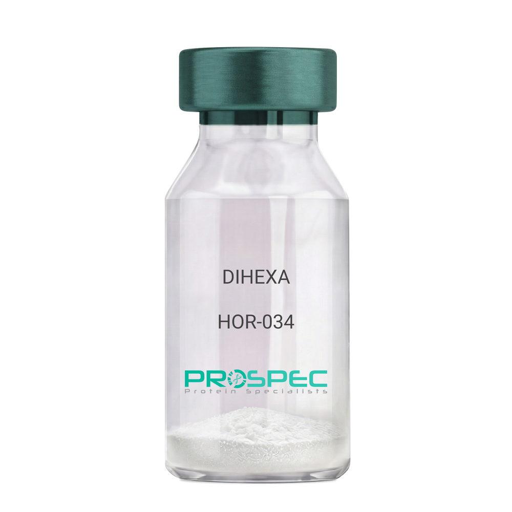
DIHEXA
Product # :
HOR-034Price :
Quantity :
Shipping Method :
Shipped at Room temp
More Info
- description
- formulation
- purity
- More Info
- Citations
Description
DIHEXA Synthetic is a single, non-glycosylated polypeptide chain containing 3 amino acids, having a molecular mass of 504.28 Dalton and a Molecular formula of C27H44N4O5.
Formulation
The protein was lyophilized with no additives.
Purity
Greater than 97.0% as determined by analysis by RP-HPLC.
Citations
More Info
-
Physical Appearance
Sterile Filtered White lyophilized (freeze-dried) powder.
-
Stability
Lyophilized DIHEXA although stable at room temperature for 3 weeks, should be stored desiccated below -18°C. Upon reconstitution DIHEXA should be stored at 4°C between 2-7 days and for future use below -18°C. For long term storage it is recommended to add a carrier protein (0.1% HSA or BSA). Please prevent freeze-thaw cycles.
-
Solubility
It is recommended to reconstitute the lyophilized DIHEXA in sterile 18MΩ-cm H2O not less than 100 µg/ml, which can then be further diluted to other aqueous solutions.
-
Amino Acid Sequence
Hexanoyl-Tyr-Ile-Ahx-NH2.
-
Background
Dihexa, also known as N-hexanoic-Tyr-Ile-(6) aminohexanoic amide, is a potent and orally active small peptide that has been the focus of significant research due to its potential neurogenic and neuroprotective effects. This compound, derived from angiotensin IV, has been shown to possess a wide range of biological activities, including enhancing cognitive function, promoting neurogenesis, and potentially mitigating the effects of neurodegenerative diseases.
Dihexa's primary mechanism of action involves its interaction with hepatocyte growth factor (HGF) and its receptor, c-Met. By mimicking the effects of HGF, Dihexa can stimulate the c-Met receptor, leading to a cascade of events that promote neurogenesis and synaptic plasticity. Studies by Benoist et al. (2014) have demonstrated that Dihexa can enhance cognitive function in rats, suggesting potential applications in cognitive enhancement and the treatment of cognitive disorders.
In addition to its neurogenic effects, Dihexa has been shown to possess neuroprotective properties. Research by Kawas et al. (2017) found that Dihexa could protect neurons from apoptosis, suggesting potential applications in the treatment of neurodegenerative diseases such as Alzheimer's and Parkinson's disease.
Given its neurogenic and neuroprotective effects, Dihexa has been proposed as a potential therapeutic agent for a variety of conditions, including cognitive disorders, neurodegenerative diseases, and stroke. For instance, a study by Harding et al. (2018) found that Dihexa could improve outcomes in animal models of stroke, indicating its potential as a therapeutic agent in stroke recovery.
While research on Dihexa is promising, it is important to note that most studies have been conducted in animals or in vitro. More research is needed to fully understand the potential effects and applications of Dihexa in humans. However, the existing body of research suggests that Dihexa could be a promising tool in the treatment of cognitive disorders and neurodegenerative diseases.
ProSpec's products are furnished for LABORATORY RESEARCH USE ONLY. The product may not be used as drugs, agricultural or pesticidal products, food additives or household chemicals.
- View Data Sheet
Name :
Prolactin Mouse, HisDescription:
Prolactin Mouse Recombinant, His Tag
AV290867, Gha1, Prl1a1, Growth hormone a1, Mammotropin, Luterotropic hormone, Lutetropin, PRL.
Product # :
CYT-1060Price :
Quantity :
Shipping Method :
Shipped with Ice Packs
More Info
- description
- source
- formulation
- purity
- More Info
- Citations
Description
Prolactin Mouse produced in E.Coli is a single, non-glycosylated, polypeptide chain containing 222 amino acids (30-228a.a.) and having a molecular mass of 25 kDa. Prolactin Mouse protein is fused to a 23 amino acid His tag at N-terminus and is purified by standard chromatography.
Source
Escherichia Coli.
Formulation
The Prolactin Mouse solution (0.25mg/ml) contains Phosphate Buffered Saline (pH 7.4) and 20% glycerol.
Purity
Greater than 90% as determined by SDS-PAGE.
Citations
More Info
-
Introduction
Prolactin is a neuroendocrine hormone synthesized primarily by the pituitary gland but also a variety of other cell types including the placenta, brain and uterus. Its primary function is to promote and maintain lactation but has also been shown to have a role in breast cancer development, regulation of reproductive function and immunoregulation.
-
Synonyms
AV290867, Gha1, Prl1a1, Growth hormone a1, Mammotropin, Luterotropic hormone, Lutetropin, PRL.
-
Physical Appearance
Sterile filtered colorless solution.
-
Stability
Store at 4°C if entire vial will be used within 2-4 weeks. Store, frozen at -20°C for longer periods of time. For long term storage it is recommended to add a carrier protein (0.1% HSA or BSA). Avoid multiple freeze-thaw cycles.
-
Amino Acid Sequence
MGSSHHHHHH SSGLVPRGSH MGSQPLPICS AGDCQTSLRE LFDRVVILSH YIHTLYTDMF IEFDKQYVQD REFMVKVIND CPTSSLATPE DKEQALKVPP EVLLNLILSL VQSSSDPLFQ LITGVGGIQE APEYILSRAK EIEEQNKQLL EGVEKIISQA YPEAKGNGIY VWSQLPSLQ GVDEESKILS LRNTIRCLRR DSHKVDNFLK VLRCQIAHQN NC.
ProSpec's products are furnished for LABORATORY RESEARCH USE ONLY. The product may not be used as drugs, agricultural or pesticidal products, food additives or household chemicals.
- View Data Sheet
Name :
MMP2 HumanDescription:
Matrix Metalloproteinase-2 Human Recombinant
kDa gelatinase, Gelatinase A, Matrix metalloproteinase-2, MMP-2, TBE-1, MMP2, CLG4A, CLG4, MONA, MMP-II.
Product # :
ENZ-769Price :
Quantity :
Shipping Method :
Shipped with Ice Packs
More Info
- description
- source
- formulation
- purity
- More Info
- Citations
Description
MMP2 Human Recombinant produced in E.coli is a single, non-glycosylated polypeptide chain containing 576 amino acids (110-660a.a) and having a molecular mass of 64.7kDa. MMP2 is fused to a 25 amino acid His-tag at N-terminus & purified by proprietary chromatographic techniques.
Source
Escherichia Coli.
Formulation
The MMP2 solution (0.25mg/ml) contains 20mM Tris-HCl buffer (pH 8.0), 0.15M NaCl and 10% glycerol.
Purity
Greater than 95% as determined by SDS-PAGE.
Citations
More Info
-
Introduction
Matrix metalloproteinase-2 (MMP-2) is involved in endometrial menstrual breakdown, regulation of vascularization and the inflammatory response. MMP-2 contains a number of distinct domains: a prodomain that is cleaved upon activation; a catalytic domain containing the zinc binding site; a fibronectin like domain believed to have a role in substrate targeting; and a carboxyl terminal (hemopexin like) domain containing 2 N-linked glycosylation. The MMP-2 can degrade an extensive array of substrates including type IV, V, VII and X collagens as well as gelatin type I. In addition, MMP-2 interacts with THBS2, TIMP2, Thrombospondin 1, CCL7 and TIMP4. MMP-2 autocatalytic cleavage in the C-terminal generates the anti-angiogenic peptide, PEX. This process seems to be made possible by binding integrinv/beta3. Defects in the MMP-2 are the cause of Torg-Winchester syndrome (TWS), aka multicentric osteolysis nodulosis and arthropathy (MONA).
-
Synonyms
kDa gelatinase, Gelatinase A, Matrix metalloproteinase-2, MMP-2, TBE-1, MMP2, CLG4A, CLG4, MONA, MMP-II.
-
Physical Appearance
Sterile Filtered colorless solution.
-
Stability
Store at 4°C if entire vial will be used within 2-4 weeks. Store, frozen at -20°C for longer periods of time. For long term storage it is recommended to add a carrier protein (0.1% HSA or BSA). Avoid multiple freeze-thaw cycles.
-
Amino Acid Sequence
MGSSHHHHHH SSGLVPRGSH MGSEFYNFFP RKPKWDKNQI TYRIIGYTPD LDPETVDDAF ARAFQVWSDV TPLRFSRIHD GEADIMINFG RWEHGDGYPF DGKDGLLAHA FAPGTGVGGD SHFDDDELWT LGEGQVVRVK YGNADGEYCK FPFLFNGKEY NSCTDTGRSD GFLWCSTTYN FEKDGKYGFC PHEALFTMGG NAEGQPCKFP FRFQGTSYDS CTTEGRTDGY RWCGTTEDYD RDKKYGFCPE TAMSTVGGNS EGAPCVFPFT FLGNKYESCT SAGRSDGKMW CATTANYDDD RKWGFCPDQG YSLFLVAAHE FGHAMGLEHS QDPGALMAPI YTYTKNFRLS QDDIKGIQEL YGASPDIDLG TGPTPTLGPV TPEICKQDIV FDGIAQIRGE IFFFKDRFIW RTVTPRDKPM GPLLVATFWP ELPEKIDAVY EAPQEEKAVF FAGNEYWIYS ASTLERGYPK PLTSLGLPPD VQRVDAAFNW SKNKKTYIFA GDKFWRYNEV KKKMDPGFPK LIADAWNAIP DNLDAVVDLQ GGGHSYFFKG AYYLKLENQS LKSVKFGSIK SDWLGC.
ProSpec's products are furnished for LABORATORY RESEARCH USE ONLY. The product may not be used as drugs, agricultural or pesticidal products, food additives or household chemicals.
- View Data Sheet
Name :
FGF23 HumanDescription:
Fibroblast Growth Factor-23 Human Recombinant
Tumor-derived hypophosphatemia-inducing factor, HYPF, ADHR, HPDR2, PHPTC, FGF23, FGF-23, Fibroblast Growth Factor-23.
Product # :
CYT-020Price :
Quantity :
Shipping Method :
Shipped at Room temp
More Info
- description
- source
- formulation
- purity
- biological activity
- More Info
- Citations
Description
Fibroblast Growth Factor-23 Human Recombinant produced in E.Coli is a single, non-glycosylated polypeptide chain containing a total of 228 amino acids and having a molecular mass of 22.5kDa. The FGF-23 is and purified by chromatographic techniques.
Source
Escherichia Coli.
Formulation
The FGF-23 protein (0.5mg/ml) was lyophilized from a 0.2µm filtered concentrated solution in PBS, pH 7.4.
Purity
Greater than 95.0% as determined by SDS-PAGE.
Biological Activity
The biological activity of FGF-23 was measured in a cell proliferation assay using NIH/3T3 mouse embryonic fibroblasts. The ED50 for this effect is typically 0.05-0.5µg/ml in the presence of 5µg/ml of Recombinant Mouse Klotho and 10 µg/ml of HPR.
Citations
More Info
-
Introduction
FGF-23 is a member of the fibroblast growth factor (FGF) family. FGF family members possess broad mitogenic and cell survival activities and are involved in a variety of biological processes including embryonic development, cell growth, morphogenesis, tissue repair, tumor growth and invasion. FGF-23 inhibits renal tubular phosphate transport. The FGF-23 gene was identified by its mutations associated with autosomal dominant hypophosphatemic rickets (ADHR), an inherited phosphate wasting disorder. Abnormally high level expression of FGF-23 was found in oncogenic hypophosphatemic osteomalacia (OHO), a phenotypically similar disease caused by abnormal phosphate metabolism. FGF-23 mutations have also been shown to cause familial tumoral calcinosis with hyperphosphatemia.
-
Synonyms
Tumor-derived hypophosphatemia-inducing factor, HYPF, ADHR, HPDR2, PHPTC, FGF23, FGF-23, Fibroblast Growth Factor-23.
-
Physical Appearance
Sterile Filtered white lyophilized powder.
-
Stability
Lyophilized FGF-23 although stable at room temperature for 3 weeks, should be stored desiccated below -18°C. Upon reconstitution FGF-23 should be stored at 4°C between 2-7 days and for future use below -18°C.For long term storage it is recommended to add a carrier protein (0.1% HSA or BSA).Please prevent freeze-thaw cycles.
-
Solubility
It is recommended to reconstitute the lyophilized FGF-23 in sterile 18M-cm H2O not less than 100µg/ml, which can then be further diluted to other aqueous solutions.
-
Amino Acid Sequence
MYPNASPLLG SSWGGLIHLY TATARNSYHL QIHKNGHVDG APHQTIYSAL MIRSEDAGFV VITGVMSRRY LCMDFRGNIF GSHYFDPENC RFQHQTLENG YDVYHSPQYH FLVSLGRAKR AFLPGMNPPP YSQFLSRRNE IPLIHFNTPI PRRHTRSAED DSERDPLNVL KPRARMTPAP ASCSQELPSA EDNSPMASDP LGVVRGGRVN THAGGTGPEG CRPFAKFI.
-
Background
Before it was discovered in 2000, there was a hypothesis that a similar type of protein existed that performed many of the functions we see in FGF23. This was originally referred to as phosphatonin. Various effects were described and noted by researchers including inhibition of production and inhibition of secretion of parathyroid hormone. Derived from the bone Fibroblast Growth Factor 23 is a phosphaturic hormone. It increases phosphate excretion when acting on the kidney and also suppresses the biosynthesis of 1,25(OH)2D3.
Mechanism
Despite various research studies into the topic, many of the mechanisms for the regulations of FGF23 production remain a mystery to the scientific community. While we know that mutations in PHEX, ENPP1 and DMP1 result in the increased expression of FGF23, it is unclear why this occurs. This also means that currently, it is not possible to regulate the production of FGF23 either. We also do not know how signals from FGF23 regulate vitamin D metabolism. However, understanding these types of mechanisms could offer information needed to provide better treatment for deranged bone and mineral metabolism.Interactions
Molecular interactions involving FGF-23, vitamin D and klotho do provide the solution needed to regulate phosphate levels within the body. Furthermore, an interaction between Vitamin D and FGF3 can have an impact on renal phosphate balance. As well as this, when in the presence of klotho, FGF3 does actually increase bioactivity and begins to change systemic phosphate homeostasis.Function
Based on research it seems that the main function for FGF23 is the regulation of phosphate concentration in plasma. It seems to be secreted from the osteocytes due to elevated levels . When acting upon the kidneys, the hormone reduces the expression of NPT2. This is a sodium-phosphate cotransporter found in the proximal tube.
As such, it appears as though FGF23 is able to reduce the reabsorption, all the while maxing the excretion of phosphate. It has also been suggested that the hormone is able to suppress 1-alpha-hydroxylase. If this is the case, it can limit its potential to activate vitamin D and thus impair the ability for calcium absorption.Structure
FGF243 is located on the chromosome 12. It is composed of three exons. The crystal structure of FGF23 is completely different from the common conformation typically adopted by paracrine-acting FGFs. Instead, there is a conformation of the HPR region between beta strands 10 and 12. As well as this, there is a cleft between the other HPR-binding region, the beta1-beta12 loop and this one. This comes before a direct interaction between HPR sulfate and FGF23s backbone atoms. Due to this, endocrine function is benefitted and HPR-binding affinity is reduced for the lipangs.
Certain mutations that cause the protein to be completely resistant to proteolytic cleavage does trigger a surge in activity of the protein and the renal phosphate loss typically found in certain human diseases including hypophosphatemic rickets.
Studies have also revealed that FGF23 is overproduced in certain tumors including phosphaturic mesenchymal tumors. Furthermore a reduced level of activity for this protein is believed to lead to higher phosphate levels and familial tumor calcinosis clinical syndrome.What is the molecular weight/Mw of FGF23 HUMAN Protein?
FGF23 HUMAN Protein has a total Mw of 22.5kDa.
What is the source or expression system of FGF23 HUMAN Protein?
Escherichia Coli.
What is the Purity of FGF23 HUMAN Protein?
FGF23 HUMAN Protein is >95X% pure as determined by SDS-PAGE.
What is the Biological Activity of FGF23 HUMAN Protein?
The biological activity of FGF-23 was measured in a cell proliferation assay using NIH/3T3 mouse embryonic fibroblasts. The ED50 for this effect is typically 0.05-0.5µg/ml in the presence of 5µg/ml of Recombinant Mouse Klotho and 10 µg/ml of HPR.
What is the amino acid sequence of FGF23 HUMAN Protein?
MYPNASPLLG SSWGGLIHLY TATARNSYHL QIHKNGHVDG APHQTIYSAL MIRSEDAGFV VITGVMSRRY LCMDFRGNIF GSHYFDPENC RFQHQTLENG YDVYHSPQYH FLVSLGRAKR AFLPGMNPPP YSQFLSRRNE IPLIHFNTPI PRRHTRSAED DSERDPLNVL KPRARMTPAP ASCSQELPSA EDNSPMASDP LGVVRGGRVN THAGGTGPEG CRPFAKFI.
What applications can FGF23 HUMAN Protein be used in?
FGF23 HUMAN Protein can probably be used in western blot, ELISA and Lateral Flow.
What is the endotoxin level for FGF23 HUMAN Protein?
The endotoxin level is minimal, FGF23 HUMAN Protein was purified using conventional chromatography techniques.
ProSpec's products are furnished for LABORATORY RESEARCH USE ONLY. The product may not be used as drugs, agricultural or pesticidal products, food additives or household chemicals.