Search results
1000 results found for “Exosome Component”
Name
Description
Product #
Price
Quantity
Shipping Method
- View Data Sheet
Name :
Omentin Human, HisDescription:
Omentin Human Recombinant, His Tag
Intelectin-1, HL1, LFR, HL-1, INTL, ITLN, hIntL.
Product # :
CYT-551Price :
Quantity :
Shipping Method :
Shipped at Room temp
More Info
- description
- source
- formulation
- purity
- More Info
Description
Omentin Human Recombinant is produced in E.Coli by recombinant DNA technology is a single, polypeptide chain containing 294 amino acids and having a molecular mass of 32.7 kDa. Recombinant Human Omentin contains His tag fused at N-Terminus.Intelectin is purified by proprietary chromatographic techniques.
Source
Escherichia Coli.
Formulation
Each mg of lyophilized powder contains 20mM Tris & 50mM NaCl pH-8.0.
Purity
Greater than 95.0% as determined by(a) Analysis by RP-HPLC.
(b) Analysis by SDS-PAGE.More Info
-
Introduction
Omentin is a recently recognized gene highly localized to the mental tissue (visceral adipose tissue). Omentin is present in the stromal vascular cells in the adipose tissue rather than in the adipocytes. Omentin is predominantly expressed in the visceral adipose tissue than the subcutaneous tissue, with the omentin mRNA being 150 times higher in the visceral adipose tissue. Omentin has also been detected in human blood using western blot analysis, and seems to increase insulin-stimulated glucose uptake in 3T3-L1 adipocytes in mice. Omentin seems to increase Akt phosphorylation irrespective of insulin presence. Its role in glucose metabolism and obesity remains to be described; an insulin-sensitizing action is possible.Differences in Omentin expression has been noted in adipose tissue from normals and patients with inflammatory bowel disease although its significance is unknown.
-
Synonyms
Intelectin-1, HL1, LFR, HL-1, INTL, ITLN, hIntL.
-
Physical Appearance
Sterile Filtered White lyophilized (freeze-dried) powder.
-
Stability
Lyophilized Intelectin His Tag although stable at room temperature for 3 weeks, should be stored desiccated below -18°C. Upon reconstitution Intelectin should be stored at 4°C between 2-7 days and for future use below -18°C.For long term storage it is recommended to add a carrier protein (0.1% HSA or BSA).Please prevent freeze-thaw cycles.
-
Solubility
It is recommended to reconstitute the lyophilized Omentin His Tag in sterile 18MΩ-cm H2O not less than 100µg/ml, which can then be further diluted to other aqueous solutions.
-
Amino Acid Sequence
MRGSHHHHHH GMASTDEANT YFKEWTCSSS PSLPRSCKEI KDECPSAFDG LYFLRTENGV IYQTFCDMTS GGGGWTLVAS VHENDMRGKC TVGDRWSSQQ GSKAVYPEGD GNWANYNTFG SAEAATSDDY KNPGYYDIQA KDLGIWHVPN KSPMQHWRNS SLLRYRTDTG FLQTLGHNLF GIYQKYPVKY GEGKCWTDNG PVIPVVYDFG DAQKTASYYS PYGQREFTAG FVQFRVFNNE RAANALCAGM RVTGCNTEHH CIGGGGYFPE ASPQQCGDFS GFDWSGYGTH VGYS.
-
Applications
Elisa
Western blot.
ProSpec's products are furnished for LABORATORY RESEARCH USE ONLY. The product may not be used as drugs, agricultural or pesticidal products, food additives or household chemicals.
- View Data Sheet
Name :
CPEB1 HumanDescription:
Cytoplasmic Polyadenylation Element Binding Protein 1 Human Recombinant
Cytoplasmic Polyadenylation Element Binding Protein 1, CPE-Binding Protein 1, CPE-BP1, HCPEB-1, CPEB, Cytoplasmic Polyadenylation Element-Binding Protein 1, CPEB-1, H-CEBP, CEBP, CPEB1.
Product # :
PRO-1952Price :
Quantity :
Shipping Method :
Shipped with Ice Packs
More Info
- description
- source
- formulation
- purity
- More Info
Description
CPEB1 Human Recombinant produced in E.coli is a single, non-glycosylated polypeptide chain containing 584 amino acids (1-561) and having a molecular mass of 64.5 kDa.CPEB1 is fused to a 23 amino acid His-tag at N-terminus & purified by proprietary chromatographic techniques.
Source
Escherichia Coli.
Formulation
The CPEB1 solution (1mg/ml) contains 20mM Tris-HCl buffer (pH 8.0), 0.4M Urea and 10% glycerol.
Purity
Greater than 85.0% as determined by SDS-PAGE.
More Info
-
Introduction
Cytoplasmic Polyadenylation Element Binding Protein 1 (CPEB1) belongs to the cytoplasmic polyadenylation element (CPE) binding protein family, whose members regulate translation of cyclin B1 during embryonic cell divisions. The CPEB1 is a highly conserved protein which binds to a specific RNA sequence called the CPE found in the 3' UTR of some mRNAs. Analogous proteins in Xenopus and mouse function to stimulate cytoplasmic polyadenylation of dormant mRNAs with short polyA tails, resulting in their translation.
-
Synonyms
Cytoplasmic Polyadenylation Element Binding Protein 1, CPE-Binding Protein 1, CPE-BP1, HCPEB-1, CPEB, Cytoplasmic Polyadenylation Element-Binding Protein 1, CPEB-1, H-CEBP, CEBP, CPEB1.
-
Physical Appearance
Sterile Filtered colorless solution.
-
Stability
Store at 4°C if entire vial will be used within 2-4 weeks. Store, frozen at -20°C for longer periods of time. For long term storage it is recommended to add a carrier protein (0.1% HSA or BSA).Avoid multiple freeze-thaw cycles.
-
Amino Acid Sequence
MGSSHHHHHH SSGLVPRGSH MGSMAFPLEE EAGRIKDCWD NQEAPALSTC SNANIFRRIN AILDNSLDFS RVCTTPINRG IHDHLPDFQD SEETVTSRML FPTSAQESSR GLPDANDLCL GLQSLSLTGW DRPWSTQDSD SSAQSSTHSV LSMLHNPLGN VLGKPPLSFL PLDPLGSDLV DKFPAPSVRG SRLDTRPILD SRSSSPSDSD TSGFSSGSDH LSDLISSLRI SPPLPFLSLS GGGPRDPLKM GVGSRMDQEQ AALAAVTPSP TSASKRWPGA SVWPSWDLLE APKDPFSIER EARLHRQAAA VNEATCTWSG QLPPRNYKNP IYSCKVFLGG VPWDITEAGL VNTFRVFGSL SVEWPGKDGK HPRCPPKGYV YLVFELEKSV RSLLQACSHD PLSPDGLSEY YFKMSSRRMR CKEVQVIPWV LADSNFVRSP SQRLDPSRTV FVGALHGMLN AEALAAILND LFGGVVYAGI DTDKHKYPIG SGRVTFNNQR SYLKAVSAAF VEIKTTKFTK KVQIDPYLED SLCHICSSQP GPFFCRDQVC FKYFCRSCWH WRHSMEGLRH HSPLMRNQKN RDSS.
ProSpec's products are furnished for LABORATORY RESEARCH USE ONLY. The product may not be used as drugs, agricultural or pesticidal products, food additives or household chemicals.
- View Data Sheet
Name :
PEBP1 MouseDescription:
Phosphatidylethanolamine Binding Protein 1 Mouse Recombinant
Phosphatidylethanolamine-binding protein 1, PEBP-1, HCNPpp.
Product # :
PRO-2230Price :
Quantity :
Shipping Method :
Shipped with Ice Packs
More Info
- description
- source
- formulation
- purity
- More Info
Description
PEBP1 Mouse Recombinant produced in E.Coli is a single, non-glycosylated polypeptide chain containing 210 amino acids (1-187 a.a) and having a molecular mass of 23.2kDa. PEBP1 is fused to a 23 amino acid His-tag at N-terminus & purified by proprietary chromatographic techniques.
Source
Escherichia Coli.
Formulation
PEBP1 protein solution (1mg/ml) containing Phosphate Buffered Saline (pH7.4) and 10% glycerol.
Purity
Greater than 95.0% as determined by SDS-PAGE.
More Info
-
Introduction
PEBP1 (Phosphatidylethanolamine binding protein 1) belongs to the phosphatidylethanolamine-binding protein family and a serine protease inhibitor that inhibits thrombin, neuropsin. PEBP1 plays a key modulatory part in several protein kinase signaling cascades. PKC phosphorylates PEBP1, resulting in the release of Raf-1 and activation of MEK and ERK. PEBP1 is expressed in many tissues and implicated in the regulation of such physiological processes as membrane biosynthesis, spermatogenesis, neural development, and metastasis suppression.
PEBP1 binds ATP, opioids and phosphatidylethanolamine, however it has lower affinity for phosphatidylinositol and phosphatidylcholine. PEBP1 may also be involved in the function of the presynaptic cholinergic neurons of the CNS. PEBP1 increases the production of choline acetyltransferase although not acetylcholinesterase. Furtheremore, PEBP1 functions in potentially sequestering toxic compounds, including locostatin which may have harmful effects on cells.
Loss of PEBP1 expression may have a significant role as prognostic marker in Gastrointestinal stromal tumors. In addition, PEBP1 is found differentially expressed in the Wernicke's Area from schizophrenia patients. PEBP1 is also, an invasion suppressor protein in nasopharyngeal carcinoma. -
Synonyms
Phosphatidylethanolamine-binding protein 1, PEBP-1, HCNPpp.
-
Physical Appearance
Sterile Filtered colorless solution.
-
Stability
Store at 4°C if entire vial will be used within 2-4 weeks. Store, frozen at -20°C for longer periods of time. For long term storage it is recommended to add a carrier protein (0.1% HSA or BSA).Avoid multiple freeze-thaw cycles.
-
Amino Acid Sequence
MGSSHHHHHH SSGLVPRGSH MGSMAADISQ WAGPLCLQEV DEPPQHALRV DYAGVTVDEL GKVLTPTQVM NRPSSISWDG LDPGKLYTLV LTDPDAPSRK DPKFREWHHF LVVNMKGNDI SSGTVLSDYV GSGPPSGTGL HRYVWLVYEQ EQPLSCDEPI LSNKSGDNRG KFKVETFRKK YNLGAPVAGT CYQAEWDDYV PKLYEQLSGK.
ProSpec's products are furnished for LABORATORY RESEARCH USE ONLY. The product may not be used as drugs, agricultural or pesticidal products, food additives or household chemicals.
- View Data Sheet
Name :
C1q Human, LiquidDescription:
Complement Component C1q Human, Liquid
Component C1q, Complement C1q, Complement Component C1q, C1q.
Product # :
PRO-554LPrice :
Quantity :
Shipping Method :
Shipped with Ice Packs
More Info
- description
- source
- formulation
- purity
- More Info
Description
Human Complement Component C1q produced in Human plasma having a molecular mass of 410 kDa.
Source
Human Plasma.
Formulation
10 mM HEPES and 300mM NaCl, pH 7.2.
Purity
Greater than 95.0% as determined by SDS-PAGE.
More Info
-
Introduction
C1q is the first component of the classical pathway of complement activation. C1q along with the enzymatically active components C1r and C1s forms the C1 complex. When C1 binds to immunoglobulins in the form of immune complexes, it leads to activation of C1r and C1s proteases and a further activates the classical pathway of complement.
C1q is a glycoprotein that belongs to the collectin family, having a molecular weight of about 410-462 kDa. C1q is a hexamer composed of globular heads attached to collagen-like triple-helix tails. The globular heads of C1q exclusively bind to the CH2 domain of IgG molecules or the CH3 domain of IgM. Each heavy chain of the immunoglobulin molecule contains a single binding site for C1q. Given that C1q must bind to no less than two heavy chains in order to alter its conformation and activate C1r and C1s, its activation follows only after binding to immunoglobulins in the form of immune complexes bound to multivalent antigens. C1q’s main physiological role is in the clearance of immune complexes and apoptotic bodies from the organism. Interruption of this process may lead to development of autoimmunity. Individuals with genetic deficiencies of C1q or other components of the classical pathway are at risk to develop SLE. C1q specifically binds to apoptotic bodies of human keratinocytes, vascular endothelial cells and lymphocytes. Complement components C1q and bound C3 mediate the clearance of apoptotic bodies. Hence, C1q may advance the clearance of autoantigens, avoiding stimulation of the immune system. Nonetheless, an extended exposition of the immune system to the neoepitope exposed on C1q molecules bound to immune complexes or apoptotic bodies could ultimately lead to an autoimmune response against C1q itself and to an altered complement function. C1q deficiency may also lead to disruption of the negative selection of autoreactive B cells. C1q along with other specific recognition proteins bind to the highly conserved lupus antigens (dsDNA and nuclear proteins) and activate the complement system. Autoantibodies against C1q (anti-C1q) are found in a number of autoimmune and infectious diseases like glomerulonephritis (GN) and lupus erythematosus (SLE), these antibodies are significant in clinical practice due to their negative predictive value. -
Synonyms
Component C1q, Complement C1q, Complement Component C1q, C1q.
-
Physical Appearance
Sterile Filtered solution.
-
Stability
Human C1q is stable at 4°C if entire vial will be used within 2-4 weeks. Store, frozen below -20°C for longer periods of time. For long term storage it is recommended to add a carrier protein (0.1% HSA or BSA). Avoid multiple freeze-thaw cycles.
-
Human Virus Test
Plasma from each donor has been tested and found negative for antibody to HIV-1, HIV-2, HCV and HBSAG.
ProSpec's products are furnished for LABORATORY RESEARCH USE ONLY. The product may not be used as drugs, agricultural or pesticidal products, food additives or household chemicals.
- View Data Sheet
Name :
LY6D HumanDescription:
Lymphocyte Antigen 6 Complex, Locus D Human Recombinant
Lymphocyte Antigen 6 Complex Locus D, E48 Antigen, Ly-6D, Lymphocyte Antigen 6D.
Product # :
PRO-1838Price :
Quantity :
Shipping Method :
Shipped with Ice Packs
More Info
- description
- source
- formulation
- purity
- More Info
Description
LY6D Human Recombinant produced in E.coli is a single, non-glycosylated polypeptide chain containing 101 amino acids (21-98) and having a molecular mass of 10.8 kDa. LY6D is fused to a 23 amino acid His-tag at N-terminus.
Source
Escherichia Coli.
Formulation
The LY6D solution (0.5mg/ml) contains 20mM Tris-HCl buffer (pH 8.0), 0.15M NaCl and 50% glycerol.
Purity
Greater than 90% as determined by SDS-PAGE.
More Info
-
Introduction
LY6D encloses 1 UPAR/Ly6 domain and is only expressed at the keratinocyte of stratified squamous epithelia and the outer cell surface of transitional epithelia. LY6D performs as a specification marker at earliest stage specification of lymphocytes between B- and T-cell developments.
-
Synonyms
Lymphocyte Antigen 6 Complex Locus D, E48 Antigen, Ly-6D, Lymphocyte Antigen 6D.
-
Physical Appearance
Sterile Filtered clear solution.
-
Stability
Store at 4°C if entire vial will be used within 2-4 weeks. Store, frozen at -20°C for longer periods of time. For long term storage it is recommended to add a carrier protein (0.1% HSA or BSA).Avoid multiple freeze-thaw cycles.
-
Amino Acid Sequence
MGSSHHHHHH SSGLVPRGSH MGSLRCHVCT SSSNCKHSVV CPASSRFCKT TNTVEPLRGN LVKKDCAESC TPSYTLQGQV SSGTSSTQCC QEDLCNEKLH N
ProSpec's products are furnished for LABORATORY RESEARCH USE ONLY. The product may not be used as drugs, agricultural or pesticidal products, food additives or household chemicals.
- View Data Sheet
Name :
CST3 MouseDescription:
Cystatin-C Mouse Recombinant
Post G-globulin, CST 3, CST3, Gamma-Trace, Cystatin 3, Amyloid Angiopathy and Cerebral Hemorrhage, Cystatin-C precursor, neuroendocrine basic polypeptide.
Product # :
PRO-597Price :
Quantity :
Shipping Method :
Shipped at Room temp
More Info
- description
- source
- formulation
- purity
- More Info
Description
Cystatin-C Murine Recombinant produced in E.Coli is a single, non-glycosylated, polypeptide chain containing 134 amino acids and having a molecular mass of 15kDa. The Mouse Cystatin-C is fused to His tag at N-Terminus.The Mouse Cystatin-C is purified by proprietary chromatographic techniques.
Source
Escherichia Coli.
Formulation
The sterile filtered concentrated (0.5mg/ml) protein solution was lyophilized with 20mM Tris & 50mM NaCl pH-7.5.
Purity
Greater than 95% as determined by SDS-PAGE.
More Info
-
Introduction
Cystatins are a superfamily of cysteine proteinase inhibitors found in both plants and animals. They comprise a group of proteinase inhibitors, widely distributed in tissues and body fluids, and form tight complexes with cysteine proteases such as cathepsin B, H, L and S. Cystatin C, a secreted molecule of this family, is of interest from biochemical, medicine and evolutionary points of view. Cystatin C, with molecular weight of 13260 Da, is composed of 120 amino acids, lacks carbohydrate and has two disulfide bridges located near the carboxyl terminus. Cystatin C is increased in patients with malignant diseases, and is related to the insufficiency of renal function and appears to be a better marker than creatinine. On the other hand, low levels of cystatin C involve cause the breakdown of the elastic laminae and, subsequently, the atherosclerosis and abdominal aortic aneurysm.
-
Synonyms
Post G-globulin, CST 3, CST3, Gamma-Trace, Cystatin 3, Amyloid Angiopathy and Cerebral Hemorrhage, Cystatin-C precursor, neuroendocrine basic polypeptide.
-
Physical Appearance
Sterile Filtered White lyophilized (freeze-dried) powder.
-
Stability
Store lyophilized protein at -20°C. Aliquot the product after reconstitution to avoid repeated freezing/thawing cycles. Reconstituted protein can be stored at 4°C for a limited period of time.
-
Solubility
Add deionized water to a working concentration of 0.5mg/ml and let the lyophilized pellet dissolve completely.
-
Amino Acid Sequence
MRGSHHHHHH GMASATPKQG PRMLGAPEEA DANEEGVRRA LDFAVSEYNK GSNDAYHSRA IQVVRARKQL VAGVNYFLDV EMGRTTCTKS QTNLTDCPFH DQPHLMRKAL CSFQIYSVPW KGTHSLTKFSCKNA.
ProSpec's products are furnished for LABORATORY RESEARCH USE ONLY. The product may not be used as drugs, agricultural or pesticidal products, food additives or household chemicals.
- View Data Sheet
Name :
TROVE2 Human, His BiotinDescription:
TROVE Domain Family Member 2 Human Recombinant, His Tag Biotinylated
60 kDa SS-A/Ro ribonucleoprotein, 60 kDa ribonucleoprotein Ro, RoRNP, 60 kDa Ro protein, Ro 60 kDa autoantigen, TROVE domain family member 2, Sjoegren syndrome type A antigen, SS-A, Sjoegren syndrome antigen A2, TROVE2, RO60, SSA2, RO-60.
Product # :
PRO-2560Price :
Quantity :
Shipping Method :
Shipped with Ice Packs
More Info
- description
- source
- formulation
- purity
- More Info
Description
TROVE2 Human Recombinant Biotin produced in E.Coli is a single, non-glycosylated, polypeptide chain having a molecular mass of 56kDa. The TROVE2 is expressed with a 6xHis tag at N-terminus and purified by proprietary chromatographic techniques.
Source
Escherichia Coli.
Formulation
The protein solution contains 20mM HEPES (pH 8.0), 20% Glycerol and 150mM NaCl.
Purity
Greater than 89% as determined by Capillary Electrophoresis.
More Info
-
Introduction
Ro 60 kDa autoantigen is an RNA-binding protein that binds to several small cytoplasmic RNA molecules known as Y RNAs. SSA2 may stabilize these RNAs from degradation. Sera taken from patients with Systemic Lupus Erythematosus (SLE) often contains antibodies that react with the normal cellular SSA2 protein as if this antigen was foreign.
-
Synonyms
60 kDa SS-A/Ro ribonucleoprotein, 60 kDa ribonucleoprotein Ro, RoRNP, 60 kDa Ro protein, Ro 60 kDa autoantigen, TROVE domain family member 2, Sjoegren syndrome type A antigen, SS-A, Sjoegren syndrome antigen A2, TROVE2, RO60, SSA2, RO-60.
-
Physical Appearance
Sterile Filtered clear solution.
-
Stability
Store at 4°C if entire vial will be used within 2-4 weeks. Store, frozen at -20°C for longer periods of time. Avoid multiple freeze-thaw cycles.
ProSpec's products are furnished for LABORATORY RESEARCH USE ONLY. The product may not be used as drugs, agricultural or pesticidal products, food additives or household chemicals.
- View Data Sheet
Name :
TCP1 HumanDescription:
T-Complex 1 Human Recombinant
T-complex protein 1 subunit alpha, TCP-1-alpha, CCT-alpha, TCP1, CCT1, CCTA, D6S230E.
Product # :
PRO-276Price :
Quantity :
Shipping Method :
Shipped with Ice Packs
More Info
- description
- source
- formulation
- purity
- More Info
Description
TCP1 Human Recombinant fused with a 20 amino acid His tag at N-terminus produced in E.Coli is a single, non-glycosylated, polypeptide chain containing 576 amino acids (1-556 a.a.) and having a molecular mass of 62.5kDa. The TCP1 is purified by proprietary chromatographic techniques.
Source
Escherichia Coli.
Formulation
The TCP1 solution (0.5 mg/ml) contains 20mM Tris-HCl Buffer (pH 8.0), 1mM DTT, 0.1mM PMSF and 10% Glycerol.
Purity
Greater than 80.0% as determined by SDS-PAGE.
More Info
-
Introduction
TCP1 is a molecular chaperone that is a member of the chaperonin containing TCP1 complex (CCT), also known as the TCP1 ring complex (TRiC). This complex consists of 2 identical stacked rings, each containing eight different proteins. Unfolded polypeptides penetrate the central cavity of the complex and are folded in an ATP-dependent manner. The TCP1 protein is found in the cytosol as a subunit of a hetero-oligomeric chaperone. TCP1 has a significant function in maintaining cellular homoeostasis by assisting the folding of many proteins such as the cytoskeletal components actin and tubulin.
-
Synonyms
T-complex protein 1 subunit alpha, TCP-1-alpha, CCT-alpha, TCP1, CCT1, CCTA, D6S230E.
-
Physical Appearance
Sterile Filtered colorless solution.
-
Stability
Store at 4°C if entire vial will be used within 2-4 weeks. Store, frozen at -20°C for longer periods of time. For long term storage it is recommended to add a carrier protein (0.1% HSA or BSA). Avoid multiple freeze-thaw cycles.
-
Amino Acid Sequence
MGSSHHHHHH SSGLVPRGSH MEGPLSVFGD RSTGETIRSQ NVMAAASIAN IVKSSLGPVG LDKMLVDDIG DVTITNDGAT ILKLLEVEHP AAKVLCELAD LQDKEVGDGT TSVVIIAAEL LKNADELVKQ KIHPTSVISG YRLACKEAVR YINENLIVNT DELGRDCLIN AAKTSMSSKI IGINGDFFAN MVVDAVLAIK YTDIRGQPRY PVNSVNILKA HGRSQMESML ISGYALNCVV GSQGMPKRIV NAKIACLDFS LQKTKMKLGV QVVITDPEKL DQIRQRESDI TKERIQKILA TGANVILTTG GIDDMCLKYF VEAGAMAVRR VLKRDLKRIA KASGATILST LANLEGEETF EAAMLGQAEE VVQERICDDE LILIKNTKAR TSASIILRGA NDFMCDEMER SLHDALCVVK RVLESKSVVP GGGAVEAALS IYLENYATSM GSREQLAIAE FARSLLVIPN TLAVNAAQDS TDLVAKLRAF HNEAQVNPER KNLKWIGLDL SNGKPRDNKQ AGVFEPTIVK VKSLKFATEA AITILRIDDL IKLHPESKDD KHGSYEDAVH SGALND.
ProSpec's products are furnished for LABORATORY RESEARCH USE ONLY. The product may not be used as drugs, agricultural or pesticidal products, food additives or household chemicals.
- View Data Sheet
Name :
CALR3 HumanDescription:
Calreticulin 3 Human Recombinant
CRT2, FLJ25355, MGC26577, Calreticulin-3, Calreticulin-2, Calsperin, CALR3.
Product # :
PRO-1879Price :
Quantity :
Shipping Method :
Shipped with Ice Packs
More Info
- description
- source
- formulation
- purity
- More Info
Description
CALR3 Human Recombinant produced in E.Coli is a single, non-glycosylated polypeptide chain containing 366 amino acids (20-384 a.a) and having a molecular mass of 43kDa. CALR3 is purified by proprietary chromatographic techniques.
Source
Escherichia Coli.
Formulation
CALR3 protein solution (1mg/ml) containing 20mM Tris-HCl buffer (pH 8.0), 0.4M Urea, and 10% glycerol.
Purity
Greater than 90.0% as determined by SDS-PAGE.
More Info
-
Introduction
Calreticulin 3 (CALR3) belongs to the calreticulin (CRT) family which consists of calcium binding chaperones that are localized to the endoplasmic reticulum or sarcoplasmic reticulum in eukaryotes. CALR3, which is expressed mainly in testis, takes part in protein-folding, oligomeric assembly and quality control in the ER. CALR3 plays an important role as a cancer-testis antigen due to its frequent expression in many cancers.
-
Synonyms
CRT2, FLJ25355, MGC26577, Calreticulin-3, Calreticulin-2, Calsperin, CALR3.
-
Physical Appearance
Sterile Filtered colorless solution.
-
Stability
Store at 4°C if entire vial will be used within 2-4 weeks. Store, frozen at -20°C for longer periods of time. For long term storage it is recommended to add a carrier protein (0.1% HSA or BSA).Avoid multiple freeze-thaw cycles.
-
Amino Acid Sequence
MTVYFQEEFL DGEHWRNRWL QSTNDSRFGH FRLSSGKFYG HKEKDKGLQT TQNGRFYAIS ARFKPFSNKG KTLVIQYTVK HEQKMDCGGG YIKVFPADID QKNLNGKSQY YIMFGPDICG FDIKKVHVIL HFKNKYHENK KLIRCKVDGF THLYTLILRP DLSYDVKIDG QSIESGSIEY DWNLTSLKKE TSPAESKDWE QTKDNKAQDW EKHFLDASTS KQSDWNGDLD GDWPAPMLQK PPYQDGLKPE GIHKDVWLHR KMKNTDYLTQ YDLSEFENIG AIGLELWQVR SGTIFDNFLI TDDEEYADNF GKATWGETKG PEREMDAIQA KEEMKKAREE EEEELLSGKI NRHEHYFNQF HRRNEL.
ProSpec's products are furnished for LABORATORY RESEARCH USE ONLY. The product may not be used as drugs, agricultural or pesticidal products, food additives or household chemicals.
- View Data Sheet
Name :
CALM2 HumanDescription:
Calmodulin-2 Human Recombinant
PHKD, CAMII, PHKD2, Calmodulin-2, CALM2, CALM1 protein, Phosphorylase kinase delta.
Product # :
PRO-618Price :
Quantity :
Shipping Method :
Shipped with Ice Packs
More Info
- description
- source
- formulation
- purity
- More Info
Description
Recombinant CALM2 produced in E.Coli is a single, non-glycosylated polypeptide chain containing 149 amino acids and having a molecular mass of 16 kDa. CALM2 is purified by conventional chromatography techniques.
Source
Escherichia Coli.
Formulation
The CALM2 solution (1mg/ml) contains 20mM Tris-HCl, pH-7.5.
Purity
Greater than 90.0% as determined by:
(a) Analysis by RP-HPLC.
(b) Analysis by SDS-PAGE.More Info
-
Introduction
Calmodulin-2 acts as an intracellular calcium sensor protein. When the intracellular Ca2+ concentration increases, calmodulin can bind up to four Ca2+, changing its conformation and regulating cellular functions such as activation or inhibition of a large number of enzymes, ion channels, and receptors. P53 protein stimulates CALM2 gene expression in 041 cells. CALM-2 is involved in the processes of Ca(2+)-induced neuronal cell death and the blockage of calmodulin attenuates brain injury after cerebral ischemia. Calmodulin-2 mediates the control of a large number of enzymes and other proteins by ca(2+). among the enzymes to be stimulated by the calmodulin-ca(2+) complex are a number of protein kinases and phosphatases.
-
Synonyms
PHKD, CAMII, PHKD2, Calmodulin-2, CALM2, CALM1 protein, Phosphorylase kinase delta.
-
Physical Appearance
Sterile filtered colorless solution.
-
Stability
Store at 4°C if entire vial will be used within 2-4 weeks. Store, frozen at -20°C for longer periods of time. For long term storage it is recommended to add a carrier protein (0.1% HSA or BSA). Avoid multiple freeze-thaw cycles.
-
Amino Acid Sequence
MADQLTEEQI AEFKEAFSLF DKDGDGTITT KELGTVMRSL GQNPTEAELQ DMINEVDADG NGTIDFPEFL TMMARKMKDT DSEEEIREAF RVFDKDGNGY ISAAELRHVM TNLGEKLTDE EVDEMIREAD IDGDGQVNYE EFVQMMTAK.
ProSpec's products are furnished for LABORATORY RESEARCH USE ONLY. The product may not be used as drugs, agricultural or pesticidal products, food additives or household chemicals.
- View Data Sheet
Name :
CDH11 HumanDescription:
Cadherin 11 Human Recombinant
CAD11, CDHOB, OB, OSF-4, Cadherin 11, Osteoblast cadherin, OB-cadherin.
Product # :
PRO-1368Price :
Quantity :
Shipping Method :
Shipped with Ice Packs
More Info
- description
- source
- formulation
- purity
- More Info
Description
CDH11 Human Recombinant produced in E.Coli is a single, non-glycosylated polypeptide chain containing 601 amino acids (54-617 a.a) and having a molecular mass of 66.2kDa. CDH11 is fused to a 37 amino acid His-tag at N-terminus & purified by proprietary chromatographic techniques.
Source
E.coli.
Formulation
CDH11 protein solution (1mg/ml) containing 20mM Tris-HCl buffer (pH8.0), 10% glycerol and 0.1M NaCl.
Purity
Greater than 85% as determined by SDS-PAGE.
More Info
-
Introduction
Cadherin 11 (CDH11) belongs to the cadherin superfamily (integral membrane proteins) which mediate calcium-dependent cell-cell adhesion. CDH11 is defined based on lacking a HAV cell adhesion recognition sequence specific to type I cadherins. CDH11 protein’s expression in osteoblastic cell lines, and its upregulation during differentiation, suggests a specific function in bone development and maintenance. CDH11 contributes to the sorting of heterogeneous cell types.
-
Synonyms
CAD11, CDHOB, OB, OSF-4, Cadherin 11, Osteoblast cadherin, OB-cadherin.
-
Physical Appearance
Sterile Filtered colorless solution.
-
Stability
Store at 4°C if entire vial will be used within 2-4 weeks. Store, frozen at -20°C for longer periods of time. For long term storage it is recommended to add a carrier protein (0.1% HSA or BSA).Avoid multiple freeze-thaw cycles.
-
Amino Acid Sequence
MRGSHHHHHH GMASMTGGQQ MGRDLYDDDD KDRWGSMGWV WNQFFVIEEY TGPDPVLVGR LHSDIDSGDG NIKYILSGEG AGTIFVIDDK SGNIHATKTL DREERAQYTL MAQAVDRDTN RPLEPPSEFI VKVQDINDNP PEFLHETYHA NVPERSNVGT SVIQVTASDA DDPTYGNSAK LVYSILEGQP YFSVEAQTGI IRTALPNMDR EAKEEYHVVI QAKDMGGHMG GLSGTTKVMI TLTDVNDNPP KFPQSVYQMS VSEAAVPGEE VGRVKAKDPD IGENGLVTYN IVDGDGMESF EITTDYETQE GVIKLKKPVD FETKRAYSLK VEAANVHIDP KFISNGPFKD TVTVKIAVED ADEPPMFLAP SYIHEVQENA AAGTVVGRVH AKDPDAANSP IRYSIDRHTD LDRFFTINPE DGFIKTTKPL DREETAWLNI TVFAAEIHNR HQEAKVPVAI RVLDVNDNAP KFAAPYEGFI CESDQTKPLS NQPIVTISAD DKDDTANGPR FIFSLPPEII HNPNFTVRDN RDNTAGVYAR RGGFSRQKQD LYLLPIVISD GGIPPMSSTN TLTIKVCGCD VNGALLSCNA EAYILNAGLS T.
ProSpec's products are furnished for LABORATORY RESEARCH USE ONLY. The product may not be used as drugs, agricultural or pesticidal products, food additives or household chemicals.
- View Data Sheet
Name :
SERPINA5 HumanDescription:
Serpin Peptidase Inhibitor Clade A Member 5 Human Recombinant
PAI3, PCI, PROCI, PLANH3, Protein-C Inhibitor, Serpin A5, Plasminogen activator inhibitor 3, PAI-3, SERPINA5.
Product # :
PRO-749Price :
Quantity :
Shipping Method :
Shipped with Ice Packs
More Info
- description
- source
- formulation
- purity
- More Info
Description
SERPINA5 Human Recombinant fused with a 20 amino acid His tag at N-terminus produced in E.Coli is a single, non-glycosylated, polypeptide chain containing 408 amino acids (20-406 a.a.) and having a molecular mass of 45.9 kDa.The SERPINA5 is purified by proprietary chromatographic techniques.
Source
Escherichia Coli.
Formulation
The SERPINA5 solution (0.5mg/ml) contains 20mM Tris-HCl buffer pH-8, 1mM DTT and 10% glycerol.
Purity
Greater than 90.0% as determined by SDS-PAGE.
More Info
-
Introduction
SERPINA5 up regulates TAFI activation by inhibiting the protein C activation. SERPINA5 is a significant regulator in the equilibrium between coagulation and fibrinolysis by differentially inhibiting the activation of TAFI and of Protein-C. SERPINA5 belongs to the serpin serine proteinase inhibitor family. SERPINA5 protein inhibits plasminogen activators as well as activated protein C.
SERPINA5 is secreted in plasma & liver. SERPINA5 is involved in cell inflammation, proliferation, apoptosis, tumour cell migration, invasion, and metastasis. Moreover, SERPINA5 controls the invasive potential of renal cell carcinoma by inhibiting urinary plasminogen activator secreted by the cells. SERPINA5 participtes in regulating key serine proteases which are involved in metastatic prostate disease. -
Synonyms
PAI3, PCI, PROCI, PLANH3, Protein-C Inhibitor, Serpin A5, Plasminogen activator inhibitor 3, PAI-3, SERPINA5.
-
Physical Appearance
Sterile Filtered colorless solution.
-
Stability
Store at 4°C if entire vial will be used within 2-4 weeks.Store, frozen at -20°C for longer periods of time. For long term storage it is recommended to add a carrier protein (0.1% HSA or BSA).Avoid multiple freeze-thaw cycles.
-
Amino Acid Sequence
MGSSHHHHHH SSGLVPRGSH MHRHHPREMK KRVEDLHVGA TVAPSSRRDF TFDLYRALAS AAPSQNIFFS PVSISMSLAM LSLGAGSSTK MQILEGLGLN LQKSSEKELH RGFQQLLQEL NQPRDGFQLS LGNALFTDLV VDLQDTFVSA MKTLYLADTF PTNFRDSAGA MKQINDYVAK QTKGKIVDLL KNLDSNAVVI MVNYIFFKAK WETSFNHKGT QEQDFYVTSE TVVRVPMMSR EDQYHYLLDR NLSCRVVGVP YQGNATALFI LPSEGKMQQV ENGLSEKTLR KWLKMFKKRQ LELYLPKFSI EGSYQLEKVL PSLGISNVFT SHADLSGISN HSNIQVSEMV HKAVVEVDES GTRAAAATGT IFTFRSARLN SQRLVFNRPF LMFIVDNNIL FLGKVNRP.
ProSpec's products are furnished for LABORATORY RESEARCH USE ONLY. The product may not be used as drugs, agricultural or pesticidal products, food additives or household chemicals.
- View Data Sheet
Name :
SGCB HumanDescription:
Sarcoglycan Beta Human Recombinant
Beta-sarcoglycan, Beta-SG, 43 kDa dystrophin-associated glycoprotein, 43DAG, A3b, LGMD2E, Sarcoglycan, Beta (43kDa Dystrophin-Associated Glycoprotein), Limb Girdle Muscular Dystrophy 2E (Non-Linked Families), SGC.
Product # :
PRO-2211Price :
Quantity :
Shipping Method :
Shipped with Ice Packs
More Info
- description
- source
- formulation
- purity
- More Info
Description
SGCB Human Recombinant produced in E.Coli is a single, non-glycosylated polypeptide chain containing 255 amino acids (87-318 a.a) and having a molecular mass of 27.8kDa.SGCB is fused to a 23 amino acid His-tag at N-terminus & purified by proprietary chromatographic techniques.
Source
Escherichia Coli.
Formulation
SGCB protein solution (0.5mg/ml) containing 20mM Tris-HCl buffer(pH8.0) and 10% glycerol.
Purity
Greater than 90.0% as determined by SDS-PAGE.
More Info
-
Introduction
Sarcoglycan Beta, also known as SGCB, is a part of the sarcoglycan family. SGCB belongs to the sarcoglycan complex which is a subcomplex of the dystrophin-glycoprotein complex that connects between the F-actin cytoskeleton and the extracellular matrix.
-
Synonyms
Beta-sarcoglycan, Beta-SG, 43 kDa dystrophin-associated glycoprotein, 43DAG, A3b, LGMD2E, Sarcoglycan, Beta (43kDa Dystrophin-Associated Glycoprotein), Limb Girdle Muscular Dystrophy 2E (Non-Linked Families), SGC.
-
Physical Appearance
Sterile Filtered colorless solution.
-
Stability
Store at 4°C if entire vial will be used within 2-4 weeks. Store, frozen at -20°C for longer periods of time. For long term storage it is recommended to add a carrier protein (0.1% HSA or BSA). Avoid multiple freeze-thaw cycles.
-
Amino Acid Sequence
MGSSHHHHHH SSGLVPRGSH MGSWAVIRIG PNGCDSMEFH ESGLLRFKQV SDMGVIHPLY KSTVGGRRNE NLVITGNNQP IVFQQGTTKL SVENNKTSIT SDIGMQFFDP RTQNILFSTD YETHEFHLPS GVKSLNVQKA STERITSNAT SDLNIKVDGR AIVRGNEGVF IMGKTIEFHM GGNMELKAEN SIILNGSVMV STTRLPSSSS GDQLGSGDWV RYKLCMCADG TLFKVQVTSQ NMGCQISDNP CGNTH.
ProSpec's products are furnished for LABORATORY RESEARCH USE ONLY. The product may not be used as drugs, agricultural or pesticidal products, food additives or household chemicals.
- View Data Sheet
Name :
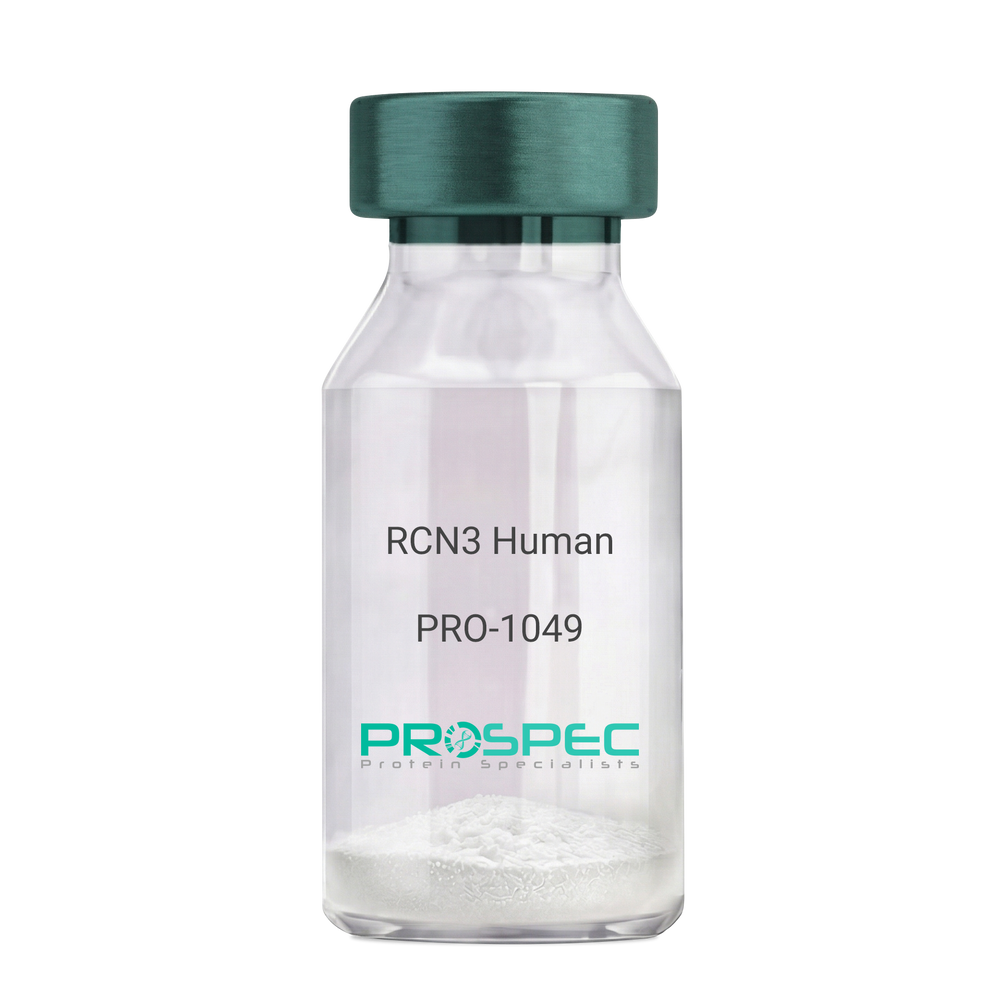
RCN3 HumanDescription:
Reticulocalbin 3 Human Recombinant
Reticulocalbin-3, EF-hand calcium-binding protein RLP49, RCN3, RLP49.
Product # :
PRO-1049Price :
Quantity :
Shipping Method :
Shipped with Ice Packs
More Info
- description
- source
- formulation
- purity
- More Info
Description
RCN3 Human Recombinant produced in E.Coli is a single, non-glycosylated polypeptide chain containing 333 amino acids (21-328 a.a) and having a molecular mass of 37.9kDa.RCN3 is fused to a 25 amino acid His-tag at N-terminus & purified by proprietary chromatographic techniques.
Source
Escherichia Coli.
Formulation
RCN3 protein solution (1mg/ml) containing 20mM Tris-HCl buffer (pH 8.0), 1mM DTT, 10% glycerol and 0.1M NaCl.
Purity
Greater than 95.0% as determined by SDS-PAGE.
More Info
-
Introduction
Reticulocalbin 3 (RCN3) belongs to the CREC (cab45/reticulocalbin/ ERC45/calumenin) family. RCN3 contains 5 Arg-Xaa-Xaa-Arg motifs, which function as target sequences of SPCs (subtilisin-like proprotein convertases), which is a family of serine endoproteases that proteolytically activate proproteins. The synthesis of PACE4 is induced by association and coexpression with RCN3.
-
Synonyms
Reticulocalbin-3, EF-hand calcium-binding protein RLP49, RCN3, RLP49.
-
Physical Appearance
Sterile Filtered colorless solution.
-
Stability
Store at 4°C if entire vial will be used within 2-4 weeks. Store, frozen at -20°C for longer periods of time. For long term storage it is recommended to add a carrier protein (0.1% HSA or BSA).Avoid multiple freeze-thaw cycles.
-
Amino Acid Sequence
MGSSHHHHHH SSGLVPRGSH MGSHMKPSPD AGPHGQGRVH QAAPLSDAPH DDAHGNFQYD HEAFLGREVA KEFDQLTPEE SQARLGRIVD RMDRAGDGDG WVSLAELRAW IAHTQQRHIR DSVSAAWDTY DTDRDGRVGW EELRNATYGH YAPGEEFHDV EDAETYKKML ARDERRFRVA DQDGDSMATR EELTAFLHPE EFPHMRDIVI AETLEDLDRN KDGYVQVEEY IADLYSAEPG EEEPAWVQTE RQQFRDFRDL NKDGHLDGSE VGHWVLPPAQ DQPLVEANHL LHESDTDKDG RLSKAEILGN WNMFVGSQAT NYGEDLTRHH DEL.
ProSpec's products are furnished for LABORATORY RESEARCH USE ONLY. The product may not be used as drugs, agricultural or pesticidal products, food additives or household chemicals.
- View Data Sheet
Name :
RGS10 HumanDescription:
Regulator of G-Protein Signaling 10 Human Recombinant
Regulator Of G-Protein Signaling 10, RGS10.
Product # :
PRO-890Price :
Quantity :
Shipping Method :
Shipped with Ice Packs
More Info
- description
- source
- formulation
- purity
- More Info
Description
RGS10 Human Recombinant produced in E.coli is a single, non-glycosylated polypeptide chain containing 205 amino acids (1-181) and having a molecular mass of 23.7 kDa.The RGS10 is fused to a 24 amino acid His-Tag at N-terminus and purified by proprietary chromatographic techniques.
Source
E.coli.
Formulation
The RGS10 solution (1mg/ml) contains 20mM Tris-HCl buffer (pH 8.0), 1mM DTT, 0.1M NaCl and 10% glycerol.
Purity
Greater than 90% as determined by SDS-PAGE.
More Info
-
Introduction
RGS10 is a member of the RGS family that regulate molecules which perform as GTPase activating proteins (GAPs) for G alpha subunits of heterotrimeric G proteins. RGS proteins can disengage G protein subunits of the Gi alpha, Go alpha and Gq alpha subtypes. They drive G proteins into their inactive GDP-bound forms. All RGS proteins share a conserved 120-amino acid sequence termed the RGS domain. The nucleus localized RGS10 is associates specifically with the activated forms of the two related G-protein subunits, G-alphai3 and G-alphaz but is unable to cooperate with the structurally and functionally distinct G-alpha subunits.
-
Synonyms
Regulator Of G-Protein Signaling 10, RGS10.
-
Physical Appearance
Sterile Filtered colorless solution.
-
Stability
Store at 4°C if entire vial will be used within 2-4 weeks. Store, frozen at -20°C for longer periods of time. For long term storage it is recommended to add a carrier protein (0.1% HSA or BSA).Avoid multiple freeze-thaw cycles.
-
Amino Acid Sequence
MGSSHHHHHH SSGLVPRGSH MGSHMFNRAV SRLSRKRPPS DIHDSDGSSS SSHQSLKSTA KWAASLENLL EDPEGVKRFR EFLKKEFSEE NVLFWLACED FKKMQDKTQM QEKAKEIYMT FLSSKASSQV NVEGQSRLNE KILEEPHPLM FQKLQDQIFN LMKYDSYSRF LKSDLFLKHK RTEEEEEDLP DAQTAAKRAS RIYNT
ProSpec's products are furnished for LABORATORY RESEARCH USE ONLY. The product may not be used as drugs, agricultural or pesticidal products, food additives or household chemicals.
- View Data Sheet
Name :
ITGB3BP HumanDescription:
Integrin Beta 3 Binding Protein Human Recombinant
CENP-R, CENPR, HSU37139, NRIF3, TAP20, Centromere protein R, Beta-3-endonexin, Integrin beta-3-binding protein, Nuclear receptor-interacting factor 3, ITGB3BP.
Product # :
PRO-1435Price :
Quantity :
Shipping Method :
Shipped with Ice Packs
More Info
- description
- source
- formulation
- purity
- More Info
Description
ITGB3BP Human Recombinant produced in E. coli is a single polypeptide chain containing 239 amino acids (1-216) and having a molecular mass of 27.1kDa. ITGB3BP is fused to a 23 amino acid His-tag at N-terminus & purified by proprietary chromatographic techniques.
Source
E.coli.
Formulation
The ITGB3BP solution (0.5mg/ml) contains 20mM Tris-HCl buffer (pH 8.0), 0.4M Urea and 10% glycerol.
Purity
Greater than 90% as determined by SDS-PAGE.
More Info
-
Introduction
Integrin Beta 3 Binding Protein (ITGB3BP1) is a broadly expressed protein which contains a DD1 or RepD1 domain and an LXXLL motif. ITGB3BP1 functions as a nuclear receptor coactivator while induced by estrogen. ITGB3BP1 is also relates with the CENP-A-CAD complex that is engaged in mitotic progression, the assembly of kinetochore proteins and chromosome segregation. ITGB3BP1 behave s as a transcriptional corepressor via its interaction with the NFKB1 NF-kappa-B subunit by interfering with the transactivation domain of NFKB1.
-
Synonyms
CENP-R, CENPR, HSU37139, NRIF3, TAP20, Centromere protein R, Beta-3-endonexin, Integrin beta-3-binding protein, Nuclear receptor-interacting factor 3, ITGB3BP.
-
Physical Appearance
Sterile Filtered colorless solution.
-
Stability
Store at 4°C if entire vial will be used within 2-4 weeks. Store, frozen at -20°C for longer periods of time. For long term storage it is recommended to add a carrier protein (0.1% HSA or BSA).Avoid multiple freeze-thaw cycles.
-
Amino Acid Sequence
MGSSHHHHHH SSGLVPRGSH MGSMPFAPVA QARVQWHDFR SLQHLLPAFK RFSCLSLGSS WDYSVKRSLK LDGLLEENSF DPSKITRKKS VITYSPTTGT CQMSLFASPT SSEEQKHRNG LSNEKRKKLN HPSLTESKES TTKDNDEFMM LLSKVEKLSE EIMEIMQNLS SIQALEGSRE LENLIGISCA SHFLKREMQK TKELMTKVNK QKLFEKSTGL PHKASRHLDS YEFLKAILN.
ProSpec's products are furnished for LABORATORY RESEARCH USE ONLY. The product may not be used as drugs, agricultural or pesticidal products, food additives or household chemicals.
- View Data Sheet
Name :
LILRA3 HumanDescription:
Leukocyte Immunoglobulin Like Receptor A3 Human Recombinant
LILRA3, ILT6, LIR4, LIR-4, LILRA3, CD85e, Leukocyte immunoglobulin-like receptor subfamily A member 3, leukocyte immunoglobulin-like receptor subfamily A member 3 isoform 1, CD85 antigen-like family member E, Immunoglobulin-like transcript 6, Leukocyte immunoglobulin-like receptor 4, Monocyte inhibitory receptor HM43/HM31.
Product # :
PRO-2831Price :
Quantity :
Shipping Method :
Shipped with Ice Packs
More Info
- description
- source
- formulation
- purity
- More Info
Description
LILRA3 Human Recombinant is a single, glycosylated, polypeptide chain (24-439 a.a) containing a total of 658 amino acids and having a molecular mass of 72.2 kDa. LILRA3 is fused to a 239 a.a hIgG-his-Tag at C-terminus and is purified by proprietary chromatographic techniques.
Source
HEK293 Cells.
Formulation
The LILRA3 solution (0.5mg/ml) contains 10% Glycerol and Phosphate-Buffered Saline (pH 7.4).
Purity
Greater than 95.0% as determined by SDS-PAGE.
More Info
-
Synonyms
LILRA3, ILT6, LIR4, LIR-4, LILRA3, CD85e, Leukocyte immunoglobulin-like receptor subfamily A member 3, leukocyte immunoglobulin-like receptor subfamily A member 3 isoform 1, CD85 antigen-like family member E, Immunoglobulin-like transcript 6, Leukocyte immunoglobulin-like receptor 4, Monocyte inhibitory receptor HM43/HM31.
-
Physical Appearance
Sterile Filtered colorless solution.
-
Stability
Store at 4°C if entire vial will be used within 2-4 weeks. Store, frozen at -20°C for longer periods of time. For long term storage it is recommended to add a carrier protein (0.1% HSA or BSA). Avoid multiple freeze-thaw cycles.
-
Amino Acid Sequence
DGSGPLPKPT LWAEPGSVIT QGSPVTLRCQ GSLETQEYHL YREKKTALWI TRIPQELVKK GQFPILSITW EHAGRYCCIY GSHTAGLSES SDPLELVVTG AYSKPTLSAL PSPVVTSGGN VTIQCDSQVA FDGFILCKEG EDEHPQCLNS HSHARGSSRA IFSVGPVSPS RRWSYRCYGY DSRAPYVWSL PSDLLGLLVP GVSKKPSLSV QPGPVVAPGE KLTFQCGSDA GYDRFVLYKE WGRDFLQRPG RQPQAGLSQA NFTLGPVSRS YGGQYTCSGA YNLSSEWSAP SDPLDILITG QIRARPFLSV RPGPTVASGE NVTLLCQSQG GMHTFLLTKE GAADSPLRLK SKRQSHKYQA EFPMSPVTSA HAGTYRCYGS LSSNPYLLTH PSDPLELVVS GAAETLSPPQ NKSDSKAGEL EPKSCDKTHT CPPCPAPELL GGPSVFLFPP KPKDTLMISR TPEVTCVVVD VSHEDPEVKF NWYVDGVEVH NAKTKPREEQ YNSTYRVVSV LTVLHQDWLN GKEYKCKVSN KALPAPIEKT ISKAKGQPRE PQVYTLPPSR DELTKNQVSL TCLVKGFYPS DIAVEWESNG QPENNYKTTP PVLDSDGSFF LYSKLTVDKS RWQQGNVFSC SVMHEALHNH YTQKSLSLSP GKHHHHHH.
-
Background
Leukocyte Immunoglobulin Like Receptor A3 (LILRA3) acts as a soluble receptor for class I MHC antigens. LILRA3 binds classical and non-classical HLA class I molecules and exhibits high affinity for monocyte surfaces, effectively suppressing LPS-induced TNF-alpha production. Unlike many of its family, LILRA3 has no transmembrane domain.
ProSpec's products are furnished for LABORATORY RESEARCH USE ONLY. The product may not be used as drugs, agricultural or pesticidal products, food additives or household chemicals.
- View Data Sheet
Name :
CALR HumanDescription:
Calreticulin Human Recombinant
cC1qR, CRT, FLJ26680, RO, SSA, CRP55, Calreticulin, ERp60, CRTC, CALR.
Product # :
PRO-813Price :
Quantity :
Shipping Method :
Shipped with Ice Packs
More Info
- description
- source
- formulation
- purity
- More Info
Description
CALR Human Recombinant produced in E.Coli is a single, non-glycosylated, polypeptide chain containing 421 amino acids (18-417 a.a.) and having a molecular mass of 48.7 kDa. CALR protein is fused to a 21 amino acid His-Tag at N-terminus and purified by standard chromatography.
Source
Escherichia Coli.
Formulation
CALR Human solution containing 20mM Tris-HCl pH-8, 1mM DTT, 0.1M NaCl and 10% glycerol.
Purity
Greater than 85% as determined by SDS-PAGE.
More Info
-
Introduction
CALR is a multifunctional protein that acts as a main Ca(2+)-binding (storage) protein in the lumen of the endoplasmic reticulum. Calreticulin is localized in the nucleus, and participates in transcription regulation. Calreticulin binds to the synthetic peptide KLGFFKR, which is nearly identical to an amino acid sequence in the DNA-binding domain of the superfamily of nuclear receptors. CALR binds to antibodies in specific sera of systemic lupus and Sjogren patients which have anti-Ro/SSA antibodies, it is well conserved among species, and it is positioned in the endoplasmic and sarcoplasmic reticulum where it binds calcium. The amino terminus of CALR interacts with the DNA-binding domain of the glucocorticoid receptor and prevents the receptor from binding to its specific glucocorticoid response element. CALR reduces the binding of androgen receptor to its hormone-responsive DNA element and inhibits androgen receptor and retinoic acid receptor transcriptional activities in vivo, as well as retinoic acid-induced neuronal differentiation. Therefore, CALR acts as a significant modulator of the regulation of gene transcription by nuclear hormone receptors.
-
Synonyms
cC1qR, CRT, FLJ26680, RO, SSA, CRP55, Calreticulin, ERp60, CRTC, CALR.
-
Physical Appearance
Sterile filtered colorless solution.
-
Stability
Store at 4°C if entire vial will be used within 2-4 weeks. Store, frozen at -20°C for longer periods of time. For long term storage it is recommended to add a carrier protein (0.1% HSA or BSA).Avoid multiple freeze-thaw cycles.
-
Amino Acid Sequence
MGSSHHHHHH SSGLVPRGSH MEPAVYFKEQ FLDGDGWTSR WIESKHKSDF GKFVLSSGKF YGDEEKDKGL QTSQDARFYA LSASFEPFSN KGQTLVVQFT VKHEQNIDCG GGYVKLFPNS LDQTDMHGDS EYNIMFGPDI CGPGTKKVHV IFNYKGKNVL INKDIRCKDD EFTHLYTLIV RPDNTYEVKI DNSQVESGSL EDDWDFLPPK KIKDPDASKP EDWDERAKID DPTDSKPEDW DKPEHIPDPD AKKPEDWDEE MDGEWEPPVI QNPEYKGEWK PRQIDNPDYK GTWIHPEIDN PEYSPDPSIY AYDNFGVLGL DLWQVKSGTI FDNFLITNDE AYAEEFGNET WGVTKAAEKQ MKDKQDEEQR LKEEEEDKKR KEEEEAEDKE DDEDKDEDEE DEEDKEEDEE EDVPGQAKDE L.
ProSpec's products are furnished for LABORATORY RESEARCH USE ONLY. The product may not be used as drugs, agricultural or pesticidal products, food additives or household chemicals.
- View Data Sheet
Name :
SERPINC1 HumanDescription:
Serpin Peptidase Inhibitor, Clade C Member 1 Human Recombinant
AT3, AT3D, ATIII, THPH7, Antithrombin-III, Serpin C1, SERPINC1.
Product # :
PRO-1877Price :
Quantity :
Shipping Method :
Shipped with Ice Packs
More Info
- description
- source
- formulation
- purity
- More Info
Description
SERPINC1 Human Recombinant produced in E.Coli is a single, non-glycosylated polypeptide chain containing 455 amino acids (33-464 a.a) and having a molecular mass of 51.4kDa. SERPINC1 is fused to a 23 amino acid His-tag at N-terminus & purified by proprietary chromatographic techniques.
Source
Escherichia Coli.
Formulation
SERPINC1 protein solution (0.5mg/ml) containing 20mM Tris-HCl buffer (pH 8.0), 0.4M Urea and 10% glycerol.
Purity
Greater than 85.0% as determined by SDS-PAGE.
More Info
-
Introduction
Serpin Peptidase Inhibitor, Clade C Member 1 (SERPINC1), which is a part of the serpin superfamily, is a plasma protease inhibitor. SERPINC1 inhibits thrombin and other activated serine proteases of the coagulation system, and regulates the blood coagulation cascade. SERPINC1 also inhibits Thrombin and Factors IXa, Xa and XIa. Deficiencies in SERPINC1 can cause ATIII deficiency, an autosomal dominant disease which might lead to a hereditary thrombophilia.
-
Synonyms
AT3, AT3D, ATIII, THPH7, Antithrombin-III, Serpin C1, SERPINC1.
-
Physical Appearance
Sterile Filtered colorless solution.
-
Stability
Store at 4°C if entire vial will be used within 2-4 weeks. Store, frozen at -20°C for longer periods of time. For long term storage it is recommended to add a carrier protein (0.1% HSA or BSA).Avoid multiple freeze-thaw cycles.
-
Amino Acid Sequence
MGSSHHHHHH SSGLVPRGSH MGSHGSPVDI CTAKPRDIPM NPMCIYRSPE KKATEDEGSE QKIPEATNRR VWELSKANSR FATTFYQHLA DSKNDNDNIF LSPLSISTAF AMTKLGACND TLQQLMEVFK FDTISEKTSD QIHFFFAKLN CRLYRKANKS SKLVSANRLF GDKSLTFNET YQDISELVYG AKLQPLDFKE NAEQSRAAIN KWVSNKTEGR ITDVIPSEAI NELTVLVLVN TIYFKGLWKS KFSPENTRKE LFYKADGESC SASMMYQEGK FRYRRVAEGT QVLELPFKGD DITMVLILPK PEKSLAKVEK ELTPEVLQEW LDELEEMMLV VHMPRFRIED GFSLKEQLQD MGLVDLFSPE KSKLPGIVAE GRDDLYVSDA FHKAFLEVNE EGSEAAASTA VVIAGRSLNP NRVTFKANRP FLVFIREVPL NTIIFMGRVA NPCVK.
ProSpec's products are furnished for LABORATORY RESEARCH USE ONLY. The product may not be used as drugs, agricultural or pesticidal products, food additives or household chemicals.
- View Data Sheet
Name :
FTH1 HumanDescription:
Ferritin Human Recombinant, Heavy Chain
Ferritin heavy chain, Cell proliferation-inducing gene 15 protein, FTH1, FHC, FTH, PLIF, FTHL6, PIG15, MGC104426.
Product # :
PRO-658Price :
Quantity :
Shipping Method :
Shipped with Ice Packs
More Info
- description
- source
- formulation
- purity
- More Info
Description
FTH1 Human Recombinant produced in E.Coli is a single, non-glycosylated polypeptide chain containing 183 amino acids and having a molecular mass of 21 kDa.
Source
Escherichia Coli.
Formulation
The FTH1 protein solution contains 20mM Tris-HCl pH-7.5, 1mM DTT and 10% glycerol.
Purity
Greater than 95.0% as determined by SDS-PAGE.
More Info
-
Introduction
Ferritin is a fairly large, iron-storage heteropolymeric protein composed of 2 subunit types, light Ferritin & heavy Ferritin polypeptides, which is expressed in most kinds of cells and co-assemble in different proportion in a tissue-specific manner. Ferritin is composed of 24 self-assembled polypeptide subunits of the heavy and light ferritin chains and is characterized by the capacity to remove Fe (II) from solution in the presence of oxygen.
Ferritin light polypeptide protein is the main intracellular iron storage protein in prokaryotes and eukaryotes. Variation in ferritin subunit composition influence the rates of iron uptake and release in various tissues. A key function of ferritin is the storage of iron in a soluble and nontoxic state. Defects in this light chain ferritin gene are associated with several neurodegenerative diseases and hyper ferrit anemia-cataract syndrome.
Ferritin stores iron in a soluble, nontoxic, readily accessible form. Ferritin is needed for iron homeostasis. Iron is taken up in the ferrous form and deposited as ferric hydroxides after it has been oxidized. -
Synonyms
Ferritin heavy chain, Cell proliferation-inducing gene 15 protein, FTH1, FHC, FTH, PLIF, FTHL6, PIG15, MGC104426.
-
Physical Appearance
Sterile Filtered colorless solution.
-
Stability
Store at 4°C if entire vial will be used within 2-4 weeks. Store, frozen at -20°C for longer periods of time. For long term storage it is recommended to add a carrier protein (0.1% HSA or BSA).Avoid multiple freeze-thaw cycles.
-
Amino Acid Sequence
MTTASTSQVR QNYHQDSEAA INRQINLELY ASYVYLSMSY YFDRDDVALK NFAKYFLHQS HEEREHAEKL MKLQNQRGGR IFLQDIKKPD CDDWESGLNA MECALHLEKN VNQSLLELHK LATDKNDPHL CDFIETHYLN EQVKAIKELG DHVTNLRKMG APESGLAEYL FDKHTLGDSD NES.
ProSpec's products are furnished for LABORATORY RESEARCH USE ONLY. The product may not be used as drugs, agricultural or pesticidal products, food additives or household chemicals.
- View Data Sheet
Name :
ATG5 HumanDescription:
Autophagy Related 5 Human Recombinant
Autophagy protein 5, APG5-like, Apoptosis-specific protein, ATG5, ASP, APG5, APG5L, hAPG5.
Product # :
PRO-454Price :
Quantity :
Shipping Method :
Shipped with Ice Packs
More Info
- description
- source
- formulation
- purity
- More Info
Description
ATG5 produced in E.Coli is a single, non-glycosylated polypeptide chain containing 295 amino acids (1-275a.a.) and having a molecular mass of 34.6kDa.ATG5 is fused to a 20 amino acid His-tag at N-terminus & purified by proprietary chromatographic techniques.
Source
Escherichia Coli.
Formulation
ATG5 at a concentration of 0.25mg/ml containing 20mM Tris-HCl, pH 8.0, 1mM DTT , 0.2M NaCl and 40% glycerol.
Purity
Greater than 85.0% as determined by SDS-PAGE.
More Info
-
Introduction
ATG5 is a member of the autophagy-related (ATG) genes family. Autophagy is a catabolic process for the autophagosomic-lysosomal degradation of bulk cytoplasmic contents. ATG5 protein is essential for autophagy. It conjugates to ATG12 and associates with isolation membrane to form autophagosomes. In addition ATG5 takes a vital part in the apoptotic process and it is expressed relatively late in the apoptotic process, downstream of caspase activity.
-
Synonyms
Autophagy protein 5, APG5-like, Apoptosis-specific protein, ATG5, ASP, APG5, APG5L, hAPG5.
-
Physical Appearance
Sterile Filtered clear solution.
-
Stability
Store at 4°C if entire vial will be used within 2-4 weeks. Store, frozen at -20°C for longer periods of time. For long term storage it is recommended to add a carrier protein (0.1% HSA or BSA).Avoid multiple freeze-thaw cycles.
-
Amino Acid Sequence
MGSSHHHHHH SSGLVPRGSH MTDDKDVLRD VWFGRIPTCF TLYQDEITER EAEPYYLLLPRVSYLTLVTD KVKKHFQKVM RQEDISEIWF EYEGTPLKWH YPIGLLFDLL ASSSALPWNITVHFKSFPEK DLLHCPSKDA IEAHFMSCMK EADALKHKSQ VINEMQKKDH KQLWMGLQND RFDQFWAINR KLMEYPAEEN GFRYIPFRIY QTTTERPFIQ KLFRPVAADG QLHTLGDLLKEVCPSAIDPE DGEKKNQVMI HGIEPMLETP LQWLSEHLSY PDNFLHISII PQPTD
ProSpec's products are furnished for LABORATORY RESEARCH USE ONLY. The product may not be used as drugs, agricultural or pesticidal products, food additives or household chemicals.
- View Data Sheet
Name :
EGFL6 MouseDescription:
EGF Like Domain Multiple 6 Mouse Recombinant
Epidermal growth factor-like protein 6, EGF-L6, Egfl6, Maeg.
Product # :
CYT-1105Price :
Quantity :
Shipping Method :
Shipped with Ice Packs
More Info
- description
- source
- formulation
- purity
- More Info
Description
EGFL6 Mouse Recombinant produced in Sf9 Insect cells is a single, glycosylated polypeptide chain containing 273 amino acids (287-550a.a.) and having a molecular mass of 31.1kDa (Molecular size on SDS-PAGE will appear at approximately 28-40kDa).EGFL6 is expressed with a 9 amino acid His tag at C-Terminus and purified by proprietary chromatographic techniques.
Source
Sf9, Insect cells.
Formulation
EGFL6 protein solution ( 0.5mg/ml ) contains Phosphate Buffered Saline (pH 7.4) containing 10% glycerol.
Purity
Greater than 95.0% as determined by SDS-PAGE.
More Info
-
Introduction
Epidermal Growth Factorlike Domain Multiple 6 (EGFL6) belongs to the EGF repeat superfamily of proteins, whose members are involved in the regulation of cell cycle, proliferation, and developmental processes. EGFL6 gene product contains a signal peptide, suggesting that EGFL6 is secreted; an EGF repeat region consisting of four complete EGF-like repeats and 1 partial EGF-like repeat, 3 of which have a calcium-binding consensus sequence; an arg-gly-asp integrin association motif; and a MAM domain, which is assumed to have an adhesive function. Within shared regions, human EGFL6 shares 75% and 78% amino acid sequence identity with the mouse and rat orthologs, respectively. EGFL6 is expressed in various fetal tissues during early development such as the lung, heart, liver, spleen, cochlea and the placenta, as well as meningioma tumors.
-
Synonyms
Epidermal growth factor-like protein 6, EGF-L6, Egfl6, Maeg.
-
Physical Appearance
Sterile filtered colorless solution.
-
Stability
Store at 4°C if entire vial will be used within 2-4 weeks. Store, frozen at -20°C for longer periods of time. For long term storage it is recommended to add a carrier protein (0.1% HSA or BSA).Avoid multiple freeze-thaw cycles.
-
Amino Acid Sequence
ADLTMKKKVK LKMVTPRPAS TRVPKVNLPY SSEEGVSRGR NYDGEQKKKE EGKRERLEEE
KGEKTLRNEV EQERTLRGDV FSPKVNEAED LDLVYVQRKE LNSKLKHKDL NISVDCSFDL
GVCDWKQDRE DDFDWHPADR DNDVGYYMAV PALAGHKKNI GRLKLLLPNL TPQSNFCLLF
DYRLAGDKVG KLRVFVKNSN NALAWEETKN EDGRWRTGKI QLYQGIDTTK SVIFEAERGK GKTGEIAVDG VLLVSGLCPD DFLSVEGHHH HHH. -
Background
Title: EGF-Like Domain Multiple 6 Mouse Recombinant: Insights into its Biological Significance and Potential Applications
Abstract:
EGF-Like Domain Multiple 6 (EGFL6) is a critical protein involved in various biological processes, including development, tissue homeostasis, and cancer progression. This research paper provides a comprehensive analysis of mouse recombinant EGFL6, focusing on its production, characterization, and potential applications in studying its biological functions. The paper highlights the significance of EGFL6 in cellular processes and its role in disease pathogenesis. Furthermore, it discusses ongoing research and potential therapeutic applications of recombinant EGFL6 in cancer and regenerative medicine. The information presented in this paper aims to enhance our understanding of mouse recombinant EGFL6 and its utility as a research tool and a potential therapeutic agent.Introduction:
EGF-Like Domain Multiple 6 (EGFL6) is a secreted protein that belongs to the epidermal growth factor (EGF) family. Mouse recombinant EGFL6, produced through genetic engineering techniques, provides a valuable tool for investigating its biological functions and potential therapeutic applications.Production and Characterization:
Recombinant EGFL6 is typically generated using expression systems such as bacteria or mammalian cells. The protein is then purified and characterized to ensure its structural integrity and functional activity. Rigorous quality control measures are implemented to confirm the specificity and potency of the recombinant EGFL6.Biological Significance:
EGFL6 plays a crucial role in diverse cellular processes, including angiogenesis, tissue regeneration, and cell proliferation. It is involved in the modulation of signaling pathways, such as the Wnt/β-catenin pathway, and interacts with extracellular matrix components. Recombinant EGFL6 offers a valuable tool for investigating the molecular mechanisms underlying its biological functions and its involvement in disease pathogenesis.Role in Cancer:
EGFL6 is implicated in cancer progression and metastasis. It promotes tumor angiogenesis, invasion, and resistance to chemotherapy. Studies utilizing recombinant EGFL6 can contribute to a better understanding of its role in tumor microenvironment remodeling and the development of targeted therapeutic strategies.Therapeutic Implications:
Given its involvement in various cellular processes and disease pathogenesis, EGFL6 has emerged as a potential therapeutic target. Recombinant EGFL6-based therapies, such as antibody-based approaches or small molecule inhibitors, hold promise for cancer treatment and regenerative medicine. Ongoing research is focused on developing strategies to modulate EGFL6 activity for therapeutic benefit.Conclusion:
Mouse recombinant EGFL6 serves as a valuable research tool for studying its biological functions and exploring its therapeutic potential. Its production, characterization, and applications in understanding cellular processes and disease pathogenesis contribute to our knowledge of EGFL6 biology and the development of targeted interventions. Continued research and clinical investigations exploring the therapeutic applications of recombinant EGFL6 offer promising avenues for improving outcomes in cancer and regenerative medicine.What is the molecular weight/Mw of EGFL6 MOUSE Protein?
EGFL6 MOUSE Protein has a total Mw of 31.1kDa.
What is the source or expression system of EGFL6 MOUSE Protein?
Sf9, Insect cells.
What is the Purity of EGFL6 MOUSE Protein?
EGFL6 MOUSE Protein is >95% pure as determined by SDS-PAGE.
What is the Biological Activity of EGFL6 MOUSE Protein?
The biological functionality of EGFL6 MOUSE Protein will be determined in the future.
What is the amino acid sequence of EGFL6 MOUSE Protein?
EGFL6 MOUSE Protein is composed from 273 amino acids.
What applications can EGFL6 MOUSE Protein be used in?
EGFL6 MOUSE Protein can probably be used in western blot, ELISA and Lateral Flow.
What is the endotoxin level for EGFL6 MOUSE Protein?
The endotoxin level is minimal, EGFL6 MOUSE Protein was purified using conventional chromatography techniques.
ProSpec's products are furnished for LABORATORY RESEARCH USE ONLY. The product may not be used as drugs, agricultural or pesticidal products, food additives or household chemicals.
- View Data Sheet
Name :
PSMB6 HumanDescription:
Proteasome Subunit Beta Type 6 Human Recombinant
Proteasome subunit beta type-6, Macropain delta chain, Multicatalytic endopeptidase complex delta chain, Proteasome delta chain, Proteasome subunit Y, PSMB6, LMPY, Y, DELTA.
Product # :
ENZ-223Price :
Quantity :
Shipping Method :
Shipped with Ice Packs
More Info
- description
- source
- formulation
- purity
- More Info
Description
PSMB6 Human Recombinant produced in E.coli is a single, non-glycosylated polypeptide chain containing 226 amino acids (35-239) and having a molecular mass of 24.2kDa.PSMB6 is fused to a 21 amino acid His-tag at N-terminus & purified by proprietary chromatographic techniques.
Source
Escherichia Coli.
Formulation
The PSMB6 solution (1mg/ml) contains 20mM Tris-HCl buffer (pH 8.0), 10% glycerol, 1mM DTT and 0.15M NaCl.
Purity
Greater than 85.0% as determined by SDS-PAGE.
More Info
-
Introduction
PSMB6 belongs to the proteasome B-type family (T1B family) which is a 20S core beta subunit in the proteasome. The PSMB6 is not present in the immunoproteasome and is replaced by PSMB9. The proteasome is a multicatalytic proteinase complex with an extremely ordered ring-shaped 20S core structure. The core structure is comprised of 4 rings of 28 non-identical subunits; 2 rings are composed of 7 alpha subunits and 2 rings are composed of 7 beta subunits.
-
Synonyms
Proteasome subunit beta type-6, Macropain delta chain, Multicatalytic endopeptidase complex delta chain, Proteasome delta chain, Proteasome subunit Y, PSMB6, LMPY, Y, DELTA.
-
Physical Appearance
Sterile Filtered colorless solution.
-
Stability
Store at 4°C if entire vial will be used within 2-4 weeks. Store, frozen at -20°C for longer periods of time. For long term storage it is recommended to add a carrier protein (0.1% HSA or BSA).Avoid multiple freeze-thaw cycles.
-
Amino Acid Sequence
MGSSHHHHHH SSGLVPRGSH MTTIMAVQFD GGVVLGADSR TTTGSYIANR VTDKLTPIHD RIFCCRSGSA ADTQAVADAV TYQLGFHSIE LNEPPLVHTA ASLFKEMCYR YREDLMAGII IAGWDPQEGG QVYSVPMGGM MVRQSFAIGG SGSSYIYGYV DATYREGMTK EECLQFTANA LALAMERDGS SGGVIRLAAI AESGVERQVL LGDQIPKFAV ATLPPA.
ProSpec's products are furnished for LABORATORY RESEARCH USE ONLY. The product may not be used as drugs, agricultural or pesticidal products, food additives or household chemicals.
- View Data Sheet
Name :
NUDT5 HumanDescription:
Nudix Type Motif 5 Human Recombinant
hYSAH1, YSA1, YSA1H, ADP-sugar pyrophosphatase, EC=3.6.1.-, EC=3.6.1.13, Nucleoside diphosphate-linked moiety X motif 5, Nudix motif 5, HSPC115.
Product # :
ENZ-547Price :
Quantity :
Shipping Method :
Shipped with Ice Packs
More Info
- description
- source
- formulation
- purity
- More Info
Description
NUDT5 Recombinant Human produced in E.Coli is a single, non-glycosylated polypeptide chain containing 239 amino acids (1-219 a.a.) and having a molecular mass of 26.5 kDa. The NUDT5 is fused to a 20 amino acid His-Tag at N-terminus and purified by proprietary chromatographic techniques.
Source
Escherichia Coli.
Formulation
NUDT5 Human 1mg/ml solution containing 20mM Tris-HCl pH-8, 1mM DTT, 0.1M NaCl & 10% glycerol.
Purity
Greater than 85.0% as determined by SDS-PAGE.
More Info
-
Introduction
NUDT5 is part of the nudix hydrolase family which remove toxic nucleotide derivatives from the cell. NUDT5 hydrolyzes ADP-ribose and ADP-mannose in the presence of magnesium, and in addition hydrolyzes nucleotide sugars with decreasing activity such as ADP-glucose and diadenosine diphosphate. As a nudix hydrolase, NUDT5 holds a central nudix motif and functions to eliminate toxic nucleotide metabolites from the cell while maintaining the levels of signaling nucleotides.
NUDT5 is broadly expressed but is most abundant in liver as a homodimer. -
Synonyms
hYSAH1, YSA1, YSA1H, ADP-sugar pyrophosphatase, EC=3.6.1.-, EC=3.6.1.13, Nucleoside diphosphate-linked moiety X motif 5, Nudix motif 5, HSPC115.
-
Physical Appearance
Sterile filtered colorless solution.
-
Stability
Store at 4°C if entire vial will be used within 2-4 weeks. Store, frozen at -20°C for longer periods of time. For long term storage it is recommended to add a carrier protein (0.1% HSA or BSA).Avoid multiple freeze-thaw cycles.
-
Amino Acid Sequence
MGSSHHHHHH SSGLVPRGSH MESQEPTESS QNGKQYIISE ELISEGKWVK LEKTTYMDPT GKTRTWESVK RTTRKEQTAD GVAVIPVLQR TLHYECIVLV KQFRPPMGGY CIEFPAGLID DGETPEAAAL RELEEETGYK GDIAECSPAV CMDPGLSNCT IHIVTVTING DDAENARPKP KPGDGEFVEV ISLPKNDLLQ RLDALVAEEH LTVDARVYSY ALALKHANAK PFEVPFLKF.
ProSpec's products are furnished for LABORATORY RESEARCH USE ONLY. The product may not be used as drugs, agricultural or pesticidal products, food additives or household chemicals.