Skip to content

High-quality research proteins.

  • 1-800-805-6531

  • Mon - Sat 8.00-18.00, Sun - Closed

  • info@prospecbio.com

  • PO Box 6591 East Brunswick NJ 08816

  • Products
    • Cytokines
    • Growth factors
    • Chemokines
    • Cd antigens
    • Neurotrophins
    • Hormones
    • Enzymes
    • Viral antigens
    • Recombinant proteins
    • Natural proteins
    • Monoclonal antibodies
    • Polyclonal antibodies
    • Thermal Packaging
  • About
    • Company
    • Founder
    • Contribution
  • Customers
    • Eastern Asia
    • Eastern Europe
    • North America
    • South America
    • South East Asia
    • Southern Asia
    • Western Europe
  • Ordering
    • Ordering Method
    • Ordering - Credit Card
    • Ordering - PO
    • Shipping Fees
    • FAQs
  • Services
    • DNA Cloning
    • Protein Expression
    • Monoclonal Antibody
    • Fermentation
    • Technology License
  • Contact us
Log in

Country/region

  • USD $ | Albania
  • USD $ | Andorra
  • USD $ | Argentina
  • USD $ | Armenia
  • USD $ | Australia
  • USD $ | Austria
  • USD $ | Azerbaijan
  • USD $ | Belarus
  • USD $ | Belgium
  • USD $ | Bolivia
  • USD $ | Brazil
  • USD $ | Bulgaria
  • USD $ | Cambodia
  • USD $ | Canada
  • USD $ | Chile
  • USD $ | China
  • USD $ | Colombia
  • USD $ | Costa Rica
  • USD $ | Croatia
  • USD $ | Cyprus
  • USD $ | Czechia
  • USD $ | Denmark
  • USD $ | Estonia
  • USD $ | Finland
  • USD $ | France
  • USD $ | Georgia
  • USD $ | Germany
  • USD $ | Greece
  • USD $ | Hong Kong SAR
  • USD $ | Hungary
  • USD $ | Iceland
  • USD $ | India
  • USD $ | Ireland
  • USD $ | Israel
  • USD $ | Italy
  • USD $ | Japan
  • USD $ | Kazakhstan
  • USD $ | Latvia
  • USD $ | Liechtenstein
  • USD $ | Lithuania
  • USD $ | Madagascar
  • USD $ | Malta
  • USD $ | Mexico
  • USD $ | Moldova
  • USD $ | Monaco
  • USD $ | Netherlands
  • USD $ | New Zealand
  • USD $ | North Macedonia
  • USD $ | Norway
  • USD $ | Panama
  • USD $ | Paraguay
  • USD $ | Peru
  • USD $ | Philippines
  • USD $ | Poland
  • USD $ | Portugal
  • USD $ | Romania
  • USD $ | Singapore
  • USD $ | Slovakia
  • USD $ | Slovenia
  • USD $ | South Africa
  • USD $ | South Korea
  • USD $ | Spain
  • USD $ | Sri Lanka
  • USD $ | Sweden
  • USD $ | Switzerland
  • USD $ | Taiwan
  • USD $ | Thailand
  • USD $ | Türkiye
  • USD $ | Ukraine
  • USD $ | United Kingdom
  • USD $ | United States
  • USD $ | Uruguay
  • USD $ | Vatican City
  • USD $ | Venezuela
  • USD $ | Vietnam
  • Facebook
  • Instagram
logoProSpec - Recombinant Protein Manufacturer for academic and R&D industry
Become a distributor
Account Log in
Wishlist
Cart Cart(0)
  • Products
    +
    • Cytokines
    • Growth factors
    • Chemokines
    • Cd antigens
    • Neurotrophins
    • Hormones
    • Enzymes
    • Viral antigens
    • Recombinant proteins
    • Natural proteins
    • Monoclonal antibodies
    • Polyclonal antibodies
    • Thermal Packaging
  • About
    +
    • Company
    • Founder
    • Contribution
  • Customers
    +
    • Eastern Asia
    • Eastern Europe
    • North America
    • South America
    • South East Asia
    • Southern Asia
    • Western Europe
  • Ordering
    +
    • Ordering Method
    • Ordering - Credit Card
    • Ordering - PO
    • Shipping Fees
    • FAQs
  • Services
    +
    • DNA Cloning
    • Protein Expression
    • Monoclonal Antibody
    • Fermentation
    • Technology License
  • Contact us

Item added to your cart

View cart
View All
  • Cytokines
  • Growth factors
  • Chemokines
  • Cd antigens
  • Neurotrophins
  • Hormones
  • Enzymes
  • Viral antigens
  • Recombinant proteins
  • Natural proteins
  • Monoclonal antibodies
  • Polyclonal antibodies
  • Thermal Packaging
  • Tumor Necrosis Factor

    Tumor Necrosis Factor

    View All
  • 4-1BB

    4-1BB

    View All
  • Adiponectin

    Adiponectin

    View All
  • AIF1

    AIF1

    View All
  • Other Cytokines

    Other Cytokines

    View All
  • AITRL

    AITRL

    View All
  • Angiopoietin

    Angiopoietin

    View All
  • Apolipoprotein

    Apolipoprotein

    View All
  • B-Cell Activating Factor

    B-Cell Activating Factor

    View All
  • Beta Defensin

    Beta Defensin

    View All
  • Bone Morphogenetic Protein

    Bone Morphogenetic Protein

    View All
  • B type Natriuretic Peptide

    B type Natriuretic Peptide

    View All
  • BST

    BST

    View All
  • Betacellulin

    Betacellulin

    View All
  • Cardiotrophin

    Cardiotrophin

    View All
  • Activin

    Activin

    View All
  • Fibroblast Growth Factor

    Fibroblast Growth Factor

    View All
  • Other Growth Factors

    Other Growth Factors

    View All
  • CSF

    CSF

    View All
  • CTGF

    CTGF

    View All
  • IGFBP

    IGFBP

    View All
  • VEGF Protein

    VEGF Protein

    View All
  • EGF

    EGF

    View All
  • Epigen

    Epigen

    View All
  • Erythropoietin

    Erythropoietin

    View All
  • Insulin-Like Growth Factor

    Insulin-Like Growth Factor

    View All
  • Growth Hormone

    Growth Hormone

    View All
  • HDGF

    HDGF

    View All
  • Hepatocyte Growth Factor

    Hepatocyte Growth Factor

    View All
  • Insulin

    Insulin

    View All
  • BCA-1/ BLC (CXCL13)

    BCA-1/ BLC (CXCL13)

    View All
  • BRAK (CXCL14)

    BRAK (CXCL14)

    View All
  • C-10 (CCL6)

    C-10 (CCL6)

    View All
  • Other Chemokines

    Other Chemokines

    View All
  • MEC (CCL28)

    MEC (CCL28)

    View All
  • LD78-beta (CCL3L1)

    LD78-beta (CCL3L1)

    View All
  • CTACK (CCL27)

    CTACK (CCL27)

    View All
  • CXCL16

    CXCL16

    View All
  • CXCL17

    CXCL17

    View All
  • Platelet Factor-4 (CXCL4)

    Platelet Factor-4 (CXCL4)

    View All
  • ENA-78 (CXCL5)

    ENA-78 (CXCL5)

    View All
  • Eotaxin (CCL11,24,26)

    Eotaxin (CCL11,24,26)

    View All
  • Exodus-2 (CCL21)

    Exodus-2 (CCL21)

    View All
  • Fractalkine (CX3CL1)

    Fractalkine (CX3CL1)

    View All
  • CXCL6

    CXCL6

    View All
  • Other CD Antigens

    Other CD Antigens

    View All
  • CD14

    CD14

    View All
  • CD164

    CD164

    View All
  • CD1

    CD1

    View All
  • CD2

    CD2

    View All
  • CD200

    CD200

    View All
  • CD207

    CD207

    View All
  • CD226

    CD226

    View All
  • CD23

    CD23

    View All
  • CD244

    CD244

    View All
  • CD247

    CD247

    View All
  • CD27

    CD27

    View All
  • CD274

    CD274

    View All
  • CD300

    CD300

    View All
  • CD33

    CD33

    View All
  • Other Neurotrophins

    Other Neurotrophins

    View All
  • Beta-NGF

    Beta-NGF

    View All
  • BDNF

    BDNF

    View All
  • CDNF

    CDNF

    View All
  • CNTF

    CNTF

    View All
  • GDNF

    GDNF

    View All
  • Glia Maturation Factor

    Glia Maturation Factor

    View All
  • MANF

    MANF

    View All
  • Midkine

    Midkine

    View All
  • Neuroglobin

    Neuroglobin

    View All
  • Neurotrophic factors

    Neurotrophic factors

    View All
  • Neuregulin

    Neuregulin

    View All
  • Neuropilin

    Neuropilin

    View All
  • Pigment Epithelium-Derived Factor

    Pigment Epithelium-Derived Factor

    View All
  • Pleiotrophin

    Pleiotrophin

    View All
  • Peptide Hormones

    Peptide Hormones

    View All
  • Other Hormones

    Other Hormones

    View All
  • HCG

    HCG

    View All
  • Endothelin

    Endothelin

    View All
  • Exendin

    Exendin

    View All
  • FSH

    FSH

    View All
  • Glucagon

    Glucagon

    View All
  • GHRP

    GHRP

    View All
  • GLP

    GLP

    View All
  • Thyrostimulin

    Thyrostimulin

    View All
  • Inhibin A

    Inhibin A

    View All
  • LHRH

    LHRH

    View All
  • Melanotan

    Melanotan

    View All
  • Procalcitonin

    Procalcitonin

    View All
  • PTH

    PTH

    View All
  • Peroxiredoxin

    Peroxiredoxin

    View All
  • Peptidase

    Peptidase

    View All
  • Synthetase

    Synthetase

    View All
  • Transferase

    Transferase

    View All
  • Hydrolase

    Hydrolase

    View All
  • Dehydrogenase

    Dehydrogenase

    View All
  • ACE2

    ACE2

    View All
  • Esterase

    Esterase

    View All
  • Protein Kinases

    Protein Kinases

    View All
  • Other Enzymes

    Other Enzymes

    View All
  • Phosphatase

    Phosphatase

    View All
  • Deaminase

    Deaminase

    View All
  • Oxygenase

    Oxygenase

    View All
  • Adenylate Kinase

    Adenylate Kinase

    View All
  • Lyase

    Lyase

    View All
  • Chagas

    Chagas

    View All
  • SARS

    SARS

    View All
  • Influenza

    Influenza

    View All
  • ASFV

    ASFV

    View All
  • Astrovirus

    Astrovirus

    View All
  • Borrelia

    Borrelia

    View All
  • Helicobacter Pylori

    Helicobacter Pylori

    View All
  • Chikungunya

    Chikungunya

    View All
  • Chlamydia

    Chlamydia

    View All
  • Cytomegalo

    Cytomegalo

    View All
  • Dengue

    Dengue

    View All
  • EBV

    EBV

    View All
  • Ebola

    Ebola

    View All
  • Feline Leukemia Virus

    Feline Leukemia Virus

    View All
  • Hepatitis A

    Hepatitis A

    View All
  • Other

    Other

    View All
  • Actin

    Actin

    View All
  • ADAM

    ADAM

    View All
  • Complement Component

    Complement Component

    View All
  • Ag85

    Ag85

    View All
  • Eukaryotic Translation Initiation Factor

    Eukaryotic Translation Initiation Factor

    View All
  • Anterior Gradient Protein

    Anterior Gradient Protein

    View All
  • Heat Shock Protein

    Heat Shock Protein

    View All
  • Alpha-2-HS-Glycoprotein

    Alpha-2-HS-Glycoprotein

    View All
  • Hemoglobin

    Hemoglobin

    View All
  • Allergy

    Allergy

    View All
  • Anaplasma

    Anaplasma

    View All
  • Angiogenin

    Angiogenin

    View All
  • Ankyrin Repeat Domain

    Ankyrin Repeat Domain

    View All
  • Annexin

    Annexin

    View All
  • Other Natural Proteins

    Other Natural Proteins

    View All
  • Aprotinin

    Aprotinin

    View All
  • Transferrin

    Transferrin

    View All
  • Avidin

    Avidin

    View All
  • Anti Coagulation Factors

    Anti Coagulation Factors

    View All
  • Natural Albumin

    Natural Albumin

    View All
  • Natural Coagulation Factors

    Natural Coagulation Factors

    View All
  • Fibronectin

    Fibronectin

    View All
  • Thrombin

    Thrombin

    View All
  • Anti Human Enzyme

    Anti Human Enzyme

    View All
  • Other Monoclonal Antibodies

    Other Monoclonal Antibodies

    View All
  • Anti Human Cytokine

    Anti Human Cytokine

    View All
  • Anti Human Heat Shock Protein

    Anti Human Heat Shock Protein

    View All
  • Anti Mouse Lymphocyte

    Anti Mouse Lymphocyte

    View All
  • Anti Human Chemokine

    Anti Human Chemokine

    View All
  • Anti Human Lymphocyte

    Anti Human Lymphocyte

    View All
  • Anti Viral Monoclonal

    Anti Viral Monoclonal

    View All
  • Anti Mouse Cytokine

    Anti Mouse Cytokine

    View All
  • Anti-GST Monoclonal

    Anti-GST Monoclonal

    View All
  • Other Polyclonal Antibodies

    Other Polyclonal Antibodies

    View All
  • Anti Viral

    Anti Viral

    View All
  • Anti-GST Monoclonal

    Anti-GST Monoclonal

    View All
Back

Search results

1000 results found for “pparg”

Name

Description

Product #

Price

Quantity

Shipping Method

  • View Data Sheet

    Name :

    EG VEGF Human

    Description:

    Endocrine Gland Vascular Endothelial Growth Factor Human Recombinant

    PK1, PRK1, Prokineticin 1, EG-VEGF.

    Product # :

    CYT-338

    Price :

    Quantity :

    Shipping Method :

    Room Temp Icon

    Shipped at Room temp

    Add To Cart

    More Info

    • description
    • source
    • formulation
    • purity
    • biological activity
    • More Info

    Description

    EG-VEGF Human Recombinant produced in E.Coli is a single, non-glycosylated, polypeptide chain containing 86 amino acids and having a molecular mass of 9.7kDa. The EG-VEGF is purified by proprietary chromatographic techniques.

    Source

    Escherichia Coli.

    Formulation

    The protein was lyophilized from a concentrated (1mg/ml) solution containing 0.1% Trifluoroacetic Acid (TFA).

    Purity

    Greater than 95.0% as determined by SDS-PAGE.

    Biological Activity

    The activity as determined by the dose-dependent proliferation of MIA PaCa-2 cells is typically 1-4 μg/ml.

    More Info

    • Introduction

      Endocrine gland-derived vascular endothelial growth factor (EG-VEGF) induces proliferation, migration, and fenestration in capillary endothelial cells derived from endocrine glands. Its expression is induced by hypoxia and is restricted to the steroidogenic glands (ovary, testis, adrenal, and placenta). Its expression is often complementary to the expression of VEGF (MIM 192240), suggesting that these molecules function in a coordinated manner. EG-VEGF potently contracts gastrointestinal (gi) smooth muscle. Induces proliferation, migration and fenestration (the formation of membrane discontinuities) in capillary endothelial cells derived from endocrine glands. Has little or no effect on a variety of other endothelial and non-endothelial cell types.

    • Synonyms

      PK1, PRK1, Prokineticin 1, EG-VEGF.

    • Physical Appearance

      Sterile Filtered White lyophilized (freeze-dried) powder.

    • Stability

      Lyophilized EG-VEGF Human Recombinant although stable at room temperature for 3 weeks, should be stored desiccated below -18°C. Upon reconstitution EG-VEGF should be stored at 4°C between 2-7 days and for future use below -18°C.Please prevent freeze-thaw cycles.

    • Solubility

      It is recommended to reconstitute the lyophilized Endocrine Gland Vascular Endothelial Growth Factor in sterile 18MΩ-cm H2O not less than 100µg/ml, which can then be further diluted to other aqueous solutions.

    • Amino Acid Sequence

      AVITGACERD VQCGAGTCCA ISLWLRGLRM CTPLGREGEE CHPGSHKVPF FRKRKHHTCP CLPNLLCSRF PDGRYRCSMD LKNINF.

    • Background

      Title: Endocrine Gland Vascular Endothelial Growth Factor Human Recombinant: Insights into its Role in Endocrine Disorders and Therapeutic Applications

      Abstract:


      Endocrine Gland Vascular Endothelial Growth Factor (EG-VEGF) is a unique angiogenic factor that plays a crucial role in the development and function of endocrine glands. This research paper provides a comprehensive analysis of human recombinant EG-VEGF, focusing on its production, characterization, and potential applications in endocrine disorders. The paper highlights the significance of EG-VEGF in endocrine gland angiogenesis and explores its role in the pathogenesis of endocrine-related diseases. Furthermore, it discusses ongoing research and clinical trials investigating the therapeutic potential of recombinant EG-VEGF in endocrine disorders and related conditions. The information presented in this paper aims to enhance our understanding of human recombinant EG-VEGF and its utility as a research tool and a potential therapeutic agent.

      Introduction:


      Endocrine Gland Vascular Endothelial Growth Factor (EG-VEGF) is a growth factor specifically expressed in endocrine tissues. Human recombinant EG-VEGF, produced through genetic engineering techniques, offers a valuable tool for studying its angiogenic properties and exploring its potential therapeutic applications in endocrine disorders.

      Production and Characterization:


      Recombinant EG-VEGF is typically generated using mammalian cell expression systems. The protein is then purified and characterized to ensure its structural integrity and functional activity. Rigorous quality control measures are implemented to confirm the specificity and potency of the recombinant EG-VEGF.

      Role in Endocrine Disorders:


      EG-VEGF is involved in the regulation of endocrine gland angiogenesis, which is critical for their development, hormone secretion, and overall function. Dysregulation of EG-VEGF signaling has been implicated in various endocrine disorders, including preeclampsia, gestational trophoblastic diseases, and adrenal disorders. Recombinant EG-VEGF serves as a valuable tool for investigating the mechanisms underlying EG-VEGF-mediated angiogenesis and its potential implications in endocrine-related diseases.

      Therapeutic Implications:


      Manipulation of angiogenesis holds promise as a therapeutic approach in various endocrine disorders. Recombinant EG-VEGF offers potential therapeutic applications in promoting neovascularization and restoring endocrine gland function. Ongoing research and clinical trials are investigating the therapeutic potential of recombinant EG-VEGF in conditions such as hypopituitarism, ovarian disorders, and other endocrine-related pathologies.

      Conclusion:


      Human recombinant EG-VEGF represents a valuable research tool and a potential therapeutic agent. Its production, characterization, and applications in endocrine disorders contribute to our understanding of endocrine gland angiogenesis and the development of targeted therapeutic interventions. Continued research and clinical trials exploring the therapeutic potential of recombinant EG-VEGF offer promising avenues for improving outcomes in endocrine disorders and related conditions.

      What is the molecular weight/Mw of EG-VEGF Protein?
      EG-VEGF Protein has a total Mw of 9.7kDa.

      What is the source or expression system of EG-VEGF Protein?
      Escherichia Coli.

      What is the Purity of EG-VEGF Protein?
      EG-VEGF Protein is >95% pure as determined by SDS-PAGE.

      What is the Biological Activity of EG-VEGF Protein?
      The activity as determined by the dose-dependent proliferation of MIA PaCa-2 cells is typically 1-4 μg/ml.

      What is the amino acid sequence of EG-VEGF Protein?
      AVITGACERD VQCGAGTCCA ISLWLRGLRM CTPLGREGEE CHPGSHKVPF FRKRKHHTCP CLPNLLCSRF PDGRYRCSMD LKNINF.

      What applications can EG-VEGF Protein be used in?
      EG-VEGF Protein can probably be used in western blot, ELISA and Lateral Flow.

      What is the endotoxin level for EG-VEGF Protein?
      The endotoxin level is minimal, EG-VEGF Protein was purified using conventional chromatography techniques.

    ProSpec's products are furnished for LABORATORY RESEARCH USE ONLY. The product may not be used as drugs, agricultural or pesticidal products, food additives or household chemicals.

    Eg Vegf Human
  • View Data Sheet

    Name :

    PSMG4 Human

    Description:

    Proteasome Assembly Chaperone 4 Human Recombinant

    Proteasome (prosome, macropain) assembly chaperone 4, C6orf86, hPAC4, chromosome 6 open reading frame 86, bA506K6.2.

    Product # :

    PRO-989

    Price :

    Quantity :

    Shipping Method :

    Ice Icon

    Shipped with Ice Packs

    Add To Cart

    More Info

    • description
    • source
    • formulation
    • purity
    • More Info

    Description

    PSMG4 Human Recombinant produced in E.Coli is a single, non-glycosylated polypeptide chain containing 143 amino acids (1-123 a.a.) and having a molecular mass of 15.9kDa.PSMG4 is fused to a 20 amino acid His-tag at N-terminus & purified by proprietary chromatographic techniques.

    Source

    E.coli.

    Formulation

    PSMG4 protein solution (0.5mg/ml) containing 20mM Tris-HCl buffer (pH8.0), 100mM NaCl, 1mM DTT and 10% glycerol.

    Purity

    Greater than 95% as determined by SDS-PAGE.

    More Info

    • Introduction

      PSMG4 is a chaperone protein that enhances assembly of the 20S proteasome. PSMG4 cooperates with alpha and beta subunits of the 20S proteasome and with PSMG3 but disconnects when the POMP is recruited before the formation of half-proteasomes.

    • Synonyms

      Proteasome (prosome, macropain) assembly chaperone 4, C6orf86, hPAC4, chromosome 6 open reading frame 86, bA506K6.2.

    • Physical Appearance

      Sterile Filtered colorless solution.

    • Stability

      Store at 4°C if entire vial will be used within 2-4 weeks. Store, frozen at -20°C for longer periods of time. For long term storage it is recommended to add a carrier protein (0.1% HSA or BSA).Avoid multiple freeze-thaw cycles.

    • Amino Acid Sequence

      MGSSHHHHHH SSGLVPRGSH MEGLVVAAGG DVSLHNFSAR LWEQLVHFHV MRLTDSLFLW VGATPHLRNL AVAMCSRYDS IPVSTSLLGD TSDTTSTGLA QRLARKTNKQ VFVSYNLQNT DSNFALLVEN RIKEEMEAFP EKF

    ProSpec's products are furnished for LABORATORY RESEARCH USE ONLY. The product may not be used as drugs, agricultural or pesticidal products, food additives or household chemicals.

    Psmg4 Human
  • View Data Sheet

    Name :

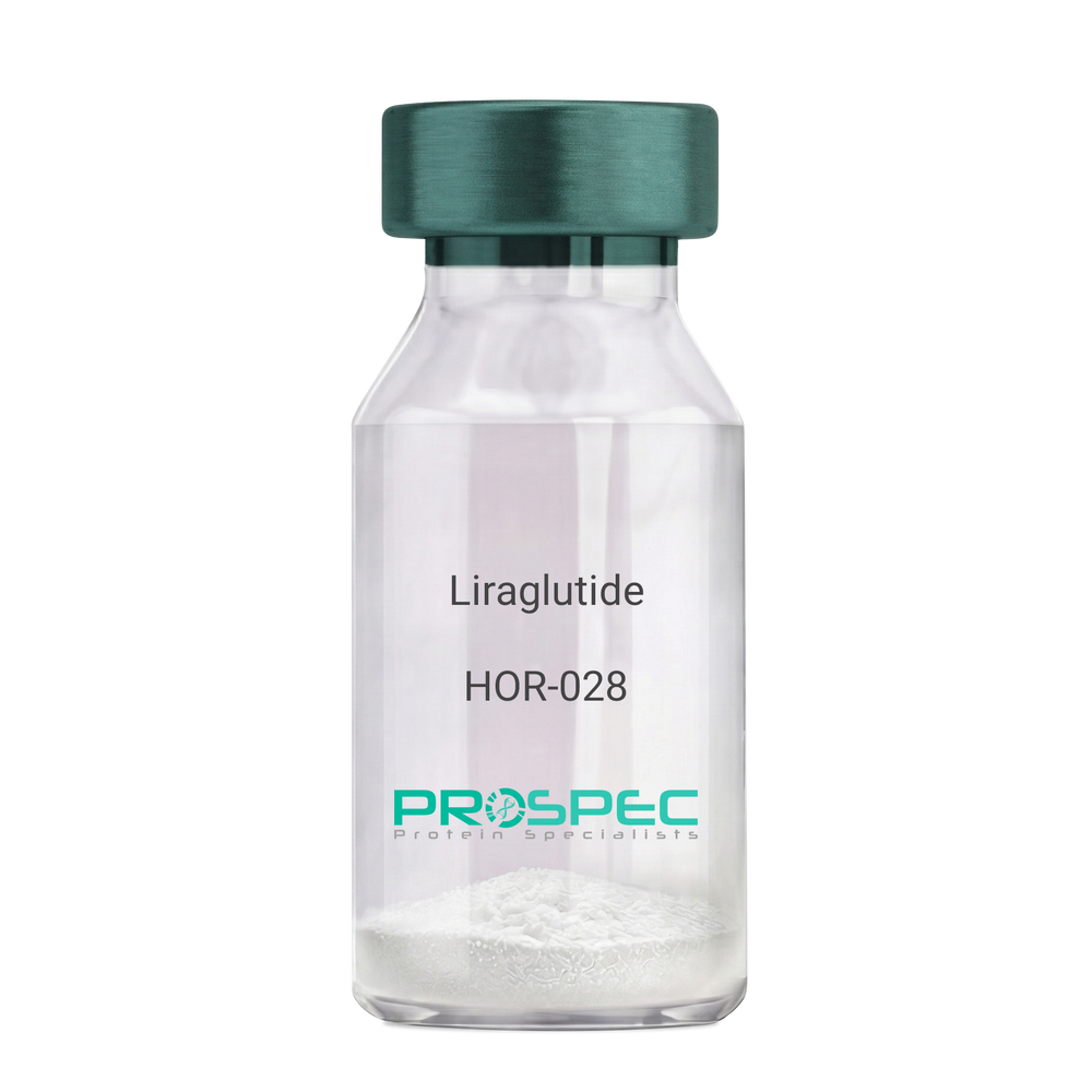
    Liraglutide

    Description:

    Liraglutide

    Product # :

    HOR-028

    Price :

    Quantity :

    Shipping Method :

    Room Temp Icon

    Shipped at Room temp

    Add To Cart

    More Info

    • description
    • formulation
    • purity
    • More Info
    • HPLC, MS

    Description

    Liraglutide peptide is a single, non-glycosylated polypeptide chain containing 31 amino acids, having a molecular mass of 3751 Dalton and a Molecular formula of C172H265N43O51.

    Formulation

    The protein (1mg/ml) was lyophilized with no additives.

    Purity

    Greater than 97% as determined by RP-HPLC.

    HPLC, MS

    liraglutide hplc - Product image 1
    liraglutide mass spec - Product image 2

    More Info

    • Introduction

      Liraglutide is a long-acting GLP1 analogue sharing 97% a.a. homology to human GLP1. Liraglutide differs from GLP1 by the substitution of arginine for lysine at position 34. While GLP1 is degraded swiftly by DPP4 resulting in short life of insulin, Liraglutide contains a fatty acid that binds to albumin and prolongs the half-life of the structure. liraglutide causes insulin to secret in the presence of increasing glucose levels. Due toits receptor site, liraglutide inhibits glucagon secretion and delays gastric emptying.

    • Physical Appearance

      Sterile Filtered White lyophilized (freeze-dried) powder.

    • Stability

      Store the lyophilized Liraglutide between 2-8°C, do not freeze. Upon reconstitution Lypressin should be stored at 4°C between 2-7 days and for future use below -18°C. Please prevent freeze-thaw cycles.

    • Solubility

      It is recommended to reconstitute the lyophilized Liraglutide in sterile 18MΩ-cm H2O not less than 100 µg/ml, which can then be further diluted to other aqueous solutions.
      Lypressin is also soluble in 1% Acetic Acid.

    • Amino Acid Sequence

      H-His-Ala-Glu-Gly-Thr-Phe-Thr-Ser-Asp-Val-Ser-Ser-Tyr-Leu-Glu-Gly-Gln-Ala-Ala-Lys(γ-Glu-palmitoyl)-Glu-Phe-Ile-Ala-Trp-Leu-Val-Arg-Gly-Arg-Gly-OH

    ProSpec's products are furnished for LABORATORY RESEARCH USE ONLY. The product may not be used as drugs, agricultural or pesticidal products, food additives or household chemicals.

    Liraglutide
  • View Data Sheet

    Name :

    HMGA2 Human

    Description:

    High Mobility Group AT-Hook 2 Human Recombinant

    High Mobility Group AT-Hook 2, HMGIC, High-Mobility Group (Nonhistone Chromosomal) Protein Isoform I-C, High Mobility Group AT-Hook Protein, BABL, HMGI-C, LIPO, STQTL9, High Mobility Group Protein HMGI-C, HMGA2.

    Product # :

    PRO-1469

    Price :

    Quantity :

    Shipping Method :

    Ice Icon

    Shipped with Ice Packs

    Add To Cart

    More Info

    • description
    • source
    • formulation
    • purity
    • More Info

    Description

    HMGA2 Human Recombinant produced in E.Coli is a single, non-glycosylated polypeptide chain containing 117 amino acids (1-109 a.a) and having a molecular mass of 12.8kDa.HMGA2 is fused to an 8 amino acid His-tag at C-terminus & purified by proprietary chromatographic techniques.

    Source

    Escherichia Coli.

    Formulation

    HMGA2 protein solution (0.25mg/ml) containing 20mM Tris-HCl buffer (pH 8.0), 0.2M NaCl, 50% glycerol, 2mM DTT and 250mM Imidazole.

    Purity

    Greater than 80.0% as determined by SDS-PAGE.

    More Info

    • Introduction

      High Mobility Group AT-Hook 2 (HMGA2) is a member of the non-histone chromosomal high mobility group (HMG) family. HMG proteins perform as architectural factors and are necessary components of the enhancesome. Thisprotein contains structural DNA-binding domains and may function as a transcriptional regulating factor. In addition, identification ofthe deletion, amplification, and rearrangement of this gene that are associated with myxoid liposarcoma proposes arole in adipogenesis and mesenchymal differentiation. A gene knock out study of the mouse counterpartdemonstrated that this gene is implicated in diet-induced obesit.Among the Diseases associated with HMGA2 are diffuse lipomatosis, and 12q14 microdeletion syndrome.

    • Synonyms

      High Mobility Group AT-Hook 2, HMGIC, High-Mobility Group (Nonhistone Chromosomal) Protein Isoform I-C, High Mobility Group AT-Hook Protein, BABL, HMGI-C, LIPO, STQTL9, High Mobility Group Protein HMGI-C, HMGA2.

    • Physical Appearance

      Sterile Filtered clear solution.

    • Stability

      Store at 4°C if entire vial will be used within 2-4 weeks. Store, frozen at -20°C for longer periods of time. For long term storage it is recommended to add a carrier protein (0.1% HSA or BSA).Avoid multiple freeze-thaw cycles.

    • Amino Acid Sequence

      MSARGEGAGQ PSTSAQGQPA APAPQKRGRG RPRKQQQEPT GEPSPKRPRG RPKGSKNKSP SKAAQKKAEA TGEKRPRGRP RKWPQQVVQK KPAQEETEET SSQESAEEDL EHHHHHH.

    ProSpec's products are furnished for LABORATORY RESEARCH USE ONLY. The product may not be used as drugs, agricultural or pesticidal products, food additives or household chemicals.

    Hmga2 Human
  • View Data Sheet

    Name :

    KAAG1 Human

    Description:

    Kidney Associated Antigen 1 Human Recombinant

    Kidney-associated antigen 1, RU2 antisense gene protein, KAAG1, RU2AS, MGC78738.

    Product # :

    PRO-192

    Price :

    Quantity :

    Shipping Method :

    Ice Icon

    Shipped with Ice Packs

    Add To Cart

    More Info

    • description
    • source
    • formulation
    • purity
    • More Info

    Description

    KAAG1 Human Recombinant fused with a 20 amino acid His tag at N-terminus produced in E.Coli is a single, non-glycosylated, polypeptide chain containing 104 amino acids (1-84 a.a.) and having a molecular mass of 11.1kDa. The KAAG1 is purified by proprietary chromatographic techniques.

    Source

    Escherichia Coli.

    Formulation

    The KAAG1 solution (0.5 mg/ml) contains 20mM Tris-HCl buffer (pH 8.0), 1mM DTT, 10% glycerol and 100mM NaCl.

    Purity

    Greater than 90.0% as determined by SDS-PAGE.

    More Info

    • Introduction

      Kidney associated antigen 1 (KAAG1) is expressed in testis and kidney, and, at lower levels, in urinary bladder and liver. KAAG1 is expressed by numerous tumors of various histologic origin, including melanomas, sarcomas and colorectal carcinomas.

    • Synonyms

      Kidney-associated antigen 1, RU2 antisense gene protein, KAAG1, RU2AS, MGC78738.

    • Physical Appearance

      Sterile Filtered colorless solution.

    • Stability

      Store at 4°C if entire vial will be used within 2-4 weeks. Store, frozen at -20°C for longer periods of time. For long term storage it is recommended to add a carrier protein (0.1% HSA or BSA).Avoid multiple freeze-thaw cycles.

    • Amino Acid Sequence

      MGSSHHHHHH SSGLVPRGSH MDDDAAPRVE GVPVAVHKHA LHDGLRQVAG PGAAAAHLPR WPPPQLAASR REAPPLSQRP HRTQGAGSPP ETNEKLTNPQ VKEK.

    ProSpec's products are furnished for LABORATORY RESEARCH USE ONLY. The product may not be used as drugs, agricultural or pesticidal products, food additives or household chemicals.

    Kaag1 Human
  • View Data Sheet

    Name :

    RGS2 Human

    Description:

    Regulator of G-Protein Signaling 2 Human Recombinant

    Regulator of G-protein Signaling 2 24kDa, G0S8, G0/G1 switch regulatory protein 8, Cell growth-inhibiting gene 31 protein, RGS2.

    Product # :

    PRO-182

    Price :

    Quantity :

    Shipping Method :

    Ice Icon

    Shipped with Ice Packs

    Add To Cart

    More Info

    • description
    • source
    • formulation
    • purity
    • More Info

    Description

    RGS2 produced in E.Coli is a single, non-glycosylated polypeptide chain containing 231 amino acids (1-211a.a.) and having a molecular mass of 26.5 kDa. RGS2 is fused to a 20 amino acid His-tag at N-terminus & purified by proprietary chromatographic techniques.

    Source

    Escherichia Coli.

    Formulation

    The RGS2 protein solution (0.25mg/1ml) is formulated in 20mM Tris-HCl buffer (pH8.0), 100mM NaCl, 2mM DTT and 10% glycerol.

    Purity

    Greater than 90.0% as determined by SDS-PAGE.

    More Info

    • Introduction

      RGS2 belongs to the RGS family which are regulatory molecules that act as GTPase activating proteins for G alpha subunits of heterotrimeric G proteins. RGS2 inhibits signal transduction by increasing the GTPase activity of G protein alpha subunits and as a result driving them into their inactive GDP-bound form.

    • Synonyms

      Regulator of G-protein Signaling 2 24kDa, G0S8, G0/G1 switch regulatory protein 8, Cell growth-inhibiting gene 31 protein, RGS2.

    • Physical Appearance

      Sterile filtered colorless solution.

    • Stability

      Store at 4°C if entire vial will be used within 2-4 weeks. Store, frozen at -20°C for longer periods of time. For long term storage it is recommended to add a carrier protein (0.1% HSA or BSA).Avoid multiple freeze-thaw cycles.

    • Amino Acid Sequence

      MGSSHHHHHH SSGLVPRGSH MQSAMFLAVQ HDCRPMDKSA GSGHKSEEKR EKMKRTLLKD WKTRLSYFLQ NSSTPGKPKT GKKSKQQAFI KPSPEEAQLW SEAFDELLAS KYGLAAFRAF LKSEFCEENI EFWLACEDFK KTKSPQKLSS KARKIYTDFI EKEAPKEINI DFQTKTLIAQ NIQEATSGCF TTAQKRVYSL MENNSYPRFL ESEFYQDLCK KPQITTEPHA T

    ProSpec's products are furnished for LABORATORY RESEARCH USE ONLY. The product may not be used as drugs, agricultural or pesticidal products, food additives or household chemicals.

    Rgs2 Human
  • View Data Sheet

    Name :

    Protein A/G

    Description:

    Protein A/G Recombinant

    Product # :

    PRO-646

    Price :

    Quantity :

    Shipping Method :

    Room Temp Icon

    Shipped at Room temp

    Add To Cart

    More Info

    • description
    • source
    • formulation
    • purity
    • More Info

    Description

    The recombinant Protein A/G consists of 5 IgG-binding regions of protein A and 2 of protein G, which corresponds to the Protein A and G domains that are included in the recombinant sequence. Cell wall binding region, cell membrane binding region and albumin binding region have been removed from the recombinant Protein A/G to ensure the maximum specific IgG binding. The Protein A portion is from Staphylococcus aureus segments E, D, A, B and C. The Protein G portion is from Streptococcus segments C1 and C3. The fusion protein has a predicted molecular mass of 47.7kDa and containing 429 amino acids.

    Source

    Escherichia coli.

    Formulation

    Lyophilized white Powder containing no additives.

    Purity

    >97% as determined by SDS-PAGE and RP-HPLC.

    More Info

    • Introduction

      Recombinant Protein A/G fusion protein joins IgG binding domains of both Protein A and Protein G.
      Protein A/G includes four Fc binding domains from Protein A and two from Protein G, yielding a final mass of 50.4 kDa. The binding dependency to pH of Protein A/G lower than Protein A, but has the additive properties of Protein A and G together. Protein A/G binds to all subclasses of human IgG, making it helpful for purifying polyclonal or monoclonal IgG antibodies whose subclasses have not been identifieed. Protein A/G binds to IgA, IgE, IgM and IgD. Protein A/G binds to all subclasses of mouse IgG excluding mouse IgA, IgM or serum albumin. This permits Protein A/G to be used in purification and detection of mouse monoclonal IgG antibodies, with no interference from IgA, IgM and serum albumin. Mouse monoclonal antibodies normally have a stronger affinity to the chimeric Protein A/G than to either Protein A or Protein G. Protein A/G also has been used for purification of macaque IgG.

    • Stability

      After reconstitution, aliquot and store at -20°C. Avoid repeated freeze/thaw cycles.

    • Solubility

      It is recommended to reconstitute the lyophilized Protein-A/G in sterile 18M-cm H2O not less than 0.1mg/ml, which can then be further diluted to other aqueous solutions.

    • Amino Acid Sequence

      NAAQHDEAQQ NAFYQVLNMP NLNADQRNGF IQSLKDDPSQ SANVLGEAQK LNDSQAPKAD AQQNNFNKDQ QSAFYEILNM PNLNEAQRNG FIQSLKDDPS QSTNVLGEAK KLNESQAPKA DNNFNKEQQN AFYEILNMPN LNEEQRNGFI QSLKDDPSQS ANLLSEAKKL NESQAPKADN KFNKEQQNAF YEILHLPNLN EEQRNGFIQS LKDDPSQSAN LLAEAKKLND AQAPKADNKF NKEQQNAFYE ILHLPNLTEE QRNGFIQSLK DDPSVSKEIL AEAKKLNDAQ APKEEDSLEG SGSGTYKLIL NGKTLKGETT TEAVDAATAE KVFKQYANDN GVDGEWTYDD ATKTFTVTEK PEVIDASELT PAVTTYKLVI NGKTLKGETT TKAVDAETAE KAFKQYANDN GVDGVWTYDD ATKTFTVTE.

    ProSpec's products are furnished for LABORATORY RESEARCH USE ONLY. The product may not be used as drugs, agricultural or pesticidal products, food additives or household chemicals.

    Protein A G
  • View Data Sheet

    Name :

    Protein G

    Description:

    Protein G Recombinant

    Product # :

    PRO-402

    Price :

    Quantity :

    Shipping Method :

    Room Temp Icon

    Shipped at Room temp

    Add To Cart

    More Info

    • description
    • source
    • formulation
    • purity
    • More Info
    • sds-page

    Description

    The Protein G is a single, non-glycosylated protein contains 200 amino acids having a molecular mass of 21.8kDa. The Protein-G migrates on SDS-PAGE around 32kDa.

    Source

    Escherichia Coli.

    Formulation

    Lyophilized white powder containing no additives.

    Purity

    >96% as determined by SDS-PAGE and RP-HPLC.

    sds-page

    Protein-G sds-page - Product image 1

    More Info

    • Physical Appearance

      Sterile Filtered White lyophilized (freeze-dried) powder.

    • Stability

      Lyophilized Recombinant Protein G although stable at room temperature for 3 weeks, should be stored desiccated below -18°C. Upon reconstitution Protein G should be stored at 4°C between 2-7 days and for future use below -18°C.Please prevent freeze-thaw cycles.

    • Solubility

      Reconstitution with deionized water or PBS.

    • Amino Acid Sequence

      LPKTDTYKLILNGKTLKGETTTEAVDAATAEKVFKQYANDNGVDGEWTYDDAT KTFTVTEKPEVIDASELTPAVTTYKLVINGKTLKGETTTEAVDAATAEKVFK QYANDNGVDGEWTYDDATKTFTVTEKPEVIDASELTPAVTTYKLVINGKTL KGETTTKAVDAETAEKAFKQYANDNGVDGVWTYDDATKTFTVTE.

    ProSpec's products are furnished for LABORATORY RESEARCH USE ONLY. The product may not be used as drugs, agricultural or pesticidal products, food additives or household chemicals.

    Protein G
  • View Data Sheet

    Name :

    T.pallidum p15

    Description:

    Treponema pallidum p15 Recombinant

    Product # :

    TRP-246

    Price :

    Quantity :

    Shipping Method :

    Ice Icon

    Shipped with Ice Packs

    Add To Cart

    More Info

    • description
    • source
    • formulation
    • purity
    • More Info

    Description

    The E.Coli derived recombinant protein contains the Trp. Pallidum p15 immunodominant regions. The protein contains beta-galactosidase (114 kDa) fused at the N-terminus.

    Source

    Escherichia Coli.

    Formulation

    20mM Tris-HCl pH-8, 10mM B-ME and 8M urea.

    Purity

    Treponema Pallidum protein is >95% pure as determined by 10% PAGE (coomassie staining).

    More Info

    • Introduction

      Treponema pallidum is a gram-negative spirochaete bacterium and is considered to be metabolically crippled. There are at least four known subspecies: T. pallidum pallidum, T. pallidum pertenue, T. pallidum carateum and T. pallidum endemicum. The helical structure of T. pallidum pallidum allows it to move in a corkscrew motion through viscous mediums such as mucus. Treponema pallidum sub sp. pallidum has one of the smallest bacterial genomes at 1.14 million base pairs (Mb) and has limited metabolic capabilities, reflecting its adaptation through genome reduction to the rich environment of mammalian tissue.

    • Stability

      Protein should be stored at 4°C.Refrigerate Upon arrival. DO NOT FREEZE.

    • Applications

      Treponema Pallidum protein is suitable for ELISA and Western blots, excellent antigen for detection of Trp. Pallidum with minimal specificity problems.

    • Specificity

      Immunoreactive with sera of Trp. Pallidum infected individuals.

    • Purification Method

      Treponema Pallidum protein was purified by proprietary chromatographic technique.

    ProSpec's products are furnished for LABORATORY RESEARCH USE ONLY. The product may not be used as drugs, agricultural or pesticidal products, food additives or household chemicals.

    Tpallidum P15
  • View Data Sheet

    Name :

    HBcAg (1-149)

    Description:

    Hepatitis B Virus Core (1-149 a.a) Recombinant

    Product # :

    HBV-234

    Price :

    Quantity :

    Shipping Method :

    Ice Icon

    Shipped with Ice Packs

    Add To Cart

    More Info

    • description
    • source
    • formulation
    • purity
    • More Info

    Description

    HBcAg subtype adw2 produced in E.Coli, is a single, non-glycosylated, polypeptide chain containing 1-149 amino acids and having a molecular weight of approximately 20kDa. HBcAg adw2 1-149 protamine-like domain was truncated in order to enhance the differentiation of anti-HBe in ELISA test. HBcAg is fused to a his tag and purified by proprietary chromatographic techniques.

    Source

    Escherichia Coli.

    Formulation

    Tris base and 10mM K2CO3.

    Purity

    Greater than 90% as determined by SDS-PAGE.

    More Info

    • Introduction

      Hepatitis B is one of a few known non-retroviralviruses which employ reverse transcriptionas a part of its replication process. (HIV, a completely unrelated virus, also uses reverse transcription, but it is a retrovirus.) HBV invades the cell by binding to surface receptor and become internalized. The viral core particles then migrate to the hepatocyte nucleus and the partially double-stranded, relaxed circular genomes (RC-DNA) are repaired to form a covalently closed circular DNA (cccDNA), which is the template for viral genomic and sub-genomic RNAs by cellular RNA polymerase II. Of these, the pregenomic RNA(pgRNA is selectively packaged into progeny capsids and is then reverse-transcribed into new RC-DNA. The core can either bud into the endoplasmic reticulum to be enveloped or exported from the cell or recycled back into the genome for conversion to cccDNA.

    • Physical Appearance

      Sterile filtered colorless solution.

    • Stability

      Store at 4°C if entire vial will be used within 2-4 weeks. Store, frozen at -20°C for longer periods of time.For long term storage it is recommended to add a carrier protein (0.1% HSA or BSA). Avoid multiple freeze-thaw cycles.

    • Amino Acid Sequence

      MDIDPYKEFG ATVELLSFLP SDFFPSVRDL LDTASALYRE ALESPEHCSP HHTALRQAIL CWGELMTLAT WVGNNLEDPA SRDLVVNYVN TNVGLKIRQL LWFHISCLTF GRETVLEYLV SFGVWIRTPP AYRPPNAPIL

    ProSpec's products are furnished for LABORATORY RESEARCH USE ONLY. The product may not be used as drugs, agricultural or pesticidal products, food additives or household chemicals.

    Hbcag 1 149 Aa
  • View Data Sheet

    Name :

    T.pallidum p41

    Description:

    Treponema pallidum p41 Recombinant

    Product # :

    TRP-242

    Price :

    Quantity :

    Shipping Method :

    Ice Icon

    Shipped with Ice Packs

    Add To Cart

    More Info

    • description
    • source
    • formulation
    • purity
    • More Info

    Description

    The E.coli derived recombinant protein contains the outer membrane T.Pallidum p41 immunodominant regions. The protein is fused with a Beta-galactosidase tag.

    Source

    Escherichia Coli.

    Formulation

    10mM Tris-HCl pH8.0, 1mM EDTA, 1mMDTT and 8M urea.

    Purity

    Treponema pallidum protein is >90% pure as determined by 10% PAGE (coomassie staining).

    More Info

    • Introduction

      Treponema pallidum is a gram-negative spirochaete bacterium and is considered to be metabolically crippled. There are at least four known subspecies: T. pallidum pallidum, T. pallidum pertenue, T. pallidum carateum and T. pallidum endemicum. The helical structure of T. pallidum pallidum allows it to move in a corkscrew motion through viscous mediums such as mucus. Treponema pallidum sub sp. pallidum has one of the smallest bacterial genomes at 1.14 million base pairs (Mb) and has limited metabolic capabilities, reflecting its adaptation through genome reduction to the rich environment of mammalian tissue.

    • Stability

      T.pallidum p41 although stable at 4°C for 1 week, should be stored below -18°C. Please prevent freeze thaw cycles.

    • Applications

      Treponema pallidum is suitable for ELISA and Western blots, excellent antigen for detection of T. Pallidum with minimal specificity problems.

    • Specificity

      Immunoreactive with sera of T.Pallidum infected individuals.

    • Purification Method

      Treponema pallidum protein was purified by proprietary chromatographic technique.

    ProSpec's products are furnished for LABORATORY RESEARCH USE ONLY. The product may not be used as drugs, agricultural or pesticidal products, food additives or household chemicals.

    Tpallidum P41
  • View Data Sheet

    Name :

    Ang K1-3 Human

    Description:

    Angiostatin Kringles 1-3 Human Recombinant

    Angiostatin, Angiostatin Kringles 1-3, Ang K1-3.

    Product # :

    PRO-284

    Price :

    Quantity :

    Shipping Method :

    Room Temp Icon

    Shipped at Room temp

    Add To Cart

    More Info

    • description
    • source
    • formulation
    • purity
    • biological activity
    • More Info

    Description

    Angiostatin Human Recombinant produced in E.coli is a single, non-glycosylated polypeptide chain containing 259 amino acids and having a molecular mass of approximately 29.7 kDa. The Ang K1-3 is purified by proprietary chromatographic techniques.

    Source

    Escherichia coli.

    Formulation

    Lyophilized from a 0.2μm filtered concentrated (1.0mg/ml) solution in 20mM NaAc, pH5.5, 4% mannitol.

    Purity

    Greater than 95.0% as determined by:
    (a) Analysis by RP-HPLC.
    (b) Analysis by SDS-PAGE.

    Biological Activity

    The activity is assayed on anti-proliferation and anti-migration of endothelial cells in vitro and anti-angiogenesis in vivo. The specific activity of anti-migration of endothelial cells in vitro is 550,000 Units/mg.

    More Info

    • Introduction

      Ang K1-3 is a proteolytic fragment of plasminogen containing the first three kringle structures. A specific inhibitor of endothelial cell growth and angiogenesis. More active relative to kringles 1-4. Ang K1-3 reduces endothelial cell proliferation and acts as a potent inhibitor of angiogenesis and tumor growth.

    • Synonyms

      Angiostatin, Angiostatin Kringles 1-3, Ang K1-3.

    • Physical Appearance

      Sterile Filtered White lyophilized (freeze-dried) powder.

    • Stability

      The lyophilized Angiostatin K1-3 is stable for several weeks at 2-8°C, but should be kept at -20°C for long term storage, preferably desiccated. Upon reconstitution, the preparation is stable for up to one week at 2-8°C. For maximal stability, apportion the reconstituted preparation into working aliquots and store at -20°C to -70°C. Avoid repeated freeze/thaw cycles.

    • Solubility

      We recommend to briefly centrifuge the vial prior to opening to bring the contents to the bottom. Reconstitute in sterile distilled water or aqueous buffer containing 0.1% BSA to a concentration of 0.1-1.0 mg/ml. Stock solutions should be apportioned into working aliquots and stored at <-20°C. Further dilutions should be made in appropriate buffered solutions.

    • Amino Acid Sequence

      VYLSECKTGN GKNYRGTMSK TKNGITCQKW SSTSPHRPRF SPATHPSEGL EENYCRNPDN DPQGPWCYTT DPEKRYDYCD ILECEEECMH CSGENYDGKI SKTMSGLECQ AWDSQSPHAH GYIPSKFPNK NLKKNYCRNP DRELRPWCFT TDPNKRWELC DIPRCTTPPP SSGPTYQCLKGTGENYRGNV AVTVSGHTCQ HWSAQTPHTH NRTPENFPCK NLDENYCRNP DGKRAPWCHT TNSQVRWEYC KIPSCDSSP.

    ProSpec's products are furnished for LABORATORY RESEARCH USE ONLY. The product may not be used as drugs, agricultural or pesticidal products, food additives or household chemicals.

    Angiostatin K1 3 Human
  • View Data Sheet

    Name :

    P Selectin Human

    Description:

    P-selectin Human Recombinant

    P-selectin, Granule membrane protein 140, GMP-140, PADGEM, Leukocyte-endothelial cell adhesion molecule 3, LECAM3, CD62 antigen-like family member P, CD62P antigen, SELP, GMRP, GRMP, CD62, PSEL, CD62P, GMP140, FLJ45155.

    Product # :

    PRO-382

    Price :

    Quantity :

    Shipping Method :

    Ice Icon

    Shipped with Ice Packs

    Add To Cart

    More Info

    • description
    • source
    • formulation
    • purity
    • More Info

    Description

    P-Selectin Human Recombinant is expressed in E. coli containing 566 amino acids 197-761 fused to an amino terminal hexahistidine tag.

    Source

    Escherichia Coli.

    Formulation

    P-Selectin is supplied in 50% Glycerol.

    Purity

    Greater than 95% as determined by SDS-PAGE.
    Single band on Western Blot.

    More Info

    • Introduction

      P-Selectin also called Platelet Alpha-Granule Membrane Protein, CD62, and Granulocyte Membrane Protein GRMP belongs to a family of divalent cation-dependent carbohydrate-binding glycoproteins or adhesion molecules. Pselectin is expressed transiently on the surface of activated platelets and endothelial cells. P-Selectin is a 140 kDa protein which is stored in the alpha-granules of platelets and Weibel-Palade bodies of endothelial cells. Secreted P-selectin is thought to play a key role in the adhesion of platelets to monocytes and neutrophils during an inflammatory response. P-Selectin is a calcium-dependent receptor that binds to sialylated forms of Lewis blood group carbohydrate antigens on neutrophils and monocytes. Levels of P-Selectin may be elevated in a number of pathological conditions.

    • Synonyms

      P-selectin, Granule membrane protein 140, GMP-140, PADGEM, Leukocyte-endothelial cell adhesion molecule 3, LECAM3, CD62 antigen-like family member P, CD62P antigen, SELP, GMRP, GRMP, CD62, PSEL, CD62P, GMP140, FLJ45155.

    • Physical Appearance

      Sterile Filtered liquid formulation.

    • Stability

      Store at 4°C if entire vial will be used within 1-2 weeks. Store, frozen at -20°C for longer periods of time. Avoid multiple freeze-thaw cycles.

    • Applications

      P-Selectin can be used directly as a positive control in Western blotting, ELISA, immunoprecipitation and other immunological experiments.
      The biological activity of this product has not yet been tested.

    ProSpec's products are furnished for LABORATORY RESEARCH USE ONLY. The product may not be used as drugs, agricultural or pesticidal products, food additives or household chemicals.

    P Selectin Human
  • View Data Sheet

    Name :

    PSMB1 Human

    Description:

    Proteasome Subunit Beta Type 1 Human Recombinant

    HC5, PSC5, PMSB1, FLJ25321, KIAA1838, PSMB1, Proteasome subunit beta type-1, Proteasome component C5, Macropain subunit C5, Multicatalytic endopeptidase complex subunit C5, Proteasome gamma chain.

    Product # :

    ENZ-405

    Price :

    Quantity :

    Shipping Method :

    Ice Icon

    Shipped with Ice Packs

    Add To Cart

    More Info

    • description
    • source
    • formulation
    • purity
    • More Info

    Description

    PSMB1 Human Recombinant fused to a 37 amino acid His Tag at N-terminal produced in E.Coli is a single, non-glycosylated, polypeptide chain containing 250 amino acids (30-241) and having a molecular mass of 27.7 kDa. The PSMB1 is purified by proprietary chromatographic techniques.

    Source

    Escherichia Coli.

    Formulation

    The PSMB1 solution contains 20mM Tris-HCl pH-8, 1mM DTT and 10% glycerol.

    Purity

    Greater than 95.0% as determined by SDS-PAGE.

    More Info

    • Introduction

      PSMB1 is part of the proteasome B-type family, also identified as the T1B family, that is a 20S core beta subunit. PSMB1 is tightly linked to the TBP (TATA-binding protein) gene in human and in mouse, and is transcribed in the opposite orientation in both species.The main function of PSMB1 is its degradation activity of unnecessary or damaged proteins by proteolysis. PSMB1 is a multicatalytic proteinase complex which is characterized by its ability to cleave peptides with arg, phe, tyr, leu, and glu adjacent to the leaving group at neutral or slightly basic ph. the proteasome has an atp-dependent proteolytic activity.

    • Synonyms

      HC5, PSC5, PMSB1, FLJ25321, KIAA1838, PSMB1, Proteasome subunit beta type-1, Proteasome component C5, Macropain subunit C5, Multicatalytic endopeptidase complex subunit C5, Proteasome gamma chain.

    • Physical Appearance

      Sterile Filtered clear colorless solution.

    • Stability

      Store at 4°C if entire vial will be used within 2-4 weeks. Store, frozen at -20°C for longer periods of time. For long term storage it is recommended to add a carrier protein (0.1% HSA or BSA).Avoid multiple freeze-thaw cycles.

    • Amino Acid Sequence

      MRGSHHHHHH GMASMTGGQQ MGRDLYDDDD KDRWGSHMFS PYVFNGGTIL AIAGEDFAIV ASDTRLSEGF SIHTRDSPKC YKLTDKTVIG CSGFHGDCLT LTKIIEARLK MYKHSNNKAM TTGAIAAMLS TILYSRRFFP YYVYNIIGGL DEEGKGAVYS FDPVGSYQRD SFKAGGSASA MLQPLLDNQV GFKNMQNVEH VPLSLDRAMR LVKDVFISAA ERDVYTGDAL RICIVTKEGI REETVSLRKD.

    ProSpec's products are furnished for LABORATORY RESEARCH USE ONLY. The product may not be used as drugs, agricultural or pesticidal products, food additives or household chemicals.

    Psmb1 Human
  • View Data Sheet

    Name :

    PSMB6 Human

    Description:

    Proteasome Subunit Beta Type 6 Human Recombinant

    Proteasome subunit beta type-6, Macropain delta chain, Multicatalytic endopeptidase complex delta chain, Proteasome delta chain, Proteasome subunit Y, PSMB6, LMPY, Y, DELTA.

    Product # :

    ENZ-223

    Price :

    Quantity :

    Shipping Method :

    Ice Icon

    Shipped with Ice Packs

    Add To Cart

    More Info

    • description
    • source
    • formulation
    • purity
    • More Info

    Description

    PSMB6 Human Recombinant produced in E.coli is a single, non-glycosylated polypeptide chain containing 226 amino acids (35-239) and having a molecular mass of 24.2kDa.PSMB6 is fused to a 21 amino acid His-tag at N-terminus & purified by proprietary chromatographic techniques.

    Source

    Escherichia Coli.

    Formulation

    The PSMB6 solution (1mg/ml) contains 20mM Tris-HCl buffer (pH 8.0), 10% glycerol, 1mM DTT and 0.15M NaCl.

    Purity

    Greater than 85.0% as determined by SDS-PAGE.

    More Info

    • Introduction

      PSMB6 belongs to the proteasome B-type family (T1B family) which is a 20S core beta subunit in the proteasome. The PSMB6 is not present in the immunoproteasome and is replaced by PSMB9. The proteasome is a multicatalytic proteinase complex with an extremely ordered ring-shaped 20S core structure. The core structure is comprised of 4 rings of 28 non-identical subunits; 2 rings are composed of 7 alpha subunits and 2 rings are composed of 7 beta subunits.

    • Synonyms

      Proteasome subunit beta type-6, Macropain delta chain, Multicatalytic endopeptidase complex delta chain, Proteasome delta chain, Proteasome subunit Y, PSMB6, LMPY, Y, DELTA.

    • Physical Appearance

      Sterile Filtered colorless solution.

    • Stability

      Store at 4°C if entire vial will be used within 2-4 weeks. Store, frozen at -20°C for longer periods of time. For long term storage it is recommended to add a carrier protein (0.1% HSA or BSA).Avoid multiple freeze-thaw cycles.

    • Amino Acid Sequence

      MGSSHHHHHH SSGLVPRGSH MTTIMAVQFD GGVVLGADSR TTTGSYIANR VTDKLTPIHD RIFCCRSGSA ADTQAVADAV TYQLGFHSIE LNEPPLVHTA ASLFKEMCYR YREDLMAGII IAGWDPQEGG QVYSVPMGGM MVRQSFAIGG SGSSYIYGYV DATYREGMTK EECLQFTANA LALAMERDGS SGGVIRLAAI AESGVERQVL LGDQIPKFAV ATLPPA.

    ProSpec's products are furnished for LABORATORY RESEARCH USE ONLY. The product may not be used as drugs, agricultural or pesticidal products, food additives or household chemicals.

    Psmb6 Human
  • View Data Sheet

    Name :

    T.pallidum TmpA partial

    Description:

    Treponema pallidum TmpA (partial) Recombinant

    Product # :

    TRP-245

    Price :

    Quantity :

    Shipping Method :

    Ice Icon

    Shipped with Ice Packs

    Add To Cart

    More Info

    • description
    • source
    • formulation
    • purity
    • More Info

    Description

    The E.Coli derived 68kDa recombinant protein contains the Trp. Pallidum TmpA immunodominant regions, 23-41 amino acids and 288-325 amino acids.

    Source

    Escherichia Coli.

    Formulation

    10mM Tris-HCl pH8.0, 20mM DDT, 1mM EDTA and 8M urea.

    Purity

    Protein is >95% pure as determined by 10% PAGE (coomassie staining).

    More Info

    • Introduction

      Treponema pallidum is a gram-negative spirochaete bacterium and is considered to be metabolically crippled. There are at least four known subspecies: T. pallidum pallidum, T. pallidum pertenue, T. pallidum carateum and T. pallidum endemicum. The helical structure of T. pallidum pallidum allows it to move in a corkscrew motion through viscous mediums such as mucus. Treponema pallidum sub sp. pallidum has one of the smallest bacterial genomes at 1.14 million base pairs (Mb) and has limited metabolic capabilities, reflecting its adaptation through genome reduction to the rich environment of mammalian tissue.

    • Stability

      T.pallidum TmpA partial although stable at 4°C for 1 week, should be stored below -18°C. Please prevent freeze thaw cycles.

    • Applications

      This protein binds to murine anti-TmpA protein monoclonal antibodies and Treponema pallidum converted human serum polyclonal antibodies in ELISA and Western Elisa. Dot Blots and Lateral Flow immunochromatographic diagnostics test.

    • Specificity

      Immunoreactive with sera of Trp. Pallidum infected individuals.

    • Purification Method

      Purified by proprietary chromatographic technique.

    ProSpec's products are furnished for LABORATORY RESEARCH USE ONLY. The product may not be used as drugs, agricultural or pesticidal products, food additives or household chemicals.

    Tpallidum Tmpa Partial
  • View Data Sheet

    Name :

    PA2G4 Antibody

    Description:

    Proliferation-associated protein 2G4, Mouse Anti Human

    Proliferation-associated protein 2G4, Cell cycle protein p38-2G4 homolog, hG4-1, ErbB3-binding protein 1, PA2G4, EBP1, p38-2G4.

    Product # :

    ANT-655

    Price :

    Quantity :

    Shipping Method :

    Ice Icon

    Shipped with Ice Packs

    Add To Cart

    More Info

    • formulation
    • More Info

    Formulation

    1mg/ml containing PBS, pH-7.4, 10% Glycerol and 0.02% Sodium Azide.

    More Info

    • Introduction

      PA2G4 belongs to the peptidase M24C family and functions as an RNA-binding protein involved in cellular proliferation and differentiation processes. PA2G4 is a component of pre-ribosomal ribonucleoprotein complexes, participating in ribosome assembly and regulating the later steps of rRNA processing. Also, PA2G4 interacts with ErbB-3 and may function as a modulator of the ErbB-3 mediated signal transduction pathway by regulating the effects of Neuregulin-1. Furthermore, PA2G4 is a transcriptional co-repressor of androgen receptor-regulated genes and other cell cycle regulatory genes through its interactions with histone deacetylases. PA2G4 is implicated in growth inhibition and the induction of differentiation of human cancer cells. In addition, PA2G4 mediates cap-independent translation of specific viral IRESs (internal ribosomal entry site). PA2G4 associates with 28S, 18S and 5.8S mature rRNAs, several rRNA precursors and probably U3 small nucleolar RNA.

    • Synonyms

      Proliferation-associated protein 2G4, Cell cycle protein p38-2G4 homolog, hG4-1, ErbB3-binding protein 1, PA2G4, EBP1, p38-2G4.

    • Physical Appearance

      Sterile filtered colorless solution.

    • Immunogen

      Anti-human PA2G4 mAb, is derived from hybridization of mouse F0 myeloma cells with spleen cells from BALB/c mice immunized with a recombinant human PA2G4 protein 1-394 amino acids purified from E. coli.

    • Ig Subclass

      Mouse IgG1 heavy chain and k light chain.

    • Clone

      PAT5E7AT.

    • Applications

      The antibody has been tested by ELISA, Western blot analysis, Flow cytometry and ICC/IF to assure specificity and reactivity. Since application varies, however, each investigation should be titrated by the reagent to obtain optimal results.

    • Type

      Mouse Anti Human Monoclonal.

    • Storage Procedures

      For periods up to 1 month store at 4°C, for longer periods of time, store at -20°C. Prevent freeze thaw cycles.

    • Purification Method

      PA2G4 antibody was purified from mouse ascitic fluids by protein-A affinity chromatography.

    ProSpec's products are furnished for LABORATORY RESEARCH USE ONLY. The product may not be used as drugs, agricultural or pesticidal products, food additives or household chemicals.

    Pa2G4 Antibody
  • View Data Sheet

    Name :

    PSMD11 PAT2C7AT Antibody

    Description:

    26S proteasome non-ATPase regulatory subunit 11 clone PAT2C7AT Mouse Anti Human

    26S proteasome non-ATPase regulatory subunit 11, 26S proteasome regulatory subunit S9, 26S proteasome regulatory subunit RPN6, 26S proteasome regulatory subunit p44.5, PSMD11, S9, Rpn6, p44.5, MGC3844.

    Product # :

    ANT-675

    Price :

    Quantity :

    Shipping Method :

    Ice Icon

    Shipped with Ice Packs

    Add To Cart

    More Info

    • formulation
    • More Info

    Formulation

    1mg/ml containing PBS, pH-7.4, 10% Glycerol and 0.02% Sodium Azide.

    More Info

    • Introduction

      The 26S proteasome is a multicatalytic proteinase complex with a well ordered structure composed of two complexes- a 20S core and a 19S regulator. PSMD11 is a non-ATPase subunit of the 19S regulator. The 20S core is composed of four rings of 28 non-identical subunits; two rings are composed of 7 alpha subunits and two rings are composed of 7 beta subunits. The 19S regulator is composed of a base that contains six ATPase subunits and two non-ATPase subunits, and a lid that contains up to ten non-ATPase subunits. Proteasomes are spread throughout eukaryotic cells at a high concentration and cleave peptides in an ATP/ubiquitin-dependent process in a non-lysosomal pathway. An indispensable function of a modified proteasome, the immunoproteasome, is the processing of class I MHC peptides.

    • Synonyms

      26S proteasome non-ATPase regulatory subunit 11, 26S proteasome regulatory subunit S9, 26S proteasome regulatory subunit RPN6, 26S proteasome regulatory subunit p44.5, PSMD11, S9, Rpn6, p44.5, MGC3844.

    • Physical Appearance

      Sterile filtered colorless solution.

    • Immunogen

      Anti-human PSMD11 mAb, is derived from hybridization of mouse F0 myeloma cells with spleen cells from BALB/c mice immunized with recombinant human PSMD11 PAT2C7AT amino acids 1-422 purified from E. coli.

    • Ig Subclass

      Mouse IgG2b heavy chain and ? light chain.

    • Clone

      PAT2C7AT.

    • Applications

      PSMD11 antibody has been tested by ELISA, Western blot analysis, Flow cytometry and ICC/IF to assure specificity and reactivity. Since application varies, however, each investigation should be titrated by the reagent to obtain optimal results.

    • Type

      Mouse Anti Human Monoclonal.

    • Storage Procedures

      For periods up to 1 month store at 4°C, for longer periods of time, store at -20°C. Prevent freeze thaw cycles.

    • Purification Method

      PSMD11 antibody was purified from mouse ascitic fluids by protein-A affinity chromatography.

    ProSpec's products are furnished for LABORATORY RESEARCH USE ONLY. The product may not be used as drugs, agricultural or pesticidal products, food additives or household chemicals.

    Psmd11 Pat2C7At Antibody
  • View Data Sheet

    Name :

    S100A8 Human

    Description:

    S100 Calcium Binding Protein A8 Human Recombinant

    Calgranulin A, MRP8, CAGA, CGLA, CFAG, Protein S100-A8, S100 calcium-binding protein A8, Migration inhibitory factor-related protein 8, MRP-8, p8, Cystic fibrosis antigen, Leukocyte L1 complex light chain, Calprotectin L1L subunit, Urinary stone protein band A, S100A8, MIF, NIF, L1Ag, CP-10, MA387, 60B8AG.

    Product # :

    PRO-800

    Price :

    Quantity :

    Shipping Method :

    Ice Icon

    Shipped with Ice Packs

    Add To Cart

    More Info

    • description
    • source
    • formulation
    • purity
    • More Info

    Description

    S100A8 Human Recombinant produced in E.Coli is a single, non-glycosylated, polypeptide chain containing 93 amino acids (1-93 a.a.) and having a molecular mass of 10.8 kDa. The S100A8 is purified by proprietary chromatographic techniques.

    Source

    Escherichia Coli.

    Formulation

    The S100A8 solution (0.5mg/ml) contains 20mM Tris-HCl pH-8, 1mM DTT and 10% glycerol.

    Purity

    Greater than 90% as determined by SDS-PAGE.

    More Info

    • Introduction

      S100A8 is a part of the S100 family of proteins containing 2 EF-hand calcium-binding motifs. S100 proteins are localized in the cytoplasm and/or nucleus of a broad range of cells, and participate in the regulation of cellular processes such as cell cycle progression and differentiation. S100A8 plays a role in the inhibition of casein kinase and as a cytokine. S100A8 altered expression is related with cystic fibrosis disease. S100A8 is a calcium-binding protein that has antimicrobial activity against bacteria and fungi.S100A8 is crucial for resistance towards invasion by pathogenic bacteria. S100A8 up-regulates transcription of genes that are under the control of NF-kappa-B. S100A8 plays a role in the development of endotoxic shock in response to bacterial lipopolysaccharide. S100A8 endorses tubulin polymerization and promotes phagocyte migration and infiltration of granulocytes at sites of wounding. S100A8 takes part as a pro-inflammatory mediator in acute and chronic inflammation and up-regulates the release of IL8 and cell-surface expression of ICAM1.

    • Synonyms

      Calgranulin A, MRP8, CAGA, CGLA, CFAG, Protein S100-A8, S100 calcium-binding protein A8, Migration inhibitory factor-related protein 8, MRP-8, p8, Cystic fibrosis antigen, Leukocyte L1 complex light chain, Calprotectin L1L subunit, Urinary stone protein band A, S100A8, MIF, NIF, L1Ag, CP-10, MA387, 60B8AG.

    • Physical Appearance

      Sterile Filtered clear colorless solution.

    • Stability

      Store at 4°C if entire vial will be used within 2-4 weeks.Store, frozen at -20°C for longer periods of time. For long term storage it is recommended to add a carrier protein (0.1% HSA or BSA).Avoid multiple freeze-thaw cycles.

    • Amino Acid Sequence

      MLTELEKALN SIIDVYHKYS LIKGNFHAVY RDDLKKLLET ECPQYIRKKG ADVWFKELDI NTDGAVNFQE FLILVIKMGV AAHKKSHEES HKE.

    ProSpec's products are furnished for LABORATORY RESEARCH USE ONLY. The product may not be used as drugs, agricultural or pesticidal products, food additives or household chemicals.

    S100A8 Human
  • View Data Sheet

    Name :

    ATG10 Human

    Description:

    Autophagy Related 10 Human Recombinant

    Autophagy Related Protein 10, ATG10 Autophagy Related 10 Homolog (S. Cerevisiae), Ubiquitin-Like-Conjugating Enzyme ATG10, APG10 Autophagy 10-Like (S. Cerevisiae), APG10-Like, APG10L, Pp12616, DKFZP586I0418, FLJ13954, EC 6.3.2.-.

    Product # :

    PRO-1234

    Price :

    Quantity :

    Shipping Method :

    Ice Icon

    Shipped with Ice Packs

    Add To Cart

    More Info

    • description
    • source
    • formulation
    • purity
    • More Info

    Description

    ATG10 Human Recombinant produced in E. coli is a single polypeptide chain containing 243 amino acids (1-220) and having a molecular mass of 27.7 kDa.ATG10 is fused to a 23 amino acid His-tag at N-terminus & purified by proprietary chromatographic techniques.

    Source

    E.coli.

    Formulation

    The ATG10 solution (1mg/1ml) contains 20mM Tris-HCl buffer (pH 8.0), 0.1M NaCl, 1mM DTT and 10% glycerol.

    Purity

    Greater than 95% as determined by SDS-PAGE.

    More Info

    • Introduction

      Ubiquitin-like-conjugating enzyme ATG10 (ATG10) is a 220 amino acid protein which localizes to the cytoplasm and has a role in autophagy, specifically acting as an E2-like enzyme providing Atg recognition sites during autophagosome synthesis. ATG10 functions as an E2-like enzyme which catalyzes the conjugation of ATG12 to ATG5, which is required for autophagy. In addition, ATG10 interacts with ATG12 in human embryonic kidney cells in the presence of ATG7. ATG10 probably serves as an ATG5-recognition molecule. Furthermore, ATG10 has a role in adenovirus-mediated cell lysis.

    • Synonyms

      Autophagy Related Protein 10, ATG10 Autophagy Related 10 Homolog (S. Cerevisiae), Ubiquitin-Like-Conjugating Enzyme ATG10, APG10 Autophagy 10-Like (S. Cerevisiae), APG10-Like, APG10L, Pp12616, DKFZP586I0418, FLJ13954, EC 6.3.2.-.

    • Physical Appearance

      Sterile Filtered colorless solution.

    • Stability

      Store at 4°C if entire vial will be used within 2-4 weeks. Store, frozen at -20°C for longer periods of time. For long term storage it is recommended to add a carrier protein (0.1% HSA or BSA).Avoid multiple freeze-thaw cycles.

    • Amino Acid Sequence

      MGSSHHHHHH SSGLVPRGSH MGSMEEDEFI GEKTFQRYCA EFIKHSQQIG DSWEWRPSKD CSDGYMCKIH FQIKNGSVMS HLGASTHGQT CLPMEEAFEL PLDDCEVIET AAASEVIKYE YHVLYSCSYQ VPVLYFRASF LDGRPLTLKD IWEGVHECYK MRLLQGPWDT ITQQEHPILG QPFFVLHPCK TNEFMTPVLK NSQKINKNVN YITSWLSIVG PVVGLNLPLS YAKATSQDER NVP

    ProSpec's products are furnished for LABORATORY RESEARCH USE ONLY. The product may not be used as drugs, agricultural or pesticidal products, food additives or household chemicals.

    Atg10 Human
  • View Data Sheet

    Name :

    PFN1 Human

    Description:

    Profilin-1 Human Recombinant

    Profilin-1, Profilin I, PFN1.

    Product # :

    PRO-528

    Price :

    Quantity :

    Shipping Method :

    Ice Icon

    Shipped with Ice Packs

    Add To Cart

    More Info

    • description
    • source
    • formulation
    • purity
    • More Info

    Description

    PFN1 Human Recombinant produced in E.Coli is a single, non-glycosylated, polypeptide chain containing 140 amino acids (1-140 a.a.) and having a molecular mass of 15kDa.The PFN1 is purified by proprietary chromatographic techniques.

    Source

    Escherichia Coli.

    Formulation

    The PFN1 protein solution contains 20mM Tris-HCl buffer (pH8.0) and 10% glycerol.

    Purity

    Greater than 95.0% as determined by SDS-PAGE.

    More Info

    • Introduction

      Profilin1 (PFN1) is a ubiquitous actin monomer-binding protein which is a member of the profilin family. PFN1 significantly boosts skin wound healing in-vitro and in-vivo which may be mediated by purinergic receptors. PFN1 is also active in endothelial cell migration and vessel sprouting. PFN1 is thought to control actin polymerization in response to extracellular signals. PFN1 binds to actin and affects the formation of the cytoskeleton. In addition, PFN1 has an important role in the regulation of epithelial cell-cell adhesion. At high concentrations, profilin averts the polymerization of actin, while at low concentrations it enhances the polymerization. PFN1 gene deletion is linked to Miller-Dieker syndrome.

    • Synonyms

      Profilin-1, Profilin I, PFN1.

    • Physical Appearance

      Sterile Filtered colorless solution.

    • Stability

      Store at 4°C if entire vial will be used within 2-4 weeks. Store, frozen at -20°C for longer periods of time. For long term storage it is recommended to add a carrier protein (0.1% HSA or BSA).Avoid multiple freeze-thaw cycles.

    • Amino Acid Sequence

      MAGWNAYIDN LMADGTCQDA AIVGYKDSPS VWAAVPGKTF VNITPAEVGV LVGKDRSSFY VNGLTLGGQK CSVIRDSLLQ DGEFSMDLRT KSTGGAPTFN VTVTKTDKTL VLLMGKEGVH GGLINKKCYE MASHLRRSQY.

    ProSpec's products are furnished for LABORATORY RESEARCH USE ONLY. The product may not be used as drugs, agricultural or pesticidal products, food additives or household chemicals.

    Pfn1 Human
  • View Data Sheet

    Name :

    PFN1 Rat

    Description:

    Profilin-1 Rat Recombinant

    Profilin-1, Profilin I.

    Product # :

    PRO-2231

    Price :

    Quantity :

    Shipping Method :

    Ice Icon

    Shipped with Ice Packs

    Add To Cart

    More Info

    • description
    • source
    • formulation
    • purity
    • More Info

    Description

    PFN1 Rat Recombinant produced in E.Coli is a single, non-glycosylated polypeptide chain containing 164 amino acids (1-140 a.a) and having a molecular mass of 17.5kDa. PFN1 is fused to a 24 amino acid His-tag at N-terminus & purified by proprietary chromatographic techniques.

    Source

    Escherichia Coli.

    Formulation

    PFN1 protein solution (1mg/ml) containing Phosphate Buffered Saline (pH 7.4) and 10% glycerol 1mM DTT.

    Purity

    Greater than 95.0% as determined by SDS-PAGE.

    More Info

    • Introduction

      Profilin-1 also known as Pfn1 is a ubiquitous actin monomer-binding protein which is a member of the profilin family. Pfn1 significantly enhances skin wound healing in-vitro as well as in-vivo which is mediated by purinergic receptors. Furthermore, Pfn1 is also active in endothelial cell migration and vessel sprouting. Pfn1 is considered to regulate actin polymerization in response to extracellular signals.

    • Synonyms

      Profilin-1, Profilin I.

    • Physical Appearance

      Sterile Filtered colorless solution.

    • Stability

      Store at 4°C if entire vial will be used within 2-4 weeks. Store, frozen at -20°C for longer periods of time. For long term storage it is recommended to add a carrier protein (0.1% HSA or BSA).Avoid multiple freeze-thaw cycles.

    • Amino Acid Sequence

      MGSSHHHHHH SSGLVPRGSH MGSHMAGWNA YIDSLMADGT CQDAAIVGYK DSPSVWAAVP GKTFVSITPA EVGVLVGKDR SSFFVNGLTL GGQKCSVIRD SLLQDGEFTM DLRTKSTGGA PTFNVTVTMT AKTLVLLMGK EGVHGGLINK KCYEMASHLR RSQY.

    ProSpec's products are furnished for LABORATORY RESEARCH USE ONLY. The product may not be used as drugs, agricultural or pesticidal products, food additives or household chemicals.

    Pfn1 Rat
  • View Data Sheet

    Name :

    PLA2G12 Human

    Description:

    Secreted Phospholipase A2-XII Human Recombinant

    Group XIIA secretory phospholipase A2, EC 3.1.1.4, Phosphatidylcholine 2-acylhydrolase GXII, GXII sPLA2, PLA2G12, sPLA2-XII, PLA2G12A.

    Product # :

    ENZ-330

    Price :

    Quantity :

    Shipping Method :

    Room Temp Icon

    Shipped at Room temp

    Add To Cart

    More Info

    • description
    • source
    • formulation
    • purity
    • More Info

    Description

    Secreted Phospholipase A2-XII Human Recombiannt was produced with N-terminal His-Tag. PLA2G12 His-Tagged Fusion Protein is 20.6 kDa containing 167 amino acid residues of the human secreted phospholipase A2-XII and 16 additional amino acid residues – His-Tag (underlined).

    Source

    Escherichia Coli.

    Formulation

    Filtered (0.4µm) and lyophilized from 0.5 mg/ml in 0.01M Tris buffer pH 8.6.

    Purity

    Greater than 95% as determined by SDS PAGE.

    More Info

    • Introduction

      Phospholipase A2 (PLA2) catalyzes the hydrolysis of the sn-2 position of membrane glycerophospholipids to liberate arachidonic acid (AA), a precursor of eicosanoids including prostaglandins and leukotrienes. The same reaction also produces lysophosholipids, which represent another class of lipid mediators.
      The secretory PLA2 (sPLA2) family, in which 10 isozymes have been identified, consists of low molecular weight, Ca2+-requiring secretory enzymes that have been implicated in a number of biological processes, such as modification of eicosanoid generation, inflammation, and host defense.
      This enzyme has been proposed to hydrolyze phosphatidylcholine (PC) in lipoproteins to liberate lyso- PC and free fatty acids in the arterial wall, thereby facilitating the accumulation of bioactive lipids and modified lipoproteins in atherosclerotic foci.
      In mice, sPLA2 expression significantly influences HDL particle size and composition and demonstrate that an induction of sPLA2 is required for the decrease in plasma HDL cholesterol in response to inflammatory stimuli. Instillation of bacteria into the bronchi was associated with surfactant degradation and a decrease in large:small ratio of surfactant aggregates in rats.

    • Synonyms

      Group XIIA secretory phospholipase A2, EC 3.1.1.4, Phosphatidylcholine 2-acylhydrolase GXII, GXII sPLA2, PLA2G12, sPLA2-XII, PLA2G12A.

    • Physical Appearance

      Filtered White lyophilized (freeze-dried) powder.

    • Stability

      Store lyophilized protein at -20°C. Aliquot the product after reconstitution to avoid repeated freezing/thawing cycles. Reconstituted protein can be stored at 4°C for a limited period of time; it does not show any change after two weeks at 4°C.

    • Solubility

      It is recommended to add deionized water to prepare a working stock solution of approximately 0.5mg/ml and let the lyophilized pellet dissolve completely. Product is not sterile! Please filter the product by an appropriate sterile filter before using it on cell culture.

    • Amino Acid Sequence

      MRGSHHHHHH GMASHMQEQA QTTDWRATLK TIRNGVHKID TYLNAALDLL GGEDGLCQYK CSDGSKPFPR YGYKPSPPNG CGSPLFGVHL NIGIPSLTKC CNQHDRCYET CGKSKNDCDE EFQYCLSKIC RDVQKTLGLTQ HVQACETTVE LLFDSVIHLG CKPYLDSQRA ACRCHYEEKT DL

    • Applications

      Western blotting.

    ProSpec's products are furnished for LABORATORY RESEARCH USE ONLY. The product may not be used as drugs, agricultural or pesticidal products, food additives or household chemicals.

    Pla2G12 Human
  • View Data Sheet

    Name :

    Polcalcin Phl p 7

    Description:

    Pollen Allergen Phl p 7 Recombinant

    Polcalcin Phl p 7, Calcium-binding pollen allergen Phl p 7, P7, Phl p 7.

    Product # :

    ALR-015

    Price :

    Quantity :

    Shipping Method :

    Ice Icon

    Shipped with Ice Packs

    Add To Cart

    More Info

    • description
    • source
    • formulation
    • purity
    • More Info

    Description

    Recombinant Polcalcin Phl p 7 produced in SF9 is a glycosylated, polypeptide chain having a calculated molecular mass of 9.0 kDa. Polcalcin Phl p 7 is purified by proprietary chromatographic techniques.

    Source

    Sf9 insect cells.

    Formulation

    Polcalcin Phl p 7 is supplied in 20mM HEPES buffer pH-8.0, 0.2M NaCl, 1mM CaCl2 and 20% Glycerol.

    Purity

    Greater than 95.0% as determined by SDS-PAGE.

    More Info

    • Introduction

      Phl p 7.0101 is a secondary allergen of timothy grass pollen, which is non-glycosylated protein. Grass pollen-sensitized individuals show IgE antibodies in their blood system. Phl p 7.0101 is a calcium binding protein with similar sequence to pollen antigens that exist in different plants, hence, cross-reactions are possible.

    • Synonyms

      Polcalcin Phl p 7, Calcium-binding pollen allergen Phl p 7, P7, Phl p 7.

    • Physical Appearance

      Sterile Filtered clear solution.

    • Stability

      Store at 4°C if entire vial will be used within 2-4 weeks. Store, frozen at -20°C for longer periods of time. Avoid multiple freeze-thaw cycles.

    • Immunological Functions

      1. Binds IgE type human antibodies. 2. Immunodot test with positive/negative sera panels.

    ProSpec's products are furnished for LABORATORY RESEARCH USE ONLY. The product may not be used as drugs, agricultural or pesticidal products, food additives or household chemicals.

    Polcalcin Phl P 7
  • 1
  • …
  • 11
  • 12
  • 13
  • 14
  • 15
  • …
  • 42
Your browser does not support the video tag.

Products

  • Cytokines
  • Growth Factors
  • Chemokines
  • CD Antigens
  • Neurotrophins
  • Hormones
  • Enzymes
  • Viral Antigens
  • Recombinant Proteins
  • Natural Proteins
  • Monoclonal Antibodies
  • Polyclonal Antibodies
  • Thermal Packaging

About

  • Company
  • Founder
  • Contribution
  • Contact Us
  • My Account

Ordering

  • Ordering Method
  • Ordering - Credit Card
  • Ordering PO
  • Shipping Fees
  • FAQ's

Legal

  • Terms & Conditions
  • Privacy
  • Site Map
  • info@prospecbio.com
  • Facebook
  • Instagram
  • Linkedin Profile

© 2026 ProSpec-Tany TechnoGene Ltd. All Rights Reserved.

  • Choosing a selection results in a full page refresh.
  • Opens in a new window.