Search results
1000 results found for “Angiogenin”
Name
Description
Product #
Price
Quantity
Shipping Method
- View Data Sheet
Name :
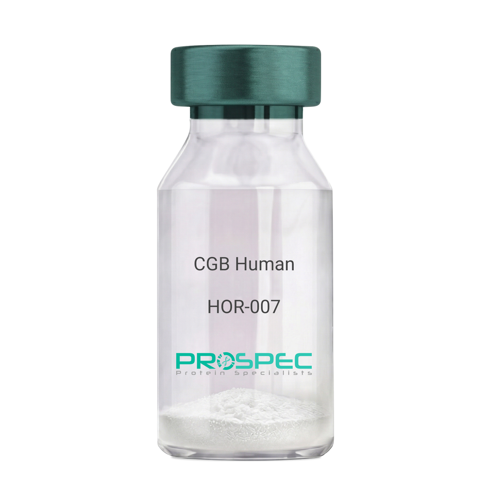
CG Beta HumanDescription:
CGB Human Recombinant
CGB3, CGB5, CGB7, CGB8, hCGB, CG-beta, CGB.
Product # :
HOR-007Price :
Quantity :
Shipping Method :
Shipped with Ice Packs
More Info
- description
- source
- formulation
- purity
- More Info
- Citations
Description
CGB Human Recombinant produced in E.Coli is a single, non-glycosylated polypeptide chain containing 168 amino acids (21-165 a.a.) and having a molecular mass of 17.9kDa. CGB is fused to a 23 amino acid His-tag at N-terminus & purified by proprietary chromatographic techniques.
Source
Escherichia Coli.
Formulation
CGB protein solution (0.5mg/ml) containing 20mM Tris-HCl buffer (pH 8.0), 0.4M Urea and 10% glycerol.
Purity
Greater than 85.0% as determined by SDS-PAGE.
Citations
More Info
-
Introduction
Glycoprotein hormones are heterodimers consisting of a common alpha subunit and a unique beta subunit which confers biological specificity. CGB is a member of the glycoprotein hormone beta chain family and is the beta 3 subunit of (CG). CG is produced by the trophoblastic cells of the placenta and stimulates the ovaries. The beta subunit of CG is encoded by 6 genes which are arranged in tandem and inverted pairs on chromosome 19q13.3 and contiguous with the luteinizing hormone beta subunit gene.
-
Synonyms
CGB3, CGB5, CGB7, CGB8, hCGB, CG-beta, CGB.
-
Physical Appearance
Sterile filtered colorless solution.
-
Stability
Store at 4°C if entire vial will be used within 2-4 weeks. Store, frozen at -20°C for longer periods of time. For long term storage it is recommended to add a carrier protein (0.1% HSA or BSA).Avoid multiple freeze-thaw cycles.
-
Amino Acid Sequence
MGSSHHHHHH SSGLVPRGSH MGSSKEPLRP RCRPINATLA VEKEGCPVCI TVNTTICAGY CPTMTRVLQG VLPALPQVVC NYRDVRFESI RLPGCPRGVN PVVSYAVALS CQCALCRRST TDCGGPKDHP LTCDDPRFQD SSSSKAPPPS LPSPSRLPGP SDTPILPQ.
-
Background
What is the molecular weight/Mw of CG BETA HUMAN Protein?
CG BETA HUMAN Protein has a total Mw of 17.9kDa.
What is the source or expression system of CG BETA HUMAN Protein?
Escherichia Coli.
What is the Purity of CG BETA HUMAN Protein?
CG BETA HUMAN Protein is > 85% pure as determined by SDS-PAGE.
What is the Biological Activity of CG BETA HUMAN Protein?
The biological functionality of CG BETA HUMAN Protein will be determined in the future.
What is the amino acid sequence of CG BETA HUMAN Protein?
MGSSHHHHHH SSGLVPRGSH MGSSKEPLRP RCRPINATLA VEKEGCPVCI TVNTTICAGY CPTMTRVLQG VLPALPQVVC NYRDVRFESI RLPGCPRGVN PVVSYAVALS CQCALCRRST TDCGGPKDHP LTCDDPRFQD SSSSKAPPPS LPSPSRLPGP SDTPILPQ.
What applications can CG BETA HUMAN Protein be used in?
CG BETA HUMAN Protein can probably be used in western blot, ELISA and Lateral Flow.
What is the endotoxin level for CG BETA HUMAN Protein?
The endotoxin level is minimal, CG BETA HUMAN Protein was purified using conventional chromatography techniques.
Who supplies recombinant CG BETA HUMAN that is in stock and available to order?
ProSpec has the protein in stock and available for immediate delivery.
Where can I order bulk mg quantities of CG BETA HUMAN?
ProSpec supplies bulk mg quantities at affordable prices that meets research and academic budgets.
ProSpec's products are furnished for LABORATORY RESEARCH USE ONLY. The product may not be used as drugs, agricultural or pesticidal products, food additives or household chemicals.
- View Data Sheet
Name :
ANXA10 Human (1-162)Description:
Annexin A10 (1-162 a.a.) Human Recombinant
anxa-10.
Product # :
PRO-2837Price :
Quantity :
Shipping Method :
Shipped at Room temp
More Info
- description
- source
- formulation
- purity
- More Info
- Citations
Description
The ANXA10 Human is created as a recombinant protein with a 4kda N-terminal fusion of His Tag. The ANXA10 His-Tagged Fusion Protein, produced in E. coli, is a 21kDa protein containing 162 amino acid residues of the ANXA10 Human, 1-162 amino acids.
Source
Escherichia Coli.
Formulation
Each mg was lyophilized with 1xPBS, 0.4% SDS and 4mM DTT.
Purity
Greater than 90% as determined by SDS-PAGE.
Citations
More Info
-
Synonyms
anxa-10.
-
Physical Appearance
Filtered White lyophilized (freeze-dried) powder.
-
Stability
Store lyophilized ANXA10 at -20°C. Aliquot the product after reconstitution to avoid repeated freezing/thawing cycles. Reconstituted protein can be stored at 4°C for a limited period of time; it does not show any change after two weeks at 4°C.
-
Solubility
It is recommended to add deionized water to prepare a working stock solution of approximately 0.5mg/ml and let the lyophilized pellet dissolve completely. Product is not sterile! Please filter the product by an appropriate sterile filter before using it on cell culture.
-
Background
Annexin A10 also known as ANXA10 is a part of the annexin family of calcium-binding proteins which members own a conserved core domain and a unique amino-terminal region which may convene binding specificity. The ANXA10 protein contains 4 annexin domains and plays a role in the regulation of cellular growth and signal transduction pathways throughout the cell.
ProSpec's products are furnished for LABORATORY RESEARCH USE ONLY. The product may not be used as drugs, agricultural or pesticidal products, food additives or household chemicals.
- View Data Sheet
Description:
Leptin Super Antagonist Human Recombinant
Product # :
CYT-1238Price :
Quantity :
Shipping Method :
Shipped at Room temp
More Info
- description
- source
- formulation
- purity
- biological activity
- More Info
- Citations
Description
Super Leptin Antagonist Human Recombinant is a single polypeptide chain containing 146 amino acids. Super Human Leptin Antagonist was mutated, resulting in D23L/L39A/D40A/F41A super human leptin antagonist that was purified by proprietary chromatographic techniques.
Source
Escherichia coli.
Formulation
The protein was lyophilized from a concentrated (1mg/ml) solution with 0.003mM NaHCO3.
Purity
Greater than 98.0% as determined by:
(a) Gel filtration analysis.
(b) Analysis by SDS-PAGE.
Biological Activity
ProSpec’s super human leptin antagonist is capable of inhibiting leptin-induced proliferation of BAF/3 cells stably transfected with the long form of human leptin.
Citations
More Info
-
Physical Appearance
White lyophilized (freeze-dried) powder.
-
Stability
Lyophilized Super Leptin Antagonist although stable at room temperature for several weeks, should be stored desiccated below -18°C. Upon reconstitution of super human leptin antagonist at > 0.1 mg/ml and up to 2 mg/ml and filter sterilization super human leptin antagonist can be stored at 4C or even room temperature for several weeks making it suitable for long term infusion studies using osmotic pumps. At lower concentration addition of a carrier protein (0.1% HSA or BSA) is suggested. Please prevent freeze-thaw cycles.
-
Solubility
It is recommended to reconstitute the lyophilized Super Leptin Antagonist in sterile 0.4% NaHCO3 adjusted to pH 8-9, not less than 100µg/ml, which can then be further diluted to other aqueous solutions.
-
Amino Acid Sequence
The sequence of the first five N-terminal amino acids was determined and was found to be Ala-Val-Pro-Ile-Gln.
-
Background
Leptin is a hormone which takes part in regulating body weight, metabolism and reproductive function. Leptin is a~16 kDa protein which is encoded by the obese gene. leptin is expressed predominantly by adipocytes, which fits with the idea that body weight is sensed as the total mass of fat in the body. Smaller amounts of leptin are also secreted by cellsin the epithelium of the stomach and in the placenta. Leptin receptors are highly expressed in areas of the hypothalamus which regulates body weight, as well as in T lymphocytes and vascular endothelial cells.
ProSpec's products are furnished for LABORATORY RESEARCH USE ONLY. The product may not be used as drugs, agricultural or pesticidal products, food additives or household chemicals.
- View Data Sheet
Name :
Adiponectin Human, HEKDescription:
Adiponectin Human Recombinant, HEK
Acrp30, AdipoQ, GBP-28, APM-1, ACDC.
Product # :
CYT-434Price :
Quantity :
Shipping Method :
Shipped at Room temp
More Info
- description
- source
- formulation
- purity
- biological activity
- More Info
- sds-page
- Citations
Description
Acrp30 Human Recombinant produced in HEK cells is a glycosylated polypeptide chain containing 234 amino acids (19-244a.a.) and having a molecular mass of 25.5kDa. Acrp30 is expressed with a FLAG tag at C-Terminus and purified by proprietary chromatographic techniques.
Source
HEK293 (Human Embryonic Kidney cell line).
Formulation
Filtered (0.4µm) and lyophilized from 0.5mg/ml in 50mM phosphate Buffer, 75mM NaCl, pH 7.4.
Purity
Greater than 98% as determined by HPLC and SDS PAGE.
Biological Activity
In vitro gluconeogenesis assay in primary hepatocytes was performed, showing the Adiponectin human derived from mammalian cells can inhibit glucose production. The ED50 was ~6 µg/ml.
sds-page
Citations
More Info
-
Introduction
Adiponectin is a recently discovered 244 amino acid protein, the product of the apM1 gene, which is physiologically active and specifically and highly expressed in adipose cells (Adipokine). The protein belongs to the soluble defense collagen super family; it has a collagen-like domain structurally homologous with collagen VIII and X and complement factor C1q-like globular domain. APM-1 forms homotrimers, which are the building blocks for higher order complexes found circulating in serum.
-
Synonyms
Acrp30, AdipoQ, GBP-28, APM-1, ACDC.
-
Physical Appearance
White lyophilized (freeze-dried) powder.
-
Stability
For long term, store lyophilized AdipoQ at -20°C. Aliquot the product after reconstitution to avoid repeated freezing/thawing cycles. Reconstituted protein can be stored at 4°C for a limited period of time; it does not show any change after two weeks at 4°C. The lyophilized protein remains stable for 24 months when stored at -20°C.
-
Solubility
Add deionized water to prepare a working stock solution of approximately 0.5mg/ml and let the lyophilized pellet dissolve completely. Product not sterile! Please filter the product by an appropriate sterile filter before using it in cell culture.
-
Amino Acid Sequence
ETTTQGPGVL LPLPKGACTG WMAGIPGHPG HNGAPGRDGR DGTPGEKGEK GDPGLIGPKG DIGETGVPGA EGPRGFPGIQ GRKGEPGEGA YVYRSAFSVG LETYVTIPNM PIRFTKIFYN QQNHYDGSTG KFHCNIPGLY YFAYHIVYMK DVKVSLFKKD KAMLFTYDQY QENNVDQASG SVLLHLEVGD QVWLQVYGEG ERNGLYADND NDSTFTGFLL YHDTNDYKDDDDK.
-
Background
What is the molecular weight/Mw of adiponectin Protein?
Adiponectin Protein has a total Mw of 25.5kDa.
What is the source or expression system of adiponectin Protein?
HEK293.
What is the Purity of adiponectin Protein?
adiponectin Protein is >98% pure as determined by SDS-PAGE.
What is the Biological Activity of adiponectin Protein?
In vitro gluconeogenesis assay in primary hepatocytes was performed, showing the Adiponectin human derived from mammalian cells can inhibit glucose production. The ED50 was ~6 µg/ml.
What is the amino acid sequence of adiponectin Protein?
ETTTQGPGVL LPLPKGACTG WMAGIPGHPG HNGAPGRDGR DGTPGEKGEK GDPGLIGPKG DIGETGVPGA EGPRGFPGIQ GRKGEPGEGA YVYRSAFSVG LETYVTIPNM PIRFTKIFYN QQNHYDGSTG KFHCNIPGLY YFAYHIVYMK DVKVSLFKKD KAMLFTYDQY QENNVDQASG SVLLHLEVGD QVWLQVYGEG ERNGLYADND NDSTFTGFLL YHDTNDYKDDDDK.
What applications can adiponectin Protein be used in?
adiponectin Protein can probably be used in western blot, ELISA and Lateral Flow.
What is the endotoxin level for adiponectin Protein?
The endotoxin level is minimal, adiponectin Protein was purified using conventional chromatography techniques.
ProSpec's products are furnished for LABORATORY RESEARCH USE ONLY. The product may not be used as drugs, agricultural or pesticidal products, food additives or household chemicals.
- View Data Sheet
Name :
ACVR2A HumanDescription:
Actv Receptor Type 2A Human Recombinant
ACVR2A, ACTRIIA, ACTR-IIA,
Product # :
CYT-976Price :
Quantity :
Shipping Method :
Shipped with Ice Packs
More Info
- description
- source
- formulation
- purity
- More Info
- sds-page
- Citations
Description
ACVR2A Human Recombinant produced in Sf9 Baculovirus cells is a single, glycosylated polypeptide chain containing 124 amino acids (20-135a.a.) and having a molecular mass of 14.4kDa (Molecular size on SDS-PAGE will appear at approximately 28kDa.ACVR2A is fused to 6 amino acid His-tag at C-terminus & purified by proprietary chromatographic techniques.
Source
Sf9, Baculovirus cells.
Formulation
ACVR2A protein solution (0.5mg/ml) containing Phosphate Buffered Saline (pH 7.4) and 10% glycerol.
Purity
Greater than 95% as determined by SDS-PAGE.
sds-page
Citations
More Info
-
Introduction
ACVR2A takes part in various biological processes including mesoderm induction, neural cell differentiation, bone remodeling, hematopoiesis, carcinogenesis, and inflammation. ACVR2A which is a receptor for Actv A, Actv B and inhibin A mediates induction of adipogenesis by GDF6.
-
Synonyms
ACVR2A, ACTRIIA, ACTR-IIA,
-
Physical Appearance
Sterile Filtered colorless solution.
-
Stability
Store at 4°C if entire vial will be used within 2-4 weeks. Store, frozen at -20°C for longer periods of time. For long term storage it is recommended to add a carrier protein (0.1% HSA or BSA).Avoid multiple freeze-thaw cycles.
-
Amino Acid Sequence
AILGRSETQE CLFFNANWEK DRTNQTGVEP CYGDKDKRRH CFATWKNISG SIEIVKQGCW LDDINCYDRT DCVEKKDSPE VYFCCCEGNM CNEKFSYFPE MEVTQPTSNP VTPKPPLEHH HHHH.
-
Background
A Comprehensive Examination of the Activin Receptor Type 2A Human Recombinant: Biological Functions and Therapeutic Possibilities
1. Abstract
This paper delves into the complex world of Activin Receptor Type 2A Human Recombinant (ACVR2A), a crucial element of the Transforming Growth Factor-beta (TGF-beta) signaling pathway. The structure, biological implications, and signaling pathway of ACVR2A are all extensively reviewed. The potential for ACVR2A as a therapeutic target in various pathological conditions is also explored.
2. Introduction
The ACVR2A, a receptor protein vital to the TGF-beta signaling pathway, plays a significant role in a multitude of biological processes, including embryogenesis, cell differentiation, and homeostasis. Understanding the intricate operations of ACVR2A could open the door to innovative therapeutic strategies.
3. Structure and Signaling of ACVR2A
As a transmembrane serine/threonine kinase receptor, ACVR2A is characterized by a ligand-binding extracellular domain and an intracellular domain responsible for signal transduction. Upon binding of specific ligands like activin, ACVR2A interacts with type I receptors to trigger phosphorylation events, leading to the activation of downstream SMAD signaling pathways.
4. Biological Functions of ACVR2A
ACVR2A plays a substantial role in a wide range of biological processes. These include embryonic development, cell differentiation, bone growth, immune responses, and homeostasis. Furthermore, ACVR2A is instrumental in follicle-stimulating hormone (FSH) regulation, highlighting its importance in reproduction.
5. ACVR2A in Disease Pathology
Impairments in ACVR2A signaling have been linked to several diseases, including various cancers and reproductive disorders. Mutations in the ACVR2A gene have been implicated in tumor progression, underscoring the receptor's role in cell proliferation and differentiation.
6. Therapeutic Potential of ACVR2A
The centrality of ACVR2A in critical biological processes and disease pathology suggests its therapeutic potential. By modulating ACVR2A signaling, it may be possible to intervene in diseases characterized by aberrant TGF-beta signaling. Additionally, ACVR2A antagonists are being studied for their potential in cancer treatment.
7. Conclusion and Future Perspectives
Our comprehension of ACVR2A's functions has substantially increased in recent years, yet much remains to be discovered. Further research into ACVR2A's precise molecular mechanisms and involvement in disease will undoubtedly yield new therapeutic strategies.
What is the molecular weight / Mw of ACVR2A Protein?
ACVR2A Protein has a total Mw of 14.4kDa.
What is the source or expression system of ACVR2A Protein?
Sf9, Baculovirus cells.
What is the Purity of ACVR2A Protein?
ACVR2A Protein is >95% pure as determined by SDS-PAGE.
What is the Biological Activity of ACVR2A Protein?
The biological functionality of ACVR2A Protein will be determined in the future.
What is the amino acid sequence of ACVR2A Protein?
AILGRSETQE CLFFNANWEK DRTNQTGVEP CYGDKDKRRH CFATWKNISG SIEIVKQGCW LDDINCYDRT DCVEKKDSPE VYFCCCEGNM CNEKFSYFPE MEVTQPTSNP VTPKPPLEHH HHHH.
What applications can ACVR2A Protein be used in?
ACVR2A Protein can probably be used in western blot, ELISA and Lateral Flow.
What is the endotoxin level for ACVR2A Protein?
The endotoxin level is minimal, ACVR2A Protein was purified using conventional chromatography techniques.
ProSpec's products are furnished for LABORATORY RESEARCH USE ONLY. The product may not be used as drugs, agricultural or pesticidal products, food additives or household chemicals.
- View Data Sheet
Name :
Ag85ADescription:
Mycobacterium Tuberculosis major secretory protein Antigen 85A Recombinant
Ronectin-binding protein A, Mycolyl transferase 85A.
Product # :
PRO-1076Price :
Quantity :
Shipping Method :
Shipped with Ice Packs
More Info
- description
- source
- formulation
- purity
- More Info
- Citations
Description
Recombinant Mycobacterium tuberculosis Ag85A (43-338 a.a) produced in Hi-5 cells is a single, glycosylated polypeptide chain having a molecular mass of 32.8kDa (305 a.a in total).Antigen 85A is fused to a 6 amino acid His tag at C-terminus and purified by conventional chromatographic techniques.
Source
Baculovirus
Formulation
The Ag85A solution (0.5mg/1ml) contains 20mM Tris-HCl buffer (pH 8.0) and 10% glycerol.
Purity
Greater than 95% as determined by SDS-PAGE.
Citations
More Info
-
Introduction
Antigen 85A is a member of the antigen 85 complex (Antigen 85A, B, C). The enzymes of the antigen 85 complex have mycolyltransferase activity and catalyze the synthesis of the very rich glycolipid of the mycobacterial cell wall, the cord factor (trehalose 6,6'-dimycolate, TDM). The cord factor is vital for the integrity of the mycobacterial cell wall and pathogenesis of the bacillus. TDM is synthesized from two molecules of trehalose-6'-monomycolate (TMM) by Antigen 85A.
-
Synonyms
Ronectin-binding protein A, Mycolyl transferase 85A.
-
Physical Appearance
Sterile Filtered colorless solution.
-
Stability
Store at 4°C if entire vial will be used within 2-4 weeks. Store, frozen at -20°C for longer periods of time. For long term storage it is recommended to add a carrier protein (0.1% HSA or BSA).Avoid multiple freeze-thaw cycles.
-
Amino Acid Sequence
ADPAFSRPGL PVEYLQVPSP SMGRDIKVQF QSGGANSPAL YLLDGLRAQD DFSGWDINTP AFEWYDQSGL SVVMPVGGQS SFYSDWYQPA CGKAGCQTYK WETFLTSELP GWLQANRHVK PTGSAVVGLS MAASSALTLA IYHPQQFVYA GAMSGLLDPS QAMGPTLIGL AMGDAGGYKA SDMWGPKEDP AWQRNDPLLN VGKLIANNTR VWVYCGNGKP SDLGGNNLPA KFLEGFVRTS NIKFQDAYNA GGGHNGVFDF PDSGTHSWEY WGAQLNAMKP DLQRALGATP NTGPAPQGAH HHHHH.
ProSpec's products are furnished for LABORATORY RESEARCH USE ONLY. The product may not be used as drugs, agricultural or pesticidal products, food additives or household chemicals.
- View Data Sheet
Name :
HIF1A Human, HisDescription:
Hypoxia-Inducible Factor-1 Alpha Human Recombinant, His Tag
Hypoxia-inducible factor 1 alpha, HIF-1 alpha, HIF1 alpha, ARNT-interacting protein, Member of PAS protein 1, Basic-helix-loop-helix-PAS protein MOP1, HIF1A, MOP1, HIF1, PASD8, HIF-1A.
Product # :
PRO-415Price :
Quantity :
Shipping Method :
Shipped with Ice Packs
More Info
- description
- source
- formulation
- purity
- More Info
- Citations
Description
HIF1A Recombinant Human produced in E.Coli is a single, non-glycosylated polypeptide chain containing 231 amino acids (576-785 a.a.) and having a molecular mass of 25.1 kDa. The HIF1A is fused to a 21 amino acid His-Tag at N-terminus and purified by proprietary chromatographic techniques.
Source
Escherichia Coli.
Formulation
HIF1A Human solution containing 20mM Tris-HCl pH-8, 1mM DTT, 0.2M NaCl & 10% glycerol.
Purity
Greater than 85.0% as determined by SDS-PAGE.
Citations
More Info
-
Introduction
HIF1A has a role as a master transcriptional monitor of the adaptive response to hypoxia. Under hypoxic conditions HIF1A activates the transcription of over 40 genes, including, erythropoietin, glucose transporters, glycolytic enzymes, vascular endothelial growth factor, and genes whose protein products increase oxygen release or facilitate metabolic adaptation to hypoxia. HIF1A functions as an essential role in embryonic vascularization, tumor angiogenesis and pathophysiology of ischemic disease.
-
Synonyms
Hypoxia-inducible factor 1 alpha, HIF-1 alpha, HIF1 alpha, ARNT-interacting protein, Member of PAS protein 1, Basic-helix-loop-helix-PAS protein MOP1, HIF1A, MOP1, HIF1, PASD8, HIF-1A.
-
Physical Appearance
Sterile filtered colorless solution.
-
Stability
Store at 4°C if entire vial will be used within 2-4 weeks. Store, frozen at -20°C for longer periods of time. For long term storage it is recommended to add a carrier protein (0.1% HSA or BSA).Avoid multiple freeze-thaw cycles.
-
Amino Acid Sequence
MGSSHHHHHH SSGLVPRGSH MSFDQLSPLE SSSASPESAS PQSTVTVFQQ TQIQEPTANA TTTTATTDEL KTVTKDRMED IKILIASPSP THIHKETTSA TSSPYRDTQS RTASPNRAGK GVIEQTEKSH PRSPNVLSVA LSQRTTVPEE ELNPKILALQ NAQRKRKMEH DGSLFQAVGI GTLLQQPDDH AATTSLSWKR VKGCKSSEQN GMEQKTIILI PSDLACRLLG Q.
ProSpec's products are furnished for LABORATORY RESEARCH USE ONLY. The product may not be used as drugs, agricultural or pesticidal products, food additives or household chemicals.
- View Data Sheet
Name :
AIF1 HumanDescription:
Allograft Inflammatory Factor 1 Human Recombinant
AIF-1, Allograft inflammatory factor 1, Em:AF129756.17, G1, IBA1, Ionized calcium-binding adapter molecule 1, IRT-1, Protein G1, AIF1.
Product # :
CYT-697Price :
Quantity :
Shipping Method :
Shipped with Ice Packs
More Info
- description
- source
- formulation
- purity
- More Info
- sds-page
- Citations
Description
AIF1 produced in E.Coli is a single, non-glycosylated polypeptide chain containing 167 amino acids (1-147a.a.) and having a molecular mass of 18.9kDa. AIF1 is fused to a 20 amino acid His-tag at N-terminus & purified by proprietary chromatographic techniques.
Source
E. Coli.
Formulation
The AIF1 protein solution (0.5mg/1ml) is formulated in 20mM Tris-HCl buffer (pH8.0)
2mM DTT, 200mM NaCl and 20% glycerol.Purity
Greater than 95% as determined by SDS PAGE.
sds-page
Citations
More Info
-
Introduction
Human AIF1 protein shares 98% homology/identity with that of rat. AIF1 is expressed in macrophages and neutrophils. The expression of AIF1 transcripts is upregulated by IFN-g in rat macrophages. AIF1 is expressed selectively in human macrophage-like cell lines, and in a subset of CD68(+) macrophages in the interstitial and perivascular spaces of human heart allografts. In quiescent cultured human vascular smooth muscle cells synthesis of AIF1 is induced by IFN-g, IL1b, and conditioned medium of T-cells. Overexpression of AIF1 in human VSMCs results in enhanced growth of these cells. AIF1 is expressed during apoptosis rat mammary gland and ventral prostate tissues. Allograft Inflammatory Factor 1 is expressed by several tumor-associated activated macrophages and microglial cells in rat and human gliomas. There is an evident relationship of AIF1-expressing activated macrophages and microglial cells with tumor malignancy in humans.
-
Synonyms
AIF-1, Allograft inflammatory factor 1, Em:AF129756.17, G1, IBA1, Ionized calcium-binding adapter molecule 1, IRT-1, Protein G1, AIF1.
-
Stability
Store AIF1 at 4°C if entire vial will be used within 2-4 weeks. Store, frozen at -20°C for longer periods of time.For long term storage it is recommended to add a carrier protein (0.1% HSA or BSA).Avoid multiple freeze-thaw cycles.
-
Amino Acid Sequence
MGSSHHHHHH SSGLVPRGSH MSQTRDLQGG KAFGLLKAQQ EERLDEINKQ FLDDPKYSSD EDLPSKLEGF KEKYMEFDLN GNGDIDIMSL KRMLEKLGVP KTHLELKKLI GEVSSGSGET FSYPDFLRMM LGKRSAILKM ILMYEEKARE KEKPTGPPAK KAISELP.
-
Background
Allograft Inflammatory Factor 1 Human Recombinant: Uncovering its Role in Immune Responses and Therapeutic Prospects
1. Abstract
This paper explores the Allograft Inflammatory Factor 1 Human Recombinant (AIF-1), a cytoplasmic, IFN-gamma-inducible calcium-binding protein involved in inflammation and immunity. We review the structure, biological roles, and involvement of AIF-1 in disease pathology. The therapeutic potential of AIF-1 in immune-related disorders is also explored.
2. Introduction
AIF-1, also known as IBA1, plays an important role in immune responses. It is associated with various immune cells, particularly macrophages, and has been implicated in numerous inflammatory and immune-related diseases. Understanding the function of AIF-1 could aid the development of novel therapeutic strategies.
3. Structure and Signaling of AIF-1
AIF-1 is a small 17 kDa protein with an EF-hand calcium-binding motif. Although the precise mechanism by which AIF-1 exerts its functions is not entirely clear, it is known to regulate the activation, migration, and proliferation of macrophages, key cells involved in immune responses.
4. Biological Functions of AIF-1
AIF-1 has been shown to play key roles in macrophage activation and function, which are central to inflammation and immunity. It is also implicated in cell survival, proliferation, and differentiation.
5. AIF-1 in Disease Pathology
AIF-1 has been associated with a range of inflammatory and immune-related diseases, including rheumatoid arthritis, atherosclerosis, and multiple sclerosis. It is also implicated in several cancers, further underscoring its broad physiological and pathological relevance.
6. Therapeutic Potential of AIF-1
Given its pivotal role in immune responses, AIF-1 presents an intriguing target for therapeutic interventions in immune-related diseases. Modulating the activity of AIF-1 could potentially alleviate pathological inflammation and autoimmunity.
7. Conclusion and Future Perspectives
Our knowledge of AIF-1 and its functions has significantly improved in recent years, but much remains to be discovered. Further research into AIF-1's exact molecular mechanisms and roles in disease will undoubtedly contribute to the development of novel therapeutic strategies.
What is the molecular weight/Mw of AIF1 Protein?
AIF1 Protein has a total Mw of 18.9kDa.
What is the source or expression system of AIF1 Protein?
Escherichia Coli.
What is the Purity of AIF1 Protein?
AIF1 Protein is >95% pure as determined by SDS-PAGE.
What is the Biological Activity of AIF1 Protein?
The biological functionality of AIF1 Protein will be determined in the future.
What is the amino acid sequence of AIF1 Protein?
MGSSHHHHHH SSGLVPRGSH MSQTRDLQGG KAFGLLKAQQ EERLDEINKQ FLDDPKYSSD EDLPSKLEGF KEKYMEFDLN GNGDIDIMSL KRMLEKLGVP KTHLELKKLI GEVSSGSGET FSYPDFLRMM LGKRSAILKM ILMYEEKARE KEKPTGPPAK KAISELP.
What applications can AIF1 Protein be used in?
AIF1 Protein can probably be used in western blot, ELISA and Lateral Flow.
What is the endotoxin level for AIF1 Protein?
The endotoxin level is minimal, AIF1 Protein was purified using conventional chromatography techniques.
ProSpec's products are furnished for LABORATORY RESEARCH USE ONLY. The product may not be used as drugs, agricultural or pesticidal products, food additives or household chemicals.
- View Data Sheet
Name :
STMN2 HumanDescription:
Stathmin Like-2 Human Recombinant
SCG10, STMB2, SCGN10, AI159727, STMN-2, Stathmin-2, Superior cervical ganglion-10 protein, Protein SCG10, STMN2, SGC10, stathmin-like 2.
Product # :
PRO-752Price :
Quantity :
Shipping Method :
Shipped with Ice Packs
More Info
- description
- source
- formulation
- purity
- More Info
- Citations
Description
Recombinant Human Stathmin Like-2 produced in E.Coli is a single, non-glycosylated polypeptide chain containing 142 amino acids (39-179 a.a) and having a molecular mass of 16.4 kDa. STMN2 is purified by conventional chromatography techniques.
Source
Escherichia Coli.
Formulation
The STMN2 protein solution contains 50mM MES, pH-6.0, 0.1mM PMSF, 1 mM EDTA and 10% Glycerol.
Purity
Greater than 85.0% as determined by SDS-PAGE.
Citations
More Info
-
Introduction
STMN2 is a neuronal growth-associated protein which shares a majority of its amino acid sequence with the phosphoprotein stathmin. STMN2 is involved in neuronal differentiation and in modulating membrane interaction with the cytoskeleton through neurite outgrowth. STMN2 is necessary for maintaining the anchorage-independent growth state of beta-catenin/TCF-activated hepatoma cells. SCG10 is a novel indicator of osteogenesis and osteoblast that takes part in the regulation of the adipocyte/osteoblast balance. STMN2 plays a role as an effector downstream of Rnd1 to regulate axon extensions by modulating microtubule organization.
-
Synonyms
SCG10, STMB2, SCGN10, AI159727, STMN-2, Stathmin-2, Superior cervical ganglion-10 protein, Protein SCG10, STMN2, SGC10, stathmin-like 2.
-
Physical Appearance
Sterile filtered colorless solution.
-
Stability
Store at 4°C if entire vial will be used within 2-4 weeks. Store, frozen at -20°C for longer periods of time. For long term storage it is recommended to add a carrier protein (0.1% HSA or BSA).Avoid multiple freeze-thaw cycles.
-
Amino Acid Sequence
MDMEVKQINK RASGQAFELI LKPPSPISEA PRTLASPKKK DLSLEEIQKK LEAAEERRKS QEAQVLKQLA EKREHEREVL QKALEENNNF SKMAEEKLIL KMEQIKENRE ANLAAIIERL QEKERHAAEV RRNKELQVEL SG.
ProSpec's products are furnished for LABORATORY RESEARCH USE ONLY. The product may not be used as drugs, agricultural or pesticidal products, food additives or household chemicals.
- View Data Sheet
Name :
CCNG1 HumanDescription:
Cyclin G1 Human Recombinant
Cyclin-G1, Cyclin-G, CCNG1, CCNG, CYCG1.
Product # :
PRO-1005Price :
Quantity :
Shipping Method :
Shipped with Ice Packs
More Info
- description
- source
- formulation
- purity
- More Info
- Citations
Description
CCNG1 Human Recombinant produced in E.Coli is a single, non-glycosylated polypeptide chain containing 315 amino acids (1-295 a.a.) and having a molecular mass of 36.2kDa. CCNG1 is fused to a 20 amino acid His-tag at N-terminus & purified by proprietary chromatographic techniques.
Source
Escherichia Coli.
Formulation
CCNG1 protein solution (0.25mg/ml) containing 20mM Tris-HCl buffer (pH8.0), 50% glycerol, 0.2M NaCl and 5mM DTT.
Purity
Greater than 85% as determined by SDS-PAGE.
Citations
More Info
-
Introduction
Cyclin-G1 (CCNG1) belongs to the cyclin family and contains the cyclin box. CCNG1 may have a part in growth regulation and is associated with G2/M phase arrest in response to DNA damage. CCNG1 may be an intermediate by which p53 mediates its role as an inhibitor of cellular proliferation. The CCNG1 protein lacks the protein destabilizing (PEST) sequence which is present in other family members.
-
Synonyms
Cyclin-G1, Cyclin-G, CCNG1, CCNG, CYCG1.
-
Physical Appearance
Sterile Filtered clear colorless solution.
-
Stability
Store at 4°C if entire vial will be used within 2-4 weeks. Store, frozen at -20°C for longer periods of time. For long term storage it is recommended to add a carrier protein (0.1% HSA or BSA).Avoid multiple freeze-thaw cycles.
-
Amino Acid Sequence
MGSSHHHHHH SSGLVPRGSH MIEVLTTTDS QKLLHQLNAL LEQESRCQPK VCGLRLIESA HDNGLRMTAR LRDFEVKDLL SLTQFFGFDT ETFSLAVNLL DRFLSKMKVQ PKHLGCVGLS CFYLAVKSIE EERNVPLATD LIRISQYRFT VSDLMRMEKI VLEKVCWKVK ATTAFQFLQL YYSLLQENLP LERRNSINFE RLEAQLKACH CRIIFSKAKP SVLALSIIAL EIQAQKCVEL TEGIECLQKH SKINGRDLTF WQELVSKCLT EYSSNKCSKP NVQKLKWIVS GRTARQLKHS YYRITHLPTI PEMVP.
ProSpec's products are furnished for LABORATORY RESEARCH USE ONLY. The product may not be used as drugs, agricultural or pesticidal products, food additives or household chemicals.
- View Data Sheet
Name :
CHGA Human, HisDescription:
Chromogranin-A Human Recombinant, His Tag
CGA, CHGA, Vasostatin-2, Pituitary secretory protein I, SP-I.
Product # :
PRO-699Price :
Quantity :
Shipping Method :
Shipped with Ice Packs
More Info
- description
- source
- formulation
- purity
- More Info
- Citations
Description
Recombinant Human CHGA produced in E.Coli is a single, non-glycosylated polypeptide chain containing 460 amino acids (19-457 a.a) and having a molecular mass of 51.2kDa (Molecular weight on SDS-PAGE will appear higher). Chromgranin-A is fused to 21 amino acid His Tag at N-terminus and purified by proprietary chromatographic techniques.
Source
Escherichia Coli.
Formulation
The CHGA protein (0.5mg/ml) contains 20mM Tris-HCl buffer pH-7.5, 2mM EDTA, 0.1mM PMSF and 10% glycerol.
Purity
Greater than 80.0% as determined by Analysis by SDS-PAGE.
Citations
More Info
-
Introduction
Chromgranin-A is part of the neuroendocrine secretory protein family. CHGA is located in secretory vesicles of neurons and endocrine cells. Chromgranin-A is a precursor to three biologically active peptides; vasostatin, pancreastatin, and parastatin. These peptides act as autocrine or paracrine negative modulators of the neuroendocrine system. Other peptides, including chromostatin, beta-granin, WE-14 and GE-25, are also derived from the full-length protein. Chromgranin-A has numerous biological activities on some tissues and organs and exerts a large spectrum of homeostatic actions, including antifungal and antimicrobial effect, modulation of cell adhesion, and inhibition of parathyroid hormone secretion.
-
Synonyms
CGA, CHGA, Vasostatin-2, Pituitary secretory protein I, SP-I.
-
Physical Appearance
Sterile filtered colorless solution.
-
Stability
Store at 4°C if entire vial will be used within 2-4 weeks. Store, frozen at -20°C for longer periods of time. For long term storage it is recommended to add a carrier protein (0.1% HSA or BSA). Avoid multiple freeze-thaw cycles.
-
Amino Acid Sequence
MGSSHHHHHH SSGLVPRGSH MLPVNSPMNK GDTEVMKCIV EVISDTLSKP SPMPVSQECF ETLRGDERIL SILRHQNLLK ELQDLALQGA KERAHQQKKH SGFEDELSEV LENQSSQAEL KEAVEEPSSK DVMEKREDSK EAEKSGEATD GARPQALPEP MQESKAEGNN QAPGEEEEEE EEATNTHPPA SLPSQKYPGP QAEGDSEGLS QGLVDREKGL SAEPGWQAKR EEEEEEEEEA EAGEEAVPEE EGPTVVLNPH PSLGYKEIRK GESRSEALAV DGAGKPGAEE AQDPEGKGEQ EHSQQKEEEE EMAVVPQGLF RGGKSGELEQ EEERLSKEWE DSKRWSKMDQ LAKELTAEKR LEGQEEEEDN RDSSMKLSFR ARAYGFRGPG PQLRRGWRPS SREDSLEAGL PLQVRGYPEE KKEEEGSANR RPEDQELESL SAIEAELEKV AHQLQALRRG.
ProSpec's products are furnished for LABORATORY RESEARCH USE ONLY. The product may not be used as drugs, agricultural or pesticidal products, food additives or household chemicals.
- View Data Sheet
Name :
VCAM1 MouseDescription:
Vascular cell adhesion molecule 1 Mouse Recombinant
V-CAM 1, VCAM-1, CD106, Vcam1, Vcam-1, Vascular cell adhesion protein 1.
Product # :
PRO-2257Price :
Quantity :
Shipping Method :
Shipped with Ice Packs
More Info
- description
- source
- formulation
- purity
- More Info
- Citations
Description
VCAM1 Mouse Recombinant produced in Sf9 Baculovirus cells is a single, glycosylated polypeptide chain containing 682 amino acids (25-698 a.a) and having a molecular mass of 75.4kDa (Migrates at 70-100kDa on SDS-PAGE under reducing conditions). VCAM1 is fused to an 8 amino acid His-tag at C-terminus & purified by proprietary chromatographic techniques.
Source
Sf9, Baculovirus cells.
Formulation
VCAM1 protein solution (0.5mg/ml) containing Phosphate Buffered Saline (pH 7.4) and 10% glycerol.
Purity
Greater than 95.0% as determined by SDS-PAGE.
Citations
More Info
-
Introduction
VCAM1 is a member of the Ig superfamily. It is a cell surface sialoglycoprotein expressed by cytokine activated endothelium. VCAM-1 contains 6 or 7 immunoglobulin domains, and is expressed on both large and small vessels only after the endothelial cells are stimulated by cytokines. The protein has a number of functions including the regulation of leukocyte migration, leukocyte endothelial cell adhesion and signal transduction and may play a role in a number of inflammatory diseases (artherosclerosis and rheumatoid arthritis).
-
Synonyms
V-CAM 1, VCAM-1, CD106, Vcam1, Vcam-1, Vascular cell adhesion protein 1.
-
Physical Appearance
Sterile Filtered colorless solution.
-
Stability
Store at 4°C if entire vial will be used within 2-4 weeks. Store, frozen at -20°C for longer periods of time. For long term storage it is recommended to add a carrier protein (0.1% HSA or BSA).Avoid multiple freeze-thaw cycles.
-
Amino Acid Sequence
FKIEISPEYK TIAQIGDSMA LTCSTTGCES PLFSWRTQID SPLNAKVRTE GSKSVLTMEP VSFENEHSYL CTATCGSGKL ERSIHVDIYS FPKDPEIQFS GPLEVGKPVT VKCLAPDIYP VYRLEIDLFK GDQLMNRQEF SSEEMTKSLE TKSLEVTFTP VIEDIGKALV CRAKLHIDQI DSTLKERETV KELQVYISPR NTTISVHPST RLQEGGAVTM TCSSEGLPAP EIFWGRKLDN EVLQLLSGNA TLTLIAMRME DSGVYVCEGV NLIGRDKAEV ELVVQEKPFI VDISPGSQVA AQVGDSVVLT CAAIGCDSPS FSWRTQTDSP LNGVVRNEGA KSTLVLSSVG FEDEHSYLCA VTCLQRTLEK RTQVEVYSFP EDPVIKMSGP LVHGRPVTVN CTVPNVYPFD HLEIELLKGE TTLMKKYFLE EMGIKSLETK ILETTFIPTI EDTGKSLVCL ARLHSGEMES EPKQRQSVQP LYVNVAPKET TIWVSPSPIL EEGSPVNLTC SSDGIPAPKI LWSRQLNNGE LQPLSENTTL TFMSTKRDDS GIYVCEGINE AGISRKSVEL IIQVSPKDIQ LTVFPSKSVK EGDTVIISCT CGNVPETWII LKKKAKTGDM VLKSVDGSYT IRQAQLQDAG IYECESKTEV GSQLRSLTLD VKGKEHNKNY FSPELEHHHH HH.
ProSpec's products are furnished for LABORATORY RESEARCH USE ONLY. The product may not be used as drugs, agricultural or pesticidal products, food additives or household chemicals.
- View Data Sheet
Name :
PDGF AA HumanDescription:
Platelet-Derived Growth Factor AA Human Recombinant
Glioma-derived growth factor, GDGF, Osteosarcoma-derived Growth Factor, ODGF, PDGF-AA, PDGF-1.
Product # :
CYT-341Price :
Quantity :
Shipping Method :
Shipped at Room temp
More Info
- description
- source
- formulation
- purity
- biological activity
- More Info
- Citations
Description
Platelet-Derived Growth Factor AA Human Recombinant is a homodimeric, non-glycosylated, polypeptide chain containing 2 x 125 amino acids and having a total molecular mass of 28511 Dalton. PDGF-AA is purified by proprietary chromatographic techniques.
Source
Escherichia Coli.
Formulation
Lyophilized without any additives.
Purity
Greater than 95.0% as determined by SDS-PAGE.
Biological Activity
The ED50, calculated by the dose-dependent proliferation of murine 3T3 indicator cells is < 0.32ng/ml, corresponding to a Specific Activity of 3,125x103units/mg.Citations
More Info
-
Introduction
PDGF-AA, PDGF-BB and PDGF-AB, are potent mitogens for a variety of cell types including smooth muscle cells, connective tissue cells, bone and cartilage cells, and some blood cells. The PDGF is stored in platelet alpha-granules and released upon platelet activation. The PDGF is involved in a number of biological processes, including hyperplasia, chemotaxis, embryonic neuron development, and respiratory tubule epithelial cell development. Two distinct signaling receptors used by PDGF
have been identified and named PDGFR-alpha and PDGFR-beta. PDGFR-alpha is high-affinity receptor for each of the three PDGF forms. On the other hand, PDGFR-beta interacts with only PDGF-BB and PDGF-AB. -
Synonyms
Glioma-derived growth factor, GDGF, Osteosarcoma-derived Growth Factor, ODGF, PDGF-AA, PDGF-1.
-
Physical Appearance
Sterile Filtered White lyophilized (freeze-dried) powder.
-
Stability
Lyophilized Platelet-Derived Growth Factor AA although stable at room temperature for 3 weeks, should be stored desiccated below -18°C. Upon reconstitution PDGF-AA should be stored at 4°C between 2-7 days and for future use below -18°C.For long term storage it is recommended to add a carrier protein (0.1% HSA or BSA).Please prevent freeze-thaw cycles.
-
Solubility
It is recommended to reconstitute the lyophilized Platelet-derived Growth Factor-AA in sterile 18MΩ-cm H2O not less than 100µg/ml, which can then be further diluted to other aqueous solutions.
-
Amino Acid Sequence
SIEEAVPAVC KTRTVIYEIP RSQVDPTSAN FLIWPPCVEV KRCTGCCNTS SVKCQPSRVH HRSVKVAKVE YVRKKPKLKE VQVRLEEHLE CACATTSLNP DYREEDTGRP RESGKKRKRK RLKPT.
ProSpec's products are furnished for LABORATORY RESEARCH USE ONLY. The product may not be used as drugs, agricultural or pesticidal products, food additives or household chemicals.
- View Data Sheet
Name :
IFNG MonkeyDescription:
Interferon-gamma Recombinant Rhesus Macaque
Immune IFN, type II IFN, T cell IFN, MAF, IFNG, IFG, IFI, IFN-gamma.
Product # :
CYT-1122Price :
Quantity :
Shipping Method :
Shipped at Room temp
More Info
- description
- source
- formulation
- purity
- biological activity
- More Info
- Citations
Description
Interferon-gamma Rhesus Macaque Recombinant produced in E.Coli is a non-glycosylated polypeptide chain containing 142 amino acid and having a molecular mass of approximately 16.8kDa.IFNG is purified by proprietary chromatographic techniques.
Source
Escherichia Coli.
Formulation
Lyophilized from a 0.2μm filtered concentrated solution in PBS, pH7.4.
Purity
Greater than 97.0% as determined by:
(a) Analysis by RP-HPLC.
(b) Analysis by SDS-PAGE.Biological Activity
The ED50 as determined by an anti-viral assay using human HeLa cells infected with encephalomyocarditis (EMC) virus is < 20.0 ng/ml, corresponding to a specific activity of > 5.0 × 10⁴ IU/mg.
Citations
More Info
-
Introduction
IFN-gamma produced by lymphocytes activated by specific antigens / mitogens.
IFN-gamma, on top of having antiviral activity, has important immunoregulatory functions, it is a potent activator of macrophages, and has antiproliferative effects on transformed cells and it can potentiate the antiviral and antitumor effects of the type I interferons. -
Synonyms
Immune IFN, type II IFN, T cell IFN, MAF, IFNG, IFG, IFI, IFN-gamma.
-
Physical Appearance
Sterile Filtered White lyophilized (freeze-dried) powder.
-
Stability
Lyophilized IFNG although stable at room temperature for 3 weeks, should be stored desiccated below -18°C. Upon reconstitution Interferon-gamma Rhesus Macaque should be stored at 4°C between 2-7 days and for future use below -18°C.Please prevent freeze-thaw cycles.
-
Solubility
It is recommended to reconstitute the lyophilized Interferon-gamma Rhesus Macaque in sterile 18MΩ-cm H2O not less than 100µg/ml, which can then be further diluted to other aqueous solutions.
-
Amino Acid Sequence
QDPYVKEAEN LKKYFNAGDP DVADNGTLFL DILRNWKEES DRKIMQSQIV SFYFKLFKNF KDDQRIQKSV ETIKEDINVK FFNSNKKKRD DFEKLTNYSV TDSNVQRKAV HELIQVMAEL SPAAKIGKRK RSQMFRGRRA SQ.
-
Background
What is the molecular weight/Mw of IFNG MONKEY Protein?
IFNG MONKEY Protein has a total Mw of 16.8kDa.
What is the source or expression system of IFNG MONKEY Protein?
Escherichia Coli.
What is the Purity of IFNG MONKEY Protein?
IFNG MONKEY Protein is >97% pure as determined by SDS-PAGE.
What is the Biological Activity of IFNG MONKEY Protein?
The ED50 as determined by an anti-viral assay using human HeLa cells infected with encephalomyocarditis (EMC) virus is < 20.0 ng/ml, corresponding to a specific activity of > 5.0 × 10⁴ IU/mg.
What is the amino acid sequence of IFNG MONKEY Protein?
QDPYVKEAEN LKKYFNAGDP DVADNGTLFL DILRNWKEES DRKIMQSQIV SFYFKLFKNF KDDQRIQKSV ETIKEDINVK FFNSNKKKRD DFEKLTNYSV TDSNVQRKAV HELIQVMAEL SPAAKIGKRK RSQMFRGRRA SQ.
What applications can IFNG MONKEY Protein be used in?
IFNG MONKEY Protein can probably be used in western blot, ELISA and Lateral Flow.
What is the endotoxin level for IFNG MONKEY Protein?
The endotoxin level is minimal, IFNG MONKEY Protein was purified using conventional chromatography techniques.
ProSpec's products are furnished for LABORATORY RESEARCH USE ONLY. The product may not be used as drugs, agricultural or pesticidal products, food additives or household chemicals.
- View Data Sheet
Name :
BMP8B HumanDescription:
Bone Morphogenetic protein-8b Human Recombinant
Bone morphogenetic protein 8B, BMP-8, BMP-8B, Osteogenic protein 2, OP-2, BMP8B, BMP8, Bone Morphogenetic protein-8b, OP2.
Product # :
CYT-830Price :
Quantity :
Shipping Method :
Shipped with Ice Packs
More Info
- description
- source
- formulation
- purity
- More Info
- sds-page
- Citations
Description
BMP8B Human Recombinant produced in E.Coli is a single, non-glycosylated polypeptide chain containing 162 amino acids (264-402a.a.) and having a molecular mass of 18.1kDa.BMP8B is fused to a 23 amino acid His-tag at N-terminus & purified by proprietary chromatographic techniques.
Source
E.coli.
Formulation
BMP8B protein solution (1mg/ml) containing 20mM Tris-HCl buffer (pH8.0), 0.4M Urea and 10% glycerol.
Purity
Greater than 90% as determined by SDS-PAGE.
sds-page
Citations
More Info
-
Introduction
Bone Morphogenetic protein-8b (BMP8B) belongs to a family of secreted signaling molecules which can induce ectopic bone growth. BMP8B is known for having a possible bone inductive activity as it is related to BMP5 and BMP7. BMP8B is the osteoinductive factor accountable for epithelial osteogenesis.
-
Synonyms
Bone morphogenetic protein 8B, BMP-8, BMP-8B, Osteogenic protein 2, OP-2, BMP8B, BMP8, Bone Morphogenetic protein-8b, OP2.
-
Physical Appearance
Sterile Filtered colorless solution.
-
Stability
Store at 4°C if entire vial will be used within 2-4 weeks. Store, frozen at -20°C for longer periods of time. For long term storage it is recommended to add a carrier protein (0.1% HSA or BSA).Avoid multiple freeze-thaw cycles.
-
Amino Acid Sequence
MGSSHHHHHH SSGLVPRGSH MGSAVRPLRR RQPKKSNELP QANRLPGIFD DVHGSHGRQV CRRHELYVSF QDLGWLDWVI APQGYSAYYC EGECSFPLDS CMNATNHAIL QSLVHLMMPD AVPKACCAPT KLSATSVLYY DSSNNVILRK HRNMVVKACG CH.
-
Background
Bone Morphogenetic Protein-8B Human Recombinant: Unveiling the Potential for Regenerative Medicine and Tissue Engineering
Abstract:
Bone Morphogenetic Protein-8B (BMP-8B) human recombinant is a key member of the bone morphogenetic protein family, renowned for its crucial role in tissue development, regeneration, and repair. This research paper aims to provide a comprehensive analysis of BMP-8B, including its characteristics, signaling pathways, and potential therapeutic applications. Furthermore, innovative methodologies for the production and optimization of BMP-8B human recombinant are proposed, shedding light on its future implications in the field of regenerative medicine and tissue engineering.
Introduction:
Regenerative medicine and tissue engineering offer promising solutions to address the challenges of tissue repair and regeneration. BMP-8B, a prominent member of the BMP family, plays a vital role in orchestrating cellular responses during tissue development and healing. This paper delves into the distinctive features of BMP-8B and presents novel approaches for the production and optimization of BMP-8B human recombinant, aiming to unleash its therapeutic potential in various regenerative contexts.
Characteristics and Signaling Pathways:
BMP-8B is a secreted growth factor belonging to the transforming growth factor-beta (TGF-β) superfamily. It exerts its biological effects by binding to specific cell surface receptors, thereby initiating intricate intracellular signaling cascades. BMP-8B signaling pathways, including Smad-dependent and Smad-independent pathways, regulate crucial processes such as cell differentiation, proliferation, and extracellular matrix synthesis, influencing tissue development and repair.
Production of BMP-8B Human Recombinant:
Efficient production methodologies are essential for harnessing the therapeutic potential of BMP-8B human recombinant. Various recombinant protein expression systems, such as mammalian cells or baculovirus-insect cell systems, have been utilized for the production of functional BMP-8B. Optimization strategies, including codon optimization, signal peptide engineering, and protein folding enhancement, have been employed to improve the yield and bioactivity of BMP-8B recombinant protein.
Potential Therapeutic Applications:
BMP-8B human recombinant holds immense promise in the field of regenerative medicine and tissue engineering. Its involvement in bone and cartilage formation, muscle regeneration, and wound healing makes it a potential candidate for the treatment of skeletal disorders, muscle injuries, and chronic wounds. Furthermore, the ability of BMP-8B to modulate cell behavior and tissue remodeling highlights its broader therapeutic applications in diverse regenerative processes.
Conclusion:
BMP-8B human recombinant emerges as a crucial regulator in regenerative medicine and tissue engineering, offering significant potential for tissue repair and regeneration. Optimizing production methodologies and further unraveling its signaling mechanisms will enhance its therapeutic applications. Given its involvement in bone, cartilage, and muscle formation, as well as wound healing, BMP-8B human recombinant represents a valuable tool for promoting tissue regeneration and addressing the unmet clinical needs in regenerative medicine.
What is the molecular weight/Mw of BMP8B Protein?
BMP8B Protein has a total Mw of 18.1kDa.
What is the source or expression system of BMP8B Protein?
Escherichia Coli.
What is the Purity of BMP8B Protein?
BMP8B Protein is >90% pure as determined by SDS-PAGE.
What is the Biological Activity of BMP8B Protein?
The biological functionality of BMP8B Protein will be determined in the future.
What is the amino acid sequence of BMP8B Protein?
MGSSHHHHHH SSGLVPRGSH MGSAVRPLRR RQPKKSNELP QANRLPGIFD DVHGSHGRQV CRRHELYVSF QDLGWLDWVI APQGYSAYYC EGECSFPLDS CMNATNHAIL QSLVHLMMPD AVPKACCAPT KLSATSVLYY DSSNNVILRK HRNMVVKACG CH.
What applications can BMP8B Protein be used in?
BMP8B Protein can probably be used in western blot, ELISA and Lateral Flow.
What is the endotoxin level for BMP8B Protein?
The endotoxin level is minimal, BMP8B Protein was purified using conventional chromatography techniques.
ProSpec's products are furnished for LABORATORY RESEARCH USE ONLY. The product may not be used as drugs, agricultural or pesticidal products, food additives or household chemicals.
- View Data Sheet
Name :
FGF 8 HumanDescription:
Fibroblast Growth Factor-8 Human Recombinant
FGF8B, FGF-8B, FGF8-B, KAL6, HBGF-8, HBGF8, AIGF, HBGF-8, MGC149376, fibroblast growth factor 8.
Product # :
CYT-839Price :
Quantity :
Shipping Method :
Shipped at Room temp
More Info
- description
- source
- formulation
- purity
- biological activity
- More Info
- Citations
Description
FGF 8 Human Recombinant produced in E.Coli is a non-glycosylated polypeptide chain containing 194 amino acids and having a total molecular mass of 22.5kDa.
Source
Escherichia Coli.
Formulation
Lyophilized from a sterile 0.2 micron filtered aqueous solution containing 0.1% TFA.
Purity
Greater than 95.0% as determined by analysis by SDS-PAGE.
Biological Activity
The ED50, as determined by its ability to induce proliferation of NR6-R 3T3, is 0.915ng/ml, corresponding to a specific activity of 1.1x10⁶units/mg.
Citations
More Info
-
Introduction
FGF8 is part of the fibroblast growth factor family. FGF family members have wide mitogenic and cell survival activities, and participate in a variety of biological processes, including embryonic development, cell growth, morphogenesis, tissue repair, tumor growth and invasion. FGF8 supports androgen and anchorage independent growth of mammary tumor cells. FGF8 over expression increases tumor growth and angiogensis. The adult expression of FGF-8 gene is restricted to testes and ovaries. FGF8 functions as an embryonic epithelial factor. FGF8 takes part in midbrain and limb development, organogenesis, embryo gastrulation and left-right axis determination.
-
Synonyms
FGF8B, FGF-8B, FGF8-B, KAL6, HBGF-8, HBGF8, AIGF, HBGF-8, MGC149376, fibroblast growth factor 8.
-
Physical Appearance
Sterile Filtered White lyophilized (freeze-dried) powder.
-
Stability
Lyophilized FGF-8 although stable at room temperature for 3 weeks, should be stored desiccated below -18°C. Upon reconstitution FGF-8 should be stored at 4°C between 2-7 days and for future use below -18°C. For long term storage it is recommended to add a carrier protein (0.1% HSA or BSA). Please prevent freeze-thaw cycles.
-
Solubility
It is recommended to reconstitute the lyophilized FGF-8 in sterile 18MΩ-cm H2O not less than 100µg/ml, which can then be further diluted to other aqueous solutions.
-
Amino Acid Sequence
MQVTVQSSPN FTQHVREQSL VTDQLSRRLI RTYQLYSRTS GKHVQVLANK RINAMAEDGDPFAKLIVETD TFGSRVRVRG AETGLYICMN KKGKLIAKSN GKGKDCVFTE IVLENNYTAL QNAKYEGWYM AFTRKGRPRK GSKTRQHQRE VHFMKRLPRG HHTTEQSLRF EFLNYPPFTR SLRGSQRTWA PEPR.
-
Background
What is the molecular weight/Mw of FGF8 Protein?
FGF8 Protein has a total Mw of 22.5kDa.
What is the source or expression system of FGF8 Protein?
Escherichia Coli.
What is the Purity of FGF8 Protein?
FGF8 Protein is >95% pure as determined by SDS-PAGE.
What is the Biological Activity of FGF8 Protein?
The ED50, as determined by its ability to induce proliferation of NR6-R 3T3, is 0.915ng/ml, corresponding to a specific activity of 1.1x10⁶units/mg.
What is the amino acid sequence of FGF8 Protein?
MQVTVQSSPN FTQHVREQSL VTDQLSRRLI RTYQLYSRTS GKHVQVLANK RINAMAEDGDPFAKLIVETD TFGSRVRVRG AETGLYICMN KKGKLIAKSN GKGKDCVFTE IVLENNYTAL QNAKYEGWYM AFTRKGRPRK GSKTRQHQRE VHFMKRLPRG HHTTEQSLRF EFLNYPPFTR SLRGSQRTWA PEPR.
What applications can FGF8 Protein be used in?
FGF8 Protein can probably be used in western blot, ELISA and Lateral Flow.
What is the endotoxin level for FGF8 Protein?
The endotoxin level is minimal, FGF8 Protein was purified using conventional chromatography techniques.
ProSpec's products are furnished for LABORATORY RESEARCH USE ONLY. The product may not be used as drugs, agricultural or pesticidal products, food additives or household chemicals.
- View Data Sheet
Name :
CALB2 HumanDescription:
Calbindin-2 Human Recombinant
Calretinin, CR, CALB2, CAB29, CAL2, CaBP29K, 29 kDa calbindin.
Product # :
PRO-401Price :
Quantity :
Shipping Method :
Shipped with Ice Packs
More Info
- description
- source
- formulation
- purity
- More Info
- Citations
Description
CALB2 Human Recombinant produced in E.Coli is a single, non-glycosylated, polypeptide chain containing 291 amino acids (1-271 a.a.) and having a molecular mass of 33.7kDa.The CALB2 is fused to a 20 amino acid His-Tag at N-terminus and purified by proprietary chromatographic techniques.
Source
Escherichia Coli.
Formulation
The CALB2 1mg/ml protein solution contains 20mM Tris-HCl buffer pH-8 & 10% glycerol.
Purity
Greater than 95% as determined by SDS-PAGE.
Citations
More Info
-
Introduction
Calretinin is an intracellular calcium-binding protein belonging to the troponin C superfamily characterized by a structural motif described as the EF-hand domain. The immunohistochemical detection of calretinin in developing cerebellum is restricted to the later stages indicated by weak staining from week 21 of gestation, in Purkinje and basket cells and in neurons of the dentate nucleus. The intensity of staining increases as the cerebellum matures. In tumors, calretinin has been detected in mesotheliomas and some pulmonary adenocarcinomas.
-
Synonyms
Calretinin, CR, CALB2, CAB29, CAL2, CaBP29K, 29 kDa calbindin.
-
Physical Appearance
Sterile Filtered colorless solution.
-
Stability
Store at 4°C if entire vial will be used within 2-4 weeks. Store, frozen at -20°C for longer periods of time. For long term storage it is recommended to add a carrier protein (0.1% HSA or BSA).Avoid multiple freeze-thaw cycles.
-
Amino Acid Sequence
MGSSHHHHHH SSGLVPRGSH MAGPQQQPPY LHLAELTASQ FLEIWKHFDA DGNGYIEGKE LENFFQELEK ARKGSGMMSK SDNFGEKMKE FMQKYDKNSD GKIEMAELAQ ILPTEENFLL CFRQHVGSST EFMEAWRKYD TDRSGYIEAN ELKGFLSDLL KKANRPYDEP KLQEYTQTIL RMFDLNGDGK LGLSEMSRLL PVQENFLLKF QGMKLTSEEF NAIFTFYDKD RSGYIDEHEL DALLKDLYEK NKKEMNIQQL TNYRKSVMSL AEAGKLYRKD LEIVLCSEPP M.
ProSpec's products are furnished for LABORATORY RESEARCH USE ONLY. The product may not be used as drugs, agricultural or pesticidal products, food additives or household chemicals.
- View Data Sheet
Name :
HaptoglobinDescription:
Haptoglobin Human Recombinant
Haptoglobin, HP, BP, HPA1S, MGC111141, HP2-ALPHA-2.
Product # :
PRO-567Price :
Quantity :
Shipping Method :
Shipped at Room temp
More Info
- description
- source
- formulation
- purity
- More Info
- Citations
Description
Haptoglobin Human Recombinant produced in E.Coli is a single, non-glycosylated, polypeptide chain containing fusion protein with His tag and having a total Mw of 33 kDa (4 kDa His-tag).
Source
Escherichia Coli.
Formulation
Each mg was lyophilized with 1xPBS, 0.4% SDS and 4mM DTT.
Purity
Greater than 90.0% as determined by SDS-PAGE.
Citations
More Info
-
Introduction
Haptoglobin is a glycoprotein which is synthesized in the liver and circulates in the blood. Haptoglobin is produced typically by hepatocytes but also by other tissues: e.g. skin, lung, and kidney. It is a positive acute phase protein that binds free hemoglobin and removes it from the circulation to prevent kidney injury, and iron loss following hemolysis. The haptoglobin-hemoglobin complex is subsequently removed by the reticuloendothelial system (generally the spleen). As the reticuloendothelial system removes the haptoglobin-hemoglobin complex from the body, haptoglobin levels are reduced in hemolytic anaemias. In the course of binding hemoglobin, haptoglobin sequesters the iron inside hemoglobin, preventing iron-utilizing bacteria from benefitting from hemolysis.
Haptoglobin consists of two A- and two B-chains, connected by disulfide bonds. Three major haptoglobin phenotypes are known to exist (Hp 1-1, Hp 2-1, and Hp 2-2). Hp 1-1 is biologically the most effective in binding free hemoglobin and suppressing inflammatory responses associated with free hemoglobin. Hp 2-2 is biologically the least active, and Hp 2-1 is moderately active. Haptoglobin’s molecular mass ranges from 8-200 kDa.
Reduced levels can be seen in haemolysis and impaired liver function. High levels are a marker for acute or chronic inflammation. Ahaptoglobinemia or hypohaptoglobinemia are caused by mutations in the haptoglobin gene and/or its regulatory regions. Haptoglobin is also linked to diabetic nephropathy, the incidence of coronary artery disease in type 1 diabetes, Crohn's disease, inflammatory disease behavior, primary sclerosing cholangitis, susceptibility to idiopathic Parkinson's disease, and a reduced incidence of Plasmodium falciparum malaria. -
Synonyms
Haptoglobin, HP, BP, HPA1S, MGC111141, HP2-ALPHA-2.
-
Physical Appearance
Sterile Filtered White lyophilized (freeze-dried) powder.
-
Stability
Lyophilized Haptoglobin although stable at room temperature for 3 weeks, should be stored desiccated below -18°C. Upon reconstitution Haptoglobin should be stored at 4°C between 2-7 days and for future use below -18°C.For long term storage it is recommended to add a carrier protein (0.1% HSA or BSA).Please prevent freeze-thaw cycles.
-
Solubility
It is recommended to reconstitute the lyophilized Haptoglobin in sterile 18MΩ-cm H2O not less than 100µg/ml, which can then be further diluted to other aqueous solutions.
-
Amino Acid Sequence
L ILGGHLDAKG SFPWQAKMVS HHNLTTGATL INEQWLLTTA KNLFLNHSEN ATAKDIAPTL TLYVGKKQLV EIEKVVLHPN YSQVDIGLIK LKQKVSVNER VMPICLPSKD YAEVGRVGYV SGWGRNANFK FTDHLKYVML PVADQDQCIR HYEGSTVPEK KTPKSPVGVQ PILNEHTFCA GMSKYQEDTC YGDAGSAFAV HDLEEDTWYA TGILSFDKSC AVAEYGVYVK VTSIQDWVQK TIAEN
ProSpec's products are furnished for LABORATORY RESEARCH USE ONLY. The product may not be used as drugs, agricultural or pesticidal products, food additives or household chemicals.
- View Data Sheet
Name :
SERPINE1 HumanDescription:
Plasminogen Activator Inhibitor-1 Human Recombinant
PAI-1, PAI1, PLANH1, SERPINE1, PAIE, PLASMINOGEN ACTIVATOR INHIBITOR, BETA-MIGRATING ENDOTHELIAL-CELL-DERIVED TYPE.
Product # :
ENZ-357Price :
Quantity :
Shipping Method :
Shipped with Ice Packs
More Info
- description
- source
- formulation
- purity
- biological activity
- More Info
- Citations
Description
SERPINE1 Human Recombinant fused to an N-terminal His-Tag produced in E.Coli is a single, non-glycosylated polypeptide chain containing 400 amino acids (24-402) and having a molecular mass of 45kDa.SERPINE1 is fused to a 21 amino acid His-Tag at N-terminus and purified by proprietary chromatographic techniques.
Source
Escherichia Coli.
Formulation
50mM NaAc (pH 5.5), 10% glycerol and 0.1M NaCl.
Purity
Greater than 95.0% as determined by SDS-PAGE.
Biological Activity
The IC50 for this effect is less than 3nM, measured by its ability to inhibit uPA cleavage of the substrate Z-GGRAMC.
Citations
More Info
-
Introduction
Plasminogen activator inhibitor-1 is the principal inhibitor of tissue plasminogen activator(tPA) and uPA, the activators of plasminogenand hence fibrinolysis(the physiological breakdown of blood clots). It is a serine protease inhibitor(serpin) protein (SERPINE1). The other PAI, plasminogen activator inhibitor-2(PAI-2) is secreted by the placentaand only present in significant amounts during pregnancy. In addition, protease nexinacts as an inhibitor of tPA. SERPINE1, however, is the main inhibitor of the plasminogen activators.
-
Synonyms
PAI-1, PAI1, PLANH1, SERPINE1, PAIE, PLASMINOGEN ACTIVATOR INHIBITOR, BETA-MIGRATING ENDOTHELIAL-CELL-DERIVED TYPE.
-
Physical Appearance
Sterile Filtered clear colorless solution.
-
Stability
Store at 4°C if entire vial will be used within 2-4 weeks. Store, frozen at -20°C for longer periods of time. For long term storage it is recommended to add a carrier protein (0.1% HSA or BSA).Avoid multiple freeze-thaw cycles.
-
Amino Acid Sequence
MGSSHHHHHH SSGLVPRGSH MVHHPPSYVA HLASDFGVRV FQQVAQASKD RNVVFSPYGVASVLAMLQLT TGGETQQQIQ AAMGFKIDDK GMAPALRHLY KELMGPWNKD EISTTDAIFVQRDLKLVQGF MPHFFRLFRS TVKQVDFSEV ERARFIINDW VKTHTKGMIS NLLGKGAVDQLTRLVLVNAL YFNGQWKTPF PDSSTHRRLF HKSDGSTVSV PMMAQTNKFN YTEFTTPDGHYYDILELPYH GDTLSMFIAA PYEKEVPLSA LTNILSAQLI SHWKGNMTRL PRLLVLPKFSLETEVDLRKP LENLGMTDMF RQFQADFTSL SDQEPLHVAQ ALQKVKIEVN ESGTVASSSTAVIVSARMAP EEIIMDRPFL FVVRHNPTGT VLFMGQVMEP.
ProSpec's products are furnished for LABORATORY RESEARCH USE ONLY. The product may not be used as drugs, agricultural or pesticidal products, food additives or household chemicals.
- View Data Sheet
Name :
Adiponectin (108-244) HumanDescription:
Adiponectin (108-244 a.a.) Human Recombinant
Acrp30, AdipoQ, GBP-28, APM-1, ACDC.
Product # :
CYT-073Price :
Quantity :
Shipping Method :
Shipped with Ice Packs
More Info
- description
- source
- formulation
- purity
- More Info
- sds-page
- Citations
Description
Acrp30 Human Recombinant produced in E.Coli is a single, non-glycosylated polypeptide chain containing 158 amino acids (108-244 a.a.) and having a molecular mass of 18.1kDa.Acrp30 is fused to a 21 amino acid His-tag at N-terminus & purified by proprietary chromatographic techniques.
Source
Escherichia Coli.
Formulation
Acrp30 protein solution (1mg/ml) contains 20mM Tris-HCl buffer (pH 8.0), 10% glycerol and 0.4M Urea.
Purity
Greater than 80.0% as determined by SDS-PAGE.
sds-page
Citations
More Info
-
Introduction
Adiponectin is an adipocyte specific secreted protein that circulates in the plasma. It is induced during adipocyte differentiation and its secretion is stimulated by insulin. Mouse adiponectin shares about 83% amino acid identity with that human. Adiponectin plays a role in various physiological processes such as energy homeostasis and obesity. Adiponectin is reduced in obese humans, and decreased level is associated with insulin resistance and hyperinsulinemia.
-
Synonyms
Acrp30, AdipoQ, GBP-28, APM-1, ACDC.
-
Stability
Store at 4°C if entire vial will be used within 2-4 weeks. Store, frozen at -20°C for longer periods of time. For long term storage it is recommended to add a carrier protein (0.1% HSA or BSA).Avoid multiple freeze-thaw cycles.
-
Amino Acid Sequence
MGSSHHHHHH SSGLVPRGSH MAYVYRSAFS VGLETYVTIP NMPIRFTKIF YNQQNHYDGS TGKFHCNIPG LYYFAYHITV YMKDVKVSLF KKDKAMLFTY DQYQENNVDQ ASGSVLLHLE VGDQVWLQVY GEGERNGLYA DNDNDSTFTG FLLYHDTN.
-
Background
What is the molecular weight / Mw of ADIPONECTIN Protein?
ADIPONECTIN Protein has a total Mw of 18.1kDa.What is the source or expression system of ADIPONECTIN Protein?
Escherichia Coli.
What is the Purity of ADIPONECTIN Protein?
ADIPONECTIN Protein is >80% pure as determined by SDS-PAGE.
What is the Biological Activity of ADIPONECTIN Protein?
The biological functionality of ADIPONECTIN Protein will be determined in the future.
What is the amino acid sequence of ADIPONECTIN Protein? MGSSHHHHHH SSGLVPRGSH MAYVYRSAFS VGLETYVTIP NMPIRFTKIF YNQQNHYDGS TGKFHCNIPG LYYFAYHITV YMKDVKVSLF KKDKAMLFTY DQYQENNVDQ ASGSVLLHLE VGDQVWLQVY GEGERNGLYA DNDNDSTFTG FLLYHDTN..
What applications can ADIPONECTIN Protein be used in?
ADIPONECTIN Protein can probably be used in western blot, ELISA and Lateral Flow.
What is the endotoxin level for ADIPONECTIN Protein?
The endotoxin level is minimal, ADIPONECTIN Protein was purified using conventional chromatography techniques
ProSpec's products are furnished for LABORATORY RESEARCH USE ONLY. The product may not be used as drugs, agricultural or pesticidal products, food additives or household chemicals.
- View Data Sheet
Name :
PLGF3 HumanDescription:
Placental Growth Factor-3 Human Recombinant
Placental Growth Factor, Placental Growth Factor Vascular Endothelial Growth Factor-Related Protein, PGFL, PLGF, Placental Growth Factor-Like, Placenta Growth Factor, SHGC-10760, D12S1900, PlGF-2, PGF.
Product # :
CYT-969Price :
Quantity :
Shipping Method :
Shipped at Room temp
More Info
- description
- source
- formulation
- purity
- More Info
- Citations
Description
PLGF3 Human Recombinant produced in E.Coli is a non-glycosylated homodimer containing 2x204 amino acids and having a total molecular mass of 45.8kDa.
Source
Escherichia Coli.
Formulation
Lyophilized from a sterile (0.2µm) filtered aqueous solution containing 0.1% Trifluoroacetic Acid (TFA).
Purity
Greater than 95.0% as determined by SDS-PAGE.
Citations
More Info
-
Introduction
PLGF is a growth factor active in angiogenesis, and endothelial cell growth, stimulating their proliferation and migration. It binds to receptor vegfr-1/flt1.
-
Synonyms
Placental Growth Factor, Placental Growth Factor Vascular Endothelial Growth Factor-Related Protein, PGFL, PLGF, Placental Growth Factor-Like, Placenta Growth Factor, SHGC-10760, D12S1900, PlGF-2, PGF.
-
Physical Appearance
Sterile Filtered White lyophilized (freeze-dried) powder.
-
Stability
Lyophilized PLGF3 although stable at room temperature for 3 weeks, should be stored desiccated below -18°C. Upon reconstitution PLGF-3 should be stored at 4°C between 2-7 days and for future use below -18°C. For long term storage it is recommended to add a carrier protein (0.1% HSA or BSA). Please prevent freeze-thaw cycles.
-
Solubility
It is recommended to reconstitute the lyophilized PLGF-3 in sterile 18M-cm H2O not less than 100µg/ml, which can then be further diluted to other aqueous solutions.
-
Amino Acid Sequence
MLPAVPPQQW ALSAGNGSSE VEVVPFQEVW GRSYCRALER LVDVVSEYPS EVEHMFSPSC VSLLRCTGCC GDENLHCVPV ETANVTMQLL KIRSGDRPSY VELTFSQHVR CECRHSPGRQ SPDMPGDFRA DAPSFLPPRR SLPMLFRMEW GCALTGSQSA VWPSSPVPEE IPRMHPGRNG KKQQRKPLRE KMKPERCGDA VPRR.
ProSpec's products are furnished for LABORATORY RESEARCH USE ONLY. The product may not be used as drugs, agricultural or pesticidal products, food additives or household chemicals.
- View Data Sheet
Name :
FLT1 D4 HumanDescription:
Vascular Endothelial Growth Factor Receptor-1 D4 Human Recombinant
FLT-1, FLT1, Tyrosine-protein kinase receptor FLT, Flt-1, Tyrosine-protein kinase FRT, Fms-like tyrosine kinase 1, VEGFR-1.
Product # :
PKA-235Price :
Quantity :
Shipping Method :
Shipped at Room temp
More Info
- description
- source
- formulation
- purity
- biological activity
- More Info
- Citations
Description
Soluble FLT1 D1-4 Human Recombinant produced in baculovirus is monomeric, glycosylated, polypeptide containing 457 amino acids and having a molecular mass of 55 kDa. The soluble receptor protein contains only the first 4 extracellular domains, which contain all the information necessary for binding of VEGF.The VEGFR1 is purified by proprietary chromatographic techniques.
Source
Insect Cells.
Formulation
FLT1 D1-4 was lyophilized from a concentrated (1mg/ml) sterile solution containing no additives.
Purity
Greater than 90.0% as determined by(a)Analysis by RP-HPLC.
(b)Analysis by SDS-PAGE.Biological Activity
The activity of FLT1D1-4 was determined by its ability to abolish the binding of iodinated VEGF to solid surfaces or cell surfaces, and in Far-Western and cross-linking experiments with iodinated VEGF.Citations
More Info
-
Introduction
Endothelial cells express three different vascular endothelial growth factor (VEGF) receptors, belonging to the family of receptor tyrosine kinases (RTKs). They are named VEGFR-1 (Flt-1), VEGFR-2 (KDR/Flk-1), VEGFR-3 (Flt-4). Their expression is almost exclusively restricted to endothelial cells, but VEGFR-1 can also be found on monocytes, dendritic cells and on trophoblast cells. The flt-1 gene was first described in 1990. The receptor contains seven immunoglobulin-like extracellular domains, a single transmembrane region and an intracellular splited tyrosine kinase domain. Compared to VEGFR-2 the Flt-1 receptor has a higher affinity for VEGF but a weaker signaling activity. VEGFR-1 thus leads not to proliferation of endothelial cells, but mediates signals for differentiation. Interestingly a naturally occuring soluble variant of VEGFR-1 (sVEGFR-1) was found in HUVE supernatants in 1996, which is generated by alternative splicing of the flt-1 mRNA. The biological functions of sVEGFR-1 still are not clear, but it seems to be an endogenous regulator of angiogenesis, binding VEGF with the same affinity as the full-length receptor.
-
Synonyms
FLT-1, FLT1, Tyrosine-protein kinase receptor FLT, Flt-1, Tyrosine-protein kinase FRT, Fms-like tyrosine kinase 1, VEGFR-1.
-
Physical Appearance
Sterile Filtered White lyophilized (freeze-dried) powder.
-
Stability
Lyophilized FLT-1 although stable at room temperature for 3 weeks, should be stored desiccated below -18°C. Upon reconstitution FLT1 should be stored at 4°C between 2-7 days and for future use below -18°C.For long term storage it is recommended to add a carrier protein (0.1% HSA or BSA).Please prevent freeze-thaw cycles.
-
Solubility
It is recommended to reconstitute the lyophilized FLT1 D4 in sterile water not less than 100 µg/ml, which can then be further diluted to other aqueous solutions.
ProSpec's products are furnished for LABORATORY RESEARCH USE ONLY. The product may not be used as drugs, agricultural or pesticidal products, food additives or household chemicals.
- View Data Sheet
Name :
GDF6 HumanDescription:
Bone Morphogenetic protein-13 Human Recombinant
Growth Differentiation Factor 6, Growth/Differentiation Factor 16, Bone Morphogenetic Protein 13, BMP-13, BMP13, GDF-6, Klippel-Feil Malformation, Segmentation Syndrome 1, Klip-Feil Malformation, Klippel-Feil Syndrome, MCOPCB6, SCDO4, CDMP2, LCA17, MCOP4, GDF16, KFS1, KFSL, SGM1, KFM, KFS, GDF6.
Product # :
CYT-938Price :
Quantity :
Shipping Method :
Shipped at Room temp
More Info
- description
- source
- formulation
- purity
- biological activity
- More Info
- Citations
Description
BMP13 Human Recombinant produced in E.coli is a non-glycosylated disulfide linked homodimer containing 2 chains of 120 amino acids and having a molecular mass of 27.1kDa.The BMP-13 is purified by proprietary chromatographic techniques.
Source
Escherichia Coli.
Formulation
BMP-13 protein was lyophilized from a 0.2µm filtered concentrated solution in 30% Acetonitrile and 0.1% TFA.
Purity
Greater than 95.0% as determined by: (a) Analysis by HPLC. (b) Analysis by SDS-PAGE.
Biological Activity
The ED50 as determined by inducing alkaline phosphatase production of murine ATDC5 cells is less than 2.0µg/ml, corresponding to a specific activity of > 500IU/mg.Citations
More Info
-
Introduction
Growth/differentiation factors (GDF1-GDF15) belong to the BMP family of TGF-beta superfamily proteins. These factors are produced as inactive preproproteins which are subsequently cleaved and assembled into active secreted homodimers. BMP13 is a growth factor which controls proliferation and cellular differentiation in the retina and bone formation. BMP13 has a central role in regulating apoptosis during retinal development. GDF proteins are vital during embryonic development, particularly in the skeletal, nervous, and muscular systems. BMP13 gene mutations result in colobomata, which are congenital abnormalities in ocular development, and in Klippel-Feil syndrome (KFS), which is a congenital disorder of spinal segmentation.
-
Synonyms
Growth Differentiation Factor 6, Growth/Differentiation Factor 16, Bone Morphogenetic Protein 13, BMP-13, BMP13, GDF-6, Klippel-Feil Malformation, Segmentation Syndrome 1, Klip-Feil Malformation, Klippel-Feil Syndrome, MCOPCB6, SCDO4, CDMP2, LCA17, MCOP4, GDF16, KFS1, KFSL, SGM1, KFM, KFS, GDF6.
-
Physical Appearance
Sterile Filtered White lyophilized (freeze-dried) powder.
-
Stability
Lyophilized BMP13 although stable at room temperature for 3 weeks, should be stored desiccated below -18°C. Upon reconstitution BMP-13 should be stored at 4°C between 2-7 days and for future use below -18°C. For long term storage it is recommended to add a carrier protein (0.1% HSA or BSA). Please prevent freeze-thaw cycles.
-
Solubility
It is recommended to reconstitute the lyophilized BMP13 in sterile 18M-cm H2O not less than 100µg/ml, which can then be further diluted to other aqueous solutions.
-
Amino Acid Sequence
TAFASRHGKR HGKKSRLRCS KKPLHVNFKE LGWDDWIIAP LEYEAYHCEG VCDFPLRSHL EPTNHAIIQT LMNSMDPGST PPSCCVPTKL TPISILYIDA GNNVVYKQYE DMVVESCGCR.
-
Background
Bone Morphogenetic Protein-13 Human Recombinant: Unraveling its Potential in Tissue Engineering and Regenerative Medicine
Abstract:
Bone Morphogenetic Protein-13 (BMP-13) human recombinant is a pivotal member of the bone morphogenetic protein family, known for its crucial role in tissue development, regeneration, and repair. This research paper aims to provide a comprehensive analysis of BMP-13, including its characteristics, signaling pathways, and potential therapeutic applications. Furthermore, innovative methodologies for the production and optimization of BMP-13 human recombinant are proposed, shedding light on its future implications in the field of tissue engineering and regenerative medicine.
Introduction:
Tissue engineering and regenerative medicine hold great promise in addressing tissue repair and regeneration challenges. BMP-13, a prominent member of the BMP family, plays a vital role in orchestrating cellular responses during tissue development and healing. This paper explores the distinctive features of BMP-13 and presents novel approaches for the production and optimization of BMP-13 human recombinant, aiming to unlock its therapeutic potential in various regenerative contexts.
Characteristics and Signaling Pathways:
BMP-13 is a secreted growth factor belonging to the transforming growth factor-beta (TGF-β) superfamily. It exerts its biological effects by binding to specific cell surface receptors, initiating intricate intracellular signaling cascades. BMP-13 signaling pathways, including Smad-dependent and Smad-independent pathways, regulate critical processes such as cell differentiation, proliferation, and extracellular matrix synthesis, influencing tissue development and repair.
Production of BMP-13 Human Recombinant:
Efficient production methodologies are crucial for harnessing the therapeutic potential of BMP-13 human recombinant. Various recombinant protein expression systems, such as mammalian cells or baculovirus-insect cell systems, have been utilized for the production of functional BMP-13. Optimization strategies, including codon optimization, signal peptide engineering, and protein folding enhancement, have been employed to improve the yield and bioactivity of BMP-13 recombinant protein.
Potential Therapeutic Applications:
BMP-13 human recombinant holds immense promise in the field of tissue engineering and regenerative medicine. Its involvement in cartilage formation, osteogenesis, and tissue repair makes it a potential candidate for the treatment of musculoskeletal disorders, joint injuries, and cartilage defects. Furthermore, the ability of BMP-13 to modulate cell behavior and tissue remodeling indicates its wider therapeutic applications in diverse regenerative processes.
Conclusion:
BMP-13 human recombinant emerges as a crucial regulator in tissue engineering and regenerative medicine, offering significant potential for tissue repair and regeneration. Optimizing production methodologies and further unraveling its signaling mechanisms will undoubtedly enhance its therapeutic applications. Given its involvement in cartilage and bone formation, as well as tissue repair, BMP-13 human recombinant represents a valuable tool for promoting tissue regeneration and addressing the unmet clinical needs in regenerative medicine.
What is the molecular weight/Mw of GDF6 Protein?
GDF6 Protein has a total Mw of 27.1kDa.
What is the source or expression system of GDF6 Protein?
Escherichia Coli.
What is the Purity of GDF6 Protein?
GDF6 Protein is >95% pure as determined by SDS-PAGE.
What is the Biological Activity of GDF6 Protein?
The ED50 as determined by inducing alkaline phosphatase production of murine ATDC5 cells is less than 2.0µg/ml, corresponding to a specific activity of > 500IU/mg.
What is the amino acid sequence of GDF6 Protein?
TAFASRHGKR HGKKSRLRCS KKPLHVNFKE LGWDDWIIAP LEYEAYHCEG VCDFPLRSHL EPTNHAIIQT LMNSMDPGST PPSCCVPTKL TPISILYIDA GNNVVYKQYE DMVVESCGCR.
What applications can GDF6 Protein be used in?
GDF6 Protein can probably be used in western blot, ELISA and Lateral Flow.
What is the endotoxin level for GDF6 Protein?
The endotoxin level is minimal, GDF6 Protein was purified using conventional chromatography techniques.
ProSpec's products are furnished for LABORATORY RESEARCH USE ONLY. The product may not be used as drugs, agricultural or pesticidal products, food additives or household chemicals.
- View Data Sheet
Name :
FLT1 Human, HEK ActiveDescription:
Vascular Endothelial Growth Factor receptor-1 Human Recombinant, HEK Active
FLT-1, FLT1, Tyrosine-protein kinase receptor FLT, Flt-1, Tyrosine-protein kinase FRT, Fms-like tyrosine kinase 1, VEGFR-1.
Product # :
PKA-134Price :
Quantity :
Shipping Method :
Shipped with Ice Packs
More Info
- description
- source
- formulation
- purity
- biological activity
- More Info
- Citations
Description
FLT1 Human Recombinant is a single, glycosylated polypeptide chain containing 535 amino acids (27-328a.a) and having a molecular mass of 60.3kDa (calculated). FLT1 is fused to a 233 amino acid hIgG-Tag at C-terminus and is purified by proprietary chromatographic techniques.
Source
HEK293
Formulation
FLT1 protein solution (0.5mg/ml) contains 10% Glycerol and Phosphate-Buffered Saline (pH 7.4).
Purity
Greater than 90.0% as determined by SDS-PAGE.
Biological Activity
The ED50 range ≤ 60ng/ml and is measured by its ability to inhibit proliferation using HUVEC human umbilical vein endothelial cells in the
presence of Human VEGF165.Citations
More Info
-
Synonyms
FLT-1, FLT1, Tyrosine-protein kinase receptor FLT, Flt-1, Tyrosine-protein kinase FRT, Fms-like tyrosine kinase 1, VEGFR-1.
-
Physical Appearance
Filtered clear solution.
-
Stability
Store at 4°C if entire vial will be used within 2-4 weeks.Store, frozen at -20°C for longer periods of time.Avoid multiple freeze-thaw cycles.
-
Amino Acid Sequence
SKLKDPELSL KGTQHIMQAG QTLHLQCRGE AAHKWSLPEM VSKESERLSI TKSACGRNGK QFCSTLTLNT AQANHTGFYS CKYLAVPTSK KKETESAIYI FISDTGRPFV EMYSEIPEII HMTEGRELVI PCRVTSPNIT VTLKKFPLDT LIPDGKRIIW DSRKGFIISN ATYKEIGLLT CEATVNGHLY KTNYLTHRQT NTIIDVQIST PRPVKLLRGH TLVLNCTATT PLNTRVQMTW SYPDEKNKRA SVRRRIDQSN SHANIFYSVL TIDKMQNKDK GLYTCRVRSG PSFKSVNTSV HILEPKSCDK THTCPPCPAP ELLGGPSVFL FPPKPKDTLM ISRTPEVTCV VVDVSHEDPE VKFNWYVDGV EVHNAKTKPR EEQYNSTYRV VSVLTVLHQD WLNGKEYKCK VSNKALPAPI EKTISKAKGQ PREPQVYTLP PSRDELTKNQ VSLTCLVKGF YPSDIAVEWE SNGQPENNYK TTPPVLDSDG SFFLYSKLTV DKSRWQQGNV FSCSVMHEAL HNHYTQKSLS LSPGK.
-
Background
VEGFR-1 (Vascular Endothelial Growth Factor Receptor-1), also known as Flt-1 (Fms-like tyrosine kinase 1), is a critical receptor involved in angiogenesis and vascular development. This research paper delves into the structure, function, and therapeutic implications of VEGFR-1, shedding light on its multifaceted role in various physiological and pathological processes.
VEGFR-1 is a transmembrane receptor tyrosine kinase belonging to the VEGF receptor family. It is primarily expressed on endothelial cells and plays a pivotal role in mediating the cellular responses to VEGF ligands. Upon ligand binding, VEGFR-1 initiates intracellular signaling cascades that regulate endothelial cell proliferation, migration, and survival, ultimately contributing to the formation of new blood vessels.
The structure of VEGFR-1 comprises distinct domains, including an extracellular ligand-binding domain, a transmembrane domain, and an intracellular tyrosine kinase domain. The extracellular domain facilitates the interaction between VEGF ligands and the receptor, while the intracellular domain transduces downstream signals by phosphorylating specific tyrosine residues.
VEGFR-1 exhibits not only ligand-dependent but also ligand-independent functions. In addition to its role as a VEGF receptor, it can act as a decoy receptor, sequestering VEGF and modulating the bioavailability of VEGF ligands. This unique property allows VEGFR-1 to regulate VEGF signaling and influence angiogenic processes.
The signaling pathways activated by VEGFR-1 are diverse and intricate, involving multiple downstream effectors, such as PI3K/AKT, MAPK/ERK, and STAT proteins. These pathways regulate endothelial cell behaviors, including proliferation, migration, and differentiation, which are crucial for angiogenesis. Perturbations in VEGFR-1 signaling have been implicated in various pathological conditions, including cancer, retinopathy, and inflammatory disorders.
The therapeutic targeting of VEGFR-1 has gained considerable attention for its potential in managing angiogenesis-related diseases. Inhibitors specifically designed to block VEGFR-1 have been developed to suppress aberrant angiogenesis and impede tumor growth. Moreover, VEGFR-1-based therapies have been explored for ocular diseases like wet age-related macular degeneration (AMD) and diabetic retinopathy, aiming to alleviate pathological neovascularization.
The availability of VEGFR-1 human recombinant proteins has facilitated in-depth research and the development of potential therapeutic interventions. Recombinant VEGFR-1 proteins serve as valuable tools for investigating VEGF-VEGFR-1 interactions, screening drug candidates, and elucidating the underlying molecular mechanisms of VEGFR-1-mediated signaling pathways.
ProSpec's products are furnished for LABORATORY RESEARCH USE ONLY. The product may not be used as drugs, agricultural or pesticidal products, food additives or household chemicals.