Search results
1000 results found for “heparanase”
Name
Description
Product #
Price
Quantity
Shipping Method
- View Data Sheet
Name :
HPSE ActiveDescription:
Recombinant Human Heparanase-1 Active
Product # :
ENZ-1032Price :
Quantity :
Shipping Method :
Shipped with Ice Packs
More Info
- description
- formulation
- purity
- biological activity
- More Info
Description
Heparanase Active Enzyme is produced in CHO cells.The protein is purified by several orthogonal chromatography steps.
Formulation
Heparanase Active Enzyme is supplied in20mM Acetate buffer and 750mM NaCl pH 5.4.
Purity
Greater than 95.0% as determined by:
(a) Analysis by RP-HPLC.
(b) Analysis by SDS-PAGE.Biological Activity
The specific activity of Heparanase Active Enzyme in-house standard is about 0.7 Units (1 unit = 1 μmole of reducing ends of heparan sulfate substrate formed per minute per mg Heparanase Active Enzyme at 37°C). The enzymatic activity of each Heparanase Active Enzyme batch is comparable to the standard as determined by activity assay in which immobilized heparan, released due to heparanase activity, is quantified colorimetrically. Recommended reaction buffer: 20 mM Citrate Phosphate buffer, pH 5.4; 50mM NaCl; 1mM CaCl2.
More Info
-
Introduction
Heparanase is an endo β-D-glucuronidase, which degrades heparan sulfate side chains of heparan sulfate proteoglycans (HSPGs) in the extracellular matrix. Heparanase plays an important role in ECM degradation, facilitating the migration and extravasation of tumor cells and inflammatory leukocytes (1,2,3). Upon degradation, heparanase releases growth factors and cytokines that stimulate cell proliferation and chemotaxis (4,5). Heparanase is a heterodimer comprised of a 50 kDa subunit harboring the active site and a 8 kDa subunit. It is produced as a latent 65 kDa precursor and proteolytically processed to its active form (1,6). Heparanase is highly expressed in myeloid leukocytes (i.e. neutrophils) in platelets and in human placenta. Human heparanase was found to be upregulated in various types of primary tumors, correlating in some cases with increased tumor invasiveness and vascularity and with poor prospective survival (7,8).
-
Data Sheet
To view the FULL VERSION data sheet click Heparanase-1 Active Enzyme
-
Specificity
Heparanase Active Enzyme is identified by Western blot analysis with polyclonalrabbit anti-HPA1 antibodies as 2 subunits of 8-kDa and 50-kDa.
-
Shipping Conditions
Heparanase Active Enzyme is shipped frozen on dry ice unless stated otherwise by the customer. Shipping fees to N. America and W. Europe is $400 Shipping fees to Asia, Australia and E. Europe is $500
-
Storage Procedures
Store at –80ºC, avoid repeated freeze-thaw cycles.
ProSpec's products are furnished for LABORATORY RESEARCH USE ONLY. The product may not be used as drugs, agricultural or pesticidal products, food additives or household chemicals.
- View Data Sheet
Name :
HPSE HumanDescription:
Heparanase-1 Human Recombinant
Heparanase, HPA, HSE1, HPA1, HPR1, HPSE1, heparanase-1, EC 3.2.1.166, Endo-glucoronidase, HEP, Heparanase-1, Hpa1.
Product # :
ENZ-778Price :
Quantity :
Shipping Method :
Shipped with Ice Packs
More Info
- description
- source
- formulation
- purity
- More Info
Description
HPSE Human Recombinant produced in E.Coli is a single, non-glycosylated polypeptide chain containing 531 amino acids (36-543 a.a) and having a molecular mass of 60kDa.HPSE is fused to a 23 amino acid His-tag at N-terminus.
Source
Escherichia Coli.
Formulation
HPSE protein solution (1mg/ml) containing 20mM Tris-HCl (pH 8.0), 10% glycerol and 0.4M Urea.
Purity
Greater than 85% as determined by SDS-PAGE.
More Info
-
Introduction
HPSE also known as Heparanase-1 is an enzyme which cleaves heparan sulfate proteoglycans to allow cell movement through changing the extracellular matrix. In fact, the Heparan sulfate proteoglycans are major components of the basement membrane and extracellular matrix. In addition, this cleavage can release bioactive molecules from the extracellular matrix. HPSE is significant for the overall degradation of proteins in lysosomes.
-
Synonyms
Heparanase, HPA, HSE1, HPA1, HPR1, HPSE1, heparanase-1, EC 3.2.1.166, Endo-glucoronidase, HEP, Heparanase-1, Hpa1.
-
Physical Appearance
Sterile Filtered clear solution.
-
Stability
Store at 4°C if entire vial will be used within 2-4 weeks. Store, frozen at -20°C for longer periods of time.For long term storage it is recommended to add a carrier protein (0.1% HSA or BSA).Please avoid freeze thaw cycles.
-
Amino Acid Sequence
MGSSHHHHHH SSGLVPRGSH MGSQDVVDLD FFTQEPLHLV SPSFLSVTID ANLATDPRFL ILLGSPKLRT LARGLSPAYL RFGGTKTDFL IFDPKKESTF EERSYWQSQV NQDICKYGSI PPDVEEKLRL EWPYQEQLLL REHYQKKFKN STYSRSSVDV LYTFANCSGL DLIFGLNALL RTADLQWNSS NAQLLLDYCS SKGYNISWEL GNEPNSFLKK ADIFINGSQL GEDFIQLHKL LRKSTFKNAK LYGPDVGQPR RKTAKMLKSF LKAGGEVIDS VTWHHYYLNG RTATKEDFLN PDVLDIFISS VQKVFQVVES TRPGKKVWLG ETSSAYGGGA PLLSDTFAAG FMWLDKLGLS ARMGIEVVMR QVFFGAGNYH LVDENFDPLP DYWLSLLFKK LVGTKVLMAS VQGSKRRKLR VYLHCTNTDN PRYKEGDLTL YAINLHNVTK YLRLPYPFSN KQVDKYLLRP LGPHGLLSKS VQLNGLTLKM VDDQTLPPLM EKPLRPGSSL GLPAFSYSFF VIRNAKVAAC I.
ProSpec's products are furnished for LABORATORY RESEARCH USE ONLY. The product may not be used as drugs, agricultural or pesticidal products, food additives or household chemicals.
- View Data Sheet
Description:
Heparanase 1 (HPA1), Polyclonal Rabbit Anti-Human
Product # :
ANT-155Price :
Quantity :
Shipping Method :
Shipped with Ice Packs
More Info
- source
- formulation
- More Info
Source
Polyclonal rabbit anti-human HPA1 is a Protein G affinity purified polyclonal antibody raised against the 50 kDa-8 kDa Heparanase heterodimer.
Formulation
Each vial contains 0.29 mg or 0.57 mg of antibody in 50 or 100 µL, respectively, of 20 mM sodium phosphate; 150 mM NaCl; pH 7.2, containing 0. 01% Thimerosal.
More Info
-
Introduction
Heparanase is an endo ß-D-glucuronidase, which degrades heparan sulfate side chains of heparan sulfate proteoglycans (HSPGs) in the extracellular matrix. Heparanase plays an important role in ECM degradation, facilitating the migration and extravasation of tumor cells and inflammatory leukocytes (1,2,3). Upon degradation, heparanase releases growth factors and cytokines that stimulate cell proliferation and chemotaxis (4,5). Heparanase is a heterodimer comprised of a 50 kDa subunit harboring the active site and a 8 kDa subunit. It is produced as a latent 65 kDa precursor and proteolytically processed to its active form (1,6). Heparanase is highly expressed in myeloid leukocytes (i.e. neutrophils) in platelets and in human placenta. Human heparanase was found to be upregulated in various types of primary tumors, correlating in some cases with increased tumor invasiveness and vascularity and with poor prospective survival (7,8).
-
Stability
Store at 4°C. For extended storage, freeze in working aliquots at -20°C.Avoid repeated freeze-thaw cycles.
-
Applications
Western blot
Immunohistochemistry -
Data Sheet
To view the FULL VERSION click Heparanase-1 Polyclonal Rabbit :
-
Patent Protected Countries
Polyclonal and monoclonal Anti-heparanase antibodies and their uses are protected by US. Patents No. 6,177,545; 6,531,129, additional US patent applications and patents and patent applications worldwide.
-
Specificity
Western blot analysis: The antibody reacts with the 65 kDa precursor as well as the 50 kDa and 8 kDa subunits of human or mouse Heparanase.Immunohistochemistry: The antibody interacts with Heparanase in paraffin sections and blood smears.Recommended dilution range for Western blot analysis: 1:2000.Recommended dilution range for immunohistochemistry: 1:100.
ProSpec's products are furnished for LABORATORY RESEARCH USE ONLY. The product may not be used as drugs, agricultural or pesticidal products, food additives or household chemicals.
- View Data Sheet
Name :
HPSE WBDescription:
Recombinant Human Heparanase-1 WB Control
Product # :
ENZ-261Price :
Quantity :
Shipping Method :
Shipped with Ice Packs
More Info
- description
- formulation
- More Info
Description
Recombinant Heparanase protein HPA1 is produced in CHO cells.The protein is purified by several orthogonal chromatography steps.
Formulation
Concentration: 1μg /ml
Content: 100 ng
Buffer: LDS-PAGE buffer
[140 mM Tris buffer pH 8.5, 10% Glycerol, 2% LDS, 0.015% EDTA, 1.88% (v/v) of 1% Serva Blue G250 and 0.625% (v/v) of 1% Phenol red].More Info
-
Introduction
Heparanase is an endo β-D-glucuronidase, which degrades heparan sulfate side chains of heparan sulfate proteoglycans (HSPGs) in the extracellular matrix. Heparanase plays an important role in ECM degradation, facilitating the migration and extravasation of tumor cells and inflammatory leukocytes (1,2,3). Upon degradation, heparanase releases growth factors and cytokines that stimulate cell proliferation and chemotaxis (4,5). Heparanase is a heterodimer comprised of a 50 kDa subunit harboring the active site and a 8 kDa subunit. It is produced as a latent 65 kDa precursor and proteolytically processed to its active form (1,6). Heparanase is highly expressed in myeloid leukocytes (i.e. neutrophils) in platelets and in human placenta. Human heparanase was found to be upregulated in various types of primary tumors, correlating in some cases with increased tumor invasiveness and vascularity and with poor prospective survival (7,8).
-
Applications
Positive control for western blot analysis.
-
Preparation protocol
Use 20 μl of recombinant human heparanase 1 (HPA1) per lane, as a control for using monoclonal anti HPA 1 clone HP3/17 antibodies (Cat. No.: Ins-AB-04001) or polyclonal rabbit anti HPA1 antibody (Cat. No.: Ins-AB-04002).
-
Data Sheet
To view the FULL VERSION data sheet click Heparanase-1 WB Protein:
-
Storage Procedures
Store at –20ºC, avoid repeated freeze-thaw cycles.
ProSpec's products are furnished for LABORATORY RESEARCH USE ONLY. The product may not be used as drugs, agricultural or pesticidal products, food additives or household chemicals.
- View Data Sheet
Name :
Heparanase 1 Clone HP3/17Description:
Heparanase 1 (HPA1), Monoclonal Anti-Human Antibody
Product # :
ANT-154Price :
Quantity :
Shipping Method :
Shipped with Ice Packs
More Info
- source
- formulation
- purity
- More Info
Source
Mab HP3/17 is a Protein G affinity purified monoclonal antibody raised against a polypeptide from the 50 kDa subunit of Heparanase.
Formulation
Each vial contains 50, 100 or 150 μg in 14, 28 or 42 μl respectively, of 0.22 micron filtered solution of 20 mM Sodium Phosphate; 150 mM NaCl; pH 7.2, containing 0.01% Thimerosal.
Purity
>98% on SDS-PAGE when loaded 50 μg/lane.
More Info
-
Introduction
Heparanase is an endo-β-D-glucuronidase, which degrades heparan sulfate side chains of heparan sulfate proteoglycans (HSPGs) in the extracellular matrix. Heparanase plays an important role in ECM degradation, facilitating the migration and extravasation of tumor cells and inflammatory leukocytes (1,2,3). Upon degradation, heparanase releases growth factors and cytokines that stimulate cell proliferation and chemotaxis (4,5). Heparanase is a heterodimer comprised of a 50 kDa subunit harboring the active site and a 8 kDa subunit. It is produced as a latent 65 kDa precursor and proteolytically processed to its active form (1,6). Heparanase is highly expressed in myeloid leukocytes (i.e. neutrophils) in platelets and in human placenta. Human heparanase was found to be upregulated in various types of primary tumors, correlating in some cases with increased tumor invasiveness and vascularity and with poor prospective survival (7,8).
-
Stability
Store at 4°C. Stable for six months from the date of shipment. For extended storage, freeze in working aliquots at -20°C. Avoid repeated freeze-thaw cycles.
-
Ig Subclass
Mouse IgG2Bκ
-
Applications
Western blot
Immunohistochemistry -
Data Sheet
To view the FULL VERSION click Monoclonal Anti-Human Heparanase 1:
-
Patent Protected Countries
Anti-heparanase antibodies and their uses, including HP3/17 and its uses, are protected by US. Patents No. 6,177,545; 6,531,129, additional US patent applications and patents and patent applications worldwide.
-
Specificity
HP3/17 reacts with the 50 kDa subunit and with the 65 kDa precursor of human or mouse Heparanase by Western blotting and immunohistochemistry.Recommended dilution range for Western blot analysis: 1:4000.Recommended dilution range for immunohistochemistry: 1:40.
ProSpec's products are furnished for LABORATORY RESEARCH USE ONLY. The product may not be used as drugs, agricultural or pesticidal products, food additives or household chemicals.
- View Data Sheet
Name :
HS3ST1 HumanDescription:
Heparan Sulfate 3-O-Sulfotransferase 1 Human Recombinant
Heparan sulfate glucosamine 3-O-sulfotransferase 1, Heparan sulfate D-glucosaminyl 3-O-sulfotransferase 1, 3-OST-1, Heparan sulfate 3-O-sulfotransferase 1, h3-OST-1, HS3ST1, 3OST, 3OST1, HS3S1.
Product # :
ENZ-744Price :
Quantity :
Shipping Method :
Shipped with Ice Packs
More Info
- description
- source
- formulation
- purity
- More Info
Description
HS3ST1 Human Recombinant produced in E.Coli is a single, non-glycosylated polypeptide chain containing 310 amino acids (21-307 a.a) and having a molecular mass of 36.2kDa.HS3ST1 is fused to a 23 amino acid His-tag at N-terminus & purified by proprietary chromatographic techniques.
Source
Escherichia Coli.
Formulation
HS3ST1 protein solution (0.25mg/ml) containing 20mM Tris-HCl buffer (pH 8.0), 0.2M NaCl, 40% glycerol and 2mM DTT.
Purity
Greater than 90.0% as determined by SDS-PAGE.
More Info
-
Introduction
Heparan Sulfate 3-O-Sulfotransferase 1 (HS3ST1), is sulfotransferase which uses 3'-phospho-5'-adenylyl sulfate (PAPS) to catalyze the transfer of a sulfo group to position 3 of glucosamine residues in heparan. HS3ST1 catalyzes the rate limiting step in the biosynthesis of heparan sulfate (HSact). This modification is a vital part in the biosynthesis of anticoagulant heparan sulfate since it concludes the structure of the antithrombin pentasaccharide binding site.
-
Synonyms
Heparan sulfate glucosamine 3-O-sulfotransferase 1, Heparan sulfate D-glucosaminyl 3-O-sulfotransferase 1, 3-OST-1, Heparan sulfate 3-O-sulfotransferase 1,
h3-OST-1, HS3ST1, 3OST, 3OST1, HS3S1. -
Physical Appearance
Sterile Filtered colorless solution.
-
Stability
Store at 4°C if entire vial will be used within 2-4 weeks. Store, frozen at -20°C for longer periods of time. For long term storage it is recommended to add a carrier protein (0.1% HSA or BSA).Avoid multiple freeze-thaw cycles.
-
Amino Acid Sequence
MGSSHHHHHH SSGLVPRGSH MGSRPAELGQ QELLRKAGTL QDDVRDGVAP NGSAQQLPQT IIIGVRKGGT RALLEMLSLH PDVAAAENEV HFFDWEEHYS HGLGWYLSQM PFSWPHQLTV EKTPAYFTSP KVPERVYSMN PSIRLLLILR DPSERVLSDY TQVFYNHMQK HKPYPSIEEF LVRDGRLNVD YKALNRSLYH VHMQNWLRFF PLRHIHIVDG DRLIRDPFPE IQKVERFLKL SPQINASNFY FNKTKGFYCL RDSGRDRCLH ESKGRAHPQV DPKLLNKLHE YFHEPNKKFF ELVGRTFDWH.
ProSpec's products are furnished for LABORATORY RESEARCH USE ONLY. The product may not be used as drugs, agricultural or pesticidal products, food additives or household chemicals.
- View Data Sheet
Name :
L-AsparaginaseDescription:
L-Asparaginase
Product # :
ENZ-287Price :
Quantity :
Shipping Method :
Shipped at Room temp
More Info
- description
- source
- formulation
- purity
- biological activity
- More Info
Description
L-asparaginase was purified from E.coli ASI.357.
Source
Escherichia Coli.
Formulation
The enzyme was lyophilized with no additives.
Purity
Greater than 96.0% as determined by SDS-PAGE.
Biological Activity
One IU of L- Asparaginase is defined as that amount of enzyme required to generate 1 µmol of ammonia per minute at pH 7.3 and 37°C.
More Info
-
Introduction
L-Asparaginase is an enzyme that depletes L-Asparagine "an important nutrient for cancer cells" resulting in cancer/tumor cell starvation. L-asparaginase is an anti-tumor agent derived from E.coli.,which can inhibit the growth of malignant cells. It is used mainly for the induction of remission in acute lymphoblastic leukaemia. Because of the lymph node origin of malignant B cells in Multiple Myeloma, L-Asparagine is an essential amino acid for their cell metabolism, and, consequently, L-Asparaginase may be of value in managing the disease.
The rationale behind asparaginase is that it takes advantage of the fact that ALL cellsare unable to synthesize the non-essential amino acidasparaginewhereas normal cells are able to make their own asparagine. These leukemic cells depend on circulating asparagine. Asparaginase however catalyzes the conversion of L-asparagine to aspartic acidand ammonia. This deprives the leukemic cell of circulating asparagine. -
Physical Appearance
Sterile Filtered White lyophilized (freeze-dried) powder.
-
Stability
Lyophilized L-Asparaginase although stable at room temperature for 3 weeks, should be stored desiccated below -18°C. Upon reconstitution L-Asparaginase should be stored at 4°C between 2-7 days and for future use below -18°C. Please prevent freeze-thaw cycles.
-
Solubility
It is recommended to reconstitute the lyophilized L-Asparaginase in 18M-cm H2O at 1mg/ml.
-
Background
L-Asparaginase Enzyme: Unraveling Its Therapeutic Potential in Cancer Treatment
Abstract: L-Asparaginase is an enzyme that plays a crucial role in cancer therapy by depleting the levels of asparagine in the blood, thereby inhibiting the growth of cancer cells.
This research paper provides a comprehensive analysis of L-Asparaginase, including its biochemical properties, mechanisms of action, therapeutic applications, and clinical implications.
The paper explores the significance of L-Asparaginase as a key enzyme in cancer treatment and highlights its potential for improving patient outcomes.
- Introduction L-Asparaginase is an enzyme widely used in cancer therapy, particularly in the treatment of acute lymphoblastic leukemia (ALL). This section introduces L-Asparaginase and its role in cancer treatment, emphasizing its importance in medical research and clinical practice.
- Biochemical Properties of L-Asparaginase L-Asparaginase belongs to the class of enzymes known as hydrolases and catalyzes the hydrolysis of asparagine to aspartic acid and ammonia. This section discusses the biochemical properties of L-Asparaginase, including its structure, catalytic mechanism, and factors influencing its activity and stability.
- Mechanisms of Action L-Asparaginase exerts its anti-cancer effects by depleting circulating asparagine, an essential amino acid for cancer cell survival. This section delves into the mechanisms of action of L-Asparaginase, including its ability to selectively target cancer cells and induce metabolic stress, leading to cell death. The impact of asparagine deprivation on cancer cell metabolism and survival is also explored.
- Therapeutic Applications of L-Asparaginase L-Asparaginase has demonstrated therapeutic efficacy in the treatment of various malignancies, including ALL and certain solid tumors. This section provides an overview of the therapeutic applications of L-Asparaginase, highlighting its use as a first-line treatment in ALL and its potential in other cancer types. The challenges and limitations associated with L-Asparaginase therapy are also discussed.
- Clinical Implications and Future Perspectives L-Asparaginase therapy has shown promising results in improving patient outcomes, but it is not without side effects and challenges. This section discusses the clinical implications of L-Asparaginase treatment, including its impact on patient survival, toxicity profile, and the development of resistance. Additionally, the future prospects of L-Asparaginase therapy, such as the development of novel formulations and combination strategies, are explored.
- Conclusion L-Asparaginase is a vital enzyme in cancer treatment, particularly in the management of ALL. This research paper has provided a comprehensive analysis of L-Asparaginase, highlighting its biochemical properties, mechanisms of action, therapeutic applications, and clinical implications. Further research on L-Asparaginase and its optimization in cancer therapy will enhance our understanding and pave the way for improved treatments.
-
Unit Definition
One unit of enzyme catalyzes hydrolyzation of 10 nanomoles of dUTP to dUMP in one hour at 85 Centigrade.
-
Specific Activity
250IU/mg.
ProSpec's products are furnished for LABORATORY RESEARCH USE ONLY. The product may not be used as drugs, agricultural or pesticidal products, food additives or household chemicals.
- View Data Sheet
Name :
AGA HumanDescription:
Aspartylglucosaminidase Human Recombinant
Aspartylglucosaminidase, AGU, ASRG, GA.
Product # :
ENZ-854Price :
Quantity :
Shipping Method :
Shipped with Ice Packs
More Info
- description
- source
- formulation
- purity
- More Info
Description
AGA Human Recombinant produced in E.Coli is a single, non-glycosylated polypeptide chain containing 346 amino acids (24-346 a.a.) and having a molecular mass of 37kDa.AGA is fused to a 23 amino acid His-tag at N-terminus & purified by proprietary chromatographic techniques.
Source
E.coli.
Formulation
AGA protein solution (0.5mg/ml) containing 20mM Tris-HCl buffer (pH 8.0) and 10% glycerol.
Purity
Greater than 90% as determined by SDS-PAGE.
More Info
-
Introduction
Aspartylglucosaminidase, also known as AGA, takes part in the catabolism of Nlinked oligosaccharides of glycoproteins. AGA is a protein coding gene which cleaves asparagine from N-acetylglucosamines in the lysosomal breakdown of glycoproteins.
-
Synonyms
Aspartylglucosaminidase, AGU, ASRG, GA.
-
Physical Appearance
Sterile Filtered colorless solution.
-
Stability
Store at 4°C if entire vial will be used within 2-4 weeks. Store, frozen at -20°C for longer periods of time. For long term storage it is recommended to add a carrier protein (0.1% HSA or BSA).Avoid multiple freeze-thaw cycles.
-
Amino Acid Sequence
MGSSHHHHHH SSGLVPRGSH MGSSSPLPLV VNTWPFKNAT EAAWRALASG GSALDAVESG CAMCEREQCD GSVGFGGSPD ELGETTLDAM IMDGTTMDVG AVGDLRRIKN AIGVARKVLE HTTHTLLVGE SATTFAQSMG FINEDLSTTA SQALHSDWLA RNCQPNYWRN VIPDPSKYCG PYKPPGILKQ DIPIHKETED DRGHDTIGMV VIHKTGHIAA GTSTNGIKFK IHGRVGDSPI PGAGAYADDT AGAAAATGNG DILMRFLPSY QAVEYMRRGE DPTIACQKVI SRIQKHFPEF FGAVICANVT GSYGAACNKL STFTQFSFMV YNSEKNQPTE EKVDCI.
ProSpec's products are furnished for LABORATORY RESEARCH USE ONLY. The product may not be used as drugs, agricultural or pesticidal products, food additives or household chemicals.
- View Data Sheet
Name :
AGA Human, sf9Description:
Aspartylglucosaminidase Human Recombinant, sf9
Aspartylglucosaminidase, Glycosylasparaginase, N4-(N-Acetyl-Beta-Glucosaminyl)-L-Asparagine Amidase, N(4)-(Beta-N-Acetylglucosaminyl)-L-Asparaginase , EC 3.5.1.26, Aspartylglucosylamine Deaspartylase, EC 3.5.1, ASRG, AGU, GA.
Product # :
ENZ-990Price :
Quantity :
Shipping Method :
Shipped with Ice Packs
More Info
- description
- source
- formulation
- purity
- More Info
Description
AGA produced in Sf9 Baculovirus cells is a single, glycosylated polypeptide chain containing 332 amino acids (24-346 a.a.) and having a molecular mass of 35.7kDa (Molecular size on SDS-PAGE will appear at approximately 18-57kDa). AGA is expressed with a 6 amino acid His tag at C-Terminus and purified by proprietary chromatographic techniques.
Source
Sf9, Baculovirus cells.
Formulation
AGA protein solution (0.25mg/ml) contains Phosphate Buffered Saline (pH 7.4) and 10% glycerol.
Purity
Greater than 90.0% as determined by SDS-PAGE.
More Info
-
Introduction
Aspartylglucosaminidase, also known as AGA, takes part in the catabolism of Nlinked oligosaccharides of glycoproteins. AGA is a protein coding gene which cleaves asparagine from N-acetylglucosamines in the lysosomal breakdown of glycoproteins.
-
Synonyms
Aspartylglucosaminidase, Glycosylasparaginase, N4-(N-Acetyl-Beta-Glucosaminyl)-L-Asparagine Amidase, N(4)-(Beta-N-Acetylglucosaminyl)-L-Asparaginase , EC 3.5.1.26, Aspartylglucosylamine Deaspartylase, EC 3.5.1, ASRG, AGU, GA.
-
Physical Appearance
Sterile Filtered colorless solution.
-
Stability
Store at 4°C if entire vial will be used within 2-4 weeks. Store, frozen at -20°C for longer periods of time. For long term storage it is recommended to add a carrier protein (0.1% HSA or BSA). Avoid multiple freeze-thaw cycles.
-
Amino Acid Sequence
ADPSSPLPLV VNTWPFKNAT EAAWRALASG GSALDAVESG CAMCEREQCD GSVGFGGSPD ELGETTLDAM IMDGTTMDVG AVGDLRRIKN AIGVARKVLE HTTHTLLVGE SATTFAQSMG FINEDLSTTA SQALHSDWLA RNCQPNYWRN VIPDPSKYCG PYKPPGILKQ DIPIHKETED DRGHDTIGMV VIHKTGHIAA GTSTNGIKFK IHGRVGDSPI PGAGAYADDT AGAAAATGNG DILMRFLPSY QAVEYMRRGE DPTIACQKVI SRIQKHFPEF FGAVICANVT GSYGAACNKL STFTQFSFMV YNSEKNQPTE EKVDCIHHHH HH.
ProSpec's products are furnished for LABORATORY RESEARCH USE ONLY. The product may not be used as drugs, agricultural or pesticidal products, food additives or household chemicals.
- View Data Sheet
Name :
LIPG Human, HEKDescription:
Lipase Endothelial Human Recombinant, HEK
LIPG, Lipase Endothelial, EDL, EL, Endothelial Cell-Derived Lipase, EC 3.1.1.3, PRO719, Endothelial Lipase, Lipoprotein Lipase H, EC 3.1.1.
Product # :
ENZ-810Price :
Quantity :
Shipping Method :
Shipped at Room temp
More Info
- description
- source
- formulation
- purity
- More Info
Description
LIPG Human Recombinant produced in HEK cells is a single, glycosylated, polypeptide chain (Ser21-Pro500) containing a total of 490 amino acids, having a calculated molecular mass of 55.8kDa. LIPG is fused to a 2 aa N-terminal linker, a 2 aa C-terminal linker and a 6 aa His tag at C-Terminus.
Source
HEK 293.
Formulation
LIPG was filtered (0.4 µm) and lyophilized from a solution in phosphate buffered saline pH 7.5 (PBS), 1% (w/v) Sucrose and 4% (w/v) Mannitol.
Purity
Greater than 85.0% as determined by SDS-PAGE.
More Info
-
Introduction
Lipase Endothelial (LIPG) has extensive phospholipase activity and may be involved in lipoprotein metabolism and vascular biology. The LIPG protein is considered a member of the TG lipase family through its sequence and characteristic lid region which provides substrate specificity for enzymes of the TG lipase family. In addition, the LIPG has triglyceride lipase activities. LIPG hydrolyzes HDLs more efficiently than other lipoproteins. LIPG also binds heparin.
-
Synonyms
LIPG, Lipase Endothelial, EDL, EL, Endothelial Cell-Derived Lipase, EC 3.1.1.3, PRO719, Endothelial Lipase, Lipoprotein Lipase H, EC 3.1.1.
-
Physical Appearance
Filtered White lyophilized (freeze-dried) powder.
-
Stability
Store lyophilized protein at -20°C. Aliquot the product after reconstitution to avoid repeated freezing/thawing cycles. Reconstituted protein can be stored at 4°C for a limited period of time; it does not show any change after two weeks at 4°C.
-
Solubility
It is recommended to add deionized water to prepare a working stock solution of approximately 0.5 mg/ml and let the lyophilized pellet dissolve completely. LIPG is not sterile! Please filter the product by an appropriate sterile filter before using it in the cell culture.
-
Amino Acid Sequence
ASSPVPFGPE GRLEDKLHKP KATQTEVKPS VRFNLRTSKD PEHEGCYLSV GHSQPLEDCS FNMTAKTFFI IHGWTMSGIF ENWLHKLVSA LHTREKDANV VVVDWLPLAH QLYTDAVNNT RVVGHSIARM LDWLQEKDDF SLGNVHLIGY SLGAHVAGYA GNFVKGTVGR ITGLDPAGPM FEGADIHKRL SPDDADFVDV LHTYTRSFGL SIGIQMPVGH IDIYPNGGDF QPGCGLNDVL GSIAYGTITE VVKCEHERAV HLFVDSLVNQ DKPSFAFQCT DSNRFKKGIC LSCRKNRCNS IGYNAKKMRN KRNSKMYLKT RAGMPFRVYH YQMKIHVFSY KNMGEIEPTF YVTLYGTNAD SQTLPLEIVE RIEQNATNTF LVYTEEDLGD LLKIQLTWEG ASQSWYNLWK EFRSYLSQPR NPGRELNIRR IRVKSGETQR KLTFCTEDPE NTSISPGREL WFRKCRDGWR MKNETSPTVE LP KLHHHHHH.
ProSpec's products are furnished for LABORATORY RESEARCH USE ONLY. The product may not be used as drugs, agricultural or pesticidal products, food additives or household chemicals.
- View Data Sheet
Name :
ASRGL1 HumanDescription:
ASRGL1 Human Recombinant
ALP, ALP1, CRASH, ,Beta-aspartyl-peptidase, Isoaspartyl dipeptidase.
Product # :
ENZ-837Price :
Quantity :
Shipping Method :
Shipped with Ice Packs
More Info
- description
- source
- formulation
- purity
- More Info
Description
ASRGL1 Human Recombinant produced in E.Coli is a single, non-glycosylated polypeptide chain containing 331 amino acids (1-308 a.a) and having a molecular mass of 34.4kDa.ASRGL1 is fused to a 23 amino acid His-tag at N-terminus & purified by proprietary chromatographic techniques.
Source
Escherichia Coli.
Formulation
ASRGL1 protein solution (0.5mg/ml) containing Phosphate buffer saline, (pH 7.4) ,10% glycerol and 1mM DTT.
Purity
Greater than 90.0% as determined by SDS-PAGE.
More Info
-
Introduction
ASRGL1 is a 308 amino acid protein which is a member of the Ntn-hydrolase family. ASRGL1 is an autoantigenic protein which is present in the mid-piece of sperm after obstruction of the male reproductive tract. ASRGL1 is expressed highly in the testis, but is also expressed in the brain, kidney and gastrointestinal tissues. High levels of ASRGL1 are also detected in ovarian, uterine and mammary tumors in comparison with normal tissues of the same origin.
-
Synonyms
ALP, ALP1, CRASH, ,Beta-aspartyl-peptidase, Isoaspartyl dipeptidase.
-
Physical Appearance
Sterile Filtered colorless solution.
-
Stability
Store at 4°C if entire vial will be used within 2-4 weeks. Store, frozen at -20°C for longer periods of time. For long term storage it is recommended to add a carrier protein (0.1% HSA or BSA).Avoid multiple freeze-thaw cycles.
-
Amino Acid Sequence
MGSSHHHHHH SSGLVPRGSH MGSMNPIVVV HGGGAGPISK DRKERVHQGM VRAATVGYGI LREGGSAVDA VEGAVVALED DPEFNAGCGS VLNTNGEVEM DASIMDGKDL SAGAVSAVQC IANPIKLARL VMEKTPHCFL TDQGAAQFAA AMGVPEIPGE KLVTERNKKR LEKEKHEKGA QKTDCQKNLG TVGAVALDCK GNVAYATSTG GIVNKMVGRV GDSPCLGAGG YADNDIGAVS TTGHGESILK VNLARLTLFH IEQGKTVEEA ADLSLGYMKS RVKGLGGLIV VSKTGDWVAK WTSTSMPWAA AKDGKLHFGI DPDDTTITDL P.
ProSpec's products are furnished for LABORATORY RESEARCH USE ONLY. The product may not be used as drugs, agricultural or pesticidal products, food additives or household chemicals.
- View Data Sheet
Name :
LGMN MouseDescription:
Legumain Mouse Recombinant
Legumain, Asparaginyl endopeptidase, Protease, cysteine 1.
Product # :
ENZ-933Price :
Quantity :
Shipping Method :
Shipped with Ice Packs
More Info
- description
- source
- formulation
- purity
- More Info
- sds-page
Description
LGMN produced in Sf9 Baculovirus cells is a single, glycosylated polypeptide chain containing 426 amino acids (18-435a.a.) and having a molecular mass of 48.6kDa. LGMN is expressed with an 8 amino acid His tag at C-Terminus and purified by proprietary chromatographic techniques.
Source
Sf9, Baculovirus cells.
Formulation
LGMN protein solution (0.5mg/ml) contains Phosphate Buffered Saline (pH 7.4) and 10% glycerol.
Purity
Greater than 90.0% as determined by SDS-PAGE.
sds-page
More Info
-
Introduction
Legumain, also known as LGMN is a lysosomal cysteine protease which is found in all mouse tissues, however it was mainly abundant in the kidney as well as placenta. LGMN plays an essential role in the endosomal/lysosomal degradation system as the Legumain deficiency causes the accumulation of pro cathepsins B, H & L, another group of lysosomal cysteine proteases. Furthermore, over expression of LGMN in tumors is important for invasion/metastasis.
-
Synonyms
Legumain, Asparaginyl endopeptidase, Protease, cysteine 1.
-
Physical Appearance
Sterile filtered colorless solution.
-
Stability
Store at 4°C if entire vial will be used within 2-4 weeks.Store, frozen at -20°C for longer periods of time. For long term storage it is recommended to add a carrier protein (0.1% HSA or BSA).Avoid multiple freeze-thaw cycles.
-
Amino Acid Sequence
VPVGVDDPED GGKHWVVIVA GSNGWYNYRH QADACHAYQI IHRNGIPDEQ IIVMMYDDIA NSEENPTPGV VINRPNGTDV YKGVLKDYTG EDVTPENFLA VLRGDAEAVK GKGSGKVLKS GPRDHVFIYF TDHGATGILV FPNDDLHVKD LNKTIRYMYE HKMYQKMVFY IEACESGSMM NHLPDDINVY ATTAANPKES SYACYYDEER GTYLGDWYSV NWMEDSDVED LTKETLHKQY HLVKSHTNTS HVMQYGNKSI STMKVMQFQG MKHRASSPIS LPPVTHLDLT PSPDVPLTIL KRKLLRTNDV KESQNLIGQI QQFLDARHVI EKSVHKIVSL LAGFGETAER HLSERTMLTA HDCYQEAVTH FRTHCFNWHS VTYEHALRYL YVLANLCEAP YPIDRIEMAM DKVCLSHYLE HHHHHH.
ProSpec's products are furnished for LABORATORY RESEARCH USE ONLY. The product may not be used as drugs, agricultural or pesticidal products, food additives or household chemicals.
- View Data Sheet
Name :
HEXA Human, Sf9Description:
Hexosaminidase A Human Recombinant, SF9
Hexosaminidase A (Alpha Polypeptide), N-Acetyl-Beta-Glucosaminidase Subunit Alpha, Beta-N-Acetylhexosaminidase Subunit Alpha, Hexosaminidase Subunit A, EC 3.2.1.52, TSD, Beta-Hexosaminidase Subunit Alpha, GM2 Gangliosidosis, Tay Sachs Disease, EC 3.2.1, Beta-hexosaminidase subunit alpha, Beta-N-acetylhexosaminidase subunit alpha, Hexosaminidase subunit A, N-acetyl-beta-glucosaminidase subunit alpha.
Product # :
ENZ-881Price :
Quantity :
Shipping Method :
Shipped with Ice Packs
More Info
- description
- source
- formulation
- purity
- More Info
Description
HEXA produced in Sf9 Baculovirus cells is a single, glycosylated polypeptide chain containing 513 amino acids (23-529a.a.) and having a molecular mass of 59.2kDa. (Molecular size on SDS-PAGE will appear at approximately 50-70kDa). HEXA is expressed with a 6 amino acid His tag at C-Terminus and purified by proprietary chromatographic techniques.
Source
Sf9, Baculovirus cells.
Formulation
HEXA protein solution (0. 5mg/ml) contains phosphate buffered saline (pH7.4).
Purity
Greater than 90% as determined by SDS-PAGE.
More Info
-
Introduction
HEXA is the alpha subunit of the lysosomal enzyme beta-hexosaminidase which, combined with the cofactor GM2 activator protein, catalyzes the degradation of the ganglioside GM2, and other molecules having N-acetyl hexosamines terminus. The two subunits composing Beta-hexosaminidase, alpha and beta, belong to the glycosyl hydrolases family and are encoded by distinct genes. Alpha subunit gene mutations can cause Tay-Sachs disease (GM2-gangliosidosis type I).
-
Synonyms
Hexosaminidase A (Alpha Polypeptide), N-Acetyl-Beta-Glucosaminidase Subunit Alpha, Beta-N-Acetylhexosaminidase Subunit Alpha, Hexosaminidase Subunit A, EC 3.2.1.52, TSD, Beta-Hexosaminidase Subunit Alpha, GM2 Gangliosidosis, Tay Sachs Disease, EC 3.2.1, Beta-hexosaminidase subunit alpha, Beta-N-acetylhexosaminidase subunit alpha, Hexosaminidase subunit A, N-acetyl-beta-glucosaminidase subunit alpha.
-
Physical Appearance
Sterile Filtered colorless solution.
-
Stability
Store at 4°C if entire vial will be used within 2-4 weeks. Store, frozen at -20°C for longer periods of time. For long term storage it is recommended to add a carrier protein (0.1% HSA or BSA).Avoid multiple freeze-thaw cycles.
-
Amino Acid Sequence
LWPWPQNFQT SDQRYVLYPN NFQFQYDVSS AAQPGCSVLD EAFQRYRDLL FGSGSWPRPY LTGKRHTLEK NVLVVSVVTP GCNQLPTLES VENYTLTIND DQCLLLSETV WGALRGLETF SQLVWKSAEG TFFINKTEIE DFPRFPHRGL LLDTSRHYLP LSSILDTLDV MAYNKLNVFH WHLVDDPSFP YESFTFPELM RKGSYNPVTH IYTAQDVKEV IEYARLRGIR VLAEFDTPGH TLSWGPGIPG LLTPCYSGSE PSGTFGPVNP SLNNTYEFMS TFFLEVSSVF PDFYLHLGGD EVDFTCWKSN PEIQDFMRKK GFGEDFKQLE SFYIQTLLDI VSSYGKGYVV WQEVFDNKVK IQPDTIIQVW REDIPVNYMK ELELVTKAGF RALLSAPWYL NRISYGPDWK DFYIVEPLAF EGTPEQKALV IGGEACMWGE YVDNTNLVPR LWPRAGAVAE RLWSNKLTSD LTFAYERLSH FRCELLRRGV QAQPLNVGFC EQEFEQTHHH HHH.
ProSpec's products are furnished for LABORATORY RESEARCH USE ONLY. The product may not be used as drugs, agricultural or pesticidal products, food additives or household chemicals.
- View Data Sheet
Name :
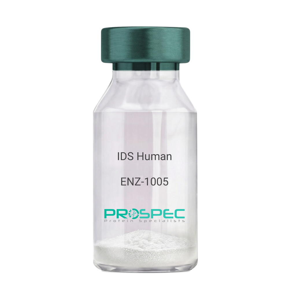
IDS HumanDescription:
Iduronate 2-Sulfatase Human Recombinant
Iduronate 2-Sulfatase, Alpha-L-Iduronate Sulfate Sulfatase, SIDS, Iduronate 2-Sulfatase 14 KDa Chain, Iduronate 2-Sulfatase 42 KDa Chain, Hunter Syndrome, EC 3.1.6.13, MPS2, Iduronate 2-sulfatase, Alpha-L-iduronate sulfate sulfatase.
Product # :
ENZ-1005Price :
Quantity :
Shipping Method :
Shipped with Ice Packs
More Info
- description
- source
- formulation
- purity
- More Info
Description
IDS Human Recombinant produced in Sf9 Baculovirus cells is a single, glycosylated polypeptide chain containing 533 amino acids (26-550a.a) and having a molecular mass of 60.3kDa. (Molecular size on SDS-PAGE will appear at approximately 35-70kDa). IDS is fused to an 8 amino acid His-tag at C-terminus & purified by proprietary chromatographic techniques.
Source
Sf9, Baculovirus cells.
Formulation
IDS protein solution (0.25mg/ml) containing Phosphate Buffered Saline (pH 7.4) and 10% glycerol.
Purity
Greater than 90% as determined by SDS-PAGE.
More Info
-
Introduction
Iduronate 2-Sulfatase also known as IDS, belongs to the highly-conserved sulfatase family of enzymes which catalyze the hydrolysis of O-sulfate and N-salfate esters from a variety of substrates. IDS is essential for the lysosomal degradation of the glycosaminoglycans (GAG) heparan sulfate as well as dermatan sulfate. Furthermore, IDS hydrolyzes the 2-sulfate group of the IDS units of the GAG.
-
Synonyms
Iduronate 2-Sulfatase, Alpha-L-Iduronate Sulfate Sulfatase, SIDS, Iduronate 2-Sulfatase 14 KDa Chain, Iduronate 2-Sulfatase 42 KDa Chain, Hunter Syndrome, EC 3.1.6.13, MPS2, Iduronate 2-sulfatase, Alpha-L-iduronate sulfate sulfatase.
-
Physical Appearance
Sterile Filtered clear solution.
-
Stability
Store at 4°C if entire vial will be used within 2-4 weeks. Store, frozen at -20°C for longer periods of time. For long term storage it is recommended to add a carrier protein (0.1% HSA or BSA).Avoid multiple freeze-thaw cycles.
-
Amino Acid Sequence
SETQANSTTD ALNVLLIIVD DLRPSLGCYG DKLVRSPNID QLASHSLLFQ NAFAQQAVCA PSRVSFLTGR RPDTTRLYDF NSYWRVHAGN FSTIPQYFKE NGYVTMSVGK VFHPGISSNH TDDSPYSWSF PPYHPSSEKY ENTKTCRGPD GELHANLLCP VDVLDVPEGT LPDKQSTEQA IQLLEKMKTS ASPFFLAVGY HKPHIPFRYP KEFQKLYPLE NITLAPDPEV PDGLPPVAYN PWMDIRQRED VQALNISVPY GPIPVDFQRK IRQSYFASVS YLDTQVGRLL SALDDLQLAN STIIAFTSDH GWALGEHGEW AKYSNFDVAT HVPLIFYVPG RTASLPEAGE KLFPYLDPFD SASQLMEPGR QSMDLVELVS LFPTLAGLAG LQVPPRCPVP SFHVELCREG KNLLKHFRFR DLEEDPYLPG NPRELIAYSQ YPRPSDIPQW NSDKPSLKDI KIMGYSIRTI DYRYTVWVGF NPDEFLANFS DIHAGELYFV DSDPLQDHNM YNDSQGGDLF QLLMPLEHHH HHH.
ProSpec's products are furnished for LABORATORY RESEARCH USE ONLY. The product may not be used as drugs, agricultural or pesticidal products, food additives or household chemicals.
- View Data Sheet
Name :
HERC5 HumanDescription:
HECT and RLD Domain Containing E3 Ubiquitin Protein Ligase 5 Human Recombinant
HERC5, HECT and RLD Domain Containing E3 Ubiquitin Protein Ligase 5, CEB1, Hect Domain and RLD 5, Cyclin-E-Binding Protein 1, CEBP1, HECT Domain and RCC1-Like Domain-Containing Protein 5, E3 ISG15--Protein Ligase HERC5, Probable E3 Ubiquitin-Protein Ligase HERC5, EC 6.3.2.- , EC 6.3.2.
Product # :
ENZ-797Price :
Quantity :
Shipping Method :
Shipped with Ice Packs
More Info
- description
- source
- formulation
- purity
- More Info
Description
HERC5 Human Recombinant produced in E.Coli is a single, non-glycosylated polypeptide chain containing 367 amino acids (681-1024 a.a.) and having a molecular mass of 43kDa. HERC5 is fused to a 23 amino acid His-tag at N-terminus.
Source
Escherichia Coli.
Formulation
HERC5 protein solution (1mg/ml) contains 20mM Tris-HCl buffer (pH 8.0), 0.4M Urea and 10% glycerol.
Purity
Greater than 90.0% as determined by SDS-PAGE.
More Info
-
Introduction
HECT and RLD Domain Containing E3 Ubiquitin Protein Ligase 5 (HERC5) is a member of the HERC family of ubiquitin ligases, found in a cluster of HERC family genes on chromosome 4. HERC5 is a protein with a HECT domain and 5 RCC1 repeats. The HERC5 protein localizes to the cytoplasm and perinuclear region and serves as an INF-induced E3 protein ligase that mediates ISGylation of protein targets. HERC5 exhibits antiviral activity towards HIV-1, influenza A virus and human papillomavirus. HERC5 is a major E3 ligase for ISG15 conjugation. HERC5 also serves as a positive regulator of innate antiviral response in cells induced by INF. Pro-inflammatory cytokines upregulate HERC5 in endothelial cells. HERC5 is physically connected with polyribosomes, broadly modifies recently synthesized proteins in a cotranslational fashion.
-
Synonyms
HERC5, HECT and RLD Domain Containing E3 Ubiquitin Protein Ligase 5, CEB1, Hect Domain and RLD 5, Cyclin-E-Binding Protein 1, CEBP1, HECT Domain and RCC1-Like Domain-Containing Protein 5, E3 ISG15--Protein Ligase HERC5, Probable E3 Ubiquitin-Protein Ligase HERC5, EC 6.3.2.- , EC 6.3.2.
-
Physical Appearance
Sterile Filtered colorless solution.
-
Stability
Store at 4°C if entire vial will be used within 2-4 weeks. Store, frozen at -20°C for longer periods of time. For long term storage it is recommended to add a carrier protein (0.1% HSA or BSA).Avoid multiple freeze-thaw cycles.
-
Amino Acid Sequence
MGSSHHHHHH SSGLVPRGSH MGSFDLTVRR NHLIEDVLNQ LSQFENEDLR KELWVSFSGE IGYDLGGVKK EFFYCLFAEM IQPEYGMFMY PEGASCMWFP VKPKFEKKRY FFFGVLCGLS LFNCNVANLP FPLALFKKLL DQMPSLEDLK ELSPDLGKNL QTLLDDEGDN FEEVFYIHFN VHWDRNDTNL IPNGSSITVN QTNKRDYVSK YINYIFNDSV KAVYEEFRRG FYKMCDEDII KLFHPEELKD VIVGNTDYDW KTFEKNARYE PGYNSSHPTI VMFWKAFHKL TLEEKKKFLV FLTGTDRLQM KDLNNMKITF CCPESWNERD PIRALTCFSV LFLPKYSTME TVEEALQEAI NNNRGFG.
ProSpec's products are furnished for LABORATORY RESEARCH USE ONLY. The product may not be used as drugs, agricultural or pesticidal products, food additives or household chemicals.
- View Data Sheet
Name :
HYAL1 HumanDescription:
Hyaluronidase Human Recombinant
Hyaluronidase-1, Hyal-1,Hyaluronoglucosaminidase-1, Lung carcinoma protein 1, LuCa-1, HYAL1, Hyaluronidase 1, Hyaluronoglucosaminidase 1, Hyaluronoglucosaminidase1, LUCA 1,MPS9, NAT6, Plasma hyaluronidase, Tumor suppressor LUCA 1.
Product # :
ENZ-1155Price :
Quantity :
Shipping Method :
Shipped with Ice Packs
More Info
- description
- source
- formulation
- purity
- More Info
Description
HYAL1 Human Recombinant produced in HEK cells is a single, glycosylated, polypeptide chain (22-435 a.a) containing a total of 420 amino acids, having a molecular mass of 46.9 kDa. HYAL1 is fused to a 6 amino acid His-tag at C-terminus,and is purified by proprietary chromatographic techniques.
Source
HEK293 Cells.
Formulation
The HYAL1 solution (0.25mg/ml) contains 10% Glycerol and Phosphate-Buffered Saline (pH 7.4).
Purity
Greater than 90.0% as determined by SDS-PAGE.
More Info
-
Introduction
Hyaluronidase-1 or HYAL1 is a protein, part of the endolytic glycoside hydrolase proteins group. Human hyaluronidases proteins, are 5 endoβNacetylhexosaminidases (including HYAL1, HYAL2, HYAL3). Hyaluronidase-1 causes degradation to hyaluronic acid in the extracellular matrix of somatic tissues. HYAL1 needs an acidic environment and is the most common hyaluronidase in the plasma. Mutations in this protein can lead to mucopolysaccharidosis type IX and hyaluronidase deficiency.
-
Synonyms
Hyaluronidase-1, Hyal-1,Hyaluronoglucosaminidase-1, Lung carcinoma protein 1, LuCa-1, HYAL1, Hyaluronidase 1, Hyaluronoglucosaminidase 1, Hyaluronoglucosaminidase1, LUCA 1,MPS9, NAT6, Plasma hyaluronidase, Tumor suppressor LUCA 1.
-
Physical Appearance
Sterile Filtered colorless solution.
-
Stability
Store at 4°C if entire vial will be used within 2-4 weeks. Store, frozen at -20°C for longer periods of time. For long term storage it is recommended to add a carrier protein (0.1% HSA or BSA).Avoid multiple freeze-thaw cycles.
-
Amino Acid Sequence
FRGPLLPNRP FTTVWNANTQ WCLERHGVDV DVSVFDVVAN PGQTFRGPDM TIFYSSQLGT YPYYTPTGEP VFGGLPQNAS LIAHLARTFQ DILAAIPAPD FSGLAVIDWE AWRPRWAFNW DTKDIYRQRS RALVQAQHPD WPAPQVEAVA QDQFQGAARA WMAGTLQLGR ALRPRGLWGF YGFPDCYNYD FLSPNYTGQC PSGIRAQNDQ LGWLWGQSRA LYPSIYMPAV LEGTGKSQMY VQHRVAEAFR VAVAAGDPNL PVLPYVQIFY DTTNHFLPLD ELEHSLGESA AQGAAGVVLW VSWENTRTKE SCQAIKEYMD TTLGPFILNV TSGALLCSQA LCSGHGRCVR RTSHPKALLL LNPASFSIQL TPGGGPLSLR GALSLEDQAQ MAVEFKCRCY PGWQAPWCER KSMWHHHHHH
ProSpec's products are furnished for LABORATORY RESEARCH USE ONLY. The product may not be used as drugs, agricultural or pesticidal products, food additives or household chemicals.
- View Data Sheet
Name :
CNDP1 Human, ActiveDescription:
CNDP Dipeptidase 1 Human Recombinant, Active
Carnosine Dipeptidase 1 (Metallopeptidase M20 Family), Glutamate Carboxypeptidase-Like Protein 2, CNDP Dipeptidase 1, Serum Carnosinase, Carnosinase 1, CPGL2, CN1, Carnosine Dipeptidase 1, EC 3.4.13.20, HsT2308.
Product # :
ENZ-1022Price :
Quantity :
Shipping Method :
Shipped with Ice Packs
More Info
- description
- source
- formulation
- purity
- biological activity
- More Info
Description
CNDP1 Human Recombinant produced in Sf9 Baculovirus cells is a single, glycosylated polypeptide chain containing 489 amino acids (27-507 a.a.) and having a molecular mass of 54.9kDa (Migrates at 50-70kDa on SDS-PAGE under reducing conditions).CNDP1 is expressed with a 8 amino acid His tag at C-Terminus and purified by proprietary chromatographic techniques.
Source
Sf9, Insect cells.
Formulation
CNDP1 protein solution (0.25mg/ml) contains Phosphate Buffered Saline (pH 7.4) and 10% glycerol.
Purity
Greater than 95.0% as determined by SDS-PAGE.
Biological Activity
Specific activity is >3,000 pmol/min/ug, and as measured by the hydrolysis of carnosine per minute at pH6.8 at 25°C.More Info
-
Introduction
CNDP Dipeptidase 1, also known as CNDP1 is a member of the peptidase M20A family. CNDP1 Mannheim which is the shortest allelic form has been more common in the absence of nephropathy in addition to being associated with lower serum carnosinase levels. Furthermore, Carnosine inhibited the increased production of fibronectin as well as collagen type VI in podocytes and the increased production of TGF-beta in mesangial cells. Diabetic patients with the CNDP1 Mannheim variant are less at risk for nephropathy. In addition, on renal cells carnosine protects against the adverse effects of high glucose levels.
-
Synonyms
Carnosine Dipeptidase 1 (Metallopeptidase M20 Family), Glutamate Carboxypeptidase-Like Protein 2, CNDP Dipeptidase 1, Serum Carnosinase, Carnosinase 1, CPGL2, CN1, Carnosine Dipeptidase 1, EC 3.4.13.20, HsT2308.
-
Physical Appearance
Sterile filtered colorless solution.
-
Stability
Store at 4°C if entire vial will be used within 2-4 weeks.Store, frozen at -20°C for longer periods of time. For long term storage it is recommended to add a carrier protein (0.1% HSA or BSA).Avoid multiple freeze-thaw cycles.
-
Amino Acid Sequence
SPSPPPALLE KVFQYIDLHQ DEFVQTLKEW VAIESDSVQP VPRFRQELFR MMAVAADTLQ RLGARVASVD MGPQQLPDGQ SLPIPPVILA ELGSDPTKGT VCFYGHLDVQ PADRGDGWLT DPYVLTEVDG KLYGRGATDN KGPVLAWINA VSAFRALEQD LPVNIKFIIE GMEEAGSVAL EELVEKEKDR FFSGVDYIVI SDNLWISQRK PAITYGTRGN SYFMVEVKCR DQDFHSGTFG GILHEPMADL VALLGSLVDS SGHILVPGIY DEVVPLTEEE INTYKAIHLD LEEYRNSSRV EKFLFDTKEE ILMHLWRYPS LSIHGIEGAF DEPGTKTVIP GRVIGKFSIR LVPHMNVSAV EKQVTRHLED VFSKRNSSNK MVVSMTLGLH PWIANIDDTQ YLAAKRAIRT VFGTEPDMIR DGSTIPIAKM FQEIVHKSVV LIPLGAVDDG EHSQNEKINR WNYIEGTKLF AAFFLEMAQL HLEHHHHHH.
ProSpec's products are furnished for LABORATORY RESEARCH USE ONLY. The product may not be used as drugs, agricultural or pesticidal products, food additives or household chemicals.
- View Data Sheet
Name :
PCOLCE HumanDescription:
Procollagen C-Endopeptidase Enhancer Human Recombinant
Procollagen C-Endopeptidase Enhancer, Procollagen C-Proteinase Enhancer 1, Type I Procollagen COOH-Terminal Proteinase Enhancer, Type 1 Procollagen C-Proteinase Enhancer Protein, Procollagen COOH-Terminal Proteinase Enhancer 1, PCPE-1, PCPE1, Procollagen, Type 1, COOH-Terminal Proteinase Enhancer, Procollagen C-Endopeptidase Enhancer 1, COOH-Terminal Proteinase Enhancer, Procollagen, Type 1, PCPE, Procollagen C-endopeptidase enhancer 1.
Product # :
ENZ-863Price :
Quantity :
Shipping Method :
Shipped with Ice Packs
More Info
- description
- source
- formulation
- purity
- More Info
Description
PCOLCE Human Recombinant produced in E.Coli is a single, non-glycosylated polypeptide chain containing 447 amino acids (26-449 a.a) and having a molecular mass of 47.9kDa. PCOLCE is fused to a 23 amino acid His-tag at N-terminus & purified by proprietary chromatographic techniques.
Source
Escherichia Coli.
Formulation
PCOLCE protein solution (1mg/ml) containing 20mM Tris-HCl buffer (pH 8.0) and 10% glycerol.
Purity
Greater than 85.0% as determined by SDS-PAGE.
More Info
-
Introduction
Procollagen C-endopeptidase enhancer 1, also known as PCOLCE binds to the C-terminal propeptide of type I procollagen and enhances procollagen C-proteinase activity. In addition, C-terminal processed part of PCPE (CT-PCPE) has a metalloproteinase inhibitory activity. Among the diseases which are associated with PCOLCE: bone fracture & oculopharyngeal muscular dystrophy.
-
Synonyms
Procollagen C-Endopeptidase Enhancer, Procollagen C-Proteinase Enhancer 1, Type I Procollagen COOH-Terminal Proteinase Enhancer, Type 1 Procollagen C-Proteinase Enhancer Protein, Procollagen COOH-Terminal Proteinase Enhancer 1, PCPE-1, PCPE1, Procollagen, Type 1, COOH-Terminal Proteinase Enhancer, Procollagen C-Endopeptidase Enhancer 1, COOH-Terminal Proteinase Enhancer, Procollagen, Type 1, PCPE, Procollagen C-endopeptidase enhancer 1.
-
Physical Appearance
Sterile filtered colorless solution.
-
Stability
Store at 4°C if entire vial will be used within 2-4 weeks. Store, frozen at -20°C for longer periods of time. For long term storage it is recommended to add a carrier protein (0.1% HSA or BSA).Avoid multiple freeze-thaw cycles.
-
Amino Acid Sequence
MGSSHHHHHH SSGLVPRGSH MGSQTPNYTR PVFLCGGDVK GESGYVASEG FPNLYPPNKE CIWTITVPEG QTVSLSFRVF DLELHPACRY DALEVFAGSG TSGQRLGRFC GTFRPAPLVA PGNQVTLRMT TDEGTGGRGF LLWYSGRATS GTEHQFCGGR LEKAQGTLTT PNWPESDYPP GISCSWHIIA PPDQVIALTF EKFDLEPDTY CRYDSVSVFN GAVSDDSRRL GKFCGDAVPG SISSEGNELL VQFVSDLSVT ADGFSASYKT LPRGTAKEGQ GPGPKRGTEP KVKLPPKSQP PEKTEESPSA PDAPTCPKQC RRTGTLQSNF CASSLVVTAT VKSMVREPGE GLAVTVSLIG AYKTGGLDLP SPPTGASLKF YVPCKQCPPM KKGVSYLLMG QVEENRGPVL PPESFVVLHR PNQDQILTNL SKRKCPSQPV RAAASQD.
ProSpec's products are furnished for LABORATORY RESEARCH USE ONLY. The product may not be used as drugs, agricultural or pesticidal products, food additives or household chemicals.
- View Data Sheet
Name :
LPL Human, HEKDescription:
Lipoprotein Lipase Human Recombinant, HEK
Lipoprotein lipase, LPL, LIPD, HDLCQ11.
Product # :
ENZ-087Price :
Quantity :
Shipping Method :
Shipped at Room temp
More Info
- description
- source
- formulation
- More Info
Description
The Recombinant Human LPL produced in HEK293 cell line has a molecular mass of 51.8kDa containing 461 amino acid residues of the human LPL (Ala28-Gly475, variant Asn > Ser318) and fused to a 13 a.a. Flag-tag at N-terminus.
Source
HEK293 (Human Embryonic Kidney cell line).
Formulation
LPL was filtered (0.4 µm) and lyophilized from 0.5mg/ml in 20mM Tris buffer and 50mM NaCl, pH 7.5.
More Info
-
Introduction
LPL is a lipoprotein lipase, which is expressed in the heart, muscle, and adipose tissue. LPL acts as a homodimer, and has the dual functions of triglyceride hydrolase and ligand/bridging factor for receptor-mediated lipoprotein uptake. Type I hyperlipoproteinemia is a result of severe mutations which cause LPL deficiency, whereas less extreme mutations in LPL are linked to many disorders of lipoprotein metabolism. Lipoprotein lipase (LPL) is a fundamental enzyme in plasma triglyceride hydrolysis and is secreted by macrophages in the subendothelial space. LPL also promotes the development of atherosclerosis through facilitation of monocyte adhesion to endothelial cells, stimulation of tumor necrosis factor alpha (TNF) secretion and induction of vascular smooth muscle cell proliferation.
-
Synonyms
Lipoprotein lipase, LPL, LIPD, HDLCQ11.
-
Physical Appearance
Filtered white lyophilized powder.
-
Stability
Store lyophilized protein at -20°C. Aliquot the product after reconstitution to avoid repeated freezing/thawing cycles. Reconstituted protein can be stored at 4°C for a limited period of time; it does not show any change after two weeks at 4°C.
-
Solubility
It is recommended to add deionized water to prepare a working stock solution of approximately 0.5 mg/ml and let the lyophilized pellet dissolve completely. Product is not sterile! Please filter the product by an appropriate sterile filter before using it in the cell culture.
-
Amino Acid Sequence
HVDYKDDDDK PAGADQRRDF IDIESKFALR TPEDTAEDTC HLIPGVAESV ATCHFNHSSK TFMVIHGWTV TGMYESWVPK ADQRRDF IDIESKFALR TPEDTAEDTC HLIPGVAESV ATCHFNHSSK TFMVIHGWTV TGMYESWVPK LVAALYKREP DSNVIVVDWL SRAQEHYPVS AGYTKLVGQD VARFINWMEE EFNYPLDNVH LLGYSLGAHA AGIAGSLTNK KVNRITGLDP AGPNFEYAEA PSRLSPDDAD FVDVLHTFTR GSPGRSIGIQ KPVGHVDIYP NGGTFQPGCN IGEAIRVIAE RGLGDVDQLV KCSHERSIHL FIDSLLNEEN PSKAYRCSSK EAFEKGLCLS CRKNRCNNLG YEISKVRAKR SSKMYLKTRS QMPYKVFHYQ VKIHFSGTES ETHTNQAFEI SLYGTVAESE NIPFTLPEVS TNKTYSFLIY TEVDIGELLM LKLKWKSDSY FSWSDWWSSP GFAIQKIRVK AGETQKKVIF CSREKVSHLQ KGKAPAVFVK CHDKSLNKKS G.
ProSpec's products are furnished for LABORATORY RESEARCH USE ONLY. The product may not be used as drugs, agricultural or pesticidal products, food additives or household chemicals.
- View Data Sheet
Name :
Enterokinase Bovine HisDescription:
Enteropeptidase/ Enterokinase Bovine Recombinant His Tag
Enteropeptidase, EC 3.4.21.9, Enterokinase, Serine protease 7, ENTK, MGC133046.
Product # :
ENZ-655Price :
Quantity :
Shipping Method :
Shipped with Ice Packs
More Info
- description
- source
- formulation
- More Info
Description
Enterokinase Bovine Recombinant produced in E.Coli is a single, non-glycosylated, polypeptide chain containing 241 amino acids with a 6 × His at C-terminus and having a molecular mass of 28.0kDa.The Enterokinase Bovine is purified by proprietary chromatographic techniques.
Source
Escherichia Coli.
Formulation
Bovine EK is supplied in 50mM Tris-HCl, pH 8.0, 0.5M NaCl and 50% glycerol.
More Info
-
Introduction
Enteropeptidase or enterokinase is an enzymeinvolved in human digestion. It is produced by cells in the duodenum wall, and is secreted from duodenum's glands, the crypts of Lieberk?hn, whenever ingested food enters the duodenum from the stomach. Enteropeptidase has the critical job of turning trypsinogen(a zymogen) to trypsin, indirectly activating a number of pancreaticdigestive enzymes.
Enteropeptidase is a serine proteaseenzyme(EC3.4.21.9). Enteropeptidase is a part of the Chymotrypsin-clan of serine proteases, and is structurally similar to these proteins. -
Synonyms
Enteropeptidase, EC 3.4.21.9, Enterokinase, Serine protease 7, ENTK, MGC133046.
-
Physical Appearance
Sterile liquid solution.
-
Stability
One year when stored at -20°C. Please avoid freeze-thaw cycles.
-
Unit Definition
One unit is defined as the amount of enzyme needed to cleave 50ug of fusion protein in 16 hours to 95% completion at 25°C in a buffer containing 25mM Tris-HCl, pH 7.6, 50mM NaCl, and 2mM CaCl2.
ProSpec's products are furnished for LABORATORY RESEARCH USE ONLY. The product may not be used as drugs, agricultural or pesticidal products, food additives or household chemicals.
- View Data Sheet
Name :
MMP-2 Human, HEKDescription:
Matrix Metalloproteinase-2 Human Recombinant, HEK
72 kDa type IV collagenase, 72 kDa gelatinase, Gelatinase A, Matrix metalloproteinase-2, MMP-2, TBE-1, MMP2, CLG4A, CLG4, MONA, MMP-II.
Product # :
ENZ-100Price :
Quantity :
Shipping Method :
Shipped with Ice Packs
More Info
- description
- source
- formulation
- purity
- biological activity
- More Info
- sds-page
Description
MMP-2 Human Recombinant produced in HEK293 cells is a proform of the Human MMP-2 (Ala30-Cys660) and fused with a ployhistide tag at the C-terminus, having an Mw of 71kDa. MMP-2 is purified by proprietary chromatographic techniques.
Source
HEK293 cells.
Formulation
The MMP-2 is supplied as a 0.2µm filtered solution in 20mM Tris-HCl, 150mM NaCl and 0.05% Brij 35, pH 7.4.
Purity
Greater than 95% as determined by SDS-PAGE.
Biological Activity
The activity was measured by its ability to cleave fluorogenic peptide substrate, Mca-PLGL-Dpa-AR-NH2 (RND,Catalog # ES001)., The specific activity is > 1,000 pmoles/min/µg.
Recombinant Human MMP-2 protein pro form needs to be activated with p-aminophenylmercuric acetate (APMA).
Activation Protocol:
1. Dilute MMP2 to 100µg/ml in the Assay Buffer: 50mM Tris, 10mM CaCl2, 150mM NaCl, 0.05% (w/v) and Brij 35, pH 7.5.
2. Activate MMP2 by adding APMA to a final concentration of 1mM. (Sigma, Catalog # A9563) and 100mM stock in DMSO.
3. Incubate at 37°C for 1 hour.sds-page
More Info
-
Introduction
Matrix metalloproteinase-2 (MMP-2) is a type IV collagenase, which is involved in endometrial menstrual breakdown, regulation of vascularization and the inflammatory response. MMP-2 contains a number of distinct domains: a prodomain that is cleaved upon activation; a catalytic domain containing the zinc binding site; a fibronectin like domain believed to have a role in substrate targeting; and a carboxyl terminal (hemopexin like) domain containing 2 N-linked glycosylation. The MMP-2 can degrade an extensive array of substrates including type IV, V, VII and X collagens as well as gelatin type I. In addition, MMP-2 interacts with THBS2, TIMP2, Thrombospondin 1, CCL7 and TIMP4. MMP-2 autocatalytic cleavage in the C-terminal generates the anti-angiogenic peptide, PEX. This process seems to be made possible by binding integrinv/beta3. Defects in the MMP-2 are the cause of Torg-Winchester syndrome (TWS), aka multicentric osteolysis nodulosis and arthropathy (MONA).
-
Synonyms
72 kDa type IV collagenase, 72 kDa gelatinase, Gelatinase A, Matrix metalloproteinase-2, MMP-2, TBE-1, MMP2, CLG4A, CLG4, MONA, MMP-II.
-
Physical Appearance
The MMP-2 is supplied as a sterile Filtered colorless solution.
-
Stability
Store MMP-2 at 4°C if entire vial will be used within 2-4 weeks. Store frozen at -20°C for longer periods of time.Avoid multiple freeze-thaw cycles.
ProSpec's products are furnished for LABORATORY RESEARCH USE ONLY. The product may not be used as drugs, agricultural or pesticidal products, food additives or household chemicals.
- View Data Sheet
Name :
HMGCL HumanDescription:
3-Hydroxymethyl-3-Methylglutaryl-CoA Lyase Human Recombinant
Hydroxymethylglutaryl-CoA lyase mitochondrial, HL, HMG-CoA lyase, 3-hydroxy-3-methylglutarate-CoA lyase, HMGCL.
Product # :
ENZ-218Price :
Quantity :
Shipping Method :
Shipped with Ice Packs
More Info
- description
- source
- formulation
- purity
- More Info
Description
HMGCL Human Recombinant produced in E.coli is a single, non-glycosylated polypeptide chain containing 323 amino acids (28-325) and having a molecular mass of 34.2kDa.HMGCL is fused to a 25 amino acid His-tag at N-terminus & purified by proprietary chromatographic techniques.
Source
Escherichia Coli.
Formulation
The HMGCL solution (1mg/ml) contains 20mM Tris-HCl buffer (pH8.0), 10% glycerol and 0.4M Urea.
Purity
Greater than 85.0% as determined by SDS-PAGE.
More Info
-
Introduction
Hydroxymethylglutaryl-CoA lyase (HMGCL) is a mitochondrial matrix protein which is a member of the HMG-CoA lyase family. HMGCL is a homodimer and participates in leucine catabolism and ketogenesis, the hepatic synthesis of ketone bodies which, during fasting, provides a major source of energy for the heart, brain and kidney. More precisely, HMGCL catalyzes the final step of these processes, the cleavage of 3-hydroxy-3-methylglutaryl-CoA to acetoacetic acid and acetyl-CoA.
-
Synonyms
Hydroxymethylglutaryl-CoA lyase mitochondrial, HL, HMG-CoA lyase, 3-hydroxy-3-methylglutarate-CoA lyase, HMGCL.
-
Physical Appearance
Sterile Filtered colorless solution.
-
Stability
Store at 4°C if entire vial will be used within 2-4 weeks. Store, frozen at -20°C for longer periods of time. For long term storage it is recommended to add a carrier protein (0.1% HSA or BSA).Avoid multiple freeze-thaw cycles.
-
Amino Acid Sequence
MGSSHHHHHH SSGLVPRGSH MGSHMTLPKR VKIVEVGPRD GLQNEKNIVS TPVKIKLIDM LSEAGLSVIE TTSFVSPKWV PQMGDHTEVL KGIQKFPGIN YPVLTPNLKG FEAAVAAGAK EVVIFGAASE LFTKKNINCS IEESFQRFDA ILKAAQSANI SVRGYVSCAL GCPYEGKISP AKVAEVTKKF YSMGCYEISL GDTIGVGTPG IMKDMLSAVM QEVPLAALAV HCHDTYGQAL ANTLMALQMG VSVVDSSVAG LGGCPYAQGA SGNLATEDLV YMLEGLGIHT GVNLQKLLEA GNFICQALNR KTSSKVAQAT CKL.
ProSpec's products are furnished for LABORATORY RESEARCH USE ONLY. The product may not be used as drugs, agricultural or pesticidal products, food additives or household chemicals.
- View Data Sheet
Name :
NAPSA HumanDescription:
Napsin A Aspartic Peptidase Human Recombinant
Napsin A Aspartic Peptidase, NAP1, NAPA, Kidney-Derived Aspartic Protease-Like Protein, Aspartyl Protease 4, TA01/TA02, Napsin-1, SNAPA, Asp 4, ASP4, CTB-191K22.6, EC 3.4.23.15, Pronapsin A, EC 3.4.23.5, EC 3.4.23.3, EC 3.4.23.-, EC 3.4.23, Napsin-A, KDAP, KAP,Napsin-A.
Product # :
ENZ-841Price :
Quantity :
Shipping Method :
Shipped with Ice Packs
More Info
- description
- source
- formulation
- purity
- More Info
Description
NAPSA Human Recombinant produced in E.Coli is a single, non-glycosylated polypeptide chain containing 380 amino acids (64-420 a.a) and having a molecular mass of 40.9kDa. NAPSA is fused to a 23 amino acid His-tag at N-terminus & purified by proprietary chromatographic techniques.
Source
Escherichia Coli.
Formulation
NAPSA protein solution (0.25mg/ml) containing 20mM Tris-HCl (pH8.0) and 10% glycerol.
Purity
Greater than 90.0% as determined by SDS-PAGE.
More Info
-
Introduction
Napsin A Aspartic Peptidase, also known as NAPSA is a member of the peptidase A1 family. NAPSA is involved in the processing of pneumocyte surfactant precursors. Furthermore the activation peptides of aspartic proteinases take part as inhibitors of the active site. These peptide segments/pro-parts are considered essential for correct folding, targeting, as well as control of the activation of aspartic proteinase zymogens. The pronapsin A gene is expressed mostly in lung and kidney. In addition, NAPSA translation product is expected to be a fully functional, glycosylated aspartic proteinase precursor which contains an RGD motif as well as an additional 18 residues at its C-terminus.
-
Synonyms
Napsin A Aspartic Peptidase, NAP1, NAPA, Kidney-Derived Aspartic Protease-Like Protein, Aspartyl Protease 4, TA01/TA02, Napsin-1, SNAPA, Asp 4, ASP4, CTB-191K22.6, EC 3.4.23.15, Pronapsin A, EC 3.4.23.5, EC 3.4.23.3, EC 3.4.23.-, EC 3.4.23, Napsin-A, KDAP, KAP,Napsin-A.
-
Physical Appearance
Sterile filtered colorless solution.
-
Stability
Store at 4°C if entire vial will be used within 2-4 weeks. Store, frozen at -20°C for longer periods of time. For long term storage it is recommended to add a carrier protein (0.1% HSA or BSA).Avoid multiple freeze-thaw cycles.
-
Amino Acid Sequence
MGSSHHHHHH SSGLVPRGSH MGSKPIFVPL SNYRDVQYFG EIGLGTPPQN FTVAFDTGSS NLWVPSRRCH FFSVPCWLHH RFDPKASSSF QANGTKFAIQ YGTGRVDGIL SEDKLTIGGI KGASVIFGEA LWEPSLVFAF AHFDGILGLG FPILSVEGVR PPMDVLVEQG LLDKPVFSFY LNRDPEEPDG GELVLGGSDP AHYIPPLTFV PVTVPAYWQI HMERVKVGPG LTLCAKGCAA ILDTGTSLIT GPTEEIRALH AAIGGIPLLA GEYIILCSEI PKLPAVSFLL GGVWFNLTAH DYVIQTTRNG VRLCLSGFQA LDVPPPAGPF WILGDVFLGT YVAVFDRGDM KSSARVGLAR ARTRGADLGW GETAQAQFPG.
ProSpec's products are furnished for LABORATORY RESEARCH USE ONLY. The product may not be used as drugs, agricultural or pesticidal products, food additives or household chemicals.
- View Data Sheet
Name :
IDE Human, ActiveDescription:
Insulin-Degrading Enzyme Human Recombinant
Insulin-Degrading Enzyme, Abeta-Degrading Protease, Insulysin, EC 3.4.24.56, Insulinase, Insulin Protease, INSULYSIN, EC 3.4.24, IDE, insulin-degrading enzyme isoform 1.
Product # :
ENZ-1192Price :
Quantity :
Shipping Method :
Shipped with Ice Packs
More Info
- description
- source
- formulation
- purity
- biological activity
- More Info
Description
IDE Human, Active Human Recombinant produced in E.Coli is a single, non-glycosylated, polypeptide chain (42-1019 a.a) containing a total of 984 amino acids, having a molecular mass of 114 kDa. IDE is fused to a 6 amino acid His-tag at C-terminus and is purified by proprietary chromatographic techniques.
Source
Escherichia Coli.
Formulation
The IDE solution (0.5mg/ml) contains 10% Glycerol, 100mM NaCl, 0.05% Brij35 and 20mM Tris-HCl buffer (pH 7.5).
Purity
Greater than 90.0% as determined by SDS-PAGE.
Biological Activity
Specific activity is greater than 3,000 pmol/min/ug in which 1 unit will convert 1.0 pmole of Mca-RPPGFSAFK(Dnp)-OH to MCA-Pro-Leu-OH per minute at pH 7.5 at 25°C.
More Info
-
Synonyms
Insulin-Degrading Enzyme, Abeta-Degrading Protease, Insulysin, EC 3.4.24.56, Insulinase, Insulin Protease, INSULYSIN, EC 3.4.24, IDE, insulin-degrading enzyme isoform 1.
-
Physical Appearance
Filtered colorless solution.
-
Stability
Store at 4°C if entire vial will be used within 2-4 weeks. Store, frozen at -20°C for longer periods of time. For long term storage it is recommended to add a carrier protein (0.1% HSA or BSA). Avoid multiple freeze-thaw cycles.
-
Amino Acid Sequence
MNNPAIKRIG NHITKSPEDK REYRGLELAN GIKVLLISDP TTDKSSAALD VHIGSLSDPP NIAGLSHFCE HMLFLGTKKY PKENEYSQFL SEHAGSSNAF TSGEHTNYYF DVSHEHLEGA LDRFAQFFLC PLFDESCKDR EVNAVDSEHE KNVMNDAWRL FQLEKATGNP KHPFSKFGTG NKYTLETRPN QEGIDVRQEL LKFHSAYYSS NLMAVCVLGR ESLDDLTNLV VKLFSEVENK NVPLPEFPEH PFQEEHLKQL YKIVPIKDIR NLYVTFPIPD LQKYYKSNPG HYLGHLIGHE GPGSLLSELK SKGWVNTLVG GQKEGARGFM FFIINVDLTE EGLLHVEDII LHMFQYIQKL RAEGPQEWVF QECKDLNAVA FRFKDKERPR GYTSKIAGIL HYYPLEEVLT AEYLLEEFRP DLIEMVLDKL RPENVRVAIV SKSFEGKTDR TEEWYGTQYK QEAIPDEVIK KWQNADLNGK FKLPTKNEFI PTNFEILPLE KEATPYPALI KDTAMSKLWF KQDDKFFLPK ACLNFEFFSP FAYVDPLHCN MAYLYLELLK DSLNEYAYAA ELAGLSYDLQ NTIYGMYLSV KGYNDKQPIL LKKIIEKMAT FEIDEKRFEI IKEAYMRSLN NFRAEQPHQH AMYYLRLLMT EVAWTKDELK EALDDVTLPR LKAFIPQLLS RLHIEALLHG NITKQAALGI MQMVEDTLIE HAHTKPLLPS
QLVRYREVQL PDRGWFVYQQ RNEVHNNCGI EIYYQTDMQS TSENMFLELF CQIISEPCFN TLRTKEQLGY IVFSGPRRAN GIQGLRFIIQ SEKPPHYLES RVEAFLITME KSIEDMTEEA FQKHIQALAI RRLDKPKKLS AECAKYWGEI ISQQYNFDRD NTEVAYLKTL TKEDIIKFYK EMLAVDAPRR HKVSVHVLAR EMDSCPVVGE FPCQNDINLS QAPALPQPEV IQNMTEFKRG LPLFPLVKPH INFMAAKLHH HHHH. -
Background
Insulin-degrading enzyme (IDE) is a crucial protease that plays a significant role in maintaining glucose homeostasis by degrading insulin and other bioactive peptides. Dysregulation of IDE has been implicated in various metabolic disorders, particularly type 2 diabetes mellitus. IDE is also associated with the clearance of amyloid-beta peptides in the brain, making it relevant to Alzheimer's disease pathology. Studying the recombinant form of IDE is fundamental to understanding its functional mechanisms and exploring potential avenues for therapeutic interventions.
The primary goal of this research is to express and purify recombinant IDE using diverse expression systems. Recombinant DNA techniques will be employed to construct expression vectors containing the IDE gene, followed by expression in bacterial, yeast, or mammalian cell-based systems. The recombinant IDE will be purified using affinity chromatography or other appropriate methods, facilitating subsequent biochemical and biophysical characterization.
The second objective is to investigate the substrate specificity and catalytic activity of the purified IDE. In vitro enzymatic assays will be conducted to analyse the ability of the recombinant IDE to degrade insulin and other potential substrates. The effects of various factors, such as pH, temperature, and potential modulators, on IDE activity will be evaluated. Additionally, the interactions between IDE and its substrates will be explored using binding assays.
The third objective is to elucidate the three-dimensional structure of the IDE recombinant using techniques like X-ray crystallography or nuclear magnetic resonance (NMR) spectroscopy. Structural insights into the active site and binding pockets of IDE will provide valuable information for understanding its substrate recognition and catalytic mechanisms. This knowledge could be instrumental in designing targeted therapeutic compounds.
By characterizing the IDE recombinant, this research aims to contribute to our understanding of its role in insulin metabolism, glucose regulation, and potential therapeutic applications. The findings from this study may have implications for the development of novel treatments for diabetes and other related disorders.
ProSpec's products are furnished for LABORATORY RESEARCH USE ONLY. The product may not be used as drugs, agricultural or pesticidal products, food additives or household chemicals.