Search results
1000 results found for “glycoprotein nmb”
Name
Description
Product #
Price
Quantity
Shipping Method
- View Data Sheet
Name :
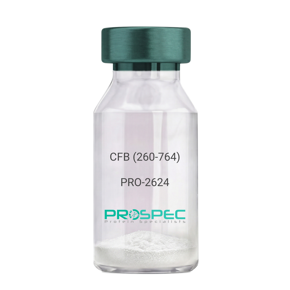
CFB (260-764) Human, Sf9Description:
Complement Factor B (260-764 a.a.) Human Recombinant, Sf9
CFB, C3/C5 convertase, Glycine-rich beta glycoprotein, GBG, PBF2, Properdin factor B, BF, BFD, AHUS4, ARMD14, CFAB, CFBD, FB, FBI12, GBG, H2-Bf.
Product # :
PRO-2624Price :
Quantity :
Shipping Method :
Shipped with Ice Packs
More Info
- description
- source
- formulation
- purity
- More Info
Description
CFB Human Recombinant produced in Sf9 Baculovirus cells is a single, glycosylated polypeptide chain containing 514 amino acids (260-764 a.a) and having a molecular mass of 58.1kDa.CFB is fused to an 9 amino acid His-tag at C-terminus & purified by proprietary chromatographic techniques.
Source
Sf9, Baculovirus cells.
Formulation
CFB protein solution (0.5mg/ml) containing Phosphate Buffered Saline (pH 7.4) and 20% glycerol.
Purity
Greater than 90.0% as determined by SDS-PAGE.
More Info
-
Introduction
Factor B is a protein, build from a single chain that circulates in the blood. Complement factor D cleaves and activates factor B, thus creating a catalytic subunit Bb and noncatalytic chain Ba. Subunit Bb is a serine protease that can bind to C3b and forms alternative pathway C3 convertase. Subunit Ba acts as an inhibitor for proliferation of lymphocytes, when Bb is part of the lymphocytes’ proliferation. The gene that codes for this protein is located on chromosome six.
-
Synonyms
CFB, C3/C5 convertase, Glycine-rich beta glycoprotein, GBG, PBF2, Properdin factor B, BF, BFD, AHUS4, ARMD14, CFAB, CFBD, FB, FBI12, GBG, H2-Bf.
-
Physical Appearance
Sterile Filtered colorless solution.
-
Stability
Store at 4°C if entire vial will be used within 2-4 weeks. Store, frozen at -20°C for longer periods of time. For long term storage it is recommended to add a carrier protein (0.1% HSA or BSA).Avoid multiple freeze-thaw cycles.
-
Amino Acid Sequence
ADPKIVLDPS GSMNIYLVLD GSDSIGASNF TGAKKCLVNL IEKVASYGVK PRYGLVTYAT
YPKIWVKVSE ADSSNADWVT KQLNEINYED HKLKSGTNTK KALQAVYSMM SWPDDVPPEG
WNRTRHVIIL MTDGLHNMGG DPITVIDEIR DLLYIGKDRK NPREDYLDVY VFGVGPLVNQ
VNINALASKK DNEQHVFKVK DMENLEDVFY QMIDESQSLS LCGMVWEHRK GTDYHKQPWQ
AKISVIRPSK GHESCMGAVV SEYFVLTAAH CFTVDDKEHS IKVSVGGEKR DLEIEVVLFH
PNYNINGKKE AGIPEFYDYD VALIKLKNKL KYGQTIRPIC LPCTEGTTRA LRLPPTTTCQ
QQKEELLPAQ DIKALFVSEE EKKLTRKEVY IKNGDKKGSC ERDAQYAPGY DKVKDISEVV
TPRFLCTGGV SPYADPNTCR GDSGGPLIVH KRSRFIQVGV ISWGVVDVCK NQKRQKQVPA HARDFHINLF QVLPWLKEKL QDEDLGFLHH HHHH
ProSpec's products are furnished for LABORATORY RESEARCH USE ONLY. The product may not be used as drugs, agricultural or pesticidal products, food additives or household chemicals.
- View Data Sheet
Name :
b NGF Human, HEKDescription:
beta Nerve Growth Factor Human Recombinant, HEK
Beta Polypeptide, NGF, NGFB, HSAN5, Beta-NGF, MGC161426, MGC161428.
Product # :
CYT-079Price :
Quantity :
Shipping Method :
Shipped at Room temp
More Info
- description
- source
- formulation
- purity
- biological activity
- More Info
Description
BNGF Human Recombinant produced in HEK293 is a noncovalently disulfide linked homodimer, glycosylated, polypeptide chain (Ser122-Arg239) containing 2 identical 118 amino acids and having a molecular mass of 26.5 kDa.
Source
HEK293 cells.
Formulation
The b-NGF was lyophilized from 1mg/ml in 20mM PB and 0.25M NaCl pH-7.5.
Purity
Greater than 97% as obsereved by SDS-PAGE.
Biological Activity
The specific activity was determined by the dose-dependent stimulation of the proliferation of human TF-1 cells (human erythroleukemic indicator cell line), the ED50 is <0.04-0.4ng/ml.
More Info
-
Introduction
NGF-beta has nerve growth stimulating activity and the complex is involved in the regulation of growth and the differentiation of sympathetic and certain sensory neurons. Mutations in this gene have been associated with hereditary sensory and autonomic neuropathy, type 5 (HSAN5), and dysregulation of this gene's expression is associated with allergic rhinitis.
-
Synonyms
Beta Polypeptide, NGF, NGFB, HSAN5, Beta-NGF, MGC161426, MGC161428.
-
Physical Appearance
Sterile Filtered White lyophilized (freeze-dried) powder.
-
Stability
Lyophilized b-NGF although stable at room temperature for 3 weeks, should be stored desiccated below -18°C. Upon reconstitution b-NGF should be stored at 4°C between 2-7 days and for future use below -18°C.For long term storage it is recommended to add a carrier protein (0.1% HSA or BSA).Please prevent freeze-thaw cycles.
-
Solubility
It is recommended to reconstitute the lyophilized b-NGF in sterile distilled pyrogen free water at a concentration of 0.25mg/ml.
-
Background
What is the molecular weight/Mw of B NGF Protein?
B NGF Protein has a total Mw of 26.5kDa.
What is the source or expression system of B NGF Protein?
HEK293 cells.
What is the Purity of B NGF Protein?
B NGF Protein is >97% pure as determined by SDS-PAGE.
What is the Biological Activity of B NGF Protein?
The specific activity was determined by the dose-dependent stimulation of the proliferation of human TF-1 cells (human erythroleukemic indicator cell line), the ED50 is <0.04-0.4ng/ml.
What is the amino acid sequence of B NGF Protein?
B NGF Protein is composed from 118 amino acids.
What applications can B NGF Protein be used in?
B NGF Protein can probably be used in western blot, ELISA and Lateral Flow.
What is the endotoxin level for B NGF Protein?
The endotoxin level is minimal, B NGF Protein was purified using conventional chromatography techniques.
ProSpec's products are furnished for LABORATORY RESEARCH USE ONLY. The product may not be used as drugs, agricultural or pesticidal products, food additives or household chemicals.
- View Data Sheet
Name :
F8 ProteinDescription:
Coagulation Factor-VIII Human Recombinant
Coagulation factor VIII, Procoagulant component, Antihemophilic factor, AHF, F8, F8C, F8B, HEMA, FVIII, DXS1253E, F8 protein.
Product # :
PRO-318Price :
Quantity :
Shipping Method :
Shipped at Room temp
More Info
- description
- source
- formulation
- purity
- biological activity
- More Info
Description
Antihemophilic Facor Human Recombinant produced in CHO is a glycosylated polypeptide chain having 2332 amino acids. The Factor-VIII is purified by proprietary chromatographic techniques.
Source
CHO cells (Chinese Hamster Ovarian Cells).
Formulation
Each 250IU vial was lyophilized from a solution containing 8mg Tween-80, 112mM NaCl, 40mg Mannitol, 10mg Trehalose, 1ng VWF and 4.2mM CaCl2.
Purity
Greater than 97.0% as determined by SDS-PAGE.
Biological Activity
The specific activity was found to be 7,058 IU/mg.More Info
-
Introduction
Coagulation factor VIII participates in the intrinsic pathway of blood coagulation; factor VIII is a cofactor for factor IXa which, in the presence of Ca+2 and phospholipids, converts factor X to the activated form Xa. This gene produces two alternatively spliced transcripts. Transcript variant 1 encodes a large glycoprotein, isoform a, which circulates in plasma and associates with von Willebrand factor in a noncovalent complex. This protein undergoes multiple cleavage events. Transcript variant 2 encodes a putative small protein, isoform b, which consists primarily of the phospholipid binding domain of factor VIIIc. This binding domain is essential for coagulant activity. Defects in this gene results in hemophilia A, a common recessive X-linked coagulation disorder.
-
Synonyms
Coagulation factor VIII, Procoagulant component, Antihemophilic factor, AHF, F8, F8C, F8B, HEMA, FVIII, DXS1253E, F8 protein.
-
Physical Appearance
Sterile Filtered White lyophilized (freeze-dried) powder.
-
Stability
Lyophilized Factor-VIII although stable at room temperature for 3 weeks, should be stored desiccated below -18°C. Upon reconstitution Factor-VIII should be stored at 4°C between 2-7 days and for future use below -18°C.Please prevent freeze-thaw cycles.
-
Solubility
It is recommended to reconstitute 250IU lyophilized Factor-VIII in 5ml sterile 18M-cm H2O, which can then be further diluted to other aqueous solutions.
ProSpec's products are furnished for LABORATORY RESEARCH USE ONLY. The product may not be used as drugs, agricultural or pesticidal products, food additives or household chemicals.
- View Data Sheet
Name :
MCAM HumanDescription:
Melanoma Cell Adhesion Molecule Human Recombinant
Cell surface glycoprotein MUC18, Cell surface glycoprotein P1H12, Melanoma cell adhesion molecule, Melanoma-associated antigen A32, Melanoma-associated antigen MUC18, S-endo 1 endothelial-associated antigen, CD146, MCAM, MUC18, Cell Surface Glycoprotein MUC18, Melanoma Adhesion Molecule, CD146 Antigen, CD146, Melanoma Cell Adhesion Molecule, S-Endo 1 Endothelial-Associated Antigen, Melanoma-Associated Antigen MUC18, Cell Surface Glycoprotein P1H12, Melanoma-Associated Antigen A32, Gicerin.
Product # :
PRO-2503Price :
Quantity :
Shipping Method :
Shipped with Ice Packs
More Info
- description
- source
- formulation
- purity
- More Info
Description
MCAM produced in Sf9 Insect cells is a single, glycosylated polypeptide chain containing 547 amino acids (24-559 a.a.) and having a molecular mass of 61.0kDa (Molecular size on SDS-PAGE will appear at approximately 70-100kDa).MCAM is expressed with a 6 amino acid His tag at C-Terminus and purified by proprietary chromatographic techniques.
Source
Sf9, Insect cells.
Formulation
MCAM protein solution (0.5mg/ml) contains Phosphate Buffered Saline (pH 7.4) and 10% glycerol.
Purity
Greater than 95.0% as determined by SDS-PAGE.
More Info
-
Introduction
Cell surface glycoprotein MUC18 (MCAM), is an integral membrane glycoprotein which is part of the immunoglobulin superfamily. MCAM is related with a variety of carcinomas such as tumor progression, metastasis and is also implicated in embryonic neural development. In Addition, MCAM takes part in cell adhesion, as well as in cohesion of the endothelial monolayer at the intercellular junctions in vascular tissue.
-
Synonyms
Cell surface glycoprotein MUC18, Cell surface glycoprotein P1H12, Melanoma cell adhesion molecule, Melanoma-associated antigen A32, Melanoma-associated antigen MUC18, S-endo 1 endothelial-associated antigen, CD146, MCAM, MUC18, Cell Surface Glycoprotein MUC18, Melanoma Adhesion Molecule, CD146 Antigen, CD146, Melanoma Cell Adhesion Molecule, S-Endo 1 Endothelial-Associated Antigen, Melanoma-Associated Antigen MUC18, Cell Surface Glycoprotein P1H12, Melanoma-Associated Antigen A32, Gicerin.
-
Physical Appearance
Sterile filtered colorless solution.
-
Stability
Store at 4°C if entire vial will be used within 2-4 weeks. Store, frozen at -20°C for longer periods of time. For long term storage it is recommended to add a carrier protein (0.1% HSA or BSA).Avoid multiple freeze-thaw cycles.
-
Amino Acid Sequence
VPGEAEQPAP ELVEVEVGST ALLKCGLSQS QGNLSHVDWF SVHKEKRTLI FRVRQGQGQS EPGEYEQRLS LQDRGATLAL TQVTPQDERI FLCQGKRPRS QEYRIQLRVY KAPEEPNIQV NPLGIPVNSK EPEEVATCVG RNGYPIPQVI WYKNGRPLKE EKNRVHIQSS QTVESSGLYT LQSILKAQLV KEDKDAQFYC ELNYRLPSGN HMKESREVTV PVFYPTEKVW LEVEPVGMLK EGDRVEIRCL ADGNPPPHFS ISKQNPSTRE AEEETTNDNG VLVLEPARKE HSGRYECQGL DLDTMISLLS EPQELLVNYV SDVRVSPAAP ERQEGSSLTL TCEAESSQDL EFQWLREETG QVLERGPVLQ LHDLKREAGG GYRCVASVPS IPGLNRTQLV NVAIFGPPWM AFKERKVWVK ENMVLNLSCE ASGHPRPTIS WNVNGTASEQ DQDPQRVLST LNVLVTPELL ETGVECTASN DLGKNTSILF LELVNLTTLT PDSNTTTGLS TSTASPHTRA NSTSTERKLP EPESRGAAAL EHHHHHH.
ProSpec's products are furnished for LABORATORY RESEARCH USE ONLY. The product may not be used as drugs, agricultural or pesticidal products, food additives or household chemicals.
- View Data Sheet
Name :
CD58 Antibody, FITCDescription:
CD58, Mouse Anti-Human, FITC
CD58, LFA-3, Ag3, Surface glycoprotein LFA-3.
Product # :
ANT-279Price :
Quantity :
Shipping Method :
Shipped at Room temp
More Info
- formulation
- More Info
Formulation
1mg/ml in PBS (after reconstitution).
More Info
-
Introduction
CD58 is part of the immunoglobulin superfamily. CD58 is a ligand of the T lymphocyte CD2 protein, and is invloved in adhesion and activation of T lymphocytes. CD58 is localized to the plasma membrane.
-
Synonyms
CD58, LFA-3, Ag3, Surface glycoprotein LFA-3.
-
Solubility
Reconstitute with of H20. Mix gently, wash the sides of the vial and wait 30-60 seconds before use.
-
Immunogen
Human Erythrocytes.
-
Ig Subclass
Mouse IgG1.
-
Clone
hCD58.
-
Applications
Staining antibody. For staining, use 10µl/1,000,000 cells.
-
Available Conjugates
This antibody is also available unconjugated to and conjugated to Biotin. For staining with biotin or FITC-conjugated antibody use 5-10µl/1,000,000 cells.
-
Type
Mouse Anti Human Monoclonal.
-
Storage Procedures
Lyophilized: store at 4°C. After reconstitution, if not intended for use within a month, aliquot and store at -20°C.
-
Purification Method
Ion-Exchange Column.
ProSpec's products are furnished for LABORATORY RESEARCH USE ONLY. The product may not be used as drugs, agricultural or pesticidal products, food additives or household chemicals.
- View Data Sheet
Name :
CD200R1 MouseDescription:
CD200 Receptor 1 Mouse Recombinant
Cell surface glycoprotein CD200 receptor 1, CD200 cell surface glycoprotein receptor, Cell surface glycoprotein OX2 receptor 1, Cd200r1, Mox2r, Ox2r.
Product # :
PRO-2363Price :
Quantity :
Shipping Method :
Shipped with Ice Packs
More Info
- description
- source
- formulation
- purity
- More Info
Description
CD200R1 produced in Sf9 Insect cells is a single, glycosylated polypeptide chain containing 455 amino acids (26-238a.a.) and having a molecular mass of 50.4kDa. (Molecular size on SDS-PAGE will appear at approximately 70-100kDa).CD200R1 is expressed with a 239 amino acid hIgG His tag at C-Terminus and purified by proprietary chromatographic techniques.
Source
Sf9, Insect cells.
Formulation
CD200R1 protein solution (0.5mg/ml) contains Phosphate Buffered Saline (pH 7.4) and 10% glycerol.
Purity
Greater than 95.0% as determined by SDS-PAGE.
More Info
-
Introduction
CD200R1, also known as cell surface glycoprotein CD200 receptor 1, is an isoform of CD200 receptors which is expressed on cells of the myeloid lineage. CD200R1 inhibitory signaling pathway has been involved in taking a prominent role in limiting inflammation in a broad range of inflammatory diseases. CD200R1 signaling inhibits the expression of proinflammatory molecules including tumor necrosis factor, interferons, in addition to inducible nitric oxide synthase in response to selected stimuli.
-
Synonyms
Cell surface glycoprotein CD200 receptor 1, CD200 cell surface glycoprotein receptor, Cell surface glycoprotein OX2 receptor 1, Cd200r1, Mox2r, Ox2r.
-
Physical Appearance
Sterile Filtered colorless solution.
-
Stability
Store at 4°C if entire vial will be used within 2-4 weeks. Store, frozen at -20°C for longer periods of time. For long term storage it is recommended to add a carrier protein (0.1% HSA or BSA).Avoid multiple freeze-thaw cycles.
-
Amino Acid Sequence
ADPTDKNQTT QNNSSSPLTQ VNTTVSVQIG TKALLCCFSI PLTKAVLITW IIKLRGLPSC TIAYKVDTKT NETSCLGRNI TWASTPDHSP ELQISAVTLQ HEGTYTCETV TPEGNFEKNY DLQVLVPPEV TYFPEKNRSA VCEAMAGKPA AQISWSPDGD CVTTSESHSN GTVTVRSTCH WEQNNVSDVS CIVSHLTGNQ SLSIELSRGG NQSLRPLEPK SCDKTHTCPP CPAPELLGGP SVFLFPPKPK DTLMISRTPE VTCVVVDVSH EDPEVKFNWY VDGVEVHNAK TKPREEQYNS TYRVVSVLTV LHQDWLNGKE YKCKVSNKAL PAPIEKTISK AKGQPREPQV YTLPPSRDEL TKNQVSLTCL VKGFYPSDIA VEWESNGQPE NNYKTTPPVL DSDGSFFLYS KLTVDKSRWQ QGNVFSCSVM HEALHNHYTQ KSLSLSPGKH HHHHH.
ProSpec's products are furnished for LABORATORY RESEARCH USE ONLY. The product may not be used as drugs, agricultural or pesticidal products, food additives or household chemicals.
- View Data Sheet
Name :
CoV-NL63Description:
Coronavirus NL63 Recombinant
Product # :
SARS-003Price :
Quantity :
Shipping Method :
Shipped with Ice Packs
More Info
- description
- source
- formulation
- purity
- More Info
Description
Recombinant Coronavirus NL63 c-terminal nucleoprotein produced in E. coli contains 221-340 amino acids and fused to a 6 a.a. His tag a c-terminal having a total of 130 a.a..
Source
Escherichia Coli.
Formulation
Coronavirus NL63 protein solution is supplied in PBS and 25mM K2CO3.
Purity
Protein is >95% pure as determined by 10% SDS-PAGE (coomassie staining).
More Info
-
Introduction
Human coronavirus NL63 also known as HCoV-NL63 is a type of coronavirus that was identified in 2004. Among the associated diseases are mild to moderate upper respiratory tract infections, bronchiolitis, severe lower respiratory tract infection and croup. Coronavirus NL63 appears mainly in young children, the elderly and immunocompromised patients with acute respiratory illness. Coronavirus NL63 has a seasonal association in temperate climates. Coronavirus NL63 is not an emerging virus, but rather one that continually circulates the human population. Transmission of HCoV-NL63 is likely through droplet expulsion from the respiratory tract, which may be cottages when close personal contact occurs. Coronavirus NL63 is able to endure for up to7 days in respiratory secretions and remains infective at room temperature. Once the virus has entered the host it binds to cellular receptors using spike proteins, similar to those found in HIV-1. Coronavirus NL63 is able to use Angiotensin-converting enzyme 2 (ACE2) as an entry receptor to target cells. Since coronavirus NL63 is a positive single-stranded RNA virus, the processes of replication via transcription and translation can be carried out in the cytoplasm of the infected cell.
-
Physical Appearance
Sterile Filtered clear solution.
-
Stability
Store at 4°C if entire vial will be used within 2-4 weeks. Store, frozen at -20°C for longer periods of time. For long term storage it is recommended to add a carrier protein (0.1% HSA or BSA). Avoid multiple freeze-thaw cycles.
-
Amino Acid Sequence
GSSQPRADKPSQLKKPRWKRVPTREENVIQCFGPRDFNHNMGDSDLVQNGVDA
KGFPQLAELIPNQAALFFDSEVSTDEVGDNVQITYTYKMLVAKDNKNLPKFIEQISAF
TKPSSIKEMQSLEHHHHHH
ProSpec's products are furnished for LABORATORY RESEARCH USE ONLY. The product may not be used as drugs, agricultural or pesticidal products, food additives or household chemicals.
- View Data Sheet
Name :
CoV-2 S1 (16-685), FcDescription:
Coronavirus 2019 Spike Glycoprotein-S1 (16-685 a.a.), Fc Recombinant
Product # :
SARS-027Price :
Quantity :
Shipping Method :
Shipped at Room temp
More Info
- description
- source
- formulation
- purity
- More Info
Description
The HEK293 derived recombinant protein contains the Coronavirus 2019 CoV-2 Spike Glycoprotein S1, Wuhan-Hu-1 strain, amino acids 16-685 fused to Fc tag at C-terminal.
Source
HEK293 Cells.
Formulation
CoV-2 S1 protein is supplied in sodium chloride, glycine, arginine pH-7.5 & 5% trehalose.
Purity
Protein is >95% pure as determined SDS-PAGE.
More Info
-
Introduction
A human infecting coronavirus (viral pneumonia) called 2019 novel coronavirus, 2019-nCoV was found in the fish market at the city of Wuhan, Hubei province of China on December 2019.
The 2019-nCoV shares an 87% identity to the 2 bat-derived severe acute respiratory syndrome 2018 SARS-CoV-2 located in Zhoushan of eastern China. 2019-nCoV has an analogous receptor-BD-structure to that of 2018 SARS-CoV, even though there is a.a. diversity so thus the 2019-nCoV might bind to ACE2 receptor protein (angiotensin-converting enzyme 2) in humans.
While bats are possibly the host of 2019-nCoV, researchers suspect that animal from the ocean sold at the seafood market was an intermediate host. RSCU analysis proposes that the 2019-nCoV is a recombinant within the viral spike glycoprotein between the bat coronavirus and an unknown coronavirus.
-
Physical Appearance
Sterile Filtered White lyophilized (freeze-dried) powder.
-
Stability
Lyophilized Cov-2 Spike S1 Glycoprotein although stable at room temperature for 3 weeks, should be stored desiccated below -18°C. Upon reconstitution CoV2 Spike protein should be stored at 4°C between 2-7 days and for future use below -18°C.For long term storage it is recommended to add a carrier protein (0.1% HSA or BSA).Please prevent freeze-thaw cycles.
-
Purification Method
Purified by Protein-G chromatographic technique.
ProSpec's products are furnished for LABORATORY RESEARCH USE ONLY. The product may not be used as drugs, agricultural or pesticidal products, food additives or household chemicals.
- View Data Sheet
Name :
NCF1 HumanDescription:
Neutrophil Cytosolic Factor 1 Human Recombinant
NCF1A, NOXO2, p47phox, SH3PXD1A .
Product # :
PRO-488Price :
Quantity :
Shipping Method :
Shipped with Ice Packs
More Info
- description
- source
- formulation
- purity
- More Info
Description
NCF1 Recombinant Human produced in E.Coli is a single, non-glycosylated polypeptide chain containing 398 amino acids (1-390 a.a.) and having a molecular mass of 45.7 kDa. The NCF1 is fused to an 8 amino acid His-Tag at C-terminus and purified by proprietary chromatographic techniques.
Source
Escherichia Coli.
Formulation
0.5mg/ml solution containing 20mM Tris-HCl pH-8, 1mM DTT, 0.1M NaCl & 20% glycerol.
Purity
Greater than 90.0% as determined by SDS-PAGE.
More Info
-
Introduction
NCF1 is a cytosolic subunit protein of neutrophil NADPH oxidase which a
Multi-component enzyme that is activates production of superoxide anion. NCF1, along with NCF2 and a membrane bound cytochrome b558, is necessary for activation of the latent NADPH oxidase necessary for superoxide production. Mutations in this NCF1 have been related with chronic granulomatous disease. -
Synonyms
NCF1A, NOXO2, p47phox, SH3PXD1A .
-
Physical Appearance
Sterile filtered colorless solution.
-
Stability
Store at 4°C if entire vial will be used within 2-4 weeks. Store, frozen at -20°C for longer periods of time. For long term storage it is recommended to add a carrier protein (0.1% HSA or BSA).Avoid multiple freeze-thaw cycles.
-
Amino Acid Sequence
MGDTFIRHIA LLGFEKRFVP SQHYVYMFLV KWQDLSEKVV YRRFTEIYEF HKTLKEMFPI EAGAINPENR IIPHLPAPKW FDGQRAAENR QGTLTEYCST LMSLPTKISR CPHLLDFFKV RPDDLKLPTD NQTKKPETYL MPKDGKSTAT DITGPIILQT YRAIANYEKT SGSEMALSTG DVVEVVEKSE SGWWFCQMKA KRGWIPASFL EPLDSPDETE DPEPNYAGEP YVAIKAYTAV EGDEVSLLEG EAVEVIHKLL DGWWVIRKDDVTGYFPSMYL QKSGQDVSQA QRQIKRGAPP RRSSIRNAHS IHQRSRKRLS QDAYRRNSVR FLQQRRRQAR PGPQSPGSPL EEERQTQRSK PQPAVPPRPS ADLILNRCSE STKRKLASAV VEHHHHHH.
ProSpec's products are furnished for LABORATORY RESEARCH USE ONLY. The product may not be used as drugs, agricultural or pesticidal products, food additives or household chemicals.
- View Data Sheet
Name :
NCEH1 HumanDescription:
Neutral Cholesterol Ester Hydrolase 1 Human Recombinant
AADACL1, NCEH, Neutral cholesterol ester hydrolase 1, Arylacetamide deacetylase-like, KIAA1363, NCEH1.
Product # :
PRO-1393Price :
Quantity :
Shipping Method :
Shipped with Ice Packs
More Info
- description
- source
- formulation
- purity
- More Info
Description
NCEH1 Human Recombinant produced in E.Coli is a single, non-glycosylated polypeptide chain containing 298 amino acids (1-275a.a) and having a molecular mass of 33.6kDa. NCEH1 is fused to a 23 amino acid His-tag at N-terminus & purified by proprietary chromatographic techniques.
Source
E.coli.
Formulation
NCEH1 protein solution (1mg/ml) containing 20mM Tris-HCl buffer (pH 8.0), 0.4M urea and 10% glycerol.
Purity
Greater than 90% as determined by SDS-PAGE.
More Info
-
Introduction
Neutral cholesterol ester hydrolase 1 (NCEH1), hydrolyzes 2-acetyl monoalkylglycerol ether, the penultimate precursor of the pathway for de novo synthesis of platelet-activating factor. NCEH1 is responsible for cholesterol ester hydrolysis in macrophages, by this means contributing to the development of atherosclerosis. NCEH1 contributes also to cancer pathogenesis by promoting tumor cell migration. NCEH1 is involved in organ detoxification by hydrolyzing exogenous organophosphorus compounds.
-
Synonyms
AADACL1, NCEH, Neutral cholesterol ester hydrolase 1, Arylacetamide deacetylase-like, KIAA1363, NCEH1.
-
Physical Appearance
Sterile Filtered colorless solution.
-
Stability
Store at 4°C if entire vial will be used within 2-4 weeks. Store, frozen at -20°C for longer periods of time. For long term storage it is recommended to add a carrier protein (0.1% HSA or BSA).Avoid multiple freeze-thaw cycles.
-
Amino Acid Sequence
MGSSHHHHHH SSGLVPRGSH MGSMAEELNA VIVSIEYRLV PKVYFPEQIH DVVRATKYFL KPEVLQKYMV DPGRICISGD SAGGNLAAAL GQQFTQDASL KNKLKLQALI YPVLQALDFN TPSYQQNVNT PILPRYVMVK YWVDYFKGNY DFVQAMIVNN HTSLDVEEAA AVRARLNWTS LLPASFTKNY KPVVQTTGNA RIVQELPQLL DARSAPLIAD QAVLQLLPKT YILTCEHDVL RDDGIMYAKR LESAGVEVTL DHFEDGFHGC MIFTSWPTNF SVGIRTRNSY IKWLDQNL.
ProSpec's products are furnished for LABORATORY RESEARCH USE ONLY. The product may not be used as drugs, agricultural or pesticidal products, food additives or household chemicals.
- View Data Sheet
Name :
DNAJB8 HumanDescription:
DnaJ (Hsp40) Homolog, Subfamily B, Member 8 Human Recombinant
DnaJ homolog subfamily B member 8, DNAJB8, DJ6.
Product # :
HSP-050Price :
Quantity :
Shipping Method :
Shipped with Ice Packs
More Info
- description
- source
- formulation
- purity
- More Info
Description
DNAJB8 Human Recombinant produced in E.Coli is a single, non-glycosylated polypeptide chain containing 252 amino acids (1-232 a.a.) and having a molecular mass of 27.8kDa.DNAJB8 is fused to a 20 amino acid His-tag at N-terminus & purified by proprietary chromatographic techniques.
Source
Escherichia Coli.
Formulation
The DNAJB8 solution (0.25mg/ml) 20mM Tris-HCl buffer (pH8.0), 40% glycerol, 0.15M NaCl and 1mM DTT.
Purity
Greater than 90% as determined by SDS-PAGE.
More Info
-
Introduction
DnaJ homolog subfamily B member 8 (DNAJB8) is superior suppressors of aggregation and toxicity of disease-associated polyglutamine proteins.DNAJB8 is a member of the DNAJ/HSP40 family of proteins that regulate chaperone activity. DNAJB8 is (de)acetylated at 2 conserved C-terminal lysines which aren’t involved in substrate binding, but do have a role in suppressing protein aggregation. DNAJB8 has been implicated as a cancer-testis antigen and as a cancer stem-like cell antigen involved in renal cell carcinoma.
-
Synonyms
DnaJ homolog subfamily B member 8, DNAJB8, DJ6.
-
Physical Appearance
Sterile Filtered colorless solution.
-
Stability
Store at 4°C if entire vial will be used within 2-4 weeks. Store, frozen at -20°C for longer periods of time. For long term storage it is recommended to add a carrier protein (0.1% HSA or BSA).Avoid multiple freeze-thaw cycles.
-
Amino Acid Sequence
MGSSHHHHHH SSGLVPRGSH MANYYEVLGV QASASPEDIK KAYRKLALRW HPDKNPDNKE EAEKKFKLVS EAYEVLSDSK KRSLYDRAGC DSWRAGGGAS TPYHSPFDTG YTFRNPEDIF REFFGGLDPF SFEFWDSPFN SDRGGRGHGL RGAFSAGFGE FPAFMEAFSS FNMLGCSGGS HTTFSSTSFG GSSSGSSGFK SVMSSTEMIN GHKVTTKRIV ENGQERVEVE EDGQLKSVTV NGKEQLKWMD SK.
ProSpec's products are furnished for LABORATORY RESEARCH USE ONLY. The product may not be used as drugs, agricultural or pesticidal products, food additives or household chemicals.
- View Data Sheet
Name :
Ebola Zaire ProteinDescription:
Ebola Zaire Recombinant Protein
Product # :
EVD-078Price :
Quantity :
Shipping Method :
Shipped with Ice Packs
More Info
- description
- source
- formulation
- purity
- More Info
Description
Nucleoprotein (NP) of Ebola virus has strong antigenicity in immune reactions. C-terminal of EBO-Z nucleoprotein was expressed and purified from E. coli, it migrated at 15kDa on SDS-PAGE and pI is 4.87. Ebola Zaire is fused to a c-terminal his tag and purified by a proprietary chromatographic technique.
Source
Escherichia Coli.
Formulation
Ebola Zaire protein solution is supplied in Phosphate buffer and 0.02% sodium azide.
Purity
>95% pure as determined by 12% SDS-PAGE (Coomassie blue stain).
More Info
-
Introduction
Ebolavirus (EVD) belongs to the Filoviridae family of proteins which is comprised of a single-strand, non-infectious RNA genome. EVD genome is about 19,000 base pairs long and covers 7 genes in the order 3'-UTR-NP-VP35-VP40-GP-VP30-VP24-L-5'-UTR. There are 4 different ebolaviruses such as: Zaire (EBO-Z), Sudan (EBO-S), Cote d’Ivoire (EBO-CI) and Reston (EBO-R) that differ in amino acid sequence and location of where the gene overlaps. Similar to filoviruses and ebolavirions, EVD is a filamentous element that could appear in 3 forms: shepherd's crook, U shape or a 6 shape. Ebolaviruses may be coiled, toroid, or branched. Most ebolavirions are 80nm wideand 14,000nm long.
-
Stability
Store at 4°C if entire vial will be used within 2-4 weeks. Store, frozen at -20°C for longer periods of time. For long term storage it is recommended to add a carrier protein (0.1% HSA or BSA).Avoid multiple freeze-thaw cycles.
-
Applications
Immunoassay.
ProSpec's products are furnished for LABORATORY RESEARCH USE ONLY. The product may not be used as drugs, agricultural or pesticidal products, food additives or household chemicals.
- View Data Sheet
Name :
GNGT1 HumanDescription:
Guanine Nucleotide Binding Protein Gamma Transducing 1 Human Recombinant
Guanine Nucleotide Binding Protein (G Protein), Gamma Transducing Activity Polypeptide 1, Transducin Gamma Chain, Guanine Nucleotide-Binding Protein G(T) Subunit Gamma-T1, GNG1, Guanine nucleotide-binding protein G(T) subunit gamma-T1.
Product # :
PRO-2084Price :
Quantity :
Shipping Method :
Shipped with Ice Packs
More Info
- description
- source
- formulation
- purity
- More Info
Description
GNGT1 Human Recombinant produced in E.Coli is a single, non-glycosylated polypeptide chain containing 94 amino acids (1-71 a.a) and having a molecular mass of 10.6kDa. GNGT1 is fused to a 23 amino acid His-tag at N-terminus & purified by proprietary chromatographic techniques.
Source
Escherichia Coli.
Formulation
GNGT1 protein solution (1mg/ml) containing 20mM Tris-HCl buffer (pH8.0), 10% glycerol, 0.1M NaCl and 1mM EDTA.
Purity
Greater than 95.0% as determined by SDS-PAGE.
More Info
-
Introduction
Guanine Nucleotide Binding Protein Gamma Transducing 1, also known as GNGT1 is amember of the G protein gamma family. GNGT1 is implicated in various transmembrane signaling systems as a modulator or transducer. In addition GNGT1 (beta & gamma chains) is required for the GTPase activity, for replacement of GDP by GTP, and for G protein- effector interaction.
-
Synonyms
Guanine Nucleotide Binding Protein (G Protein), Gamma Transducing Activity Polypeptide 1, Transducin Gamma Chain, Guanine Nucleotide-Binding Protein G(T) Subunit Gamma-T1, GNG1, Guanine nucleotide-binding protein G(T) subunit gamma-T1.
-
Physical Appearance
Sterile Filtered colorless solution.
-
Stability
Store at 4°C if entire vial will be used within 2-4 weeks. Store, frozen at -20°C for longer periods of time. For long term storage it is recommended to add a carrier protein (0.1% HSA or BSA).Avoid multiple freeze-thaw cycles.
-
Amino Acid Sequence
MGSSHHHHHH SSGLVPRGSH MGSMPVINIE DLTEKDKLKM EVDQLKKEVT LERMLVSKCC EEVRDYVEER SGEDPLVKGI PEDKNPFKEL KGGC.
ProSpec's products are furnished for LABORATORY RESEARCH USE ONLY. The product may not be used as drugs, agricultural or pesticidal products, food additives or household chemicals.
- View Data Sheet
Name :
FABP4 ProteinDescription:
Fatty Acid Binding Protein 4 Human Recombinant
Fatty acid-binding protein adipocyte, AFABP, Fatty acid-binding protein 4, Adipocyte lipid-binding protein, ALBP, A-FABP, FABP4.
Product # :
PRO-416Price :
Quantity :
Shipping Method :
Shipped at Room temp
More Info
- description
- source
- formulation
- purity
- More Info
Description
14.7kDa protein containing 132 amino acid residues.
Source
Escherichia Coli.
Formulation
Sterile filtered and lyophilized from 0.5 mg/ml in 0.05M Acetate buffer pH4.
Purity
Greater than 90% as determined by SDS-PAGE.
More Info
-
Introduction
Adipocyte fatty acid binding protein FABP4 is a 15 kDa member of the intracellular fatty acid binding protein (FABP) family, which is known for the ability to bind fatty acids and related compounds (bile acids or retinoids) in an internal cavity. FABP4 is expressed in a differentiation-dependent fashion in adipocytes and is a critical gene in the regulation of the biological function of these cells.
In mice, targeted mutations in FABP4 provide significant protection from hyperinsulinemia and insulin resistance in the context of both dietary and genetic obesity. Adipocytes obtained from FABP4-deficient mice also have reduced efficiency of ipolysis in vitro and in vivo, and these mice exhibited moderately improved systemic dyslipidemia. Recent studies also demonstrated FABP4 expression in macrophages upon differentiation and activation. In these cells, FABP4 modulates inflammatory responses and cholesterol ester accumulation, and total or macrophage-specific FABP4 deficiency confers dramatic protection against atherosclerosis in the apoE-/- mice. These results indicate a central role for FABP4 in the development of major components of the metabolic syndrome through its distinct actions in adipocytes and macrophages. -
Synonyms
Fatty acid-binding protein adipocyte, AFABP, Fatty acid-binding protein 4, Adipocyte lipid-binding protein, ALBP, A-FABP, FABP4.
-
Physical Appearance
Sterile Filtered White lyophilized (freeze-dried) powder.
-
Stability
Store lyophilized protein at -20°C. Aliquot the product after reconstitution to avoid repeated freezing/thawing cycles. Reconstituted protein can be stored at 4°C for a limited period of time; it does not show any change after two weeks at 4°C.
-
Solubility
0.1M Acetate buffer pH4 and let the lyophilized pellet dissolve completely. For conversion into higher pH value, we recommend intensive dilution by relevant buffer to a concentration of 10μg/ml. In higher concentrations the solubility of this antigen is limited.
-
Amino Acid Sequence
MCDAFVGTWK LVSSENFDDY MKEVGVGFAT RKVAGMAKPN MIISVNGDVI TIKSESTFKN TEISFILGQE FDEVTADDRK VKSTITLDGG VLVHVQKWDG KSTTIKRKRE DDKLVVECVM KGVTSTRVYE RA.
-
Specificity
The amino acid sequence of the recombinant human FABP4 is 100% homologous to the amino acid sequence of the human FABP4.
-
Purification Method
Two-step procedure using size exclusion chromatography before and after refolding.
ProSpec's products are furnished for LABORATORY RESEARCH USE ONLY. The product may not be used as drugs, agricultural or pesticidal products, food additives or household chemicals.
- View Data Sheet
Name :
CFB HumanDescription:
Complement Factor B Human Recombinant
Complement factor B (EC:3.4.21.47), C3/C5 convertase, Glycine-rich beta glycoprotein, GBG, PBF2, Properdin factor B, Complement factor B Ba fragment, Complement factor B Bb fragment, CFB, Complement Factor B, BFD, AHUS4, BF, BFD, CFAB, FB, FBI12, H2-Bf.
Product # :
PRO-1813Price :
Quantity :
Shipping Method :
Shipped with Ice Packs
More Info
- description
- source
- formulation
- purity
- More Info
Description
CFB Human Recombinant produced in E.coli is a single, non-glycosylated polypeptide chain containing 528 amino acids (260-764) and having a molecular mass of 59.4 kDa.CFB is fused to a 23 amino acid His-Tag at N-terminus and purified by proprietary chromatographic techniques.
Source
Escherichia Coli.
Formulation
The CFB solution (1mg/ml) contains 20mM Tris-HCl buffer (pH 8.0), 0.4M UREA and 10% glycerol.
Purity
Greater than 85.0% as determined by SDS-PAGE.
More Info
-
Introduction
Complement Factor B, also known as CFB, encodes complement factor B which is a component of the alternative pathway of complement activation. Factor B circulates in the blood as a single chain polypeptide. Once the alternative pathway is activated it is cleaved by complement factor D yielding the noncatalytic chain Ba and the catalytic subunit Bb. The active subunit Bb is a serine protease which connects with C3b to form the alternative pathway C3 convertase. Also, Bb is involved in the proliferation of preactivated B lymphocytes, while Ba inhibits their proliferation.
-
Synonyms
Complement factor B (EC:3.4.21.47), C3/C5 convertase, Glycine-rich beta glycoprotein, GBG, PBF2, Properdin factor B, Complement factor B Ba fragment, Complement factor B Bb fragment, CFB, Complement Factor B, BFD, AHUS4, BF, BFD, CFAB, FB, FBI12, H2-Bf.
-
Physical Appearance
Sterile Filtered colorless solution.
-
Stability
Store at 4°C if entire vial will be used within 2-4 weeks. Store, frozen at -20°C for longer periods of time. For long term storage it is recommended to add a carrier protein (0.1% HSA or BSA).Avoid multiple freeze-thaw cycles.
-
Amino Acid Sequence
MGSSHHHHHH SSGLVPRGSH MGSKIVLDPS GSMNIYLVLD GSDSIGASNF TGAKKCLVNL IEKVASYGVK PRYGLVTYAT YPKIWVKVSE ADSSNADWVT KQLNEINYED HKLKSGTNTK KALQAVYSMM SWPDDVPPEG WNRTRHVIIL MTDGLHNMGG DPITVIDEIR DLLYIGKDRK NPREDYLDVY VFGVGPLVNQ VNINALASKK DNEQHVFKVK DMENLEDVFY QMIDESQSLS LCGMVWEHRK GTDYHKQPWQ AKISVIRPSK GHESCMGAVV SEYFVLTAAH CFTVDDKEHS IKVSVGGEKR DLEIEVVLFH PNYNINGKKE AGIPEFYDYD VALIKLKNKL KYGQTIRPIC LPCTEGTTRA LRLPPTTTCQ QQKEELLPAQ DIKALFVSEE EKKLTRKEVY IKNGDKKGSC ERDAQYAPGY DKVKDISEVV TPRFLCTGGV SPYADPNTCR GDSGGPLIVH KRSRFIQVGV ISWGVVDVCK NQKRQKQVPA HARDFHINLF QVLPWLKEKL QDEDLGFL.
ProSpec's products are furnished for LABORATORY RESEARCH USE ONLY. The product may not be used as drugs, agricultural or pesticidal products, food additives or household chemicals.
- View Data Sheet
Name :
NKp46 HumanDescription:
Natural Cytotoxicity Receptor NKp46 Human Recombinant
Natural cytotoxicity triggering receptor 1, Natural killer cell p46-related protein, hNKp46, NK-p46, NKp46, NK cell-activating receptor, Lymphocyte antigen 94 homolog, CD335 antigen, NCR1, LY94, NCRNKp46, CD335.
Product # :
PRO-432Price :
Quantity :
Shipping Method :
Shipped with Ice Packs
More Info
- description
- source
- formulation
- purity
- More Info
Description
NKp46 Human Recombinant produced in E.Coli is a single, non-glycosylated polypeptide chain containing 235 amino acids (22-255) and having a molecular mass of 26.6kDa. NKp46 is purified by proprietary chromatographic techniques.
Source
Escherichia Coli.
Formulation
NKp46 (1mg/ml) contains phosphate buffered saline (pH7.4) & 1mM EDTA.
Purity
Greater than 95.0% as determined by SDS-PAGE.
More Info
-
Introduction
A natural cytotoxicity receptor (NCR) NKp46 has been shown to represent a novel NK cell-specific molecule involved in human NK cell activation. The natural cytotoxicity receptors (NCRs) are a recently characterized family of Ig-like activation receptors that appear to be major triggering receptors in tumor cell recognition. The three known NCRs include NKp46 and NKp30, which are expressed on circulating NKcells, and NKp44, which is expressed only on activating NK cells. NKp46 has been implicated in NK cell-mediated lysis of several autologous tumor cells, pathogen-infected cell lines and mononuclear phagocytes infected with an intracellular bacterium. The Lysis of tumor cells by NK-cells involves recognition by NKp46 of heparan sulfate moieties of membrane heparan sulfate proteoglycans. Furthermore, NKp46 is a surface receptor involved in NK-cell cell death by apoptosis. NKp46 has two extracellular Ig-like domains followed by a ~40 residue stalk region, a type I transmembrane domain, and a short cytoplasmic tail. The extracellular Ig-like domain of NKp46 (22-255aa) is purified by FPLC gel-filtration chromatography, after refolding of the isolated inclusion bodies in a redox buffer. In addition, engagement of the antigen with the monoclonal antibody stimulates intracellular calcium levels and the synthesis of cytokines. CD59 is an NKp46 coreceptor (by physical association) together they activate cytotoxicity of human NK-cells, their engagement results in tyrosine phosphorylation of CD3-zeta chains associated with NKp46. Reduced cell surface expression of NKp46 and other NK-cell receptors is linked to the impaired NK-cell cytolytic function in viremic HIV-1 infection.
-
Synonyms
Natural cytotoxicity triggering receptor 1, Natural killer cell p46-related protein, hNKp46, NK-p46, NKp46, NK cell-activating receptor, Lymphocyte antigen 94 homolog, CD335 antigen, NCR1, LY94, NCRNKp46, CD335.
-
Physical Appearance
Sterile filtered colorless solution.
-
Stability
Store at 4°C if entire vial will be used within 2-4 weeks. Store, frozen at -20°C for longer periods of time. For long term storage it is recommended to add a carrier protein (0.1% HSA or BSA). Avoid multiple freeze-thaw cycles.
-
Amino Acid Sequence
MQQQTLPKPF IWAEPHFMVP KEKQVTICCQ GNYGAVEYQL HFEGSLFAVD RPKPPERINKVKFYIPDMNS RMAGQYSCIY RVGELWSEPS NLLDLVVTEM YDTPTLSVHP GPEVISGEEV TFYCRLDTAT SMFLLLKEGR SSHVQRGYGK VQAEFPLGPV TTAHRGTYRX FGSYNNHAWSFPSEPVKLLV TGDIENTSLA PEDPTFSADT WGTYLLTTET GLQKDHALWD HTAQN.
ProSpec's products are furnished for LABORATORY RESEARCH USE ONLY. The product may not be used as drugs, agricultural or pesticidal products, food additives or household chemicals.
- View Data Sheet
Name :
CEA ProteinDescription:
Carcinoembryonic Antigen Human
CEACAM5, Meconium Antigen 100, Carcinoembryonic Antigen, CD66e Antigen, CD66e, Carcinoembryonic Antigen, CEA, oncofetal antigen.
Product # :
PRO-2801Price :
Quantity :
Shipping Method :
Shipped with Ice Packs
More Info
- description
- source
- formulation
- purity
- More Info
Description
CEA produced from patient source colon carcinoma liver metastatic tissue can be used as general marker in screening and monitoring malignant disease states.
Source
Liver tissue.
Formulation
CEA protein solution contains 0.1M PBS, pH 7.4, 0.09 % NaN3 and 2 % methyl-mannoside.
Purity
Greater than 95.0% as determined by SDS-PAGE.
More Info
-
Synonyms
CEACAM5, Meconium Antigen 100, Carcinoembryonic Antigen, CD66e Antigen, CD66e, Carcinoembryonic Antigen, CEA, oncofetal antigen.
-
Physical Appearance
Sterile Filtered colorless solution.
-
Stability
Store at 4°C if entire vial will be used within 2-4 weeks. Store, frozen at -20°C for longer periods of time. For long term storage it is recommended to add a carrier protein (0.1% HSA or BSA). Avoid multiple freeze-thaw cycles.
-
Applications
Blood samples from tissue donors were tested and found to be negative for HBsAg, HIV-1 and HIV-2 antibodies and HCV.
-
Background
Carcinoembryonic Antigen, commonly known as CEA, is a glycoprotein that was initially identified as a tumor marker. Over the years, research into CEA has unveiled its intricate involvement in various physiological processes, not only in cancer but also in the context of normal development and inflammatory conditions. This research aims to delve into the multifaceted roles of CEA, exploring its structural intricacies, regulatory mechanisms, and its implications in health, disease, and beyond.
Structural Complexity of CEA:
CEA, belonging to the immunoglobulin superfamily, is a complex glycoprotein featuring multiple structural domains. Its diverse forms and glycosylation patterns contribute to its functional versatility. CEA is primarily expressed in fetal tissues, but its presence is often detected in adults under pathological conditions, especially in various types of cancer.
CEA in Cancer Biology:
CEA was first recognized as a biomarker for colorectal cancer, but its overexpression is not limited to this context. Elevated CEA levels have been associated with several other malignancies, including breast, lung, and pancreatic cancers. CEA’s involvement in cancer biology ranges from promoting angiogenesis and metastasis to inhibiting immune responses, making it a critical player in tumor progression and evasion.
Beyond Cancer: CEA in Development and Inflammation:
While CEA’s role in cancer is prominent, recent studies have uncovered its participation in normal physiological processes. During embryonic development, CEA is involved in cell adhesion, contributing to tissue organization and morphogenesis. Additionally, CEA expression can be induced in inflammatory conditions, suggesting its involvement in immune responses and tissue repair mechanisms.
CEA as a Diagnostic and Therapeutic Target:
The diverse expression patterns of CEA in various diseases make it a valuable diagnostic tool. CEA assays are widely used for cancer screening, monitoring disease progression, and assessing treatment efficacy. Moreover, CEA’s presence on the surface of cancer cells has made it a target for immunotherapy, enabling the development of targeted therapies aimed at specifically eradicating CEA-positive tumor cells.
ProSpec's products are furnished for LABORATORY RESEARCH USE ONLY. The product may not be used as drugs, agricultural or pesticidal products, food additives or household chemicals.
- View Data Sheet
Name :
NPPC HumanDescription:
Natriuretic Peptide C Human Recombinant
Natriuretic Peptide Precursor C, C-Type Natriuretic Peptide, CNP2, CNP.
Product # :
CYT-760Price :
Quantity :
Shipping Method :
Shipped with Ice Packs
More Info
- description
- source
- formulation
- purity
- More Info
Description
NPPC Human Recombinant produced in E.coli is a single, non-glycosylated polypeptide chain containing 126 amino acids (24-126) and having a molecular mass of 13.2kDa.NPPC is fused to a 23 amino acid His-tag at N-terminus & purified by proprietary chromatographic techniques.
Source
Escherichia Coli.
Formulation
The NPPC solution (0.25mg/ml) contains 20mM Tris-HCl buffer (pH 8.0), 0.15M NaCl and 10% glycerol.
Purity
Greater than 85% as determined by SDS-PAGE.
More Info
-
Introduction
NPPC is proteolytically managed to create a secreted hormone of the natriuretic peptide family. NPPC is vasoactive and natriuretic and controls the evolution and differentiation of cartilaginous growth plate chondrocytes.
-
Synonyms
Natriuretic Peptide Precursor C, C-Type Natriuretic Peptide, CNP2, CNP.
-
Physical Appearance
Sterile Filtered clear solution.
-
Stability
Store at 4°C if entire vial will be used within 2-4 weeks. Store, frozen at -20°C for longer periods of time. For long term storage it is recommended to add a carrier protein (0.1% HSA or BSA).Avoid multiple freeze-thaw cycles.
-
Amino Acid Sequence
MGSSHHHHHH SSGLVPRGSH MGSKPGAPPK VPRTPPAEEL AEPQAAGGGQ KKGDKAPGGG GANLKGDRSR LLRDLRVDTK SRAAWARLLQ EHPNARKYKG ANKKGLSKGC FGLKLDRIGS MSGLGC
ProSpec's products are furnished for LABORATORY RESEARCH USE ONLY. The product may not be used as drugs, agricultural or pesticidal products, food additives or household chemicals.
- View Data Sheet
Name :
NXPH1 HumanDescription:
Neurexophilin 1 Human Recombinant
Nbla00697, NPH1, Neurexophilin-1, NXPH1.
Product # :
PRO-1859Price :
Quantity :
Shipping Method :
Shipped with Ice Packs
More Info
- description
- source
- formulation
- purity
- More Info
Description
NXPH1 Human Recombinant produced in E. coli is. a single polypeptide chain containing 273 amino acids (22-271) and having a molecular mass of 31kDa. NXPH1 is fused to a 23 amino acid His-tag at N-terminus & purified by proprietary chromatographic techniques.
Source
Escherichia Coli.
Formulation
The NXPH1 solution (0.5mg/ml) contains 20mM Tris-HCl buffer (pH 8.0), 0.4M UREA and 10% glycerol.
Purity
Greater than 90% as determined by SDS-PAGE.
More Info
-
Introduction
Neurexophilin 1 (NXPH1) is a part of the neurexophilin family which encodes a secreted protein with a variable N terminal domain, an extremely conserved, N-glycosylated central domain, a short linker region, and a cysteine-rich Cterminal domain. NXPH1 shapes a very tight complex with alpha neurexins, a group of proteins which promote adhesion among dendrites and axons.
-
Synonyms
Nbla00697, NPH1, Neurexophilin-1, NXPH1.
-
Physical Appearance
Sterile Filtered clear solution.
-
Stability
Store at 4°C if entire vial will be used within 2-4 weeks. Store frozen at -20°C for longer periods of time. For long term storage it is recommended to add a carrier protein (0.1% HSA or BSA).Avoid multiple freeze-thaw cycles.
-
Amino Acid Sequence
MGSSHHHHHH SSGLVPRGSH MGSANLTNGG KSELLKSGSS KSTLKHIWTE SSKDLSISRL LSQTFRGKEN DTDLDLRYDT PEPYSEQDLW DWLRNSTDLQ EPRPRAKRRP IVKTGKFKKM FGWGDFHSNI KTVKLNLLIT GKIVDHGNGT FSVYFRHNST GQGNVSVSLV PPTKIVEFDL AQQTVIDAKD SKSFNCRIEY EKVDKATKNT LCNYDPSKTC YQEQTQSHVS WLCSKPFKVI CIYISFYSTD YKLVQKVCPD YNYHSDTPYF PSG.
ProSpec's products are furnished for LABORATORY RESEARCH USE ONLY. The product may not be used as drugs, agricultural or pesticidal products, food additives or household chemicals.
- View Data Sheet
Name :
NEFL HumanDescription:
Neurofilament Light Human Recombinant
Neurofilament light polypeptide, NF-L, NEFL, NF68, NFL, 68 kDa neurofilament protein.
Product # :
PRO-2584Price :
Quantity :
Shipping Method :
Shipped at Room temp
More Info
- description
- source
- formulation
- purity
- More Info
Description
NEFL Human Recombinant produced in E.Coli is a single, non-glycosylated, polypeptide chain (2-543 a.a) containing 551 amino acids including a 9 a.a N-terminal His tag. The total molecular mass is 62.5kDa (calculated).
Source
Escherichia Coli.
Formulation
NEFL filtered (0.4 µm) and lyophilized from 0.5mg/ml solution in 15mM Tris and 85mM Glycine, pH 8.5.
Purity
Greater than 90.0% as determined by SDS-PAGE.
More Info
-
Introduction
NEFL or Neurofilament light polypeptide is a protein that is encoded through the NEFL gene. NEFL is correlated to a disease called Charcot–Marie–Tooth. The protein’s light subunit is determined by immunoassays in the plasma and cerebrospinal fluid, if present, it can indicate on axonal damage in neurological diseases. By doing so, NEFL can act as a marker for Huntington's disease, Amyotrophic Lateral Sclerosis and multiple sclerosis.
-
Synonyms
Neurofilament light polypeptide, NF-L, NEFL, NF68, NFL, 68 kDa neurofilament protein.
-
Physical Appearance
Filtered White lyophilized (freeze-dried) powder.
-
Stability
Store lyophilized protein at -20°C. Aliquot the product after reconstitution to avoid repeated freezing/thawing cycles. Reconstituted protein can be stored at 4°C for a limited period of time; it does not show any change after two weeks at 4°C.
-
Solubility
It is recommended to add deionized water to prepare a working stock solution of approximately 0.5mg/ml and let the lyophilized pellet dissolve completely.
-
Amino Acid Sequence
MKHHHHHHAS SFSYEPYYST SYKRRYVETP RVHISSVRSG YSTARSAYSS YSAPVSSSLS VRRSYSSSSG SLMPSLENLD LSQVAAISND LKSIRTQEKA QLQDLNDRFA SFIERVHELE QQNKVLEAEL LVLRQKHSEP SRFRALYEQE IRDLRLAAED ATNEKQALQG EREGLEETLR NLQARYEEEV LSREDAEGRL MEARKGADEA ALARAELEKR IDSLMDEISF LKKVHEEEIA ELQAQIQYAQ ISVEMDVTKP DLSAALKDIR AQYEKLAAKN MQNAEEWFKS RFTVLTESAA KNTDAVRAAK DEVSESRRLL KAKTLEIEAC RGMNEALEKQ LQELEDKQNA DISAMQDTIN KLENELRTTK SEMARYLKEY QDLLNVKMAL DIEIAAYRKL LEGEETRLSF TSVGSITSGY SQSSQVFGRS AYGGLQTSSY LMSTRSFPSY YTSHVQEEQI EVEETIEAAK AEEAKDEPPS EGEAEEEEKD KEEAEEEEAA EEEEAAKEES EEAKEEEEGG EGEEGEETKE AEEEEKKVEG AGEEQAAKKK D.
ProSpec's products are furnished for LABORATORY RESEARCH USE ONLY. The product may not be used as drugs, agricultural or pesticidal products, food additives or household chemicals.
- View Data Sheet
Name :
CD14 Human, CHODescription:
CD14 Human Recombinant, CHO
Monocyte differentiation antigen CD14, Myeloid cell-specific leucine-rich glycoprotein, CD14.
Product # :
PRO-536Price :
Quantity :
Shipping Method :
Shipped at Room temp
More Info
- description
- source
- formulation
- purity
- biological activity
- More Info
Description
The CD14 is produced from human CD14 transfected CHO-cells. Before transfection the complete human CD14-cDNA was amplified by PCR and cloned into expression vector p-POL-DHFR. The myeloid differentiation antigen CD14 acts as the major receptor for bacterial LPS. The dominant form of the recombinant wild type CD14 is the 50-kDa protein containing 335 amino acids and lacks the GPI-Anchor.
Source
CHO-cells.
Formulation
CD14 was lyophilized from a concentrated protein solution (1.0 mg/ml) containing phosphate-buffered saline, pH 7.2.
Purity
Greater than 95.0% as determined by SDS-PAGE.
Biological Activity
Up to 20 µg/ml CD14 inhibit binding of FITC-LPS (0.5µg/ml) to 600,000 CD14+CHO transfectants (FACS).More Info
-
Introduction
CD14 (also known lipopolysaccharide (LPS) receptor) is expressed strongly on monocytes and macrophage and weakly on the surface of neutrophils. CD14 is anchored to cells by linkage to glycosylphosphatidylinositol (GPI) and functions as a high affinity receptor for complexes of LPS and LPS binding protein (LBP). Soluble CD14, also binding to LPS, acts at physiological concentration as an LPS agonist and has, at higher concentrations, an LPS antagonizing effect in cell activation. CD14 has been shown to bind apoptotic cells.
-
Synonyms
Monocyte differentiation antigen CD14, Myeloid cell-specific leucine-rich glycoprotein, CD14.
-
Physical Appearance
Sterile Filtered White lyophilized (freeze-dried) powder.
-
Stability
Lyophilized CD14 although stable at room temperature for 3 weeks, should be stored desiccated below -18°C. Upon reconstitution CD14 should be stored at 4°C between 2-7 days and for future use below -18°C.For long term storage it is recommended to add a carrier protein (0.1% HSA or BSA).Please prevent freeze-thaw cycles.
-
Solubility
It is recommended to reconstitute the lyophilized CD14 in sterile 18MΩ-cm H2O not less than 100µg/ml. Further dilutions should be made with phosphate buffered saline (PBS).
ProSpec's products are furnished for LABORATORY RESEARCH USE ONLY. The product may not be used as drugs, agricultural or pesticidal products, food additives or household chemicals.
- View Data Sheet
Name :
UMOD PorcineDescription:
Uromodulin Porcine
Tamm-Horsfall urinary glycoprotein, THP, FJHN, HNFJ, THGP, MCKD2, ADMCKD2, UMOD, Uromodulin.
Product # :
ENZ-733Price :
Quantity :
Shipping Method :
Shipped at Room temp
More Info
- description
- source
- formulation
- More Info
Description
Porcine Uromodulin is a 97kDa glycoprotein which is produced in the thick ascending limb of Henle´s loop and early distal convoluted tubules of the nephron.
Source
Porcine Urine.
Formulation
The UMOD protein was lyophilized from 0.4µm filtered solution at a concentration of 0.5mg/ml containing deionized water.
More Info
-
Introduction
Uromodulin is the most abundant protein in normal urine. Its secretion in urine follows proteolytic cleavage of the ectodomain of its glycosyl phosphatidylinosital-anchored counterpart that is situated on the luminal cell surface of the loop of Henle. Uromodulin plays a role as a constitutive inhibitor of calcium crystallization in renal fluids. Secretion of uromodulin in urine provides protection against urinary tract infections caused by uropathogenic bacteria. Defects in Uromodulin expression are associated with the autosomal dominant renal disorders medullary cystic kidney disease-2 (MCKD2) and familial juvenile hyperuricemic nephropathy (FJHN). These disorders are characterized by juvenile onset of hyperuricemia, gout, and progressive renal failure. While several transcript variants may exist for this gene, the full-length natures of only two have been described to date. UMOD is involved in regulating the circulating activity of cytokines as it binds to il-1, il-2 and tnf with high affinity.
-
Synonyms
Tamm-Horsfall urinary glycoprotein, THP, FJHN, HNFJ, THGP, MCKD2, ADMCKD2, UMOD, Uromodulin.
-
Physical Appearance
Filtered White lyophilized (freeze-dried) powder.
-
Stability
Store lyophilized protein at -20°C. Aliquot the product after reconstitution to avoid repeated freezing/thawing cycles. Reconstituted protein can be stored at 4°C for a limited period of time; it does not show any change after two weeks at 4°C.
-
Solubility
Add deionized water to prepare a working stock solution of approximately 0.5mg/mL and let the lyophilized pellet dissolve completely. Product is not sterile! Please filter the product by an appropriate sterile filter before using it in the cell culture.
ProSpec's products are furnished for LABORATORY RESEARCH USE ONLY. The product may not be used as drugs, agricultural or pesticidal products, food additives or household chemicals.
- View Data Sheet
Name :
SARS MERS RBD, ActiveDescription:
SARS MERS Spike Receptor Binding Domain Recombinant, Active
Middle East respiratory syndrome coronavirus, Human betacoronavirus 2c EMC/2012, MERS-CoV, MERS, MERSCoV RBD, MERS RBD, receptor binding domain, RBD, Spike RBD protein, Spike glycoprotein, S glycoprotein, E2, Peplomer protein
Product # :
SARS-060Price :
Quantity :
Shipping Method :
Shipped with Ice Packs
More Info
- description
- source
- formulation
- purity
- biological activity
- More Info
Description
SARS MERS RBD Recombinant produced in Sf9 Baculovirus cells is a single, glycosylated polypeptide chain containing 258 amino acids (358-606 aa) and having a molecular mass of 28.2kDa. SARS MERS RBD is fused to a 6 amino acid His-tag at C-terminus & purified by proprietary chromatographic techniques.
Source
Sf9, Baculovirus cells.
Formulation
The SARS MERS RBD solution (0.5mg/ml) contains 10% glycerol and Phosphate-Buffered Saline (pH 7.4).
Purity
Greater than 90.0% as determined by SDS-PAGE.
Biological Activity
Measured by its binding ability in a functional ELISA with Human DPPIV/CD26 (CAT# enz-1187).
More Info
-
Synonyms
Middle East respiratory syndrome coronavirus, Human betacoronavirus 2c EMC/2012, MERS-CoV, MERS, MERSCoV RBD, MERS RBD, receptor binding domain, RBD, Spike RBD protein, Spike glycoprotein, S glycoprotein, E2, Peplomer protein
-
Physical Appearance
Sterile filtered colorless solution.
-
Stability
Store at 4°C if entire vial will be used within 2-4 weeks.Store, frozen at -20°C for longer periods of time.For long term storage it is recommended to add a carrier protein (0.1% HSA or BSA).Avoid multiple freeze-thaw cycles.
-
Amino Acid Sequence
ADPSGVYSVS SFEAKPSGSV VEQAEGVECD FSPLLSGTPP QVYNFKRLVF TNCNYNLTKL LSLFSVNDFT CSQISPAAIA SNCYSSLILD YFSYPLSMKS DLSVSSAGPI SQFNYKQSFS NPTCLILATV PHNLTTITKP LKYSYINKCS RLLSDDRTEV PQLVNANQYS PCVSIVPSTV WEDGDYYRKQ LSPLEGGGWL VASGSTVAMT EQLQMGFGIT VQYGTDTNSV CPKLEFANDT KIASQLGNCV EYHHHHHH.
-
Background
The severe acute respiratory syndrome coronavirus (SARS-CoV) and Middle East respiratory syndrome coronavirus (MERS-CoV) have posed significant global health threats in recent years. Central to their pathogenesis is the interaction between the viral spike proteins and host cell receptors. This research aims to investigate the receptor binding domain (RBD) of SARS and MERS spike proteins and its implications for viral entry and the development of therapeutic interventions. Understanding the molecular mechanisms underlying viral-host interactions can pave the way for targeted therapeutic strategies against these deadly coronaviruses.
Structure and Function of SARS and MERS Spike RBD:
The spike proteins of SARS-CoV and MERS-CoV are critical for viral entry into host cells. These proteins consist of two subunits: S1, responsible for receptor binding, and S2, involved in membrane fusion. The receptor binding domain (RBD) within the S1 subunit specifically interacts with host cell receptors, enabling viral attachment and entry. The RBDs of SARS and MERS spike proteins exhibit unique structural features and binding affinities for their respective receptors.
Interaction with ACE2 and DPP4 Receptors:
The SARS-CoV spike protein RBD interacts with the angiotensin-converting enzyme 2 (ACE2) receptor, which is abundantly expressed in the respiratory tract. The binding of SARS-CoV RBD to ACE2 facilitates viral entry into host cells. On the other hand, the MERS-CoV spike protein RBD interacts with the dipeptidyl peptidase 4 (DPP4) receptor, predominantly expressed in the lungs and other tissues. The ACE2 and DPP4 receptors play crucial roles in determining the host range and tissue tropism of SARS and MERS coronaviruses.
Implications for Viral Pathogenesis:
The binding of SARS and MERS spike RBDs to their respective receptors triggers conformational changes in the spike protein, leading to membrane fusion and subsequent viral entry. This process is crucial for viral replication and the spread of infection within the host. The specificity and affinity of the RBD-receptor interaction influence viral tropism, tissue damage, and disease severity. Understanding the determinants of RBD-receptor binding can provide insights into viral pathogenesis and potential therapeutic targets.
Development of Therapeutic Interventions:
The RBD of SARS and MERS spike proteins represents a promising target for the development of antiviral therapeutics. Several strategies have been explored, including monoclonal antibodies and small molecule inhibitors, to disrupt the RBD-receptor interaction and inhibit viral entry. These approaches aim to block the binding interface between the spike RBD and the host receptor, thereby preventing viral attachment and entry. Additionally, vaccine development efforts have focused on generating neutralizing antibodies against the RBD to elicit protective immune responses.
Conclusion:
The investigation of the receptor binding domain (RBD) of SARS and MERS spike proteins sheds light on the molecular mechanisms underlying viral entry and pathogenesis. The specific interactions between the spike RBD and host cell receptors play a crucial role in determining viral tropism and tissue damage. Targeting the RBD-receptor interaction holds promise for the development of effective therapeutics against SARS-CoV, MERS-CoV, and potentially other related coronaviruses. Further research and development efforts are needed to exploit the potential of the spike RBD as a therapeutic target and to combat future coronavirus outbreaks.
ProSpec's products are furnished for LABORATORY RESEARCH USE ONLY. The product may not be used as drugs, agricultural or pesticidal products, food additives or household chemicals.
- View Data Sheet
Name :
NSMCE1 HumanDescription:
Non-SMC Element 1 Human Recombinant
Non-SMC Element 1 Homolog (S. Cerevisiae), NSE1, Non-Structural Maintenance of Chromosomes Element 1 Homolog, Non-SMC Element 1 Homolog, EC 6.3.2.-.
Product # :
PRO-1617Price :
Quantity :
Shipping Method :
Shipped with Ice Packs
More Info
- description
- source
- formulation
- purity
- More Info
Description
NSMCE1 Human Recombinant produced in E.coli is a single, non-glycosylated polypeptide chain containing 289 amino acids (1-266) and having a molecular mass of 33.0kDa.NSMCE1 is fused to a 23 amino acid His-tag at N-terminus & purified by proprietary chromatographic techniques.
Source
Escherichia Coli.
Formulation
The NSMCE1 solution contains 20mM Tris-HCl buffer (pH 8.0), 0.15M NaCl, 1mM DTT and 20% glycerol.
Purity
Greater than 95% as determined by SDS-PAGE.
More Info
-
Introduction
NSMCE1 is a component of the SMC5-SMC6 complex, a complex involved in DNA double-strand breaks by homologous recombination. The complex may promote sister chromatid homologous recombination by recruiting the SMC1-SMC3 cohesin complex to double-strand breaks. The complex is required for telomere maintenance via recombination in ALT (alternative lengthening of telomeres) cell lines and mediates sumoylation of shelterin complex (telosome) components which is proposed to lead to shelterin complex disassembly in ALT- associated PML bodies (APBs). NSMCE1 has in vitro ubiquitin ligase activity in presence of NDNL2.
-
Synonyms
Non-SMC Element 1 Homolog (S. Cerevisiae), NSE1, Non-Structural Maintenance of Chromosomes Element 1 Homolog, Non-SMC Element 1 Homolog, EC 6.3.2.-.
-
Physical Appearance
Sterile filtered colorless solution.
-
Stability
Store at 4°C if entire vial will be used within 2-4 weeks. Store, frozen at -20°C for longer periods of time. For long term storage it is recommended to add a carrier protein (0.1% HSA or BSA).Avoid multiple freeze-thaw cycles.
-
Amino Acid Sequence
MGSSHHHHHH SSGLVPRGSH MGSMQGSTRR MGVMTDVHRR FLQLLMTHGV LEEWDVKRLQ THCYKVHDRN ATVDKLEDFI NNINSVLESL YIEIKRGVTE DDGRPIYALV NLATTSISKM ATDFAENELD LFRKALELII DSETGFASST NILNLVDQLK GKKMRKKEAE QVLQKFVQNK WLIEKEGEFT LHGRAILEME QYIRETYPDA VKICNICHSL LIQGQSCETC GIRMHLPCVA KYFQSNAEPR CPHCNDYWPH EIPKVFDPEK ERESGVLKSN KKSLRSRQH
ProSpec's products are furnished for LABORATORY RESEARCH USE ONLY. The product may not be used as drugs, agricultural or pesticidal products, food additives or household chemicals.