Search results
1000 results found for “Parvalbumin”
Name
Description
Product #
Price
Quantity
Shipping Method
- View Data Sheet
Name :
Pleiotrophin Human, HisDescription:
Pleiotrophin Human Recombinant, His Tag
PTN, Heparin Affin Regulatory Protein, HARP, Heparin-binding growth factor-8, HBGF-8, Osteoblast-Specific Factor-1, OSF-1, Heparin-binding growth-associated molecule, HB-GAM, HBNF-1 Heparin-binding brain mitogen, Heparin-binding neurite outgrowth-promoting factor 1, HBBM, NEGF1.
Product # :
CYT-451Price :
Quantity :
Shipping Method :
Shipped at Room temp
More Info
- description
- source
- formulation
- purity
- More Info
Description
Pleiotrophin Human Recombinant contains His-Tagged Fusion Protein, produced in E. coli, its molecular weight is 17.3 kDa protein containing 136 amino acid residues of the OSF-1 human and 16 additional amino acid residues - HisTag, thrombin cleavage site (underlined).
Source
Escherichia Coli.
Formulation
Filtered and lyophilized from 0.5 mg/ml in 0.1M phosphate buffer and 0.1M NaCl, pH 7.2.
Purity
Greater than 95% as determined by SDS-PAGE.
More Info
-
Introduction
Pleiotrophin (Osteoblast-Specific Factor-1, OSF-1) contains 136 amino acid residues. The sequence is very rich in cationic amino acids (24% of the residues); lysine cluster sequences are found in the N-terminal and C-terminal ends of the structure.
The OSF-1 gene was shown by Northern blotting analysis to be expressed in mouse calvarial osteoblast-enriched cells and in mouse brain tissues, but not in thymus, spleen, kidney, liver, lung, testis or heart. Pleiotrophin has the ability to promote adhesion, migration, expansion, and differentiation of human osteoprogenitor cells. In addition to certain types of cancer, the embryonic growth and differentiation factor pleiotrophin is found also in adults in inflammatory diseases. In osteoarthritis, pleiotrophin is especially expressed in early stages, and its concentrations in the synovial fluid could serve as a marker for the progress of the disease. Pleitrophin might be involved in cartilage repair in osteoarthritis, in particular, in earlier stages. -
Synonyms
PTN, Heparin Affin Regulatory Protein, HARP, Heparin-binding growth factor-8, HBGF-8, Osteoblast-Specific Factor-1, OSF-1, Heparin-binding growth-associated molecule, HB-GAM, HBNF-1 Heparin-binding brain mitogen, Heparin-binding neurite outgrowth-promoting factor 1, HBBM, NEGF1.
-
Physical Appearance
Filtered White lyophilized (freeze-dried) powder.
-
Stability
Store lyophilized protein at -20°C. Aliquot the product after reconstitution to avoid repeated freezing/thawing cycles. Reconstituted protein can be stored at 4°C for a limited period of time; it does not show any change after two weeks at 4°C.
-
Solubility
Add PBS pH 7.2 and let the lyophilized pellet dissolve completely.
-
Amino Acid Sequence
MKHHHHHHHM LVPRGSGKKE KPEKKVKKSD CGEWQWSVCV PTSGDCGLGT REGTRTGAEC KQTMKTQRCK IPCNWKKQFG AECKYQFQAW GECDLNTALK TRTGSLKRAL HNAECQKTVT ISKPCGKLTK PKPQAESKKK KKEGKKQEKM LD.
ProSpec's products are furnished for LABORATORY RESEARCH USE ONLY. The product may not be used as drugs, agricultural or pesticidal products, food additives or household chemicals.
- View Data Sheet
Name :
DDX39A HumanDescription:
DEAD Box Protein 39A Human Recombinant
DEAD (Asp-Glu-Ala-Asp) box polypeptide 39A, BAT1, BAT1L, DDX39, DDXL, URH49, ATP-dependent RNA helicase DDX39A, DEAD box protein 39, Nuclear RNA helicase URH49, DDX39A.
Product # :
PRO-1990Price :
Quantity :
Shipping Method :
Shipped with Ice Packs
More Info
- description
- source
- formulation
- purity
- More Info
Description
DDX39A Human Recombinant produced in E.Coli is a single, non-glycosylated polypeptide chain containing 274 amino acids (1-249 a.a) and having a molecular mass of 31kDa. DDX39A is fused to a 25 amino acid His-tag at N-terminus & purified by proprietary chromatographic techniques.
Source
Escherichia Coli.
Formulation
DDX39A protein solution (1mg/ml) containing 20mM Tris-HCl buffer (pH 8.0), 10% glycerol and 0.4M Urea.
Purity
Greater than 80.0% as determined by SDS-PAGE.
More Info
-
Introduction
DDX39A which is a part of the DEAD box protein family is characterized by the conserved motif Asp-Glu-Ala-Asp. This pattern is implicated in a various cellular processes involving alteration of RNA secondary structure, nuclear and mitochondrial splicing, and ribosome and spliceosome assembly. Several members of the DEAD box protein family are taking part in embryogenesis, spermatogenesis, and cellular growth and division.
-
Synonyms
DEAD (Asp-Glu-Ala-Asp) box polypeptide 39A, BAT1, BAT1L, DDX39, DDXL, URH49, ATP-dependent RNA helicase DDX39A, DEAD box protein 39, Nuclear RNA helicase URH49, DDX39A.
-
Physical Appearance
Sterile Filtered colorless solution.
-
Stability
Store at 4°C if entire vial will be used within 2-4 weeks. Store, frozen at -20°C for longer periods of time. For long term storage it is recommended to add a carrier protein (0.1% HSA or BSA).Avoid multiple freeze-thaw cycles.
-
Amino Acid Sequence
MGSSHHHHHH SSGLVPRGSH MGSEFMAEQD VENDLLDYDE EEEPQAPQES TPAPPKKDIK GSYVSIHSSG FRDFLLKPEL LRAIVDCGFE HPSEVQHECI PQAILGMDVL CQAKSGMGKT AVFVLATLQQ IEPVNGQVTV LVMCHTRELA FQISKEYERF SKYMPSVKVS VFFGGLSIKK DEEVLKKNCP HVVVGTPGRI LALVRNRSFS LKNVKHFVLD ECDKMLEQLD MRRDVQEIFR LTPHEKQCMM FSATLSKDIR PVCRKFMQDP MEVF.
ProSpec's products are furnished for LABORATORY RESEARCH USE ONLY. The product may not be used as drugs, agricultural or pesticidal products, food additives or household chemicals.
- View Data Sheet
Name :
APP HumanDescription:
Amyloid beta (A4) Precursor Protein Human Recombinant
Amyloid beta A4 protein, ABPP, APPI, APP, Alzheimer disease amyloid protein, Cerebral vascular amyloid peptide, CVAP, PreA4, Protease nexin-II, PN-II, APP, A4, AD1, AAA, PN2, ABETA, CTFgamma.
Product # :
PRO-1080Price :
Quantity :
Shipping Method :
Shipped with Ice Packs
More Info
- description
- source
- formulation
- purity
- More Info
Description
APP Human Recombinant produced in E.Coli is a single, non-glycosylated polypeptide chain containing 308 amino acids (18-289 a.a) and having a molecular mass of 34.7kDa (Molecular size on SDS-PAGE will appear higher).APP is fused to a 36 amino acid His-tag at N-terminus & purified by proprietary chromatographic techniques.
Source
Escherichia Coli.
Formulation
APP protein solution (0.5mg/ml) containing 20mM Tris-HCl buffer (pH8.0), 20% glycerol, 0.1M NaCl and 1mM DTT.
Purity
Greater than 85.0% as determined by SDS-PAGE.
More Info
-
Introduction
Amyloid beta A4 protein (APP) functions as a cell surface receptor and transmembrane precursor protein which is cleaved by secretases to form a number of peptides. A number of these peptides are secreted and can bind to the acetyltransferase complex APBB1/TIP60 to stimulate transcriptional activation, whereas others form the protein basis of the amyloid plaques found in the brains of patients with Alzheimer disease. APP gene mutations are implicated in autosomal dominant Alzheimer disease and cerebroarterial amyloidosis (cerebral amyloid angiopathy).
-
Synonyms
Amyloid beta A4 protein, ABPP, APPI, APP, Alzheimer disease amyloid protein, Cerebral vascular amyloid peptide, CVAP, PreA4, Protease nexin-II, PN-II, APP, A4, AD1, AAA, PN2, ABETA, CTFgamma.
-
Physical Appearance
Sterile Filtered colorless solution.
-
Stability
Store at 4°C if entire vial will be used within 2-4 weeks. Store, frozen at -20°C for longer periods of time. For long term storage it is recommended to add a carrier protein (0.1% HSA or BSA).Avoid multiple freeze-thaw cycles.
-
Amino Acid Sequence
MRGSHHHHHH GMASMTGGQQ MGRDLYDDDD KDRWGSLEVP TDGNAGLLAE PQIAMFCGRL NMHMNVQNGK WDSDPSGTKT CIDTKEGILQ YCQEVYPELQ ITNVVEANQP VTIQNWCKRG RKQCKTHPHF VIPYRCLVGE FVSDALLVPD KCKFLHQERM DVCETHLHWH TVAKETCSEK STNLHDYGML LPCGIDKFRG VEFVCCPLAE ESDNVDSADA EEDDSDVWWG GADTDYADGS EDKVVEVAEE EEVAEVEEEE ADDDEDDEDG DEVEEEAEEP YEEATERTTS IATTTTTTTE SVEEVVRE.
ProSpec's products are furnished for LABORATORY RESEARCH USE ONLY. The product may not be used as drugs, agricultural or pesticidal products, food additives or household chemicals.
- View Data Sheet
Name :
SNCA A30P HumanDescription:
Alpha Synuclein A30P Human Recombinant
Alpha-synuclein, Non-A beta component of AD amyloid, Non-A4 component of amyloid precursor, NACP, PD1, PARK1, PARK4, MGC110988, a-Synuclein, SNCA.
Product # :
PRO-158Price :
Quantity :
Shipping Method :
Shipped with Ice Packs
More Info
- description
- source
- formulation
- purity
- More Info
Description
A-Synuclein A30P Human Recombinant which is a Parkinson’s disease-related point mutant, produced in E.Coli is a single, non-glycosylated polypeptide chain of 140 amino acids having a molecular mass of 14.4kDa (molecular size on SDS-PAGE will appear higher). The Recombinant Human a-Synuclein A30P is purified by proprietary chromatographic techniques.
Source
Escherichia Coli.
Formulation
The SNCA A30P protein solution (1mg/ml) contains 20mM Tris-HCl buffer pH 7.5 and 100mM NaCl.
Purity
Greater than 95.0% as determined by SDS-PAGE.
More Info
-
Introduction
a-Synuclein (amino acids 1-140), an acidic neuronal protein of 140 amino acids, is extremely heat-resistant and is natively unfolded with an extended structure primarily composed of random coils. a-synuclein has been suggested to be implicated in the pathogenesis of Parkinson’s disease and related neurodegenerative disorders, and more recently, to be an important regulatory component of vesicular transport in neuronal cells. Moreover, recent studies have shown that a-synuclein has chaperone activity and that this activity is lost upon removing its C-terminal acidic tail (amino acids 96-140).
-
Synonyms
Alpha-synuclein, Non-A beta component of AD amyloid, Non-A4 component of amyloid precursor, NACP, PD1, PARK1, PARK4, MGC110988, a-Synuclein, SNCA.
-
Physical Appearance
Sterile filtered colorless solution.
-
Stability
Store at 4°C if entire vial will be used within 2-4 weeks. Store, frozen at -20°C for longer periods of time. For long term storage it is recommended to add a carrier protein (0.1% HSA or BSA). Avoid multiple freeze-thaw cycles.
-
Amino Acid Sequence
MDVFMKGLSK AKEGVVAAAE KTKQGVAEAP GKTKEGVLYV GSKTKEGVVH GVATVAEKTK EQVTNVGGAV VTGVTAVAQK TVEGAGSIAA ATGFVKKDQL GKNEEGAPQE GILEDMPVDP DNEAYEMPSE EGYQDYEPEA.
ProSpec's products are furnished for LABORATORY RESEARCH USE ONLY. The product may not be used as drugs, agricultural or pesticidal products, food additives or household chemicals.
- View Data Sheet
Name :
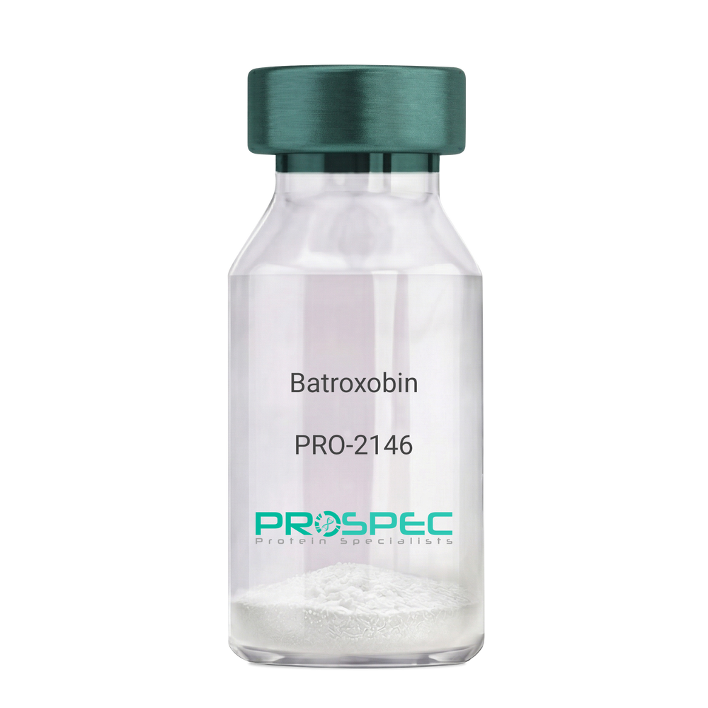
BatroxobinDescription:
Batroxobin
Thrombin-like enzyme batroxobin, EC 3.4.21.74, BX, Bothrops atrox serine proteinase, Venombin-A, Batroxobin.
Product # :
PRO-2146Price :
Quantity :
Shipping Method :
Shipped at Room temp
More Info
- description
- formulation
- More Info
Description
Batroxobin, isolated from Bothrops atrox snake venom, has an Mw of approximately 43kDa.
Formulation
The Batroxobin protein was lyophilized from a concentrated (1mg/ml) solution with no additives.
More Info
-
Introduction
Batroxobin is a serin protease that reduces fibronogen levels and is originally extracted from snake venom of Bothrops Atrox. Batroxobin is used in defibrinogenation and thrombolysis and also has an effect on c-fos gene and growth factor.
Batroxobin can efficiently restrain proliferation of VSMCs, by blocking the release and uptake of Ca2+, thus influencing [Ca2+]i.
Batroxobin converts fibrinogen to fibrin through the restricted release of fibrinopeptide-A from fibrinogen to promote blood to clot. Unlike thrombin, it is not affected by heparin and hirudin. -
Synonyms
Thrombin-like enzyme batroxobin, EC 3.4.21.74, BX, Bothrops atrox serine proteinase, Venombin-A, Batroxobin.
-
Physical Appearance
Sterile Filtered white lyophilized powder.
-
Stability
Store the lyophilized Batroxobin between 2-8°C. Do not freeze!
-
Solubility
It is recommended to reconstitute the lyophilized Batroxobin in sterile 18MΩ-cm H2O not less than 100µg/ml or more than 10mg/ml solutions.
-
Unit Definition
100BU [Batroxobin Units]=1mg.
ProSpec's products are furnished for LABORATORY RESEARCH USE ONLY. The product may not be used as drugs, agricultural or pesticidal products, food additives or household chemicals.
- View Data Sheet
Name :
SRGN HumanDescription:
Serglycin Human Recombinant
Serglycin, PRG, PRG1, PPG, Proteoglycan 1 secretory granule, Hematopoetic proteoglycan core protein, Platelet proteoglycan core protein, proteoglycan protein core for mast cell secretory granule.
Product # :
PRO-965Price :
Quantity :
Shipping Method :
Shipped with Ice Packs
More Info
- description
- source
- formulation
- purity
- More Info
Description
SRGN Human Recombinant produced in E.coli is a single, non-glycosylated polypeptide chain containing 156 amino acids (28-158) and having a molecular mass of 17.4 kDa (Molecular weight on SDS-PAGE will appear higher).SRGN is fused to a 25 amino acid His-tag at N-terminus & purified by proprietary chromatographic techniques.
Source
E.coli.
Formulation
The SRGN solution (1mg/ml) contains 20mM Tris-HCl buffer (pH 8.0), 2mM DTT, 0.15M NaCl and 10% glycerol.
Purity
Greater than 90% as determined by SDS-PAGE.
More Info
-
Introduction
SRGN is identified as a hematopoietic cell granule proteoglycan. Proteoglycans stored in the secretory granules of various hematopoietic cells also hold a protease-resistant peptide core, and is vital for neutralizing hydrolytic enzymes. SRGN is related to the macromolecular complex of granzymes and perforin that acts as a intermediary of granule-mediated apoptosis.
-
Synonyms
Serglycin, PRG, PRG1, PPG, Proteoglycan 1 secretory granule, Hematopoetic proteoglycan core protein, Platelet proteoglycan core protein, proteoglycan protein core for mast cell secretory granule.
-
Physical Appearance
Sterile Filtered colorless solution.
-
Stability
Store at 4°C if entire vial will be used within 2-4 weeks. Store, frozen at -20°C for longer periods of time. For long term storage it is recommended to add a carrier protein (0.1% HSA or BSA).Avoid multiple freeze-thaw cycles.
-
Amino Acid Sequence
MGSSHHHHHH SSGLVPRGSH MGSHMYPTRR ARYQWVRCNP DSNSANCLEE KGPMFELLPG ESNKIPRLRT DLFPKTRIQD LNRIFPLSED YSGSGFGSGS GSGSGSGSGF LTEMEQDYQL VDESDAFHDN LRSLDRNLPS DSQDLGQHGL EEDFML.
ProSpec's products are furnished for LABORATORY RESEARCH USE ONLY. The product may not be used as drugs, agricultural or pesticidal products, food additives or household chemicals.
- View Data Sheet
Name :
PF 4 ProteinDescription:
Platelet Factor-4 Human (CXCL4)
CXCL4, PF-4, PF4, Iroplact, Oncostatin-A, SCYB4, MGC138298.
Product # :
CHM-234Price :
Quantity :
Shipping Method :
Shipped at Room temp
More Info
- description
- source
- formulation
- purity
- More Info
Description
Human PF-4 a 7.8 kDa protein consisting of 70 amino acid residues.
Source
Human Platelets.
Formulation
The CXCL4 protein was lyophilized in PBS buffer pH-7.4.
Purity
Greater than 95.0% as determined by:
(a) Analysis by RP-HPLC.
(b) Analysis by SDS-PAGE.More Info
-
Introduction
Platelet factor-4 is a 70-amino acid protein that is released from the alpha-granules of activated platelets and binds with high affinity to heparin. Its major physiologic role appears to be neutralization of heparin-like molecules on the endothelial surface of blood vessels, thereby inhibiting local antithrombin III activity and promoting coagulation. As a strong chemoattractant for neutrophils and fibroblasts, PF4 probably has a role in inflammation and wound repair. Oncostatin-A is a member of the CXC chemokine family. Human PF4 is used for the proof of heparin-induced thrombocytopenia. Furthermore it is used as an inhibitor in the angiogenesis during tumor therapy.
-
Synonyms
CXCL4, PF-4, PF4, Iroplact, Oncostatin-A, SCYB4, MGC138298.
-
Physical Appearance
Sterile Filtered white lyophilized powder.
-
Stability
Human CXCL4 although stable at 25°C 1 week, should be stored desiccated below -18°C. Please prevent freeze-thaw cycles.
-
Solubility
It is recommended to reconstitute the lyophilized CXCL4 in sterile 18MΩ-cm H2O not less than 100µg/ml, which can then be further diluted to other aqueous solutions.
-
Amino Acid Sequence
The sequence of the first four N-terminal amino acids was determined and was found to be Glu-Ala-Glu-Glu.
ProSpec's products are furnished for LABORATORY RESEARCH USE ONLY. The product may not be used as drugs, agricultural or pesticidal products, food additives or household chemicals.
- View Data Sheet
Name :
PFN1 HumanDescription:
Profilin-1 Human Recombinant
Profilin-1, Profilin I, PFN1.
Product # :
PRO-528Price :
Quantity :
Shipping Method :
Shipped with Ice Packs
More Info
- description
- source
- formulation
- purity
- More Info
Description
PFN1 Human Recombinant produced in E.Coli is a single, non-glycosylated, polypeptide chain containing 140 amino acids (1-140 a.a.) and having a molecular mass of 15kDa.The PFN1 is purified by proprietary chromatographic techniques.
Source
Escherichia Coli.
Formulation
The PFN1 protein solution contains 20mM Tris-HCl buffer (pH8.0) and 10% glycerol.
Purity
Greater than 95.0% as determined by SDS-PAGE.
More Info
-
Introduction
Profilin1 (PFN1) is a ubiquitous actin monomer-binding protein which is a member of the profilin family. PFN1 significantly boosts skin wound healing in-vitro and in-vivo which may be mediated by purinergic receptors. PFN1 is also active in endothelial cell migration and vessel sprouting. PFN1 is thought to control actin polymerization in response to extracellular signals. PFN1 binds to actin and affects the formation of the cytoskeleton. In addition, PFN1 has an important role in the regulation of epithelial cell-cell adhesion. At high concentrations, profilin averts the polymerization of actin, while at low concentrations it enhances the polymerization. PFN1 gene deletion is linked to Miller-Dieker syndrome.
-
Synonyms
Profilin-1, Profilin I, PFN1.
-
Physical Appearance
Sterile Filtered colorless solution.
-
Stability
Store at 4°C if entire vial will be used within 2-4 weeks. Store, frozen at -20°C for longer periods of time. For long term storage it is recommended to add a carrier protein (0.1% HSA or BSA).Avoid multiple freeze-thaw cycles.
-
Amino Acid Sequence
MAGWNAYIDN LMADGTCQDA AIVGYKDSPS VWAAVPGKTF VNITPAEVGV LVGKDRSSFY VNGLTLGGQK CSVIRDSLLQ DGEFSMDLRT KSTGGAPTFN VTVTKTDKTL VLLMGKEGVH GGLINKKCYE MASHLRRSQY.
ProSpec's products are furnished for LABORATORY RESEARCH USE ONLY. The product may not be used as drugs, agricultural or pesticidal products, food additives or household chemicals.
- View Data Sheet
Name :
GNLY HumanDescription:
Granulysin Human Recombinant
LAG2, Lymphokine LAG-2, TLA519, NKG5, LAG2, D2S69E, Granulysin, T-cell activation protein 519, GNLY, D2S69E.
Product # :
PRO-852Price :
Quantity :
Shipping Method :
Shipped at Room temp
More Info
- description
- source
- formulation
- purity
- More Info
Description
GNLY Human Recombinant produced in E.Coli is a single, non-glycosylated, polypeptide chain containing 159 amino acids and fused to a double His Tag (N+C terminus) and having a total molecular mass of 18.1 kDa.The GNLY is purified by proprietary chromatographic techniques.
Source
Escherichia Coli.
Formulation
The Granulysin protein was lyophilized from a concentrated (1mg/ml) solution containing no additives.
Purity
Greater than 95.0% as determined by SDS-PAGE.
More Info
-
Introduction
GNLY is part of the SAPLIP family and is located in the cytotoxic granules of T cells, which are discharged upon antigen stimulation. GNLY is localized in cytotoxic granules of cytotoxic T lymphocytes and natural killer cells, and it has antimicrobial activity against M. tuberculosis and other organisms. GNLY is an antimicrobial protein that kills intracellular pathogens. GNLY is active against a wide range of microbes, including Gram-positive and Gram-negative bacteria, fungi, and parasites. Kills Mycobacterium tuberculosis.
-
Synonyms
LAG2, Lymphokine LAG-2, TLA519, NKG5, LAG2, D2S69E, Granulysin, T-cell activation protein 519, GNLY, D2S69E.
-
Physical Appearance
Sterile Filtered White lyophilized (freeze-dried) powder.
-
Stability
Lyophilized Granulysin although stable at room temperature for 3 weeks, should be stored desiccated below -18°C. Upon reconstitution Granulysin should be stored at 4°C between 2-7 days and for future use below -18°C. For long term storage it is recommended to add a carrier protein (0.1% HSA or BSA). Please prevent freeze-thaw cycles.
-
Solubility
It is recommended to reconstitute the lyophilized Granulysin in sterile 18MΩ-cm H2O not less than 100µg/ml, which can then be further diluted to other aqueous solutions.
-
Amino Acid Sequence
MGSSHHHHHHSSGLVPRGSHMMEGLVFSRLSPEYYD
LARAHLRDEEKSCPCLAQEGPQGDLLTKTQELGRDYR
TCLTIVQKLKKMVDKPTQRSVSNAATRVCRTGRSRWR
DVCRNFMRRYQSRVTQGLVAGETAQQICEDLRLCIPS
TGPLGSHHHHHH.
ProSpec's products are furnished for LABORATORY RESEARCH USE ONLY. The product may not be used as drugs, agricultural or pesticidal products, food additives or household chemicals.
- View Data Sheet
Name :
Vaspin HumanDescription:
Vaspin Recombinant Human
Serpin A12 precursor, Visceral adipose-specific serpin, Visceral adipose tissue- derived serine protease inhibitor, Vaspin, OL-64, SERPINA12, Serine (or cysteine) proteinase inhibitor, clade A, antitrypsin, alpha-1 antiproteinase.
Product # :
CYT-1132Price :
Quantity :
Shipping Method :
Shipped at Room temp
More Info
- description
- source
- formulation
- purity
- More Info
Description
Vaspin Human Recombinant produced in E.Coli is a single, non- glycosylated polypeptide chain containing 394 amino acids and having a molecular mass of 45.1kDa. Vaspin is purified by proprietary chromatographic techniques.
Source
Escherichia Coli.
Formulation
Lyophilized from a 0.2μm filtered concentrated solution in 20mM Tris-HCl, pH 8.0, 150mM NaCl and 0.02 % Tween-20.
Purity
Greater than 98.0% as determined by:
(a) Analysis by RP-HPLC.
(b) Analysis by SDS-PAGE.More Info
-
Introduction
Vaspin (visceral adipose-specific SERPIN) is a newly identified adipokine, which is a member of serine protease inhibitor family. Vaspin is also a unique insulin sensitizing adipocytokine in obesity. A recent publication indicates that induction of human vaspin mRNA expression in adipose tissue is regulated in a fat depot-specific manner and could be associated with parameters of obesity, insulin resistance, and glucose metabolism.
-
Synonyms
Serpin A12 precursor, Visceral adipose-specific serpin, Visceral adipose tissue- derived serine protease inhibitor, Vaspin, OL-64, SERPINA12, Serine (or cysteine) proteinase inhibitor, clade A, antitrypsin, alpha-1 antiproteinase.
-
Physical Appearance
Sterile Filtered White lyophilized (freeze-dried) powder.
-
Stability
Lyophilized Vaspin although stable at room temperature for 3 weeks, should be stored desiccated below -18°C. Upon reconstitution Vaspin should be stored at 4°C between 2-7 days and for future use below -18°C.Please prevent freeze-thaw cycles.
-
Solubility
It is recommended to reconstitute the lyophilized Vaspin in sterile 18MΩ-cm H2O not less than 100µg/ml, which can then be further diluted to other aqueous solutions.
-
Amino Acid Sequence
LKPSFSPRNY KALSEVQGWK QRMAAKELAR QNMDLGFKLL KKLAFYNPGR NIFLSPLSIS TAFSMLCLGA QDSTLDEIKQ GFNFRKMPEK DLHEGFHYII HELTQKTQDL KLSIGNTLFI DQRLQPQRKF LEDAKNFYSA ETILTNFQNL EMAQKQINDF ISQKTHGKIN NLIENIDPGT VMLLANYIFF RARWKHEFDP NVTKEEDFFL EKNSSVKVPM MFRSGIYQVG YDDKLSCTIL EIPYQKNITA IFILPDEGKL KHLEKGLQVD TFSRWKTLLS RRVVDVSVPR LHMTGTFDLK KTLSYIGVSK IFEEHGDLTK IAPHRSLKVG EAVHKAELKM DERGTEGAAG TGAQTLPMET PLVVKIDKPY LLLIYSEKIP SVLFLGKIVN PIGK.
ProSpec's products are furnished for LABORATORY RESEARCH USE ONLY. The product may not be used as drugs, agricultural or pesticidal products, food additives or household chemicals.
- View Data Sheet
Name :
MAP1LC3B HumanDescription:
Microtubule-Associated Protein 1 Light Chain 3 Beta Human Recombinant
Microtubule-Associated Protein 1 Light Chain 3 beta, ATG8F, Autophagy-related ubiquitin-like modifier LC3 B, MAP1 light chain 3-like protein 2, MAP1A/MAP1B LC3 B, LC3B, MAP1A/1BLC3, MAP1ALC3.
Product # :
PRO-076Price :
Quantity :
Shipping Method :
Shipped with Ice Packs
More Info
- description
- source
- formulation
- purity
- More Info
Description
MAP1LC3B produced in E.Coli is a single, non-glycosylated polypeptide chain containing 140 amino acids (1-120a.a.) and having a molecular mass of 16.2kDa.MAP1LC3B is fused to a 20 amino acid His-tag at N-terminus & purified by proprietary chromatographic techniques.
Source
Escherichia Coli.
Formulation
The MAP1LC3B protein solution (0.5mg/1ml) is formulated in 20mM Tris-HCl buffer (pH8.0)
1mM DTT, 100mM NaCl and 20% glycerol.Purity
Greater than 95% as determined by SDS-PAGE.
More Info
-
Introduction
MAP1LC3B is a member of the MAP1 LC3 family. MAP1LC3B is a subunit of neuronal microtubule-associated MAP1A and MAP1B proteins, that are involved in microtubule assembly and important for neurogenesis. In addition, MAP1LC3B takes part in formation of autophagosomal vacuoles and is expressed mainly in heart, testis, brain and skeletal muscle.
-
Synonyms
Microtubule-Associated Protein 1 Light Chain 3 beta, ATG8F, Autophagy-related ubiquitin-like modifier LC3 B, MAP1 light chain 3-like protein 2, MAP1A/MAP1B LC3 B, LC3B, MAP1A/1BLC3, MAP1ALC3.
-
Physical Appearance
Sterile Filtered clear solution.
-
Stability
Store at 4°C if entire vial will be used within 2-4 weeks. Store, frozen at -20°C for longer periods of time. For long term storage it is recommended to add a carrier protein (0.1% HSA or BSA).Avoid multiple freeze-thaw cycles.
-
Amino Acid Sequence
MGSSHHHHHH SSGLVPRGSH MPSEKTFKQR RTFEQRVEDV RLIREQHPTK IPVIIERYKG EKQLPVLDKT KFLVPDHVNM SELIKIIRRR LQLNANQAFF LLVNGHSMVS VSTPISEVYE SEKDEDGFLY MVYASQETFG
ProSpec's products are furnished for LABORATORY RESEARCH USE ONLY. The product may not be used as drugs, agricultural or pesticidal products, food additives or household chemicals.
- View Data Sheet
Name :
CRYBB1 HumanDescription:
Crystallin Beta B1 Human Recombinant
EC 1.17.4.1, RR2M, Beta-B1 crystallin, CATCN3.
Product # :
HSP-033Price :
Quantity :
Shipping Method :
Shipped with Ice Packs
More Info
- description
- source
- formulation
- purity
- More Info
Description
CRYBB1 Recombinant Human produced in E.Coli is a single, non-glycosylated polypeptide chain containing 260 amino acids (1-252 a.a.) and having a molecular mass of 29.1 kDa. The CRYBB1 is fused to an 8 amino acid His-Tag at C-terminus and purified by proprietary chromatographic techniques.
Source
Escherichia Coli.
Formulation
The CRYBB1 solution (1mg/ml) contains 20mM Tris-HCl pH-8, 1mM DTT & 10% glycerol.
Purity
Greater than 90.0% as determined by SDS-PAGE.
More Info
-
Introduction
Crystallins are the main structural proteins of the vertebrate eye lens, where they maintain the transparency and refractive index of the lens. Crystallins are divided into 3 fprotein families, α, β, & γ families. Because lens central fiber cells lose their nuclei during development, these crystallins are prepared and then retained throughout life, making them extremely stable proteins. CRYBB1 is a beta basic group member and undergoes extensive cleavage at its N-terminal extension during lens maturation.
-
Synonyms
EC 1.17.4.1, RR2M, Beta-B1 crystallin, CATCN3.
-
Physical Appearance
Sterile filtered colorless solution.
-
Stability
Store at 4°C if entire vial will be used within 2-4 weeks. Store, frozen at -20°C for longer periods of time.For long term storage it is recommended to add a carrier protein (0.1% HSA or BSA).Avoid multiple freeze-thaw cycles.
-
Amino Acid Sequence
MSQAAKASAS ATVAVNPGPD TKGKGAPPAG TSPSPGTTLA PTTVPITSAK AAELPPGNYR LVVFELENFQ GRRAEFSGEC SNLADRGFDR VRSIIVSAGP WVAFEQSNFR GEMFILEKGE YPRWNTWSSS YRSDRLMSFR PIKMDAQEHK ISLFEGANFK GNTIEIQGDD APSLWVYGFS DRVGSVKVSS GTWVGYQYPG YRGYQYLLEP GDFRHWNEWG AFQPQMQSLR RLRDKQWHLE GSFPVLATEP PKRSHHHHHH.
ProSpec's products are furnished for LABORATORY RESEARCH USE ONLY. The product may not be used as drugs, agricultural or pesticidal products, food additives or household chemicals.
- View Data Sheet
Name :
CEA ProteinDescription:
Carcinoembryonic Antigen Human
CEACAM5, Meconium Antigen 100, Carcinoembryonic Antigen, CD66e Antigen, CD66e, Carcinoembryonic Antigen, CEA, oncofetal antigen.
Product # :
PRO-2801Price :
Quantity :
Shipping Method :
Shipped with Ice Packs
More Info
- description
- source
- formulation
- purity
- More Info
Description
CEA produced from patient source colon carcinoma liver metastatic tissue can be used as general marker in screening and monitoring malignant disease states.
Source
Liver tissue.
Formulation
CEA protein solution contains 0.1M PBS, pH 7.4, 0.09 % NaN3 and 2 % methyl-mannoside.
Purity
Greater than 95.0% as determined by SDS-PAGE.
More Info
-
Synonyms
CEACAM5, Meconium Antigen 100, Carcinoembryonic Antigen, CD66e Antigen, CD66e, Carcinoembryonic Antigen, CEA, oncofetal antigen.
-
Physical Appearance
Sterile Filtered colorless solution.
-
Stability
Store at 4°C if entire vial will be used within 2-4 weeks. Store, frozen at -20°C for longer periods of time. For long term storage it is recommended to add a carrier protein (0.1% HSA or BSA). Avoid multiple freeze-thaw cycles.
-
Applications
Blood samples from tissue donors were tested and found to be negative for HBsAg, HIV-1 and HIV-2 antibodies and HCV.
-
Background
Carcinoembryonic Antigen, commonly known as CEA, is a glycoprotein that was initially identified as a tumor marker. Over the years, research into CEA has unveiled its intricate involvement in various physiological processes, not only in cancer but also in the context of normal development and inflammatory conditions. This research aims to delve into the multifaceted roles of CEA, exploring its structural intricacies, regulatory mechanisms, and its implications in health, disease, and beyond.
Structural Complexity of CEA:
CEA, belonging to the immunoglobulin superfamily, is a complex glycoprotein featuring multiple structural domains. Its diverse forms and glycosylation patterns contribute to its functional versatility. CEA is primarily expressed in fetal tissues, but its presence is often detected in adults under pathological conditions, especially in various types of cancer.
CEA in Cancer Biology:
CEA was first recognized as a biomarker for colorectal cancer, but its overexpression is not limited to this context. Elevated CEA levels have been associated with several other malignancies, including breast, lung, and pancreatic cancers. CEA’s involvement in cancer biology ranges from promoting angiogenesis and metastasis to inhibiting immune responses, making it a critical player in tumor progression and evasion.
Beyond Cancer: CEA in Development and Inflammation:
While CEA’s role in cancer is prominent, recent studies have uncovered its participation in normal physiological processes. During embryonic development, CEA is involved in cell adhesion, contributing to tissue organization and morphogenesis. Additionally, CEA expression can be induced in inflammatory conditions, suggesting its involvement in immune responses and tissue repair mechanisms.
CEA as a Diagnostic and Therapeutic Target:
The diverse expression patterns of CEA in various diseases make it a valuable diagnostic tool. CEA assays are widely used for cancer screening, monitoring disease progression, and assessing treatment efficacy. Moreover, CEA’s presence on the surface of cancer cells has made it a target for immunotherapy, enabling the development of targeted therapies aimed at specifically eradicating CEA-positive tumor cells.
ProSpec's products are furnished for LABORATORY RESEARCH USE ONLY. The product may not be used as drugs, agricultural or pesticidal products, food additives or household chemicals.
- View Data Sheet
Name :
Car b 1.0109Description:
Pollen allergen Car b 1 isoform Recombinant
Pollen allergen Car b 1 isoform, Car b 1, Car b 1.0109.
Product # :
PRO-2284Price :
Quantity :
Shipping Method :
Shipped with Ice Packs
More Info
- description
- source
- formulation
- purity
- More Info
Description
Recombinant Pollen allergen Car b 1 isoform produced in E.coli is a non-glycosylated, polypeptide chain having a calculated molecular mass of 18kDa. Car b 1.0109 is expressed with a 10xHis tag at N-terminus and purified by proprietary chromatographic techniques.
Source
Escherichia Coli.
Formulation
Car b 1.0109 is supplied in 20mM HEPES buffer pH-8.0, 20% Glycerol and 200mM NaCl.
Purity
Greater than 95.0% as determined by SDS-PAGE.
More Info
-
Introduction
Pollen allergen Car b 1 isoform (Car b 1.0109) causes an allergic reaction in humans.
-
Synonyms
Pollen allergen Car b 1 isoform, Car b 1, Car b 1.0109.
-
Physical Appearance
Sterile Filtered clear solution.
-
Stability
Store at 4°C if entire vial will be used within 2-4 weeks. Store, frozen at -20°C for longer periods of time. Avoid multiple freeze-thaw cycles.
-
Immunological Functions
1. Binds IgE type human antibodies. 2. Immunodot test with positive/negative sera panels.
-
Molar extinction coefficient
8940; A280(1mg/ml)=0.477
ProSpec's products are furnished for LABORATORY RESEARCH USE ONLY. The product may not be used as drugs, agricultural or pesticidal products, food additives or household chemicals.
- View Data Sheet
Name :
MAPT Human 412a.a.Description:
Microtubule-Associated Protein Tau 412 a.a. Human Recombinant
Microtubule-associated protein tau, isoform CRA_f, Neurofibrillary tangle protein, Paired helical filament-tau, PHF-tau, MAPT, MAPTL, MTBT1, TAU, MSTD, PPND, DDPAC, MTBT2, FTDP-17, FLJ31424, MGC138549.
Product # :
PRO-210Price :
Quantity :
Shipping Method :
Shipped with Ice Packs
More Info
- description
- source
- formulation
- purity
- More Info
Description
MAPT Human Recombinant (Isoform 5) fused with a 20 amino acid His tag at N-terminus produced in E.Coli is a single, non-glycosylated, polypeptide chain containing 432 amino acids (1-412 a.a.) and having a molecular mass of 45.1kDa (Molecular size on SDS-PAGE will appear higher). The MAPT is purified by proprietary chromatographic techniques.
Source
Escherichia Coli.
Formulation
The MAPT solution (0.5 mg/ml) contains 20mM Tris-HCl buffer (pH8.0) and 20% glycerol.
Purity
Greater than 80.0% as determined by SDS-PAGE.
More Info
-
Introduction
Microtubule-associated protein tau (MAPT or Tau) is a protein that stabilizes microtubules. MAPT is abundant in neurons in the central nervous system and is less common elsewhere. When MAPT is defective, and no longer stabilizes microtubules properly, it can result in dementias, such as Alzheimer's disease.
-
Synonyms
Microtubule-associated protein tau, isoform CRA_f, Neurofibrillary tangle protein, Paired helical filament-tau, PHF-tau, MAPT, MAPTL, MTBT1, TAU, MSTD, PPND, DDPAC, MTBT2, FTDP-17, FLJ31424, MGC138549.
-
Physical Appearance
Sterile Filtered colorless solution.
-
Stability
Store at 4°C if entire vial will be used within 2-4 weeks. Store, frozen at -20°C for longer periods of time. For long term storage it is recommended to add a carrier protein (0.1% HSA or BSA).Avoid multiple freeze-thaw cycles.
-
Amino Acid Sequence
MGSSHHHHHH SSGLVPRGSH MAEPRQEFEV MEDHAGTYGL GDRKDQGGYT MHQDQEGDTD AGLKESPLQT PTEDGSEEPG SETSDAKSTP TAEAEEAGIG DTPSLEDEAA GHVTQARMVS KSKDGTGSDD KKAKGADGKT KIATPRGAAP PGQKGQANAT RIPAKTPPAP KTPPSSGEPP KSGDRSGYSS PGSPGTPGSR SRTPSLPTPP TREPKKVAVV RTPPKSPSSA KSRLQTAPVP MPDLKNVKSK IGSTENLKHQ PGGGKVQIIN KKLDLSNVQS KCGSKDNIKH VPGGGSVQIV YKPVDLSKVT SKCGSLGNIH HKPGGGQVEV KSEKLDFKDR VQSKIGSLDN ITHVPGGGNK KIETHKLTFR ENAKAKTDHG AEIVYKSPVV SGDTSPRHLS NVSSTGSIDM VDSPQLATLA DEVSASLAKQ GL.
ProSpec's products are furnished for LABORATORY RESEARCH USE ONLY. The product may not be used as drugs, agricultural or pesticidal products, food additives or household chemicals.
- View Data Sheet
Name :
ANXA10 Human (1-162)Description:
Annexin A10 (1-162 a.a.) Human Recombinant
anxa-10.
Product # :
PRO-2837Price :
Quantity :
Shipping Method :
Shipped at Room temp
More Info
- description
- source
- formulation
- purity
- More Info
Description
The ANXA10 Human is created as a recombinant protein with a 4kda N-terminal fusion of His Tag. The ANXA10 His-Tagged Fusion Protein, produced in E. coli, is a 21kDa protein containing 162 amino acid residues of the ANXA10 Human, 1-162 amino acids.
Source
Escherichia Coli.
Formulation
Each mg was lyophilized with 1xPBS, 0.4% SDS and 4mM DTT.
Purity
Greater than 90% as determined by SDS-PAGE.
More Info
-
Synonyms
anxa-10.
-
Physical Appearance
Filtered White lyophilized (freeze-dried) powder.
-
Stability
Store lyophilized ANXA10 at -20°C. Aliquot the product after reconstitution to avoid repeated freezing/thawing cycles. Reconstituted protein can be stored at 4°C for a limited period of time; it does not show any change after two weeks at 4°C.
-
Solubility
It is recommended to add deionized water to prepare a working stock solution of approximately 0.5mg/ml and let the lyophilized pellet dissolve completely. Product is not sterile! Please filter the product by an appropriate sterile filter before using it on cell culture.
-
Background
Annexin A10 also known as ANXA10 is a part of the annexin family of calcium-binding proteins which members own a conserved core domain and a unique amino-terminal region which may convene binding specificity. The ANXA10 protein contains 4 annexin domains and plays a role in the regulation of cellular growth and signal transduction pathways throughout the cell.
ProSpec's products are furnished for LABORATORY RESEARCH USE ONLY. The product may not be used as drugs, agricultural or pesticidal products, food additives or household chemicals.
- View Data Sheet
Name :
Gliadin Gamma WheatDescription:
Gliadin Gamma Wheat Recombinant
Product # :
PRO-2148Price :
Quantity :
Shipping Method :
Shipped with Ice Packs
More Info
- description
- source
- formulation
- purity
- More Info
Description
Recombinant Wheat Gliadin Gamma protein produced in E.Coli and fused to a 6 His Tag at C-terminus, having a theoretical Mw of 37945.14 Dalton, pI 7.70.Purified by proprietary chromatographic technique.
Source
Escherichia Coli.
Formulation
Gliadin Gamma protein solution (1mg/ml) in 10mM Tris-HCl pH 7.2.
Purity
Protein is >90% pure.
More Info
-
Introduction
Wheat Gliadin and related gluten components from barley, rye and possibly oats can cause an abnormal immune response called Celiac disease which is a chronic gastrointestinal disorder. Celiac disease characteristics are flattening of the jejunal mucosa and intestinal lesions of variable severity in hereditarily inclined individuals. Even though Celiac disease is not a classic autoimmune disease it is related to anti-tissue transglutaminase antibodies and gliadin antibodies tests are most recommended in screening populations at risk for CD and other gluten-sensitive enteropathies. In the past, serologic tests for gliadin antibodies usually were not very precise and were not enough for accurate diagnosis due to missing deamidated epitopes within the authentic gliadin fraction traditionally used in diagnostic test kits. ProSpec's deamidated Gliadin isoform matches to the deamidated neo-epitopes, which in the natural antigen are formed by transglutaminase-mediated glutamine side chain deamidation.
-
Physical Appearance
Sterile Filtered clear solution.
-
Stability
Gliadin Gamma although stable at 4°C for 1 week, should be stored below -18°C. Please prevent freeze thaw cycles.
-
Amino Acid Sequence
MKTLLILTILAMAITIGTANIQVDPSGQVQWLQQQLVPQLQQPLSQQPQQTFPQPQQTFPH
QPQQQVPQPQQPQQPFLQPQQPFPQQPQQPFPQTQQPQQPFPQQPQQPFPQTQQPQQ
PFPQQPQQPFPQTQQPQQPFPQLQQPQQPFPQPQQQLPQPQQPQQSFPQQQRPFIQPSL
QQQLNCKNILLQQSKPASLVSSLWSIIWPQSDCQVMRQQCCQQLAQIPQQLQCAAIHSVVH
SIIMQQQQQQQQQQGIDIFLPLSQHEQVGQGSLVQGQGIIQPQQPAQLEAIRSLVLQTLPSM
CNVYVPPECSIMRAPFASIVAGIGGQHHHHHH.
ProSpec's products are furnished for LABORATORY RESEARCH USE ONLY. The product may not be used as drugs, agricultural or pesticidal products, food additives or household chemicals.
- View Data Sheet
Name :
NEFH BovineDescription:
Neurofilament Heavy Chain Bovine
Neurofilament light polypeptide, NF-L, NEFL, NF68, NFL, 68 kDa neurofilament protein.
Product # :
PRO-2787Price :
Quantity :
Shipping Method :
Shipped at Room temp
More Info
- description
- source
- formulation
- purity
- More Info
Description
NEFH Bovine having a calculated molecular mass of 200 kDa, pI-5.5.
Source
Bovine spinal cord.
Formulation
NEFH was lyophilized from a 1mg/ml solution containing 10mM sodium phosphate buffer pH 7.5, 6M urea, 1mM EDTA, 2mM DTT and 10mM methylammonium chloride.
Purity
Greater than 95.0% as determined by SDS-PAGE.
More Info
-
Synonyms
Neurofilament light polypeptide, NF-L, NEFL, NF68, NFL, 68 kDa neurofilament protein.
-
Physical Appearance
Filtered White lyophilized (freeze-dried) powder.
-
Stability
Store the lyophilized NEFH between 2-8°C, do not freeze. Upon reconstitution NEFH should be stored at -20°C.Please prevent freeze-thaw cycles.
-
Solubility
It is recommended to reconstitute the lyophilized NEFH in sterile 18MΩ-cm H2O not less than 100µg/ml, which can then be further diluted to other aqueous solutions.
-
Background
Neurofilament heavy chain (NEFH) is a vital structural protein in neurons, predominantly found in the central and peripheral nervous systems. Although extensive research has been conducted on NEFH in humans and rodents, the investigation of NEFH in bovine nervous tissues presents an emerging area with substantial potential for advancing our understanding of neuronal biology in larger mammals and its applications in veterinary medicine and neurobiology.
Bovine nervous tissues, including the brain and spinal cord, are of particular interest due to their relevance in cattle health, neuroscience, and the food industry. This research aims to provide a comprehensive exploration of NEFH in bovine nervous tissues, elucidating its functions, structural significance, and potential applications.
The primary objective of this research is to elucidate the role of NEFH in bovine nervous tissues, particularly in maintaining neuronal structural integrity and axonal function. In vitro and ex vivo experiments, utilizing bovine neuronal cell cultures and tissue specimens, will be conducted to investigate how NEFH contributes to neuronal morphology, axonal transport, and overall neuronal resilience. Understanding these mechanisms is fundamental for deciphering the complexities of neuronal biology in bovine species.
The second objective is to assess the relevance of bovine NEFH in veterinary medicine. Studies involving bovine models will be conducted to evaluate the impact of NEFH mutations or variations on neuronal health, disease susceptibility, and neurodegenerative conditions. These investigations may provide valuable insights into potential applications in cattle health, the development of diagnostic tools for neurological disorders, and strategies for enhancing animal welfare.
The third objective is to explore the potential applications of bovine NEFH in neurobiology and biotechnology. Research will investigate the use of bovine NEFH-expressing cells as models for studying neuronal-related diseases and for developing tissue engineering approaches in veterinary medicine and biotechnology.
By delving into the functions and roles of NEFH in bovine nervous tissues, this research aims to expand our knowledge of neuronal biology, its implications for veterinary medicine, and its potential applications in neurobiology, cattle health, and biotechnology.
ProSpec's products are furnished for LABORATORY RESEARCH USE ONLY. The product may not be used as drugs, agricultural or pesticidal products, food additives or household chemicals.
- View Data Sheet
Name :
KRT18 BovineDescription:
Cytokeratin-18 Bovine
Keratin type I cytoskeletal 18, Cytokeratin-18, CK-18, Keratin-18, K18, KRT18,CYK18,Cell proliferation-inducing gene 46 protein.
Product # :
PRO-2785Price :
Quantity :
Shipping Method :
Shipped at Room temp
More Info
- description
- source
- formulation
- purity
- More Info
Description
KRT18 Bovine having a calculated molecular mass of 45 kDa, pI-5.4.
Source
Bovine liver.
Formulation
KRT18 was lyophilized from a 1mg/ml solution containing 30mM Tris/HCI pH 8, 9M urea, 2mM EDTA, 2mM DTT and 10mM methylammonium chloride.
Purity
Greater than 95.0% as determined by SDS-PAGE.
More Info
-
Synonyms
Keratin type I cytoskeletal 18, Cytokeratin-18, CK-18, Keratin-18, K18, KRT18,CYK18,Cell proliferation-inducing gene 46 protein.
-
Physical Appearance
Sterile Filtered White lyophilized (freeze-dried) powder.
-
Stability
Store the lyophilized KRT18 between 2-8°C, do not freeze. Upon reconstitution KRT18 should be stored at -20°C.Please prevent freeze-thaw cycles.
-
Solubility
It is recommended to reconstitute the lyophilized KRT18 in sterile 18MΩ-cm H2O not less than 100µg/ml, which can then be further diluted to other aqueous solutions.
-
Background
Keratin-18 (K18) is an intermediate filament protein that plays a vital role in maintaining the structural integrity of epithelial cells. Extensive research has been conducted on K18 in human and murine models, shedding light on its functions and implications for various epithelial tissues.
However, the study of K18 in bovine tissues is an emerging area with potential for advancing our understanding of epithelial cell biology and its applications in veterinary medicine and biotechnology. Bovine tissues, such as the liver and gastrointestinal tract, are of particular interest due to their relevance in cattle production and food safety.
This research aims to provide a comprehensive exploration of K18 in bovine tissues, elucidating its functions, structural significance, and potential applications.
The primary objective of this research is to elucidate the role of K18 in bovine tissues, particularly in maintaining the structural integrity of epithelial cells.In vitro and ex vivo experiments, utilizing bovine epithelial cell cultures and tissue specimens, will be conducted to investigate how K18 contributes to cellular morphology, cytoskeletal organization, and tissue resilience. Understanding these mechanisms is fundamental for deciphering the complexities of epithelial cell biology in bovine species.
The second objective is to assess the relevance of bovine K18 in veterinary medicine and cattle production. Studies involving bovine models will be conducted to evaluate the impact of K18 mutations or variations on tissue health, disease susceptibility, and meat quality. These investigations may provide valuable insights into potential applications in cattle breeding and food safety.
The third objective is to explore the potential biotechnological applications of bovine K18. Research will investigate the use of K18-expressing bovine cells as models for studying epithelial-related diseases and for developing tissue engineering approaches for veterinary medicine and biotechnology.
By delving into the functions and roles of K18 in bovine tissues, this research aims to expand our knowledge of epithelial cell biology, its implications for veterinary medicine, and its potential applications in biotechnology and cattle production.
ProSpec's products are furnished for LABORATORY RESEARCH USE ONLY. The product may not be used as drugs, agricultural or pesticidal products, food additives or household chemicals.
- View Data Sheet
Name :
Resistin Human (64-110)Description:
Resistin (64-110) Human Recombinant
Cysteine-rich secreted protein FIZZ3, Adipose tissue-specific secretory factor, ADSF.
Product # :
CYT-1232Price :
Quantity :
Shipping Method :
Shipped at Room temp
More Info
- description
- source
- formulation
- purity
- More Info
Description
The Resistin Human is created as a recombinant protein with a 4kda N-terminal fusion of His Tag. The Resistin His-Tagged Fusion Protein, produced in E. coli, is a 12kDa protein containing 47 amino acid residues of the Resistin Human, 64-110 amino acids.
Source
Escherichia Coli.
Formulation
Each mg was lyophilized with 1xPBS, 0.4% SDS and 4mM DTT.
Purity
Greater than 80% as determined by SDS-PAGE.
More Info
-
Synonyms
Cysteine-rich secreted protein FIZZ3, Adipose tissue-specific secretory factor, ADSF.
-
Physical Appearance
Filtered White lyophilized (freeze-dried) powder.
-
Stability
Store lyophilized Resistin at -20°C. Aliquot the product after reconstitution to avoid repeated freezing/thawing cycles. Reconstituted protein can be stored at 4°C for a limited period of time; it does not show any change after two weeks at 4°C.
-
Solubility
It is recommended to add deionized water to prepare a working stock solution of approximately 0.5mg/ml and let the lyophilized pellet dissolve completely. Product is not sterile! Please filter the product by an appropriate sterile filter before using it on cell culture.
-
Background
Human resistin is an adipokine primarily secreted by adipose tissue, mainly in response to obesity and inflammatory conditions.
Resistin Function
Resistin takes part in insulin resistance, which can be the cause of the development of type 2 diabetes. Resistin can also affect glucose metabolism and insulin signalling.
Regulation
Levels of resistin are influenced by factors such as inflammation, obesity and certain hormones. It tends to increase in conditions associated with obesity and metabolic syndrome.
Clinical Relevance
Elevated levels of resistin have been associated with obesity-related conditions, cardiovascular diseases, and metabolic disorders. Resisting is considered as a potential biomarker for these conditions.
Resistin Mechanism
Resistin promotes insulin resistance through different pathways such as the modulation of inflammatory processes and the inhibition of insulin signaling in target tissues like liver and muscle.
Research
Ongoing studies are exploring resistin’s role in metabolic regulation, the exact mechanisms of action of resistin and its potential as a therapeutic target for treating metabolic diseases.
Overall, resistin is a critical factor in metabolic health, mainly in the context of diabetes and obesity.
ProSpec's products are furnished for LABORATORY RESEARCH USE ONLY. The product may not be used as drugs, agricultural or pesticidal products, food additives or household chemicals.
- View Data Sheet
Name :
Desmin ChickenDescription:
Desmin Chicken Gizzard
Desmin, DES, CSM1, CSM2, CMD1I, FLJ12025, FLJ39719, FLJ41013, FLJ41793.
Product # :
PRO-2783Price :
Quantity :
Shipping Method :
Shipped at Room temp
More Info
- description
- source
- formulation
- purity
- More Info
Description
Desmin Chicken having a calculated molecular mass of 53 kDa, pI-5.4.
Source
Chicken gizzard.
Formulation
Desmin was lyophilized from a 1mg/ml solution containing 10 mM sodium phosphate buffer pH 7.5, 6M urea, 1mM EDTA, 2mM DTT and 10mM methylammonium chloride.
Purity
Greater than 98.0% as determined by SDS-PAGE.
More Info
-
Synonyms
Desmin, DES, CSM1, CSM2, CMD1I, FLJ12025, FLJ39719, FLJ41013, FLJ41793.
-
Physical Appearance
Sterile Filtered White lyophilized (freeze-dried) powder.
-
Stability
Store the lyophilized Desmin between 2-8°C, do not freeze. Upon reconstitution Desmin should be stored at -20°C. Please prevent freeze-thaw cycles.
-
Solubility
It is recommended to reconstitute the lyophilized Desmin in sterile 18MΩ-cm H2O not less than 100µg/ml, which can then be further diluted to other aqueous solutions.
-
Background
Desmin, an intermediate filament protein, plays a fundamental role in maintaining the structural integrity and function of muscle cells. While extensive research has been conducted on desmin in mammals, the study of desmin in chickens is an emerging area with considerable potential for advancing our understanding of muscle biology. Chickens are valuable model organisms for studying muscle development, growth, and regeneration due to their relatively simple muscular system and economic significance in poultry production. This research aims to provide a comprehensive exploration of desmin in chickens, shedding light on its functions and implications for muscle structure and function.
The primary objective of this research is to elucidate the role of desmin in chicken muscle structure and development. In vitro and in vivo experiments, utilizing chicken cell cultures and embryonic models, will be conducted to investigate how desmin contributes to the organization of muscle fibers, sarcomere assembly, and myofibrillogenesis. Understanding these mechanisms is fundamental for deciphering the complexities of muscle development in chickens.
The second objective is to assess the clinical and economic relevance of desmin in poultry production. Studies involving broiler chickens will be conducted to evaluate the impact of desmin mutations or variations on muscle growth, meat quality, and disease susceptibility. These investigations may provide valuable insights into potential strategies for enhancing poultry production efficiency and meat quality.
The third objective is to explore the potential applications of desmin in biotechnology and tissue engineering. Research will investigate the use of desmin-expressing chicken cells as models for studying muscle-related diseases and for developing tissue engineering approaches for muscle repair and regeneration.
By delving into the functions and roles of desmin in chickens, this research aims to expand our knowledge of muscle biology, its implications for poultry production, and its potential applications in biotechnology and regenerative medicine.
ProSpec's products are furnished for LABORATORY RESEARCH USE ONLY. The product may not be used as drugs, agricultural or pesticidal products, food additives or household chemicals.
- View Data Sheet
Name :
SERPINA1Description:
Alpha 1 Antitrypsin Human
Alpha-1-antitrypsin, Alpha-1 protease inhibitor, Alpha-1-antiproteinase, SERPINA1, A1AT, PI, A1A, AAT, PI1, MGC9222, PRO2275, MGC23330.
Product # :
PRO-456Price :
Quantity :
Shipping Method :
Shipped at Room temp
More Info
- description
- formulation
- purity
- More Info
Description
SERPINA1 extracted from human serum and having a 54kDa molecular weight is purified by proprietary chromatographic techniques.
Formulation
SERPINA1 is 0.02M NH4HCO3.
Purity
Greater than 96% as determined by SDS-PAGE.
More Info
-
Introduction
SERPINA1 is secreted and is a serine protease inhibitor which its targets include elastase, plasmin, collagenase, thrombin, leucocytic proteases, trypsin, chymotrypsin, and plasminogen activator. Defects in SERPINA1 gene can cause emphysema or liver disease. Antral SERPINA1 expression is particularly induced by H. pylori infection. lung and prostate cancers have shown a significant increase in SERPINA1 serum levels compared with healthy controls though breast cancers did not show a significant change. SERPINA1 is an endogenous inhibitor of serine proteases and inhibits the catalytic domain of human recombinant matriptase in vitro. Rise in SERPINA1 occurs as an acute phase response to tissue necrosis and inflammation. mutations in SERPINA1 and SLC11A1 genes change the balance between elastase produced by leukocytes during phagocytosis.
-
Synonyms
Alpha-1-antitrypsin, Alpha-1 protease inhibitor, Alpha-1-antiproteinase, SERPINA1, A1AT, PI, A1A, AAT, PI1, MGC9222, PRO2275, MGC23330.
-
Physical Appearance
Sterile Filtered White lyophilized (freeze-dried) powder.
-
Stability
Lyophilized SERPINA1 should be stored at 4°C.
-
Solubility
It is recommended to reconstitute the lyophilized SERPINA1 in 0.15M NaCl, which
can then be further diluted to other aqueous solutions.
ProSpec's products are furnished for LABORATORY RESEARCH USE ONLY. The product may not be used as drugs, agricultural or pesticidal products, food additives or household chemicals.
- View Data Sheet
Name :
GDNF HumanDescription:
Glial-Derived Neurotrophic Factor Human Recombinant
ATF1, ATF2, HFB1-GDNF, GDNF.
Product # :
CYT-305Price :
Quantity :
Shipping Method :
Shipped at Room temp
More Info
- description
- source
- formulation
- purity
- biological activity
- More Info
- sds-page
Description
Glial derived Neurotrophic Factor Human Recombinant produced in E.Coli is a non-glycosylated disulfide-linked homodimer containing 2 x 135 amino acids and having a total molecular mass of approximately 30kDa. GDNF is purified by proprietary chromatographic techniques.
Source
Escherichia Coli.
Formulation
GDNF was lyophilized from a 0.2µm filtered concentrated solution in 1×PBS, pH 7.4 and 5% Trehalose.
Purity
Greater than 95.0% as determined by analysis by SDS-PAGE.
Biological Activity
The ED50 was determined by the proliferation of rat C6 cells is < 0.1 ng/ml, corresponding to a specific activity of > 1.0x10⁷ units/mg.
sds-page
More Info
-
Introduction
GDNF promotes the survival and differentiation of minergic neurons in culture, and is able to prevent apoptosis of motor neurons induced by axotomy. The encoded protein is processed to a mature secreted form that exists as a homodimer. The mature form of the protein is a ligand for the product of the RET (rearranged during transfection) protooncogene. In addition to the transcript encoding GDNF, two additional alternative transcripts encoding distinct proteins, referred to as astrocyte-derived trophic factors, have also been described. Mutations in this gene may be associated with Hirschsprung disease.
GDNF enhances survival and morphological differentiation of minergic neurons and increases their high-affinity uptake. -
Synonyms
ATF1, ATF2, HFB1-GDNF, GDNF.
-
Physical Appearance
Sterile Filtered White lyophilized (freeze-dried) powder.
-
Stability
Lyophilized Glial-derived Neurotrophic Factor although stable at room temperature for 3 weeks, should be stored desiccated below -18°C. Upon reconstitution GDNF should be stored at 4°C between 2-7 days and for future use below -18°C.For long term storage it is recommended to add a carrier protein (0.1% HSA or BSA).Please prevent freeze-thaw cycles.
-
Solubility
It is recommended to reconstitute the lyophilized Glial Derived Neurotrophic Factor in sterile 18MΩ-cm H2O not less than 100µg/ml, which can then be further diluted to other aqueous solutions.
-
Amino Acid Sequence
SPDKQMAVLP RRERNRQAAA ANPENSRGKG RRGQRGKNRG CVLTAIHLNV TDLGLGYETK EELIFRYCSG SCDAAETTYD KILKNLSRNR RLVSDKVGQA CCRPIAFDDD LSFLDDNLVY HILRKHSAKR CGCI.
-
Background
What is the molecular weight/Mw of GDNF HUMAN Protein?
GDNF HUMAN Protein has a total Mw of 30kDa.
What is the source or expression system of GDNF HUMAN Protein?
Escherichia Coli.
What is the Purity of GDNF HUMAN Protein?
GDNF HUMAN Protein is >95% pure as determined by SDS-PAGE.
What is the Biological Activity of GDNF HUMAN Protein?
The ED50 was determined by the proliferation of rat C6 cells is < 0.1 ng/ml, corresponding to a specific activity of > 1.0x10⁷ units/mg.
What is the amino acid sequence of GDNF HUMAN Protein?
SPDKQMAVLP RRERNRQAAA ANPENSRGKG RRGQRGKNRG CVLTAIHLNV TDLGLGYETK EELIFRYCSG SCDAAETTYD KILKNLSRNR RLVSDKVGQA CCRPIAFDDD LSFLDDNLVY HILRKHSAKR CGCI.
What applications can GDNF HUMAN Protein be used in?
GDNF HUMAN Protein can probably be used in western blot, ELISA and Lateral Flow.
What is the endotoxin level for GDNF HUMAN Protein?
The endotoxin level is minimal, GDNF HUMAN Protein was purified using conventional chromatography techniques.
ProSpec's products are furnished for LABORATORY RESEARCH USE ONLY. The product may not be used as drugs, agricultural or pesticidal products, food additives or household chemicals.
- View Data Sheet
Name :
AMBPDescription:
Alpha-1 Microglobulin Human Recombinant
Alpha-1-microglobulin/bikunin precursor, HCP, ITIL, ITI, EDC1, HI30, IATIL, ITILC, UTI, A1M, bikunin, complex-forming glycoprotein heterogeneous in charge, growth-inhibiting protein 19, inter-alpha-trypsin inhibitor light chain, protein AMBP, protein HC, trypstatin, uristatin, uronic-acid-rich protein.
Product # :
PRO-957Price :
Quantity :
Shipping Method :
Shipped with Ice Packs
More Info
- description
- source
- formulation
- purity
- More Info
Description
AMBP Human Recombinant produced in E.coli is a single, non-glycosylated polypeptide chain containing 205 amino acids (20-203) and having a molecular mass of 23.1 kDa.AMBP is fused to a 21 amino acid His-tag at N-terminus & purified by proprietary chromatographic techniques.
Source
E.coli.
Formulation
The AMBP solution (0.5mg/ml) contains 20mM Tris-HCl buffer (pH 8.0), 1mM DTT, 0.1M NaCl and 10% glycerol.
Purity
Greater than 90% as determined by SDS-PAGE.
More Info
-
Introduction
Alpha 1-microglobulin (A1M) is a lipocalin superfamily member (kernal lipocalins). A1M is a low molecular weight protein component of plasma. A1M is distributed in plasma and extravascular compartments of all organs. Alpha-1 Microglobulin is found in mammals, birds, amphibians and fish. The primary sites of A1M synthesis are the liver and the kidney. Around the opening of the lipocalin pocket three lysyl residues are situated; those residues carry yellow-brown modification derived from the binding and degradation of heme and kynurenin (a tryptophan metabolite). A1-Microglobulin’s reductase and dehydrogenase have broad biological substrate specificity properties due to its’ free cysteine side-chain which is located in a flexible loop. Alpha-1-microglobulin is glycosylated by three separate carbohydrate chains: two complex carbohydrates which are N-linked to asparagines at residues 17 and 96, and the other simple carbohydrate which is O-linked to threonine at position 5. The carbohydrates comprise 22% of the total molecular mass of the protein. The glycosylation varies between species.
A1M exists in two forms- a free form and complexed to other macromolecules: in humans- complexed to immunoglobulin A (IgA), in rat- complexed to alpha-1-inhibitor-3. Free A1M is exceptionally heterogeneous in charge (therefore also known as protein HC), and is found tightly linked to a chromophore. The free Alpha-1-microglobulin is a monomeric protein composed of one 188 residue polypeptide and contains three cysteines, two of which (residues 75 and 173) form a conserved intra-molecular disulphide link. The chromophoric group is covalently bound to the free cysteine residue at position 34. A1M binds retinol as a major ligand, but this is probably distinct from its covalent chromophore.
Half of all human plasma A1M (approximately 0.03mg/ml) forms a 1:1 complex with about 5% of plasma immunoglobulin A. The resulting macromolecular complexes’ molecular weight is 200000, and a plasma concentration of 0.3mg/ml. The complex can exhibit both antibody activity and affect many of the biological actions of free Alpha-1-microglobulin.
Alpha-1-microglobulin was first discovered in pathological human urine.
It was suggested that A1M might be involved in tissue defense against reactive oxygen species, oxidation by heme and kynurenin. Evidence also suggests that A1M functions in the regulation of the immune system. Other functions include inhibition of stimulation of cultured lymphocytes by protein antigens; induction of cell division of lymphocytes, a mitogenic effect that can either be enhanced or inhibited by the action of other plasma components; inhibition of neutrophil granulocyte migration in vitro; and inhibition of chemotaxis. -
Synonyms
Alpha-1-microglobulin/bikunin precursor, HCP, ITIL, ITI, EDC1, HI30, IATIL, ITILC, UTI, A1M, bikunin, complex-forming glycoprotein heterogeneous in charge, growth-inhibiting protein 19, inter-alpha-trypsin inhibitor light chain, protein AMBP, protein HC, trypstatin, uristatin,
uronic-acid-rich protein. -
Physical Appearance
Sterile Filtered colorless solution.
-
Stability
Store at 4°C if entire vial will be used within 2-4 weeks. Store, frozen at -20°C for longer periods of time. For long term storage it is recommended to add a carrier protein (0.1% HSA or BSA).Avoid multiple freeze-thaw cycles.
-
Amino Acid Sequence
MGSSHHHHHH SSGLVPRGSH MGPVPTPPDN IQVQENFNIS RIYGKWYNLA IGSTCPWLKK IMDRMTVSTL VLGEGATEAE ISMTSTRWRK GVCEETSGAY EKTDTDGKFL YHKSKWNITM ESYVVHTNYD EYAIFLTKKF SRHHGPTITA KLYGRAPQLR ETLLQDFRVV AQGVGIPEDS IFTMADRGEC VPGEQEPEPI LIPRV.
ProSpec's products are furnished for LABORATORY RESEARCH USE ONLY. The product may not be used as drugs, agricultural or pesticidal products, food additives or household chemicals.