Search results
1000 results found for “Calumenin”
Name
Description
Product #
Price
Quantity
Shipping Method
- View Data Sheet
Name :
S100A5 HumanDescription:
S100 Calcium Binding Protein A5 Human Recombinant
Protein S100-A5, Protein S-100D, S100 calcium-binding protein A5, S100A5, S100D.
Product # :
PRO-147Price :
Quantity :
Shipping Method :
Shipped with Ice Packs
More Info
- description
- source
- formulation
- purity
- More Info
Description
S100A5 Human Recombinant fused with a 20 amino acid His tag at N-terminus produced in E.Coli is a single, non-glycosylated, polypeptide chain containing 112 amino acids (1-92 a.a.) and having a molecular mass of 12.9kDa. The S100A5 is purified by proprietary chromatographic techniques.
Source
Escherichia Coli.
Formulation
The S100A5 solution (1mg/1ml) contains 20mM Tris-HCl buffer (pH 8.0), 1mM DTT, 30% glycerol, 0.1M NaCl and 0.1mM PMSF.
Purity
Greater than 90.0% as determined by SDS-PAGE.
More Info
-
Introduction
S100 calcium binding protein A5 (S100A5) belongs to the S100 family of proteins containing 2 EF-hand calcium-binding motifs. S100 family members are localized in the cytoplasm and/or nucleus of a wide range of cells, and are involved in the regulation of a number of cellular processes such as cell cycle progression and differentiation. S100A5 has a Ca2+ affinity 20-100 fold higher than the other S100 proteins investigated under identical conditions. Furthermore, S100A5 protein binds Zn2+ and Cu2+, and Cu2+ strongly which harms the binding of Ca2+. S100A5 is expressed in very limited regions of the adult brain.
-
Synonyms
Protein S100-A5, Protein S-100D, S100 calcium-binding protein A5, S100A5, S100D.
-
Physical Appearance
Sterile Filtered colorless solution.
-
Stability
Store at 4°C if entire vial will be used within 2-4 weeks. Store, frozen at -20°C for longer periods of time. For long term storage it is recommended to add a carrier protein (0.1% HSA or BSA).Avoid multiple freeze-thaw cycles.
-
Amino Acid Sequence
MGSSHHHHHH SSGLVPRGSH METPLEKALT TMVTTFHKYS GREGSKLTLS RKELKELIKK ELCLGEMKES SIDDLMKSLD KNSDQEIDFK EYSVFLTMLC MAYNDFFLED NK.
ProSpec's products are furnished for LABORATORY RESEARCH USE ONLY. The product may not be used as drugs, agricultural or pesticidal products, food additives or household chemicals.
- View Data Sheet
Name :
S100A6 HumanDescription:
S100 Calcium Binding Protein A6 Human Recombinant
Protein S100-A6, Calcyclin, Growth factor-inducible protein 2A9, MLN 4, Prolactin receptor-associated protein, PRA, S100 calcium-binding protein A6, S100A6, CACY, 2A9, 5B10, CABP.
Product # :
PRO-148Price :
Quantity :
Shipping Method :
Shipped with Ice Packs
More Info
- description
- source
- formulation
- purity
- More Info
Description
S100A6 Human Recombinant fused with a 20 amino acid His tag at N-terminus produced in E.Coli is a single, non-glycosylated, polypeptide chain containing 110 amino acids (1-90 a.a.) and having a molecular mass of 12.3kDa. The S100A6 is purified by proprietary chromatographic techniques.
Source
Escherichia Coli.
Formulation
The S100A6 solution (1 mg/ml) contains 20mM Tris-HCl buffer (pH8.0), 0.1M NaCl, 1mM DTT and 30% glycerol.
Purity
Greater than 95.0% as determined by SDS-PAGE.
More Info
-
Introduction
S100A6 is a member of the S100 family of proteins containing 2 EF-hand calcium-binding motifs. S100 proteins are localized either in the cytoplasm or the nucleus of a wide range of cells. S100 proteins are involved in the regulation of a number of cellular processes such as cell cycle progression and differentiation. There are at least 13 members in the S100 gene family, which are located as a cluster on chromosome 1q21. S100A6 function in stimulation of prolactin secretion and exocytosis. Chromosomal rearrangements and altered expression of the S100A6 gene are implicated in melanoma.
-
Synonyms
Protein S100-A6, Calcyclin, Growth factor-inducible protein 2A9, MLN 4, Prolactin receptor-associated protein, PRA, S100 calcium-binding protein A6, S100A6, CACY, 2A9, 5B10, CABP.
-
Physical Appearance
Sterile Filtered colorless solution.
-
Stability
Store at 4°C if entire vial will be used within 2-4 weeks. Store, frozen at -20°C for longer periods of time. For long term storage it is recommended to add a carrier protein (0.1% HSA or BSA).Avoid multiple freeze-thaw cycles.
-
Amino Acid Sequence
MGSSHHHHHH SSGLVPRGSH MACPLDQAIG LLVAIFHKYS GREGDKHTLS KKELKELIQK ELTIGSKLQD AEIARLMEDL DRNKDQEVNF QEYVTFLGAL ALIYNEALKG.
ProSpec's products are furnished for LABORATORY RESEARCH USE ONLY. The product may not be used as drugs, agricultural or pesticidal products, food additives or household chemicals.
- View Data Sheet
Name :
S100b Mouse, HisDescription:
S100 Calcium Binding Protein B Mouse Recombinant, His Tag
Protein S100-B, S-100 protein beta chain, S-100 protein subunit beta, S100 calcium-binding protein B, Bpb, AI850290, MGC74317, S100b.
Product # :
PRO-853Price :
Quantity :
Shipping Method :
Shipped with Ice Packs
More Info
- description
- source
- formulation
- purity
- More Info
Description
S100B Mouse Recombinant produced in E.Coli is a single, non-glycosylated polypeptide chain containing 92 amino acids (1-112 a.a.) and having a molecular mass of 12.8 kDa. The Mouse S100B is fused to a 20 amino acid His-Tag at N-terminus and purified by proprietary chromatographic techniques.
Source
Escherichia Coli.
Formulation
S100B Mouse solution containing 20mM Tris-HCl pH-8, 1mM DTT & 10% glycerol.
Purity
Greater than 85.0% as determined by SDS-PAGE.
More Info
-
Introduction
S100B faintly binds calcium but binds zinc extremely strongly-distinct binding sites with different affinities exist for both ions on each monomer. Physiological concentrations of potassium ion antagonize the binding of both divalent cations, especially affecting high-affinity calcium-binding sites. S100B binds to and initiates the activation of STK38 by releasing autoinhibitory intramolecular interactions within the kinase. Interaction with AGER after myocardial infarction participates in myocyte apoptosis by activating ERK1/2 and p53/TP53 signaling.
-
Synonyms
Protein S100-B, S-100 protein beta chain, S-100 protein subunit beta, S100 calcium-binding protein B, Bpb, AI850290, MGC74317, S100b.
-
Physical Appearance
Sterile filtered colorless solution.
-
Stability
Store at 4°C if entire vial will be used within 2-4 weeks. Store, frozen at -20°C for longer periods of time. For long term storage it is recommended to add a carrier protein (0.1% HSA or BSA).Avoid multiple freeze-thaw cycles.
-
Amino Acid Sequence
MGSSHHHHHH SSGLVPRGSH MSELEKAMVA LIDVFHQYSG REGDKHKLKK SELKELINNE LSHFLEEIKE QEVVDKVMET LDEDGDGECD FQEFMAFVAM VTTACHEFFE HE.
ProSpec's products are furnished for LABORATORY RESEARCH USE ONLY. The product may not be used as drugs, agricultural or pesticidal products, food additives or household chemicals.
- View Data Sheet
Name :
BMP 7 Human, HisDescription:
Bone Morphogenetic Protein-7 Human Recombinant, His Tag
Osteogenic Protein 1, OP-1, BMP-7, Bone morphogenetic protein 7, BMP7, OP1.
Product # :
CYT-629Price :
Quantity :
Shipping Method :
Shipped with Ice Packs
More Info
- description
- source
- formulation
- purity
- More Info
- sds-page
Description
BMP7 Human Recombinant produced in E.Coli is a monomeric, non-glycosylated, polypeptide chain containing 148 amino acids (293-431) and having a molecular mass of 16.8 kDa. The BMP-7 is fused to 8 amino acid His Tag at C-terminus & purified by proprietary chromatographic techniques.
Source
Escherichia Coli.
Formulation
BMP-7 protein (0.5mg/ml) solution contains 10mM sodium citrate pH3.5 and 10% glycerol.
Purity
Greater than 95.0% as determined by SDS-PAGE.
sds-page
More Info
-
Introduction
The bone morphogenetic proteins (BMPs) are a family of secreted signaling molecules that can induce ectopic bone growth. Many BMPs are part of the transforming growth factor-beta (TGFB) superfamily. BMPs were originally identified by an ability of demineralized bone extract to induce endochondral osteogenesis in vivo in an extraskeletal site. Based on its expression early in embryogenesis, the BMP encoded by this gene has a proposed role in early development. In addition, the fact that this BMP is closely related to BMP5 and BMP7 has lead to speculation of possible bone inductive activity.
-
Synonyms
Osteogenic Protein 1, OP-1, BMP-7, Bone morphogenetic protein 7, BMP7, OP1.
-
Physical Appearance
Sterile Filtered colorless solution.
-
Stability
Store at 4°C if entire vial will be used within 2-4 weeks. Store, frozen at -20°C for longer periods of time.For long term storage it is recommended to add a carrier protein (0.1% HSA or BSA).Please avoid freeze thaw cycles.
-
Amino Acid Sequence
MSTGSKQRSQ NRSKTPKNQE ALRMANVAEN SSSDQRQACK KHELYVSFRD LGWQDWIIAP EGYAAYYCEG ECAFPLNSYM NATNHAIVQTLVHFINPETV PKPCCAPTQL NAISVLYFDD SSNVILKKYR NMVVRACGCH LEHHHHHH.
-
Background
Research Paper on Bone Morphogenetic Protein-7 Human Recombinant, His Tag, Monomer, HEK
Abstract:
Step into the fascinating world of Bone Morphogenetic Protein-7 Human Recombinant, His Tag, Monomer (BMP-7 HR) in Human Embryonic Kidney Cells (HEK). In this research paper, we embark on an exciting journey to uncover the wonders of BMP-7 HR and its significance in cellular differentiation. As a pivotal member of the transforming growth factor-beta (TGF-β) superfamily, BMP-7 HR holds immense potential in tissue regeneration and development. Join us as we delve into the intricate molecular mechanisms of BMP-7 HR signaling in HEK cells while also exploring its friendly interactions with key cytokines, including Tumor Necrosis Factor-alpha (TNF-α) and Tumor Necrosis Factor-alpha Superfamily Member 2 (TNFα SF2 or TNFSF2).
Introduction:
Welcome to the world of BMP-7 HR! In this section, we introduce the remarkable BMP-7 HR and its essential role in guiding cellular differentiation. Let's get to know our loyal companion, Human Embryonic Kidney Cells (HEK), as they help us unveil the secrets of BMP-7 HR signaling.
BMP-7 HR Signaling in HEK Cells:
Be amazed by the graceful dance of BMP-7 HR signaling within HEK cells! Uncover the captivating process of ligands binding to specific receptors, setting the stage for both the canonical SMAD-dependent and non-canonical SMAD-independent pathways. This harmonious interplay orchestrates various cellular processes, including gene transcription, cell proliferation, and differentiation.
Influential Role in Cellular Differentiation:
Watch in awe as BMP-7 HR takes center stage as a master conductor of cellular differentiation within HEK cells. Marvel at its ability to promote osteogenic differentiation, leading to the expression of vital osteogenic markers like RUNX2 and Osteocalcin. But that's not all! Join us in exploring BMP-7 HR's versatile nature, influencing other forms of differentiation, such as chondrogenic and adipogenic pathways.
Interplay with Key Cytokines:
Uncover the intriguing interactions between BMP-7 HR and key cytokines like TNF-α and TNFSF2. Witness how BMP-7 HR modulates the expression and activity of these cytokines, hinting at potential cross-talk between BMP-7 HR and inflammatory pathways, fostering a harmonious cellular environment.
Therapeutic Implications and Tissue Regeneration:
The therapeutic potential of BMP-7 HR in tissue regeneration comes to the forefront. Together, we explore the exciting possibilities of utilizing BMP-7 HR in regenerative medicine, offering hope for healing and tissue repair. As we navigate this path, we also address challenges, such as optimal dosage, innovative delivery methods, and safety considerations, ensuring the best outcomes.
Conclusion:
As we conclude our exploration of BMP-7 HR in HEK cells, we stand in awe of its role in guiding cellular differentiation and tissue regeneration. Equipped with this knowledge, we look forward to a future where BMP-7 HR opens doors to innovative applications in regenerative medicine, making a positive impact on human health and well-being.
What is the molecular weight/Mw of BMP7 Protein?
BMP7 Protein has a total Mw of 16.8kDa.
What is the source or expression system of BMP7 Protein?
Escherichia Coli.
What is the Purity of BMP7 Protein?
BMP7 Protein is >95% pure as determined by SDS-PAGE.
What is the Biological Activity of BMP7 Protein?
The biological functionality of BMP7 Protein will be determined in the future.
What is the amino acid sequence of BMP7 Protein?
MSTGSKQRSQ NRSKTPKNQE ALRMANVAEN SSSDQRQACK KHELYVSFRD LGWQDWIIAP EGYAAYYCEG ECAFPLNSYM NATNHAIVQTLVHFINPETV PKPCCAPTQL NAISVLYFDD SSNVILKKYR NMVVRACGCH LEHHHHHH.
What applications can BMP7 Protein be used in?
BMP7 Protein can probably be used in western blot, ELISA and Lateral Flow.
What is the endotoxin level for BMP7 Protein?
The endotoxin level is minimal, BMP7 Protein was purified using conventional chromatography techniques.
ProSpec's products are furnished for LABORATORY RESEARCH USE ONLY. The product may not be used as drugs, agricultural or pesticidal products, food additives or household chemicals.
- View Data Sheet
Name :
CCL6 MouseDescription:
C-10 Mouse Recombinant (CCL6)
Small inducible cytokine A6, CCL6, C10 protein, c10, MRP-1, Scya6, chemokine (C-C motif) ligand 6.
Product # :
CHM-307Price :
Quantity :
Shipping Method :
Shipped at Room temp
More Info
- description
- source
- formulation
- purity
- biological activity
- More Info
Description
C-10 Mouse Recombinant produced in E.Coli is a single,non-glycosylated, polypeptide chain containing 95 amino acids and having a molecular mass of 10.7kDa.The CCL6 is purified by proprietary chromatographic techniques.
Source
Escherichia Coli.
Formulation
CCL6 Mouse was lyophilized from a 0.2µm filtered concentrated solution in 1×PBS, pH 7.4.
Purity
Greater than 95.0% as determined by:
(a) Analysis by RP-HPLC.
(b) Analysis by SDS-PAGE.Biological Activity
The Biological activity was determined by its ability to chemoattract human CCR1 transfected BaF3 mouse proB cells using a concentration range of 0.05-0.25µg/ml.
More Info
-
Introduction
Chemokine (C-C motif) ligand 6 (CCL6) is a small cytokine belonging to the CC chemokine family that has only been identified in rodents.
In mice, CCL6 is expressed in cells from neutrophil and macrophage lineages, and can be greatly induced under conditions suitable for myeloid cell differentiation. It is highly expressed in bone marrow cultures that have been stimulated with the cytokine GM-CSF. Some low levels of gene expression also occur in certain cell lines of myeloid origin (e.g. the immature myeloid cell lines DA3 and 32D cl3, and the macrophage cell line P388D) that can also be greatly induced in culture with GM-CSF. However, in activated T cell lines, expression of CCL6 is greatly reduced. CCL6 can also be induced in the mouse lung by the cytokine interleukin 13. Mouse CCL6 is located on chromosome 11. The cell surface receptor for CCL6 is believed to be the chemokine receptor CCR1. -
Synonyms
Small inducible cytokine A6, CCL6, C10 protein, c10, MRP-1, Scya6, chemokine (C-C motif) ligand 6.
-
Physical Appearance
Sterile Filtered White lyophilized (freeze-dried) powder.
-
Stability
Lyophilized C10 protein although stable at room temperature for 3 weeks, should be stored desiccated below -18°C. Upon reconstitution CCL6 should be stored at 4°C between 2-7 days and for future use below -18°C. For long term storage it is recommended to add a carrier protein (0.1% HSA or BSA).Please prevent freeze-thaw cycles.
-
Solubility
It is recommended to reconstitute the lyophilized C10 protein in sterile 18MΩ-cm H2O not less than 100µg/ml, which can then be further diluted to other aqueous solutions.
-
Amino Acid Sequence
GLIQEIEKED RRYNPPIIHQ GFQDTSSDCC FSYATQIPCK RFIYYFPTSG GCIKPGIIFI SRRGTQVCAD PSDRRVQRCL STLKQGPRSG NKVIA.
-
Background
What is the molecular weight/Mw of CCL6 MOUSE Protein?
CCL6 MOUSE Protein has a total Mw of 10.7kDa.
What is the source or expression system of CCL6 MOUSE Protein?
Escherichia Coli.
What is the Purity of CCL6 MOUSE Protein?
CCL6 MOUSE Protein is >95% pure as determined by SDS-PAGE.
What is the Biological Activity of CCL6 MOUSE Protein?
The Biological activity was determined by its ability to chemoattract human CCR1 transfected BaF3 mouse proB cells using a concentration range of 0.05-0.25µg/ml.
What is the amino acid sequence of CCL6 MOUSE Protein?
GLIQEIEKED RRYNPPIIHQ GFQDTSSDCC FSYATQIPCK RFIYYFPTSG GCIKPGIIFI SRRGTQVCAD PSDRRVQRCL STLKQGPRSG NKVIA.
What applications can CCL6 MOUSE Protein be used in?
CCL6 MOUSE Protein can probably be used in western blot, ELISA and Lateral Flow.
What is the endotoxin level for CCL6 MOUSE Protein?
The endotoxin level is minimal, CCL6 MOUSE Protein was purified using conventional chromatography techniques.
ProSpec's products are furnished for LABORATORY RESEARCH USE ONLY. The product may not be used as drugs, agricultural or pesticidal products, food additives or household chemicals.
- View Data Sheet
Name :
TACSTD2 HumanDescription:
Tumor-Associated Calcium Signal Transducer 2 Human Recombinant
EGP-1, EGP1, GA733-1, GA7331, GP50, M1S1, TROP2, Cell surface glycoprotein Trop-2, Membrane component chromosome 1 surface marker 1, Pancreatic carcinoma marker protein GA733-1.
Product # :
PRO-157Price :
Quantity :
Shipping Method :
Shipped with Ice Packs
More Info
- description
- source
- formulation
- purity
- More Info
Description
TACSTD2 Human Recombinant produced in E.Coli is a single, non-glycosylated polypeptide chain containing 267 amino acids (31-274a.a.) and having a molecular mass of 29.9kDa. TACSTD2 is fused to a 23 amino acid His-tag at N-terminus & purified by proprietary chromatographic techniques.
Source
E.coli.
Formulation
TACSTD2 protein solution (1mg/ml) contains 20mM Tris-HCl buffer (pH 8.0), 0.4M urea and 10% glycerol.
Purity
Greater than 90% as determined by SDS-PAGE.
More Info
-
Introduction
Tumor-associated calcium signal transducer 2 (TACSTD2), is a member of to the EPCAM family. TACSTD2 is an intronless gene which encodes a carcinoma-associated antigen. TACSTD2 may act as a growth factor receptor and as a cell surface receptor that transduces calcium signals. Mutations of TACSTD2 have been associated with gelatinous drop-like corneal dystrophy.
-
Synonyms
EGP-1, EGP1, GA733-1, GA7331, GP50, M1S1, TROP2, Cell surface glycoprotein Trop-2, Membrane component chromosome 1 surface marker 1, Pancreatic carcinoma marker protein GA733-1.
-
Physical Appearance
Sterile Filtered colorless solution.
-
Stability
Store at 4°C if entire vial will be used within 2-4 weeks. Store, frozen at -20°C for longer periods of time. For long term storage it is recommended to add a carrier protein (0.1% HSA or BSA).Avoid multiple freeze-thaw cycles.
-
Amino Acid Sequence
MGSSHHHHHH SSGLVPRGSH MGSQDNCTCP TNKMTVCSPD GPGGRCQCRA LGSGMAVDCS TLTSKCLLLK ARMSAPKNAR TLVRPSEHAL VDNDGLYDPD CDPEGRFKAR QCNQTSVCWC VNSVGVRRTD KGDLSLRCDE LVRTHHILID LRHRPTAGAF NHSDLDAELR RLFRERYRLH PKFVAAVHYE QPTIQIELRQ NTSQKAAGDV DIGDAAYYFE RDIKGESLFQ GRGGLDLRVR GEPLQVERTL IYYLDEIPPK FSMKRLT.
ProSpec's products are furnished for LABORATORY RESEARCH USE ONLY. The product may not be used as drugs, agricultural or pesticidal products, food additives or household chemicals.
- View Data Sheet
Name :
TARC HumanDescription:
Thymus & Activation Regulated Chemokine Human Recombinant (CCL17)
C-C motif chemokine 17, Small-inducible cytokine A17, Thymus and activation-regulated chemokine, CC chemokine TARC, ABCD-2, CCL17, CCL-17, SCYA17, TARC, A-152E5.3, MGC138271, MGC138273.
Product # :
CHM-240Price :
Quantity :
Shipping Method :
Shipped at Room temp
More Info
- description
- source
- formulation
- purity
- biological activity
- More Info
Description
CCL17 Human Recombinant produced in E.Coli is a single,non-glycosylated, polypeptide chain containing 71 amino acids and having a molecular mass of 8 kDa. The TARC is purified by proprietary chromatographic techniques.
Source
Escherichia Coli.
Formulation
The protein was lyophilized from a concentrated (0.5mg/ml) solution containing 20mM PBS & 150mM NaCl pH-7.4.
Purity
Greater than 97.0% as determined by:
(a) Analysis by RP-HPLC.
(b) Analysis by SDS-PAGE.Biological Activity
Determined by its ability to chemoattract human T-Lymphocytes using a concentration range of 1.0-10.0 ng/ml corresponding to a Specific Activity of 100,000-1,000,000IU/mg.More Info
-
Introduction
TARC cDNA encodes a 94 amino acid precursor protein with a 23 amino acid residue signal peptide that is cleaved off to generate the 71 amino acid residue mature secreted protein. Along with CC chemokine family members, CCL-17 has approximately 24-29% amino acid sequence identity with RANTES, MIP-1a, MIP-1b, MCP-1, MCP-2, MCP-3 and I-309. TARC is expressed in thymus, and at a lower level in the lung, colon, and small intestine. TARC is in addition transiently expressed in stimulated peripheral blood mononuclear cells. Recombinant TARC has been shown to be chemotactic for T cell lines but not monocytes or neutrophils. CCL-17 was recently identified to be a specific functional ligand for CCR4, a receptor that is selectively expressed on T cells. CCL17 is one of quite a few Cys-Cys (CC) cytokine genes clustered on the q arm of chromosome 16. CCL17 shows chemotactic activity for T lymphocytes, but not monocytes or granulocytes. CCL17 binds to chemokine receptors CCR4 and CCR8. This chemokine plays important roles in T cell development in thymus as well as in trafficking and activation of mature T cells.
-
Synonyms
C-C motif chemokine 17, Small-inducible cytokine A17, Thymus and activation-regulated chemokine, CC chemokine TARC, ABCD-2, CCL17, CCL-17, SCYA17, TARC, A-152E5.3, MGC138271, MGC138273.
-
Physical Appearance
Sterile Filtered White lyophilized (freeze-dried) powder.
-
Stability
Lyophilized TARC although stable at room temperature for 3 weeks, should be stored desiccated below -18°C. Upon reconstitution TARC should be stored at 4°C between 2-7 days and for future use below -18°C.For long term storage it is recommended to add a carrier protein (0.1% HSA or BSA).Please prevent freeze-thaw cycles.
-
Solubility
It is recommended to reconstitute the lyophilized CCL17 in sterile 18MΩ-cm H2O not less than 100µg/ml, which can then be further diluted to other aqueous solutions.
-
Amino Acid Sequence
ARGTNVGRECCLEYFKGAIPLRKLKTWYQTSEDCSRD
AIVFVTVQGRAICSDPNNK RVKNAVKYLQSLERS.
ProSpec's products are furnished for LABORATORY RESEARCH USE ONLY. The product may not be used as drugs, agricultural or pesticidal products, food additives or household chemicals.
- View Data Sheet
Name :
LGALS7 MouseDescription:
Galectin-7 Mouse Recombinant
Galectin-7, Gal-7, HKL-14, PI7, p53-induced gene 1 protein, LGALS7, PIG1, LGALS7B, GAL7, LGALS7A.
Product # :
CYT-184Price :
Quantity :
Shipping Method :
Shipped with Ice Packs
More Info
- description
- source
- formulation
- purity
- More Info
- SDS-PAGE
Description
LGALS7 mouse Recombinant produced E. coli is a single polypeptide chain containing 159 amino acids (1-136) and having a molecular mass of 17.6kDa.LGALS7 is fused to a 23 amino acid His-tag at N-terminus & purified by proprietary chromatographic techniques.
Source
Escherichia Coli.
Formulation
The LGALS7 solution (1mg/ml) contains 20mM Tris-HCl buffer (pH 8.0), 0.1M NaCl, 20% glycerol and 2mM DTT.
Purity
Greater than 95.0% as determined by SDS-PAGE.
SDS-PAGE
More Info
-
Introduction
Galectins are a family of animal lectins with an affinity for beta-galactosides. This family has at least 14 identified members. Galectins share similarities in the CRD (the carbohydrate recognition domain). Galectins are synthesized as cytosolic proteins. Though localized principally in the cytoplasm and lacking a classical signal peptide, galectins can also be stimulated to secretion by non-classical pathways or alternatively targeted to the nucleus. Galectins are involved in modulating cell-cell and cell-matrix interactions. Human Galectin-7 belongs to the prototypical Galectins containing a single CRD, which is initially identified in human epidermis as a monomer. The Galectin-7 expression is induced by tumor suppressor protein p53 and associated with apoptosis. Galectin-7 is a pro-apoptotic protein which functions intracellularlly upstream of JNK activation and mitochondrial cytochrome c release. The correlation of Galectin-7 with the UV-induced apoptosis of keratinocytes presents a critical mechanism in the maintenance of epidermal homeostasis. Human Galectin-7 is localized in both nucleus and cytoplasm.
-
Synonyms
Galectin-7, Gal-7, HKL-14, PI7, p53-induced gene 1 protein, LGALS7, PIG1, LGALS7B, GAL7, LGALS7A.
-
Physical Appearance
Sterile Filtered colorless solution.
-
Stability
Store at 4°C if entire vial will be used within 2-4 weeks. Store, frozen at -20°C for longer periods of time. For long term storage it is recommended to add a carrier protein (0.1% HSA or BSA).Avoid multiple freeze-thaw cycles.
-
Amino Acid Sequence
MGSSHHHHHH SSGLVPRGSH MGSMSATQHK TSLPQGVRVG TVMRIRGMVP DQAGRFHVNL LCGEEQGADA ALHFNPRLDT SEVVFNTKEQ GKWGREERGT GIPFERGQPF EVLLIATEEG FKAVVGDDEY LHFHHRMPPA RVRLVEVGGD VQLHSVKIF.
-
Background
What is the molecular weight/Mw of LGALS7 MOUSE Protein?
LGALS7 MOUSE Protein has a total Mw of 17.6kDa.
What is the source or expression system of LGALS7 MOUSE Protein?
Escherichia Coli.
What is the Purity of LGALS7 MOUSE Protein?
LGALS7 MOUSE Protein is >95% pure as determined by SDS-PAGE.
What is the Biological Activity of LGALS7 MOUSE Protein?
The biological functionality of LGALS7 MOUSE Protein will be determined in the future.
What is the amino acid sequence of LGALS7 MOUSE Protein?
MGSSHHHHHH SSGLVPRGSH MGSMSATQHK TSLPQGVRVG TVMRIRGMVP DQAGRFHVNL LCGEEQGADA ALHFNPRLDT SEVVFNTKEQ GKWGREERGT GIPFERGQPF EVLLIATEEG FKAVVGDDEY LHFHHRMPPA RVRLVEVGGD VQLHSVKIF.
What applications can LGALS7 MOUSE Protein be used in?
LGALS7 MOUSE Protein can probably be used in western blot, ELISA and Lateral Flow.
What is the endotoxin level for LGALS7 MOUSE Protein?
The endotoxin level is minimal, LGALS7 MOUSE Protein was purified using conventional chromatography techniques.
ProSpec's products are furnished for LABORATORY RESEARCH USE ONLY. The product may not be used as drugs, agricultural or pesticidal products, food additives or household chemicals.
- View Data Sheet
Name :
CFL1 HumanDescription:
Cofilin-1 Human Recombinant
CFL-1, CFL1, Cofilin1, Cofilin-1, Cofilin, non-muscle isoform, 18 kDa phosphoprotein, p18.
Product # :
PRO-591Price :
Quantity :
Shipping Method :
Shipped with Ice Packs
More Info
- description
- source
- formulation
- purity
- More Info
Description
Cofilin1 Human Recombinant (aa 1-166) fused to a 20 aa N-terminal His-Tag produced in E.Coli is a single, non-glycosylated polypeptide chain containing a total of 186 amino acids and having a molecular mass of 20kDa.
Source
Escherichia Coli.
Formulation
The protein solution contains 20mM Tris pH-8 and 10% glycerol.
Purity
Greater than 95.0% as determined by Analysis by SDS-PAGE.
More Info
-
Introduction
Cofilin is a family of actin-binding proteins which disassembles actin filaments. It is a widely distributed intracellular actin-modulating protein that binds and depolymerizes filamentous F-actin and inhibits the polymerization of monomeric G-actin in a pH-dependent manner. It is involved in the translocation of actin-cofilin complex from cytoplasm to nucleus.
-
Synonyms
CFL-1, CFL1, Cofilin1, Cofilin-1, Cofilin, non-muscle isoform, 18 kDa phosphoprotein, p18.
-
Physical Appearance
Sterile Filtered colorless solution.
-
Stability
Store at 4°C if entire vial will be used within 2-4 weeks. Store, frozen at -20°C for longer periods of time. For long term storage it is recommended to add a carrier protein (0.1% HSA or BSA).Please prevent freeze-thaw cycles.
-
Amino Acid Sequence
MGSSHHHHHH SSGLVPRGSH MASGVAVSDG VIKVFNDMKV RKSSTPEEVK KRKKAVLFCLSEDKKNIILE EGKEILVGDV GQTVDDPYAT FVKMLPDKDC RYALYDATYE TKESKKEDLVFIFWAPESAP LKSKMIYASS KDAIKKKLTG IKHELQANCY EEVKDRCTLA EKLGGSAVIS LEGKPL.
ProSpec's products are furnished for LABORATORY RESEARCH USE ONLY. The product may not be used as drugs, agricultural or pesticidal products, food additives or household chemicals.
- View Data Sheet
Name :
Recoverin HumanDescription:
Recoverin Human Recombinant
RCV1, Cancer-associated retinopathy protein, Protein CAR, RCVRN, Recoverin.
Product # :
PRO-441Price :
Quantity :
Shipping Method :
Shipped with Ice Packs
More Info
- description
- source
- formulation
- purity
- More Info
Description
Recoverin Human Recombinant produced in E.Coli is a single, non-glycosylated polypeptide chain containing 200 amino acids & having a molecular mass of 23kDa.
Source
Escherichia Coli.
Formulation
The protein (1mg/ml) contains 20mM Tris-HCl pH 8.0, 1mM EDTA, 2mM MgCl2 and 10% Glycerol.
Purity
Greater than 95.0% as determined by SDS-PAGE.
More Info
-
Introduction
Recoverin is a member of the recoverin family of neuronal calcium sensors. Recoverin is a heterogeneously acylated calcium-binding and intracellular signal transduction 23kDa protein in the photoreceptor cells of retina. Recoverin contains four EF-hands, of which two bind Ca. Ca-induced extrusion of the acyl group from a hydrophobic cleft in the protein drives the translocation of recoverin from solution to the disc membrane. Recoverin may prolong the termination of the phototransduction cascade in the retina by blocking the phosphorylation of photo-activated rhodopsin. Recoverin plays a key role in the inhibition of rhodopsin kinase, a molecule that regulates the phosphorylation of rhodopsin. This in due course controls the ability of the eye to adapt to, and recover from, exposure to the presence of light. Recoverin is a detectable serologic protein that is expressed in patients with cancer-associated retinopathy, a paraneoplastic syndrome.
-
Synonyms
RCV1, Cancer-associated retinopathy protein, Protein CAR, RCVRN, Recoverin.
-
Physical Appearance
Sterile filtered colorless solution.
-
Stability
Store at 4°C if entire vial will be used within 2-4 weeks. Store, frozen at -20°C for longer periods of time. For long term storage it is recommended to add a carrier protein (0.1% HSA or BSA). Avoid multiple freeze-thaw cycles.
-
Amino Acid Sequence
MGNSKSGALS KEILEELQLN TKFSEEELCS WYQSFLKDCP TGRITQQQFQ SIYAKFFPDT DPKAYAQHVF RSFDSNLDGT LDFKEYVIAL HMTTAGKTNQ KLEWAFSLYD VDGNGTISKNEVLEIVMAIF KMITPEDVKL LPDDENTPEK RAEKIWKYFG KNDDDKLTEK EFIEGTLANK EILRLIQFEP QKVKEKMKNA.
ProSpec's products are furnished for LABORATORY RESEARCH USE ONLY. The product may not be used as drugs, agricultural or pesticidal products, food additives or household chemicals.
- View Data Sheet
Name :
CXCL7 95 a.a HumanDescription:
Neutrophil Activating Protein-2 (CXCL7) Human Recombinant, 95 a.a.
Platelet basic protein, PBP, Small inducible cytokine B7, CXCL7, Leukocyte-derived growth factor, LDGF, Macrophage-derived growth factor, MDGF, pro-platelet basic protein (chemokine (C-X-C motif) ligand 7), TC1, TC2, TGB, TGB1, B-TG1, CTAP3, NAP-2, SCYB7, THBGB, LA-PF4, THBGB1, Beta-TG, CTAPIII, CTAP-III.
Product # :
CHM-277Price :
Quantity :
Shipping Method :
Shipped with Ice Packs
More Info
- description
- source
- formulation
- purity
- More Info
Description
NAP 2 Human Recombinant produced in E.coli is a single, non-glycosylated polypeptide chain containing 95 amino acids (35-128) and having a molecular mass of 10.3 kDa.The NAP 2 is purified by proprietary chromatographic techniques.
Source
Escherichia Coli.
Formulation
NAP 2 protein 1mg/ml is supplied in 20mM Tris-HCL, pH-7.5, 1mM DTT and 10% Glycerol.
Purity
Greater than 90.0% as determined by SDS-PAGE.
More Info
-
Introduction
Chemokine (C-X-C motif) ligand (CXCL7) is a small cytokine belonging to the CXC chemokine family. It is a protein that is released in large amounts from platelets following their activation. It stimulates various processes including mitogenesis, synthesis of extracellular matrix, glucose metabolism and synthesis of plasminogen activator.
-
Synonyms
Platelet basic protein, PBP, Small inducible cytokine B7, CXCL7, Leukocyte-derived growth factor, LDGF, Macrophage-derived growth factor, MDGF, pro-platelet basic protein (chemokine (C-X-C motif) ligand 7), TC1, TC2, TGB, TGB1, B-TG1, CTAP3, NAP-2, SCYB7, THBGB, LA-PF4, THBGB1, Beta-TG, CTAPIII, CTAP-III.
-
Physical Appearance
NAP 2 is supplied as a sterile filtered clear solution.
-
Stability
Store at 4°C if entire vial will be used within 2-4 weeks. Store, frozen at -20°C for longer periods of time. For long term storage it is recommended to add a carrier protein (0.1% HSA or BSA).Avoid multiple freeze-thaw cycles.
-
Amino Acid Sequence
MSSTKGQTKR NLAKGKEESL DSDLYAELRC MCIKTTSGIH PKNIQSLEVI GKGTHCNQVE VIATLKDGRK ICLDPDAPRI KKIVQKKLAG DESAD
-
Background
What is the molecular weight/Mw of CXCL7 95 A.A HUMAN Protein?
CXCL7 95 A.A HUMAN Protein has a total Mw of 10.3kDa.
What is the source or expression system of CXCL7 95 A.A HUMAN Protein?
Escherichia Coli.
What is the Purity of CXCL7 95 A.A HUMAN Protein?
CXCL7 95 A.A HUMAN Protein is > 90% pure as determined by SDS-PAGE.
What is the Biological Activity of CXCL7 95 A.A HUMAN Protein?
The biological functionality of CXCL7 95 A.A HUMAN Protein will be determined in the future.
What is the amino acid sequence of CXCL7 95 A.A HUMAN Protein?
MSSTKGQTKR NLAKGKEESL DSDLYAELRC MCIKTTSGIH PKNIQSLEVI GKGTHCNQVE VIATLKDGRK ICLDPDAPRI KKIVQKKLAG DESAD
What applications can CXCL7 95 A.A HUMAN Protein be used in?
CXCL7 95 A.A HUMAN Protein can probably be used in western blot, ELISA and Lateral Flow.
What is the endotoxin level for CXCL7 95 A.A HUMAN Protein?
The endotoxin level is minimal, CXCL7 95 A.A HUMAN Protein was purified using conventional chromatography techniques.
ProSpec's products are furnished for LABORATORY RESEARCH USE ONLY. The product may not be used as drugs, agricultural or pesticidal products, food additives or household chemicals.
- View Data Sheet
Name :
ThymalinDescription:
Thymulin
Product # :
HOR-047Price :
Quantity :
Shipping Method :
Shipped at Room temp
More Info
- description
- formulation
- purity
- More Info
Description
Thymulin Synthetic is a single, non-glycosylated polypeptide chain containing 9 amino acids, having a molecular mass of 858 Dalton and a Molecular formula of C33H54N12O15.
Formulation
The protein was lyophilized with no additives.
Purity
Greater than 97.0% as determined by analysis by RP-HPLC.
More Info
-
Physical Appearance
Sterile Filtered White lyophilized (freeze-dried) powder.
-
Stability
Lyophilized Thymulin although stable at room temperature for 3 weeks, should be stored desiccated below -18°C. Upon reconstitution Thymulin should be stored at 4°C between 2-7 days and for future use below -18°C. For long term storage it is recommended to add a carrier protein (0.1% HSA or BSA). Please prevent freeze-thaw cycles.
-
Solubility
It is recommended to reconstitute the lyophilized Thymulin in sterile 18MΩ-cm H2O not less than 100 µg/ml, which can then be further diluted to other aqueous solutions.
-
Amino Acid Sequence
Pyr-Ala-Lys-Ser-Gln-Gly-Gly-Ser-Asn-OH.
-
Background
Thymulin, a nonapeptide hormone, is produced primarily by the thymus gland and has been recognized for its pivotal role in the immune system. It plays a significant role in the maturation and differentiation of T lymphocytes, which are crucial for immune function. Beyond its immune-related functions, research has increasingly unveiled the diverse physiological roles of thymulin. This study aims to comprehensively investigate thymulin, shedding light on its immunological functions and exploring its potential applications in various aspects of health and medicine.
The primary objective of this research is to elucidate the mechanisms underlying thymulin's role in immune regulation. In vitro and in vivo experiments will be conducted to explore thymulin's interactions with immune cells, its impact on T cell development and function, and its potential modulation of immune responses. Understanding these mechanisms is fundamental for harnessing thymulin's immunomodulatory properties.
The second objective is to assess the clinical relevance of thymulin in immune-related disorders. Clinical trials and studies involving individuals with autoimmune diseases, immunodeficiencies, and age-related immune decline will be conducted to evaluate the potential therapeutic applications of thymulin. These investigations may offer insights into the use of thymulin as an immunomodulatory agent in various clinical settings.
The third objective is to explore the broader implications of thymulin in health and medicine. Research will investigate its potential roles in areas beyond immunology, such as neuroprotection, wound healing, and tissue regeneration. Understanding the multifaceted properties of thymulin may open new avenues for therapeutic interventions in various medical specialties.
By delving into the diverse functions of thymulin, this research aims to expand our knowledge of its physiological roles and clinical applications. The findings may have implications for the development of innovative approaches in immunology and healthcare, ultimately benefiting patients affected by immune-related disorders and other medical conditions.
What is the molecular weight/Mw of THYMALIN Protein?
THYMALIN Protein has a total Mw of 0.85kDa.
What is the Purity of THYMALIN Protein?
THYMALIN Protein is >97% pure as determined by SDS-PAGE.
What is the Biological Activity of THYMALIN Protein?
The biological functionality of THYMALIN Protein will be determined in the future.
What is the amino acid sequence of THYMALIN Protein?
Pyr-Ala-Lys-Ser-Gln-Gly-Gly-Ser-Asn-OH.
What applications can THYMALIN Protein be used in?
THYMALIN Protein can probably be used in western blot, ELISA and Lateral Flow.
What is the endotoxin level for THYMALIN Protein?
The endotoxin level is minimal, THYMALIN Protein was purified using conventional chromatography techniques.
ProSpec's products are furnished for LABORATORY RESEARCH USE ONLY. The product may not be used as drugs, agricultural or pesticidal products, food additives or household chemicals.
- View Data Sheet
Name :
NUCB2 HumanDescription:
Nucleobindin-2 Human Recombinant
Nucleobindin-2, DNA-binding protein NEFA, Gastric cancer antigen Zg4, NUCB2, NEFA, Nesfatin.
Product # :
PRO-492Price :
Quantity :
Shipping Method :
Shipped at Room temp
More Info
- description
- source
- formulation
- purity
- More Info
Description
The Recombinant Human NUCB2 (Nesfatin) produced in E.coli has a molecular mass of 9.7kDa containing 82 amino acid residues of the human NUCB2.
Source
Escherichia Coli.
Formulation
The NUCB2 protein was lyophilized from a 0.2µm filtered concentrated solution in 1×PBS, pH 7.4.
Purity
Greater than 95.0% as determined by:
(a) Analysis by RP-HPLC.
(b) Analysis by SDS-PAGE.More Info
-
Introduction
Nucleobindin-2 (also known as NUCB2 or Nesfatin) is a EF-hand calcium-binding protein. Nucleobindin-2 takes part in calcium homeostasis and is a multifunctional protein that interacts with Ca(2+) nucleic acids & various regulatory proteins in different signaling pathways. NUCB2 (Nesfatin) is localized in neuronal perikarya and dendrites of mouse brain.
-
Synonyms
Nucleobindin-2, DNA-binding protein NEFA, Gastric cancer antigen Zg4, NUCB2, NEFA, Nesfatin.
-
Physical Appearance
Sterile Filtered White lyophilized (freeze-dried) powder.
-
Stability
Lyophilized NUCB2 although stable at room temperature for 3 weeks, should be stored desiccated below -18°C. Upon reconstitution NUCB2 should be stored at 4°C between 2-7 days and for future use below -18°C.For long term storage it is recommended to add a carrier protein (0.1% HSA or BSA).Please prevent freeze-thaw cycles.
-
Solubility
It is recommended to reconstitute the lyophilized NUCB2 in sterile 18M-cm H2O not less than 100µg/ml, which can then be further diluted to other aqueous solutions.
-
Amino Acid Sequence
VPIDIDKTKV QNIHPVESAK IEPPDTGLYY DEYLKQVIDV LETDKHFREK LQKADIEEIK SGRLSKELDL VSHHVRTKLD EL.
ProSpec's products are furnished for LABORATORY RESEARCH USE ONLY. The product may not be used as drugs, agricultural or pesticidal products, food additives or household chemicals.
- View Data Sheet
Name :
LMNA RatDescription:
Lamin A/C Rat Recombinant
Prelamin-A/C, Lamin-A/C, 70 kDa lamin, LMNA, LMN1, Renal carcinoma antigen NY-REN-32, Progerin.
Product # :
PRO-2667Price :
Quantity :
Shipping Method :
Shipped with Ice Packs
More Info
- description
- source
- formulation
- purity
- More Info
Description
LMNA Rat Recombinant fused with a His tag produced in E.Coli is a single, non-glycosylated, polypeptide chain containing 614 amino acids and having a molecular mass of 68.0kDa. The LMNA is purified by proprietary chromatographic techniques.
Source
Escherichia Coli.
Formulation
The LMNA solution contains 20mM Tris-HCl pH 7.5, 1mM DTT, 0.3M NaCl, 1.5mM EDTA and 10%(v/v) glycerol.
Purity
Greater than 90.0% as determined by SDS-PAGE.
More Info
-
Introduction
Lamin-A is a major component of the nuclear lamina, a dynamic meshwork located just under the nuclear envelope and it is encoded by lamin A/C gene (LMNA).
Lamin-A is synthesized as Prelamin A, a longer precursor that in vivo goes through a serial post-translational modifications that lead to mature Lamin A.
Diverse mutations in the Lamin A/C gene are associated with different diseases that are collectively called laminophaties, including Emery-Dreifuss muscular dystrophy, familiar partial lipodystrophy, limb girdle muscular dystrophy, dilated cardiomyopathy, Charcot-Marie-Tooth disease, and Hutchinson-Gilford progeria syndrome. -
Synonyms
Prelamin-A/C, Lamin-A/C, 70 kDa lamin, LMNA, LMN1, Renal carcinoma antigen NY-REN-32, Progerin.
-
Physical Appearance
Sterile filtered colorless solution.
-
Stability
Store at 4°C if entire vial will be used within 2-4 weeks.Store, frozen at -20°C for longer periods of time.For long term storage it is recommended to add a carrier protein (0.1% HSA or BSA).Avoid multiple freeze-thaw cycles.
-
Amino Acid Sequence
METPSQRRAT RSGAQASSTP LSPTRITRLQ EKEDLQELND RLAVYIDRVR SLETENAGLR LRITESEEVV SREVSGIKAA YEAELGDARK TLDSVAKERA RLQLELSKVR EEFKELKARN TKKEGDLLAA QARLKDLEAL LNSKEAALST ALSEKRTLEG ELHDLRGQVA KLEAALGEAK KQLQDEMLRR VDAENRLQTL KEELDFQKNI YSEELRETKR RHETRLVEID NGKQREFESR LADALQELRA QHEDQVEQYK KELEKTYSAK LDNARQSAER NSNLVGAAHE ELQQSRIRID SLSAQLSQLQ KQLAAKEAKL RDLEDSLARE RDTSRRLLAE KEREMAEMRA RMQQQLDEYQ ELLDIKLALD MEIHAYRKLL EGEEERLRLS PSPTSQRSRG RASSHSSQSQ GGGSVTKKRK LESSESRSSF SQHARTSGRV AVEEVDEEGK FVRLRNKSNE DQSMGNWQIR RQNGDDPLMT YRFPPKFTLK AGQVVTIWAS GAGATHSPPT DLVWKAQNTW GCGSSLRTAL INATGEEVAM RKLVRSLTMV EDDEDEDGDD LLHHHHGSHC SSSGDPAEYN LRSRTVLCGT CGQPADKASA SGSGAQVSSQ NCSIM
ProSpec's products are furnished for LABORATORY RESEARCH USE ONLY. The product may not be used as drugs, agricultural or pesticidal products, food additives or household chemicals.
- View Data Sheet
Name :
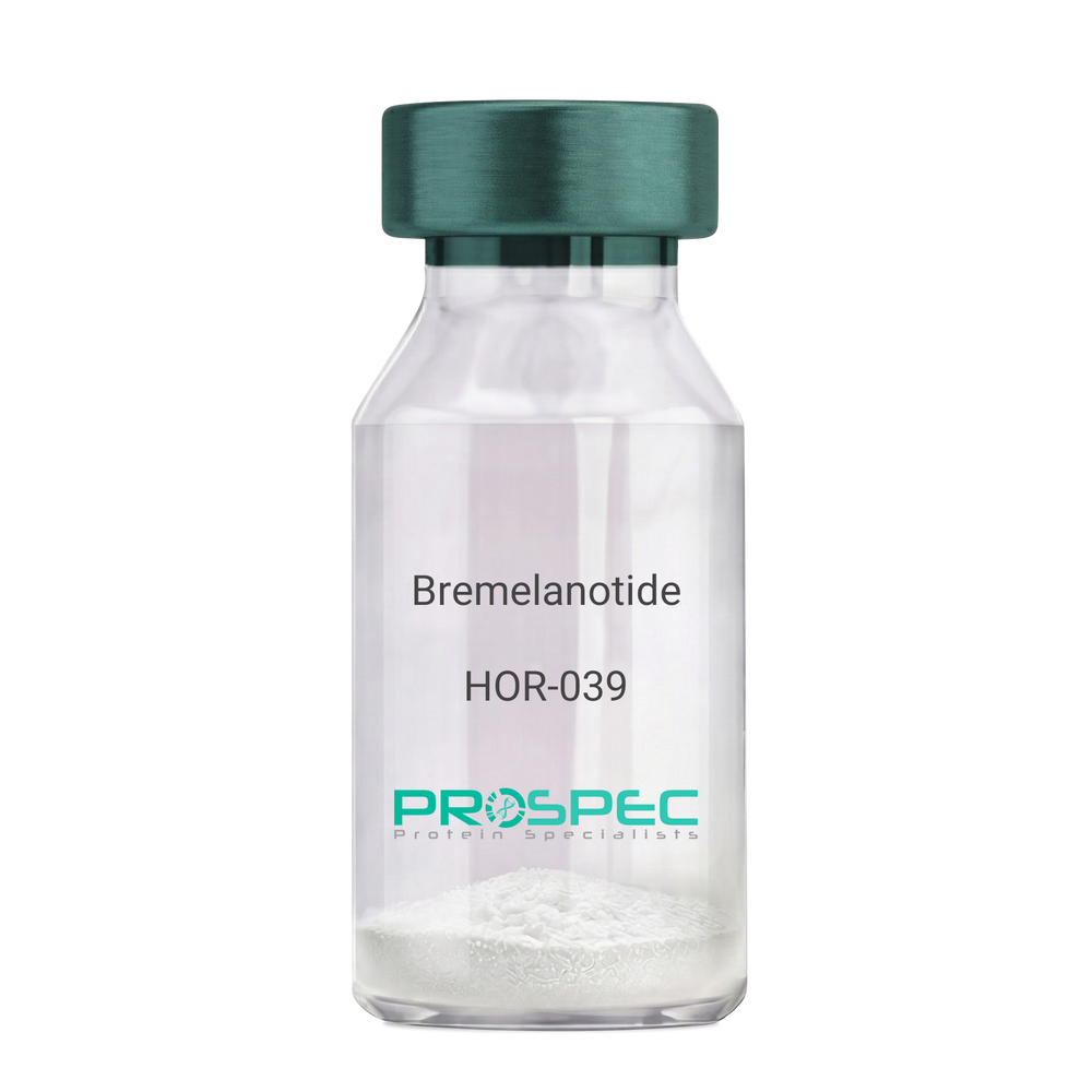
BremelanotideDescription:
Bremelanotide
PT-141, Rekynda, bremelanotide acetate.
Product # :
HOR-039Price :
Quantity :
Shipping Method :
Shipped at Room temp
More Info
- description
- formulation
- purity
- More Info
Description
Bremelanotide Synthetic is a single, non-glycosylated polypeptide chain containing 6 amino acids, having a molecular mass of 1025.16 Dalton and a Molecular formula of C50H68N40O10.
Formulation
The protein was lyophilized with no additives.
Purity
Greater than 97.0% as determined by analysis by RP-HPLC.
More Info
-
Synonyms
PT-141, Rekynda, bremelanotide acetate.
-
Physical Appearance
Sterile Filtered White lyophilized (freeze-dried) powder.
-
Stability
Lyophilized Bremelanotide although stable at room temperature for 3 weeks, should be stored desiccated below -18°C. Upon reconstitution Bremelanotide should be stored at 4°C between 2-7 days and for future use below -18°C. For long term storage it is recommended to add a carrier protein (0.1% HSA or BSA). Please prevent freeze-thaw cycles.
-
Solubility
It is recommended to reconstitute the lyophilized Bremelanotide in sterile 18MΩ-cm H2O not less than 100 µg/ml, which can then be further diluted to other aqueous solutions.
-
Amino Acid Sequence
Ac-Nle-cyclo(-Asp-His-D-Phe-Arg-Trp-Lys)-OH.
-
Background
Bremelanotide (PT-141), a synthetic heptapeptide, is a novel therapeutic agent primarily utilized for the treatment of sexual dysfunction. This paper provides an exhaustive review of Bremelanotide, detailing its biochemical structure, mechanism of action, therapeutic applications, and future prospects in clinical medicine.
Bremelanotide (PT-141), derived from Melanotan II, is a synthetic peptide developed for its role in treating sexual dysfunction in both men and women (Clayton et al., 2016). This paper presents a comprehensive examination of Bremelanotide, its biological properties, and potential therapeutic uses.
Bremelanotide acts as a non-selective agonist of the melanocortin receptors, primarily MC3R and MC4R, present in the central nervous system. The peptide initiates its action in the hypothalamus, leading to downstream effects on sexual desire and arousal (King et al., 2007).
The US Food and Drug Administration approved Bremelanotide for the treatment of premenopausal women with hypoactive sexual desire disorder (HSDD) in 2019. In clinical trials, it demonstrated efficacy in enhancing sexual desire and reducing distress associated with HSDD (Kingsberg et al., 2019).
While the current focus of Bremelanotide is on treating sexual dysfunction, its future potential may extend to other areas due to its unique mechanism of action. Further research is needed to explore the full therapeutic potential of this intriguing peptide. In conclusion, Bremelanotide represents a significant advance in sexual dysfunction therapeutics, with potential implications extending beyond this realm.
ProSpec's products are furnished for LABORATORY RESEARCH USE ONLY. The product may not be used as drugs, agricultural or pesticidal products, food additives or household chemicals.
- View Data Sheet
Name :
METRN MouseDescription:
Meteorin Mouse Recombinant
Meteorin, Hypoxia/reoxygenation regulatory factor, Metrn, 1810034B16Rik, Hyrac.
Product # :
PRO-2241Price :
Quantity :
Shipping Method :
Shipped with Ice Packs
More Info
- description
- source
- formulation
- purity
- More Info
Description
METRN produced in Sf9 Baculovirus cells is a single, glycosylated polypeptide chain containing 276 amino acids (22-291 a.a.) and having a molecular mass of 30.2kDa (Migrates at 28-40kDa on SDS-PAGE under reducing conditions). METRN is expressed with a 6 amino acid His tag at C-Terminus and purified by proprietary chromatographic techniques.
Source
Sf9, Baculovirus cells.
Formulation
METRN protein solution (0.25mg/ml) contains Phosphate Buffered Saline (pH 7.4) and 10% glycerol.
Purity
Greater than 90.0% as determined by SDS-PAGE.
More Info
-
Introduction
Meteorin (METRN) is involved in both glial cell differentiation and axonal network formation during neurogenesis. METRN promotes astrocyte differentiation and transforms cerebellar astrocytes into radial glia. Moreover, the METRN protein stimulates axonal extension in small and intermediate neurons of sensory ganglia by activating nearby satellite glia.
-
Synonyms
Meteorin, Hypoxia/reoxygenation regulatory factor, Metrn, 1810034B16Rik, Hyrac.
-
Physical Appearance
Sterile Filtered colorless solution.
-
Stability
Store at 4°C if entire vial will be used within 2-4 weeks. Store, frozen at -20°C for longer periods of time. For long term storage it is recommended to add a carrier protein (0.1% HSA or BSA). Avoid multiple freeze-thaw cycles.
-
Amino Acid Sequence
GYSEDRCSWR GSGLTQEPGS VGQLTLDCTE GAIEWLYPAG ALRLTLGGPD PGTRPSIVCL RPERPFAGAQ VFAERMTGNL ELLLAEGPDL AGGRCMRWGP RERRALFLQA TPHRDISRRV AAFRFELHED QRAEMSPQAQ GLGVDGACRP CSDAELLLAA CTSDFVIHGT IHGVAHDTEL QESVITVVVA RVIRQTLPLF KEGSSEGQGR ASIRTLLRCG VRPGPGSFLF MGWSRFGEAW LGCAPRFQEF SRVYSAALTT HLNPCEMALD HHHHHH.
ProSpec's products are furnished for LABORATORY RESEARCH USE ONLY. The product may not be used as drugs, agricultural or pesticidal products, food additives or household chemicals.
- View Data Sheet
Name :
TARC RatDescription:
Thymus and Activation Regulated Chemokine (CCL17) Rat Recombinant
Thymus and activation-regulated chemokine, CCL17, SCYA17, TARC.
Product # :
CHM-012Price :
Quantity :
Shipping Method :
Shipped at Room temp
More Info
- description
- source
- formulation
- purity
- biological activity
- More Info
Description
TARC Rat Recombinant produced in E.Coli is a non-glycosylated, polypeptide chain containing 70 amino acids and having a molecular mass of 8.1kDa. The TARC Rat is purified by proprietary chromatographic techniques.
Source
Escherichia Coli.
Formulation
The protein was lyophilized from a 0.2µm filtered concentrated solution in 1×PBS, pH7.4.
Purity
Greater than 97.0% as determined by
(a) Analysis by RP-HPLC.
(b) Analysis by SDS-PAGE.Biological Activity
Determined by its ability to chemoattract human T-Lymphocytes using a concentration range of 1.0-10.0 ng/ml.More Info
-
Introduction
TARC cDNA encodes a 94 amino acid precursor protein with a 23 amino acid residue signal peptide that is cleaved off to generate the 71 amino acid residue mature secreted protein. Along with CC chemokine family members, CCL-17 has approximately 24-29% amino acid sequence identity with RANTES, MIP-1a, MIP-1b, MCP-1, MCP-2, MCP-3 and I-309. TARC is expressed in thymus, and at a lower level in the lung, colon, and small intestine. TARC is in addition transiently expressed in stimulated peripheral blood mononuclear cells. Recombinant TARC has been shown to be chemotactic for T cell lines but not monocytes or neutrophils. CCL-17 was recently identified to be a specific functional ligand for CCR4, a receptor that is selectively expressed on T cells. CCL17 is one of quite a few Cys-Cys (CC) cytokine genes clustered on the q arm of chromosome 16. CCL17 shows chemotactic activity for T lymphocytes, but not monocytes or granulocytes. CCL17 binds to chemokine receptors CCR4 and CCR8. This chemokine plays important roles in T cell development in thymus as well as in trafficking and activation of mature T cells.
-
Synonyms
Thymus and activation-regulated chemokine, CCL17, SCYA17, TARC.
-
Physical Appearance
Sterile Filtered White lyophilized (freeze-dried) powder.
-
Stability
Lyophilized TARC although stable at room temperature for 3 weeks, should be stored desiccated below -18C. Upon reconstitution TARC should be stored at 4C between 2-7 days and for future use below -18C. For long term storage it is recommended to add a carrier protein (0.1% HSA or BSA).Please prevent freeze-thaw cycles.
-
Solubility
It is recommended to reconstitute the lyophilized TARC in sterile 18M-cm H2O not less than 100µg/ml, which can then be further diluted to other aqueous solutions.
-
Amino Acid Sequence
ARATNVGREC CLDYFKGAIP IRKLVTWFRT SVECPKDAIV FETVQGRLIC TDPKDKHVKK AIRHLKNQRL.
ProSpec's products are furnished for LABORATORY RESEARCH USE ONLY. The product may not be used as drugs, agricultural or pesticidal products, food additives or household chemicals.
- View Data Sheet
Name :
SCGN RatDescription:
Secretagogin Rat Recombinant
SCGN, EF-hand calcium binding protein, Setagin, SEGN, CALBL, Secretagogin.
Product # :
PRO-657Price :
Quantity :
Shipping Method :
Shipped at Room temp
More Info
- description
- source
- formulation
- purity
- More Info
Description
Secretagogin Rat Recombinant produced in E.Coli is a single, non-glycosylated, polypeptide chain containing 286 amino acids and having a molecular mass of 33.3 kDa. The Rat SCGN is fused to a 10 a.a. His tag at N-Terminus.The protein’s amino acids sequence is identical to UniProtKB/Swiss-Prot entry Q6R556.The Rat SCGN is purified by proprietary chromatographic techniques.
Source
Escherichia Coli.
Formulation
The sterile filtered concentrated protein solution was lyophilized with 20mM Tris & 50mM NaCl pH-7.5.
Purity
Greater than 95% as determined by SDS-PAGE.
More Info
-
Introduction
SCGN is a secreted calcium-binding protein which is found in the cytoplasm. It is related to calbindin D-28K and calretinin. Secretagogin is involved in KCL-stimulated calcium flux and cell proliferation.
Secretagogin plays a role in human non-functional pituitary adenomas. -
Synonyms
SCGN, EF-hand calcium binding protein, Setagin, SEGN, CALBL, Secretagogin.
-
Physical Appearance
Sterile Filtered White lyophilized (freeze-dried) powder.
-
Stability
Store lyophilized protein at -20°C. Aliquot the product after reconstitution to avoid repeated freezing/thawing cycles. Reconstituted protein can be stored at 4°C for a limited period of time.
-
Solubility
Add deionized water to a working concentration of 0.5mg/ml and let the lyophilized pellet dissolve completely.
-
Amino Acid Sequence
MKHHHHHHAS MDNAHRQTQA HLDAACFWQI WQRFDKDEKG YIKETELDAF FDDLLAKFGI EDTLMEENVQ KMKEQLMVGH DISKEGRILM KELASMFLSE DENFLLFFRL ETPLDNSVEF MQIWRKYDAD SSGFISAAEL SNFLRDLFLH HKKVISEAEL EEYTSTMMKI FDRNKDGRLD LNDLARILAL QENFLLQFKM DASSTEERKR DFEKIFAHYD VSKTGALEGP EVDGFVKDMM ELVQPSISGV DLDKFREILL RHCDVNKDGK IQKSELALCLGLKINP.
ProSpec's products are furnished for LABORATORY RESEARCH USE ONLY. The product may not be used as drugs, agricultural or pesticidal products, food additives or household chemicals.
- View Data Sheet
Name :
BCDIN3D HumanDescription:
BCDIN3D Human Recombinant
Pre-miRNA 5'-monophosphate methyltransferase, BCDIN3 domain-containing protein, BCDIN3D.
Product # :
PRO-1262Price :
Quantity :
Shipping Method :
Shipped with Ice Packs
More Info
- description
- source
- formulation
- purity
- More Info
Description
BCDIN3D Human Recombinant produced in E.Coli is a single, non-glycosylated polypeptide chain containing 315 amino acids (1-292 a.a) and having a molecular mass of 35kDa.BCDIN3D is fused to a 23 amino acid His-tag at N-terminus & purified by proprietary chromatographic techniques.
Source
Escherichia Coli.
Formulation
BCDIN3D protein solution (0.25mg/ml) containing 20mM Tris-HCl buffer (pH 8.0), 0.2M NaCl, 40% glycerol, 1mM DTT and 2mM EDTA.
Purity
Greater than 85.0% as determined by SDS-PAGE.
More Info
-
Introduction
BCDIN3D is a member of the methyltransferase superfamily and contains 1 Bin3-type SAM domain. BCDIN3D acts in the catalysis of the transfer of a methyl group to an acceptor molecule. BCDIN3D is an O-methyltransferase which specifically dimethylates the 5' monophosphate of pre-miRNAs, serving as a negative regulator of miRNA processing. BCDIN3D mediates the methylation of pre-miR-145, as well as other pre-miRNAs.
-
Synonyms
Pre-miRNA 5'-monophosphate methyltransferase, BCDIN3 domain-containing protein, BCDIN3D.
-
Physical Appearance
Sterile Filtered colorless solution.
-
Stability
Store at 4°C if entire vial will be used within 2-4 weeks. Store, frozen at -20°C for longer periods of time. For long term storage it is recommended to add a carrier protein (0.1% HSA or BSA).Avoid multiple freeze-thaw cycles.
-
Amino Acid Sequence
MGSSHHHHHH SSGLVPRGSH MGSMAVPTEL DGGSVKETAA EEESRVLAPG AAPFGNFPHY SRFHPPEQRL RLLPPELLRQ LFPESPENGP ILGLDVGCNS GDLSVALYKH FLSLPDGETC SDASREFRLL CCDIDPVLVK RAEKECPFPD ALTFITLDFM NQRTRKVLLS SFLSQFGRSV FDIGFCMSIT MWIHLNHGDH GLWEFLAHLS SLCHYLLVEP QPWKCYRAAA RRLRKLGLHD FDHFHSLAIR GDMPNQIVQI LTQDHGMELI CCFGNTSWDR SLLLFRAKQT IETHPIPESL IEKGKEKNRL SFQKQ.
ProSpec's products are furnished for LABORATORY RESEARCH USE ONLY. The product may not be used as drugs, agricultural or pesticidal products, food additives or household chemicals.
- View Data Sheet
Name :
P Selectin HumanDescription:
P-selectin Human Recombinant
P-selectin, Granule membrane protein 140, GMP-140, PADGEM, Leukocyte-endothelial cell adhesion molecule 3, LECAM3, CD62 antigen-like family member P, CD62P antigen, SELP, GMRP, GRMP, CD62, PSEL, CD62P, GMP140, FLJ45155.
Product # :
PRO-382Price :
Quantity :
Shipping Method :
Shipped with Ice Packs
More Info
- description
- source
- formulation
- purity
- More Info
Description
P-Selectin Human Recombinant is expressed in E. coli containing 566 amino acids 197-761 fused to an amino terminal hexahistidine tag.
Source
Escherichia Coli.
Formulation
P-Selectin is supplied in 50% Glycerol.
Purity
Greater than 95% as determined by SDS-PAGE.
Single band on Western Blot.More Info
-
Introduction
P-Selectin also called Platelet Alpha-Granule Membrane Protein, CD62, and Granulocyte Membrane Protein GRMP belongs to a family of divalent cation-dependent carbohydrate-binding glycoproteins or adhesion molecules. Pselectin is expressed transiently on the surface of activated platelets and endothelial cells. P-Selectin is a 140 kDa protein which is stored in the alpha-granules of platelets and Weibel-Palade bodies of endothelial cells. Secreted P-selectin is thought to play a key role in the adhesion of platelets to monocytes and neutrophils during an inflammatory response. P-Selectin is a calcium-dependent receptor that binds to sialylated forms of Lewis blood group carbohydrate antigens on neutrophils and monocytes. Levels of P-Selectin may be elevated in a number of pathological conditions.
-
Synonyms
P-selectin, Granule membrane protein 140, GMP-140, PADGEM, Leukocyte-endothelial cell adhesion molecule 3, LECAM3, CD62 antigen-like family member P, CD62P antigen, SELP, GMRP, GRMP, CD62, PSEL, CD62P, GMP140, FLJ45155.
-
Physical Appearance
Sterile Filtered liquid formulation.
-
Stability
Store at 4°C if entire vial will be used within 1-2 weeks. Store, frozen at -20°C for longer periods of time. Avoid multiple freeze-thaw cycles.
-
Applications
P-Selectin can be used directly as a positive control in Western blotting, ELISA, immunoprecipitation and other immunological experiments.
The biological activity of this product has not yet been tested.
ProSpec's products are furnished for LABORATORY RESEARCH USE ONLY. The product may not be used as drugs, agricultural or pesticidal products, food additives or household chemicals.
- View Data Sheet
Name :
HAPLN1 HumanDescription:
Hyaluronan And Proteoglycan Link Protein 1 Human Recombinant
Hyaluronan and proteoglycan link protein 1 precursor, CRTL1, Cartilage-linking protein 1, Cartilage-link protein, Proteoglycan link protein, HAPLN1.
Product # :
PRO-2012Price :
Quantity :
Shipping Method :
Shipped with Ice Packs
More Info
- description
- source
- formulation
- purity
- More Info
Description
HAPLN1 Human Recombinant produced in E.Coli is a single, non-glycosylated polypeptide chain containing 362 amino acids (16-354 a.a.) and having a molecular mass of 40.9kDa.HAPLN1 is fused to a 23 amino acid His-tag at N-terminus & purified by proprietary chromatographic techniques.
Source
Escherichia Coli.
Formulation
HAPLN1 protein solution (1mg/ml) containing 20mM Tris-HCl buffer (pH 8.0), 0.4M Urea and 10% glycerol.
Purity
Greater than 80.0% as determined by SDS-PAGE.
More Info
-
Introduction
Hyaluronan And Proteoglycan Link Protein 1, which is also known as HAPLN1 is a part of the HAPLN family.HAPLN1 contains one Ig-like V-type domain and two Link domains. In the extracellular cartilage matrix, HAPLN1 stabilizes the aggregates of proteoglycan monomers by means of hyaluronic acid .
-
Synonyms
Hyaluronan and proteoglycan link protein 1 precursor, CRTL1, Cartilage-linking protein 1, Cartilage-link protein, Proteoglycan link protein, HAPLN1.
-
Physical Appearance
Sterile Filtered colorless solution.
-
Stability
Store at 4°C if entire vial will be used within 2-4 weeks. Store, frozen at -20°C for longer periods of time. For long term storage it is recommended to add a carrier protein (0.1% HSA or BSA).Avoid multiple freeze-thaw cycles.
-
Amino Acid Sequence
MGSSHHHHHH SSGLVPRGSH MGSDHLSDNY TLDHDRAIHI QAENGPHLLV EAEQAKVFSH RGGNVTLPCK FYRDPTAFGS GIHKIRIKWT KLTSDYLKEV DVFVSMGYHK KTYGGYQGRV FLKGGSDSDA SLVITDLTLE DYGRYKCEVI EGLEDDTVVV ALDLQGVVFP YFPRLGRYNL NFHEAQQACL DQDAVIASFD QLYDAWRGGL DWCNAGWLSD GSVQYPITKP REPCGGQNTV PGVRNYGFWD KDKSRYDVFC FTSNFNGRFY YLIHPTKLTY DEAVQACLND GAQIAKVGQI FAAWKILGYD RCDAGWLADG SVRYPISRPR RRCSPTEAAV RFVGFPDKKH KLYGVYCFRA YN.
ProSpec's products are furnished for LABORATORY RESEARCH USE ONLY. The product may not be used as drugs, agricultural or pesticidal products, food additives or household chemicals.
- View Data Sheet
Name :
S100B Human, HisDescription:
S100 Calcium Binding Protein B Human Recombinant, His Tag
Protein S100-B, S100 calcium-binding protein B, S-100 protein subunit beta, S-100 protein beta chain, S100B, NEF, S100, S100beta.
Product # :
PRO-306Price :
Quantity :
Shipping Method :
Shipped with Ice Packs
More Info
- description
- source
- formulation
- purity
- More Info
Description
S100B Human Recombinant produced in E.Coli is single, a non-glycosylated, Polypeptide chain containing 112 amino acids fragment (1-92) with a 20 amino acids N-terminal His tag and having a total molecular mass of 12.8kDa. The S100B is purified by proprietary chromatographic techniques.
Source
Escherichia Coli.
Formulation
S100B (1mg/ml) is supplied in 20mM Tris-HCl buffer (pH8.0), 1mM DTT and 10% glycerol.
Purity
Greater than 90.0% as determined by SDS-PAGE.
More Info
-
Introduction
S100b is a member of the S100 family of proteins which are a family of EF-hand calcium binding proteins that exist mostly as dimers of the 20 currently identified individual S100 monomers. The S100B homodimer is expressed in cells of the central nervous system, glial cells and in certain peripheral cells e.g. Schwann cells, melanocytes, adipocytes and chondrocytes. S100 proteins are localized either in the cytoplasm or the nucleus of a wide range of cells. S100 proteins are involved in the regulation of a number of cellular processes such as cell cycle progression and differentiation. There are at least 13 members in the S100 gene family, which are located as a cluster on chromosome 1q21; however, S100b is located at 21q22.3. The determination of S100B in serum levels may be used to monitor the extent of brain injury and malignant melanoma. S100b proteins may have a role in Neurite extension, proliferation of melanoma cells, stimulation of Ca2+ fluxes, inhibition of PKC-mediated phosphorylation, astrocytosis and axonal proliferation, and inhibition of microtubule assembly. Chromosomal rearrangements and altered expression of the S100b gene are implicated in several neurological, neoplastic, and other types of diseases, including Alzheimer's disease, Down's syndrome, epilepsy, amyotrophic lateral sclerosis, melanoma, and type I diabetes.
-
Synonyms
Protein S100-B, S100 calcium-binding protein B, S-100 protein subunit beta, S-100 protein beta chain, S100B, NEF, S100, S100beta.
-
Physical Appearance
Sterile Filtered clear solution.
-
Stability
Store at 4°C if entire vial will be used within 2-4 weeks. Store, frozen at -20°C for longer periods of time. For long term storage it is recommended to add a carrier protein (0.1% HSA or BSA).Avoid multiple freeze-thaw cycles.
-
Amino Acid Sequence
MGSSHHHHHH SSGLVPRGSH MSELEKAMVA LIDVFHQYSG REGDKHKLKK SELKELINNE LSHFLEEIKE QEVVDKVMET LDNDGDGECD FQEFMAFVAM VTTACHEFFE HE.
ProSpec's products are furnished for LABORATORY RESEARCH USE ONLY. The product may not be used as drugs, agricultural or pesticidal products, food additives or household chemicals.
- View Data Sheet
Name :
S100b MouseDescription:
S100 Calcium Binding Protein B Mouse Recombinant
Protein S100-B, S-100 protein beta chain, S-100 protein subunit beta, S100 calcium-binding protein B.
Product # :
PRO-2370Price :
Quantity :
Shipping Method :
Shipped with Ice Packs
More Info
- description
- source
- formulation
- purity
- More Info
Description
s100b Mouse Recombinant produced in E.Coli is a single, non-glycosylated, polypeptide chain containing 92 amino acids (1-92 a.a.) and having a molecular mass of 10.7kDa. The s100b is purified by proprietary chromatographic techniques.
Source
Escherichia Coli.
Formulation
The s100b protein solution (1mg/ml) contains Phosphate Buffered Saline (pH 7.4) and 10% glycerol.
Purity
Greater than 95.0% as determined by SDS-PAGE.
More Info
-
Introduction
S100b is a member of the S100 family of proteins which are a family of EF-hand calcium binding proteins that exist mostly as dimers of the 20 currently identified individual S100 monomers. The S100B homodimer is expressed in cells of the central nervous system, glial cells and in certain peripheral cells e.g. Schwann cells, melanocytes, adipocytes and chondrocytes. S100 proteins are localized either in the cytoplasm or the nucleus of a wide range of cells. S100 proteins are involved in the regulation of a number of cellular processes such as cell cycle progression and differentiation. There are at least 13 members in the S100 gene family, which are located as a cluster on chromosome 1q21; however, S100b is located at 21q22.3. The determination of S100B in serum levels may be used to monitor the extent of brain injury and malignant melanoma. S100b proteins may have a role in Neurite extension, proliferation of melanoma cells, stimulation of Ca2+ fluxes, inhibition of PKC-mediated phosphorylation, astrocytosis and axonal proliferation, and inhibition of microtubule assembly. Chromosomal rearrangements and altered expression of the S100b gene are implicated in several neurological, neoplastic, and other types of diseases, including Alzheimer's disease, Down's syndrome, epilepsy, amyotrophic lateral sclerosis, melanoma, and type I diabetes.
-
Synonyms
Protein S100-B, S-100 protein beta chain, S-100 protein subunit beta, S100 calcium-binding protein B.
-
Physical Appearance
Sterile filtered colorless solution.
-
Stability
Store at 4°C if entire vial will be used within 2-4 weeks. Store, frozen at -20°C for longer periods of time. For long term storage it is recommended to add a carrier protein (0.1% HSA or BSA).Avoid multiple freeze-thaw cycles.
-
Amino Acid Sequence
MSELEKAMVA LIDVFHQYSG REGDKHKLKK SELKELINNE LSHFLEEIKE QEVVDKVMET LDEDGDGECD FQEFMAFVAM VTTACHEFFE HE.
ProSpec's products are furnished for LABORATORY RESEARCH USE ONLY. The product may not be used as drugs, agricultural or pesticidal products, food additives or household chemicals.
- View Data Sheet
Name :
S100P HumanDescription:
S100 Calcium Binding Protein P Human Recombinant
Protein S100-P, S100 calcium-binding protein P, S100P, S100E, MIG9.
Product # :
PRO-425Price :
Quantity :
Shipping Method :
Shipped at Room temp
More Info
- description
- source
- formulation
- purity
- More Info
Description
The Recombinant Human S100P has a molecular mass of 10.4 kDa containing 95 amino acid residues of the human S100P.
Source
Escherichia Coli.
Formulation
Filtered (0.4µm) and lyophilized from 0.5 mg/ml in 0.05M Phosphate buffer pH7.2, 0.1M NaCl.
Purity
Greater than 95% as determined by SDS-PAGE.
More Info
-
Introduction
S100P was originally described as a placental protein of 95 amino acid residues shares about 50% sequence identity with the brain S100 proteins alpha and beta.
S100 proteins are small dimeric members of the EF-hand superfamily of Ca(2+) binding proteins thought to participate in mediating intracellular Ca(2+) signals by binding to and thereby regulating target proteins in a Ca(2+)-dependent manner. S100P in addition to binding Ca2+, also binds Zn2+ and Mg2+. S100P gene is located on chromosome 4p16.
S100P is dysregulated in the androgen-independent prostate cancer cell lines LNCaP-R, DU145, and PC3 and may play a role in the etiology of prostate cancer.
In ductal hyperplasias, in situ and invasive ductal carcinoma, but not in the normal tissues, S100P overexpression is an early event that might play an important role in the immortalization of human breast epithelial cells in vitro and tumor progression in vivo.
In NIH3T3 cells, the expression of S100P led to the presence of S100P in the culture medium, increased cellular proliferation, and enhanced survival following detachment from the culture substrate or after exposure to the chemotherapeutic agent 5-flurouracil. The proliferation and survival effects of S100P expression were duplicated in a time- and concentration-dependent manner by extracellular addition of purified S100P to wild-type NIH3T3 cells and correlated with the activation of Erks and NFB.
To determine the mechanisms involved in these effects, we tested the hypothesis that S100P activated RAGE (Receptor for Activated Glycation End-Products). It was found that S100P coimmunoprecipitated with RAGE. Furthermore, the effects of S100P on cell signaling, proliferation and survival were blocked by agents that interfere with RAGE including administration of an amphoterin derived peptide known to antagonize RAGE activation, anti-RAGE antibodies and by expression of a dominant negative RAGE. These data suggest that S100P can act in an autocrine manner via RAGE to stimulate cell proliferation and survival. -
Synonyms
Protein S100-P, S100 calcium-binding protein P, S100P, S100E, MIG9.
-
Physical Appearance
Filtered White lyophilized (freeze-dried) powder.
-
Stability
Store lyophilized protein at -20°C. Aliquot the product after reconstitution to avoid repeated freezing/thawing cycles. Reconstituted protein can be stored at 4°C for a limited period of time; it does not show any change after two weeks at 4°C.
-
Solubility
Add deionized H2O and let the lyophilized pellet dissolve completely.
-
Amino Acid Sequence
MTELEAAMGM IIDVFSRYSG SEGSTQTLTK GELKVLMEKE LPGFLQSGKD KDAVDKLLKD LDANGDAQVD FSEFIVFVAA ITSACHKYFE KAGLK.
ProSpec's products are furnished for LABORATORY RESEARCH USE ONLY. The product may not be used as drugs, agricultural or pesticidal products, food additives or household chemicals.