Skip to content

High-quality research proteins.

  • 1-800-805-6531

  • Mon - Sat 8.00-18.00, Sun - Closed

  • info@prospecbio.com

  • PO Box 6591 East Brunswick NJ 08816

  • Products
    • Cytokines
    • Growth factors
    • Chemokines
    • Cd antigens
    • Neurotrophins
    • Hormones
    • Enzymes
    • Viral antigens
    • Recombinant proteins
    • Natural proteins
    • Monoclonal antibodies
    • Polyclonal antibodies
    • Thermal Packaging
  • About
    • Company
    • Founder
    • Contribution
  • Customers
    • Eastern Asia
    • Eastern Europe
    • North America
    • South America
    • South East Asia
    • Southern Asia
    • Western Europe
  • Ordering
    • Ordering Method
    • Ordering - Credit Card
    • Ordering - PO
    • Shipping Fees
    • FAQs
  • Services
    • DNA Cloning
    • Protein Expression
    • Monoclonal Antibody
    • Fermentation
    • Technology License
  • Contact us
Log in

Country/region

  • USD $ | Albania
  • USD $ | Andorra
  • USD $ | Argentina
  • USD $ | Armenia
  • USD $ | Australia
  • USD $ | Austria
  • USD $ | Azerbaijan
  • USD $ | Belarus
  • USD $ | Belgium
  • USD $ | Bolivia
  • USD $ | Brazil
  • USD $ | Bulgaria
  • USD $ | Cambodia
  • USD $ | Canada
  • USD $ | Chile
  • USD $ | China
  • USD $ | Colombia
  • USD $ | Costa Rica
  • USD $ | Croatia
  • USD $ | Cyprus
  • USD $ | Czechia
  • USD $ | Denmark
  • USD $ | Estonia
  • USD $ | Finland
  • USD $ | France
  • USD $ | Georgia
  • USD $ | Germany
  • USD $ | Greece
  • USD $ | Hong Kong SAR
  • USD $ | Hungary
  • USD $ | Iceland
  • USD $ | India
  • USD $ | Ireland
  • USD $ | Israel
  • USD $ | Italy
  • USD $ | Japan
  • USD $ | Kazakhstan
  • USD $ | Latvia
  • USD $ | Liechtenstein
  • USD $ | Lithuania
  • USD $ | Madagascar
  • USD $ | Malta
  • USD $ | Mexico
  • USD $ | Moldova
  • USD $ | Monaco
  • USD $ | Netherlands
  • USD $ | New Zealand
  • USD $ | North Macedonia
  • USD $ | Norway
  • USD $ | Panama
  • USD $ | Paraguay
  • USD $ | Peru
  • USD $ | Philippines
  • USD $ | Poland
  • USD $ | Portugal
  • USD $ | Romania
  • USD $ | Singapore
  • USD $ | Slovakia
  • USD $ | Slovenia
  • USD $ | South Africa
  • USD $ | South Korea
  • USD $ | Spain
  • USD $ | Sri Lanka
  • USD $ | Sweden
  • USD $ | Switzerland
  • USD $ | Taiwan
  • USD $ | Thailand
  • USD $ | Türkiye
  • USD $ | Ukraine
  • USD $ | United Kingdom
  • USD $ | United States
  • USD $ | Uruguay
  • USD $ | Vatican City
  • USD $ | Venezuela
  • USD $ | Vietnam
  • Facebook
  • Instagram
logoProSpec - Recombinant Protein Manufacturer for academic and R&D industry
Become a distributor
Account Log in
Wishlist
Cart Cart(0)
  • Products
    +
    • Cytokines
    • Growth factors
    • Chemokines
    • Cd antigens
    • Neurotrophins
    • Hormones
    • Enzymes
    • Viral antigens
    • Recombinant proteins
    • Natural proteins
    • Monoclonal antibodies
    • Polyclonal antibodies
    • Thermal Packaging
  • About
    +
    • Company
    • Founder
    • Contribution
  • Customers
    +
    • Eastern Asia
    • Eastern Europe
    • North America
    • South America
    • South East Asia
    • Southern Asia
    • Western Europe
  • Ordering
    +
    • Ordering Method
    • Ordering - Credit Card
    • Ordering - PO
    • Shipping Fees
    • FAQs
  • Services
    +
    • DNA Cloning
    • Protein Expression
    • Monoclonal Antibody
    • Fermentation
    • Technology License
  • Contact us

Item added to your cart

View cart
View All
  • Cytokines
  • Growth factors
  • Chemokines
  • Cd antigens
  • Neurotrophins
  • Hormones
  • Enzymes
  • Viral antigens
  • Recombinant proteins
  • Natural proteins
  • Monoclonal antibodies
  • Polyclonal antibodies
  • Thermal Packaging
  • Tumor Necrosis Factor

    Tumor Necrosis Factor

    View All
  • 4-1BB

    4-1BB

    View All
  • Adiponectin

    Adiponectin

    View All
  • AIF1

    AIF1

    View All
  • Other Cytokines

    Other Cytokines

    View All
  • AITRL

    AITRL

    View All
  • Angiopoietin

    Angiopoietin

    View All
  • Apolipoprotein

    Apolipoprotein

    View All
  • B-Cell Activating Factor

    B-Cell Activating Factor

    View All
  • Beta Defensin

    Beta Defensin

    View All
  • Bone Morphogenetic Protein

    Bone Morphogenetic Protein

    View All
  • B type Natriuretic Peptide

    B type Natriuretic Peptide

    View All
  • BST

    BST

    View All
  • Betacellulin

    Betacellulin

    View All
  • Cardiotrophin

    Cardiotrophin

    View All
  • Activin

    Activin

    View All
  • Fibroblast Growth Factor

    Fibroblast Growth Factor

    View All
  • Other Growth Factors

    Other Growth Factors

    View All
  • CSF

    CSF

    View All
  • CTGF

    CTGF

    View All
  • IGFBP

    IGFBP

    View All
  • VEGF Protein

    VEGF Protein

    View All
  • EGF

    EGF

    View All
  • Epigen

    Epigen

    View All
  • Erythropoietin

    Erythropoietin

    View All
  • Insulin-Like Growth Factor

    Insulin-Like Growth Factor

    View All
  • Growth Hormone

    Growth Hormone

    View All
  • HDGF

    HDGF

    View All
  • Hepatocyte Growth Factor

    Hepatocyte Growth Factor

    View All
  • Insulin

    Insulin

    View All
  • BCA-1/ BLC (CXCL13)

    BCA-1/ BLC (CXCL13)

    View All
  • BRAK (CXCL14)

    BRAK (CXCL14)

    View All
  • C-10 (CCL6)

    C-10 (CCL6)

    View All
  • Other Chemokines

    Other Chemokines

    View All
  • MEC (CCL28)

    MEC (CCL28)

    View All
  • LD78-beta (CCL3L1)

    LD78-beta (CCL3L1)

    View All
  • CTACK (CCL27)

    CTACK (CCL27)

    View All
  • CXCL16

    CXCL16

    View All
  • CXCL17

    CXCL17

    View All
  • Platelet Factor-4 (CXCL4)

    Platelet Factor-4 (CXCL4)

    View All
  • ENA-78 (CXCL5)

    ENA-78 (CXCL5)

    View All
  • Eotaxin (CCL11,24,26)

    Eotaxin (CCL11,24,26)

    View All
  • Exodus-2 (CCL21)

    Exodus-2 (CCL21)

    View All
  • Fractalkine (CX3CL1)

    Fractalkine (CX3CL1)

    View All
  • CXCL6

    CXCL6

    View All
  • Other CD Antigens

    Other CD Antigens

    View All
  • CD14

    CD14

    View All
  • CD164

    CD164

    View All
  • CD1

    CD1

    View All
  • CD2

    CD2

    View All
  • CD200

    CD200

    View All
  • CD207

    CD207

    View All
  • CD226

    CD226

    View All
  • CD23

    CD23

    View All
  • CD244

    CD244

    View All
  • CD247

    CD247

    View All
  • CD27

    CD27

    View All
  • CD274

    CD274

    View All
  • CD300

    CD300

    View All
  • CD33

    CD33

    View All
  • Other Neurotrophins

    Other Neurotrophins

    View All
  • Beta-NGF

    Beta-NGF

    View All
  • BDNF

    BDNF

    View All
  • CDNF

    CDNF

    View All
  • CNTF

    CNTF

    View All
  • GDNF

    GDNF

    View All
  • Glia Maturation Factor

    Glia Maturation Factor

    View All
  • MANF

    MANF

    View All
  • Midkine

    Midkine

    View All
  • Neuroglobin

    Neuroglobin

    View All
  • Neurotrophic factors

    Neurotrophic factors

    View All
  • Neuregulin

    Neuregulin

    View All
  • Neuropilin

    Neuropilin

    View All
  • Pigment Epithelium-Derived Factor

    Pigment Epithelium-Derived Factor

    View All
  • Pleiotrophin

    Pleiotrophin

    View All
  • Peptide Hormones

    Peptide Hormones

    View All
  • Other Hormones

    Other Hormones

    View All
  • HCG

    HCG

    View All
  • Endothelin

    Endothelin

    View All
  • Exendin

    Exendin

    View All
  • FSH

    FSH

    View All
  • Glucagon

    Glucagon

    View All
  • GHRP

    GHRP

    View All
  • GLP

    GLP

    View All
  • Thyrostimulin

    Thyrostimulin

    View All
  • Inhibin A

    Inhibin A

    View All
  • LHRH

    LHRH

    View All
  • Melanotan

    Melanotan

    View All
  • Procalcitonin

    Procalcitonin

    View All
  • PTH

    PTH

    View All
  • Peroxiredoxin

    Peroxiredoxin

    View All
  • Peptidase

    Peptidase

    View All
  • Synthetase

    Synthetase

    View All
  • Transferase

    Transferase

    View All
  • Hydrolase

    Hydrolase

    View All
  • Dehydrogenase

    Dehydrogenase

    View All
  • ACE2

    ACE2

    View All
  • Esterase

    Esterase

    View All
  • Protein Kinases

    Protein Kinases

    View All
  • Other Enzymes

    Other Enzymes

    View All
  • Phosphatase

    Phosphatase

    View All
  • Deaminase

    Deaminase

    View All
  • Oxygenase

    Oxygenase

    View All
  • Adenylate Kinase

    Adenylate Kinase

    View All
  • Lyase

    Lyase

    View All
  • Chagas

    Chagas

    View All
  • SARS

    SARS

    View All
  • Influenza

    Influenza

    View All
  • ASFV

    ASFV

    View All
  • Astrovirus

    Astrovirus

    View All
  • Borrelia

    Borrelia

    View All
  • Helicobacter Pylori

    Helicobacter Pylori

    View All
  • Chikungunya

    Chikungunya

    View All
  • Chlamydia

    Chlamydia

    View All
  • Cytomegalo

    Cytomegalo

    View All
  • Dengue

    Dengue

    View All
  • EBV

    EBV

    View All
  • Ebola

    Ebola

    View All
  • Feline Leukemia Virus

    Feline Leukemia Virus

    View All
  • Hepatitis A

    Hepatitis A

    View All
  • Other

    Other

    View All
  • Actin

    Actin

    View All
  • ADAM

    ADAM

    View All
  • Complement Component

    Complement Component

    View All
  • Ag85

    Ag85

    View All
  • Eukaryotic Translation Initiation Factor

    Eukaryotic Translation Initiation Factor

    View All
  • Anterior Gradient Protein

    Anterior Gradient Protein

    View All
  • Heat Shock Protein

    Heat Shock Protein

    View All
  • Alpha-2-HS-Glycoprotein

    Alpha-2-HS-Glycoprotein

    View All
  • Hemoglobin

    Hemoglobin

    View All
  • Allergy

    Allergy

    View All
  • Anaplasma

    Anaplasma

    View All
  • Angiogenin

    Angiogenin

    View All
  • Ankyrin Repeat Domain

    Ankyrin Repeat Domain

    View All
  • Annexin

    Annexin

    View All
  • Other Natural Proteins

    Other Natural Proteins

    View All
  • Aprotinin

    Aprotinin

    View All
  • Transferrin

    Transferrin

    View All
  • Avidin

    Avidin

    View All
  • Anti Coagulation Factors

    Anti Coagulation Factors

    View All
  • Natural Albumin

    Natural Albumin

    View All
  • Natural Coagulation Factors

    Natural Coagulation Factors

    View All
  • Fibronectin

    Fibronectin

    View All
  • Thrombin

    Thrombin

    View All
  • Anti Human Enzyme

    Anti Human Enzyme

    View All
  • Other Monoclonal Antibodies

    Other Monoclonal Antibodies

    View All
  • Anti Human Cytokine

    Anti Human Cytokine

    View All
  • Anti Human Heat Shock Protein

    Anti Human Heat Shock Protein

    View All
  • Anti Mouse Lymphocyte

    Anti Mouse Lymphocyte

    View All
  • Anti Human Chemokine

    Anti Human Chemokine

    View All
  • Anti Human Lymphocyte

    Anti Human Lymphocyte

    View All
  • Anti Viral Monoclonal

    Anti Viral Monoclonal

    View All
  • Anti Mouse Cytokine

    Anti Mouse Cytokine

    View All
  • Anti-GST Monoclonal

    Anti-GST Monoclonal

    View All
  • Other Polyclonal Antibodies

    Other Polyclonal Antibodies

    View All
  • Anti Viral

    Anti Viral

    View All
  • Anti-GST Monoclonal

    Anti-GST Monoclonal

    View All
Back

Search results

610 results found for “vascular cell adhesion molecule”

Name

Description

Product #

Price

Quantity

Shipping Method

  • View Data Sheet

    Name :

    ITAC (63-87) Human

    Description:

    ITAC (63-87 a.a.) Human Recombinant

    ITAC, I-TAC, CXCL-11, CXCL11.

    Product # :

    CHM-049

    Price :

    Quantity :

    Shipping Method :

    Room Temp Icon

    Shipped at Room temp

    Add To Cart

    More Info

    • description
    • source
    • formulation
    • purity
    • More Info

    Description

    The I-TAC Human is created as a recombinant protein with a 4kda N-terminal fusion of His Tag. The I-TAC His-Tagged Fusion Protein, produced in E. coli, is a 9kDa protein containing 25 amino acid residues of the I-TACHuman, 63-87 amino acids.

    Source

    Escherichia Coli.

    Formulation

    Each mg was lyophilized with 1xPBS, 0.4% SDS and 4mM DTT.

    Purity

    Greater than 90% as determined by SDS-PAGE.

    More Info

    • Synonyms

      ITAC, I-TAC, CXCL-11, CXCL11.

    • Physical Appearance

      Filtered White lyophilized (freeze-dried) powder.

    • Stability

      Store lyophilized I-TAC at -20°C. Aliquot the product after reconstitution to avoid repeated freezing/thawing cycles. Reconstituted protein can be stored at 4°C for a limited period of time; it does not show any change after two weeks at 4°C.

    • Solubility

      It is recommended to add deionized water to prepare a working stock solution of approximately 0.5mg/ml and let the lyophilized pellet dissolve completely. Product is not sterile! Please filter the product by an appropriate sterile filter before using it on cell culture.

    • Background

      I-TAC is a small cytokine belongs to the CXC chemokinen family which also called inducible T-cell alpha chemoattractant and IP-9. I-TAC is expressed mainly in peripheral blood leukocytes, liver and pancreas with moderate levels in spleen, thymus and lung and low levels in small intestine, placenta and prostate. IFN-g and IFN-b induces strongly gene expression of I-TAC. The I-TAC chemokine elicits its effects on its target cells by interacting with the cell surface chemokine receptor CXCR3, with a higher affinity than do the other ligands for this receptor, CXCL9 and CXCL10.

    ProSpec's products are furnished for LABORATORY RESEARCH USE ONLY. The product may not be used as drugs, agricultural or pesticidal products, food additives or household chemicals.

    Cxcl11 Human
  • View Data Sheet

    Name :

    VBP1 Human

    Description:

    Von Hippel-Lindau Binding Protein 1 Human Recombinant

    Prefoldin subunit 3, HIBBJ46, Von Hippel-Lindau-binding protein 1, VBP-1, VHL-binding protein 1, VBP1, PFDN3, PFD3.

    Product # :

    PRO-1325

    Price :

    Quantity :

    Shipping Method :

    Ice Icon

    Shipped with Ice Packs

    Add To Cart

    More Info

    • description
    • source
    • formulation
    • purity
    • More Info

    Description

    VBP1 Human Recombinant produced in E.Coli is a single, non-glycosylated polypeptide chain containing 220 amino acids (1-197 a.a) and having a molecular mass of 25kDa.VBP1 is fused to a 23 amino acid His-tag at N-terminus & purified by proprietary chromatographic techniques.

    Source

    Escherichia Coli.

    Formulation

    VBP1 protein solution (0.5mg/ml) containing 20mM Tris-HCl buffer (pH 8.0), 0.2M NaCl, 50% glycerol and 2mM DTT.

    Purity

    Greater than 90.0% as determined by SDS-PAGE.

    More Info

    • Introduction

      Prefoldin subunit 3 (VBP1) is a member of the prefoldin subunit alpha family. VBP1 interacts with the Von Hippel-Lindau protein in order to create an intracellular complex. Since VBP1 serves as a chaperone protein, it is assumed to have a role in the transport of the Von Hippel-Lindau protein from the perinuclear granules to the nucleus or cytoplasm. VBP1 binds specifically to cytosolic chaperonin (c-CPN) and transfers target proteins to it. VBP1 also binds to nascent polypeptide chain and stimulates folding in an environment in which there are numerous competing pathways for nonnative proteins.

    • Synonyms

      Prefoldin subunit 3, HIBBJ46, Von Hippel-Lindau-binding protein 1, VBP-1, VHL-binding protein 1, VBP1, PFDN3, PFD3.

    • Physical Appearance

      Sterile Filtered colorless solution.

    • Stability

      Store at 4°C if entire vial will be used within 2-4 weeks. Store, frozen at -20°C for longer periods of time. For long term storage it is recommended to add a carrier protein (0.1% HSA or BSA).Avoid multiple freeze-thaw cycles.

    • Amino Acid Sequence

      MGSSHHHHHH SSGLVPRGSH MGSMAAVKDS CGKGEMATGN GRRLHLGIPE AVFVEDVDSF MKQPGNETAD TVLKKLDEQY QKYKFMELNL AQKKRRLKGQ IPEIKQTLEI LKYMQKKKES TNSMETRFLL ADNLYCKASV PPTDKVCLWL GANVMLEYDI DEAQALLEKN LSTATKNLDS LEEDLDFLRD QFTTTEVNMA RVYNWDVKRR NKDDSTKNKA.

    ProSpec's products are furnished for LABORATORY RESEARCH USE ONLY. The product may not be used as drugs, agricultural or pesticidal products, food additives or household chemicals.

    Vbp1 Human
  • View Data Sheet

    Name :

    CXCL13 Macaque

    Description:

    BCA-1/ BLC (CXCL13) Rhesus Macaque Recombinant

    C-X-C motif chemokine 13, Small-inducible cytokine B13, B lymphocyte chemoattractant, CXC chemokine BLC, CXCL13, BCA1, BCA-1, CXCL-13, B cell Attracting Chemokine-1, BLC, ANGIE, BLR1L, SCYB13, ANGIE2.

    Product # :

    CHM-035

    Price :

    Quantity :

    Shipping Method :

    Room Temp Icon

    Shipped at Room temp

    Add To Cart

    More Info

    • description
    • source
    • formulation
    • purity
    • More Info

    Description

    BCA-1/ BLC (CXCL13) Rhesus Macaque Recombinant produced in E.Coli is a non-glycosylated polypeptide chain containing 87 amino acid and having a molecular mass of approximately 10.3kDa.BCA1 is purified by proprietary chromatographic techniques.

    Source

    Escherichia Coli.

    Formulation

    Lyophilized from a 0.2μm filtered concentrated solution in 20mM Tris-HCl, pH 8.0 and 300mM NaCl.

    Purity

    Greater than 95.0% as determined by:
    (a) Analysis by RP-HPLC.
    (b) Analysis by SDS-PAGE.

    More Info

    • Introduction

      BCA-1 is a CXC chemokine that is highly expressed in thesecondary lymphoid organs, such as follicles of the spleen, lymph nodes, and Peyer's patches. CXCL13 promotes the migration of B lymphocytes (compared to T cells and macrophages), by stimulating calcium influx into, and chemotaxis of, cells expressing Burkitt's lymphoma receptor 1 (BLR1). BCA1 therefore function in the homing of B lymphocytes to follicles. Human BCA-1 shares a 64% amino acid sequence similarity with the mouse protein and 23 - 34% amino acid sequence identity with other known CXC chemokines. Recombinant or chemically synthesized BCA1 is a potent chemoattractant for B lymphocytes but not T lymphocytes, monocytes or neutrophils. BLR1, a G protein-coupled receptor originally isolated from Burkitt’s lymphoma cells, has now been shown to be the specific receptor for BCA1. Among cells of the hematopoietic lineages, the expression of BLR-1, now designated CXCR-5, is restricted to B lymphocytes and a subpopulation of T helper memory cells.

    • Synonyms

      C-X-C motif chemokine 13, Small-inducible cytokine B13, B lymphocyte chemoattractant, CXC chemokine BLC, CXCL13, BCA1, BCA-1, CXCL-13, B cell Attracting Chemokine-1, BLC, ANGIE, BLR1L, SCYB13, ANGIE2.

    • Physical Appearance

      Sterile Filtered White lyophilized (freeze-dried) powder.

    • Stability

      Lyophilized BCA1 although stable at room temperature for 3 weeks, should be stored desiccated below -18°C. Upon reconstitution BCA-1/ BLC (CXCL13) Rhesus Macaque should be stored at 4°C between 2-7 days and for future use below -18°C. Please prevent freeze-thaw cycles.

    • Solubility

      It is recommended to reconstitute the lyophilized BCA-1/ BLC (CXCL13) Rhesus Macaque in sterile 18MΩ-cm H2O not less than 100µg/ml, which can then be further diluted to other aqueous solutions.

    • Amino Acid Sequence

      VLEVYYTHLR CRCVQESSVF IPRRFIDRIQ ISPRGNGCPR KEIIVWKKNK SVVCVDPQAE WIQRIMEMLR KKSSSTPPVP VFKRKIP.

    • Background

      What is the molecular weight/Mw of CXCL13 MACAQUE Protein?
      CXCL13 MACAQUE Protein has a total Mw of 10.3kDa.

      What is the source or expression system of CXCL13 MACAQUE Protein?
      Escherichia Coli.

      What is the Purity of CXCL13 MACAQUE Protein?
      CXCL13 MACAQUE Protein is >95% pure as determined by SDS-PAGE.

      What is the Biological Activity of CXCL13 MACAQUE Protein?
      The biological functionality of CXCL13 MACAQUE Protein will be determined in the future.

      What is the amino acid sequence of CXCL13 MACAQUE Protein?
      VLEVYYTHLR CRCVQESSVF IPRRFIDRIQ ISPRGNGCPR KEIIVWKKNK SVVCVDPQAE WIQRIMEMLR KKSSSTPPVP VFKRKIP.

      What applications can CXCL13 MACAQUE Protein be used in?
      CXCL13 MACAQUE Protein can probably be used in western blot, ELISA and Lateral Flow.

      What is the endotoxin level for CXCL13 MACAQUE Protein?
      The endotoxin level is minimal, CXCL13 MACAQUE Protein was purified using conventional chromatography techniques.


    ProSpec's products are furnished for LABORATORY RESEARCH USE ONLY. The product may not be used as drugs, agricultural or pesticidal products, food additives or household chemicals.

    Bca1 Monkey
  • View Data Sheet

    Name :

    SDF 1b Rat

    Description:

    Stromal Cell-Derived Factor-1 beta Rat Recombinant (CXCL12)

    SDF-1, CXCL12, Pre-B cell growth-stimulating factor, PBSF, hIRH, chemokine (C-X-C motif) ligand 12, SDF1, SDF1B, TPAR1, SCYB12, SDF-1b, TLSF-b, 12-O-tetradecanoylphorbol 13-acetate repressed protein 1, Thymic lymphoma cell-stimulating factor, TLSF.

    Product # :

    CHM-248

    Price :

    Quantity :

    Shipping Method :

    Room Temp Icon

    Shipped at Room temp

    Add To Cart

    More Info

    • description
    • source
    • formulation
    • purity
    • biological activity
    • More Info

    Description

    SDF-1 beta Rat Recombinant produced in E.Coli is a non-glycosylated, Polypeptide chain containing 72 amino acids and having a molecular mass of 8.4 kDa. The Rat SDF-1b is purified by proprietary chromatographic techniques.

    Source

    Escherichia Coli.

    Formulation

    The Rat SDF1B protein was lyophilized from a concentrated (1mg/ml) sterile solution containing 20mM Phsophate buffer pH-7.4 and 0.15M NaCl.

    Purity

    Greater than 97.0% as determined by:
    (a) Analysis by RP-HPLC.
    (b) Analysis by SDS-PAGE.

    Biological Activity

    The specific activity as determined by its ability to chemoattract human peripheral blood monocytes at 100-150ng/ml corresponding to a Specific Activity of 6,667-10,000IU/mg.

    More Info

    • Introduction

      SDF-1 (stromal cell-derived factor-1) is small cytokine belonging to the chemokine family that is officially designated Chemokine (C-X-C motif) ligand 12 (CXCL12). It is produced in two forms, SDF-1?/CXCL12a and SDF-1?/CXCL12b, by alternate splicing of the same gene. Chemokines are characterized by the presence of four conserved cysteines, which form two disulfide bonds. The CXCL12 proteins belong to the group of CXC chemokines, whose initial pair of cysteines are separated by one intervening amino acid. CXCL12 is strongly chemotactic for lymphocytes and has been implicated as an important cell co-ordinator during development. During embryogenesis it directs the migration of hematopoietic cells from foetal liver to bone marrow. Mice which were knocked-out for CXCL12 gene were lethal before the birth or within just 1 hour of life. As another role, CXCL12a alters also the electrophysiology of neurons. CXCL12 was shown to be expressend in many tissues in mice (including brain, thymus, heart, lung, liver, kidney, spleen and bone marrow).
      The receptor for this chemokine is CXCR4, which was previously called fusin. This CXCL12-CXCR4 interaction used to be considered exclusive (unlike for other chemokines and their receptors), but recently it was suggested that CXCL12 is also bound by CXCR7 receptor.
      The gene for CXCL12 is located on human chromosome 10. In human and mouse both CXCL12 and CXCR4 show high identity of sequence: 99% and 90%, respectively.

    • Synonyms

      SDF-1, CXCL12, Pre-B cell growth-stimulating factor, PBSF, hIRH, chemokine (C-X-C motif) ligand 12, SDF1, SDF1B, TPAR1, SCYB12, SDF-1b, TLSF-b, 12-O-tetradecanoylphorbol 13-acetate repressed protein 1, Thymic lymphoma cell-stimulating factor, TLSF.

    • Physical Appearance

      Sterile Filtered White lyophilized (freeze-dried) powder.

    • Stability

      Lyophilized SDF-1b although stable at room temperature for 3 weeks, should be stored desiccated below -18°C. Upon reconstitution CXCL12 should be stored at 4°C between 2-7 days and for future use below -18°C.For long term storage it is recommended to add a carrier protein (0.1% HSA or BSA).Please prevent freeze-thaw cycles.

    • Solubility

      It is recommended to reconstitute the lyophilized SDF-1b in sterile 18MΩ-cm H2O not less than 100µg/ml, which can then be further diluted to other aqueous solutions.

    • Amino Acid Sequence

      KPVSLSYRCP CRFFESHVAR ANVKHLKILN TPNCALQIVA RLKSNNRQVC IDPKLKWIQE YLDKALNKRL KM.

    ProSpec's products are furnished for LABORATORY RESEARCH USE ONLY. The product may not be used as drugs, agricultural or pesticidal products, food additives or household chemicals.

    Sdf 1B Rat
  • View Data Sheet

    Name :

    Angiostatin K1-4

    Description:

    Angiostatin Kringles 1-4 Human

    Product # :

    PRO-604

    Price :

    Quantity :

    Shipping Method :

    Room Temp Icon

    Shipped at Room temp

    Add To Cart

    More Info

    • description
    • source
    • formulation
    • purity
    • biological activity
    • More Info

    Description

    Human Angiostatin kringles 1-4 is produced from Human Fluid is a glycosylated polypeptide chain which migrates as a doublet 50 kDa on SDS-PAGE. The Ang K1-4 is purified by proprietary chromatographic techniques.

    Source

    Human Fluid.

    Formulation

    Lyophilized from a (1mg/ml) solution in containing 20mM Hepes buffer pH-8.2 & 20mM NaCl.

    Purity

    Greater than 98.0% as determined by SDS-PAGE.

    Biological Activity

    Human Angiostatin Kringles 1-4 significantly inhibits basic-FGF induced endothelial cell proliferation and migration at concentration ranging from 300nM-1.0 uM.

    More Info

    • Introduction

      There are several proteolytic fragments or specific domains of proteins that act as inhibitors of angiogenesis. These include fragments of plasminogen such as Angiostatin protein kringles 1-4 and kringles 1-5, Endostatin, Restin, PEX, the N-terminal fragment of prolactin, and the Nterminally truncated platelet factor. Angiostatin is a proteolytic protein fragment of plasminogen that is comprised of the first 4 kringle regions. Angiostatin k1-4 prevents the growth of endothelial cells, and its systemic administration inhibits the growth of primary carcinomas in mice. Angiostatin Kringles 1-3 segment has a larger inhibitory activity than the Angiostatin kringles 1-4 fragment. The protease-activated angiostatin kringles 1-5 is the most potent plasminogen fragment with over 50 times larger endothelial cell specific inhibitory activity. Angiostatin kringles 1-5 systemic administration inhibits growth of fibrosarcoma and significantly reduces neovascularization.
      Angiostatin is an angiogenesis inhibitor in mouse serum and urine. Angiostatin is a 38 kDa protein fragment of the plasminogen composed of the 1st 4 kringle domains of plasminogen. Angiostatin K1-4 is also named plasminogen kringles 1-4 and PK1-4.
      Angiostatin protein is manufactured by the protelytic cleavage of plasminogen by a serine protease from several prostate carcinoma cell lines. The manufacturing of angiostatin by pancreatic cancer cells can be inhibited by TGF-beta 1 along with plasminogen activator inhibitor type-1 (PAI1).

    • Physical Appearance

      Sterile Filtered lyophilized powder.

    • Stability

      Lyophilized Angiostatin Kringles 1-4 although stable at room temperature for 3 weeks, should be stored desiccated below -18°C. Upon reconstitution Angiostatin Kringles1-4 should be stored at 4°C between 2-7 days and for future use below -18°C.For long term storage it is recommended to add a carrier protein (0.1% HSA or BSA).Please prevent freeze-thaw cycles.

    • Solubility

      It is recommended to reconstitute the lyophilized Angiostatin K1-4 in sterile 18MΩ-cm H2O not less than 100µg/ml, which can then be further diluted to other aqueous solutions.

    ProSpec's products are furnished for LABORATORY RESEARCH USE ONLY. The product may not be used as drugs, agricultural or pesticidal products, food additives or household chemicals.

    Angiostatin K1 4 Human
  • View Data Sheet

    Name :

    SDF 1a Mouse

    Description:

    Stromal Cell-Derived Factor-1 Alpha Mouse Recombinant (CXCL12)

    SDF-1, CXCL12, Pre-B cell growth-stimulating factor, PBSF, hIRH, chemokine (C-X-C motif) ligand 12, SDF1, SDF1A, TPAR1, SCYB12, SDF-1a, TLSF-a, 12-O-tetradecanoylphorbol 13-acetate repressed protein 1, Thymic lymphoma cell-stimulating factor, TLSF.

    Product # :

    CHM-324

    Price :

    Quantity :

    Shipping Method :

    Room Temp Icon

    Shipped at Room temp

    Add To Cart

    More Info

    • description
    • source
    • formulation
    • purity
    • biological activity
    • More Info

    Description

    Stromal Cell-Derived Factor-1 alpha Mouse Recombinant produced in E.Coli is a non-glycosylated, Polypeptide chain containing 68 amino acids and having a molecular mass of 8 kDa. The SDF-1a is purified by proprietary chromatographic techniques.

    Source

    Escherichia Coli.

    Formulation

    The protein was Lyophilized from a sterile filtered aqueous solution containing 0.1% Trifluoroacetic Acid (TFA).

    Purity

    Greater than 95% as determined by SDS-PAGE.

    Biological Activity

    The specific activity as determined by its ability to chemoattract human peripheral blood monocytes at 50-100 ng/ml corresponding to a Specific Activity of 10,000-20,000IU/mg.

    More Info

    • Introduction

      SDF-1 (stromal cell-derived factor-1) is small cytokine belonging to the chemokine family that is officially designated Chemokine (C-X-C motif) ligand 12 (CXCL12). It is produced in two forms, SDF-1?/CXCL12a and SDF-1?/CXCL12b, by alternate splicing of the same gene. Chemokines are characterized by the presence of four conserved cysteines, which form two disulfide bonds. The CXCL12 proteins belong to the group of CXC chemokines, whose initial pair of cysteines are separated by one intervening amino acid. CXCL12 is strongly chemotactic for lymphocytes and has been implicated as an important cell co-ordinator during development. During embryogenesis it directs the migration of hematopoietic cells from foetal liver to bone marrow. Mice which were knocked-out for CXCL12 gene were lethal before the birth or within just 1 hour of life. As another role, CXCL12a alters also the electrophysiology of neurons. CXCL12 was shown to be expressend in many tissues in mice (including brain, thymus, heart, lung, liver, kidney, spleen and bone marrow).
      The receptor for this chemokine is CXCR4, which was previously called fusin. This CXCL12-CXCR4 interaction used to be considered exclusive (unlike for other chemokines and their receptors), but recently it was suggested that CXCL12 is also bound by CXCR7 receptor.
      The gene for CXCL12 is located on human chromosome 10. In human and mouse both CXCL12 and CXCR4 show high identity of sequence: 99% and 90%, respectively.

    • Synonyms

      SDF-1, CXCL12, Pre-B cell growth-stimulating factor, PBSF, hIRH, chemokine (C-X-C motif) ligand 12, SDF1, SDF1A, TPAR1, SCYB12, SDF-1a, TLSF-a, 12-O-tetradecanoylphorbol 13-acetate repressed protein 1, Thymic lymphoma cell-stimulating factor, TLSF.

    • Physical Appearance

      Sterile Filtered White lyophilized (freeze-dried) powder.

    • Stability

      Lyophilized SDF-1a although stable at room temperature for 3 weeks, should be stored desiccated below -18°C. Upon reconstitution CXCL12 should be stored at 4°C between 2-7 days and for future use below -18°C. For long term storage it is recommended to add a carrier protein (0.1% HSA or BSA).Please prevent freeze-thaw cycles.

    • Solubility

      It is recommended to reconstitute the lyophilized Stromal Cell-Derived Factor-1a in sterile 18MΩ-cm H2O not less than 100µg/ml, which can then be further diluted to other aqueous solutions.

    • Amino Acid Sequence

      The sequence of the first five N-terminal amino acids was determined and was found to be Lys-Pro-Val-Ser-Leu.

    ProSpec's products are furnished for LABORATORY RESEARCH USE ONLY. The product may not be used as drugs, agricultural or pesticidal products, food additives or household chemicals.

    Sdf 1A Mouse
  • View Data Sheet

    Name :

    CX3CL1 Human, His

    Description:

    Fractalkine Human Recombinant (CX3CL1), His Tag

    Fractalkine, CX3CL1, Neurotactin, CX3C membrane-anchored chemokine, Small inducible cytokine D1, NTN, NTT, CXC3, CXC3C, SCYD1, ABCD-3, C3Xkine.

    Product # :

    CHM-360

    Price :

    Quantity :

    Shipping Method :

    Ice Icon

    Shipped with Ice Packs

    Add To Cart

    More Info

    • description
    • source
    • formulation
    • purity
    • More Info
    • SDS-PAGE

    Description

    Fractalkine Human Recombinant fused with a 20 amino acid His tag at N-terminus produced in E.Coli is a single, non-glycosylated, polypeptide chain containing 97 amino acids (25-100 a.a.) and having a molecular mass of 10.9kDa. The Fractalkine is purified by proprietary chromatographic techniques.

    Source

    Escherichia Coli.

    Formulation

    The Fractalkine solution (0.5 mg/ml) contains Phosphate Buffered Saline pH7.4 and 10% glycerol.

    Purity

    Greater than 90.0% as determined by SDS-PAGE.

    SDS-PAGE

    CX3CL1 Human, His-SDS-PAGE - Product image 1

    More Info

    • Introduction

      Fractalkine soluble form is chemotactic for t-cells and monocytes, but not for neutrophils. Fractalkine membrane-bound form promotes adhesion of those leukocytes to endothelial cells. Fractalkine regulates leukocyte adhesion and migration processes at the endothelium and binds to CX3CR1. Natural Human Fractalkine is produced as a long protein (373-amino acid) with an extended mucin-like stalk and a chemokine domain on top. The mucin-like stalk permits it to bind to the cell surface. Fractalkine gene is located on human chromosome 16 along with some CC chemokines known as CCL17 and CCL22.

    • Synonyms

      Fractalkine, CX3CL1, Neurotactin, CX3C membrane-anchored chemokine, Small inducible cytokine D1, NTN, NTT, CXC3, CXC3C, SCYD1, ABCD-3, C3Xkine.

    • Physical Appearance

      Sterile Filtered clear solution.

    • Stability

      Store at 4°C if entire vial will be used within 2-4 weeks.Store, frozen at -20°C for longer periods of time.For long term storage it is recommended to add a carrier protein (0.1% HSA or BSA).Avoid multiple freeze-thaw cycles.

    • Amino Acid Sequence

      MGSSHHHHHH SSGLVPRGSH MQHHGVTKCN ITCSKMTSKI PVALLIHYQQ NQASCGKRAI ILETRQHRLF CADPKEQWVK DAMQHLDRQA AALTRNG.

    • Background

      What is the molecular weight/Mw of CX3CL1 HUMAN, HIS Protein?
      CX3CL1 HUMAN, HIS Protein has a total Mw of 10.9kDa.

      What is the source or expression system of CX3CL1 HUMAN, HIS Protein?
      Escherichia Coli.

      What is the Purity of CX3CL1 HUMAN, HIS Protein?
      CX3CL1 HUMAN, HIS Protein is >90% pure as determined by SDS-PAGE.

      What is the Biological Activity of CX3CL1 HUMAN, HIS Protein?
      The biological functionality of CX3CL1 HUMAN, HIS Protein will be determined in the future.

      What is the amino acid sequence of CX3CL1 HUMAN, HIS Protein?
      MGSSHHHHHH SSGLVPRGSH MQHHGVTKCN ITCSKMTSKI PVALLIHYQQ NQASCGKRAI ILETRQHRLF CADPKEQWVK DAMQHLDRQA AALTRNG.

      What applications can CX3CL1 HUMAN, HIS Protein be used in?
      CX3CL1 HUMAN, HIS Protein can probably be used in western blot, ELISA and Lateral Flow.

      What is the endotoxin level for CX3CL1 HUMAN, HIS Protein?
      The endotoxin level is minimal, CX3CL1 HUMAN, HIS Protein was purified using conventional chromatography techniques.


    ProSpec's products are furnished for LABORATORY RESEARCH USE ONLY. The product may not be used as drugs, agricultural or pesticidal products, food additives or household chemicals.

    Fractalkine Human His
  • View Data Sheet

    Name :

    CD18 Human

    Description:

    CD18 Human Recombinant

    Integrin Subunit Beta 2, Integrin, Beta 2 (Complement Component 3 Receptor 3 And 4 Subunit), Complement Component 3 Receptor 3 And 4 Subunit, CD18, MFI7, Integrin, Beta 2 (Antigen CD18 (P95), Lymphocyte Function-Associated Antigen 1; Macrophage Antigen 1 (Mac-1) Beta Subunit), Cell Surface Adhesion Glycoprotein (LFA-1/CR3/P150,959 Beta Subunit Precursor), Cell Surface Adhesion Glycoproteins LFA-1/CR3/P150,95 Subunit Beta, Leukocyte-Associated Antigens CD18/11A, CD18/11B, CD18/11C, Leukocyte Cell Adhesion Molecule CD18, Complement Receptor C3 Beta-Subunit, Complement Receptor C3 Subunit Beta, Integrin Beta Chain, Beta 2, CD18 Antigen, LFA-1, MAC-1, LCAMB, MF17, LAD, CD18.

    Product # :

    PRO-2324

    Price :

    Quantity :

    Shipping Method :

    Ice Icon

    Shipped with Ice Packs

    Add To Cart

    More Info

    • description
    • source
    • formulation
    • purity
    • More Info

    Description

    CD18 Human Recombinant produced in Sf9 Baculovirus cells is a single, glycosylated polypeptide chain containing 686 amino acids (23-700 a.a.) and having a molecular mass of 75.9kDa (Migrates at 70-100kDa on SDS-PAGE under reducing conditions).CD18 is expressed with an 8 amino acid His tag at C-Terminus and purified by proprietary chromatographic techniques.

    Source

    Sf9, Baculovirus cells.

    Formulation

    CD18 protein solution (0.25mg/ml) containing Phosphate Buffered Saline (pH 7.4) and 10% glycerol.

    Purity

    Greater than 85.0% as determined by SDS-PAGE.

    More Info

    • Introduction

      CD18 (ITGB2 or integrin beta-2) is a receptor for ICAM1, ICAM2, ICAM3 and ICAM4. CD18 may be a member of an extended family of cell surface molecules including the fibronectin binding protein. In addition, CD18 is a complement receptor type 3 (CR3). CD18 triggers neutrophil transmigration during lung injury through PTK2B/PYK2-mediated activation.

    • Synonyms

      Integrin Subunit Beta 2, Integrin, Beta 2 (Complement Component 3 Receptor 3 And 4 Subunit), Complement Component 3 Receptor 3 And 4 Subunit, CD18, MFI7, Integrin, Beta 2 (Antigen CD18 (P95), Lymphocyte Function-Associated Antigen 1; Macrophage Antigen 1 (Mac-1) Beta Subunit), Cell Surface Adhesion Glycoprotein (LFA-1/CR3/P150,959 Beta Subunit Precursor), Cell Surface Adhesion Glycoproteins LFA-1/CR3/P150,95 Subunit Beta, Leukocyte-Associated Antigens CD18/11A, CD18/11B, CD18/11C, Leukocyte Cell Adhesion Molecule CD18, Complement Receptor C3 Beta-Subunit, Complement Receptor C3 Subunit Beta, Integrin Beta Chain, Beta 2, CD18 Antigen, LFA-1, MAC-1, LCAMB, MF17, LAD, CD18.

    • Physical Appearance

      Sterile filtered colorless solution.

    • Stability

      Store at 4°C if entire vial will be used within 2-4 weeks.Store, frozen at -20°C for longer periods of time. For long term storage it is recommended to add a carrier protein (0.1% HSA or BSA).Avoid multiple freeze-thaw cycles.

    • Amino Acid Sequence

      QECTKFKVSS CRECIESGPG CTWCQKLNFT GPGDPDSIRC DTRPQLLMRG CAADDIMDPT SLAETQEDHN GGQKQLSPQK VTLYLRPGQA AAFNVTFRRA KGYPIDLYYL MDLSYSMLDD LRNVKKLGGD LLRALNEITE SGRIGFGSFV DKTVLPFVNT HPDKLRNPCP NKEKECQPPF AFRHVLKLTN NSNQFQTEVG KQLISGNLDA PEGGLDAMMQ VAACPEEIGW RNVTRLLVFA TDDGFHFAGD GKLGAILTPN DGRCHLEDNL YKRSNEFDYP SVGQLAHKLA ENNIQPIFAV TSRMVKTYEK LTEIIPKSAV GELSEDSSNV VQLIKNAYNK LSSRVFLDHN ALPDTLKVTY DSFCSNGVTH RNQPRGDCDG VQINVPITFQ VKVTATECIQ EQSFVIRALG FTDIVTVQVL PQCECRCRDQ SRDRSLCHGK GFLECGICRC DTGYIGKNCE CQTQGRSSQE LEGSCRKDNN SIICSGLGDC VCGQCLCHTS DVPGKLIYGQ YCECDTINCE RYNGQVCGGP GRGLCFCGKC RCHPGFEGSA CQCERTTEGC LNPRRVECSG RGRCRCNVCE CHSGYQLPLC QECPGCPSPC GKYISCAECL KFEKGPFGKN CSAACPGLQL SNNPVKGRTC KERDSEGCWV AYTLEQQDGM DRYLIYVDES RECVAGPNLE HHHHHH

    ProSpec's products are furnished for LABORATORY RESEARCH USE ONLY. The product may not be used as drugs, agricultural or pesticidal products, food additives or household chemicals.

    Cd18 Human
  • View Data Sheet

    Name :

    VAMP8 Human

    Description:

    Endobrevin Human Recombinant

    VAMP8, VAMP-8, Endobrevin, Vesicle-Associated Membrane Protein 8, EDB.

    Product # :

    PRO-660

    Price :

    Quantity :

    Shipping Method :

    Ice Icon

    Shipped with Ice Packs

    Add To Cart

    More Info

    • description
    • source
    • formulation
    • purity
    • More Info

    Description

    VAMP8 Human Recombinant produced in E.Coli is a single, non-glycosylated polypeptide chain containing 96 amino acids (1-76 a.a.) and having a molecular mass of 10.9 kDa. The VAMP8 is fused to a 20 amino acid His Tag at N-terminus and purified by proprietary chromatography techniques.

    Source

    Escherichia Coli.

    Formulation

    The VAMP8 protein solution (0.25mg/ml) contains 20mM Tris pH-8, 0.1mM PMSF, 0.2M NaCl and 50% glycerol.

    Purity

    Greater than 85.0% as determined by SDS-PAGE.

    More Info

    • Introduction

      VAMP8 also called endobrevin, is the main component of a SNARE complex involved in the docking and fusion of synaptic vesicles with the presynaptic membrane. VAMP8 protein is involved in the regulatation of enzyme secretion in pancreatic acinar cells and plays a part in the abscission of the midbody during cell division, which leads to completely separate daughter cells. VAMP8 is essential for dense-granule secretion in platelets. VAMP8 is related with the perinuclear vesicular structures of the early endocytic compartment. VAMP8 interacts particularly with the soluble NSF-attachment protein (alpha-SNAP), through an VAMP8-containing SNARE complex.

    • Synonyms

      VAMP8, VAMP-8, Endobrevin, Vesicle-Associated Membrane Protein 8, EDB.

    • Physical Appearance

      Sterile Filtered colorless solution.

    • Stability

      Store at 4°C if entire vial will be used within 2-4 weeks. Store, frozen at -20°C for longer periods of time. For long term storage it is recommended to add a carrier protein (0.1% HSA or BSA).Please prevent freeze-thaw cycles.

    • Amino Acid Sequence

      MGSSHHHHHH SSGLVPRGSH MEEASEGGGN DRVRNLQSEV EGVKNIMTQN VERILARGEN LEHLRNKTED LEATSEHFKT TSQKVARKFW WKNVKM.

    ProSpec's products are furnished for LABORATORY RESEARCH USE ONLY. The product may not be used as drugs, agricultural or pesticidal products, food additives or household chemicals.

    Vamp8 Human
  • View Data Sheet

    Name :

    VAV1 Human

    Description:

    Vav 1 Guanine Nucleotide Exchange Factor Human Recombinant

    Vav 1 Guanine Nucleotide Exchange Factor, VAV, Vav 1 Oncogene, Proto-Oncogene Vav.

    Product # :

    PRO-1764

    Price :

    Quantity :

    Shipping Method :

    Ice Icon

    Shipped with Ice Packs

    Add To Cart

    More Info

    • description
    • source
    • formulation
    • purity
    • More Info

    Description

    VAV1 Human Recombinant produced in E.Coli is a single, non-glycosylated polypeptide chain containing 400 amino acids (189-565a.a) and having a molecular mass of 46.8kDa.VAV1 is fused to a 23 amino acid His-tag at N-terminus & purified by proprietary chromatographic techniques.

    Source

    Escherichia Coli.

    Formulation

    VAV1 protein solution (0.25mg/ml) containing In 20mM Tris-HCl buffer (pH 8.0), 0.2M NaCl, 30% glycerol and 2mM DTT.

    Purity

    Greater than 85% as determined by SDS-PAGE.

    More Info

    • Introduction

      Vav 1 Guanine Nucleotide Exchange Factor (VAV1), belongs to the VAV gene family. The VAV1 proteins are guanine nucleotide exchange factors (GEFs) for Rho family GTPases which activate pathways leading to actin cytoskeletal rearrangements and transcriptional alterations. VAV1 is essential in hematopoiesis, taking part in T-cell and B-cell development and activation. VAV1 has been identified as the specific binding partner of Nef proteins from HIV-1. In addition, VAV1 contains an SH2 domain, which could show its function as a substrate for tyrosine kinases. Expression of VAV1 is limited solely to cells of hematopoietic origin, as well as those of the erythroid, lymphoid and myeloid lineages. Among the diseases associated with VAV1 are cherubism, and hiv-1.

    • Synonyms

      Vav 1 Guanine Nucleotide Exchange Factor, VAV, Vav 1 Oncogene, Proto-Oncogene Vav.

    • Physical Appearance

      Sterile Filtered clear solution.

    • Stability

      Store at 4°C if entire vial will be used within 2-4 weeks. Store, frozen at -20°C for longer periods of time.For long term storage it is recommended to add a carrier protein (0.1% HSA or BSA).Please avoid freeze thaw cycles.

    • Amino Acid Sequence

      MGSSHHHHHH SSGLVPRGSH MGSMTEYDKR CCCLREIQQT EEKYTDTLGS IQQHFLKPLQ RFLKPQDIEI IFINIEDLLR VHTHFLKEMK EALGTPGAAN LYQVFIKYKE RFLVYGRYCS QVESASKHLD RVAAAREDVQ MKLEECSQRA NNGRFTLRDL LMVPMQRVLK YHLLLQELVK HTQEAMEKEN LRLALDAMRD LAQCVNEVKR DNETLRQITN FQLSIENLDQ SLAHYGRPKI DGELKITSVE RRSKMDRYAF LLDKALLICK RRGDSYDLKD FVNLHSFQVR DDSSGDRDNK KWSHMFLLIE DQGAQGYELF FKTRELKKKW MEQFEMAISN IYPENATANG HDFQMFSFEE TTSCKACQML LRGTFYQGYR CHRCRASAHK ECLGRVPPCG

    ProSpec's products are furnished for LABORATORY RESEARCH USE ONLY. The product may not be used as drugs, agricultural or pesticidal products, food additives or household chemicals.

    Vav1 Human
  • View Data Sheet

    Name :

    Tamm Horsfall

    Description:

    Recombinant Human Tamm Horsfall Glycoprotein

    Tamm-Horsfall urinary glycoprotein, THP, FJHN, HNFJ, THGP, MCKD2, ADMCKD2, UMOD, Uromodulin.


    Product # :

    ENZ-1206

    Price :

    Quantity :

    Shipping Method :

    Room Temp Icon

    Shipped at Room temp

    Add To Cart

    More Info

    • description
    • source
    • formulation
    • purity
    • More Info
    • SDS-PAGE

    Description

    Uromodulin Human Recombinant protein produced from HEK Cells, is a polypeptide chain containing 595 amino acids ( 25-613 a.a. ) and having a total Mw of 65 kDa.

    Source

    HEK293

    Formulation

    The UMOD protein was lyophilized from 0.4μm filtered solution containing 50mM NaCl, 0.02M TRIS, pH 7.5.

    Purity

    Greater than 90.0% as determined by SDS-PAGE analysis.

    SDS-PAGE

    Tamm Horsfall-SDS-PAGE - Product image 1

    More Info

    • Introduction

      Uromodulin (Tamm–Horsfall protein) is produced mainly by cells in the kidney’s thick ascending limb and is the most abundant protein in normal urine.
      Uromodulin takes part in salt and water regulation, helps prevent urinary tract infections and kidney stones, and influences inflammation and immune activity in the kidney.
      Reduced Uromodulin levels is associated with chronic kidney disease.
      UMOD mutations causes unproper protein folding and thus transported incorrectly, resulting in its accumulation inside kidney tubular cells that damages the tubules and cause kidney disease (ADTKD-UMOD), associated with high uric acid, gout, and progressive kidney failure.

    • Synonyms

      Tamm-Horsfall urinary glycoprotein, THP, FJHN, HNFJ, THGP, MCKD2, ADMCKD2, UMOD, Uromodulin.

    • Physical Appearance

      Filtered White lyophilized (freeze-dried) powder.

    • Stability

      Lyophilized UMOD although stable at room temperature for 3 weeks, should be stored at -18C. Upon reconstitution UMOD should be stored at 4C between 2-7 days and for future use below -18C.
      For long term storage it is recommended to add a carrier protein (0.1% HSA or BSA).
      Please prevent freeze-thaw cycles.

    • Solubility

      Add deionized water to prepare a working stock solution of approximately 0.5mg/ml and let the lyophilized pellet dissolve completely. Product is not sterile! Please filter the product by an appropriate sterile filter before using it in the cell culture.

    • Amino Acid Sequence

      DTSEARWCSE CHSNATCTED EAVTTCTCQE GFTGDGLTCV DLDECAIPGA HNCSANSSCV NTPGSFSCVC PEGFRLSPGL GCTDVDECAE PGLSHCHALA TCVNVVGSYL CVCPAGYRGD GWHCECSPGS CGPGLDCVPE GDALVCADPC QAHRTLDEYW RSTEYGEGYA CDTDLRGWYR FVGQGGARMA ETCVPVLRCN TAAPMWLNGT HPSSDEGIVS RKACAHWSGH CCLWDASVQV KACAGGYYVY NLTAPPECHL AYCTDPSSVE GTCEECSIDE DCKSNNGRWH CQCKQDFNIT DISLLEHRLE CGANDMKVSL GKCQLKSLGF DKVFMYLSDS RCSGFNDRDN RDWVSVVTPA RDGPCGTVLT RNETHATYSN TLYLADEIII RDLNIKINFA CSYPLDMKVS LKTALQPMVS ALNIRVGGTG MFTVRMALFQ TPSYTQPYQG SSVTLSTEAF LYVGTMLDGG DLSRFALLMT NCYATPSSNA TDPLKYFIIQ DRCPHTRDST IQVVENGESS QGRFSVQMFR FAGNYDLVYL HCEVYLCDTM NEKCKPTCSG TRFRSGSVID QSRVLNLGPI TRKGVQATVH HHHHH

    • Background

      Uromodulin is the most abundant protein in normal urine. Its secretion in urine follows proteolytic cleavage of the ectodomain of its glycosyl phosphatidylinosital-anchored counterpart that is situated on the luminal cell surface of the loop of Henle. Uromodulin plays a role as a constitutive inhibitor of calcium crystallization in renal fluids. Secretion of uromodulin in urine provides protection against urinary tract infections caused by uropathogenic bacteria. Defects in Uromodulin expression are associated with the autosomal dominant renal disorders medullary cystic kidney disease-2 (MCKD2) and familial juvenile hyperuricemic nephropathy (FJHN). These disorders are characterized by juvenile onset of hyperuricemia, gout, and progressive renal failure. While several transcript variants may exist for this gene, the full-length natures of only two have been described to date. UMOD is involved in regulating the circulating activity of cytokines as it binds to il-1, il-2 and tnf with high affinity.

      What is the molecular weight/Mw of UMOD Protein?
      UMOD Protein has a total Mw of 65kDa.

      What is the source or expression system of UMOD Protein?
      HEK293.

      What is the Purity of UMOD Protein?
      UMOD Protein is >90% pure as determined by SDS-PAGE.

      What is the Biological Activity of UMOD Protein?
      The biological functionality of UMOD Protein will be determined in the future.

      What is the amino acid sequence of UMOD Protein?
      UMOD Protein is composed from 595 amino acids.

      What applications can UMOD Protein be used in?
      UMOD Protein can probably be used in western blot, ELISA and Lateral Flow.

      What is the endotoxin level for UMOD Protein?
      The endotoxin level is minimal, UMOD Protein was purified using conventional chromatography techniques.

    ProSpec's products are furnished for LABORATORY RESEARCH USE ONLY. The product may not be used as drugs, agricultural or pesticidal products, food additives or household chemicals.

    Tamm Horsfall
  • View Data Sheet

    Name :

    EG VEGF Human

    Description:

    Endocrine Gland Vascular Endothelial Growth Factor Human Recombinant

    PK1, PRK1, Prokineticin 1, EG-VEGF.

    Product # :

    CYT-338

    Price :

    Quantity :

    Shipping Method :

    Room Temp Icon

    Shipped at Room temp

    Add To Cart

    More Info

    • description
    • source
    • formulation
    • purity
    • biological activity
    • More Info

    Description

    EG-VEGF Human Recombinant produced in E.Coli is a single, non-glycosylated, polypeptide chain containing 86 amino acids and having a molecular mass of 9.7kDa. The EG-VEGF is purified by proprietary chromatographic techniques.

    Source

    Escherichia Coli.

    Formulation

    The protein was lyophilized from a concentrated (1mg/ml) solution containing 0.1% Trifluoroacetic Acid (TFA).

    Purity

    Greater than 95.0% as determined by SDS-PAGE.

    Biological Activity

    The activity as determined by the dose-dependent proliferation of MIA PaCa-2 cells is typically 1-4 μg/ml.

    More Info

    • Introduction

      Endocrine gland-derived vascular endothelial growth factor (EG-VEGF) induces proliferation, migration, and fenestration in capillary endothelial cells derived from endocrine glands. Its expression is induced by hypoxia and is restricted to the steroidogenic glands (ovary, testis, adrenal, and placenta). Its expression is often complementary to the expression of VEGF (MIM 192240), suggesting that these molecules function in a coordinated manner. EG-VEGF potently contracts gastrointestinal (gi) smooth muscle. Induces proliferation, migration and fenestration (the formation of membrane discontinuities) in capillary endothelial cells derived from endocrine glands. Has little or no effect on a variety of other endothelial and non-endothelial cell types.

    • Synonyms

      PK1, PRK1, Prokineticin 1, EG-VEGF.

    • Physical Appearance

      Sterile Filtered White lyophilized (freeze-dried) powder.

    • Stability

      Lyophilized EG-VEGF Human Recombinant although stable at room temperature for 3 weeks, should be stored desiccated below -18°C. Upon reconstitution EG-VEGF should be stored at 4°C between 2-7 days and for future use below -18°C.Please prevent freeze-thaw cycles.

    • Solubility

      It is recommended to reconstitute the lyophilized Endocrine Gland Vascular Endothelial Growth Factor in sterile 18MΩ-cm H2O not less than 100µg/ml, which can then be further diluted to other aqueous solutions.

    • Amino Acid Sequence

      AVITGACERD VQCGAGTCCA ISLWLRGLRM CTPLGREGEE CHPGSHKVPF FRKRKHHTCP CLPNLLCSRF PDGRYRCSMD LKNINF.

    • Background

      Title: Endocrine Gland Vascular Endothelial Growth Factor Human Recombinant: Insights into its Role in Endocrine Disorders and Therapeutic Applications

      Abstract:


      Endocrine Gland Vascular Endothelial Growth Factor (EG-VEGF) is a unique angiogenic factor that plays a crucial role in the development and function of endocrine glands. This research paper provides a comprehensive analysis of human recombinant EG-VEGF, focusing on its production, characterization, and potential applications in endocrine disorders. The paper highlights the significance of EG-VEGF in endocrine gland angiogenesis and explores its role in the pathogenesis of endocrine-related diseases. Furthermore, it discusses ongoing research and clinical trials investigating the therapeutic potential of recombinant EG-VEGF in endocrine disorders and related conditions. The information presented in this paper aims to enhance our understanding of human recombinant EG-VEGF and its utility as a research tool and a potential therapeutic agent.

      Introduction:


      Endocrine Gland Vascular Endothelial Growth Factor (EG-VEGF) is a growth factor specifically expressed in endocrine tissues. Human recombinant EG-VEGF, produced through genetic engineering techniques, offers a valuable tool for studying its angiogenic properties and exploring its potential therapeutic applications in endocrine disorders.

      Production and Characterization:


      Recombinant EG-VEGF is typically generated using mammalian cell expression systems. The protein is then purified and characterized to ensure its structural integrity and functional activity. Rigorous quality control measures are implemented to confirm the specificity and potency of the recombinant EG-VEGF.

      Role in Endocrine Disorders:


      EG-VEGF is involved in the regulation of endocrine gland angiogenesis, which is critical for their development, hormone secretion, and overall function. Dysregulation of EG-VEGF signaling has been implicated in various endocrine disorders, including preeclampsia, gestational trophoblastic diseases, and adrenal disorders. Recombinant EG-VEGF serves as a valuable tool for investigating the mechanisms underlying EG-VEGF-mediated angiogenesis and its potential implications in endocrine-related diseases.

      Therapeutic Implications:


      Manipulation of angiogenesis holds promise as a therapeutic approach in various endocrine disorders. Recombinant EG-VEGF offers potential therapeutic applications in promoting neovascularization and restoring endocrine gland function. Ongoing research and clinical trials are investigating the therapeutic potential of recombinant EG-VEGF in conditions such as hypopituitarism, ovarian disorders, and other endocrine-related pathologies.

      Conclusion:


      Human recombinant EG-VEGF represents a valuable research tool and a potential therapeutic agent. Its production, characterization, and applications in endocrine disorders contribute to our understanding of endocrine gland angiogenesis and the development of targeted therapeutic interventions. Continued research and clinical trials exploring the therapeutic potential of recombinant EG-VEGF offer promising avenues for improving outcomes in endocrine disorders and related conditions.

      What is the molecular weight/Mw of EG-VEGF Protein?
      EG-VEGF Protein has a total Mw of 9.7kDa.

      What is the source or expression system of EG-VEGF Protein?
      Escherichia Coli.

      What is the Purity of EG-VEGF Protein?
      EG-VEGF Protein is >95% pure as determined by SDS-PAGE.

      What is the Biological Activity of EG-VEGF Protein?
      The activity as determined by the dose-dependent proliferation of MIA PaCa-2 cells is typically 1-4 μg/ml.

      What is the amino acid sequence of EG-VEGF Protein?
      AVITGACERD VQCGAGTCCA ISLWLRGLRM CTPLGREGEE CHPGSHKVPF FRKRKHHTCP CLPNLLCSRF PDGRYRCSMD LKNINF.

      What applications can EG-VEGF Protein be used in?
      EG-VEGF Protein can probably be used in western blot, ELISA and Lateral Flow.

      What is the endotoxin level for EG-VEGF Protein?
      The endotoxin level is minimal, EG-VEGF Protein was purified using conventional chromatography techniques.

    ProSpec's products are furnished for LABORATORY RESEARCH USE ONLY. The product may not be used as drugs, agricultural or pesticidal products, food additives or household chemicals.

    Eg Vegf Human
  • View Data Sheet

    Name :

    CXCL7 95 a.a Human

    Description:

    Neutrophil Activating Protein-2 (CXCL7) Human Recombinant, 95 a.a.

    Platelet basic protein, PBP, Small inducible cytokine B7, CXCL7, Leukocyte-derived growth factor, LDGF, Macrophage-derived growth factor, MDGF, pro-platelet basic protein (chemokine (C-X-C motif) ligand 7), TC1, TC2, TGB, TGB1, B-TG1, CTAP3, NAP-2, SCYB7, THBGB, LA-PF4, THBGB1, Beta-TG, CTAPIII, CTAP-III.

    Product # :

    CHM-277

    Price :

    Quantity :

    Shipping Method :

    Ice Icon

    Shipped with Ice Packs

    Add To Cart

    More Info

    • description
    • source
    • formulation
    • purity
    • More Info

    Description

    NAP 2 Human Recombinant produced in E.coli is a single, non-glycosylated polypeptide chain containing 95 amino acids (35-128) and having a molecular mass of 10.3 kDa.The NAP 2 is purified by proprietary chromatographic techniques.

    Source

    Escherichia Coli.

    Formulation

    NAP 2 protein 1mg/ml is supplied in 20mM Tris-HCL, pH-7.5, 1mM DTT and 10% Glycerol.

    Purity

    Greater than 90.0% as determined by SDS-PAGE.

    More Info

    • Introduction

      Chemokine (C-X-C motif) ligand (CXCL7) is a small cytokine belonging to the CXC chemokine family. It is a protein that is released in large amounts from platelets following their activation. It stimulates various processes including mitogenesis, synthesis of extracellular matrix, glucose metabolism and synthesis of plasminogen activator.

    • Synonyms

      Platelet basic protein, PBP, Small inducible cytokine B7, CXCL7, Leukocyte-derived growth factor, LDGF, Macrophage-derived growth factor, MDGF, pro-platelet basic protein (chemokine (C-X-C motif) ligand 7), TC1, TC2, TGB, TGB1, B-TG1, CTAP3, NAP-2, SCYB7, THBGB, LA-PF4, THBGB1, Beta-TG, CTAPIII, CTAP-III.

    • Physical Appearance

      NAP 2 is supplied as a sterile filtered clear solution.

    • Stability

      Store at 4°C if entire vial will be used within 2-4 weeks. Store, frozen at -20°C for longer periods of time. For long term storage it is recommended to add a carrier protein (0.1% HSA or BSA).Avoid multiple freeze-thaw cycles.

    • Amino Acid Sequence

      MSSTKGQTKR NLAKGKEESL DSDLYAELRC MCIKTTSGIH PKNIQSLEVI GKGTHCNQVE VIATLKDGRK ICLDPDAPRI KKIVQKKLAG DESAD

    • Background

      What is the molecular weight/Mw of CXCL7 95 A.A HUMAN Protein?
      CXCL7 95 A.A HUMAN Protein has a total Mw of 10.3kDa.

      What is the source or expression system of CXCL7 95 A.A HUMAN Protein?
      Escherichia Coli.

      What is the Purity of CXCL7 95 A.A HUMAN Protein?
      CXCL7 95 A.A HUMAN Protein is > 90% pure as determined by SDS-PAGE.

      What is the Biological Activity of CXCL7 95 A.A HUMAN Protein?
      The biological functionality of CXCL7 95 A.A HUMAN Protein will be determined in the future.

      What is the amino acid sequence of CXCL7 95 A.A HUMAN Protein?
      MSSTKGQTKR NLAKGKEESL DSDLYAELRC MCIKTTSGIH PKNIQSLEVI GKGTHCNQVE VIATLKDGRK ICLDPDAPRI KKIVQKKLAG DESAD

      What applications can CXCL7 95 A.A HUMAN Protein be used in?
      CXCL7 95 A.A HUMAN Protein can probably be used in western blot, ELISA and Lateral Flow.

      What is the endotoxin level for CXCL7 95 A.A HUMAN Protein?
      The endotoxin level is minimal, CXCL7 95 A.A HUMAN Protein was purified using conventional chromatography techniques.


    ProSpec's products are furnished for LABORATORY RESEARCH USE ONLY. The product may not be used as drugs, agricultural or pesticidal products, food additives or household chemicals.

    Nap 2 95 Aa Human
  • View Data Sheet

    Name :

    CX3CL1 Rat

    Description:

    Fractalkine Rat Recombinant (CX3CL1)

    Fractalkine, CX3CL1, Neurotactin, CX3C membrane-anchored chemokine, Small inducible cytokine D1, NTN, NTT, CXC3, CXC3C, SCYD1, ABCD-3, C3Xkine.

    Product # :

    CHM-005

    Price :

    Quantity :

    Shipping Method :

    Room Temp Icon

    Shipped at Room temp

    Add To Cart

    More Info

    • description
    • source
    • formulation
    • purity
    • biological activity
    • More Info

    Description

    Fractalkine Rat Recombinant produced in E.Coli is a single, non-glycosylated, polypeptide chain containing 76 amino acids and having a molecular mass of 8.7kDa.The Fractalkine is purified by proprietary chromatographic techniques.

    Source

    Escherichia Coli.

    Formulation

    Lyophilized from a 0.2µm filtered concentrated solution in PBS, pH 7.4.

    Purity

    Greater than 97.0% as determined by SDS-PAGE.

    Biological Activity

    Determined by its ability to chemoattract human monocytes using a concentration range of 5.0-10.0 ng/ml, corresponding to a specific activity of 100,000-200,000units/mg.

    More Info

    • Introduction

      Fractalkine soluble form is chemotactic for t-cells and monocytes, but not for neutrophils. Fractalkine membrane-bound form promotes adhesion of those leukocytes to endothelial cells. Fractalkine regulates leukocyte adhesion and migration processes at the endothelium and binds to CX3CR1. Natural Human Fractalkine is produced as a long protein (373-amino acid) with an extended mucin-like stalk and a chemokine domain on top. The mucin-like stalk permits it to bind to the cell surface. Fractalkine gene is located on human chromosome 16 along with some CC chemokines known as CCL17 and CCL22.

    • Synonyms

      Fractalkine, CX3CL1, Neurotactin, CX3C membrane-anchored chemokine, Small inducible cytokine D1, NTN, NTT, CXC3, CXC3C, SCYD1, ABCD-3, C3Xkine.

    • Physical Appearance

      Sterile Filtered White lyophilized (freeze-dried) powder.

    • Stability

      Lyophilized FractalkineRat although stable at room temperature for 3 weeks, should be stored desiccated below -18°C. Upon reconstitution Fractalkine should be stored at 4°C between 2-7 days and for future use below -18°C.Please prevent freeze-thaw cycles.

    • Solubility

      It is recommended to reconstitute the lyophilized Fractalkine in sterile 18MΩ-cm H2O not less than 100µg/ml, which can then be further diluted to other aqueous solutions.

    • Amino Acid Sequence

      QHLGMTKCNI TCHKMTSPIP VTLLIHYQLN QESCGKRAII LETRQHRHFC ADPKEKWVQD AMKHLDHQTA ALTRNG

    • Background

      What is the molecular weight/Mw of CX3CL1 RAT Protein?
      CX3CL1 RAT Protein has a total Mw of 8.7kDa.

      What is the source or expression system of CX3CL1 RAT Protein?
      Escherichia Coli.

      What is the Purity of CX3CL1 RAT Protein?
      CX3CL1 RAT Protein is >97% pure as determined by SDS-PAGE.

      What is the Biological Activity of CX3CL1 RAT Protein?
      Determined by its ability to chemoattract human monocytes using a concentration range of 5.0-10.0 ng/ml, corresponding to a specific activity of 100,000-200,000units/mg.

      What is the amino acid sequence of CX3CL1 RAT Protein?
      QHLGMTKCNI TCHKMTSPIP VTLLIHYQLN QESCGKRAII LETRQHRHFC ADPKEKWVQD AMKHLDHQTA ALTRNG

      What applications can CX3CL1 RAT Protein be used in?
      CX3CL1 RAT Protein can probably be used in western blot, ELISA and Lateral Flow.

      What is the endotoxin level for CX3CL1 RAT Protein?
      The endotoxin level is minimal, CX3CL1 RAT Protein was purified using conventional chromatography techniques.


    ProSpec's products are furnished for LABORATORY RESEARCH USE ONLY. The product may not be used as drugs, agricultural or pesticidal products, food additives or household chemicals.

    Fractalkine Rat
  • View Data Sheet

    Name :

    CXCL1 Human

    Description:

    GRO-Alpha Human Recombinant (CXCL1)

    Growth-regulated protein alpha, CXCL1, Melanoma growth stimulatory activity, MGSA, Neutrophil-activating protein 3, NAP-3, GRO-alpha (1-73), chemokine (C-X-C motif) ligand 1, GRO1, GROa, SCYB1, MGSA-a, MGSA alpha.

    Product # :

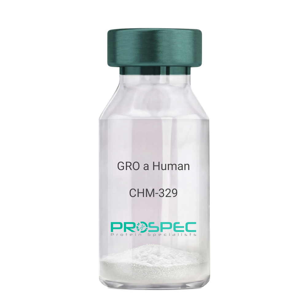
    CHM-329

    Price :

    Quantity :

    Shipping Method :

    Room Temp Icon

    Shipped at Room temp

    Add To Cart

    More Info

    • description
    • source
    • formulation
    • purity
    • biological activity
    • More Info

    Description

    GRO Alpha Human Recombinant produced in E.Coli is a single,non-glycosylated, polypeptide chain containing 73 amino acids and having a molecular mass of 7811 Dalton. The GRO-alpha is purified by proprietary chromatographic techniques.

    Source

    Escherichia Coli.

    Formulation

    Lyophilized from a 0.2µm filtered concentrated (1mg/ml) solution in 20mM PB, pH 7.4, 50mM NaCl.

    Purity

    Greater than 97.0% as determined by:
    (a) Analysis by RP-HPLC.
    (b) Analysis by SDS-PAGE.

    Biological Activity

    Determined by its ability to chemoattract human peripheral blood neutrophils using a concentration range of 10.0-100.0 ng/ml.

    More Info

    • Introduction

      Chemokine (C-X-C motif) ligand 1 (CXCL1) is a small cytokine belonging to the CXC chemokine family that was previously called GRO1 oncogene, Neutrophil-activating protein 3 (NAP-3) and melanoma growth stimulating activity, alpha (MSGA-a). It is secreted by human melanoma cells, has mitogenic properties and is implicated in melanoma pathogenesis. CXCL1 is expressed by macrophages, neutrophilsand epithelial cells, and has neutrophil chemoattractant activity. CXCL1 plays a role in spinal cord development by inhibiting the migration of oligodendrocyte precursors and is involved in the processes of angiogenesis, inflammation, wound healing, and tumorigenesis. This chemokine elicits its effects by signaling through the chemokine receptor CXCR2. The gene for CXCL1 is located on human chromosome 4 amongst genes for other CXC chemokines.

    • Synonyms

      Growth-regulated protein alpha, CXCL1, Melanoma growth stimulatory activity, MGSA, Neutrophil-activating protein 3, NAP-3, GRO-alpha (1-73), chemokine (C-X-C motif) ligand 1, GRO1, GROa, SCYB1, MGSA-a, MGSA alpha.

    • Physical Appearance

      Sterile Filtered White lyophilized (freeze-dried) powder.

    • Stability

      Lyophilized GRO-alpha although stable at room temperature for 3 weeks, should be stored desiccated below -18°C. Upon reconstitution CXCL1 should be stored at 4°C between 2-7 days and for future use below -18°C. For long term storage it is recommended to add a carrier protein (0.1% HSA or BSA).Please prevent freeze-thaw cycles.

    • Solubility

      It is recommended to reconstitute the lyophilized CXCL1 in sterile 18MΩ-cm H2O not less than 100µg/ml, which can then be further diluted to other aqueous solutions.

    • Amino Acid Sequence

      The sequence of the first five N-terminal amino acids was determined and was found to be Ala-Ser-Val-Ala-Thr.

    • Background

      What is the molecular weight/Mw of CXCL1 HUMAN Protein?
      CXCL1 HUMAN Protein has a total Mw of 7.81kDa.

      What is the source or expression system of CXCL1 HUMAN Protein?
      Escherichia Coli.

      What is the Purity of CXCL1 HUMAN Protein?
      CXCL1 HUMAN Protein is >97% pure as determined by SDS-PAGE.

      What is the Biological Activity of CXCL1 HUMAN Protein?
      Determined by its ability to chemoattract human peripheral blood neutrophils using a concentration range of 10.0-100.0 ng/ml.

      What is the amino acid sequence of CXCL1 HUMAN Protein?
      The sequence of the first five N-terminal amino acids was determined and was found to be Ala-Ser-Val-Ala-Thr.

      What applications can CXCL1 HUMAN Protein be used in?
      CXCL1 HUMAN Protein can probably be used in western blot, ELISA and Lateral Flow.

      What is the endotoxin level for CXCL1 HUMAN Protein?
      The endotoxin level is minimal, CXCL1 HUMAN Protein was purified using conventional chromatography techniques.


    ProSpec's products are furnished for LABORATORY RESEARCH USE ONLY. The product may not be used as drugs, agricultural or pesticidal products, food additives or household chemicals.

    Gro A Human
  • View Data Sheet

    Name :

    ANGPTL2 Human

    Description:

    Angiopoietin-like Protein 2 Human Recombinant

    Angiopoietin-related protein 2, Angiopoietin-like protein 2, ANGPTL2, ARP2, HARP.

    Product # :

    CYT-765

    Price :

    Quantity :

    Shipping Method :

    Ice Icon

    Shipped with Ice Packs

    Add To Cart

    More Info

    • description
    • source
    • formulation
    • purity
    • More Info
    • sds-page

    Description

    ANGPTL2 Human Recombinant produced in E.Coli is a single, non-glycosylated, polypeptide chain containing 493 amino acids ( 22-493 a.a.) including a 20 a.a N-terminal His tag. The total molecular mass is 57.1kDa (calculated).

    Source

    Escherichia Coli.

    Formulation

    ANGPTL2 protein solution (0.5mg/ml) contains 20mM Tris HCL (pH7-8) and 10% glycerol.

    Purity

    Greater than 85.0% as determined by SDS-PAGE.

    sds-page

    ANGPTL2-sds-page - Product image 1

    More Info

    • Introduction

      Angiopoietins belong to the vascular endothelial growth factor family and the only known growth factors largely specific for vascular endothelium. Angiopoietins-1, 2 and 4 partake in the formation of blood vessels. ANGPTL2 displays angiogenic effects. Angiopoietin-like Protein 2 (ANGPTL2) is an anti-diabetic factor. ANGPTL2 induces sprouting in endothelial cells through an autocrine and paracrine action. ANGPTL2 increases insulin sensitivity in adipocytes. In addition, ANGPTL2 is a mediator of chronic adipose tissue inflammation. ANGPTL2 is widely expressed in the heart, small intestine, spleen and stomach. ANGPTL2 is also found in lower levels in the colon, ovary, adrenal gland, skeletal muscle and in prostate.

    • Synonyms

      Angiopoietin-related protein 2, Angiopoietin-like protein 2, ANGPTL2, ARP2, HARP.

    • Physical Appearance

      Sterile filtered colorless solution.

    • Stability

      Store at 4°C if entire vial will be used within 2-4 weeks.Store, frozen at -20°C for longer periods of time.For long term storage it is recommended to add a carrier protein (0.1% HSA or BSA).Avoid multiple freeze-thaw cycles.

    • Amino Acid Sequence

      MGSSHHHHHH SSGLVPRGSH MGQEDGFEGT EEGSPREFIY LNRYKRAGES QDKCTYTFIV PQQRVTGAIC VNSKEPEVLL ENRVHKQELE LLNNELLKQK RQIETLQQLV EVDGGIVSEV KLLRKESRNM NSRVTQLYMQ LLHEIIRKRD NALELSQLEN RILNQTADML QLASKYKDLE HKYQHLATLA HNQSEIIAQL EEHCQRVPSA RPVPQPPPAA PPRVYQPPTY NRIINQISTN EIQSDQNLKV LPPPLPTMPT LTSLPSSTDK PSGPWRDCLQ ALEDGHDTSS IYLVKPENTN RLMQVWCDQR HDPGGWTVIQ RRLDGSVNFF RNWETYKQGF GNIDGEYWLG LENIYWLTNQ GNYKLLVTME DWSGRKVFAE YASFRLEPES EYYKLRLGRY HGNAGDSFTW HNGKQFTTLD RDHDVYTGNC AHYQKGGWWY NACAHSNLNG VWYRGGHYRS RYQDGVYWAE FRGGSYSLKK VVMMIRPNPN TFH

    • Background

      Angiopoietin-like Protein 2 Human Recombinant: A Potential Therapeutic Target for Metabolic and Cardiovascular Disorders

      Abstract:

      Angiopoietin-like protein 2 (ANGPTL2) has emerged as a crucial regulator in metabolic and cardiovascular disorders. This multifunctional protein is involved in various biological processes, including angiogenesis, adipose tissue function, and inflammation. The availability of human recombinant ANGPTL2 protein has provided researchers with a valuable tool to explore its therapeutic potential. This concise review provides an overview of the role of ANGPTL2 in metabolic and cardiovascular health and discusses the potential of ANGPTL2 human recombinant protein as a therapeutic target.

      Introduction:

      Metabolic disorders, such as obesity and type 2 diabetes, are closely associated with cardiovascular diseases and pose significant global health challenges. ANGPTL2, a member of the angiopoietin-like protein family, has gained attention for its involvement in metabolic regulation and cardiovascular homeostasis. Through interactions with various receptors and signaling pathways, ANGPTL2 influences lipid metabolism, insulin sensitivity, inflammation, and vascular integrity.

      Mechanisms of ANGPTL2 Action:

      ANGPTL2 acts through binding to integrins, toll-like receptors (TLRs), and other receptors on different cell types, including adipocytes, endothelial cells, and immune cells. By influencing angiogenesis, inflammation, and extracellular matrix remodeling, ANGPTL2 affects adipose tissue function, lipid metabolism, and insulin signaling.

      Role of ANGPTL2 in Metabolic Regulation:

      ANGPTL2 plays a critical role in metabolic regulation and the development of metabolic disorders. It promotes adipose tissue inflammation, impairs adipogenesis, and alters adipokine secretion, contributing to metabolic dysfunction. Furthermore, ANGPTL2 modulates lipid metabolism by regulating lipoprotein lipase activity, affecting triglyceride clearance, and promoting hepatic lipid accumulation.

      ANGPTL2 in Cardiovascular Health and Disease:

      Increasing evidence suggests that ANGPTL2 is implicated in cardiovascular diseases, including atherosclerosis and heart failure. ANGPTL2 promotes vascular inflammation, endothelial dysfunction, and smooth muscle cell proliferation, which contribute to atherosclerotic plaque progression and vascular remodeling. Additionally, ANGPTL2 influences cardiac remodeling and fibrosis, impacting heart failure development.

      Therapeutic Potential of ANGPTL2 Human Recombinant Protein:

      The availability of ANGPTL2 human recombinant protein opens avenues for therapeutic interventions targeting metabolic and cardiovascular disorders. Preclinical studies employing ANGPTL2 blockade or supplementation have shown promising results in improving metabolic parameters, reducing atherosclerosis, and preserving cardiac function. However, further research is needed to optimize the clinical application of ANGPTL2 human recombinant protein, including dosage, timing, and delivery methods.

      Conclusion:

      ANGPTL2 holds promise as a therapeutic target for metabolic and cardiovascular disorders. Its involvement in key biological processes makes it an attractive candidate for interventions aiming to improve metabolic health and prevent cardiovascular complications. The development of ANGPTL2 human recombinant protein provides a valuable tool for investigating its therapeutic potential further.

      What is the molecular weight/Mw of ANGPTL2 Protein?
      ANGPTL2 Protein has a total Mw of 57.1kDa.

      What is the source or expression system of ANGPTL2 Protein?
      Escherichia Coli.

      What is the Purity of ANGPTL2 Protein?
      ANGPTL2 Protein is >85% pure as determined by SDS-PAGE.

      What is the Biological Activity of ANGPTL2 Protein?
      The biological functionality of ANGPTL2 Protein will be determined in the future.

      What is the amino acid sequence of ANGPTL2 Protein?
      MGSSHHHHHH SSGLVPRGSH MGQEDGFEGT EEGSPREFIY LNRYKRAGES QDKCTYTFIV PQQRVTGAIC VNSKEPEVLL ENRVHKQELE LLNNELLKQK RQIETLQQLV EVDGGIVSEV KLLRKESRNM NSRVTQLYMQ LLHEIIRKRD NALELSQLEN RILNQTADML QLASKYKDLE HKYQHLATLA HNQSEIIAQL EEHCQRVPSA RPVPQPPPAA PPRVYQPPTY NRIINQISTN EIQSDQNLKV LPPPLPTMPT LTSLPSSTDK PSGPWRDCLQ ALEDGHDTSS IYLVKPENTN RLMQVWCDQR HDPGGWTVIQ RRLDGSVNFF RNWETYKQGF GNIDGEYWLG LENIYWLTNQ GNYKLLVTME DWSGRKVFAE YASFRLEPES EYYKLRLGRY HGNAGDSFTW HNGKQFTTLD RDHDVYTGNC AHYQKGGWWY NACAHSNLNG VWYRGGHYRS RYQDGVYWAE FRGGSYSLKK VVMMIRPNPN TFH

      What applications can ANGPTL2 Protein be used in?
      ANGPTL2 Protein can probably be used in western blot, ELISA and Lateral Flow.

      What is the endotoxin level for ANGPTL2 Protein?
      The endotoxin level is minimal, ANGPTL2 Protein was purified using conventional chromatography techniques.

    ProSpec's products are furnished for LABORATORY RESEARCH USE ONLY. The product may not be used as drugs, agricultural or pesticidal products, food additives or household chemicals.

    Angptl2 Human
  • View Data Sheet

    Name :

    CXCL7 Human

    Description:

    Neutrophil Activating Protein-2 Human Recombinant (CXCL7)

    Platelet basic protein, PBP, Small inducible cytokine B7, CXCL7, Leukocyte-derived growth factor, LDGF, Macrophage-derived growth factor, MDGF, pro-platelet basic protein (chemokine (C-X-C motif) ligand 7), TC1, TC2, TGB, TGB1, B-TG1, CTAP3, NAP-2, SCYB7, THBGB, LA-PF4, THBGB1, Beta-TG, CTAPIII, CTAP-III.

    Product # :

    CHM-274

    Price :

    Quantity :

    Shipping Method :

    Room Temp Icon

    Shipped at Room temp

    Add To Cart

    More Info

    • description
    • source
    • formulation
    • purity
    • biological activity
    • More Info

    Description

    Neutrophil Activating Protein-2 Human Recombinant produced in E.Coli is a non-glycosylated, Polypeptide chain containing 70 amino acids and having a molecular mass of 7609 Dalton. The NAP-2 is purified by proprietary chromatographic techniques.

    Source

    Escherichia Coli.

    Formulation

    The CXCL7 protein was lyophilized from a concentrated (1mg/ml) sterile solution containing no additives.

    Purity

    Greater than 97% as determined by:
    (a) Analysis by RP-HPLC.
    (b) Analysis by SDS-PAGE.

    Biological Activity

    The specific activity as determined by the ability of NAP2 to chemoattract human neurotrophils using a concentration of 1-10ng/ml corresponding to a Specific Activity of 100,000-1,000,000IU/mg.

    More Info

    • Introduction

      Chemokine (C-X-C motif) ligand (CXCL7) is a small cytokine belonging to the CXC chemokine family. It is a protein that is released in large amounts from platelets following their activation. It stimulates various processes including mitogenesis, synthesis of extracellular matrix, glucose metabolism and synthesis of plasminogen activator.

    • Synonyms

      Platelet basic protein, PBP, Small inducible cytokine B7, CXCL7, Leukocyte-derived growth factor, LDGF, Macrophage-derived growth factor, MDGF, pro-platelet basic protein (chemokine (C-X-C motif) ligand 7), TC1, TC2, TGB, TGB1, B-TG1, CTAP3, NAP-2, SCYB7, THBGB, LA-PF4, THBGB1, Beta-TG, CTAPIII, CTAP-III.

    • Physical Appearance

      Sterile Filtered White lyophilized (freeze-dried) powder.

    • Stability

      Lyophilized NAP-2 although stable at room temperature for 3 weeks, should be stored desiccated below -18°C. Upon reconstitution CXCL7should be stored at 4°C between 2-7 days and for future use below -18°C. For long term storage it is recommended to add a carrier protein (0.1% HSA or BSA).Please prevent freeze-thaw cycles.

    • Solubility

      It is recommended to reconstitute the lyophilized Neutrophil Activating Protein-2in sterile 18MΩ-cm H2O not less than 100µg/ml, which can then be further diluted to other aqueous solutions.

    • Amino Acid Sequence

      The sequence of the first five N-terminal amino acids was determined and was found to be Ala-Glu-Leu-Arg-Cys.

    • Background

      What is the molecular weight/Mw of CXCL7 HUMAN Protein?
      CXCL7 HUMAN Protein has a total Mw of 7.6kDa.

      What is the source or expression system of CXCL7 HUMAN Protein?
      Escherichia Coli.

      What is the Purity of CXCL7 HUMAN Protein?
      CXCL7 HUMAN Protein is > 97% pure as determined by SDS-PAGE.

      What is the Biological Activity of CXCL7 HUMAN Protein?
      The specific activity as determined by the ability of NAP2 to chemoattract human neurotrophils using a concentration of 1-10ng/ml corresponding to a Specific Activity of 100,000-1,000,000IU/mg.

      What is the amino acid sequence of CXCL7 HUMAN Protein?
      The sequence of the first five N-terminal amino acids was determined and was found to be Ala-Glu-Leu-Arg-Cys.

      What applications can CXCL7 HUMAN Protein be used in?
      CXCL7 HUMAN Protein can probably be used in western blot, ELISA and Lateral Flow.

      What is the endotoxin level for CXCL7 HUMAN Protein?
      The endotoxin level is minimal, CXCL7 HUMAN Protein was purified using conventional chromatography techniques.


    ProSpec's products are furnished for LABORATORY RESEARCH USE ONLY. The product may not be used as drugs, agricultural or pesticidal products, food additives or household chemicals.

    Nap2 Human
  • View Data Sheet

    Name :

    SORBS3 Human

    Description:

    Sorbin And SH3 Domain Containing 3 Human Recombinant

    Sorbin And SH3 Domain Containing 3, SCAM1, Vinexin Beta (SH3-Containing Adaptor Molecule-1), vinexin, SH3-Containing Adapter Molecule 1.

    Product # :

    PRO-1829

    Price :

    Quantity :

    Shipping Method :

    Ice Icon

    Shipped with Ice Packs

    Add To Cart

    More Info

    • description
    • source
    • formulation
    • purity
    • More Info

    Description

    SORBS3 Human Recombinant produced in E.coli is a single, non-glycosylated polypeptide chain containing 352 amino acids (1-329) and having a molecular mass of 39.1 kDa. SORBS3 is fused to a 23 amino acid His-tag at N-terminus.

    Source

    Escherichia Coli.

    Formulation

    The SORBS3 solution (0.25mg/ml) contains 20mM Tris-HCl buffer (pH 8.0), 1mM DTT and 20% glycerol.

    Purity

    Greater than 90% as determined by SDS-PAGE.

    More Info

    • Introduction

      SORBS3 is an SH3 domain-containing adaptor protein. The existence of SH3 domains in the SORBS3 protein have a role in its capability to attach to other cytoplasmic molecules and contribute to cystoskeletal organization, cell adhesion and migration, signaling, and gene expression. Various transcript variants encoding different isoforms are known for this gene.

    • Synonyms

      Sorbin And SH3 Domain Containing 3, SCAM1, Vinexin Beta (SH3-Containing Adaptor Molecule-1), vinexin, SH3-Containing Adapter Molecule 1.

    • Physical Appearance

      Sterile Filtered clear solution.

    • Stability

      Store at 4°C if entire vial will be used within 2-4 weeks. Store, frozen at -20°C for longer periods of time. For long term storage it is recommended to add a carrier protein (0.1% HSA or BSA).Avoid multiple freeze-thaw cycles.

    • Amino Acid Sequence

      MGSSHHHHHH SSGLVPRGSH MGSMADGGSP FLGRRDFVYP SSTRDPSASN GGGSPARREE KKRKAARLKF DFQAQSPKEL TLQKGDIVYI HKEVDKNWLE GEHHGRLGIF PANYVEVLPA DEIPKPIKPP TYQVLEYGEA VAQYTFKGDL EVELSFRKGE HICLIRKVNE NWYEGRITGT GRQGIFPASY VQVSREPRLR LCDDGPQLPT SPRLTAAARS ARHPSSPSAL RSPADPIDLG GQTSPRRTGF SFPTQEPRPQ TQNLGTPGPA LSHSRGPSHP LDLGTSSPNT SQIHWTPYRA MYQYRPQNED ELELREGDRV DVMQQCDDGW FVGVSRRTQK FGTFPGNYVA PV

    ProSpec's products are furnished for LABORATORY RESEARCH USE ONLY. The product may not be used as drugs, agricultural or pesticidal products, food additives or household chemicals.

    Sorbs3 Human
  • View Data Sheet

    Name :

    Vimentin Antibody

    Description:

    Vimentin, Mouse Anti Human Antibody

    Product # :

    ANT-245

    Price :

    Quantity :

    Shipping Method :

    Room Temp Icon

    Shipped at Room temp

    Add To Cart

    More Info

    • formulation
    • More Info

    Formulation

    1mg/ml in PBS (after reconstitution).

    More Info

    • Introduction

      Vimentin expression in human malignant glioma cells depends on cellular density, algorithms of drug delivery and chemo/radio treatment. Vimentin and detyrosinated microtubules provide structural support for the extensive microtentacles observed in detached tumor cells and a mechanism to promote successful metastatic spread. Primary colorectal carcinomas display aberrant expression of vimentin, and have activated Notch and TGFbeta signaling pathways. Vimentin is a strong arterial substrate for transglutaminases. Transglutaminase-mediated vimentin dimerization results in a novel unifying pathway by which vasodilatory and remodeling responses may be regulated. Ablation of vimentin expression inhibits migration and invasion of colon and breast cancer cell lines.

    • Solubility

      Reconstitute with H20. Mix gently, wash the sides of the vial and wait 30-60 seconds before use.

    • Immunogen

      Purified Human Vimentin.

    • Ig Subclass

      mouse IgG1.

    • Clone

      hVimentin.

    • Applications

      Direct ELISA, Western Blot, Immuneprecipitation, immunohistochemistry.

    • Cross reactivity

      Vimentin antibody will recognize most mammalian Vimentin except mouse and rat.

    • Titer

      For intra-cellular staining, use 5-10µl/1,000,000 cells. Wash twice with PBS/Azide before adding the appropriate amount of secondary fluorescent reagent.

    • Shipping Conditions

      Antibody is shipped lyophilized at ambient temperature.

    • Available Conjugates

      This antibody is also available conjugated to biotin and FITC.

    • Type

      Mouse Anti Human Monoclonal Antibody.

    • Storage Procedures

      In lyophilized form, for long periods, store at 4°C in a dry environment. After reconstitution, if not intended for use within a month, aliquot and store at -20°C.

    • Purification Method

      Ion exchange column.

    ProSpec's products are furnished for LABORATORY RESEARCH USE ONLY. The product may not be used as drugs, agricultural or pesticidal products, food additives or household chemicals.

    Vimentin Antibody
  • View Data Sheet

    Name :

    TEK Mouse Fc

    Description:

    TEK Tyrosine Kinase Endothelial Fc Chimera Mouse Recombinant

    Angiopoietin-1 receptor precursor, Tyrosine-protein kinase receptor TIE-2, hTIE2, Tyrosine-protein kinase receptor TEK, p140 TEK, Tunica interna endothelial cell kinase, CD202b, VMCM, VMCM1, TIE2.

    Product # :

    PKA-249

    Price :

    Quantity :

    Shipping Method :

    Room Temp Icon

    Shipped at Room temp

    Add To Cart

    More Info

    • description
    • source
    • formulation
    • purity
    • More Info

    Description

    Soluble TEK Mouse Recombinant fused with the Fc part of human IgG1 produced in CHO is a glycosylated disulfide-linked homodimer, polypeptide containing amino acids 119-740 amino acids and having a total molecular mass of 280 kDa. Mouse TIE-2/Fc monomer has a calculated molecular mass of approximately 105 kDa. As a result of glycosylation, the recombinant protein migrates as an approximately 140 kDa protein in SDS-PAGE under reducing conditions.The TEK Fc Chimera is purified by proprietary chromatographic techniques.

    Source

    CHO Cells.

    Formulation

    TEK Fc Chimera was lyophilized from a concentrated (1 mg/ml) sterile solution containing 1xPBS.

    Purity

    Greater than 90.0% as determined by SDS-PAGE.

    More Info

    • Introduction

      TIE-1 (tyrosine kinase with Ig and EGF homology domains 1) and TIE-2/Tek comprise a receptor tyrosine kinase (RTK) subfamily with unique structural characteristics: two immunoglobulin-like domains flanking three epidermal growth factor (EGF)-like domains and followed by three fibronectin type III-like repeats in the extracellular region and a split tyrosine kinase domain in the cytoplasmic region. These receptors are expressed primarily on endothelial and hematopoietic progenitor cells and play critical roles in angiogenesis, vasculogenesis and hematopoiesis. Human TIE-1 cDNA encodes a 1122 amino acid (aa) residue precursor protein with an 18 residue putative signal peptide, a 726 residue extracellular domain and a 353 residue cytoplasmic domain. Two ligands, angiopoietin-1 (Ang1) and angiopoietin-2 (Ang2), which bind TIE-2 with high-affinity have been identified. Ang2 has been reported to act as an antagonist for Ang1. Mice engineered to overexpress Ang2 or to lack Ang1 or Tie-1 display similar angiogenic defects.

    • Synonyms

      Angiopoietin-1 receptor precursor, Tyrosine-protein kinase receptor TIE-2, hTIE2, Tyrosine-protein kinase receptor TEK, p140 TEK, Tunica interna endothelial cell kinase, CD202b, VMCM, VMCM1, TIE2.

    • Physical Appearance

      Sterile Filtered White lyophilized (freeze-dried) powder.

    • Stability

      Lyophilized sTIE-2 although stable at room temperature for 3 weeks, should be stored desiccated below -18°C. Upon reconstitution TEK should be stored at 4°C between 2-7 days and for future use below -18°C.For long term storage it is recommended to add a carrier protein (0.1% HSA or BSA).Please prevent freeze-thaw cycles.

    • Solubility

      It is recommended to reconstitute the lyophilized TIE-2 Fc Chimera in sterile water not less than 100µg/ml, which can then be further diluted to other aqueous solutions.

    ProSpec's products are furnished for LABORATORY RESEARCH USE ONLY. The product may not be used as drugs, agricultural or pesticidal products, food additives or household chemicals.

    Tie 2 Mouse Fc
  • View Data Sheet

    Name :

    VAMP3 Antibody

    Description:

    Synaptobrevin-3, Mouse Anti Human

    VAMP3, VAMP-3, Cellubrevin, Vesicle-Associated Membrane Protein 3, Synaptobrevin-3, CEB, SYB3.

    Product # :

    ANT-352

    Price :

    Quantity :

    Shipping Method :

    Ice Icon

    Shipped with Ice Packs

    Add To Cart

    More Info

    • formulation
    • More Info

    Formulation

    1mg/ml containing PBS, pH-7.4, & 0.1% Sodium Azide.

    More Info

    • Introduction

      VAMP3 is present in recycling endosomes and endosome-derived vesicles. VAMP3 has been implicated in recycling of transferrin receptors to the plasma membrane, secretion of alpha-granules in platelets, recycling of T-cell receptors to the immunological synapses, and membrane trafficking during cell migration. VAMP-3 is present in human platelets and necessary for granule secretion. Synaptobrevins are the main components of a protein complex involved in the docking and/or fusion of synaptic vesicles with the presynaptic membrane. VAMP3 high homology to other VAMPs in its broad tissue distribution and subcellular localization is shown to be the human equivalent of the rodent cellubrevin. In platelets the protein resides on a compartment that is not mobilized to the plasma membrane on calcium or thrombin stimulation.

    • Synonyms

      VAMP3, VAMP-3, Cellubrevin, Vesicle-Associated Membrane Protein 3, Synaptobrevin-3, CEB, SYB3.

    • Immunogen

      Anti-human VAMP3 mAb is derived from hybridization of mouse F0 myeloma cells with spleen cells from BALB/c mice immunized with recombinant human VAMP3 amino acids 1-77 purified from E. coli.

    • Ig Subclass

      Mouse IgG2b heavy chain and κ light chain.

    • Clone

      Pk2A2AT.

    • Applications

      VAMP3 antibody has been tested by ELISA and Western blot analysis to assure specificity and reactivity. Since application varies, however, each investigation should be titrated by the reagent to obtain optimal results. Recommended dilution range for Western blot analysis is 1:1,000 ~ 2,000. Recommended starting dilution is 1:1,000.

    • Type

      Mouse Anti Human Monoclonal.

    • Storage Procedures

      For periods up to 1 month store at 4°C, for longer periods of time, store at -20°C. Prevent freeze thaw cycles.

    • Purification Method

      VAMP3 antibody was purified from mouse ascitic fluids by protein-G affinity chromatography.

    ProSpec's products are furnished for LABORATORY RESEARCH USE ONLY. The product may not be used as drugs, agricultural or pesticidal products, food additives or household chemicals.

    Vamp3 Antibody
  • View Data Sheet

    Name :

    SERPINA1 Human, Active

    Description:

    Alpha-1 Antitrypsin, Active Human Recombinant

    Alpha-1-antitrypsin, Alpha-1 protease inhibitor, Alpha-1-antiproteinase, SERPINA1, A1AT, PI, A1A, AAT, PI1, MGC9222, PRO2275, MGC23330.

    Product # :

    PRO-907

    Price :

    Quantity :

    Shipping Method :

    Room Temp Icon

    Shipped at Room temp

    Add To Cart

    More Info

    • description
    • source
    • formulation
    • purity
    • biological activity
    • More Info

    Description

    SERPINA1 Human Recombinant produced in rice is a single, non-glycosylated polypeptide chain containing 384 amino acids and having a molecular mass of 43.1 kDa.The SERPINA1 protein is purified by proprietary chromatographic techniques.

    Source

    Rice Grain (Oryza Sativa).

    Formulation

    SERPINA1 was lyophilized from a concentrated solution containing recombinant Albumin.

    Purity

    Greater than 95% as determined by SDS-PAGE.

    Biological Activity

    3~5 mg SERPINA1 will inhibit 1 mg PPE with an activity of 10.8 units per mg protein

    More Info

    • Introduction

      SERPINA1 is secreted and is a serine protease inhibitor which its targets include elastase, plasmin, collagenase, leucocytic proteases, trypsin, chymotrypsin, and plasminogen activator. Defects in SERPINA1 gene can cause emphysema or liver disease. SERPINA1 is an endogenous inhibitor of serine proteases and inhibits the catalytic domain of human recombinant matriptase in vitro. Rise in SERPINA1 occurs as an acute phase response to tissue necrosis and inflammation. mutations in SERPINA1 and SLC11A1 genes change the balance between elastase produced by leukocytes during phagocytosis.

    • Synonyms

      Alpha-1-antitrypsin, Alpha-1 protease inhibitor, Alpha-1-antiproteinase, SERPINA1, A1AT, PI, A1A, AAT, PI1, MGC9222, PRO2275, MGC23330.

    • Physical Appearance

      Sterile Filtered White lyophilized (freeze-dried) powder.

    • Stability

      Lyophilized SERPINA1 although stable at room temperature for 3 weeks, should be stored desiccated below -18°C. Upon reconstitution SERPINA1 should be stored at 4°C between 2-7 days and for future use below -18°C.For long term storage it is recommended to add a carrier protein (0.1% HSA or BSA).Please prevent freeze-thaw cycles.

    • Solubility

      It is recommended to reconstitute the lyophilized SERPINA1 in sterile 18M-cm H2O not less than 100µg/ml, which can then be further diluted to other aqueous solutions.

    ProSpec's products are furnished for LABORATORY RESEARCH USE ONLY. The product may not be used as drugs, agricultural or pesticidal products, food additives or household chemicals.

    Serpina1 Human Active
  • View Data Sheet

    Name :

    CXCL13 Mouse

    Description:

    BCA-1/BLC Mouse Recombinant (CXCL13)

    C-X-C motif chemokine 13, B lymphocyte chemoattractant, CXC chemokine BLC, Small-inducible cytokine B13, Cxcl13, Blc, Scyb13.

    Product # :

    CHM-030

    Price :

    Quantity :

    Shipping Method :

    Room Temp Icon

    Shipped at Room temp

    Add To Cart

    More Info

    • description
    • source
    • formulation
    • purity
    • biological activity
    • More Info

    Description

    CXCL13 Mouse Recombinant (22-109) produced in E.Coli is a single, non-glycosylated, polypeptide chain containing 88 amino acids and having a molecular mass of 10kDa.The BCA-1 is purified by proprietary chromatographic techniques.

    Source

    Escherichia Coli.

    Formulation

    The BCA1 protein was lyophilized from a 0.2µm filtered solution in Acetonitrile and TFA.

    Purity

    Greater than 97.0% as determined by SDS-PAGE.

    Biological Activity

    The ED50, as measured by its ability to chemoattract human CXCR5-transfected mouse BaF3 cells, is less than 2µg/ml.

    More Info

    • Introduction

      BCA-1 is a CXC chemokine that is highly expressed in thesecondary lymphoid organs, such as follicles of the spleen, lymph nodes, and Peyer's patches. CXCL13 promotes the migration of B lymphocytes (compared to T cells and macrophages), by stimulating calcium influx into, and chemotaxis of, cells expressing Burkitt's lymphoma receptor 1 (BLR1). BCA1 therefore function in the homing of B lymphocytes to follicles. Human BCA-1 shares a 64% amino acid sequence similarity with the mouse protein and 23 - 34% amino acid sequence identity with other known CXC chemokines. Recombinant or chemically synthesized BCA1 is a potent chemoattractant for B lymphocytes but not T lymphocytes, monocytes or neutrophils. BLR1, a G protein-coupled receptor originally isolated from Burkitt’s lymphoma cells, has now been shown to be the specific receptor for BCA1. Among cells of the hematopoietic lineages, the expression of BLR-1, now designated CXCR-5, is restricted to B lymphocytes and a subpopulation of T helper memory cells.

    • Synonyms

      C-X-C motif chemokine 13, B lymphocyte chemoattractant, CXC chemokine BLC, Small-inducible cytokine B13, Cxcl13, Blc, Scyb13.

    • Physical Appearance

      Sterile Filtered White lyophilized (freeze-dried) powder.

    • Stability

      Lyophilized BCA1 although stable at room temperature for 3 weeks, should be stored desiccated below -18°C. Upon reconstitution BCA1 should be stored at 4°C between 2-7 days and for future use below -18°C.For long term storage it is recommended to add a carrier protein (0.1% HSA or BSA).Please prevent freeze-thaw cycles.

    • Solubility

      It is recommended to reconstitute the lyophilized CXCL13 in sterile 18MΩ-cm H2O not less than 100µg/ml, which can then be further diluted to other aqueous solutions.

    • Amino Acid Sequence

      ILEAHYTNLK CRCSGVISTV VGLNIIDRIQ VTPPGNGCPK TEVVIWTKMK KVICVNPRAK WLQRLLRHVQ SKSLSSTPQA PVSKRRAA.

    • Background

      What is the molecular weight/Mw of CXCL13 MOUSE Protein?
      CXCL13 MOUSE Protein has a total Mw of 10kDa.

      What is the source or expression system of CXCL13 MOUSE Protein?
      Escherichia Coli.

      What is the Purity of CXCL13 MOUSE Protein?
      CXCL13 MOUSE Protein is >97% pure as determined by SDS-PAGE.

      What is the Biological Activity of CXCL13 MOUSE Protein?
      The ED50, as measured by its ability to chemoattract human CXCR5-transfected mouse BaF3 cells, is less than 2µg/ml.

      What is the amino acid sequence of CXCL13 MOUSE Protein?
      ILEAHYTNLK CRCSGVISTV VGLNIIDRIQ VTPPGNGCPK TEVVIWTKMK KVICVNPRAK WLQRLLRHVQ SKSLSSTPQA PVSKRRAA.

      What applications can CXCL13 MOUSE Protein be used in?
      CXCL13 MOUSE Protein can probably be used in western blot, ELISA and Lateral Flow.

      What is the endotoxin level for CXCL13 MOUSE Protein?
      The endotoxin level is minimal, CXCL13 MOUSE Protein was purified using conventional chromatography techniques.


    ProSpec's products are furnished for LABORATORY RESEARCH USE ONLY. The product may not be used as drugs, agricultural or pesticidal products, food additives or household chemicals.

    Bca 1 Mouse
  • View Data Sheet

    Name :

    I TAC Human, His

    Description:

    I-TAC Human Recombinant (CXCL11), His Tag

    Small inducible cytokine B11, CXCL11, Interferon-inducible T-cell alpha chemoattractant, I-TAC, Interferon-gamma-inducible protein 9, IP-9, H174, Beta-R1, chemokine (C-X-C motif) ligand 11, IP9, b-R1, SCYB11, SCYB9B, MGC102770.

    Product # :

    CHM-232

    Price :

    Quantity :

    Shipping Method :

    Ice Icon

    Shipped with Ice Packs

    Add To Cart

    More Info

    • description
    • source
    • formulation
    • purity
    • More Info

    Description

    I-TAC (CXCL11) Human Recombinant produced in E.coli is a single, non-glycosylated polypeptide chain containing 94 amino acids (22-94) and having a molecular mass of 10.6kDa.I-TAC is fused to a 21 amino acid His-tag at N-terminus & purified by proprietary chromatographic techniques.

    Source

    Escherichia Coli.

    Formulation

    The I-TAC solution (1mg/ml) contains 10mM sodium citrate(pH 3.5), 20% glycerol and 2mM DTT.

    Purity

    Greater than 90.0% as determined by SDS-PAGE.

    More Info

    • Introduction

      Chemokine (C-X-C motif) ligand 11 (CXCL11) is a small cytokine belonging to the CXC chemokine family that is also called Interferon-inducible T-cell alpha chemoattractant (I-TAC) and Interferon-gamma-inducible protein 9 (IP-9). It is highly expressed in peripheral blood leukocytes, pancreas and liver, with moderate levels in thymus, spleen and lung and low expression levels were in small intestine, placenta and prostate. Gene expression of CXCL11 is strongly induced by IFN-g and IFN-b, and weakly induced by IFN-a. This chemokine elicits its effects on its target cells by interacting with the cell surface chemokine receptor CXCR3, with a higher affinity than do the other ligands for this receptor, CXCL9 and CXCL10. CXCL11 is chemotactic for activated T cells. Its gene is located on human chromosome 4 along with many other members of the CXC chemokine family.

    • Synonyms

      Small inducible cytokine B11, CXCL11, Interferon-inducible T-cell alpha chemoattractant, I-TAC, Interferon-gamma-inducible protein 9, IP-9, H174, Beta-R1, chemokine (C-X-C motif) ligand 11, IP9, b-R1, SCYB11, SCYB9B, MGC102770.

    • Physical Appearance

      Sterile filtered colorless solution.

    • Stability

      Store at 4°C if entire vial will be used within 2-4 weeks. Store, frozen at -20°C for longer periods of time. For long term storage it is recommended to add a carrier protein (0.1% HSA or BSA).Avoid multiple freeze-thaw cycles.

    • Amino Acid Sequence

      MGSSHHHHHH SSGLVPRGSH MFPMFKRGRC LCIGPGVKAV KVADIEKASI MYPSNNCDKI EVIITLKENK GQRCLNPKSK QARLIIKKVE RKNF.

    ProSpec's products are furnished for LABORATORY RESEARCH USE ONLY. The product may not be used as drugs, agricultural or pesticidal products, food additives or household chemicals.

    I Tac Human His
  • 1
  • …
  • 5
  • 6
  • 7
  • 8
  • 9
  • …
  • 26
Your browser does not support the video tag.

Products

  • Cytokines
  • Growth Factors
  • Chemokines
  • CD Antigens
  • Neurotrophins
  • Hormones
  • Enzymes
  • Viral Antigens
  • Recombinant Proteins
  • Natural Proteins
  • Monoclonal Antibodies
  • Polyclonal Antibodies
  • Thermal Packaging

About

  • Company
  • Founder
  • Contribution
  • Contact Us
  • My Account

Ordering

  • Ordering Method
  • Ordering - Credit Card
  • Ordering PO
  • Shipping Fees
  • FAQ's

Legal

  • Terms & Conditions
  • Privacy
  • Site Map
  • info@prospecbio.com
  • Facebook
  • Instagram
  • Linkedin Profile

© 2026 ProSpec-Tany TechnoGene Ltd. All Rights Reserved.

  • Choosing a selection results in a full page refresh.
  • Opens in a new window.