Search results
1000 results found for “killer cell”
Name
Description
Product #
Price
Quantity
Shipping Method
- View Data Sheet
Name :
VTCN1 HumanDescription:
V-Set Domain Containing T Cell Activation Inhibitor 1 Human Recombinant
V-set domain-containing T-cell activation inhibitor 1, B7 homolog 4, B7-H4, B7h.5, Immune costimulatory protein B7-H4, Protein B7S1, T-cell costimulatory molecule B7x, B7H4, VTCN1, B7S1, B7X, PRO1291, RP11-229A19.4.
Product # :
PRO-1812Price :
Quantity :
Shipping Method :
Shipped with Ice Packs
More Info
- description
- source
- formulation
- purity
- More Info
Description
VTCN1 Human Recombinant produced in E.coli is a single, non-glycosylated polypeptide chain containing 258 amino acids (25-259) and having a molecular mass of 28.2 kDa.VTCN1 is fused to a 23 amino acid His-tag at N-terminus & purified by proprietary chromatographic techniques.
Source
Escherichia Coli.
Formulation
The VTCN1 solution (1mg/ml) contains 20mM Tris-HCl buffer (pH 8.0), 0.4M UREA and 10% glycerol.
Purity
Greater than 90.0% as determined by SDS-PAGE.
More Info
-
Introduction
V-Set Domain Containing T Cell Activation Inhibitor 1, also known as VCTN1, is part of the B7 costimulatory protein family. Proteins in this family are located on the surface of antigen-presenting cells and interact with ligand bound to receptors on the surface of T cells. High level of the encoded protein has been associated with tumor progression. VCTN1 also takes part in promoting epithelial cell transformation.
-
Synonyms
V-set domain-containing T-cell activation inhibitor 1, B7 homolog 4, B7-H4, B7h.5, Immune costimulatory protein B7-H4, Protein B7S1, T-cell costimulatory molecule B7x, B7H4, VTCN1, B7S1, B7X, PRO1291, RP11-229A19.4.
-
Physical Appearance
Sterile Filtered colorless solution.
-
Stability
Store at 4°C if entire vial will be used within 2-4 weeks. Store, frozen at -20°C for longer periods of time. For long term storage it is recommended to add a carrier protein (0.1% HSA or BSA).Avoid multiple freeze-thaw cycles.
-
Amino Acid Sequence
MGSSHHHHHH SSGLVPRGSH MGSLIIGFGI SGRHSITVTT VASAGNIGED GILSCTFEPD IKLSDIVIQW LKEGVLGLVH EFKEGKDELS EQDEMFRGRT AVFADQVIVG NASLRLKNVQ LTDAGTYKCY IITSKGKGNA NLEYKTGAFS MPEVNVDYNA SSETLRCEAP RWFPQPTVVW
ASQVDQGANF SEVSNTSFEL NSENVTMKVV SVLYNVTINN TYSCMIENDI AKATGDIKVT ESEIKRRSHL QLLNSKAS.
ProSpec's products are furnished for LABORATORY RESEARCH USE ONLY. The product may not be used as drugs, agricultural or pesticidal products, food additives or household chemicals.
- View Data Sheet
Name :
Adiponectin Porcine, HEKDescription:
Adiponectin Porcine Recombinant, HEK derived
Acrp30, AdipoQ, GBP-28, APM-1, ACDC.
Product # :
CYT-696Price :
Quantity :
Shipping Method :
Shipped at Room temp
More Info
- description
- source
- formulation
- purity
- More Info
Description
The Acrp30 Porcine Recombinant contains a total of 238 amino acids having a molecular Mass of 26kDa. The Porcine Adiponectin is fused to a 13 amino acid long N-terminal FLAG tag.
Source
HEK293 (Human Embryonic Kidney cell line).
Formulation
Sterile filtered and lyophilized from 0.5mg/ml in 20mM Tris buffer and 50mM NaCl pH-7.5.
Purity
Greater than 90% as determined by SDS PAGE.
More Info
-
Introduction
Adiponectin is a recently discovered 244 amino acid protein, the product of the apM1 gene, which is physiologically active and specifically and highly expressed in adipose cells (Adipokine). The protein belongs to the soluble defense collagen super family; it has a collagen-like domain structurally homologous with collagen VIII and X and complement factor C1q-like globular domain. APM-1 forms homotrimers, which are the building blocks for higher order complexes found circulating in serum.
-
Synonyms
Acrp30, AdipoQ, GBP-28, APM-1, ACDC.
-
Stability
For long term, store lyophilized AdipoQ at -20°C. Aliquot the product after reconstitution to avoid repeated freezing/thawing cycles. Reconstituted protein can be stored at 4°C for a limited period of time; it does not show any change after two weeks at 4°C.The lyophilized protein remains stable for 24 months when stored at -20°C.
-
Solubility
Add deionized water and let the lyophilized pellet dissolve completely.
-
Amino Acid Sequence
HVDYKDDDDK PAGETTEKPG ALLPMPKGAC AGWMAGIPGH PGHNGTPGRD GRDGVPGEKG EKGDTGLTGP KGDTGESGVT GVEGPRGFPG IPGRKGEPGE SAYVYRSAFS VGLETRVTVP NMPIRFTKIF YNQQNHYDVT TGKFHCNIPG LYYFSFHITV LKDVKVSLYK DKAVLFTYDQ QDKNVDQASG VLLYLEKGDQ WLQAYGDEEN GVYADNVNDS FTGFLLYHNIE.
-
Background
What is the molecular weight/Mw of ADIPONECTIN Protein?
ADIPONECTIN Protein has a total Mw of 26kDa.
What is the source or expression system of ADIPONECTIN Protein?
HEK293
What is the Purity of ADIPONECTIN Protein?
ADIPONECTIN Protein is >90% pure as determined by SDS-PAGE.
What is the Biological Activity of ADIPONECTIN Protein?
The biological functionality of ADIPONECTIN Protein will be determined in the future.
What is the amino acid sequence of ADIPONECTIN Protein?
HVDYKDDDDK PAGETTEKPG ALLPMPKGAC AGWMAGIPGH PGHNGTPGRD GRDGVPGEKG EKGDTGLTGP KGDTGESGVT GVEGPRGFPG IPGRKGEPGE SAYVYRSAFS VGLETRVTVP NMPIRFTKIF YNQQNHYDVT TGKFHCNIPG LYYFSFHITV LKDVKVSLYK DKAVLFTYDQ QDKNVDQASG VLLYLEKGDQ WLQAYGDEEN GVYADNVNDS FTGFLLYHNIE.
What applications can ADIPONECTIN Protein be used in?
ADIPONECTIN Protein can probably be used in western blot, ELISA and Lateral Flow.
What is the endotoxin level for ADIPONECTIN Protein?
The endotoxin level is minimal, ADIPONECTIN Protein was purified using conventional chromatography techniques.
ProSpec's products are furnished for LABORATORY RESEARCH USE ONLY. The product may not be used as drugs, agricultural or pesticidal products, food additives or household chemicals.
- View Data Sheet
Name :
CD105 MouseDescription:
Endoglin Mouse Recombinant
CD105, ENG, END, ORW, HHT1, ORW1, FLJ41744, Cell surface MJ7/18 antigen, Endoglin.
Product # :
CYT-424Price :
Quantity :
Shipping Method :
Shipped at Room temp
More Info
- description
- source
- formulation
- purity
- biological activity
- More Info
Description
CD105 Mouse Recombinant extracellular domain produced in baculovirus is a homodimeric, glycosylated, Polypeptide containing 581 amino acids and having a molecular mass of 61 kDa but as a result of glycosylation, migrates at 75-85 kDa under reducing conditions in SDS-PAGE. Based on N-terminal sequence analysis, the primary structure of recombinant mature Endoglin starts at Glu 26. The CD105 is fused to a C-terminal His-tag (6xHis) and purified by proprietary chromatographic techniques.
Source
Insect Cells.
Formulation
Endoglin was lyophilized from a concentrated (1mg/ml) sterile solution containing no additives.
Purity
Greater than 95.0% as determined by(a) Analysis by RP-HPLC.
(b) Analysis by SDS-PAGE.Biological Activity
Measured by its ability to bind with rhTGF-beta RII/Fc in a functional ELISA. Optimal dilutions should be determined by each laboratory for each application.More Info
-
Introduction
Endoglin is a type I membrane glycoprotein located on cell surfaces and is part of the TGF beta receptor complex.
The protein consists of a homodimer of 180 kDA with disulfide links. It has been found on endothelial cells, activated macrophages, fibroblasts, and smooth muscle cells. Endoglin has been found to be part of the TGF-beta1 receptor complex. It thus may be involved in the binding of TGF-beta1, TGF-beta3, activin-A, BMP-2, and BMP-7. Beside TGF-beta signaling endoglin may have other functions. It has been postulated that endoglin is involved in the cytoskeletal organization affecting cell morphology and migration. Endoglin has a role in the development of the cardiovascular system and in vascular remodeling. Its expression is regulated during heart development . Experimental mice without the endoglin gene die due to cardiovascular abnormalities. -
Synonyms
CD105, ENG, END, ORW, HHT1, ORW1, FLJ41744, Cell surface MJ7/18 antigen, Endoglin.
-
Physical Appearance
Sterile Filtered White lyophilized (freeze-dried) powder.
-
Stability
Lyophilized Endoglin although stable at room temperature for 3 weeks, should be stored desiccated below -18°C. Upon reconstitution CD105 should be stored at 4°C between 2-7 days and for future use below -18°C.For long term storage it is recommended to add a carrier protein (0.1% HSA or BSA).Please prevent freeze-thaw cycles.
-
Solubility
It is recommended to reconstitute the lyophilized CD-105 in sterile PBS not less than 100 µg/ml, which can then be further diluted to other aqueous solutions.
-
Amino Acid Sequence
MDRGVLPLPITLLFVIYSFVPTTGLAERVGCDLQPVDPTRGEVT
FTTSQVSEGCVAQAANAVREVHVLFLDFPGMLSHLELTLQASKQNGTETQEVF
LVLVSNKNVFVKFQAPEIPLHLAYDSSLVIFQGQPRVNITVLPSLTSRKQILDWA
ATKGAITSIAALDDPQSIVLQLGQDPKAPFLCLPEAHKDMGATLEWQPRAQTP
VQSCRLEGVSGHKEAYILRILPGSEAGPRTVTVMMELSCTSGDAILILHGPPYVS
-
Background
What is the molecular weight/Mw of CD105 Protein?
CD105 Protein has a total Mw of 61kDa.
What is the source or expression system of CD105 Protein?
Insect Cells.
What is the Purity of CD105 Protein?
CD105 Protein is >95% pure as determined by SDS-PAGE.
What is the Biological Activity of CD105 Protein?
Measured by its ability to bind with rhTGF-beta RII/Fc in a functional ELISA. Optimal dilutions should be determined by each laboratory for each application.
What is the amino acid sequence of CD105 Protein?
MDRGVLPLPITLLFVIYSFVPTTGLAERVGCDLQPVDPTRGEVT
FTTSQVSEGCVAQAANAVREVHVLFLDFPGMLSHLELTLQASKQNGTETQEVF
LVLVSNKNVFVKFQAPEIPLHLAYDSSLVIFQGQPRVNITVLPSLTSRKQILDWA
ATKGAITSIAALDDPQSIVLQLGQDPKAPFLCLPEAHKDMGATLEWQPRAQTP
VQSCRLEGVSGHKEAYILRILPGSEAGPRTVTVMMELSCTSGDAILILHGPPYVS
What applications can CD105 Protein be used in?
CD105 Protein can probably be used in western blot, ELISA and Lateral Flow.
What is the endotoxin level for CD105 Protein?
The endotoxin level is minimal, CD105 Protein was purified using conventional chromatography techniques.
ProSpec's products are furnished for LABORATORY RESEARCH USE ONLY. The product may not be used as drugs, agricultural or pesticidal products, food additives or household chemicals.
- View Data Sheet
Name :
CD62E AntibodyDescription:
E-Selectin, Mouse Anti-Human
E-selectin, Endothelial leukocyte adhesion molecule 1, ELAM-1, Leukocyte-endothelial cell adhesion molecule 2, LECAM2, CD62E antigen, SELE, ELAM1, ELAM, ESEL, CD62E.
Product # :
ANT-237Price :
Quantity :
Shipping Method :
Shipped at Room temp
More Info
- formulation
- More Info
Formulation
1mg/ml in PBS (after reconstitution).
More Info
-
Introduction
E-Selectin belongs to a family of divalent cation-dependent carbohydrate-binding glycoproteins or adhesion molecules. CD62E is expressed on the surface of endothelial cells and mediates the interaction of leukocytes and platelets with endothelial cells during an inflammatory response. CD62E is found in cytokine-stimulated endothelial cells and is accountable for the accumulation of blood leukocytes at sites of inflammation by mediating the adhesion of cells to the vascular lining. It demonstrates structural features such as the existence of lectin- and EGF-like domains followed by short consensus repeat (SCR) domains that contain 6 conserved cysteine residues. E-Selectin participates in the interaction between leukocytes and the endothelium and appears to be involved in the pathogenesis of atherosclerosis.
-
Synonyms
E-selectin, Endothelial leukocyte adhesion molecule 1, ELAM-1, Leukocyte-endothelial cell adhesion molecule 2, LECAM2, CD62E antigen, SELE, ELAM1, ELAM, ESEL, CD62E.
-
Solubility
Reconstitute with of H20. Mix gently, wash the sides of the vial and wait 30-60 seconds before use.
-
Immunogen
Purified, Activated T-Cells.
-
Ig Subclass
Mouse IgG1.
-
Clone
hCD62E.
-
Applications
Staining antibody. For staining, use 10µl/1,000,000 cells.
-
Type
Mouse Anti Human Monoclonal.
-
Storage Procedures
Lyophilized: store at 4 C. After reconstitution, if not intended for use within a month, aliquot and store at -20 C.
-
Purification Method
Ion exchange column.
ProSpec's products are furnished for LABORATORY RESEARCH USE ONLY. The product may not be used as drugs, agricultural or pesticidal products, food additives or household chemicals.
- View Data Sheet
Name :
CD27 Human, HEKDescription:
CD27 Human Recombinant, HEK
Tumor Necrosis Factor Receptor Superfamily Member 7, T-Cell Activation Antigen CD27, CD27 Molecule, CD27 Antigen, T Cell Activation Antigen S152, CD27L Receptor, TNFRSF7, S152, Tp55, T14.
Product # :
PRO-2751Price :
Quantity :
Shipping Method :
Shipped with Ice Packs
More Info
- description
- source
- formulation
- purity
- biological activity
- More Info
Description
CD27 Human Recombinant produced in HEK cells is a single, glycosylated, polypeptide chain (20-191 a.a) containing a total of 411 amino acids, having a molecular mass of 46.2kDa. CD27 is fused to a 239 amino acid hIgG-His-Tag at C-terminus,and is purified by proprietary chromatographic techniques.
Source
HEK293 Cells.
Formulation
The CD27 solution (0.5mg/ml) contains 10% Glycerol and Phosphate-Buffered Saline (pH 7.4).
Purity
Greater than 95.0% as determined by SDS-PAGE.
Biological Activity
ED50 range is ≤ 0.8 ug/ml. Defined by its binding ability in a functional ELISA with Human CD27 Ligand/TNFSF7 (CAT# cyt-953).
More Info
-
Introduction
CD27 belongs to the TNF-receptor superfamily. CD27 is necessary for initiation and long-term maintenance of T cell immunity. CD27 binds to ligand CD70, and has a crucial role in regulating B-cell activation and immunoglobulin synthesis. The CD27 receptor transduces signals which result in the activation of NF-kappaB and MAPK8/JNK. Adaptor proteins TRAF2 and TRAF5 mediate the signaling process of CD27. CD27-binding protein (SIVA), which is a proapoptotic protein, can bind to the CD27 receptor and is believed to have a significant role in the apoptosis induced by CD27.
-
Synonyms
Tumor Necrosis Factor Receptor Superfamily Member 7, T-Cell Activation Antigen CD27, CD27 Molecule, CD27 Antigen, T Cell Activation Antigen S152, CD27L Receptor, TNFRSF7, S152, Tp55, T14.
-
Physical Appearance
Sterile Filtered colorless solution.
-
Stability
Store at 4°C if entire vial will be used within 2-4 weeks. Store, frozen at -20°C for longer periods of time. For long term storage it is recommended to add a carrier protein (0.1% HSA or BSA). Avoid multiple freeze-thaw cycles.
-
Amino Acid Sequence
ATPAPKSCPE RHYWAQGKLC CQMCEPGTFL VKDCDQHRKA AQCDPCIPGV SFSPDHHTRP HCESCRHCNS GLLVRNCTIT ANAECACRNG WQCRDKECTE CDPLPNPSLT ARSSQALSPH PQPTHLPYVS EMLEARTAGH MQTLADFRQL PARTLSTHWP PQRSLCSSDF IRLEPKSCDK THTCPPCPAP ELLGGPSVFL FPPKPKDTLM ISRTPEVTCV VVDVSHEDPE VKFNWYVDGV EVHNAKTKPR EEQYNSTYRV VSVLTVLHQD WLNGKEYKCK VSNKALPAPI EKTISKAKGQ PREPQVYTLP PSRDELTKNQ VSLTCLVKGF YPSDIAVEWE SNGQPENNYK TTPPVLDSDG SFFLYSKLTV DKSRWQQGNV FSCSVMHEAL HNHYTQKSLS LSPGKHHHHH H
ProSpec's products are furnished for LABORATORY RESEARCH USE ONLY. The product may not be used as drugs, agricultural or pesticidal products, food additives or household chemicals.
- View Data Sheet
Name :
HAVCR1 Human, HEKDescription:
Hepatitis A Virus Cellular Receptor 1 Human Recombinant, HEK
Hepatitis A Virus Cellular Receptor 1, T-Cell Immunoglobulin Mucin Family Member 1, T-Cell Immunoglobulin Mucin Receptor 1, T-Cell Membrane Protein 1, Kidney Injury Molecule 1, HAVCR-1, TIMD-1, HAVCR, KIM-1, TIM-1, TIMD1, TIM1, KIM1, TIM, T-Cell Immunoglobulin And Mucin Domain-Containing Protein 1, T Cell Immunoglobin Domain And Mucin Domain Protein 1, HAVCR1.
Product # :
HAV-223Price :
Quantity :
Shipping Method :
Shipped at Room temp
More Info
- description
- source
- formulation
- purity
- More Info
Description
HAVCR1 Human Recombinant produced in HEK cells is a single, glycosylated, polypeptide chain (Ser21-Thr288) containing a total of 283 amino acids, having a calculated molecular mass of 30.5kDa. HAVCR1 is fused to a 2 aa N-terminal linker, a 2 aa C-terminal linker and a 6 aa His tag at C-Terminus.
Source
HEK 293.
Formulation
HAVCR1 was filtered (0.4µm) and lyophilized from 0.5mg/ml solution in phosphate buffered saline and 5% (w/v) Trehalose.
Purity
Greater than 90.0% as determined by SDS-PAGE.
More Info
-
Introduction
Hepatitis A virus cellular receptor 1 (HAVCR1) is a membrane receptor for both human hepatitis A virus (HHAV) and TIMD4. HAVCR1 is a type I trans-membrane structural glycoprotein located in the renal proximal tubule epithelial cells. HAVCR1 protein may be involved in the control of asthma and allergic diseases. The reference genome represents an allele which retains a MTTVP amino acid segment that presents defense against atopy in HHAV seropositive individuals.
-
Synonyms
Hepatitis A Virus Cellular Receptor 1, T-Cell Immunoglobulin Mucin Family Member 1, T-Cell Immunoglobulin Mucin Receptor 1, T-Cell Membrane Protein 1, Kidney Injury Molecule 1, HAVCR-1, TIMD-1, HAVCR, KIM-1, TIM-1, TIMD1, TIM1, KIM1, TIM, T-Cell Immunoglobulin And Mucin Domain-Containing Protein 1, T Cell Immunoglobin Domain And Mucin Domain Protein 1, HAVCR1.
-
Physical Appearance
Filtered White lyophilized (freeze-dried) powder.
-
Stability
Store lyophilized protein at -20°C. Aliquot the product after reconstitution to avoid repeated freezing/thawing cycles. Reconstituted protein can be stored at 4°C for a limited period of time; it does not show any change after two weeks at 4°C.
-
Solubility
It is recommended to add deionized water to prepare a working stock solution of approximately 0.5 mg/ml and let the lyophilized pellet dissolve completely. HAVCR1 is not sterile! Please filter the product by an appropriate sterile filter before using it in the cell culture.
-
Amino Acid Sequence
ASSVKVGGEA GPSVTLPCHY SGAVTSMCWN RGSCSLFTCQ NGIVWTNGTH VTYRKDTRYK LLGDLSRRDV SLTIENTAVS DSGVYCCRVE HRGWFNDMKI TVSLEIVPPK VTTTPIVTTV PTVTTVRTST TVPTTTTVPM TTVPTTTVPT TMSIPTTTTV LTTMTVSTTT SVPTTTSIPT TTSVPVTTTV STFVPPMPLP RQNHEPVATS PSSPQPAETH PTTLQGAIRR EPTSSPLYSY TTDGNDTVTE SSDGLWNNNQ TQLFLEHSLL TANTTKLHHH HHH.
ProSpec's products are furnished for LABORATORY RESEARCH USE ONLY. The product may not be used as drugs, agricultural or pesticidal products, food additives or household chemicals.
- View Data Sheet
Name :
CDC34 HumanDescription:
Cell Division Cycle 34 Human Recombinant
UB2R1, CDC-34, Ubiquitin-conjugating enzyme E2 R1, Ubiquitin-protein ligase R1, Ubiquitin-conjugating enzyme E2-32 kDa complementing, E2-CDC34, CDC34, EC 6.3.2.19, UBC3, UBE2R1.
Product # :
ENZ-1103Price :
Quantity :
Shipping Method :
Shipped with Ice Packs
More Info
- description
- source
- formulation
- purity
- More Info
Description
Cell Division Cycle 34 Human Recombinant produced in E.Coli is a single, non- glycosylated polypeptide chain containing 236 amino acids and having a molecular mass of 26.7kDa. CDC34 is purified by proprietary chromatographic techniques.
Source
Escherichia Coli.
Formulation
CDC34 is supplied as a 0.2 μm filtered solution conteining 50mM HEPES, pH 7.0, 10 % Glycerol, 125mM NaCl, , 5 % Trehalose and 1 mM DTT.
Purity
Greater than 95.0% as determined by:
(a) Analysis by RP-HPLC.
(b) Analysis by SDS-PAGE.More Info
-
Introduction
CDC34 takes part in the control of cell cycle and DNA replication. Cdc34 in association with different E3 complexes, including SCF, targets many different substrates for ubiquitination and degradation during cell division, signal transduction, and development. Cdc34 substrates that have been characterized include B-Myb, Wee1, MyoD, ATF5, p27Xic1, and p27Kip1. Additionally, substrates such as p21Cip1, E2F, cyclin E, and cyclin D are putative substrates of Cdc34 by virtue of their SCF requirement for proteolysis. Cdc34 is self-associate through a domain in the C-terminus, and is phosphorylated and ubiquitinylated in vivo. CDC34is useful for in vitro ubiquitinylation reactions.
-
Synonyms
UB2R1, CDC-34, Ubiquitin-conjugating enzyme E2 R1, Ubiquitin-protein ligase R1, Ubiquitin-conjugating enzyme E2-32 kDa complementing, E2-CDC34, CDC34, EC 6.3.2.19, UBC3, UBE2R1.
-
Physical Appearance
Sterile Filtered colorless solution.
-
Stability
Store at 4°C if entire vial will be used within 2-4 weeks. Store, frozen at -20°C for longer periods of time. For long term storage it is recommended to add a carrier protein (0.1% HSA or BSA).Avoid multiple freeze-thaw cycles.
-
Amino Acid Sequence
MARPLVPSSQ KALLLELKGL QEEPVEGFRV TLVDEGDLYN WEVAIFGPPN TYYEGGYFKA RLKFPIDYPY SPPAFRFLTK MWHPNIYETG DVCISILHPP VDDPQSGELP SERWNPTQNV RTILLSVISL LNEPNTFSPA NVDASVMYRK WKESKGKDRE YTDIIRKQVL GTKVDAERDG VKVPTTLAEY CVKTKAPAPD EGSDLFYDDY YEDGEVEEEA DSCFGDDEDD SGTEES.
ProSpec's products are furnished for LABORATORY RESEARCH USE ONLY. The product may not be used as drugs, agricultural or pesticidal products, food additives or household chemicals.
- View Data Sheet
Name :
KLK10 HumanDescription:
Kallikrein-10 Human Recombinant
Kallikrein-Related Peptidase 10, KLK10, PRSSL1, NES1, Normal Epithelial Cell-Specific 1, Protease Serine-Like 1, Kallikrein 10, Breast Normal Epithelial Cell Associated Serine Protease, kallikrein-10, Protease, Serine-Like 1, EC 3.4.21.- , EC 3.4.21, EC 3.4.21.74.
Product # :
ENZ-795Price :
Quantity :
Shipping Method :
Shipped with Ice Packs
More Info
- description
- source
- formulation
- purity
- More Info
Description
KLK10 Human Recombinant produced in E.coli is a single, non-glycosylated polypeptide chain containing 266 amino acids (34-276) and having a molecular mass of 29.1kDa.KLK10 is fused to a 23 amino acid His-tag at N-terminus & purified by proprietary chromatographic techniques.
Source
Escherichia Coli.
Formulation
The KLK10 solution (1mg/ml) contains 20mM Tris-HCl buffer (pH 8.0), 0.4M Urea and 10% glycerol.
Purity
Greater than 85% as determined by SDS-PAGE.
More Info
-
Introduction
Kallikrein-10 (KLK10) is one of the 15 kallikrein subfamily members located in a cluster on chromosome 19. Kallikreins are a subgroup of serine proteases having various physiological functions. KLK10 is secreted and has a role in suppression of tumorigenesis in breast and prostate cancers.
-
Synonyms
Kallikrein-Related Peptidase 10, KLK10, PRSSL1, NES1, Normal Epithelial Cell-Specific 1, Protease Serine-Like 1, Kallikrein 10, Breast Normal Epithelial Cell Associated Serine Protease, kallikrein-10, Protease, Serine-Like 1, EC 3.4.21.- , EC 3.4.21, EC 3.4.21.74.
-
Physical Appearance
Sterile Filtered colorless solution.
-
Stability
Store at 4°C if entire vial will be used within 2-4 weeks. Store, frozen at -20°C for longer periods of time. For long term storage it is recommended to add a carrier protein (0.1% HSA or BSA).Avoid multiple freeze-thaw cycles.
-
Amino Acid Sequence
MGSSHHHHHH SSGLVPRGSH MGSALLPQND TRLDPEAYGS PCARGSQPWQ VSLFNGLSFH CAGVLVDQSW VLTAAHCGNK PLWARVGDDH LLLLQGEQLR RTTRSVVHPK YHQGSGPILP RRTDEHDLML LKLARPVVLG PRVRALQLPY RCAQPGDQCQ VAGWGTTAAR RVKYNKGLTC SSITILSPKE CEVFYPGVVT NNMICAGLDR GQDPCQSDSG GPLVCDETLQ GILSWGVYPC GSAQHPAVYT QICKYMSWIN KVIRSN.
ProSpec's products are furnished for LABORATORY RESEARCH USE ONLY. The product may not be used as drugs, agricultural or pesticidal products, food additives or household chemicals.
- View Data Sheet
Name :
KCTD15 PAT2B11AT AntibodyDescription:
Potassium channel tetramerisation domain containing 15, Clone PAT2B11AT, Mouse Anti Human
BTB/POZ domain-containing protein KCTD15, Potassium channel tetramerisation domain containing 15, KCTD15, MGC2628, MGC25497.
Product # :
ANT-696Price :
Quantity :
Shipping Method :
Shipped with Ice Packs
More Info
- formulation
- More Info
Formulation
1mg/ml containing PBS, pH-7.4, 10% Glycerol and 0.02% Sodium Azide.
More Info
-
Introduction
KCTD15 protein is encoded in humans by the KCTD15 gene. KCTD15 is expressed at a high level in the brain and the hypothalamus. The potassium channel KCTD15 was identified as a genetic loci linked to higher than normal BMI in humans along with genes such as GNPDA2, MTCH2, FTO, and TMEM18. SNPs (Single nucleotide polymorphisms) in non-diabetic and diabetic patients showed that FTO was most strongly associated with obesity while MTCH2 and GNPDA2 were still notably associated with higher than normal BMI levels.
-
Synonyms
BTB/POZ domain-containing protein KCTD15, Potassium channel tetramerisation domain containing 15, KCTD15, MGC2628, MGC25497.
-
Immunogen
Anti-human KCTD15 mAb, is derived from hybridization of mouse F0 myeloma cells with spleen cells from BALB/c mice immunized with a recombinant human KCTD15 protein 1-159 amino acids purified from E. coli.
-
Ig Subclass
Mouse IgG2b heavy chain and k light chain.
-
Clone
PAT2B11AT.
-
Applications
KCTD15 antibody has been tested by ELISA, Western blot analysis, Flow cytometry and ICC/IF to assure specificity and reactivity. Since application varies, however, each investigation should be titrated by the reagent to obtain optimal results.
-
Type
Mouse Anti Human Monoclonal.
-
Storage Procedures
For periods up to 1 month store at 4°C, for longer periods of time, store at -20°C. Prevent freeze thaw cycles.
-
Purification Method
KCTD15 antibody was purified from mouse ascitic fluids by protein-G affinity chromatography.
ProSpec's products are furnished for LABORATORY RESEARCH USE ONLY. The product may not be used as drugs, agricultural or pesticidal products, food additives or household chemicals.
- View Data Sheet
Name :
CDC25A HumanDescription:
Cell Division Cycle 25A Human Recombinant
M-phase inducer phosphatase 1, Dual specificity phosphatase Cdc25A, CDC25A, CDC25A2.
Product # :
ENZ-091Price :
Quantity :
Shipping Method :
Shipped with Ice Packs
More Info
- description
- source
- formulation
- purity
- More Info
Description
CDC25A Human Recombinant fused with a 36 amino acid His tag at N-terminus produced in E.Coli is a single, non-glycosylated, polypeptide chain containing 560 amino acids (1-524 a.a.) and having a molecular mass of 63.2kDa. The CDC25A is purified by proprietary chromatographic techniques.
Source
Escherichia Coli.
Formulation
The CDC25A solution (0.25 mg/ml) contains 20mM Tris-HCl buffer (pH 8.0), 5mM DTT, 20% glycerol, 0.2M NaCl and 1mM EDTA.
Purity
Greater than 80.0% as determined by SDS-PAGE.
More Info
-
Introduction
M-phase inducer phosphatase 1 (CDC25A) belongs to the CDC25 family of phosphatases. CDC25A is essential for progression from G1 to the S phase of the cell cycle. CDC25A activates the cyclin-dependent kinase CDC2 by eliminating 2 phosphate groups. CDC25A is specifically degraded in reaction to DNA damage, which inhibits cells with chromosomal abnormalities from progressing in the course of cell division. CDC25A is an oncogene, though its exact function in oncogenesis has not been determined.
-
Synonyms
M-phase inducer phosphatase 1, Dual specificity phosphatase Cdc25A, CDC25A, CDC25A2.
-
Physical Appearance
Sterile Filtered colorless solution.
-
Stability
Store at 4°C if entire vial will be used within 2-4 weeks. Store, frozen at -20°C for longer periods of time. For long term storage it is recommended to add a carrier protein (0.1% HSA or BSA).Avoid multiple freeze-thaw cycles.
-
Amino Acid Sequence
MRGSHHHHHH GMASMTGGQQ MGRDLYDDDD KDRWGSMELG PEPPHRRRLL FACSPPPASQ PVVKALFGAS AAGGLSPVTN LTVTMDQLQG LGSDYEQPLE VKNNSNLQRM GSSESTDSGF CLDSPGPLDS KENLENPMRR IHSLPQKLLG CSPALKRSHS DSLDHDIFQL IDPDENKENE AFEFKKPVRP VSRGCLHSHG LQEGKDLFTQ RQNSAPARML SSNERDSSEP GNFIPLFTPQ SPVTATLSDE DDGFVDLLDG ENLKNEEETP SCMASLWTAP LVMRTTNLDN RCKLFDSPSL CSSSTRSVLK RPERSQEESP PGSTKRRKSM SGASPKESTN PEKAHETLHQ SLSLASSPKG TIENILDNDP RDLIGDFSKG YLFHTVAGKH QDLKYISPEI MASVLNGKFA NLIKEFVIID CRYPYEYEGG HIKGAVNLHM EEEVEDFLLK KPIVPTDGKR VIVVFHCEFS SERGPRMCRY VRERDRLGNE YPKLHYPELY VLKGGYKEFF MKCQSYCEPP SYRPMHHEDF KEDLKKFRTK SRTWAGEKSK REMYSRLKKL.
ProSpec's products are furnished for LABORATORY RESEARCH USE ONLY. The product may not be used as drugs, agricultural or pesticidal products, food additives or household chemicals.
- View Data Sheet
Name :
BAG3 HumanDescription:
BCL2-Associated Athanogene 3 Human Recombinant
BIS, CAIR-1, BAG-3, BAG Family Molecular Chaperone Regulator 3, Bcl-2-associated athanogene 3, Bcl-2-binding protein Bis, Docking protein CAIR-1, BAG3, MGC104307.
Product # :
PRO-760Price :
Quantity :
Shipping Method :
Shipped with Ice Packs
More Info
- description
- source
- formulation
- purity
- More Info
Description
BAG3 Human Recombinant produced in E.Coli is a single, non-glycosylated polypeptide chain containing 595 amino acids (1-575 a.a.) and having a molecular mass of 63.7 kDa. The BAG3 protein is fused to a 20 amino acid His Tag at N-terminus and purified by standard chromatogrpahy techniques.
Source
Escherichia Coli.
Formulation
The BAG3 protein contains 20mM Tris buffer pH-8, 1mM EDTA, 10% glycerol and 0.1mM PMSF.
Purity
Greater than 90% as determined by SDS-PAGE.
More Info
-
Introduction
BAG3 Inhibits the chaperone activity of HSP70/HSC70 by promoting substrate release. BAG3 has anti-apoptotic activity. BAG proteins participate with Hip for their binding to Hsc70/Hsp70 ATPase domain and encourage substrate release. BAG proteins have about 45 amino acid BAG domain close to the C terminus however they differ noticeably in their N-terminal regions. BAG3 includes a WW domain in the N-terminal region and a BAG domain in the C-terminal region. The BAG domains of BAG1, BAG2, and BAG3 interact particularly with the Hsc70 ATPase domain in vitro and in mammalian cells. They bind with high affinity to the ATPase domain of Hsc70 and inhibit its chaperone activity in a Hip-repressible manner. BAG3 plays a role as a protein-refolding cochaperone of the bcl2 binding protein BAG family and as upregulated in response to persistent stress of cellular calcium balance dysregulation. BAG3 has been shown to diminish stress-induced apoptosis.
-
Synonyms
BIS, CAIR-1, BAG-3, BAG Family Molecular Chaperone Regulator 3, Bcl-2-associated athanogene 3, Bcl-2-binding protein Bis, Docking protein CAIR-1, BAG3, MGC104307.
-
Physical Appearance
Sterile filtered colorless solution.
-
Stability
Store at 4°C if entire vial will be used within 2-4 weeks. Store, frozen at -20°C for longer periods of time. For long term storage it is recommended to add a carrier protein (0.1% HSA or BSA). Avoid multiple freeze-thaw cycles.
-
Amino Acid Sequence
MGSSHHHHHH SSGLVPRGSH MSAATHSPMM QVASGNGDRD PLPPGWEIKI DPQTGWPFFV DHNSRTTTWN DPRVPSEGPK ETPSSANGPS REGSRLPPAR EGHPVYPQLR PGYIPIPVLH EGAENRQVHP FHVYPQPGMQ RFRTEAAAAA PQRSQSPLRG MPETTQPDKQ CGQVAAAAAA QPPASHGPER SQSPAASDCS SSSSSASLPS SGRSSLGSHQ LPRGYISIPV IHEQNVTRPA AQPSFHQAQK THYPAQQGEY QTHQPVYHKI QGDDWEPRPL RAASPFRSSV QGASSREGSP ARSSTPLHSP SPIRVHTVVD RPQQPMTHRE TAPVSQPENK PESKPGPVGP ELPPGHIPIQ VIRKEVDSKP VSQKPPPPSE KVEVKVPPAP VPCPPPSPGP SAVPSSPKSV ATEERAAPST APAEATPPKP GEAEAPPKHP GVLKVEAILE KVQGLEQAVD NFEGKKTDKK YLMIEEYLTK ELLALDSVDP EGRADVRQAR RDGVRKVQTI LEKLEQKAID VPGQVQVYEL QPSNLEADQP LQAIMEMGAV AADKGKKNAG NAEDPHTETQ QPEATAAATS NPSSMTDTPG NPAAP.
ProSpec's products are furnished for LABORATORY RESEARCH USE ONLY. The product may not be used as drugs, agricultural or pesticidal products, food additives or household chemicals.
- View Data Sheet
Name :
VCAM1 Human, HEKDescription:
Vascular cell adhesion molecule 1 Human Recombinant, HEK
Vascular cell adhesion protein 1, V-CAM 1, INCAM-100, CD106, VCAM1, L1CAM, MGC99561, DKFZp779G2333.
Product # :
PRO-1641Price :
Quantity :
Shipping Method :
Shipped at Room temp
More Info
- description
- source
- formulation
- purity
- More Info
Description
VCAM1 Human Recombinant produced by mammalian expression system in human cells is a single polypeptide chain containing 682 amino acids (25-698). VCAM1 is fused to an 8 amino acid His-tag at C-terminus & purified by proprietary chromatographic techniques.
Source
HEK293 cells.
Formulation
VCAM1 was lyophilized from a 0.2 µM filtered solution of 20mM PB, 150mM NaCl, 2mM CaCl2, 2mM MgCl2 and 5%Threhalose, pH 7.2.
Purity
Greater than 95% as determined by SDS-PAGE.
More Info
-
Introduction
VCAM1 is a member of the Ig superfamily. It is a cell surface sialoglycoprotein expressed by cytokine activated endothelium. VCAM-1 contains 6 or 7 immunoglobulin domains, and is expressed on both large and small vessels only after the endothelial cells are stimulated by cytokines. The protein has a number of functions including the regulation of leukocyte migration, leukocyte endothelial cell adhesion and signal transduction and may play a role in a number of inflammatory diseases (artherosclerosis and rheumatoid arthritis).
-
Synonyms
Vascular cell adhesion protein 1, V-CAM 1, INCAM-100, CD106, VCAM1, L1CAM, MGC99561, DKFZp779G2333.
-
Physical Appearance
Sterile Filtered White lyophilized (freeze-dried) powder.
-
Stability
Lyophilized VCAM1 although stable at room temperature for 3 weeks, should be stored desiccated below -18°C. Upon reconstitution VCAM1 should be stored at 4°C between 2-7 days and for future use below -18°C. Please prevent freeze-thaw cycles.
-
Solubility
It is recommended to reconstitute the lyophilized VCAM1 in 1xPBS to a concentration no less than 100 µg/ml, which can then be further diluted to other aqueous solutions.
-
Amino Acid Sequence
FKIETTPESRYLAQIGDSVSLTCSTTGCESPFFSWRTQIDSPLNGKVTNEGTTS
TLTMNPVSFGNEHSYLCTATCESRKLEKGIQVEIYSFPKDPEIHLSGPLEAGKP
ITVKCSVADVYPFDRLEIDLLKGDHLMKSQEFLEDADRKSLETKSLEVTFTPVIE
DIGKVLVCRAKLHIDEMDSVPTVRQAVKELQVYISPKNTVISVNPSTKLQEGGSVT
MTCSSEGLPAPEIFWSKKLDNGNLQHLSGNATLTLIAMRMEDSGIYVCEGVNLIGKNRKEV
ELIVQEKPFTVEISPGPRIAAQIGDSVMLTCSVMGCESPSFSWRTQIDSPLSGKVRSEGTN
STLTLSPVSFENEHSYLCTVTCGHKKLEKGIQVELYSFPRDPEIEMSGGLVNGSSVTVSCKV
PSVYPLDRLEIELLKGETILENIEFLEDTDMKSLENKSLEMTFIPTIEDTGKALVCQAKLHID
DMEFEPKQRQSTQTLYVNVAPRDTTVLVSPSSILEEGSSVNMTCLSQGFPAPKILWSRQLPNGE
LQPLSENATLTLISTKMEDSGVYLCEGINQIRKAQLKDAGVYECESKNKVGSQLRSLTLDVQGRE
NNKDYFSPEVDHHHHHHAGRSRKEVELIIQVTPKDIKLTAFPSESVKEGDTVIISCTCGNVPETW
IILKKKAETGDTVLKSIDGAYTIRKAQLKDAGVYECESKNKVGSQLRSLTLDVQGRENNKDYFSPE
VDHHHHHH.
ProSpec's products are furnished for LABORATORY RESEARCH USE ONLY. The product may not be used as drugs, agricultural or pesticidal products, food additives or household chemicals.
- View Data Sheet
Name :
SDF 1b RatDescription:
Stromal Cell-Derived Factor-1 beta Rat Recombinant (CXCL12)
SDF-1, CXCL12, Pre-B cell growth-stimulating factor, PBSF, hIRH, chemokine (C-X-C motif) ligand 12, SDF1, SDF1B, TPAR1, SCYB12, SDF-1b, TLSF-b, 12-O-tetradecanoylphorbol 13-acetate repressed protein 1, Thymic lymphoma cell-stimulating factor, TLSF.
Product # :
CHM-248Price :
Quantity :
Shipping Method :
Shipped at Room temp
More Info
- description
- source
- formulation
- purity
- biological activity
- More Info
Description
SDF-1 beta Rat Recombinant produced in E.Coli is a non-glycosylated, Polypeptide chain containing 72 amino acids and having a molecular mass of 8.4 kDa. The Rat SDF-1b is purified by proprietary chromatographic techniques.
Source
Escherichia Coli.
Formulation
The Rat SDF1B protein was lyophilized from a concentrated (1mg/ml) sterile solution containing 20mM Phsophate buffer pH-7.4 and 0.15M NaCl.
Purity
Greater than 97.0% as determined by:
(a) Analysis by RP-HPLC.
(b) Analysis by SDS-PAGE.Biological Activity
The specific activity as determined by its ability to chemoattract human peripheral blood monocytes at 100-150ng/ml corresponding to a Specific Activity of 6,667-10,000IU/mg.More Info
-
Introduction
SDF-1 (stromal cell-derived factor-1) is small cytokine belonging to the chemokine family that is officially designated Chemokine (C-X-C motif) ligand 12 (CXCL12). It is produced in two forms, SDF-1?/CXCL12a and SDF-1?/CXCL12b, by alternate splicing of the same gene. Chemokines are characterized by the presence of four conserved cysteines, which form two disulfide bonds. The CXCL12 proteins belong to the group of CXC chemokines, whose initial pair of cysteines are separated by one intervening amino acid. CXCL12 is strongly chemotactic for lymphocytes and has been implicated as an important cell co-ordinator during development. During embryogenesis it directs the migration of hematopoietic cells from foetal liver to bone marrow. Mice which were knocked-out for CXCL12 gene were lethal before the birth or within just 1 hour of life. As another role, CXCL12a alters also the electrophysiology of neurons. CXCL12 was shown to be expressend in many tissues in mice (including brain, thymus, heart, lung, liver, kidney, spleen and bone marrow).
The receptor for this chemokine is CXCR4, which was previously called fusin. This CXCL12-CXCR4 interaction used to be considered exclusive (unlike for other chemokines and their receptors), but recently it was suggested that CXCL12 is also bound by CXCR7 receptor.
The gene for CXCL12 is located on human chromosome 10. In human and mouse both CXCL12 and CXCR4 show high identity of sequence: 99% and 90%, respectively. -
Synonyms
SDF-1, CXCL12, Pre-B cell growth-stimulating factor, PBSF, hIRH, chemokine (C-X-C motif) ligand 12, SDF1, SDF1B, TPAR1, SCYB12, SDF-1b, TLSF-b, 12-O-tetradecanoylphorbol 13-acetate repressed protein 1, Thymic lymphoma cell-stimulating factor, TLSF.
-
Physical Appearance
Sterile Filtered White lyophilized (freeze-dried) powder.
-
Stability
Lyophilized SDF-1b although stable at room temperature for 3 weeks, should be stored desiccated below -18°C. Upon reconstitution CXCL12 should be stored at 4°C between 2-7 days and for future use below -18°C.For long term storage it is recommended to add a carrier protein (0.1% HSA or BSA).Please prevent freeze-thaw cycles.
-
Solubility
It is recommended to reconstitute the lyophilized SDF-1b in sterile 18MΩ-cm H2O not less than 100µg/ml, which can then be further diluted to other aqueous solutions.
-
Amino Acid Sequence
KPVSLSYRCP CRFFESHVAR ANVKHLKILN TPNCALQIVA RLKSNNRQVC IDPKLKWIQE YLDKALNKRL KM.
ProSpec's products are furnished for LABORATORY RESEARCH USE ONLY. The product may not be used as drugs, agricultural or pesticidal products, food additives or household chemicals.
- View Data Sheet
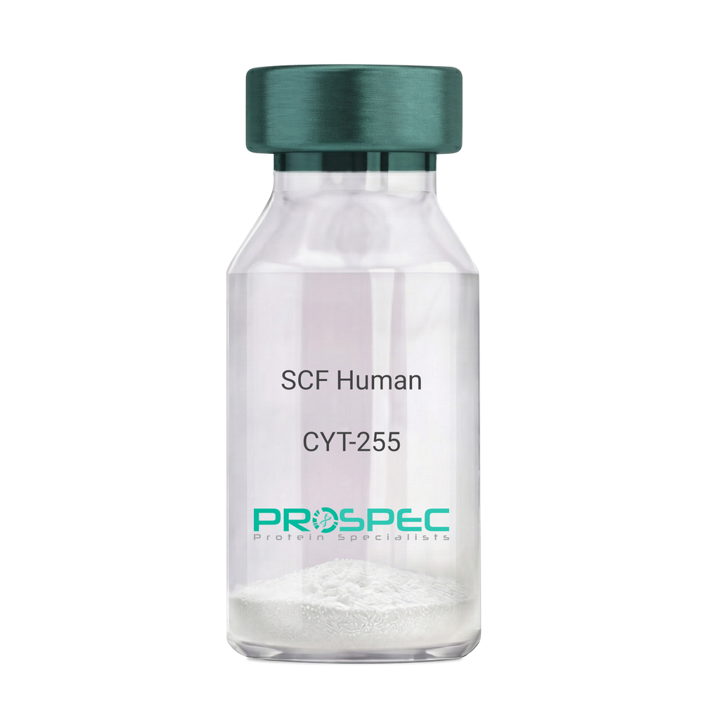
Name :
SCF HumanDescription:
Stem Cell Factor Human Recombinant
Kit ligand Precursor, C-kit ligand, SCF, Mast cell growth factor, MGF, SF, KL-1, Kitl, DKFZp686F2250.
Product # :
CYT-255Price :
Quantity :
Shipping Method :
Shipped at Room temp
More Info
- description
- source
- formulation
- purity
- biological activity
- More Info
Description
Stem Cell Factor Human Recombinant produced in E.Coli is a single, non-glycosylated polypeptide chain containing 165 amino acids and having a molecular mass of 18409 Dalton. The SCF is purified by proprietary chromatographic techniques.
Source
Escherichia Coli.
Formulation
Lyophilized from a concentrated (1mg/ml) solution in water containing 0.02% NaHCO3.
Purity
Greater than 98.0% as determined by:
(a) Analysis by SEC-HPLC.
(b) Analysis by SDS-PAGE.Biological Activity
The ED50 as determined by the dose-dependant stimulation of Human TF-1 cells is < 2 ng/ml, corresponding to a Specific Activity of 500,000 IU/mg.More Info
-
Introduction
Stem cell factor / KIT ligand (SCF) is a cytokine which binds CD117(c-Kit). SCF is also known as "steel factor" or "c-kit ligand". SCF exists in two forms, cell surface bound SCF and soluble (or free) SCF. Soluble SCF is produced by the cleavage of surface bound SCF by metalloproteases. SCF is a growth factor important for the survival, proliferation, and differentiation of hematopoietic stem cells and other hematopoietic progenitor cells. One of its roles is to change the BFU-E (burst-forming unit-erythroid) cells, which are the earliest erythrocyte precursors in the erythrocytic series, into the CFU-E (colony-forming unit-erythroid).
-
Synonyms
Kit ligand Precursor, C-kit ligand, SCF, Mast cell growth factor, MGF, SF, KL-1, Kitl, DKFZp686F2250.
-
Physical Appearance
Sterile Filtered White lyophilized (freeze-dried) powder.
-
Stability
Lyophilized KIT ligand although stable at room temperature for 3 weeks, should be stored desiccated below -18°C. Upon reconstitution SCF should be stored at 4°C between 2-7 days and for future use below -18°C.For long term storage it is recommended to add a carrier protein (0.1% HSA or BSA).Please prevent freeze-thaw cycles.
-
Solubility
It is recommended to reconstitute the lyophilized Stem Cell Factor in sterile 18MΩ-cm H2O not less than 100µg/ml, which can then be further diluted to other aqueous solutions.
-
Amino Acid Sequence
The sequence of the first five N-terminal amino acids was determined and was found to be Met-Glu-Gly-Ile-Cys.
-
Protein content
Protein quantitation was carried out by two independent methods1. UV spectroscopy at 280 nm using the absorbency value of 0.52 as the extinction coefficient for a 0.1% (1mg/ml) solution. This value is calculated by the PC GENE computer analysis program of protein sequences (IntelliGenetics). 2. Analysis by RP-HPLC, using a standard solution of Stem Cell Factor as a Reference Standard.
ProSpec's products are furnished for LABORATORY RESEARCH USE ONLY. The product may not be used as drugs, agricultural or pesticidal products, food additives or household chemicals.
- View Data Sheet
Name :
VTCN1 Human, Sf9Description:
V-Set Domain Containing T Cell Activation Inhibitor 1 Human Recombinant, Sf9
V-set domain-containing T-cell activation inhibitor 1, B7 homolog 4, B7-H4, B7h.5, Immune costimulatory protein B7-H4, Protein B7S1, T-cell costimulatory molecule B7x, B7H4, VTCN1, B7S1, B7X, PRO1291, RP11-229A19.4.
Product # :
PRO-2439Price :
Quantity :
Shipping Method :
Shipped with Ice Packs
More Info
- description
- source
- formulation
- purity
- More Info
Description
VTCN1 Human Recombinant produced in Sf9 Baculovirus cells is a single, glycosylated polypeptide chain containing 244 amino acids (25-259a.a.) and having a molecular mass of 26.9kDa (Molecular size on SDS-PAGE will appear at approximately 28-40kDa). VTCN1 is expressed with a 9 amino acids His tag at C-Terminus and purified by proprietary chromatographic techniques.
Source
Sf9, Baculovirus cells.
Formulation
VTCN1 protein solution (0.25mg/ml) contains Phosphate Buffered Saline (pH 7.4) and 10% glycerol.
Purity
Greater than 90.0% as determined by SDS-PAGE.
More Info
-
Introduction
V-Set Domain Containing T Cell Activation Inhibitor 1, also known as VCTN1, is part of the B7 costimulatory protein family. Proteins in this family are located on the surface of antigen-presenting cells and interact with ligand bound to receptors on the surface of T cells. High level of the encoded protein has been associated with tumor progression. VCTN1 also takes part in promoting epithelial cell transformation.
-
Synonyms
V-set domain-containing T-cell activation inhibitor 1, B7 homolog 4, B7-H4, B7h.5, Immune costimulatory protein B7-H4, Protein B7S1, T-cell costimulatory molecule B7x, B7H4, VTCN1, B7S1, B7X, PRO1291, RP11-229A19.4.
-
Physical Appearance
Sterile Filtered colorless solution.
-
Stability
Store at 4°C if entire vial will be used within 2-4 weeks. Store, frozen at -20°C for longer periods of time. For long term storage it is recommended to add a carrier protein (0.1% HSA or BSA).Avoid multiple freeze-thaw cycles.
-
Amino Acid Sequence
ADPLIIGFGI SGRHSITVTT VASAGNIGED GILSCTFEPD IKLSDIVIQW LKEGVLGLVH EFKEGKDELS EQDEMFRGRT AVFADQVIVG NASLRLKNVQ LTDAGTYKCY IITSKGKGNA NLEYKTGAFS MPEVNVDYNA SSETLRCEAP RWFPQPTVVW ASQVDQGANF SEVSNTSFEL NSENVTMKVV SVLYNVTINN TYSCMIENDI AKATGDIKVT ESEIKRRSHL QLLNSKASHH HHHH.
ProSpec's products are furnished for LABORATORY RESEARCH USE ONLY. The product may not be used as drugs, agricultural or pesticidal products, food additives or household chemicals.
- View Data Sheet
Name :
BLyS HumanDescription:
B-cell Activating Factor Human Recombinant
BAFF, BLYS, CD257, TALL1, THANK, ZTNF4, TALL-1, TNFSF20, TNFSF13B, B-cell Activating Factor.
Product # :
CYT-307Price :
Quantity :
Shipping Method :
Shipped at Room temp
More Info
- description
- source
- formulation
- purity
- biological activity
- More Info
Description
BAFF Human Recombinant produced in E.Coli is a single, non-glycosylated polypeptide chain containing 153 amino acids and having a molecular mass of 17007 Dalton. The BAFF is purified by proprietary chromatographic techniques.
Source
Escherichia Coli.
Formulation
Lyophilized from a 0.2µm filtered concentrated solution in PBS, pH 7.0.
Purity
Greater than 95.0% as determined by:
(a) Analysis by RP-HPLC.
(b) Analysis by SDS-PAGE.Biological Activity
The activity is determined by a mouse splenocyte survival assay. The ED50 for this effect is 0.5-2.0µg/ml.More Info
-
Introduction
BAFF binds to tnfrsf13b/taci and tnfrsf17/bcma. Tnfsf13/april binds to the same 2 receptors, together, they form a 2 ligands -2 receptors pathway involved in the stimulation of b- and t-cell function and the regulation of humoral immunity. A third b-cell specific baff-receptor (baffr/br3) promotes the survival of mature b-cells and the b-cell response.
B Lymphocyte Stimulator functions as a potent B-cell growth factor in costimulation assays.
Administration of BAFF Human recombinant to mice disrupts splenic B-cell and T-cell zones and results in elevated levels of serum immunoglobulin. -
Synonyms
BAFF, BLYS, CD257, TALL1, THANK, ZTNF4, TALL-1, TNFSF20, TNFSF13B, B-cell Activating Factor.
-
Physical Appearance
Sterile Filtered White lyophilized (freeze-dried) powder.
-
Stability
Lyophilized BAFF although stable at room temperature for 3 weeks, should be stored desiccated below -18°C. Upon reconstitution BAFF should be stored at 4°C between 2-7 days and for future use below -18°C.Please prevent freeze-thaw cycles.
-
Solubility
It is recommended to reconstitute the lyophilized BAFF in sterile 18M-cm H2O not less than 100µg/ml, which can then be further diluted to other aqueous solutions.
-
Amino Acid Sequence
MAVQGPEETV TQDCLQLIAD SETPTIQKGS YTFVPWLLSF KRGSALEEKE NKILVKETGY FFIYGQVLYT DKTYAMGHLI QRKKVHVFGD ELSLVTLFRC IQNMPETLPN NSCYSAGIAK LEEGDELQLA IPRENAQISL DGDVTFFGAL KLL.
-
Background
What is the molecular weight/Mw of BLYS Protein?
BLYS Protein has a total Mw of 17.7kDa.
What is the source or expression system of BLYS Protein?
Escherichia Coli.
What is the Purity of BLYS Protein?
BLYS Protein is >95% pure as determined by SDS-PAGE.
What is the Biological Activity of BLYS Protein?
The activity is determined by a mouse splenocyte survival assay. The ED50 for this effect is 0.5-2.0µg/ml.
What is the amino acid sequence of BLYS Protein?
MAVQGPEETV TQDCLQLIAD SETPTIQKGS YTFVPWLLSF KRGSALEEKE NKILVKETGY FFIYGQVLYT DKTYAMGHLI QRKKVHVFGD ELSLVTLFRC IQNMPETLPN NSCYSAGIAK LEEGDELQLA IPRENAQISL DGDVTFFGAL KLL.
What applications can BLYS Protein be used in?
BLYS Protein can probably be used in western blot, ELISA and Lateral Flow.
What is the endotoxin level for BLYS Protein?
The endotoxin level is minimal, BLYS Protein was purified using conventional chromatography techniques.
ProSpec's products are furnished for LABORATORY RESEARCH USE ONLY. The product may not be used as drugs, agricultural or pesticidal products, food additives or household chemicals.
- View Data Sheet
Name :
IL 3 RatDescription:
Interleukin-3 Rat Recombinant
MCGF (Mast cell growth factor), Multi-CSF, HCGF, P-cell stimulation factor, IL-3, MGC79398, MGC79399.
Product # :
CYT-383Price :
Quantity :
Shipping Method :
Shipped at Room temp
More Info
- description
- source
- formulation
- purity
- biological activity
- More Info
Description
Interleukin-3 Rat Recombinant produced in E.Coli is a single, non-glycosylated polypeptide chain containing 144 amino acids and having a molecular mass of 16.3kDa. The IL-3 is purified by proprietary chromatographic techniques.
Source
Escherichia Coli.
Formulation
Interleukin-3 Rat was lyophilized from a 0.2µm filtered concentrated solution in PBS, pH 7.4.
Purity
Greater than 97.0% as determined by:
(a) Analysis by RP-HPLC.
(b) Analysis by SDS-PAGE.Biological Activity
The ED50 as determined by the dose-dependent stimulation of the proliferation of thymidine uptake by murine MC-9 cells is < 10 ng/ml, corresponding to a specific activity of >1.0 x 10⁵ units/mg.More Info
-
Introduction
Interleukin-3 is a pleiotropic cytokine produced primarily by activated T cells.
IL-3 is thought to function via specific cell surface receptors to stimulate the proliferation, differentiation and survival of haematopoietic cell lines. IL-3 has also been shown to affect the functional activity of a variety of other cell types including mast cells, eosinophils, megakaryocytes and basophils. -
Synonyms
MCGF (Mast cell growth factor), Multi-CSF, HCGF, P-cell stimulation factor, IL-3, MGC79398, MGC79399.
-
Physical Appearance
Sterile Filtered White lyophilized (freeze-dried) powder.
-
Stability
Lyophilized Interleukin-3 although stable at room temperature for 3 weeks, should be stored desiccated below -18°C. Upon reconstitution IL3 should be stored at 4°C between 2-7 days and for future use below -18°C.For long term storage it is recommended to add a carrier protein (0.1% HSA or BSA).Please prevent freeze-thaw cycles.
-
Solubility
It is recommended to reconstitute the lyophilized Interleukin 3 in sterile 18MΩ-cm H2O not less than 100µg/ml, which can then be further diluted to other aqueous solutions.
-
Amino Acid Sequence
MISDRGSDAH HLLRTLDCRT IALEILVKLP YPQVSGLNNS DDKANLRNST LRRVNLDEFL KSQEEFDSQD TTDIKSKLQK LKCCIPAAAS DSVLPGVYNK DLDDFKKKLR FYVIHLKDLQ PVSVSRPPQP TSSSDNFRPM TVEC.
ProSpec's products are furnished for LABORATORY RESEARCH USE ONLY. The product may not be used as drugs, agricultural or pesticidal products, food additives or household chemicals.
- View Data Sheet
Name :
IL 12p40 AntibodyDescription:
Interleukin-12 p40, Rat Anti-Mouse
NKSF2, CTL maturation factor (TCMF), Cytotoxic lymphocyte maturation factor 40 kDa subunit (CLMF p40), TSF, Edodekin-alpha, IL-12 p40, IL-12B, IL-12 subunit p40, NK cell stimulatory factor chain 2.
Product # :
ANT-114Price :
Quantity :
Shipping Method :
Shipped at Room temp
More Info
- More Info
More Info
-
Introduction
Active IL-12 is a p70 disulphide-linked dimer composed of p35 and p40 subunits. The protein is a pleiotropic cytokine produced primarily by antigen presenting cells and has multiple effects on T lymphocytes and natural killer cells in terms of stimulating cytotoxicity, proliferation, production of other cytokines and Th1 subset differentiation.
-
Synonyms
NKSF2, CTL maturation factor (TCMF), Cytotoxic lymphocyte maturation factor 40 kDa subunit (CLMF p40), TSF, Edodekin-alpha, IL-12 p40, IL-12B, IL-12 subunit p40, NK cell stimulatory factor chain 2.
-
Solubility
Reconstitute with sterile H20. Mix gently, wash the sides of the vial and wait 30-60 seconds before use.
-
Immunogen
r.Murine IL-12 p40.
-
Ig Subclass
Rat IgG2a.
-
Clone
NYRmIL-12p40.
-
Applications
Direct ELISA, Western Blot, Immuneprecipitation.
-
Titer
By direct ELISA, 1:5,000 dilution will yield 0.5 O.D using alkaline phosphatase conjugated rabbit anti-mouse Ig (Jackson Laboratories).
-
Shipping Conditions
Antibody is shipped lyophilized at ambient temperature.
-
Type
Rat Anti Mouse Monoclonal.
-
Storage Procedures
In lyophilized form, for long periods, store at 4oC in a dry environment. After reconstitution, if not intended for use within a month, aliquot and store at -20oC.
-
Purification Method
Ion exchange.
ProSpec's products are furnished for LABORATORY RESEARCH USE ONLY. The product may not be used as drugs, agricultural or pesticidal products, food additives or household chemicals.
- View Data Sheet
Name :
TREM1 HumanDescription:
Triggering Receptor Expressed on Myeloid Cells 1 Human Recombinant
Triggering receptor expressed on myeloid cells 1, Triggering receptor expressed on monocytes 1, TREM-1, TREM1.
Product # :
PRO-457Price :
Quantity :
Shipping Method :
Shipped with Ice Packs
More Info
- description
- source
- formulation
- purity
- More Info
Description
TREM1 Human Recombinant produced in E.Coli is a single, non-glycosylated polypeptide chain containing 209 amino acids (21-205 a.a.) and having a molecular mass of 23.3kDa.TREM1 is fused to a 24 amino acid His-tag at N-terminus & purified by proprietary chromatographic techniques.
Source
Escherichia Coli.
Formulation
TREM1 protein solution (0.5mg/ml) containing 20mM Tris-HCl buffer (pH 8.0), 0.15M NaCl, 30% glycerol and 1mM DTT.
Purity
Greater than 85.0% as determined by SDS-PAGE.
More Info
-
Introduction
TREM1 is a transmembrane receptor protein that is involved in monocytic activation and in the inflammatory response. TREM1 is expressed on the surface of neutrophils, mature monocytes and macrophages. TREM1 stimulates neutrophil and monocyte-mediated inflammatory responses. TREM1 also triggers the release of pro-inflammatory chemokines and cytokines, as well as increased surface expression of cell activation markers. Furthermore, TREM1 is an amplifier of inflammatory responses that are triggered by bacterial and fungal infections and is a fundamental mediator of septic shock. The expression of the soluble form of TREM1 increases during sepsis and can consequently be used as a biological marker for septic shock. TREM1 is strongly expressed in acute inflammatory lesions caused by bacteria and fungi. Increased TREM1 expression on monocytes is related to both infectious and noninfectious inflammatory processes.
TREM1 is vastly expressed in adult liver, lung and spleen than in corresponding fetal tissue. TREM1 is also expressed in the lymph node, placenta, spinal cord and heart tissues. TREM1 expression is more elevated in peripheral blood leukocytes than in the bone marrow and in normal cells than malignant cells. TREM1 is expressed at low levels in the early development of the hematopoietic system and in the promonocytic stage and at high levels in mature monocytes. -
Synonyms
Triggering receptor expressed on myeloid cells 1, Triggering receptor expressed on monocytes 1, TREM-1, TREM1.
-
Physical Appearance
Sterile Filtered clear solution.
-
Stability
Store at 4°C if entire vial will be used within 2-4 weeks. Store, frozen at -20°C for longer periods of time. For long term storage it is recommended to add a carrier protein (0.1% HSA or BSA).Please avoid freeze thaw cycles.
-
Amino Acid Sequence
MGSSHHHHHH SSGLVPRGSH MGSMATKLTE EKYELKEGQT LDVKCDYTLE KFASSQKAWQ IIRDGEMPKT LACTERPSKN SHPVQVGRII LEDYHDHGLL RVRMVNLQVE DSGLYQCVIY QPPKEPHMLF DRIRLVVTKG FSGTPGSNEN STQNVYKIPP TTTKALCPLY TSPRTVTQAP PKSTADVSTP DSEINLTNVT DIIRVPVFN.
ProSpec's products are furnished for LABORATORY RESEARCH USE ONLY. The product may not be used as drugs, agricultural or pesticidal products, food additives or household chemicals.
- View Data Sheet
Name :
Dengue NS1 ST4, InsectDescription:
Dengue Virus NS1 Subtype 4 Recombinant, Insect Cells
Product # :
DEN-032Price :
Quantity :
Shipping Method :
Shipped with Ice Packs
More Info
- description
- source
- formulation
- purity
- More Info
Description
Recombinant Dengue Virus NS1 Subtype 4 produced in Insect Cells is a polypeptide chain containing amino acids 776-1130 and having a molecular weight of approximately 50kDa. Dengue NS1 ST4 is purified by proprietary chromatographic technique.
Source
Insect cells.
Formulation
Dengue NS1 ST4 protein solution in 1xD-PBS, pH7.4, 0.1% Thimerosal, 5mM EDTA, 1?g/ml of Leupeptin, Aprotinin and Pepstatin A.
Purity
Protein is >95% pure as determined by 12.5% SDS-PAGE.
More Info
-
Introduction
Caused by one of four closely related virus serotypes of the genus Flavivirus, family Flaviviridae, each serotype is sufficiently different that there is no cross-protection and epidemics caused by multiple serotypes (hyperendemicity) can occur. In cell culture experiments and mice Morpholino antisense oligos have shown specific activity against Dengue virus.
-
Physical Appearance
Sterile filtered colorless solution.
-
Stability
Store at 4°C if entire vial will be used within 2-4 weeks. Store, frozen at -20°C for longer periods of time. For long term storage it is recommended to add a carrier protein (0.1% HSA or BSA).Avoid multiple freeze-thaw cycles.
ProSpec's products are furnished for LABORATORY RESEARCH USE ONLY. The product may not be used as drugs, agricultural or pesticidal products, food additives or household chemicals.
- View Data Sheet
Name :
NFATC1 AntibodyDescription:
Nuclear factor of activated T-cells cytoplasmic 1, Mouse Anti Human
Nuclear factor of activated T-cells cytoplasmic 1, NFAT transcription complex cytosolic component, NF-ATc1, NFATc1, NF-ATc, NFATc, NFAT2, MGC138448.
Product # :
ANT-436Price :
Quantity :
Shipping Method :
Shipped with Ice Packs
More Info
- formulation
- More Info
Formulation
1mg/ml containing PBS, pH-7.4, 0.02% Sodium Azide and 10% Glycerol.
More Info
-
Introduction
NFATC1 is a component of the nuclear factor of activated T cells DNA-binding transcription complex. This complex consists of at least 2 components: a preexisting cytosolic component that translocates to the nucleus upon T cell receptor (TCR) stimulation, and an inducible nuclear component. Proteins belonging to this family of transcription factors play an essential role in inducible gene transcription during immune response. NFATC1 is an inducible nuclear component. NFATC1 functions as a major molecular target for the immunosuppressive drugs such as cyclosporin A. Different isoforms of NFATC1 may regulate inducible expression of different cytokine genes.
-
Synonyms
Nuclear factor of activated T-cells cytoplasmic 1, NFAT transcription complex cytosolic component, NF-ATc1, NFATc1, NF-ATc, NFATc, NFAT2, MGC138448.
-
Physical Appearance
Sterile filtered colorless solution.
-
Immunogen
Anti-human NFATC1 mAb is derived from hybridization of mouse F0 myeloma cells with spleen cells from BALB/c mice immunized with recombinant human NFATC1 amino acids 428-716 purified from E. coli.
-
Ig Subclass
Mouse IgG2a heavy chain and κ light chain.
-
Clone
PAT1C3AT.
-
Applications
NFATC1 antibody has been tested by ELISA and Western blot analysis to assure specificity and reactivity. Since application varies, however, each investigation should be titrated by the reagent to obtain optimal results.
-
Type
Mouse Anti Human Monoclonal.
-
Storage Procedures
For periods up to 1 month store at 4°C, for longer periods of time, store at -20°C. Prevent freeze thaw cycles.
-
Purification Method
NFATC1 antibody was purified from mouse ascitic fluids by protein-G affinity chromatography.
ProSpec's products are furnished for LABORATORY RESEARCH USE ONLY. The product may not be used as drugs, agricultural or pesticidal products, food additives or household chemicals.
- View Data Sheet
Name :
IL 3 Human, HisDescription:
Interleukin-3 Human Recombinant, His Tag
MCGF (Mast cell growth factor), Multi-CSF, HCGF, P-cell stimulation factor, IL-3, MGC79398, MGC79399.
Product # :
CYT-482Price :
Quantity :
Shipping Method :
Shipped with Ice Packs
More Info
- description
- source
- formulation
- purity
- biological activity
- More Info
Description
Interleukin-3 Human Recombinant produced in E.Coli is single, a non-glycosylated, Polypeptide chain containing 154 amino acids fragment (20-152) and having a total molecular mass of 17.3kDa and fused with a 20 aa N-terminal His tag. The IL3 His is purified by proprietary chromatographic techniques.
Source
Escherichia Coli.
Formulation
Interleukin-3 His (1mg/ml) is supplied in 20mM Tris-HCl buffer (pH 8.0), 0.2mM PMSF and 10% glycerol.
Purity
Greater than 90.0% as determined by analysis by SDS-PAGE.
Biological Activity
The ED50 for this effect is <0.53ng/ml. Measured in a cell proliferation assay using TF1 human erythroleukemic cells.More Info
-
Introduction
Interleukin-3 is a pleiotropic cytokine produced primarily by activated T cells. IL-3 is thought to function via specific cell surface receptors to stimulate the proliferation, differentiation and survival of haematopoietic cell lines. IL-3 has also been shown to affect the functional activity of a variety of other cell types including mast cells, eosinophils, megakaryocytes and basophils.
-
Synonyms
MCGF (Mast cell growth factor), Multi-CSF, HCGF, P-cell stimulation factor, IL-3, MGC79398, MGC79399.
-
Physical Appearance
Sterile Filtered clear solution.
-
Stability
Store at 4°C if entire vial will be used within 2-4 weeks. Store, frozen at -20°C for longer periods of time. For long term storage it is recommended to add a carrier protein (0.1% HSA or BSA).Avoid multiple freeze-thaw cycles.
-
Amino Acid Sequence
MGSSHHHHHH SSGLVPRGSH MAPMTQTTSL KTSWVNCSNM IDEIITHLKQ PPLPLLDFNN LNGEDQDILM ENNLRRPNLE AFNRAVKSLQ NASAIESILK NLLPCLPLAT AAPTRHPIHI KDGDWNEFRR KLTFYLKTLE NAQAQQTTLS LAIF.
ProSpec's products are furnished for LABORATORY RESEARCH USE ONLY. The product may not be used as drugs, agricultural or pesticidal products, food additives or household chemicals.
- View Data Sheet
Name :
BCL2L10 HumanDescription:
BCL2 Like 10 Human Recombinant
Bcl-2-like protein 10, Bcl2-L-10, Anti-apoptotic protein NrH, Apoptosis regulator Bcl-B, BCL2L10, BCLB, Boo, Diva, BCL-B.
Product # :
PRO-1321Price :
Quantity :
Shipping Method :
Shipped with Ice Packs
More Info
- description
- source
- formulation
- purity
- More Info
Description
BCL2L10 Human Recombinant produced in E.Coli is a single, non-glycosylated polypeptide chain containing 195 amino acids (1-172 a.a.) and having a molecular mass of 21.8kDa.BCL2L10 is fused to a 23 amino acid His-tag at N-terminus & purified by proprietary chromatographic techniques.
Source
E.coli.
Formulation
BCL2L10 protein solution (0.5mg/ml) containing 20mM Tris-HCl buffer (pH 8.0), 0.15M NaCl, 20% glycerol and 1mM DTT.
Purity
Greater than 90% as determined by SDS-PAGE.
More Info
-
Introduction
BCL2 Like 10 (BCL2L10) is a member of the BCL-2 protein family. BCL2 family members form hetero or homodimers and serve as anti or pro-apoptotic regulators which are involved in a wide variety of cellular activities. BCL2L10 includes the conserved BH4, BH1 and BH2 domains. BCL2L10 interacts with other members of BCL2 protein family including BCL2, BCL2L1/BCL-X(L), and BAX. BCL2L10 overexpression suppresses cell apoptosis possibly through the prevention of cytochrome C release from the mitochondria, and consequently activating caspase-3 activation.
-
Synonyms
Bcl-2-like protein 10, Bcl2-L-10, Anti-apoptotic protein NrH, Apoptosis regulator Bcl-B, BCL2L10, BCLB, Boo, Diva, BCL-B.
-
Physical Appearance
Sterile Filtered colorless solution.
-
Stability
Store at 4°C if entire vial will be used within 2-4 weeks. Store, frozen at -20°C for longer periods of time. For long term storage it is recommended to add a carrier protein (0.1% HSA or BSA).Avoid multiple freeze-thaw cycles.
-
Amino Acid Sequence
MGSSHHHHHH SSGLVPRGSH MGSMVDQLRE RTTMADPLRE RTELLLADYL GYCAREPGTP EPAPSTPEAA VLRSAAARLR QIHRSFFSAY LGYPGNRFEL VALMADSVLS DSPGPTWGRV VTLVTFAGTL LERGPLVTAR WKKWGFQPRL KEQEGDVARD CQRLVALLSS RLMGQHRAWL QAQGGWDGFC HFFRT.
ProSpec's products are furnished for LABORATORY RESEARCH USE ONLY. The product may not be used as drugs, agricultural or pesticidal products, food additives or household chemicals.
- View Data Sheet
Name :
CD4 Antibody, FITCDescription:
Rat Anti-Mouse CD4, FITC
gp55, HLA-2, L3 / T4, Ly-4, T cell antigen T4/LEU3, T4, sCD4, CD4mut.
Product # :
ANT-292Price :
Quantity :
Shipping Method :
Shipped at Room temp
More Info
- formulation
- More Info
Formulation
1mg/ml in PBS (after reconstitution).
More Info
-
Introduction
CD4 is a cell-surface glycoprotein found on the mature helper T cells and immature thymocytes, as well as on monocytes and macrophages. (Some cytotoxic T cells have CD4 protein as well.) Normally, about 65% of T cells in the blood are CD4+ (have CD4 protein protruding from their membrane). A mature T cell with either have CD4 or CD8, but not both. During one stage of development T cells develop CD4 and CD8 receptors, but they eventually are differentiated in the thymus and become more specialized.
-
Synonyms
gp55, HLA-2, L3 / T4, Ly-4, T cell antigen T4/LEU3, T4, sCD4, CD4mut.
-
Solubility
Reconstitute with of H20. Mix gently, wash the sides of the vial and wait 30-60 seconds before use.
-
Immunogen
Purified mouse LN CD4+ T cells.
-
Ig Subclass
Rat IgG.
-
Clone
mCD4.
-
Applications
FACS staining, immunohistochemistry, Western Blotting. For staining, use 10 µl/1,000,000 cells.
-
Available Conjugates
This antibody is also available unconjugated and conjugated to biotin.
-
Type
Rat Anti Mouse Monoclonal.
-
Storage Procedures
Lyophilized: store at 4°C. After reconstitution, if not intended for use within a month, aliquot and store at -20°C.
-
Purification Method
Ion exchange column.
ProSpec's products are furnished for LABORATORY RESEARCH USE ONLY. The product may not be used as drugs, agricultural or pesticidal products, food additives or household chemicals.