Search results
1000 results found for “Neuritin”
Name
Description
Product #
Price
Quantity
Shipping Method
- View Data Sheet
Name :
GMFB RatDescription:
Glia Maturation Factor Beta Rat Recombinant
Glia maturation factor beta, GMFB, GMF-B, GMF-beta, GMF, C79176, AI851627, D14Ertd630e, 3110001H22Rik, 3110001O16Rik.
Product # :
CYT-049Price :
Quantity :
Shipping Method :
Shipped at Room temp
More Info
- description
- source
- formulation
- purity
- More Info
Description
Glia Maturation Factor-Beta (GMF-Beta) Rat Recombinant produced in E.Coli is a signle, non-glycosylated, polypeptide chain containing 141 amino acids and having a total molecular mass of 16.6kDa. GMF-Beta is purified by proprietary chromatographic techniques.
Source
Escherichia Coli.
Formulation
Lyophilized from a 0.2µm filtered concentrated (1.0mg/ml) solution in 1×PBS, pH 7.4.
Purity
Greater than 97.0% as determined by
(a) Analysis by RP-HPLC.
(b) Analysis by SDS-PAGE.More Info
-
Introduction
GMFB is part of the GMF subfamily of the larger actin-binding protein ADF family. GMFB is phosphorylated after phorbol ester stimulation, and is crucial for the nervous system. GMFB causes brain cell differentiation, stimulates neural regeneration and inhibits tumor cell proliferation. GMFB overexpression in astrocytes results in the increase of BDNF production. GMFB expression is increased by exercise, thus BDNF is important for exercise-induction of BDNF.
-
Synonyms
Glia maturation factor beta, GMFB, GMF-B, GMF-beta, GMF, C79176, AI851627, D14Ertd630e, 3110001H22Rik, 3110001O16Rik.
-
Physical Appearance
Sterile Filtered White lyophilized (freeze-dried) powder.
-
Stability
Lyophilized GMFB although stable at room temperature for 3 weeks, should be stored desiccated below -18C. Upon reconstitution GMFB should be stored at 4C between 2-7 days and for future use below -18C.For long term storage it is recommended to add a carrier protein (0.1% HSA or BSA).Please prevent freeze-thaw cycles.
-
Solubility
It is recommended to reconstitute the lyophilized GMFB in sterile 18M-cm H2O not less than 100µg/ml, which can then be further diluted to other aqueous solutions.
-
Amino Acid Sequence
SESLVVCDVA EDLVEKLRKF RFRKETHNAA IIMKIDKDKR LVVLDEELEG VSPDELKDEL PERQPRFIVY SYKYQHDDGR VSYPLCFIFS SPLGCKPEQQ MMYAGSKNKL VQTAELTKVF EIRNTEDLTE EWLREKLGFF H.
-
Background
What is the molecular weight/Mw of GMFB RAT Protein?
GMFB RAT Protein has a total Mw of 16.6kDa.
What is the source or expression system of GMFB RAT Protein?
Escherichia Coli.
What is the Purity of GMFB RAT Protein?
GMFB RAT Protein is >97% pure as determined by SDS-PAGE.
What is the Biological Activity of GMFB RAT Protein?
The biological functionality of GMFB RAT Protein will be determined in the future.
What is the amino acid sequence of GMFB RAT Protein?
SESLVVCDVA EDLVEKLRKF RFRKETHNAA IIMKIDKDKR LVVLDEELEG VSPDELKDEL PERQPRFIVY SYKYQHDDGR VSYPLCFIFS SPLGCKPEQQ MMYAGSKNKL VQTAELTKVF EIRNTEDLTE EWLREKLGFF H.
What applications can GMFB RAT Protein be used in?
GMFB RAT Protein can probably be used in western blot, ELISA and Lateral Flow.
What is the endotoxin level for GMFB RAT Protein?
The endotoxin level is minimal, GMFB RAT Protein was purified using conventional chromatography techniques.
ProSpec's products are furnished for LABORATORY RESEARCH USE ONLY. The product may not be used as drugs, agricultural or pesticidal products, food additives or household chemicals.
- View Data Sheet
Name :
CDNF RatDescription:
CDNF Rat Recombinant
Cerebral neurotrophic factor, ARMET-like protein 1, Arginine-rich protein mutated in early stage tumors-like 1, Conserved neurotrophic factor, Cdnf, Armetl1.
Product # :
CYT-730Price :
Quantity :
Shipping Method :
Shipped at Room temp
More Info
- description
- source
- formulation
- purity
- biological activity
- More Info
Description
CDNF Rat Recombinant produced in E.coli is a single, non-glycosylated polypeptide chain containing 163 amino acids and having a molecular mass of 18.8kDa.The CDNF is purified by proprietary chromatographic techniques.
Source
Escherichia Coli.
Formulation
CDNF protein was lyophilized from a 0.2µm filtered concentrated solution in 1xPBS, pH 7.4.
Purity
Greater than 97.0% as determined by:
(a) Analysis by RP-HPLC.
(b) Analysis by SDS-PAGE.Biological Activity
CDNF Rat is able to enhance neurite outgrowth of E16-E18 rat embryonic cortical neurons when immobilized at 5-25 µg/mL on a nitrocellulose-coated microplate.More Info
-
Introduction
CDNF is a member of the ARMET family and acts as a trophic factor for neurons. CDNF inhibits the 6-hydroxy (6-OHDA)-induced degeneration of neurons. When CDNF controlled after 6-OHDA-lesioning, it reestablishes the function and inhibits the degeneration of neurons in substantia nigra. CDNF is universally expressed in neuronal and non-neuronal tissues. The highest levels in the brain are found in the optic nerve and corpus callosum.
-
Synonyms
Cerebral neurotrophic factor, ARMET-like protein 1, Arginine-rich protein mutated in early stage tumors-like 1, Conserved neurotrophic factor, Cdnf, Armetl1.
-
Physical Appearance
Sterile Filtered White lyophilized (freeze-dried) powder.
-
Stability
Lyophilized CDNF although stable at room temperature for 3 weeks, should be stored desiccated below -18°C. Upon reconstitution CDNF should be stored at 4°C between 2-7 days and for future use below -18°C. For long term storage it is recommended to add a carrier protein (0.1% HSA or BSA). Please prevent freeze-thaw cycles.
-
Solubility
It is recommended to reconstitute the lyophilized CDNF in sterile 18M-cm H2O not less than 100µg/ml, which can then be further diluted to other aqueous solutions.
-
Amino Acid Sequence
QGLEAGVRSR ADCEVCKEFL NRFYNSLLTR GIDFSVDTIE EELISFCADT KGKENRLCYY LGATKDSATK ILGEVTRPMS VHMPTVKICE KLKKMDSQIC ELKYEKKLDL ESVDLWKMRV AELKQILHSW GEECRACAEK HDYVNLIKEL APKYVETRPQ TEL.
-
Background
What is the molecular weight/Mw of CDNF Protein?
CDNF Protein has a total Mw of 18.8kDa.
What is the source or expression system of CDNF Protein?
Escherichia Coli.
What is the Purity of CDNF Protein?
CDNF Protein is >97% pure as determined by SDS-PAGE.
What is the Biological Activity of CDNF Protein?
CDNF Rat is able to enhance neurite outgrowth of E16-E18 rat embryonic cortical neurons when immobilized at 5-25 µg/mL on a nitrocellulose-coated microplate.
What is the amino acid sequence of CDNF Protein?
QGLEAGVRSR ADCEVCKEFL NRFYNSLLTR GIDFSVDTIE EELISFCADT KGKENRLCYY LGATKDSATK ILGEVTRPMS VHMPTVKICE KLKKMDSQIC ELKYEKKLDL ESVDLWKMRV AELKQILHSW GEECRACAEK HDYVNLIKEL APKYVETRPQ TEL.
What applications can CDNF Protein be used in?
CDNF Protein can probably be used in western blot, ELISA and Lateral Flow.
What is the endotoxin level for CDNF Protein?
The endotoxin level is minimal, CDNF Protein was purified using conventional chromatography techniques.
ProSpec's products are furnished for LABORATORY RESEARCH USE ONLY. The product may not be used as drugs, agricultural or pesticidal products, food additives or household chemicals.
- View Data Sheet
Name :
NRG1 (HRG-beta3) AntibodyDescription:
Neuregulin1 isoform HRG-beta3, Mouse Anti Human
Product # :
ANT-458Price :
Quantity :
Shipping Method :
Shipped with Ice Packs
More Info
- formulation
- More Info
Formulation
1mg/ml containing PBS, pH-7.4, & 0.1% Sodium Azide.
More Info
-
Introduction
NRG1 is one of 4 proteins in the neuregulin family which act on the EGFR family of receptors. NRG1 was initially identified as a 44kDa glycoprotein that interacts with the ERBB2 receptor tyrosine kinase to increase its phosphorylation on tyrosine residues. In various brain areas, NRG1 is expressed at synaptic regions and its processing can be regulated by neuronal activity. The HRG-beta3variant of NRG1, along with HRG-alpha, beta1 and beta2 variants, was identified in various normal tissues and cancer cell lines.
-
Immunogen
Anti-human NRG1 mAb is derived from hybridization of mouse F0 myeloma cells with spleen cells from BALB/c mice immunized with recombinant human NRG1 amino acids 20-241 purified from E. coli.
-
Ig Subclass
Mouse IgG1 heavy chain and κ light chain.
-
Clone
Pk1G13AT.
-
Applications
NRG1 antibody has been tested by ELISA and Western blot analysis to assure specificity and reactivity. Since application varies, however, each investigation should be titrated by the reagent to obtain optimal results. Recommended dilution range for Western blot analysis is 1:1,000 ~ 2,000. Recommended starting dilution is 1:1,000.
-
Type
Mouse Anti Human Monoclonal.
-
Storage Procedures
For periods up to 1 month store at 4°C, for longer periods of time, store at -20°C. Prevent freeze thaw cycles.
-
Purification Method
NRG1 antibody was purified from mouse ascitic fluids by protein-G affinity chromatography.
ProSpec's products are furnished for LABORATORY RESEARCH USE ONLY. The product may not be used as drugs, agricultural or pesticidal products, food additives or household chemicals.
- View Data Sheet
Name :
Periostin HumanDescription:
Periostin Human Recombinant
OSF-2, Periostin, Osteoblast Specific Factor 2, PN OSF-2, PDLPOSTN, POSTN, MGC119510, MGC119511, PN, RP11-412K4.1.
Product # :
CYT-452Price :
Quantity :
Shipping Method :
Shipped at Room temp
More Info
- description
- source
- formulation
- purity
- More Info
Description
The OSF2 His-Tagged Fusion Protein Human is produced in E. coli, and its molecular weight is 75 kDa protein containing 648 amino acid residues of the human OSF-2 and 23 additional amino acid residues - HisTag, Xa - cleavage site.
Source
Escherichia Coli.
Formulation
Filtered (0.4 µm) and lyophilized from 0.5 mg/ml in 0.05M Acetate buffer pH-4.
Purity
Greater than 90% as determined by SDS-PAGE.
More Info
-
Introduction
Periostin is a disulfide linked 90 kDa, 811 amino acid protein originally isolated as a osteoblast-specific factor that functions as a cell adhesion molecule for preosteoblasts and is thought to be involved in osteoblast recruitment, attachment and spreading. Additionally, periostin expression has previously been shown to be significantly increased by both transforming growth factor beta-1(TGFbeta1) and bone morphogenetic protein (BMP-2). OSF-2 has a typical signal sequence, followed by a cysteine-rich domain, a fourfold repeated domain and a C-terminal domain. The fourfold repeated domain of OSF-2 shows homology with the insect protein fasciclin
Periostin mRNA is expressed in the developing mouse embryonic and fetal heart, and that it is localized to the endocardial cushions that ultimately divide the primitive heart tube into a four-chambered heart. -
Synonyms
OSF-2, Periostin, Osteoblast Specific Factor 2, PN OSF-2, PDLPOSTN, POSTN, MGC119510, MGC119511, PN, RP11-412K4.1.
-
Physical Appearance
Filtered White lyophilized (freeze-dried) powder.
-
Stability
Store lyophilized protein at -20°C. Aliquot the product after reconstitution to avoid repeated freezing/thawing cycles. Reconstituted protein can be stored at 4°C for a limited period of time; it does not show any change after two weeks at 4°C.
-
Solubility
It is recommended to add 0.1M Acetate buffer pH4 to prepare a working stock solution of approximately 0.5 mg/ml and let the lyophilized pellet dissolve completely. For conversion into higher pH value, we recommend intensive dilution by relevant buffer to a concentration of 10µg/ml. In higher concentrations the solubility of this antigen is limited. Product is not sterile! Please filter the product by an appropriate sterile filter before using it in the cell culture.
-
Amino Acid Sequence
MGHHHHHHHH HHSSGHIEGR HMRNNHYDKI LAHSRIRGRD QGPNVCALQQ ILGTKKKYFS TCKNWYKKSI CGQKTTVLYE CCPGYMRMEG MKGCPAVLPI DHVYGTLGIV GATTTQRYSD ASKLREEIEG KGSFTYFAPS NEAWDNLDSD IRRGLESNVN VELLNALHSH MINKRMLTKD LKNGMIIPSM YNNLGLFINH YPNGVVTVNC ARIIHGNQIA TNGVVHVIDR VLTQIGTSIQ DFIEAEDDLS SFRAAAITSD ILEALGRDGH FTLFAPTNEA FEKLPRGVLE RFMGDKVASEALMKYHILNT LQCSESIMGG AVFETLEGNT IEIGCDGDSI TVNGIKMVNK KDIVTNNGVI HLIDQVLIPD SAKQVIELAG KQQTTFTDLV AQLGLASALR PDGEYTLLAP VNNAFSDDTL SMVQRLLKLI LQNHILKVKV GLNELYNGQI LETIGGKQLR VFVYRTAVCI ENSCMEKGSK QGRNGAIHIF REIIKPAEKS LHEKLKQDKR FSTFLSLLEA ADLKELLTQP GDWTLFVPTN DAFKGMTSEE KEILIRDKNA LQNIILYHLT PGVFIGKGFE PGVTNILKTT QGSKIFLKEV NDTLLVNELK SKESDIMTTN GVIHVVDKLL YPADTPVGND QLLEILNKLI KYIQIKFVRG STFKEIPVTV Y.
ProSpec's products are furnished for LABORATORY RESEARCH USE ONLY. The product may not be used as drugs, agricultural or pesticidal products, food additives or household chemicals.
- View Data Sheet
Name :
BNP HumanDescription:
B-type Natriuretic Peptide Human
NPPB, Natriuretic Peptide Precursor B, BNP, B-type Natriuretic Peptide.
Product # :
CYT-369Price :
Quantity :
Shipping Method :
Shipped at Room temp
More Info
- description
- formulation
- purity
- More Info
Description
B-type Natriuretic Peptide Human is a polypeptide chain containing 32 amino acids and having a molecular mass of 3464 Dalton. The molecular formula is:C143H244N50O42S4.
Formulation
The protein was lyophilized without additives.
Purity
Greater than 95.0% as determined by RP-HPLC.
More Info
-
Introduction
Natriuretic Peptide Precursor B acts as a cardiac hormone with a variety of biological actions including natriuresis, diuresis, vasorelaxation, and inhibition of renin and aldosterone secretion. It is thought to play a key role in cardiovascular homeostasis. Helps restore the body's salt and water balance. Improves heart function.
-
Synonyms
NPPB, Natriuretic Peptide Precursor B, BNP, B-type Natriuretic Peptide.
-
Physical Appearance
Sterile Filtered White lyophilized (freeze-dried) powder.
-
Stability
Lyophilized B-type Natriuretic Peptide although stable at room temperature for 3 weeks, should be stored desiccated below -18°C. Upon reconstitution B-type Natriuretic Peptide should be stored at 4°C between 2-7 days and for future use below -18°C.For long term storage it is recommended to add a carrier protein (0.1% HSA or BSA).Please prevent freeze-thaw cycles.
-
Solubility
It is recommended to reconstitute the lyophilized B-type Natriuretic Peptide in sterile 18MΩ-cm H2O not less than 100µg/ml, which can then be further diluted to other aqueous solutions.
-
Amino Acid Sequence
SPKMVQGSGCFGRKMDRISSSSGLGCKVLRRH.
-
Background
What is the molecular weight / Mw of BNP Human?
BNP Human has a total Mw of 3.4kDa.
What is the source or expression system of BNP Human?
Synthetic.
What is the Purity of BNP Human?
BNP Human is >95% pure as determined by SDS-PAGE.
What is the Biological Activity of BNP Human?
The biological functionality of BNP Human will be determined in the future.
What is the amino acid sequence of BNP Human?
SPKMVQGSGCFGRKMDRISSSSGLGCKVLRRH.
What applications can BNP Human Protein be used in?
BNP Human can probably be used in western blot, ELISA and Lateral Flow.
What is the endotoxin level for BNP Human?
The endotoxin level is minimal, BNP Human was purified using conventional chromatography techniques.
ProSpec's products are furnished for LABORATORY RESEARCH USE ONLY. The product may not be used as drugs, agricultural or pesticidal products, food additives or household chemicals.
- View Data Sheet
Name :
NECAP2 HumanDescription:
NECAP Endocytosis Associated 2 Human Recombinant
NECAP endocytosis-associated protein 2, NECAP-2, FLJ10420, Adaptin ear-binding coat-associated protein 2.
Product # :
PRO-180Price :
Quantity :
Shipping Method :
Shipped with Ice Packs
More Info
- description
- source
- formulation
- purity
- More Info
Description
NECAP2 produced in E.Coli is a single, non-glycosylated polypeptide chain containing 283 amino acids (1-263a.a.) and having a molecular mass of 30.5 kDa. NECAP2 is fused to a 20 amino acid His-tag at N-terminus & purified by proprietary chromatographic techniques.
Source
Escherichia Coli.
Formulation
The NECAP2 protein solution (1mg/1ml) is formulated in 20mM Tris-HCl buffer (pH8.0), 100mM NaCl, 2mM DTT and 10% glycerol.
Purity
Greater than 85% as determined by SDS-PAGE.
More Info
-
Introduction
NECAP2 which is involved in endocytosis, is a vital protein paralogues for clathrin-mediated membrane trafficking which are enhanced in CCV layers. NECAP2 protein colocalizes with AP-2 at the plasma membrane by binding AP-2s ?-ear domain, and interacts with AP-1, AP-2 and several GAE domain proteins termed GGA1, GGA2 and GGA3.
-
Synonyms
NECAP endocytosis-associated protein 2, NECAP-2, FLJ10420, Adaptin ear-binding coat-associated protein 2.
-
Physical Appearance
Sterile Filtered clear solution.
-
Stability
Store at 4°C if entire vial will be used within 2-4 weeks. Store, frozen at -20°C for longer periods of time. For long term storage it is recommended to add a carrier protein (0.1% HSA or BSA).Avoid multiple freeze-thaw cycles.
-
Amino Acid Sequence
MGSSHHHHHH SSGLVPRGSH MEESGYESVL CVKPDVHVYR IPPRATNRGY RAAEWQLDQP SWSGRLRITA KGQMAYIKLE DRTSGELFAQ APVDQFPGTA VESVTDSSRY FVIRIEDGNG RRAFIGIGFG DRGDAFDFNV ALQDHFKWVK QQCEFAKQAQ NPDQGPKLDL GFKEGQTIKL NIANMKKKEG AAGNPRVRPA STGGLSLLPP PPGGKTSTLI PPPGEQLAVG GSLVQPAVAP SSGGAPVPWP QPNPATADIW GDFTKSTGST SSQTQPGTGW VQF
ProSpec's products are furnished for LABORATORY RESEARCH USE ONLY. The product may not be used as drugs, agricultural or pesticidal products, food additives or household chemicals.
- View Data Sheet
Name :
NMM HumanDescription:
Non-Muscle Myosin-II Regulatory Light Chain Human Recombinant
Non-Muscle Myosin-II Regulatory Light Chain, NMM.
Product # :
PRO-366Price :
Quantity :
Shipping Method :
Shipped with Ice Packs
More Info
- description
- source
- formulation
- purity
- biological activity
- More Info
Description
Non-Muscle Myosin-II Regulatory Light Chain Human Recombinant full length expressed in E.coli.The NMM is purified by proprietary chromatographic techniques.
Source
Escherichia Coli.
Formulation
NMM protein at 0.2mg/ml in 20mM HEPES-KOH, pH7, 50mM NaCl, 1mM EDTA and 1mM DTT.
Purity
Greater than 95.0% as determined by SDS-PAGE.
Biological Activity
Assayed for phosphorilation by MLCK.
More Info
-
Introduction
Non muscle Myosins are required for cytokinesis at the end of cell division, when a contractile ring near the plasmalemma divides the cytoplasm of the daughter cells. They are involved in cytoplasmic streaming movements in tissues, and especially in activation of motile cells such as fibroblasts and macrophages. Activation of non-muscle myosin-II is achieved by phosphorylation of Ser33 in the motif KKRPQRATSN by a dedicated, Ca +2 Calmodulin regulated light chain kinase (MLCK).
-
Synonyms
Non-Muscle Myosin-II Regulatory Light Chain, NMM.
-
Physical Appearance
Sterile Filtered clear solution.
-
Stability
Store vial at -20°C to -80°C. When stored at the recommended temperature, this protein is stable for 12 months.Please prevent freeze-thaw cycles.
-
Amino Acid Sequence
MSSKKAKTKT TKKRPQRATS NVFAMFDQSQ IQEFKEAFNM IDQNRDGFID KEDLHDMLAS LGKNPTDAYL DAMMNEAPGP INFTMFLTMF GEKLNGTDPE DVIRNAFACF DEEATGTIQE DYLRELLTTM GDRFTDEEVD ELYREAPIDK KGNFNYIEFT RILKHGAKDK DD.
ProSpec's products are furnished for LABORATORY RESEARCH USE ONLY. The product may not be used as drugs, agricultural or pesticidal products, food additives or household chemicals.
- View Data Sheet
Name :
SEMA3C HumanDescription:
Semaphorin 3C Human Recombinant
Semaphorin 3C ,Semaphorin-3C, Semaphorin-3C isoform2, SEMA3C, Semaphorin-E, SEMAE, Sema E, SemE, SEME, Semaphorin E
Product # :
PRO-2750Price :
Quantity :
Shipping Method :
Shipped with Ice Packs
More Info
- description
- source
- formulation
- purity
- More Info
Description
SEMA3C Human Recombinant produced in HEK cells is a single, glycosylated, polypeptide chain (21-738 a.a) containing a total of 951 amino acids, having a molecular mass of 107.2kDa. SEMA3C is fused to a 233 amino acid hIgG-Tag at C-terminus,and is purified by proprietary chromatographic techniques.
Source
HEK293 Cells.
Formulation
The SEMA3C solution (1mg/ml) contains 10% Glycerol and Phosphate-Buffered Saline (pH 7.4).
Purity
Greater than 90.0% as determined by SDS-PAGE.
More Info
-
Introduction
SEMA3C, also known as Semaphorin 3C, is a member of the semaphorin family 3 that are grouped into 8 major classes based on phylogenetic tree analyses and structure. Class 3 have an important function after traumatic central nervous system injuries. SEMA3C regulates neuronal and non-neuronal cells associated with the traumatic injury due to their presence in the scar tissue. SEMA3C is expressed in all somatic motor neurons, in cardiac neural crest cells during development and in lung buds. The SEMA3C functions are mediated through binding to the Plexin-D1 and Neuropilin 1 or Neuropilin 2 coreceptor complex. SEMA3C activates integrins in certain cells, so besides its repulsive activities, it also acts as a chemoattractant.
-
Synonyms
Semaphorin 3C ,Semaphorin-3C, Semaphorin-3C isoform2, SEMA3C, Semaphorin-E, SEMAE, Sema E, SemE, SEME, Semaphorin E
-
Physical Appearance
Sterile Filtered colorless solution.
-
Stability
Store at 4°C if entire vial will be used within 2-4 weeks. Store, frozen at -20°C for longer periods of time. For long term storage it is recommended to add a carrier protein (0.1% HSA or BSA). Avoid multiple freeze-thaw cycles.
-
Amino Acid Sequence
GSSQPQARVY LTFDELRETK TSEYFSLSHH PLDYRILLMD EDQDRIYVGS KDHILSLNIN NISQEALSVF WPASTIKVEE CKMAGKDPTH GCGNFVRVIQ TFNRTHLYVC GSGAFSPVCT YLNRGRRSED QVFMIDSKCE SGKGRCSFNP NVNTVSVMIN EELFSGMYID FMGTDAAIFR SLTKRNAVRT DQHNSKWLSE PMFVDAHVIP DGTDPNDAKV YFFFKEKLTD NNRSTKQIHS MIARICPNDT GGLRSLVNKW TTFLKARLVC SVTDEDGPET HFDELEDVFL LETDNPRTTL VYGIFTTSSS VFKGSAVCVY HLSDIQTVFN GPFAHKEGPN HQLISYQGRI PYPRPGTCPG GAFTPNMRTT KEFPDDVVTF IRNHPLMYNS IYPIHKRPLI VRIGTDYKYT KIAVDRVNAA DGRYHVLFLG TDRGTVQKVV VLPTNNSVSG ELILEELEVF KNHAPITTMK ISSKKQQLYV SSNEGVSQVS LHRCHIYGTA CADCCLARDP YCAWDGHSCS RFYPTGKRRS AAQDVRHGNP LTQCRGFNLK AYRNAAEIVQ YGVKNNTTFL ECAPKSPQAS IKWLLQKDKD AAKEVKLNER IIATSQGLLI RSVQGSDQGL YHCIATENSF KQTIAKINFK VLDSEMVAVV TDKWSPWTWA SSVRALPFHP KDIMGAFSHS EMQMINQYCK DTRQQHQQGD ESQKMRGDYG KLKALINSLE PKSCDKTHTC PPCPAPELLG GPSVFLFPPK PKDTLMISRT PEVTCVVVDV SHEDPEVKFN WYVDGVEVHN AKTKPREEQY NSTYRVVSVL TVLHQDWLNG KEYKCKVSNK ALPAPIEKTI SKAKGQPREP QVYTLPPSRD ELTKNQVSLT CLVKGFYPSD IAVEWESNGQ PENNYKTTPP VLDSDGSFFL YSKLTVDKSR WQQGNVFSCS VMHEALHNHY TQKSLSLSPG K
ProSpec's products are furnished for LABORATORY RESEARCH USE ONLY. The product may not be used as drugs, agricultural or pesticidal products, food additives or household chemicals.
- View Data Sheet
Name :
RAET1E MouseDescription:
Retinoic Acid Early Transcript 1E Mouse Recombinant
RAE-1-epsilon, Retinoic acid early-inducible protein 1-epsilon, Raet1e.
Product # :
PRO-2362Price :
Quantity :
Shipping Method :
Shipped with Ice Packs
More Info
- description
- source
- formulation
- purity
- More Info
Description
RAET1E produced in Sf9 Insect cells is a single, glycosylated polypeptide chain containing 207 amino acids (29-227a.a.) and having a molecular mass of 23.5kDa (Molecular size on SDS-PAGE will appear at approximately 28-40kDa).RAET1E is expressed with an 8 amino acid His tag at C-Terminus and purified by proprietary chromatographic techniques.
Source
Sf9, Insect cells.
Formulation
RAET1E protein solution (0.5mg/ml) contains Phosphate Buffered Saline (pH 7.4) and 10% glycerol.
Purity
Greater than 95.0% as determined by SDS-PAGE.
More Info
-
Introduction
RAET1E is a member of the MHC class I family. MHC class I family contains main histocompatibility complex (MHC) class I-related genes positioned in a cluster on chromosome 6q24.2-q25.3. RAET1E and RAET1G protein are different from other RAET1 proteins since they have type I membrane-spanning sequences at their C termini instead glycosylphosphatidylinositol anchor sequences.RAET1E acts as a ligand for NKG2D receptor, expressed on the surface of numerous types of immune cells, involves in innate adaptive immune reactions.RAET1E delivers signals to NK cells and advances tumor immune surveillance by inducing the growth of anti-tumor cytotoxic lymphocyte.
-
Synonyms
RAE-1-epsilon, Retinoic acid early-inducible protein 1-epsilon, Raet1e.
-
Physical Appearance
Sterile Filtered clear solution.
-
Stability
Store at 4°C if entire vial will be used within 2-4 weeks. Store, frozen at -20°C for longer periods of time. For long term storage it is recommended to add a carrier protein (0.1% HSA or BSA).Avoid multiple freeze-thaw cycles.
-
Amino Acid Sequence
LDDAHSLRCN LTIKDPTSAD LPWCDVKCSV DEITILHLNN INKTMTSGDP GKMANATGKC LTQPLNDLCQ ELRDKVSNTK VDTHKTNGYP HLQVTMIYPQ SQGQTPSATW EFNISDSYFF TFYTENMSWR SANDESGVIM NKWKDDGDLV QQLKYFIPQC RQKIDEFLKQ SKEKPRSTSR SPSITQLTST SPLPPPSHSL EHHHHHH.
ProSpec's products are furnished for LABORATORY RESEARCH USE ONLY. The product may not be used as drugs, agricultural or pesticidal products, food additives or household chemicals.
- View Data Sheet
Name :
OrnipressinDescription:
Ornipressin
Product # :
HOR-036Price :
Quantity :
Shipping Method :
Shipped at Room temp
More Info
- description
- formulation
- purity
- More Info
Description
Ornipressin Synthetic is a single, non-glycosylated polypeptide chain containing 9 amino acids, having a molecular mass of 1042.19 Dalton and a Molecular formula of C45H63N13O12 S2.
Formulation
The protein was lyophilized with no additives.
Purity
Greater than 97.0% as determined by analysis by RP-HPLC.
More Info
-
Physical Appearance
Sterile Filtered White lyophilized (freeze-dried) powder.
-
Stability
Lyophilized Ornipressin although stable at room temperature for 3 weeks, should be stored desiccated below -18°C. Upon reconstitution Ornipressin should be stored at 4°C between 2-7 days and for future use below -18°C. For long term storage it is recommended to add a carrier protein (0.1% HSA or BSA). Please prevent freeze-thaw cycles.
-
Solubility
It is recommended to reconstitute the lyophilized Ornipressin in sterile 18MΩ-cm H2O not less than 100 µg/ml, which can then be further diluted to other aqueous solutions.
-
Amino Acid Sequence
H-Cys-Tyr-Phe-Gln-Asn-Cys-Pro-Orn-Gly-NH2.
-
Background
Ornipressin, a naturally occurring non-mammalian vasopressin analogue, has been recognized for its role in vasoconstriction and antidiuresis, making it an invaluable agent in certain clinical settings (Holmes et al., 2003). This paper aims to comprehensively review ornipressin's biochemical properties, its clinical applications, and potential future directions for therapeutic use.
Ornipressin is characterized by its strong vasopressin V1 receptor agonist activity, resulting in intense vasoconstriction. It also exhibits antidiuretic properties through the activation of V2 receptors in the renal collecting ducts, albeit to a lesser extent compared to vasopressin (Evans & Davidson, 1964).
Clinical applications of ornipressin are mainly driven by its potent vasoconstrictor activity. It has shown promise in managing esophageal variceal bleeding, where its vasoconstrictive properties help achieve hemostasis (Bosch et al., 2004).
Furthermore, it has been used as a rescue therapy in vasodilatory shock, where traditional vasoconstrictors may have limited effect (Morelli et al., 2005).
As our understanding of ornipressin continues to expand, future research is directed toward fine-tuning its application in various clinical scenarios and exploring potential new therapeutic roles. Current research is investigating the possible neuroprotective role of ornipressin in conditions such as cerebral ischemia (Uchida et al., 2010).Ornipressin, with its robust vasoconstrictor and antidiuretic properties, plays a crucial role in managing certain clinical situations. As we continue to explore this intriguing compound, its therapeutic potential appears to be far-reaching, warranting continued research in this field
ProSpec's products are furnished for LABORATORY RESEARCH USE ONLY. The product may not be used as drugs, agricultural or pesticidal products, food additives or household chemicals.
- View Data Sheet
Name :
VAMP1 HumanDescription:
Synaptobrevin-1 Human Recombinant
Vesicle-associated membrane protein 1, SYB1, VAMP-1, Synaptobrevin-1, VAMP1, DKFZp686H12131.
Product # :
PRO-577Price :
Quantity :
Shipping Method :
Shipped with Ice Packs
More Info
- description
- source
- formulation
- purity
- More Info
Description
VAMP1 Human Recombinant produced in E.Coli is a single, non-glycosylated polypeptide chain containing 111 amino acids (1-91) and having a molecular mass of 11.9 kDa. The VAMP-1 contains 20 amino acids His-Tag fused at N-terminus and purified by standard chromatography techniques.
Source
Escherichia Coli.
Formulation
The protein solution (1mg/ml) contains 1X PBS and 1mM EDTA.
Purity
Greater than 95.0% as determined by SDS-PAGE.
More Info
-
Introduction
Synaptobrevin 1(Vehicle-associated membrane, VAMP1) is one of the key proteins in the SNARE complex which is involved in regulated exocytosis. Synaptobrevin1 binds to t-SNAREs, syntaxin (STX) and SNAP25, after the fusion of synaptic vesicles to plasma membrane.
-
Synonyms
Vesicle-associated membrane protein 1, SYB1, VAMP-1, Synaptobrevin-1, VAMP1, DKFZp686H12131.
-
Physical Appearance
Sterile Filtered colorless solution.
-
Stability
Store at 4°C if entire vial will be used within 2-4 weeks. Store, frozen at -20°C for longer periods of time. For long term storage it is recommended to add a carrier protein (0.1% HSA or BSA).Avoid multiple freeze-thaw cycles.
-
Amino Acid Sequence
MGSSHHHHHH SSGLVPRGSH MSAPAQPPAE GTEGTAPGGG PPGPPPNMTS NRRLQQTQAQVEEVVDIIRV NVDKVLERDQ KLSELDDRAD ALQAGASQFE SSAAKLKRKY W.
ProSpec's products are furnished for LABORATORY RESEARCH USE ONLY. The product may not be used as drugs, agricultural or pesticidal products, food additives or household chemicals.
- View Data Sheet
Name :
Recoverin HumanDescription:
Recoverin Human Recombinant
RCV1, Cancer-associated retinopathy protein, Protein CAR, RCVRN, Recoverin.
Product # :
PRO-441Price :
Quantity :
Shipping Method :
Shipped with Ice Packs
More Info
- description
- source
- formulation
- purity
- More Info
Description
Recoverin Human Recombinant produced in E.Coli is a single, non-glycosylated polypeptide chain containing 200 amino acids & having a molecular mass of 23kDa.
Source
Escherichia Coli.
Formulation
The protein (1mg/ml) contains 20mM Tris-HCl pH 8.0, 1mM EDTA, 2mM MgCl2 and 10% Glycerol.
Purity
Greater than 95.0% as determined by SDS-PAGE.
More Info
-
Introduction
Recoverin is a member of the recoverin family of neuronal calcium sensors. Recoverin is a heterogeneously acylated calcium-binding and intracellular signal transduction 23kDa protein in the photoreceptor cells of retina. Recoverin contains four EF-hands, of which two bind Ca. Ca-induced extrusion of the acyl group from a hydrophobic cleft in the protein drives the translocation of recoverin from solution to the disc membrane. Recoverin may prolong the termination of the phototransduction cascade in the retina by blocking the phosphorylation of photo-activated rhodopsin. Recoverin plays a key role in the inhibition of rhodopsin kinase, a molecule that regulates the phosphorylation of rhodopsin. This in due course controls the ability of the eye to adapt to, and recover from, exposure to the presence of light. Recoverin is a detectable serologic protein that is expressed in patients with cancer-associated retinopathy, a paraneoplastic syndrome.
-
Synonyms
RCV1, Cancer-associated retinopathy protein, Protein CAR, RCVRN, Recoverin.
-
Physical Appearance
Sterile filtered colorless solution.
-
Stability
Store at 4°C if entire vial will be used within 2-4 weeks. Store, frozen at -20°C for longer periods of time. For long term storage it is recommended to add a carrier protein (0.1% HSA or BSA). Avoid multiple freeze-thaw cycles.
-
Amino Acid Sequence
MGNSKSGALS KEILEELQLN TKFSEEELCS WYQSFLKDCP TGRITQQQFQ SIYAKFFPDT DPKAYAQHVF RSFDSNLDGT LDFKEYVIAL HMTTAGKTNQ KLEWAFSLYD VDGNGTISKNEVLEIVMAIF KMITPEDVKL LPDDENTPEK RAEKIWKYFG KNDDDKLTEK EFIEGTLANK EILRLIQFEP QKVKEKMKNA.
ProSpec's products are furnished for LABORATORY RESEARCH USE ONLY. The product may not be used as drugs, agricultural or pesticidal products, food additives or household chemicals.
- View Data Sheet
Name :
AREG HumanDescription:
Amphiregulin Human Recombinant
Schwannoma-derived growth factor, Colorectum cell-derived growth factor, AR, CRDGF, SDGF, AREGB, MGC13647.
Product # :
CYT-041Price :
Quantity :
Shipping Method :
Shipped at Room temp
More Info
- description
- source
- formulation
- purity
- biological activity
- More Info
Description
Amphiregulin (AREG) Human Recombinant produced in E.Coli is a single, non-glycosylated, polypeptide chain containing 98 amino acids and having a molecular mass of 11.3 KDa.The AREG is purified by proprietary chromatographic techniques.
Source
Escherichia Coli.
Formulation
Lyophilized from a 0.2µm filtered concentrated solution in PBS, pH7.4.
Purity
Greater than 95.0% as determined by:
(a) Analysis by HPLC.
(b) Analysis by SDS-PAGE.Biological Activity
Determined by its ability to stimulate the proliferation of mouse Balb/c 3T3 cells. The expected ED50 for this effect is 5-10 ng/ml, corresponding to a specific activity of 100,000-200,000units/mg.More Info
-
Synonyms
Schwannoma-derived growth factor, Colorectum cell-derived growth factor, AR, CRDGF, SDGF, AREGB, MGC13647.
-
Physical Appearance
Sterile Filtered White lyophilized (freeze-dried) powder.
-
Stability
Lyophilized AREG although stable at room temperature for 3 weeks, should be stored desiccated below -18°C. Upon reconstitution AREG should be stored at 4°C between 2-7 days and for future use below -18°C.For long term storage it is recommended to add a carrier protein (0.1% HSA or BSA).Please prevent freeze-thaw cycles.
-
Solubility
It is recommended to reconstitute the lyophilized AREG in sterile 18M-cm H2O not less than 100µg/ml, which can then be further diluted to other aqueous solutions.
-
Amino Acid Sequence
SVRVEQVVKP PQNKTESENT SDKPKRKKKG GKNGKNRRNR KKKNPCNAEF QNFCIHGECK YIEHLEAVTC KCQQEYFGER CGEKSMKTHS MIDSSLSK.
-
Background
Amphiregulin Human Recombinant: Exploring its Role in Cancer Biology and Therapeutic Applications
Abstract:
Amphiregulin, a member of the epidermal growth factor (EGF) family, has gained significant attention in cancer research. This research paper provides an overview of Amphiregulin human recombinant, highlighting its molecular characteristics, signaling pathways, and therapeutic potential. Understanding the multifaceted role of Amphiregulin opens avenues for targeted cancer therapies. This article provides a concise analysis of Amphiregulin, emphasizing its impact on cancer biology and its therapeutic applications.Introduction:
Cancer continues to be a significant health challenge worldwide, necessitating novel therapeutic approaches. Amphiregulin, an EGF family member, has emerged as a promising target in cancer research. This paper provides an overview of Amphiregulin, shedding light on its structure, function, and therapeutic potential.Amphiregulin Signaling and Mechanisms:
Amphiregulin exerts its effects through the binding and activation of the EGF receptor (EGFR). Upon activation, a cascade of intracellular signaling pathways is triggered, including the MAPK and PI3K/AKT pathways. These pathways regulate critical cellular processes such as cell proliferation, survival, migration, and angiogenesis.Amphiregulin in Cancer Biology:
Amphiregulin has been implicated in various aspects of cancer biology, including tumor growth, metastasis, and resistance to therapy. Its overexpression is observed in several cancer types, and its role in promoting tumor growth and metastasis has been demonstrated in preclinical studies. Targeting Amphiregulin signaling shows promise in inhibiting cancer progression and overcoming therapy resistance.Therapeutic Potential of Amphiregulin Human Recombinant:
Amphiregulin human recombinant holds significant therapeutic potential in cancer treatment. Strategies aimed at blocking Amphiregulin-EGFR interactions or inhibiting downstream signaling pathways are being explored as potential therapeutic interventions. Additionally, Amphiregulin could serve as a predictive biomarker to identify patients who are more likely to respond to targeted therapies.Challenges and Future Directions:
While the therapeutic targeting of Amphiregulin shows promise, several challenges need to be addressed. Further research is required to fully understand the complex interplay between Amphiregulin and other molecular pathways in cancer biology. Additionally, the development of specific and potent inhibitors and the identification of patient selection criteria are important considerations for successful clinical translation.Conclusion:
Amphiregulin human recombinant represents a promising avenue for targeted cancer therapy. Understanding the molecular mechanisms and functional implications of Amphiregulin in cancer biology offers new opportunities for developing innovative treatments. Continued research in this field has the potential to improve patient outcomes and contribute to the advancement of personalized medicine.What is the molecular weight/Mw of AREG Protein?
AREG Protein has a total Mw of 11.3kDa.
What is the source or expression system of AREG Protein?
Escherichia Coli.
What is the Purity of AREG Protein?
AREG Protein is >95% pure as determined by SDS-PAGE.
What is the Biological Activity of AREG Protein?
Determined by its ability to stimulate the proliferation of mouse Balb/c 3T3 cells. The expected ED50 for this effect is 5-10 ng/ml, corresponding to a specific activity of 100,000-200,000units/mg.
What is the amino acid sequence of AREG Protein?
SVRVEQVVKP PQNKTESENT SDKPKRKKKG GKNGKNRRNR KKKNPCNAEF QNFCIHGECK YIEHLEAVTC KCQQEYFGER CGEKSMKTHS MIDSSLSK.
What applications can AREG Protein be used in?
AREG Protein can probably be used in western blot, ELISA and Lateral Flow.
What is the endotoxin level for AREG Protein?
The endotoxin level is minimal, AREG Protein was purified using conventional chromatography techniques.
ProSpec's products are furnished for LABORATORY RESEARCH USE ONLY. The product may not be used as drugs, agricultural or pesticidal products, food additives or household chemicals.
- View Data Sheet
Name :
PramlintideDescription:
Pramlintide
Product # :
HOR-300Price :
Quantity :
Shipping Method :
Shipped at Room temp
More Info
- description
- formulation
- purity
- More Info
Description
Pramlintide Synthetic is a single, non-glycosylated polypeptide chain containing 37 amino acids, having a molecular mass of 3949.4 Dalton and a Molecular formula of C171H267N51O53S2.
Formulation
The protein was lyophilized with no additives.
Purity
Greater than 98.0% as determined by:
(a) Analysis by RP-HPLC.
(b) Analysis by SDS-PAGE.More Info
-
Introduction
Pramlintide acetate is a hormone that is released into the bloodstream, in a similar pattern as insulin. Pramlintide aids in the absorption of glucose by slowing gastric emptying, promoting satiety, and inhibiting inappropriate secretion of glucagon, a catabolic hormone that opposes the effects of insulin.
-
Physical Appearance
Sterile Filtered White lyophilized (freeze-dried) powder.
-
Stability
Lyophilized Pramlintide although stable at room temperature for 3 weeks, should be stored desiccated below -18°C. Upon reconstitution Pramlintide should be stored at 4°C between 2-7 days and for future use below -18°C.For long term storage it is recommended to add a carrier protein (0.1% HSA or BSA).Please prevent freeze-thaw cycles.
-
Solubility
It is recommended to reconstitute the lyophilized Pramlintide in sterile 18MΩ-cm H2O not less than 100 µg/ml, which can then be further diluted to other aqueous solutions. The Pramlintide is also soluble in 1% Acetic Acid.
-
Amino Acid Sequence
KCNTATCATNRLANFLVHSSNNFGPILPPTNVGSNTY-NH2.
ProSpec's products are furnished for LABORATORY RESEARCH USE ONLY. The product may not be used as drugs, agricultural or pesticidal products, food additives or household chemicals.
- View Data Sheet
Name :
SYT3 HumanDescription:
Synaptotagmin III Human Recombinant
Synaptotagmin III, SYT3, SytIII, synaptotagmin-3.
Product # :
PRO-1937Price :
Quantity :
Shipping Method :
Shipped with Ice Packs
More Info
- description
- source
- formulation
- purity
- More Info
Description
SYT3 Human Recombinant produced in E.coli is a single, non-glycosylated polypeptide chain containing 540 amino acids (76-590) and having a molecular mass of 57.7 kDa.SYT3 is fused to a 25 amino acid His-tag at N-terminus & purified by proprietary chromatographic techniques.
Source
Escherichia Coli.
Formulation
The SYT3 solution (1mg/ml) contains 20mM Tris-HCl buffer (pH8.0), 10% glycerol and 0.4M Urea.
Purity
Greater than 85% as determined by SDS-PAGE.
More Info
-
Introduction
Synaptotagmin-3 (SYT3) is a member of the synaptotagmin family. SYT3 is involved in Ca(2+)-dependent exocytosis of secretory vesicles through Ca(2+) and phospholipid binding to the C2 domain or may function as Ca(2+) sensors in the process of vesicular trafficking and exocytosis.
-
Synonyms
Synaptotagmin III, SYT3, SytIII, synaptotagmin-3.
-
Physical Appearance
Sterile Filtered clear solution.
-
Stability
Store at 4°C if entire vial will be used within 2-4 weeks. Store, frozen at -20°C for longer periods of time. For long term storage it is recommended to add a carrier protein (0.1% HSA or BSA).Avoid multiple freeze-thaw cycles.
-
Amino Acid Sequence
MGSSHHHHHH SSGLVPRGSH MGSEFWKLCW VPWRDKGGSA VGGGPLRKDL GPGVGLAGLV GGGGHHLAAG LGGHPLLGGP HHHAHAAHHP PFAELLEPGS LGGSDTPEPS YLDMDSYPEA AAAAVAAGVK PSQTSPELPS EGGAGSGLLL LPPSGGGLPS AQSHQQVTSL APTTRYPALP RPLTQQTLTS QPDPSSEERP PALPLPLPGG EEKAKLIGQI KPELYQGTGP GGRRSGGGPG SGEAGTGAPC GRISFALRYL YGSDQLVVRI LQALDLPAKD SNGFSDPYVK IYLLPDRKKK FQTKVHRKTL NPVFNETFQF SVPLAELAQR KLHFSVYDFD RFSRHDLIGQ VVLDNLLELA EQPPDRPLWR DIVEGGSEKA DLGELNFSLC YLPTAGRLTV TIIKASNLKA MDLTGFSDPY VKASLISEGR RLKKRKTSIK KNTLNPTYNE ALVFDVAPES VENVGLSIAV VDYDCIGHNE VIGVCRVGPD AADPHGREHW AEMLANPRKP VEHWHQLVEE KTVTSFTKGS KGLSEKENSE.
ProSpec's products are furnished for LABORATORY RESEARCH USE ONLY. The product may not be used as drugs, agricultural or pesticidal products, food additives or household chemicals.
- View Data Sheet
Name :
Calumenin Human, HisDescription:
Calumenin Human Recombinant, His Tag
CALU, Crocalbin, IEF SSP 9302, FLJ90608, Calumenin.
Product # :
PRO-696Price :
Quantity :
Shipping Method :
Shipped with Ice Packs
More Info
- description
- source
- formulation
- purity
- More Info
Description
Recombinant Human Calumenin produced in E.Coli is a single, non-glycosylated polypeptide chain containing 317 amino acids (20-315 a.a) and having a molecular mass of 37.2 kDa. Calumenin is fused to a 20 amino acid His Tag at N-terminus and purified by proprietary chromatographic techniques.
Source
Escherichia Coli.
Formulation
The Calumenin protein contains 20mM Tris-HCl buffer pH-8 and 10% glycerol.
Purity
Greater than 90% as determined by Analysis by SDS-PAGE.
More Info
-
Introduction
Calumenin is a calcium-binding protein located in the endoplasmic reticulum (ER) and sarcoplasmic reticulum (SR) of mammalian tissues which plays a role in ER functions as protein folding and sorting. Calumenin belongs to a family of multiple EF-hand proteins (CERC) that include reticulocalbin, ERC-55, and Cab45 and the product of this gene. Calumenin binds 7 calcium ions having low affinity and takes part in such ER functions as protein folding and sorting.
-
Synonyms
CALU, Crocalbin, IEF SSP 9302, FLJ90608, Calumenin.
-
Physical Appearance
Sterile filtered colorless solution.
-
Stability
Store at 4°C if entire vial will be used within 2-4 weeks. Store, frozen at -20°C for longer periods of time. For long term storage it is recommended to add a carrier protein (0.1% HSA or BSA). Avoid multiple freeze-thaw cycles.
-
Amino Acid Sequence
MGSSHHHHHH SSGLVPRGSH MKPTEKKDRV HHEPQLSDKV HNDAQSFDYD HDAFLGAEEA KTFDQLTPEE SKERLGKIVS KIDGDKDGFV TVDELKDWIK FAQKRWIYED VERQWKGHDL NEDGLVSWEE YKNATYGYVL DDPDPDDGFN YKQMMVRDER RFKMADKDGD LIATKEEFTA FLHPEEYDYM KDIVVQETME DIDKNADGFI DLEEYIGDMY SHDGNTDEPE WVKTEREQFV EFRDKNRDGK MDKEETKDWI LPSDYDHAEA EARHLVYESD QNKDGKLTKE EIVDKYDLFV GSQATDFGEA LVRHDEF.
ProSpec's products are furnished for LABORATORY RESEARCH USE ONLY. The product may not be used as drugs, agricultural or pesticidal products, food additives or household chemicals.
- View Data Sheet
Name :
CX3CL1 HumanDescription:
Fractalkine Human Recombinant (CX3CL1)
Fractalkine, CX3CL1, Neurotactin, CX3C membrane-anchored chemokine, Small inducible cytokine D1, NTN, NTT, CXC3, CXC3C, SCYD1, ABCD-3, C3Xkine.
Product # :
CHM-235Price :
Quantity :
Shipping Method :
Shipped at Room temp
More Info
- description
- source
- formulation
- purity
- biological activity
- More Info
Description
Fractalkine Human Recombinant- produced in E.Coli is a single,non-glycosylated, polypeptide chain containing 76 amino acids and having a molecular mass of 8638 Dalton. The Fractalkine is purified by proprietary chromatographic techniques.
Source
Escherichia Coli.
Formulation
The CX3CL1 was lyophilized from a 0.2μm filtered concentrated (1.0 mg/ml) solution in 20mM Phosphate buffer, pH 7.4, 50mM NaCl.
Purity
Greater than 97.0% as determined by:
(a) Analysis by RP-HPLC.
(b) Analysis by SDS-PAGE.Biological Activity
The Biological activity is calculated by its ability to chemoattract human T-Lymphocytes using a concentration range of 5.0-10.0 ng/ml corresponding to a Specific Activity of 100,000-200,000IU/mg.More Info
-
Introduction
Fractalkine soluble form is chemotactic for t-cells and monocytes, but not for neutrophils. Fractalkine membrane-bound form promotes adhesion of those leukocytes to endothelial cells. Fractalkine regulates leukocyte adhesion and migration processes at the endothelium and binds to CX3CR1. Natural Human Fractalkine is produced as a long protein (373-amino acid) with an extended mucin-like stalk and a chemokine domain on top. The mucin-like stalk permits it to bind to the cell surface. Fractalkine gene is located on human chromosome 16 along with some CC chemokines known as CCL17 and CCL22.
-
Synonyms
Fractalkine, CX3CL1, Neurotactin, CX3C membrane-anchored chemokine, Small inducible cytokine D1, NTN, NTT, CXC3, CXC3C, SCYD1, ABCD-3, C3Xkine.
-
Physical Appearance
Sterile Filtered White lyophilized (freeze-dried) powder.
-
Stability
Lyophilized CX3CL1 although stable at room temperature for 3 weeks, should be stored desiccated below -18°C. Upon reconstitution CX3CL1 should be stored at 4°C between 2-7 days and for future use below -18°C.For long term storage it is recommended to add a carrier protein (0.1% HSA or BSA).Please prevent freeze-thaw cycles.
-
Solubility
It is recommended to reconstitute the lyophilized CX3CL1 in sterile 18MΩ-cm H2O not less than 100µg/ml, which can then be further diluted to other aqueous solutions.
-
Amino Acid Sequence
QHHGVTKCNITCSKMTSKIPVALLIHYQQNQASCGKRAIILETRQHRLFCADPKEQW VKDAMQHLDRQAAALTRNG.
-
Background
What is the molecular weight/Mw of CX3CL1 HUMAN Protein?
CX3CL1 HUMAN Protein has a total Mw of 8.63kDa.
What is the source or expression system of CX3CL1 HUMAN Protein?
Escherichia Coli.
What is the Purity of CX3CL1 HUMAN Protein?
CX3CL1 HUMAN Protein is >97% pure as determined by SDS-PAGE.
What is the Biological Activity of CX3CL1 HUMAN Protein?
The Biological activity is calculated by its ability to chemoattract human T-Lymphocytes using a concentration range of 5.0-10.0 ng/ml corresponding to a Specific Activity of 100,000-200,000IU/mg.
What is the amino acid sequence of CX3CL1 HUMAN Protein?
QHHGVTKCNITCSKMTSKIPVALLIHYQQNQASCGKRAIILETRQHRLFCADPKEQW VKDAMQHLDRQAAALTRNG.
What applications can CX3CL1 HUMAN Protein be used in?
CX3CL1 HUMAN Protein can probably be used in western blot, ELISA and Lateral Flow.
What is the endotoxin level for CX3CL1 HUMAN Protein?
The endotoxin level is minimal, CX3CL1 HUMAN Protein was purified using conventional chromatography techniques.
ProSpec's products are furnished for LABORATORY RESEARCH USE ONLY. The product may not be used as drugs, agricultural or pesticidal products, food additives or household chemicals.
- View Data Sheet
Name :
SYT1 HumanDescription:
Synaptotagmin I Human Recombinant
Synaptotagmin-1, Synaptotagmin I, SytI, p65, SYT1, SVP65, SYT.
Product # :
PRO-239Price :
Quantity :
Shipping Method :
Shipped with Ice Packs
More Info
- description
- source
- formulation
- purity
- More Info
Description
SYT1 produced in E.Coli is a single, non-glycosylated polypeptide chain containing 256 amino acids (136-382 a.a) and having a molecular mass of 29.5kDa.SYT1 is fused to an 8 amino acid His-tag at C-terminus & purified by proprietary chromatographic techniques.
Source
Escherichia Coli.
Formulation
SYT1 protein solution (0.25mg/ml) containing 20mM Tris-HCl buffer (pH 8.0), 2mM DTT, 20% glycerol and 100mM NaCl.
Purity
Greater than 85.0% as determined by SDS-PAGE.
More Info
-
Introduction
Synaptotagmin-1(SYT1) is a member of the synaptotagmin family, which contains two C2 domains. The synaptotagmins are integral membrane proteins of synaptic vesicles assumed to function as Ca(2+) sensors in the process of vesicular trafficking and exocytosis. SYT1 is the principal regulator responsible for allowing the human brain to release neurotransmitters. SYT1 may have a regulatory role in the membrane interactions during trafficking of synaptic vesicles at the active zone of the synapse. SYT1 binds acidic phospholipids with a specificity which entails the presence of both an acidic head group and a diacyl backbone. SYT1 can also bind to at least 3 additional proteins in a Ca2+-independent manner; these being neurexins, syntaxin and AP2.
-
Synonyms
Synaptotagmin-1, Synaptotagmin I, SytI, p65, SYT1, SVP65, SYT.
-
Physical Appearance
Sterile Filtered colorless solution.
-
Stability
Store at 4°C if entire vial will be used within 2-4 weeks. Store, frozen at -20°C for longer periods of time. For long term storage it is recommended to add a carrier protein (0.1% HSA or BSA).Avoid multiple freeze-thaw cycles.
-
Amino Acid Sequence
MEPKEEEKLG KLQYSLDYDF QNNQLLVGII QAAELPALDM GGTSDPYVKV FLLPDKKKKF ETKVHRKTLN PVFNEQFTFK VPYSELGGKT LVMAVYDFDR FSKHDIIGEF KVPMNTVDFG HVTEEWRDLQ SAEKEEQEKL GDICFSLRYV PTAGKLTVVI LEAKNLKKMD VGGLSDPYVK IHLMQNGKRL KKKKTTIKKN TLNPYYNESF SFEVPFEQIQ KVQVVVTVLD YDKIGKNDAI GKVFVGYNLE HHHHHH.
ProSpec's products are furnished for LABORATORY RESEARCH USE ONLY. The product may not be used as drugs, agricultural or pesticidal products, food additives or household chemicals.
- View Data Sheet
Name :
ASMT HumanDescription:
Acetylserotonin O-Methyltransferase Human Recombinant
HIOMT, HIOMTY, Acetylserotonin O-methyltransferase , Hydroxyindole O-methyltransferase , ASMT.
Product # :
ENZ-664Price :
Quantity :
Shipping Method :
Shipped with Ice Packs
More Info
- description
- source
- formulation
- purity
- More Info
Description
ASMT Human Recombinant produced in E.coli is a single, non-glycosylated polypeptide chain containing 318 amino acids (1-298 a.a) and having a molecular mass of 35.3kDa.ASMT is fused to a 20 amino acid His-tag at N-terminus & purified by proprietary chromatographic techniques.
Source
Escherichia Coli.
Formulation
The ASMT solution (1mg/ml) contains 20mM Tris-HCl buffer (pH 8.0) , 1M Urea and 10% glycerol.
Purity
Greater than 90.0% as determined by SDS-PAGE.
More Info
-
Introduction
ASMT is a member of the methyltransferase superfamily. ASMT participates in melatonin biosynthesis. ASMT Expressed in brain, retina and pineal gland, ASMT utilities to catalyze the final reaction in the synthesis of melatonin, particularly the conversion of S-adenosyl-L-methionine and N-acetylserotonin to S-adenosyl-Lhomocysteine and melatonin.
-
Synonyms
HIOMT, HIOMTY, Acetylserotonin O-methyltransferase , Hydroxyindole O-methyltransferase , ASMT.
-
Physical Appearance
Sterile Filtered clear solution.
-
Stability
Store at 4°C if entire vial will be used within 2-4 weeks. Store, frozen at -20°C for longer periods of time. For long term storage it is recommended to add a carrier protein (0.1% HSA or BSA).Avoid multiple freeze-thaw cycles.
-
Amino Acid Sequence
MGSSHHHHHH SSGLVPRGSH MGSSEDQAYR LLNDYANGFM VSQVLFAACE LGVFDLLAEA PGPLDVAAVA AGVRASAHGT ELLLDICVSL KLLKVETRGG KAFYRNTELS SDYLTTVSPT SQCSMLKYMG RTSYRCWGHL ADAVREGRNQ YLETFGVPAE ELFTAIYRSE GERLQFMQAL QEVWSVNGRS VLTAFDLSVF PLMCDLGGDF FKDPLPEADL YILARVLHDW ADGKCSHLLE RIYHTCKPGG GILVIESLLD EDRRGPLLTQ LYSLNMLVQT EGQERTPTHY HMLLSSAGFR DFQFKKTGAI YDAILARK
ProSpec's products are furnished for LABORATORY RESEARCH USE ONLY. The product may not be used as drugs, agricultural or pesticidal products, food additives or household chemicals.
- View Data Sheet
Name :
SRGN HumanDescription:
Serglycin Human Recombinant
Serglycin, PRG, PRG1, PPG, Proteoglycan 1 secretory granule, Hematopoetic proteoglycan core protein, Platelet proteoglycan core protein, proteoglycan protein core for mast cell secretory granule.
Product # :
PRO-965Price :
Quantity :
Shipping Method :
Shipped with Ice Packs
More Info
- description
- source
- formulation
- purity
- More Info
Description
SRGN Human Recombinant produced in E.coli is a single, non-glycosylated polypeptide chain containing 156 amino acids (28-158) and having a molecular mass of 17.4 kDa (Molecular weight on SDS-PAGE will appear higher).SRGN is fused to a 25 amino acid His-tag at N-terminus & purified by proprietary chromatographic techniques.
Source
E.coli.
Formulation
The SRGN solution (1mg/ml) contains 20mM Tris-HCl buffer (pH 8.0), 2mM DTT, 0.15M NaCl and 10% glycerol.
Purity
Greater than 90% as determined by SDS-PAGE.
More Info
-
Introduction
SRGN is identified as a hematopoietic cell granule proteoglycan. Proteoglycans stored in the secretory granules of various hematopoietic cells also hold a protease-resistant peptide core, and is vital for neutralizing hydrolytic enzymes. SRGN is related to the macromolecular complex of granzymes and perforin that acts as a intermediary of granule-mediated apoptosis.
-
Synonyms
Serglycin, PRG, PRG1, PPG, Proteoglycan 1 secretory granule, Hematopoetic proteoglycan core protein, Platelet proteoglycan core protein, proteoglycan protein core for mast cell secretory granule.
-
Physical Appearance
Sterile Filtered colorless solution.
-
Stability
Store at 4°C if entire vial will be used within 2-4 weeks. Store, frozen at -20°C for longer periods of time. For long term storage it is recommended to add a carrier protein (0.1% HSA or BSA).Avoid multiple freeze-thaw cycles.
-
Amino Acid Sequence
MGSSHHHHHH SSGLVPRGSH MGSHMYPTRR ARYQWVRCNP DSNSANCLEE KGPMFELLPG ESNKIPRLRT DLFPKTRIQD LNRIFPLSED YSGSGFGSGS GSGSGSGSGF LTEMEQDYQL VDESDAFHDN LRSLDRNLPS DSQDLGQHGL EEDFML.
ProSpec's products are furnished for LABORATORY RESEARCH USE ONLY. The product may not be used as drugs, agricultural or pesticidal products, food additives or household chemicals.
- View Data Sheet
Name :
OTOR HumanDescription:
Otoraplin Human Recombinant
Otoraplin, Fibrocyte-derived protein, Melanoma inhibitory activity-like protein, OTOR, MIAL, FDP, MIAL1, MGC126737, MGC126739.
Product # :
CYT-582Price :
Quantity :
Shipping Method :
Shipped at Room temp
More Info
- description
- source
- formulation
- purity
- More Info
Description
Otoraplin Human Recombinant produced in E.Coli is a single, non-glycosylated, polypeptide chain containing 111 amino acids and having a molecular mass of 12.7 kDa.The OTOR is purified by proprietary chromatographic techniques.
Source
Escherichia Coli.
Formulation
The OTOR protein was lyophilized from a concentrated (1mg/ml) solution containing 20mM PBS pH-7.4 and 130mM NaCl.
Purity
Greater than 98.0% as determined by(a) Analysis by RP-HPLC.
(b) Analysis by SDS-PAGE.More Info
-
Introduction
OTOR proteins is also known as fibrocyte-derived protein (Fdp) and Melanoma inhibitory activity-like (MIAL). Otoraplin is a member of the melanoma-inhibiting activity gene family. Otoraplin is a secreted 16 kDa globular protein that is expressed in the inner ear by periotic mesenchyme and developing and mature fibrocytes. OTOR is highly homologous to MIA/cartilage-derived retinoic acid-sensitive protein (CD-RAP), which is a cartilage-specific protein that is also expressed in malignant melanoma cells. The 111 amino acid mature human otoraplin contains 1 SH3 domain (46 – 107 amino acids) and a Tyr at position 50 that is reportedly sulfated. Otoraplin takes pasrt in the initiation of periotic mesenchyme chondrogenesis.
Otoraplin is secreted through the Golgi apparatus and plays a role in cartilage development and maintenance. A frequent polymorphism in the translation start codon of OTOR can abolish translation and may be associated with forms of deafness. -
Synonyms
Otoraplin, Fibrocyte-derived protein, Melanoma inhibitory activity-like protein, OTOR, MIAL, FDP, MIAL1, MGC126737, MGC126739.
-
Physical Appearance
Sterile Filtered White lyophilized (freeze-dried) powder.
-
Stability
Lyophilized OTOR Recombinant although stable at room temperature for 3 weeks, should be stored desiccated below -18°C. Upon reconstitution OTOR should be stored at 4°C between 2-7 days and for future use below -18°C.For long term storage it is recommended to add a carrier protein (0.1% HSA or BSA).Please prevent freeze-thaw cycles.
-
Solubility
It is recommended to reconstitute the lyophilized Otoraplin in sterile 18MΩ-cm H2O not less than 100µg/ml, which can then be further diluted to other aqueous solutions.
-
Amino Acid Sequence
VHGIFMDRLASKKLCADDECVYTISLASAQEDYNAPDCRFINVKKGQQIYVYS
KLVKENGAGEFWAGSVYGDGQDEMGVVGYFPRNLVKEQRVYQEATKEVPTT
DIDFFCE.
ProSpec's products are furnished for LABORATORY RESEARCH USE ONLY. The product may not be used as drugs, agricultural or pesticidal products, food additives or household chemicals.
- View Data Sheet
Name :
STX1A HumanDescription:
Syntaxin-1A Human Recombinant
STX1, HPC-1, p35-1, Syntaxin-1A, Neuron-specific antigen HPC-1, STX-1A, STX1A.
Product # :
PRO-574Price :
Quantity :
Shipping Method :
Shipped with Ice Packs
More Info
- description
- source
- formulation
- purity
- More Info
Description
Syntaxin-1A Human Recombinant fused to N-terminal His-Tag produced in E.Coli is a single, non-glycosylated polypeptide chain containing 226 amino acids (1-226) and having a molecular mass of 26.1 kDa.Recombinant Human STX1A contains N-terminal domain (Habc) and t_SNARE domain (H3 domain).
Source
Escherichia Coli.
Formulation
The protein solution contains 20mM Tris-HCl pH7.5, 10% glycerol, and 1mM DTT.
Purity
Greater than 95.0% as determined by Analysis by SDS-PAGE.
More Info
-
Introduction
Syntaxin is membrane integrated Q-SNARE protein participating in exocytosis. Syntaxin is composed of an N-terminal regulatory domain (Habc), a SNARE domain (known as H3), and a single C-terminal transmembrane domain. The SNARE (H3) domain binds to both synaptobrevin and SNAP-25 forming the core SNARE complex. Synaptic vesicles store neurotransmitters that are released during calcium-regulated exocytosis. The specificity of neurotransmitter release requires the localization of both synaptic vesicles and calcium channels to the presynaptic active zone. Syntaxins function in this vesicle fusion process. Syntaxins also serve as a substrate for botulinum neurotoxin type C, a metalloprotease that blocks exocytosis and has high affinity for a molecular complex that includes the alpha-latrotoxin receptor which produces exocytosis.
-
Synonyms
STX1, HPC-1, p35-1, Syntaxin-1A, Neuron-specific antigen HPC-1, STX-1A, STX1A.
-
Physical Appearance
Sterile Filtered colorless solution.
-
Stability
Store at 4°C if entire vial will be used within 2-4 weeks.Store, frozen at -20°C for longer periods of time. For long term storage it is recommended to add a carrier protein (0.1% HSA or BSA).Avoid multiple freeze-thaw cycles.
-
Amino Acid Sequence
MKDRTQELRT AKDSDDDDDV AVTVDRDRFM DEFFEQVEEI RGFIDKIAEN VEEVKRKHSA ILASPNPDEK TKEELEELMS DIKKTANKVR SKLKSIEQSI EQEEGLNRSS ADLRIRKTQH STLSRKFVEV MSEYNATQSD YRERCKGRIQ RQLEITGRTT TSEELEDMLE SGNPAIFASG IIMDSSISKQ ALSEIETRHS EIIKLENSIR ELHDMFMDMA MLVESQ.
ProSpec's products are furnished for LABORATORY RESEARCH USE ONLY. The product may not be used as drugs, agricultural or pesticidal products, food additives or household chemicals.
- View Data Sheet
Name :
CALM2 Human 135 a.aDescription:
Calmodulin-2 135 a.a. Human Recombinant
CALM2, CAM2, CAMB, CAMII, PHKD2, calmodulin 2.
Product # :
PRO-370Price :
Quantity :
Shipping Method :
Shipped with Ice Packs
More Info
- description
- source
- formulation
- purity
- More Info
Description
CALM2 Human Recombinant full length protein expressed in E.coli, containing 135 amino acids and having a Molecular Weight of approximately 16 kDa.
Source
Escherichia Coli.
Formulation
CALM2 at 1mg/ml in 20mM HEPES-KOH, pH7, 50mM NaCl, 1mM EDTA and 1mM DTT.
Purity
Greater than 90% as determined by SDS-PAGE.
More Info
-
Introduction
Calmodulin (CaM) is an intracellular receptor protein for Ca2+-ions. It activates the myosin light chain kinase which is the catalyst of myosin phosphorylation. CaM participates in the activation of enzymes such as cyclic nucleotide- dependent phosphodiesterase, calcineurin, ATPase, Myosin Light Chain Kinases, and CAM kinase.
-
Synonyms
CALM2, CAM2, CAMB, CAMII, PHKD2, calmodulin 2.
-
Physical Appearance
Sterile Filtered clear solution.
-
Stability
Store vial at -20°C to -80°C. When stored at the recommended temperature, this protein is stable for 12 months.Please prevent freeze-thaw cycles.
-
Amino Acid Sequence
MADQLTEEQI AEFKEAFSLF DKDGDGTITT KELGTVMRSL GQNPTEAELQ DMINEVDADG NGTIDFPEFL TMMARKMKDT DSEEEIREAF RVFDKDGNGY ISAAELRHVM TNLGEKLTDE EVDEMIREAD IDGDGQ.
ProSpec's products are furnished for LABORATORY RESEARCH USE ONLY. The product may not be used as drugs, agricultural or pesticidal products, food additives or household chemicals.
- View Data Sheet
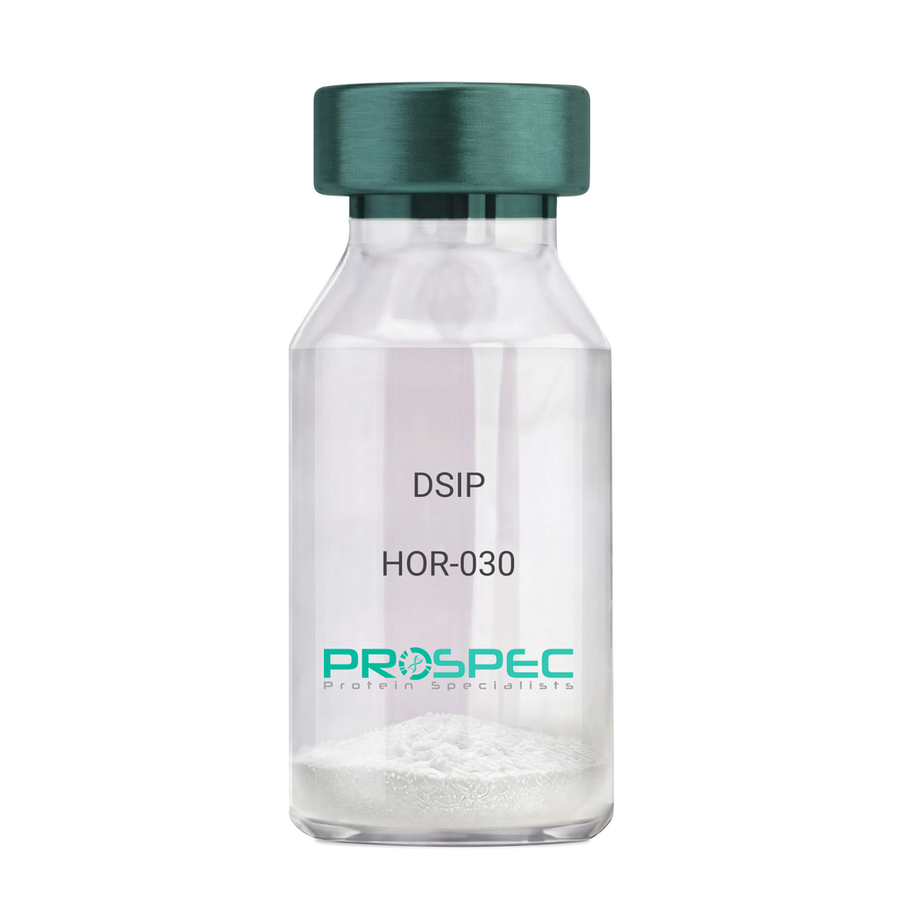
Name :
DSIPDescription:
Delta Sleep Inducing Peptide
Product # :
HOR-030Price :
Quantity :
Shipping Method :
Shipped at Room temp
More Info
- description
- formulation
- purity
- More Info
Description
DSIP Synthetic is a single, non-glycosylated polypeptide chain containing 15 amino acids, having a molecular mass of 1419.55 Dalton and a Molecular formula of C62H98N16O22.
Formulation
The protein was lyophilized with no additives.
Purity
Greater than 97.0% as determined by analysis by RP-HPLC.
More Info
-
Physical Appearance
Sterile Filtered White lyophilized (freeze-dried) powder.
-
Stability
Lyophilized DSIP although stable at room temperature for 3 weeks, should be stored desiccated below -18°C. Upon reconstitution DSIP should be stored at 4°C between 2-7 days and for future use below -18°C. For long term storage it is recommended to add a carrier protein (0.1% HSA or BSA). Please prevent freeze-thaw cycles.
-
Solubility
It is recommended to reconstitute the lyophilized DSIP in sterile 18MΩ-cm H2O not less than 100 µg/ml, which can then be further diluted to other aqueous solutions.
-
Amino Acid Sequence
H-Gly-Glu-Pro-Pro-Pro-Gly-Lys-Pro-Ala-Asp-Asp-Ala-Gly-Leu-Val-OH
-
Background
Delta Sleep-Inducing Peptide (DSIP), also known as Sleep-Promoting Peptide, is a neuropeptide that has been the subject of extensive research due to its potential role in sleep regulation, stress response, and neuroprotection. This nonapeptide, first isolated from the cerebral venous blood of rabbits during sleep, has been shown to induce slow-wave sleep, modulate pain perception, and exhibit potential antioxidant and immunomodulatory properties.
DSIP's primary function is its interaction with the sleep regulatory system. By modulating the release of certain neurotransmitters, DSIP can influence sleep patterns, particularly promoting slow-wave sleep, the most restorative stage of sleep. Studies by Kovalzon et al. (2011) have demonstrated that DSIP can enhance sleep quality in rats, suggesting potential applications in sleep disorders and the promotion of healthy sleep patterns.
In addition to its sleep-inducing effects, DSIP has been shown to possess neuroprotective properties. Research by Zolotarev et al. (2014) found that DSIP could protect neurons from oxidative stress, suggesting potential applications in the treatment of neurodegenerative diseases such as Alzheimer's and Parkinson's disease.
Given its sleep-inducing and neuroprotective effects, DSIP has been proposed as a potential therapeutic agent for a variety of conditions, including sleep disorders, chronic pain, and neurodegenerative diseases. For instance, a study by Spong et al. (2016) found that DSIP could improve sleep quality in patients with chronic insomnia, indicating its potential as a therapeutic agent in the treatment of sleep disorders.
While research on DSIP is promising, it is important to note that most studies have been conducted in animals or in vitro. More research is needed to fully understand the potential effects and applications of DSIP in humans. However, the existing body of research suggests that DSIP could be a promising tool in the treatment of sleep disorders, chronic pain, and neurodegenerative diseases.
ProSpec's products are furnished for LABORATORY RESEARCH USE ONLY. The product may not be used as drugs, agricultural or pesticidal products, food additives or household chemicals.