Search results
142 results found for “pregnancy specific beta-1-glycoprotein”
Name
Description
Product #
Price
Quantity
Shipping Method
- View Data Sheet
Name :
CoV-2-S1 (319-541), FcDescription:
Coronavirus 2019 Spike Glycoprotein-S1 Receptor Binding Domain (319-541 a.a), Fc Recombinant
Product # :
SARS-029Price :
Quantity :
Shipping Method :
Shipped at Room temp
More Info
- description
- source
- formulation
- purity
- More Info
Description
The HEK293 derived recombinant protein contains the Coronavirus 2019 Spike Glycoprotein S1 Receptor Binding Domain [ RBD ], Wuhan-Hu-1 strain, amino acids 319-541 fused to Fc tag at C-terminal.
Source
HEK293 Cells.
Formulation
CoV-S1 RBD protein is lyophilized from 1x PBS pH-7.4 + 5% trehalose.
Purity
Protein is >95% pure as determined SDS-PAGE.
More Info
-
Introduction
A human infecting coronavirus (viral pneumonia) called 2019 novel coronavirus, 2019-nCoV was found in the fish market at the city of Wuhan, Hubei province of China on December 2019.
The 2019-nCoV shares an 87% identity to the 2 bat-derived severe acute respiratory syndrome 2018 SARS-CoV-2 located in Zhoushan of eastern China. 2019-nCoV has an analogous receptor-BD-structure to that of 2018 SARS-CoV, even though there is a.a. diversity so thus the 2019-nCoV might bind to ACE2 receptor protein (angiotensin-converting enzyme 2) in humans.
While bats are possibly the host of 2019-nCoV, researchers suspect that animal from the ocean sold at the seafood market was an intermediate host. RSCU analysis proposes that the 2019-nCoV is a recombinant within the viral spike glycoprotein between the bat coronavirus and an unknown coronavirus.
-
Physical Appearance
Lyophilized freezed dried powder.
-
Stability
Lyophilized Cov-2 S1 Glycoprotein RBD although stable at room temperature for 3 weeks, should be stored desiccated below -18°C. Upon reconstitution CoV2 Spike protein should be stored at 4°C between 2-7 days and for future use below -18°C.For long term storage it is recommended to add a carrier protein (0.1% HSA or BSA).Please prevent freeze-thaw cycles.
-
Purification Method
Purified by Protein-G chromatographic technique.
ProSpec's products are furnished for LABORATORY RESEARCH USE ONLY. The product may not be used as drugs, agricultural or pesticidal products, food additives or household chemicals.
- View Data Sheet
Name :
CFB HumanDescription:
Complement Factor B Human Recombinant
Complement factor B (EC:3.4.21.47), C3/C5 convertase, Glycine-rich beta glycoprotein, GBG, PBF2, Properdin factor B, Complement factor B Ba fragment, Complement factor B Bb fragment, CFB, Complement Factor B, BFD, AHUS4, BF, BFD, CFAB, FB, FBI12, H2-Bf.
Product # :
PRO-1813Price :
Quantity :
Shipping Method :
Shipped with Ice Packs
More Info
- description
- source
- formulation
- purity
- More Info
Description
CFB Human Recombinant produced in E.coli is a single, non-glycosylated polypeptide chain containing 528 amino acids (260-764) and having a molecular mass of 59.4 kDa.CFB is fused to a 23 amino acid His-Tag at N-terminus and purified by proprietary chromatographic techniques.
Source
Escherichia Coli.
Formulation
The CFB solution (1mg/ml) contains 20mM Tris-HCl buffer (pH 8.0), 0.4M UREA and 10% glycerol.
Purity
Greater than 85.0% as determined by SDS-PAGE.
More Info
-
Introduction
Complement Factor B, also known as CFB, encodes complement factor B which is a component of the alternative pathway of complement activation. Factor B circulates in the blood as a single chain polypeptide. Once the alternative pathway is activated it is cleaved by complement factor D yielding the noncatalytic chain Ba and the catalytic subunit Bb. The active subunit Bb is a serine protease which connects with C3b to form the alternative pathway C3 convertase. Also, Bb is involved in the proliferation of preactivated B lymphocytes, while Ba inhibits their proliferation.
-
Synonyms
Complement factor B (EC:3.4.21.47), C3/C5 convertase, Glycine-rich beta glycoprotein, GBG, PBF2, Properdin factor B, Complement factor B Ba fragment, Complement factor B Bb fragment, CFB, Complement Factor B, BFD, AHUS4, BF, BFD, CFAB, FB, FBI12, H2-Bf.
-
Physical Appearance
Sterile Filtered colorless solution.
-
Stability
Store at 4°C if entire vial will be used within 2-4 weeks. Store, frozen at -20°C for longer periods of time. For long term storage it is recommended to add a carrier protein (0.1% HSA or BSA).Avoid multiple freeze-thaw cycles.
-
Amino Acid Sequence
MGSSHHHHHH SSGLVPRGSH MGSKIVLDPS GSMNIYLVLD GSDSIGASNF TGAKKCLVNL IEKVASYGVK PRYGLVTYAT YPKIWVKVSE ADSSNADWVT KQLNEINYED HKLKSGTNTK KALQAVYSMM SWPDDVPPEG WNRTRHVIIL MTDGLHNMGG DPITVIDEIR DLLYIGKDRK NPREDYLDVY VFGVGPLVNQ VNINALASKK DNEQHVFKVK DMENLEDVFY QMIDESQSLS LCGMVWEHRK GTDYHKQPWQ AKISVIRPSK GHESCMGAVV SEYFVLTAAH CFTVDDKEHS IKVSVGGEKR DLEIEVVLFH PNYNINGKKE AGIPEFYDYD VALIKLKNKL KYGQTIRPIC LPCTEGTTRA LRLPPTTTCQ QQKEELLPAQ DIKALFVSEE EKKLTRKEVY IKNGDKKGSC ERDAQYAPGY DKVKDISEVV TPRFLCTGGV SPYADPNTCR GDSGGPLIVH KRSRFIQVGV ISWGVVDVCK NQKRQKQVPA HARDFHINLF QVLPWLKEKL QDEDLGFL.
ProSpec's products are furnished for LABORATORY RESEARCH USE ONLY. The product may not be used as drugs, agricultural or pesticidal products, food additives or household chemicals.
- View Data Sheet
Name :
CFB-b HumanDescription:
Complement Factor B Fragment b Human
Complement factor B (EC:3.4.21.47), C3/C5 convertase, Glycine-rich beta glycoprotein, GBG, PBF2, Properdin factor B, Complement factor B Ba fragment, Complement factor B Bb fragment, CFB, Complement Factor B, BFD, AHUS4, BF, BFD, CFAB, FB, FBI12, H2-Bf.
Product # :
PRO-2736Price :
Quantity :
Shipping Method :
Shipped with Ice Packs
More Info
- description
- source
- formulation
- purity
- More Info
Description
CFB-b Human produced in Human Plasma having a molecular mass of 33 kDa.
Source
Human Plasma.
Formulation
CFB-b solution (1mg/ml) contains Phosphate-buffered saline, pH 7.3.
Purity
Greater than 95.0% as determined by SDS-PAGE.
More Info
-
Introduction
Complement Factor B, also known as CFB, encodes complement factor B which is a component of the alternative pathway of complement activation. Factor B circulates in the blood as a single chain polypeptide. Once the alternative pathway is activated it is cleaved by complement factor D yielding the noncatalytic chain Ba and the catalytic subunit Bb. The active subunit Bb is a serine protease which connects with C3b to form the alternative pathway C3 convertase. Also, Bb is involved in the proliferation of preactivated B lymphocytes, while Ba inhibits their proliferation.
-
Synonyms
Complement factor B (EC:3.4.21.47), C3/C5 convertase, Glycine-rich beta glycoprotein, GBG, PBF2, Properdin factor B, Complement factor B Ba fragment, Complement factor B Bb fragment, CFB, Complement Factor B, BFD, AHUS4, BF, BFD, CFAB, FB, FBI12, H2-Bf.
-
Physical Appearance
Sterile filtered solution.
-
Stability
CFB-b Human is stable at 4°C if entire vial will be used within 2-4 weeks. Store, frozen below -20°C for longer periods of time.For long term storage it is recommended to add a carrier protein (0.1% HSA or BSA).Avoid multiple freeze-thaw cycles.
-
Human Virus Test
Plasma from each donor has been tested and found negative for antibody to HIV-1, HIV-2, HCV and HBSAG.
ProSpec's products are furnished for LABORATORY RESEARCH USE ONLY. The product may not be used as drugs, agricultural or pesticidal products, food additives or household chemicals.
- View Data Sheet
Name :
CFB (26-259) HumanDescription:
Complement Factor B (26-259 a.a.) Human Recombinant
Complement factor B (EC:3.4.21.47), C3/C5 convertase, Glycine-rich beta glycoprotein, GBG, PBF2, Properdin factor B, Complement factor B Ba fragment, Complement factor B Bb fragment, CFB, Complement Factor B, BFD, AHUS4, BF, BFD, CFAB, FB, FBI12, H2-Bf.
Product # :
PRO-1860Price :
Quantity :
Shipping Method :
Shipped with Ice Packs
More Info
- description
- source
- formulation
- purity
- More Info
Description
CFB (26-259) Human Recombinant produced in E. coli is. a single polypeptide chain containing 257 amino acids and having a molecular mass of 28.4kDa. CFB is fused to a 23 amino acid His-tag at N-terminus & purified by proprietary chromatographic techniques.
Source
Escherichia Coli.
Formulation
The CFB solution (1mg/ml) contains 20mM Tris-HCl buffer (pH 8.0), 10% glycerol and 0.4M Urea.
Purity
Greater than 85.0% as determined by SDS-PAGE.
More Info
-
Introduction
Complement Factor B, also known as CFB, encodes complement factor B which is a component of the alternative pathway of complement activation. Factor B circulates in the blood as a single chain polypeptide. Once the alternative pathway is activated it is cleaved by complement factor D yielding the noncatalytic chain Ba and the catalytic subunit Bb. The active subunit Bb is a serine protease which connects with C3b to form the alternative pathway C3 convertase. Also, Bb is involved in the proliferation of preactivated B lymphocytes, while Ba inhibits their proliferation.
-
Synonyms
Complement factor B (EC:3.4.21.47), C3/C5 convertase, Glycine-rich beta glycoprotein, GBG, PBF2, Properdin factor B, Complement factor B Ba fragment, Complement factor B Bb fragment, CFB, Complement Factor B, BFD, AHUS4, BF, BFD, CFAB, FB, FBI12, H2-Bf.
-
Physical Appearance
Sterile Filtered colorless solution.
-
Stability
Store at 4°C if entire vial will be used within 2-4 weeks. Store, frozen at -20°C for longer periods of time. For long term storage it is recommended to add a carrier protein (0.1% HSA or BSA).Avoid multiple freeze-thaw cycles.
-
Amino Acid Sequence
MGSSHHHHHH SSGLVPRGSQ SHMTPWSLAR PQGSCSLEGV EIKGGSFRLL QEGQALEYVC PSGFYPYPVQ TRTCRSTGSW STLKTQDQKT VRKAECRAIH CPRPHDFENG EYWPRSPYYN VSDEISFHCY DGYTLRGSAN RTCQVNGRWS GQTAICDNGA GYCSNPGIPI GTRKVGSQYR LEDSVTYHCS RGLTLRGSQR RTCQEGGSWS GTEPSCQDSF MYDTPQEVAE AFLSSLTETI EGVDAEDGHG PGEQQKR.
ProSpec's products are furnished for LABORATORY RESEARCH USE ONLY. The product may not be used as drugs, agricultural or pesticidal products, food additives or household chemicals.
- View Data Sheet
Name :
C1QBP AntibodyDescription:
Complement Component 1, Mouse Anti Human
p32, HABP1, gC1Qr, GC1QBP, SF2p32, gC1Q-R, Complement component 1 Q subcomponent-binding protein mitochondrial, Glycoprotein gC1qBP, C1qBP, GC1q-R protein, Hyaluronan-binding protein 1, Mitochondrial matrix protein p32, p33, C1QBP.
Product # :
ANT-556Price :
Quantity :
Shipping Method :
Shipped with Ice Packs
More Info
- formulation
- More Info
Formulation
1mg/ml containing PBS, pH-7.4, 10% Glycerol and 0.02% Sodium Azide.
More Info
-
Introduction
C1QBP binds to the globular "heads" of c1q thus inhibiting c1 activation. C1QBP interacts with a wide range of ligands and is implicated in cell signaling. C1QBP associates with C1r and C1s in order to yield the first component of the serum complement system. C1QBP protein has been identified as the p32 subunit of pre-mRNA splicing factor SF2, as well as a hyaluronic acid-binding protein.
C1QBP is a new marker of tumor cells and tumor-associated macrophages/myeloid cells in hypoxic/metabolically deprived areas of tumors.
Mitochondrial C1QBP is a critical mediator of p14ARF-induced apoptosis.
C1QBP functions as a chemotactic factor for immature dendritic cells, and migration is mediated through ligation of both C1QBP and cC1qR/CR.
C1QBP overexpression successfully blocks mRNA accumulation from the adenovirus major late transcription unit (MLTU) and stimulates RNA polymerase II carboxy-terminal domain phosphorylation in virus-infected cells.
C1QBP binds with Hepacivirus core protein on CD8+ and CD4+ positive t-cells and inactivates lck and akt. -
Synonyms
p32, HABP1, gC1Qr, GC1QBP, SF2p32, gC1Q-R, Complement component 1 Q subcomponent-binding protein mitochondrial, Glycoprotein gC1qBP, C1qBP, GC1q-R protein, Hyaluronan-binding protein 1, Mitochondrial matrix protein p32, p33, C1QBP.
-
Physical Appearance
Sterile filtered colorless solution.
-
Immunogen
Anti-human C1QBP mAb, is derived from hybridization of mouse F0 myeloma cells with spleen cells from BALB/c mice immunized with a recombinant human C1QBP protein 74-282 amino acids purified from E. coli.
-
Ig Subclass
Mouse IgG1 heavy chain and k light chain.
-
Clone
PAT1G7AT.
-
Applications
C1QBP antibody has been tested by ELISA, Western blot analysis to assure specificity and reactivity. Since application varies, however, each investigation should be titrated by the reagent to obtain optimal results.
-
Type
Mouse Anti Human Monoclonal.
-
Storage Procedures
For periods up to 1 month store at 4°C, for longer periods of time, store at -20°C. Prevent freeze thaw cycles.
-
Purification Method
C1QBP antibody was purified from mouse ascitic fluids by protein-A affinity chromatography.
ProSpec's products are furnished for LABORATORY RESEARCH USE ONLY. The product may not be used as drugs, agricultural or pesticidal products, food additives or household chemicals.
- View Data Sheet
Name :
CoV-2-S1 (319-541)Description:
Coronavirus 2019 Spike Glycoprotein-S1 Receptor Binding Domain (319-541 a.a), Recombinant
Product # :
SARS-021Price :
Quantity :
Shipping Method :
Shipped with Ice Packs
More Info
- description
- source
- formulation
- purity
- biological activity
- More Info
Description
The HEK293 derived recombinant protein contains the Coronavirus 2019 Spike Glycoprotein S1 Receptor Binding Domain [ RBD ], Wuhan-Hu-1 strain, amino acids 319-541 fused to His tag at C-terminal.
Source
HEK293 Cells.
Formulation
CoV-S1 RBD protein is 1mg/ml contains 20mM sodium phosphate (NaPP), 300mM NaCl, pH7.2
Purity
Protein is >90% pure as determined SDS-PAGE.
Biological Activity
SARS CoV-2 Spike Glycoprotein S1 RBD was tested for its biological functionality by its binding to recombinant ACE2 receptor protein confirmed by structural complementation reporter assay (NanoBiT®).
More Info
-
Introduction
A human infecting coronavirus (viral pneumonia) called 2019 novel coronavirus, 2019-nCoV was found in the fish market at the city of Wuhan, Hubei province of China on December 2019.
The 2019-nCoV shares an 87% identity to the 2 bat-derived severe acute respiratory syndrome 2018 SARS-CoV-2 located in Zhoushan of eastern China. 2019-nCoV has an analogous receptor-BD-structure to that of 2018 SARS-CoV, even though there is a.a. diversity so thus the 2019-nCoV might bind to ACE2 receptor protein (angiotensin-converting enzyme 2) in humans.
While bats are possibly the host of 2019-nCoV, researchers suspect that animal from the ocean sold at the seafood market was an intermediate host. RSCU analysis proposes that the 2019-nCoV is a recombinant within the viral spike glycoprotein between the bat coronavirus and an unknown coronavirus.
-
Physical Appearance
Sterile Filtered clear solution.
-
Stability
CoV-2 S1 RBD Protein is shipped on ice packs. Upon arrival, Store at -20°C.
-
Purification Method
Purified by Metal-Afinity chromatographic technique.
ProSpec's products are furnished for LABORATORY RESEARCH USE ONLY. The product may not be used as drugs, agricultural or pesticidal products, food additives or household chemicals.
- View Data Sheet
Name :
CoV-2 S1 (1-681)Description:
Coronavirus 2019-nCoV Spike Glycoprotein-S1 (1-681), Recombinant
Product # :
SARS-045Price :
Quantity :
Shipping Method :
Shipped with Ice Packs
More Info
- description
- source
- formulation
- purity
- More Info
Description
The HEK293 derived recombinant protein contains the Coronavirus 2019-nCoV Spike Glycoprotein S1, Wuhan-Hu-1 strain, amino acids 1-681 fused to Fc tag at C-terminal having a Mw of 76 kDa.
Source
HEK293.
Formulation
CoV-2 S1protein solution is supplied PBS pH 7.4.
Purity
Protein is >95% pure as determined SDS-PAGE.
More Info
-
Introduction
A human infecting coronavirus (viral pneumonia) called 2019 novel coronavirus, 2019-nCoV was found in the fish market at the city of Wuhan, Hubei province of China on December 2019.The 2019-nCoV shares an 87% identity to the 2 bat-derived severe acute respiratory syndrome 2018 SARS-CoV-2 located in Zhoushan of eastern China. 2019-nCoV has an analogous receptor-BD-structure to that of 2018 SARS-CoV, even though there is a.a. diversity so thus the 2019-nCoV might bind to ACE2 receptor protein (angiotensin-converting enzyme 2) in humans.While bats are probably the host of 2019-nCoV, researchers suspect that animal from the ocean sold at the seafood market was an intermediate host. RSCU analysis proposes that the 2019-nCoV is a recombinant within the viral spike glycoprotein between the bat coronavirus and an unknown coronavirus.
-
Physical Appearance
Sterile Filtered clear solution.
-
Stability
Protein is shipped on ice packs. Upon arrival, Store at -20°C.
-
Purification Method
Purified by Protein-G chromatography technique.
ProSpec's products are furnished for LABORATORY RESEARCH USE ONLY. The product may not be used as drugs, agricultural or pesticidal products, food additives or household chemicals.
- View Data Sheet
Name :
CoV-2 S1 (319-541), Sf9Description:
Coronavirus 2019-nCoV Spike Glycoprotein-S1 Receptor Binding Domain Recombinant,SF9
Severe acute respiratory syndrome coronavirus 2, COVID-19, COVID-19 virus, COVID19, HCoV-19, Human coronavirus 2019, SARS-2, SARS-CoV2, SARS2, Wuhan coronavirus, Wuhan seafood market pneumonia virus, SARS-CoV-2 SP RBD, 2019-nCoV SP RBD, 2019-nCoV, 2019-nCoV; Spike RBD Protein.
Product # :
SARS-049Price :
Quantity :
Shipping Method :
Shipped with Ice Packs
More Info
- description
- source
- formulation
- purity
- biological activity
- More Info
Description
Recombinant Coronavirus 2019-nCoV Spike Glycoprotein-S1 Receptor Binding Domain is a single, glycosylated polypeptide chain containing a total of 232 amino acids (319-541) and having a calculated Mw of 26.2 kDa. CoV-2 S1 (319-541) is fused to a 6 amino acid His-tag at C-terminus,and is purified by proprietary chromatographic techniques.
Source
Sf9, Baculovirus cells.
Formulation
CoV-2 S1 (319-541) solution (0.25mg/ml) contains Phosphate-Buffered Saline (pH 7.4) containing 10% glycerol.
Purity
Greater than 95.0% as determined by SDS-PAGE.
Biological Activity
Measured by its binding ability in a functional ELISA with Human ACE-2 (CAT# enz-1159).
More Info
-
Introduction
A human infecting coronavirus (viral pneumonia) called 2019 novel coronavirus, 2019-nCoV was found in the fish market at the city of Wuhan, Hubei province of China on December 2019.The 2019-nCoV shares an 87% identity to the 2 bat-derived severe acute respiratory syndrome 2018 SARS-CoV-2 located in Zhoushan of eastern China. 2019-nCoV has an analogous receptor-BD-structure to that of 2018 SARS-CoV, even though there is a.a. diversity so thus the 2019-nCoV might bind to ACE2 receptor protein (angiotensin-converting enzyme 2) in humans.While bats are probably the host of 2019-nCoV, researchers suspect that animal from the ocean sold at the seafood market was an intermediate host. RSCU analysis proposes that the 2019-nCoV is a recombinant within the viral spike glycoprotein between the bat coronavirus and an unknown coronavirus.
-
Synonyms
Severe acute respiratory syndrome coronavirus 2, COVID-19, COVID-19 virus, COVID19, HCoV-19, Human coronavirus 2019, SARS-2, SARS-CoV2, SARS2, Wuhan coronavirus, Wuhan seafood market pneumonia virus, SARS-CoV-2 SP RBD, 2019-nCoV SP RBD, 2019-nCoV, 2019-nCoV; Spike RBD Protein.
-
Physical Appearance
Sterile Filtered colorless solution.
-
Stability
Store at 4°C if entire vial will be used within 2-4 weeks. Store, frozen at -20°C for longer periods of time. For long term storage it is recommended to add a carrier protein (0.1% HSA or BSA). Avoid multiple freeze-thaw cycles.
-
Amino Acid Sequence
ADPRVQPTES IVRFPNITNL CPFGEVFNAT RFASVYAWNR KRISNCVADY SVLYNSASFS TFKCYGVSPT KLNDLCFTNV YADSFVIRGD EVRQIAPGQT GKIADYNYKL PDDFTGCVIA WNSNNLDSKV GGNYNYLYRL FRKSNLKPFE RDISTEIYQA GSTPCNGVEG FNCYFPLQSY GFQPTNGVGY QPYRVVVLSF ELLHAPATVC GPKKSTNLVK NKCVNFHHHH HH
ProSpec's products are furnished for LABORATORY RESEARCH USE ONLY. The product may not be used as drugs, agricultural or pesticidal products, food additives or household chemicals.
- View Data Sheet
Name :
CD18 HumanDescription:
CD18 Human Recombinant
Integrin Subunit Beta 2, Integrin, Beta 2 (Complement Component 3 Receptor 3 And 4 Subunit), Complement Component 3 Receptor 3 And 4 Subunit, CD18, MFI7, Integrin, Beta 2 (Antigen CD18 (P95), Lymphocyte Function-Associated Antigen 1; Macrophage Antigen 1 (Mac-1) Beta Subunit), Cell Surface Adhesion Glycoprotein (LFA-1/CR3/P150,959 Beta Subunit Precursor), Cell Surface Adhesion Glycoproteins LFA-1/CR3/P150,95 Subunit Beta, Leukocyte-Associated Antigens CD18/11A, CD18/11B, CD18/11C, Leukocyte Cell Adhesion Molecule CD18, Complement Receptor C3 Beta-Subunit, Complement Receptor C3 Subunit Beta, Integrin Beta Chain, Beta 2, CD18 Antigen, LFA-1, MAC-1, LCAMB, MF17, LAD, CD18.
Product # :
PRO-2324Price :
Quantity :
Shipping Method :
Shipped with Ice Packs
More Info
- description
- source
- formulation
- purity
- More Info
Description
CD18 Human Recombinant produced in Sf9 Baculovirus cells is a single, glycosylated polypeptide chain containing 686 amino acids (23-700 a.a.) and having a molecular mass of 75.9kDa (Migrates at 70-100kDa on SDS-PAGE under reducing conditions).CD18 is expressed with an 8 amino acid His tag at C-Terminus and purified by proprietary chromatographic techniques.
Source
Sf9, Baculovirus cells.
Formulation
CD18 protein solution (0.25mg/ml) containing Phosphate Buffered Saline (pH 7.4) and 10% glycerol.
Purity
Greater than 85.0% as determined by SDS-PAGE.
More Info
-
Introduction
CD18 (ITGB2 or integrin beta-2) is a receptor for ICAM1, ICAM2, ICAM3 and ICAM4. CD18 may be a member of an extended family of cell surface molecules including the fibronectin binding protein. In addition, CD18 is a complement receptor type 3 (CR3). CD18 triggers neutrophil transmigration during lung injury through PTK2B/PYK2-mediated activation.
-
Synonyms
Integrin Subunit Beta 2, Integrin, Beta 2 (Complement Component 3 Receptor 3 And 4 Subunit), Complement Component 3 Receptor 3 And 4 Subunit, CD18, MFI7, Integrin, Beta 2 (Antigen CD18 (P95), Lymphocyte Function-Associated Antigen 1; Macrophage Antigen 1 (Mac-1) Beta Subunit), Cell Surface Adhesion Glycoprotein (LFA-1/CR3/P150,959 Beta Subunit Precursor), Cell Surface Adhesion Glycoproteins LFA-1/CR3/P150,95 Subunit Beta, Leukocyte-Associated Antigens CD18/11A, CD18/11B, CD18/11C, Leukocyte Cell Adhesion Molecule CD18, Complement Receptor C3 Beta-Subunit, Complement Receptor C3 Subunit Beta, Integrin Beta Chain, Beta 2, CD18 Antigen, LFA-1, MAC-1, LCAMB, MF17, LAD, CD18.
-
Physical Appearance
Sterile filtered colorless solution.
-
Stability
Store at 4°C if entire vial will be used within 2-4 weeks.Store, frozen at -20°C for longer periods of time. For long term storage it is recommended to add a carrier protein (0.1% HSA or BSA).Avoid multiple freeze-thaw cycles.
-
Amino Acid Sequence
QECTKFKVSS CRECIESGPG CTWCQKLNFT GPGDPDSIRC DTRPQLLMRG CAADDIMDPT SLAETQEDHN GGQKQLSPQK VTLYLRPGQA AAFNVTFRRA KGYPIDLYYL MDLSYSMLDD LRNVKKLGGD LLRALNEITE SGRIGFGSFV DKTVLPFVNT HPDKLRNPCP NKEKECQPPF AFRHVLKLTN NSNQFQTEVG KQLISGNLDA PEGGLDAMMQ VAACPEEIGW RNVTRLLVFA TDDGFHFAGD GKLGAILTPN DGRCHLEDNL YKRSNEFDYP SVGQLAHKLA ENNIQPIFAV TSRMVKTYEK LTEIIPKSAV GELSEDSSNV VQLIKNAYNK LSSRVFLDHN ALPDTLKVTY DSFCSNGVTH RNQPRGDCDG VQINVPITFQ VKVTATECIQ EQSFVIRALG FTDIVTVQVL PQCECRCRDQ SRDRSLCHGK GFLECGICRC DTGYIGKNCE CQTQGRSSQE LEGSCRKDNN SIICSGLGDC VCGQCLCHTS DVPGKLIYGQ YCECDTINCE RYNGQVCGGP GRGLCFCGKC RCHPGFEGSA CQCERTTEGC LNPRRVECSG RGRCRCNVCE CHSGYQLPLC QECPGCPSPC GKYISCAECL KFEKGPFGKN CSAACPGLQL SNNPVKGRTC KERDSEGCWV AYTLEQQDGM DRYLIYVDES RECVAGPNLE HHHHHH
ProSpec's products are furnished for LABORATORY RESEARCH USE ONLY. The product may not be used as drugs, agricultural or pesticidal products, food additives or household chemicals.
- View Data Sheet
Name :
Hemopexin Human, Sf9Description:
Hemopexin Human Recombinant, Sf9
Hemopexin, Beta-1B-Glycoprotein, HX, Beta-1B-glycoprotein.
Product # :
PRO-2544Price :
Quantity :
Shipping Method :
Shipped with Ice Packs
More Info
- description
- source
- formulation
- purity
- More Info
Description
Hemopexin produced in Sf9 Baculovirus cells is a single, glycosylated polypeptide chain containing 448 amino acids (24-462a.a.) and having a molecular mass of 50.4kDa. (Molecular size on SDS-PAGE will appear at approximately 50-70kDa).Hemopexin is expressed with a 9 amino acid His tag at C-Terminus and purified by proprietary chromatographic techniques.
Source
Sf9, Baculovirus cells.
Formulation
Hemopexin protein solution (0.5mg/ml) contains 10% glycerol & Phosphate Buffered Saline (pH 7.4).
Purity
Greater than 90.0% as determined by SDS-PAGE.
More Info
-
Introduction
Hemopexin (or haemopexin) is a plasma protein that binds heme with the highest affinity of any known protein. Hemopexin is generally expressed in liver, and belongs to acute phase reactants, the synthesis of which is induced after inflammation. Heme is potentially very toxic because of its ability to intercalate into lipid membrane and to generate hydroxyl radicals. Hemopexin’s function of scavenging the heme released or lost by the turnover of heme proteins such as hemoglobin defends the body from the oxidative damage that free heme can cause. Additionally, hemopexin discharges its bound ligand for internalisation upon interacting with a specific receptor located on the surface of liver cells. This hemopexin function is in order to preserve the body's iron. Hemopexin’s levels in the serum are an indication of how much heme is present in the blood. Low Hemopexin levels show that there is a lot of it in the serum. For that reason, low hemopexin levels indicate that there has been consid
-
Synonyms
Hemopexin, Beta-1B-Glycoprotein, HX, Beta-1B-glycoprotein.
-
Physical Appearance
Sterile filtered colorless solution.
-
Stability
Store at 4°C if entire vial will be used within 2-4 weeks.Store, frozen at -20°C for longer periods of time. For long term storage it is recommended to add a carrier protein (0.1% HSA or BSA). Avoid multiple freeze-thaw cycles.
-
Amino Acid Sequence
ADPTPLPPTS AHGNVAEGET KPDPDVTERC SDGWSFDATT LDDNGTMLFF KGEFVWKSHK WDRELISERW KNFPSPVDAA FRQGHNSVFL IKGDKVWVYP PEKKEKGYPK LLQDEFPGIP SPLDAAVECH RGECQAEGVL FFQGDREWFW DLATGTMKER SWPAVGNCSS ALRWLGRYYC FQGNQFLRFD PVRGEVPPRY PRDVRDYFMP CPGRGHGHRN GTGHGNSTHH GPEYMRCSPH LVLSALTSDN HGATYAFSGT HYWRLDTSRD GWHSWPIAHQ WPQGPSAVDA AFSWEEKLYL VQGTQVYVFL TKGGYTLVSG YPKRLEKEVG TPHGIILDSV DAAFICPGSS RLHIMAGRRL WWLDLKSGAQ ATWTELPWPH EKVDGALCME KSLGPNSCSA NGPGLYLIHG PNLYCYSDVE KLNAAKALPQ PQNVTSLLGC THHHHHHH.
ProSpec's products are furnished for LABORATORY RESEARCH USE ONLY. The product may not be used as drugs, agricultural or pesticidal products, food additives or household chemicals.
- View Data Sheet
Name :
APOH Human, sf9Description:
Apolipoprotein-H Human Recombinant, Sf9
Beta-2-glycoprotein 1, Beta-2-glycoprotein I, Apolipoprotein H, Apo-H, B2GPI, Beta(2)GPI, Activated protein C-binding protein, APC inhibitor, Anticardiolipin cofactor, APOH, B2G1, BG, B2GP-1, B2 Glycoprotein-I.
Product # :
PRO-388Price :
Quantity :
Shipping Method :
Shipped with Ice Packs
More Info
- description
- source
- formulation
- purity
- More Info
Description
Apolipoprotein-H Human Recombinant produced in SF9 is a glycosylated, polypeptide chain containing 326 amino acids and having a molecular mass of 38,200 Dalton (excluding glycosylation), 45kDa total mass. APOH is expressed with a -6xHis tag and purified by proprietary chromatographic techniques.
Source
Sf9 insect cells.
Formulation
APOH is supplied in 16mM HEPES buffer pH-7.2, 200mM NaCl and 20% glycerol.
Purity
Greater than 95% as determined by SDS-PAGE.
More Info
-
Introduction
Apolipoprotein H (APOH) attaches to numerous kinds of negatively charged substances for instance heparin, phospholipids, and dextran sulfate. APOH prevents activation of the intrinsic blood coagulation cascade by attaching to phospholipids on the surface of damaged cells. APOH expressed by the liver and secreted in the plasma.
-
Synonyms
Beta-2-glycoprotein 1, Beta-2-glycoprotein I, Apolipoprotein H, Apo-H, B2GPI, Beta(2)GPI, Activated protein C-binding protein, APC inhibitor, Anticardiolipin cofactor, APOH, B2G1, BG, B2GP-1, B2 Glycoprotein-I.
-
Physical Appearance
Sterile Filtered clear solution.
-
Stability
Store at 4°C if entire vial will be used within 2-4 weeks. Store, frozen at -20°C for longer periods of time. Avoid multiple freeze-thaw cycles.
ProSpec's products are furnished for LABORATORY RESEARCH USE ONLY. The product may not be used as drugs, agricultural or pesticidal products, food additives or household chemicals.
- View Data Sheet
Name :
SPARCL1 MouseDescription:
SPARC Like 1 Mouse Recombinant
Sparcl1, Ecm2, hevin, mast9, Sc1, SPARC-like protein 1, Extracellular matrix protein 2, Matrix glycoprotein Sc1.
Product # :
PRO-2617Price :
Quantity :
Shipping Method :
Shipped with Ice Packs
More Info
- description
- source
- formulation
- purity
- More Info
Description
SPARCL1 Mouse Recombinant produced in Sf9 Baculovirus cells is a single, glycosylated polypeptide chain containing 642 amino acids (17-650 a.a) and having a molecular mass of 71.7kDa.SPARCL1 is fused to an 8 amino acid His-tag at C-terminus & purified by proprietary chromatographic techniques.
Source
Sf9, Baculovirus cells.
Formulation
SPARCL1 protein solution (0.25mg/ml) containing Phosphate Buffered Saline (pH 7.4) and 10% glycerol.
Purity
Greater than 90.0% as determined by SDS-PAGE.
More Info
-
Introduction
SPARC-like protein 1 or SPARC1 is an anti-adhesive protein with a similarity to the SPARC protein’s structure. SPARC1 is highly expressed in several tissues such as brain, lungs, kidney, heart, although it is not found in the liver tissue. The protein inhibits spreading or adhesion of many substrates and is considered to lead to antiadhesive signaling that stops neuronal migration, that occurs during development or as a trauma response, consistent with production by glial and neuronal cells.
-
Synonyms
Sparcl1, Ecm2, hevin, mast9, Sc1, SPARC-like protein 1, Extracellular matrix protein 2, Matrix glycoprotein Sc1.
-
Physical Appearance
Sterile Filtered colorless solution.
-
Stability
Store at 4°C if entire vial will be used within 2-4 weeks. Store, frozen at -20°C for longer periods of time. For long term storage it is recommended to add a carrier protein (0.1% HSA or BSA).Avoid multiple freeze-thaw cycles.
-
Amino Acid Sequence
IPTSTRFLSD HSNPTTATLV TPEDATVPIA GVEATADIEN HPSDKAEKPS ALNSEEETHE
QSTEQDKTYS FEVDLKDEED GDGDLSVDPT EGTLTLDLQE GTSEPQQKSL PENGDFPATV
STSYVDPNQR ANITKGKESQ EQPVSDSHQQ PNESSKQTQD LKAEESQTQD PDIPNEEEEE
EEDEEEEEEE EPEDIGAPSD NQEEGKEPLE EQPTSKWEGN REQSDDTLEE SSQPTQISKT
EKHQSEQGNQ GQESDSEAEG EDKAAGSKEH IPHTEQQDQE GKAGLEAIGN QKDTDEKAVS
TEPTDAAVVP RSHGGAGDNG GGDDSKHGAG DDYFIPSQEF LEAERMHSLS YYLKYGGGEE
TTTGESENRR EAADNQEAKK AESSPNAEPS DEGNSREHSA GSCTNFQCKR GHICKTDPQG
KPHCVCQDPE TCPPAKILDQ ACGTDNQTYA SSCHLFATKC RLEGTKKGHQ LQLDYFGACK
SIPACTDFEV AQFPLRMRDW LKNILMQLYE PNPKHGGYLN EKQRSKVKKI YLDEKRLLAG
HPIELLLRD FKKNYHMYVY PVHWQFNELD QHPADRILTH SELAPLRASL VPMEHCITRF
FEECDPNKDK HITLKEWGHC FGIKEEDIDE NLLFLEHHHH HH
ProSpec's products are furnished for LABORATORY RESEARCH USE ONLY. The product may not be used as drugs, agricultural or pesticidal products, food additives or household chemicals.
- View Data Sheet
Name :
CD8B HumanDescription:
CD8B Human Recombinant
T Lymphocyte Surface Glycoprotein Beta Chain, CD8 Antigen, Beta Polypeptide 1 (P37), CD8b Molecule, CD8b Antigen, T-Cell Surface Glycoprotein CD8 Beta Chain, CD8B1, LYT3, LY3, LEU2, P37.
Product # :
PRO-1843Price :
Quantity :
Shipping Method :
Shipped with Ice Packs
More Info
- description
- source
- formulation
- purity
- More Info
Description
CD8B Human Recombinant produced in E.coli is a single, non-glycosylated polypeptide chain containing 172 amino acids (22-170) and having a molecular mass of 19.2 kDa. CD8B is fused to a 23 amino acid His-tag at N-terminus.
Source
Escherichia Coli.
Formulation
The CD8B solution (0.5mg/ml) contains 20mM Tris-HCl buffer (pH 8.0), 0.4M Urea and 10% glycerol.
Purity
Greater than 80% as determined by SDS-PAGE.
More Info
-
Introduction
CD8B encodes the CD8 beta chain isoforms of the CD8 antigen. CD8 is a cell surface glycoprotein which facilitates efficient cell-cell interactions within the immune system and is located on most cytotoxic T lymphocytes. The T-cell receptor on the T lymphocyte and the co-receptor CD8 antigen identify antigens exposed by an antigen presenting cell (APC) in the context of class I MHC molecules. The functional co-receptor is either a heterodimer composed of one alpha and one beta chain or a homodimer composed of two alpha chains. Both alpha and beta chains share substantial homology to immunoglobulin variable light chains.
-
Synonyms
T Lymphocyte Surface Glycoprotein Beta Chain, CD8 Antigen, Beta Polypeptide 1 (P37), CD8b Molecule, CD8b Antigen, T-Cell Surface Glycoprotein CD8 Beta Chain, CD8B1, LYT3, LY3, LEU2, P37.
-
Physical Appearance
Sterile Filtered clear solution.
-
Stability
Store at 4°C if entire vial will be used within 2-4 weeks. Store, frozen at -20°C for longer periods of time. For long term storage it is recommended to add a carrier protein (0.1% HSA or BSA).Avoid multiple freeze-thaw cycles.
-
Amino Acid Sequence
MGSSHHHHHH SSGLVPRGSH MGSLQQTPAY IKVQTNKMVM LSCEAKISLS NMRIYWLRQR QAPSSDSHHE FLALWDSAKG TIHGEEVEQE KIAVFRDASR FILNLTSVKP EDSGIYFCMI VGSPELTFGK GTQLSVVDFL PTTAQPTKKS TLKKRVCRLP RPETQKGPLC SP
ProSpec's products are furnished for LABORATORY RESEARCH USE ONLY. The product may not be used as drugs, agricultural or pesticidal products, food additives or household chemicals.
- View Data Sheet
Name :
TSH ProteinDescription:
Thyroid Stimulating Hormone Human Recombinant
Glycoprotein hormones alpha chain, Anterior pituitary glycoprotein hormones common subunit alpha, Follitropin alpha chain, Follicle-stimulating hormone alpha chain, FSH-alpha, Lutropin alpha chain, Luteinizing hormone alpha chain, LSH-alpha, Thyrotropin alpha chain, Thyroid-stimulating hormone alpha chain, TSH-alpha, Choriogonadotropin alpha chain, Chorionic gonadotrophin alpha subunit, CG-alpha, Thyrotropin subunit beta, Thyroid-stimulating hormone subunit beta, TSH-beta, TSH-B, Thyrotropin beta chain, Thyrotropin alfa.
Product # :
HOR-050Price :
Quantity :
Shipping Method :
Shipped at Room temp
More Info
- description
- source
- formulation
- purity
- biological activity
- More Info
Description
Thyroid Stimulating Hormone Human Recombinant produced in HEK 293 cells.
Source
HEK 293 cells.
Formulation
Lyophilized from a concentrated 50mM ammonium bicarbonate.
Purity
Greater than 95.0% as determined by SDS-PAGE.
Biological Activity
The protein is biologically active using Siemens Centaur CP is standardized against WHO 3rd IS 81/565.
More Info
-
Synonyms
Glycoprotein hormones alpha chain, Anterior pituitary glycoprotein hormones common subunit alpha, Follitropin alpha chain, Follicle-stimulating hormone alpha chain, FSH-alpha, Lutropin alpha chain, Luteinizing hormone alpha chain, LSH-alpha, Thyrotropin alpha chain, Thyroid-stimulating hormone alpha chain, TSH-alpha, Choriogonadotropin alpha chain, Chorionic gonadotrophin alpha subunit, CG-alpha, Thyrotropin subunit beta, Thyroid-stimulating hormone subunit beta, TSH-beta, TSH-B, Thyrotropin beta chain, Thyrotropin alfa.
-
Physical Appearance
Sterile Filtered White lyophilized (freeze-dried) powder.
-
Stability
Lyophilized Recombinant TSH although stable at room temperature for 3 weeks, should be stored desiccated below -18°C. Upon reconstitution Recombinant TSH should be stored at 4°C between 2-7 days and for future use below -18°C. For long term storage it is recommended to add a carrier protein (0.1% HSA or BSA). Please prevent freeze-thaw cycles.
-
Solubility
It is recommended to reconstitute the lyophilized recombinant TSH in sterile 10 mM Sodium Phosphate, 150 mM Sodium Chloride, 1 mg/ml BSA, 0.1% Sodium Azide (optional), pH 7.4.
-
Background
Thyroid-stimulating hormone (TSH), also known as thyrotropin, is a glycoprotein hormone produced by the anterior pituitary gland. Its primary function is to regulate thyroid gland activity by stimulating the synthesis and secretion of thyroid hormones, thyroxine (T4) and triiodothyronine (T3). Recombinant human TSH (rhTSH) has emerged as a valuable tool in clinical practice, particularly in the management of thyroid disorders and in diagnostic procedures involving the thyroid gland. This paper aims to provide an overview of research on rhTSH, including its mechanism of action, activity, and therapeutic applications.
The mechanism of action of rhTSH involves binding to the TSH receptor (TSHR) on the surface of thyroid follicular cells. This interaction activates intracellular signalling pathways, including cyclic adenosine monophosphate (cAMP) production and protein kinase A (PKA) activation. These pathways stimulate various cellular processes within thyroid follicular cells, such as iodine uptake, thyroid hormone synthesis, and secretion.
What is the source or expression system of TSH PROTEIN Protein?
HEK 293 cells.
What is the Purity of TSH PROTEIN Protein?
TSH PROTEIN Protein is >95% pure as determined by SDS-PAGE.
What is the Biological Activity of TSH PROTEIN Protein?
The protein is biologically active using Siemens Centaur CP is standardized against WHO 3rd IS 81/565.
What applications can TSH PROTEIN Protein be used in?
TSH PROTEIN Protein can probably be used in western blot, ELISA and Lateral Flow.
What is the endotoxin level for TSH PROTEIN Protein?
The endotoxin level is minimal, TSH PROTEIN Protein was purified using conventional chromatography techniques.
ProSpec's products are furnished for LABORATORY RESEARCH USE ONLY. The product may not be used as drugs, agricultural or pesticidal products, food additives or household chemicals.
- View Data Sheet
Name :
CD79B Human, Sf9Description:
CD79B Human Recombinant, Sf9
CD79b Molecule, CD79B Antigen (Immunoglobulin-Associated Beta), CD79b Molecule, Immunoglobulin-Associated Beta, Immunoglobulin-Associated B29 Protein, B-Cell-Specific Glycoprotein B29, Ig-Beta, IGB, B29, B-Cell Antigen Receptor Complex-Associated Protein Beta Chain, CD79b Antigen, CD79B, AGM6.
Product # :
PRO-2404Price :
Quantity :
Shipping Method :
Shipped with Ice Packs
More Info
- description
- source
- formulation
- purity
- More Info
Description
CD79B Human Recombinant produced in Sf9 Baculovirus cells is a single, glycosylated polypeptide chain containing 140 amino acids (29-159a.a.) and having a molecular mass of 16.3kDa (Molecular size on SDS-PAGE will appear at approximately 18-28 kDa).CD79B is expressed with a 9 amino acid His tag at C-Terminus and purified by proprietary chromatographic techniques.
Source
Sf9, Baculovirus cells.
Formulation
CD79B protein solution (0.5mg/ml) contains Phosphate Buffered Saline (pH 7.4) and 10% glycerol.
Purity
Greater than 90.0% as determined by SDS-PAGE.
More Info
-
Introduction
CD79B is a 36 kDa trans membrane glycoprotein belonging to the immunoglobulin superfamily. Heterodimers of CD79A and CD79B connect with a membrane attached immunoglobulin on the B cell surface to create the B cell antigen receptor complex (BCR). CD79A and CD79B are essential for BCR-mediated signaling and subsequently for the development and activation of B lineage cells.
-
Synonyms
CD79b Molecule, CD79B Antigen (Immunoglobulin-Associated Beta), CD79b Molecule, Immunoglobulin-Associated Beta, Immunoglobulin-Associated B29 Protein, B-Cell-Specific Glycoprotein B29, Ig-Beta, IGB, B29, B-Cell Antigen Receptor Complex-Associated Protein Beta Chain, CD79b Antigen, CD79B, AGM6.
-
Physical Appearance
Sterile Filtered colorless solution.
-
Stability
Store at 4°C if entire vial will be used within 2-4 weeks. Store, frozen at -20°C for longer periods of time. For long term storage it is recommended to add a carrier protein (0.1% HSA or BSA).Avoid multiple freeze-thaw cycles.
-
Amino Acid Sequence
ADPARSEDRY RNPKGSACSR IWQSPRFIAR KRGFTVKMHC YMNSASGNVS WLWKQEMDEN PQQLKLEKGR MEESQNESLA TLTIQGIRFE DNGIYFCQQK CNNTSEVYQG CGTELRVMGF STLAQLKQRN TLKDHHHHHH.
ProSpec's products are furnished for LABORATORY RESEARCH USE ONLY. The product may not be used as drugs, agricultural or pesticidal products, food additives or household chemicals.
- View Data Sheet
Name :
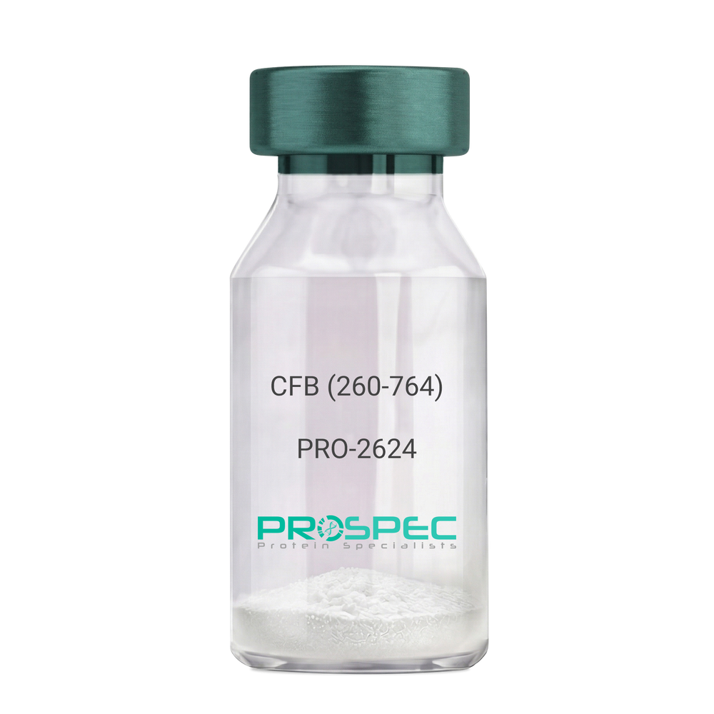
CFB (260-764) Human, Sf9Description:
Complement Factor B (260-764 a.a.) Human Recombinant, Sf9
CFB, C3/C5 convertase, Glycine-rich beta glycoprotein, GBG, PBF2, Properdin factor B, BF, BFD, AHUS4, ARMD14, CFAB, CFBD, FB, FBI12, GBG, H2-Bf.
Product # :
PRO-2624Price :
Quantity :
Shipping Method :
Shipped with Ice Packs
More Info
- description
- source
- formulation
- purity
- More Info
Description
CFB Human Recombinant produced in Sf9 Baculovirus cells is a single, glycosylated polypeptide chain containing 514 amino acids (260-764 a.a) and having a molecular mass of 58.1kDa.CFB is fused to an 9 amino acid His-tag at C-terminus & purified by proprietary chromatographic techniques.
Source
Sf9, Baculovirus cells.
Formulation
CFB protein solution (0.5mg/ml) containing Phosphate Buffered Saline (pH 7.4) and 20% glycerol.
Purity
Greater than 90.0% as determined by SDS-PAGE.
More Info
-
Introduction
Factor B is a protein, build from a single chain that circulates in the blood. Complement factor D cleaves and activates factor B, thus creating a catalytic subunit Bb and noncatalytic chain Ba. Subunit Bb is a serine protease that can bind to C3b and forms alternative pathway C3 convertase. Subunit Ba acts as an inhibitor for proliferation of lymphocytes, when Bb is part of the lymphocytes’ proliferation. The gene that codes for this protein is located on chromosome six.
-
Synonyms
CFB, C3/C5 convertase, Glycine-rich beta glycoprotein, GBG, PBF2, Properdin factor B, BF, BFD, AHUS4, ARMD14, CFAB, CFBD, FB, FBI12, GBG, H2-Bf.
-
Physical Appearance
Sterile Filtered colorless solution.
-
Stability
Store at 4°C if entire vial will be used within 2-4 weeks. Store, frozen at -20°C for longer periods of time. For long term storage it is recommended to add a carrier protein (0.1% HSA or BSA).Avoid multiple freeze-thaw cycles.
-
Amino Acid Sequence
ADPKIVLDPS GSMNIYLVLD GSDSIGASNF TGAKKCLVNL IEKVASYGVK PRYGLVTYAT
YPKIWVKVSE ADSSNADWVT KQLNEINYED HKLKSGTNTK KALQAVYSMM SWPDDVPPEG
WNRTRHVIIL MTDGLHNMGG DPITVIDEIR DLLYIGKDRK NPREDYLDVY VFGVGPLVNQ
VNINALASKK DNEQHVFKVK DMENLEDVFY QMIDESQSLS LCGMVWEHRK GTDYHKQPWQ
AKISVIRPSK GHESCMGAVV SEYFVLTAAH CFTVDDKEHS IKVSVGGEKR DLEIEVVLFH
PNYNINGKKE AGIPEFYDYD VALIKLKNKL KYGQTIRPIC LPCTEGTTRA LRLPPTTTCQ
QQKEELLPAQ DIKALFVSEE EKKLTRKEVY IKNGDKKGSC ERDAQYAPGY DKVKDISEVV
TPRFLCTGGV SPYADPNTCR GDSGGPLIVH KRSRFIQVGV ISWGVVDVCK NQKRQKQVPA HARDFHINLF QVLPWLKEKL QDEDLGFLHH HHHH
ProSpec's products are furnished for LABORATORY RESEARCH USE ONLY. The product may not be used as drugs, agricultural or pesticidal products, food additives or household chemicals.
- View Data Sheet
Name :
HLA-DRB1 Human, Sf9Description:
Major Histocompatibility Complex Class II DR Beta 1 Human Recombinant, Sf9
DRB1, HLA DRB1, HLA-DR1B, HLA-DRB1, MHC class II antigen DRB1 16, DR-16, DR16, Human Leucocyte Antigen DRB1, MHC Class II HLA-DR-Beta Cell Surface Glycoprotein, MHC Class II HLA-DRw10-Beta.
Product # :
PRO-2553Price :
Quantity :
Shipping Method :
Shipped with Ice Packs
More Info
- description
- source
- formulation
- purity
- More Info
Description
HLA-DRB1 produced in Sf9 Baculovirus cells is a single, glycosylated polypeptide chain containing 207 amino acids (30-227a.a.) and having a molecular mass of 24.0kDa. (Molecular size on SDS-PAGE will appear at approximately 28-40kDa). HLA-DRB1 is expressed with a 9 amino acid His tag at C-Terminus and purified by proprietary chromatographic techniques.
Source
Sf9, Baculovirus cells.
Formulation
HLA-DRB1 protein solution (0.25mg/ml) contains Phosphate Buffered Saline (pH 7.4) and 30% glycerol.
Purity
Greater than 85.0% as determined by SDS-PAGE.
More Info
-
Introduction
Major Histocompatibility Complex Class II DR Beta 1 also known as HLA-DRB1 ia a member of the HLA class II beta chain paralogs. Molecule class II is a heterodimer consisting of an alpha (DRA) and a beta chain (DRB), both anchored in the membrane. HLA-DRB1 takes an essential part in the immune system by presenting peptides derived from extracellular proteins. Class II molecules are expressed in antigen presenting cells (APC: B lymphocytes, dendritic cells, macrophages). Furthermore, the beta chain is approximately 26- 28 kDa. It is encoded by 6 exons. While exon one encodes the leader peptide; exons 2 and 3 encode the two extracellular domains; exon 4 encodes the transmembrane domain; and exon 5 encodes the cytoplasmic tail.
-
Synonyms
DRB1, HLA DRB1, HLA-DR1B, HLA-DRB1, MHC class II antigen DRB1 16, DR-16, DR16, Human Leucocyte Antigen DRB1, MHC Class II HLA-DR-Beta Cell Surface Glycoprotein, MHC Class II HLA-DRw10-Beta.
-
Physical Appearance
Sterile Filtered colorless solution.
-
Stability
Store at 4°C if entire vial will be used within 2-4 weeks. Store, frozen at -20°C for longer periods of time. For long term storage it is recommended to add a carrier protein (0.1% HSA or BSA). Avoid multiple freeze-thaw cycles.
-
Amino Acid Sequence
ADPGDTRPRF LWQPKRECHF FNGTERVRFL DRYFYNQEES VRFDSDVGEF RAVTELGRPD AEYWNSQKDI LEQARAAVDT YCRHNYGVVE SFTVQRRVQP KVTVYPSKTQ PLQHHNLLVC SVSGFYPGSI EVRWFLNGQE EKAGMVSTGL IQNGDWTFQT LVMLETVPRS GEVYTCQVEH PSVTSPLTVE WRARSESAQS KHHHHHH
ProSpec's products are furnished for LABORATORY RESEARCH USE ONLY. The product may not be used as drugs, agricultural or pesticidal products, food additives or household chemicals.
- View Data Sheet
Name :
CD8B Human, Sf9Description:
CD8B Human Recombinant, Sf9
CD8b Molecule, CD8 Antigen, Beta Polypeptide 1 (P37), CD8B1, T Lymphocyte Surface Glycoprotein Beta Chain, T-Cell Surface Glycoprotein CD8 Beta Chain, CD8b Antigen, Leu2, LYT3, P37, LY3, T-cell surface glycoprotein CD8 beta chain.
Product # :
PRO-2508Price :
Quantity :
Shipping Method :
Shipped with Ice Packs
More Info
- description
- source
- formulation
- purity
- More Info
Description
CD8B produced in Sf9 Baculovirus cells is a single, glycosylated polypeptide chain containing 158 amino acids (22-170a.a.) and having a molecular mass of 17.8kDa. (Molecular size on SDS-PAGE will appear at approximately 18-28kDa).CD8B is expressed with a 6 amino acid His tag at C-Terminus and purified by proprietary chromatographic techniques.
Source
Sf9, Baculovirus cells.
Formulation
CD8B protein solution (0.5mg/ml) contains 10% glycerol & Phosphate Buffered Saline (pH 7.4).
Purity
Greater than 85.0% as determined by SDS-PAGE.
More Info
-
Introduction
CD8B encodes the CD8 beta chain isoforms of the CD8 antigen. CD8 is a cell surface glycoprotein which facilitates efficient cell-cell interactions within the immune system and is located on most cytotoxic T lymphocytes. The T-cell receptor on the T lymphocyte and the co-receptor CD8 antigen identify antigens exposed by an antigen presenting cell (APC) in the context of class I MHC molecules. The functional co-receptor is either a heterodimer composed of one alpha and one beta chain or a homodimer composed of two alpha chains. Both alpha and beta chains share substantial homology to immunoglobulin variable light chains.
-
Synonyms
CD8b Molecule, CD8 Antigen, Beta Polypeptide 1 (P37), CD8B1, T Lymphocyte Surface Glycoprotein Beta Chain, T-Cell Surface Glycoprotein CD8 Beta Chain, CD8b Antigen, Leu2, LYT3, P37, LY3, T-cell surface glycoprotein CD8 beta chain.
-
Physical Appearance
Sterile filtered colorless solution.
-
Stability
Store at 4°C if entire vial will be used within 2-4 weeks.Store, frozen at -20°C for longer periods of time. For long term storage it is recommended to add a carrier protein (0.1% HSA or BSA).Avoid multiple freeze-thaw cycles.
-
Amino Acid Sequence
ADPLQQTPAY IKVQTNKMVM LSCEAKISLS NMRIYWLRQR QAPSSDSHHE FLALWDSAKG TIHGEEVEQE KIAVFRDASR FILNLTSVKP EDSGIYFCMI VGSPELTFGK GTQLSVVDFL PTTAQPTKKS TLKKRVCRLP RPETQKGPLC SPHHHHHH.
ProSpec's products are furnished for LABORATORY RESEARCH USE ONLY. The product may not be used as drugs, agricultural or pesticidal products, food additives or household chemicals.
- View Data Sheet
Name :
Hemopexin HumanDescription:
Hemopexin Human Recombinant
Hemopexin, Beta-1B-glycoprotein, HPX, Haemopexin.
Product # :
PRO-1869Price :
Quantity :
Shipping Method :
Shipped with Ice Packs
More Info
- description
- source
- formulation
- purity
- More Info
Description
Hemopexin Human Recombinant produced in E. coli is. a single polypeptide chain containing 462 amino acids (24-462) and having a molecular mass of 51.7kDa. Hemopexin is fused to a 23 amino acid His-tag at N-terminus & purified by proprietary chromatographic techniques.
Source
Escherichia Coli.
Formulation
The Hemopexin solution (1mg/ml) contains 20mM Tris-HCl buffer (pH 8.0), 0.4M Urea and 10% glycerol.
Purity
Greater than 85% as determined by SDS-PAGE.
More Info
-
Introduction
Hemopexin (or haemopexin) is a plasma protein that binds heme with the highest affinity of any known protein. Hemopexin is generally expressed in liver, and belongs to acute phase reactants, the synthesis of which is induced after inflammation. Heme is potentially very toxic because of its ability to intercalate into lipid membrane and to generate hydroxyl radicals. Hemopexin’s function of scavenging the heme released or lost by the turnover of heme proteins such as hemoglobin defends the body from the oxidative damage that free heme can cause. Additionally, hemopexin discharges its bound ligand for internalisation upon interacting with a specific receptor located on the surface of liver cells. This hemopexin function is in order to preserve the body's iron. Hemopexin’s levels in the serum are an indication of how much heme is present in the blood. Low Hemopexin levels show that there is a lot of it in the serum. For that reason, low hemopexin levels indicate that there has been consid
-
Synonyms
Hemopexin, Beta-1B-glycoprotein, HPX, Haemopexin.
-
Physical Appearance
Sterile filtered colorless solution.
-
Stability
Store at 4°C if entire vial will be used within 2-4 weeks.Store, frozen at -20°C for longer periods of time. For long term storage it is recommended to add a carrier protein (0.1% HSA or BSA). Avoid multiple freeze-thaw cycles.
-
Amino Acid Sequence
MGSSHHHHHH SSGLVPRGSH MGSTPLPPTS AHGNVAEGET KPDPDVTERC SDGWSFDATT LDDNGTMLFF KGEFVWKSHK WDRELISERW KNFPSPVDAA FRQGHNSVFL IKGDKVWVYP PEKKEKGYPK LLQDEFPGIP SPLDAAVECH RGECQAEGVL FFQGDREWFW DLATGTMKER SWPAVGNCSS ALRWLGRYYC FQGNQFLRFD PVRGEVPPRY PRDVRDYFMP CPGRGHGHRN GTGHGNSTHH GPEYMRCSPH LVLSALTSDN HGATYAFSGT HYWRLDTSRD GWHSWPIAHQ WPQGPSAVDA AFSWEEKLYL VQGTQVYVFL TKGGYTLVSG YPKRLEKEVG TPHGIILDSV DAAFICPGSS RLHIMAGRRL WWLDLKSGAQ ATWTELPWPH EKVDGALCME KSLGPNSCSA NGPGLYLIHG PNLYCYSDVE KLNAAKALPQ PQNVTSLLGC TH.
ProSpec's products are furnished for LABORATORY RESEARCH USE ONLY. The product may not be used as drugs, agricultural or pesticidal products, food additives or household chemicals.
- View Data Sheet
Name :
ADRM1 AntibodyDescription:
Adhesion Regulating Molecule 1, Mouse Anti Human
Proteasomal ubiquitin receptor ADRM1, 110 kDa cell membrane glycoprotein, Gp110, Adhesion-regulating molecule 1, ARM-1, Proteasome regulatory particle non-ATPase 13, hRpn13, Rpn13 homolog, ADRM1, ARM1, Rpn13.
Product # :
ANT-409Price :
Quantity :
Shipping Method :
Shipped with Ice Packs
More Info
- formulation
- More Info
Formulation
1mg/ml containing PBS, pH-7.4, 10% Glycerol and 0.02% Sodium Azide.
More Info
-
Introduction
Proteasomal ubiquitin receptor ADRM1 (ADRM1) functions as a proteasomal ubiquitin receptor. ADRM1 is an integral plasma membrane protein which promotes cell adhesion. ADRM1 is assumed to undergo O-linked glycosylation. ADRM1 gene expression is induced by gamma interferon in some cancer cells. ADRM1 employs the deubiquitinating enzyme UCHL5 at the 26S proteasome and promotes its activity.
-
Synonyms
Proteasomal ubiquitin receptor ADRM1, 110 kDa cell membrane glycoprotein, Gp110, Adhesion-regulating molecule 1, ARM-1, Proteasome regulatory particle non-ATPase 13, hRpn13, Rpn13 homolog, ADRM1, ARM1, Rpn13.
-
Physical Appearance
Sterile filtered colorless solution.
-
Immunogen
Anti-human ADRM1 mAb, is derived from hybridization of mouse F0 myeloma cells with spleen cells from BALB/c mice immunized with a recombinant human ADRM1 protein 1-407 amino acids purified from E. coli.
-
Ig Subclass
Mouse IgG2b heavy chain and k light chain.
-
Clone
PAT34C2AT.
-
Applications
The antibody has been tested by ELISA, Western blot analysis, Flow cytometry and ICC/IF to assure specificity and reactivity. Since application varies, however, each investigation should be titrated by the reagent to obtain optimal results.
-
Type
Mouse Anti Human Monoclonal.
-
Storage Procedures
For periods up to 1 month store at 4°C, for longer periods of time, store at -20°C. Prevent freeze thaw cycles.
-
Purification Method
ADRM1 antibody was purified from mouse ascitic fluids by protein-G affinity chromatography.
ProSpec's products are furnished for LABORATORY RESEARCH USE ONLY. The product may not be used as drugs, agricultural or pesticidal products, food additives or household chemicals.
- View Data Sheet
Name :
TSH CanineDescription:
Thyroid Stimulating Hormone Canine Recombinant
Glycoprotein hormones alpha chain, Anterior pituitary glycoprotein hormones common subunit alpha, Follitropin alpha chain, Follicle-stimulating hormone alpha chain, FSH-alpha, Lutropin alpha chain, Luteinizing hormone alpha chain, LSH-alpha, Thyrotropin alpha chain, Thyroid-stimulating hormone alpha chain, TSH-alpha, Choriogonadotropin alpha chain, Chorionic gonadotrophin alpha subunit, CG-alpha, Thyrotropin subunit beta, Thyroid-stimulating hormone subunit beta, TSH-beta, TSH-B, Thyrotropin beta chain, Thyrotropin alfa.
Product # :
HOR-049Price :
Quantity :
Shipping Method :
Shipped at Room temp
More Info
- description
- source
- formulation
- purity
- More Info
Description
TSH Canine is a heterodimeric glycoprotein consisting of 2 non-covalently linked subunits, an alpha subunit (96 a.a) and a beta subunit (118 a.a). TSH Canine is produced by co-expression of the alpha and beta subunits of TSH. Beta subunit contains alanine instead of valine at position 81.
Source
Mammalian cell line.
Formulation
TSH was lyophilized from 150mM NaCl, 10mM K-phosphate, pH 7.4, 0.1% CHAPS and 100mM D-mannitol.
Purity
Greater than 90% as determined by SDS-PAGE.
More Info
-
Synonyms
Glycoprotein hormones alpha chain, Anterior pituitary glycoprotein hormones common subunit alpha, Follitropin alpha chain, Follicle-stimulating hormone alpha chain, FSH-alpha, Lutropin alpha chain, Luteinizing hormone alpha chain, LSH-alpha, Thyrotropin alpha chain, Thyroid-stimulating hormone alpha chain, TSH-alpha, Choriogonadotropin alpha chain, Chorionic gonadotrophin alpha subunit, CG-alpha, Thyrotropin subunit beta, Thyroid-stimulating hormone subunit beta, TSH-beta, TSH-B, Thyrotropin beta chain, Thyrotropin alfa.
-
Physical Appearance
Sterile Filtered White lyophilized (freeze-dried) powder.
-
Stability
Lyophilized TSH although stable at room temperature for 3 weeks, should be stored desiccated below -18°C. Upon reconstitution Thyroid Stimulating Hormone should be stored at 4°C between 2-7 days and for future use below -18°C.
For long term storage it is recommended to add a carrier protein (0.1% HSA or BSA).
Please prevent freeze-thaw cycles.
-
Solubility
It is recommended to reconstitute the lyophilized TSH in deionized H2O not less than 100µg/ml, which can then be further diluted to other aqueous solutions.
-
Background
Thyroid Stimulating Hormone (TSH), a pivotal glycoprotein hormone secreted by the anterior pituitary gland, stands as the master regulator of thyroid function. Its intricate control over thyroid hormone synthesis and secretion makes it a linchpin in the delicate balance of the endocrine system. The study of TSH, particularly in the form of TSH Recombinant Protein, has not only provided profound insights into thyroid physiology but has also revolutionized diagnostic methods, therapeutic interventions, and our understanding of various thyroid disorders. This research delves into the multifaceted realm of TSH, exploring its structural intricacies, physiological roles, and its far-reaching implications in both clinical and research settings.
Structural Insights into TSH:
TSH, a heterodimeric protein consisting of alpha and beta subunits, possesses a distinctive structure crucial for its biological activity. The beta subunit contains a unique hinge region, allowing flexibility in its interactions with the thyroid follicular cells. Understanding these structural nuances is fundamental for comprehending TSH's receptor binding, signaling cascades, and its intricate feedback mechanisms governing thyroid hormone production.
Physiological Significance in Thyroid Regulation:
TSH orchestrates thyroid function by binding to its specific receptors on the thyroid gland, stimulating iodine uptake, thyroid hormone synthesis, and secretion. This process is indispensable for maintaining the body's metabolism, energy balance, and overall growth and development. TSH's meticulous control over thyroid activity ensures the precise release of thyroid hormones, essential for numerous physiological processes and maintaining homeostasis.
Diagnostic and Therapeutic Applications:
In the realm of diagnostics, TSH assays utilizing TSH Recombinant Protein have revolutionized the detection and monitoring of thyroid disorders, particularly hypothyroidism and hyperthyroidism. By measuring TSH levels, clinicians can assess thyroid function, enabling early diagnosis and personalized treatment strategies. Moreover, recombinant TSH finds applications in diagnostic imaging, enhancing the accuracy of radioiodine scans for thyroid cancer diagnosis and treatment.
Innovative Therapies and Future Prospects:
Recombinant TSH has paved the way for innovative therapeutic interventions, particularly in the management of thyroid cancer. Thyroid remnant ablation, a crucial step in thyroid cancer treatment, involves the administration of radioiodine following recombinant TSH stimulation, maximizing the therapeutic efficacy while minimizing the radiation exposure to surrounding tissues. Ongoing research explores the potential of TSH receptor agonists and antagonists, offering new avenues for targeted therapies in thyroid-related disorders.
TSH Recombinant Protein, with its profound influence on thyroid function and its applications in diagnostics and therapeutics, stands at the forefront of endocrine research. Its intricate roles in thyroid regulation, metabolism, and growth highlight its significance in human physiology. As our understanding of TSH deepens, it not only aids in the development of innovative treatments but also opens new avenues for exploring the complex interplay between hormones, receptors, and physiological responses. This research not only illuminates the vital role of TSH but also underscores its potential in shaping the future of thyroid-related healthcare.
What is the source or expression system of TSH CANINE Protein?
Mammalian cell line.
What is the Purity of TSH CANINE Protein?
TSH CANINE Protein is >90% pure as determined by SDS-PAGE.
What is the Biological Activity of TSH CANINE Protein?
The biological functionality of TSH CANINE Protein will be determined in the future.
What applications can TSH CANINE Protein be used in?
TSH CANINE Protein can probably be used in western blot, ELISA and Lateral Flow.
What is the endotoxin level for TSH CANINE Protein?
The endotoxin level is minimal, TSH CANINE Protein was purified using conventional chromatography techniques.
ProSpec's products are furnished for LABORATORY RESEARCH USE ONLY. The product may not be used as drugs, agricultural or pesticidal products, food additives or household chemicals.
- View Data Sheet
Name :
IL6R MouseDescription:
IL-6 Receptor Beta Mouse Recombinant
IL-6 receptor subunit beta, IL-6R subunit beta, IL-6R-beta, IL-6RB, Interleukin-6 signal transducer, Membrane glycoprotein 130, gp130, Oncostatin-M receptor subunit alpha,CD130.
Product # :
CYT-918Price :
Quantity :
Shipping Method :
Shipped with Ice Packs
More Info
- description
- source
- formulation
- purity
- More Info
Description
IL6R produced in Sf9 Insect cells is a single, glycosylated polypeptide chain containing 603 amino acids (23-617a.a.) and having a molecular mass of 67.7kDa (Molecular size on SDS-PAGE will appear at approximately 70-100kDa).IL6R is expressed with an 8 amino acid His tag at C-Terminus and purified by proprietary chromatographic techniques.
Source
Sf9, Insect cells.
Formulation
IL6R protein solution (1mg/ml) contains phosphate buffered saline (pH7.4) and 10% glycerol.
Purity
Greater than 95.0% as determined by SDS-PAGE.
More Info
-
Introduction
The IL-6 receptor complex is composed of two membrane glycoproteins: the low affinity receptor and the signaltransducing component. The soluble form of IL6R is found in the urine of healthy adult humans and the serum of HIV positive individuals, as well as in the cell culture supernatants of stimulated PBMC’s. This soluble form of IL6R results from either proteolytic cleavage from the membrane, or an isoform derived splice variant.
-
Synonyms
IL-6 receptor subunit beta, IL-6R subunit beta, IL-6R-beta, IL-6RB, Interleukin-6 signal transducer, Membrane glycoprotein 130, gp130, Oncostatin-M receptor subunit alpha,CD130.
-
Physical Appearance
Sterile filtered colorless solution.
-
Stability
Store at 4°C if entire vial will be used within 2-4 weeks.Store, frozen at -20°C for longer periods of time. For long term storage it is recommended to add a carrier protein (0.1% HSA or BSA).Avoid multiple freeze-thaw cycles.
-
Amino Acid Sequence
QLLEPCGYIY PEFPVVQRGS NFTAICVLKE ACLQHYYVNA SYIVWKTNHA AVPREQVTVI NRTTSSVTFT DVVLPSVQLT CNILSFGQIE QNVYGVTMLS GFPPDKPTNL TCIVNEGKNM LCQWDPGRET YLETNYTLKS EWATEKFPDC QSKHGTSCMV SYMPTYYVNI EVWVEAENAL GKVSSESINF DPVDKVKPTP PYNLSVTNSE ELSSILKLSW VSSGLGGLLD LKSDIQYRTK DASTWIQVPL EDTMSPRTSF TVQDLKPFTE YVFRIRSIKD SGKGYWSDWS EEASGTTYED RPSRPPSFWY KTNPSHGQEY RSVRLIWKAL PLSEANGKIL DYEVILTQSK SVSQTYTVTG TELTVNLTND RYVASLAARN KVGKSAAAVL TIPSPHVTAA YSVVNLKAFP KDNLLWVEWT PPPKPVSKYI LEWCVLSENA PCVEDWQQED ATVNRTHLRG RLLESKCYQI TVTPVFATGP GGSESLKAYL KQAAPARGPT VRTKKVGKNE AVLAWDQIPV DDQNGFIRNY SISYRTSVGK EMVVHVDSSH TEYTLSSLSS DTLYMVRMAA YTDEGGKDGP EFTFTTPKFA QGEIELEHHH HHH.
-
Background
Research Paper on IL-6 Receptor Beta Mouse Recombinant
Abstract:
The IL-6 Receptor Beta Mouse Recombinant stands as a pivotal player in deciphering the intricate realm of immune modulation. This research paper offers an in-depth exploration of its molecular intricacies and its profound implications in immunological research. By delving into its functions, synonyms including DIF, TNFA, and TNFSF2, and potential applications, we gain valuable insights into its role in shaping immune responses.
Introduction:
The IL-6 Receptor Beta Mouse Recombinant has emerged as a central focus in immunological investigations. This paper aims to provide a comprehensive understanding of its molecular attributes and its influence on immune mechanisms.
Molecular Insights and Signaling Proficiency:
Examining the molecular architecture of IL-6 Receptor Beta Mouse Recombinant, we uncover its critical role in transducing IL-6 signals. Its intricate interactions and signaling proficiency contribute to precise immune responses.
Influencing Immune Responses:
IL-6, a key cytokine, exerts remarkable effects on immune cell activation and inflammation. IL-6 Receptor Beta Mouse Recombinant serves as an essential component in the IL-6 signaling pathway, influencing immune outcomes.
Synonyms and Network Connections:
Understanding the synonyms associated with IL-6, such as DIF, TNFA, and TNFSF2, deepens our comprehension of cytokine-mediated signaling networks. The intricate interplay between IL-6 Receptor Beta Mouse Recombinant and these molecules shapes immune dynamics.
Potential Therapeutic and Research Applications:
IL-6 Receptor Beta Mouse Recombinant transcends the boundaries of laboratory research. Its potential as a therapeutic target for immune-related disorders and its utility in deciphering immune responses offer promising avenues for clinical applications.
Clinical Relevance and Future Prospects:
The clinical relevance of IL-6 Receptor Beta Mouse Recombinant is highlighted by its role in diseases characterized by dysregulated IL-6 signaling. Exploring its therapeutic potential paves the way for novel strategies to modulate immune-related conditions.
Conclusion:
In the intricate tapestry of immunology, IL-6 Receptor Beta Mouse Recombinant emerges as a central player in orchestrating immune responses. Its molecular intricacies, pivotal functions, and potential therapeutic implications position it as a cornerstone in understanding immune regulation and developing innovative interventions.
ProSpec's products are furnished for LABORATORY RESEARCH USE ONLY. The product may not be used as drugs, agricultural or pesticidal products, food additives or household chemicals.