Search results
1000 results found for “mortality factor”
Name
Description
Product #
Price
Quantity
Shipping Method
- View Data Sheet
Name :
TRAIL Human (114-281 a.a.)Description:
TRAIL/APO 2 Ligand (114-281 a.a.) Human Recombinant
Tumor necrosis factor ligand superfamily member 10, TNF-related apoptosis-inducing ligand, Protein TRAIL, Apo-2 ligand, Apo-2L, CD253 antigen, TL2, APO2L, TNFSF10.
Product # :
CYT-546Price :
Quantity :
Shipping Method :
Shipped with Ice Packs
More Info
- description
- source
- formulation
- purity
- More Info
Description
Soluble TNF-related apoptosis-inducing ligand Human Recombinant produced in E.Coli is a single, non-glycosylated polypeptide chain containing 169 amino acids (114-281) and having a molecular mass of 19.6 kDa. The sTRAIL is purified by proprietary chromatographic techniques.
Source
Escherichia Coli.
Formulation
1mg/ml in 20mM Tris-HCl pH-7.5, 300mM NaCl, 0.1mM DTT & 10% glycerol.
Purity
Greater than 95.0% as determined by SDS-PAGE.
More Info
-
Introduction
TNF-related apoptosis-inducing ligand (TRAIL) is a ligand molecule which induces apoptosis. It is a type II transmembrane protein with homology to other members of the tumor necrosis factor family.
In humans, the gene that encodes for TRAIL is located at chromosome 3q26.
TRAIL binds to the death receptors, DR4 and DR5. The process of apoptosis is caspase-8-dependent. This protein preferentially induces apoptosis in transformed and tumor cells, but does not appear to kill normal cells although it is expressed at a significant level in most normal tissues. -
Synonyms
Tumor necrosis factor ligand superfamily member 10, TNF-related apoptosis-inducing ligand, Protein TRAIL, Apo-2 ligand, Apo-2L, CD253 antigen, TL2, APO2L, TNFSF10.
-
Physical Appearance
Sterile Filtered colorless liquid.
-
Stability
Store at 4°C if entire vial will be used within 2-4 weeks. Store, frozen at -20°C for longer periods of time. For long term storage it is recommended to add a carrier protein (0.1% HSA or BSA).Avoid multiple freeze-thaw cycles.
-
Amino Acid Sequence
MVRERGPQRV AAHITGTRGR SNTLSSPNSK NEKALGRKIN SWESSRSGHS FLSNLHLRNGELVIHEKGFY YIYSQTYFRF QEEIKENTKN DKQMVQYIYK YTSYPDPILL MKSARNSCWSKDAEYGLYSI YQGGIFELKE NDRIFVSVTN EHLIDMDHEA SFFGAFLVG.
ProSpec's products are furnished for LABORATORY RESEARCH USE ONLY. The product may not be used as drugs, agricultural or pesticidal products, food additives or household chemicals.
- View Data Sheet
Name :
PGF1 MouseDescription:
Placental Growth Factor-1 Mouse Recombinant
Placenta growth factor, PlGF, PGF1.
Product # :
CYT-907Price :
Quantity :
Shipping Method :
Shipped with Ice Packs
More Info
- description
- source
- formulation
- purity
- More Info
Description
PGF1 Mouse Recombinant produced in Sf9 Baculovirus cells is a single, glycosylated polypeptide chain containing 138 amino acids (27-158a.a.) and having a molecular mass of 15.9kDa (Molecular size on SDS-PAGE will appear at approximately 18-28kDa). PGF1 is expressed with a 6 amino acid His tag at C-Terminus and purified by proprietary chromatographic techniques.
Source
Sf9, Baculovirus cells.
Formulation
PGF1 protein solution (1mg/ml) containing Phosphate Buffered Saline (pH 7.4), 0.1mM PMSF and 10% glycerol.
Purity
Greater than 85.0% as determined by SDS-PAGE.
More Info
-
Introduction
Placental Growth Factor, also known as PGF is rich in trophoblastic giant cells which are linked with the parietal yolk sac at early stages of embryogenesis. Furthermore, the secretion of PGF by trophoblastic giant cells is the signal which initiates as well as co-ordinates vascularization in the deciduum and placenta during early embryogenesis.
-
Synonyms
Placenta growth factor, PlGF, PGF1.
-
Physical Appearance
Sterile Filtered clear solution.
-
Stability
Store at 4°C if entire vial will be used within 2-4 weeks. Store, frozen at -20°C for longer periods of time. For long term storage it is recommended to add a carrier protein (0.1% HSA or BSA). Avoid multiple freeze-thaw cycles.
-
Amino Acid Sequence
AGNNSTEVEV VPFNEVWGRS YCRPMEKLVY ILDEYPDEVS HIFSPSCVLL SRCSGCCGDE GLHCVPIKTA NITMQILKIP PNRDPHFYVE MTFSQDVLCE CRPILETTKA ERRKTKGKRK RSRNSQTEEP HPHHHHHH.
ProSpec's products are furnished for LABORATORY RESEARCH USE ONLY. The product may not be used as drugs, agricultural or pesticidal products, food additives or household chemicals.
- View Data Sheet
Name :
TNF a RabbitDescription:
Tumor Necrosis Factor-Alpha Rabbit Recombinant
Tumor necrosis factor, Cachectin, TNF-alpha, Tumor necrosis factor ligand superfamily member 2, TNF-a, TNF, TNFA, TNFSF2.
Product # :
CYT-008Price :
Quantity :
Shipping Method :
Shipped at Room temp
More Info
- description
- source
- formulation
- purity
- biological activity
- More Info
Description
Tumor Necrosis Factor-a Rabbit Recombinant consists of three identical polypeptide chains of 158 amino acids combined to form a compact, bell-shaped homotrimer. TNF-alpha was produced in E.Coli is a non-glycosylated, polypeptide chain having a molecular mass of 17.4 kDa for the individual subunit. The TNF-alpha is purified by standard chromatographic techniques.
Source
Escherichia Coli.
Formulation
TNF-alpha Rabbit was lyophilized after extensive dialysis against 20mM PB, pH7.4, 300mM NaCl.
Purity
Greater than 95% as determined by:
(a) Analysis by RP-HPLC.
(b) Analysis by SDS-PAGE.Biological Activity
The ED50 as determined by the cytolysis of murine L929 cells in the presence of Actinomycin D is less than 0.03ng/ml, corresponding to a Specific Activity of 30,000,000 IU/mg.
More Info
-
Introduction
Tumor necrosis factor is a cytokine involved in systemic inflammation and is a member of a group of cytokines that all stimulate the acute phase reaction. TNF is mainly secreted by macrophages.
TNF causes apoptotic cell death, cellular proliferation, differentiation, inflammation, tumorigenesis and viral replication, TNF is also involved in lipid metabolism, and coagulation. TNF's primary role is in the regulation of immune cells.
Dysregulation and, in particular, overproduction of TNF have been implicated in a variety of human diseases- autoimmune diseases, insulin resistance, and cancer. -
Synonyms
Tumor necrosis factor, Cachectin, TNF-alpha, Tumor necrosis factor ligand superfamily member 2, TNF-a, TNF, TNFA, TNFSF2.
-
Physical Appearance
Sterile Filtered White lyophilized (freeze-dried) powder.
-
Stability
Lyophilized Tumor Necrosis Factor-a although stable at room temperature for 3 weeks, should be stored desiccated below -18°C. Upon reconstitution TNF-a should be stored at 4°C between 2-7 days and for future use below -18°C.For long term storage it is recommended to add a carrier protein (0.1% HSA or BSA).Please prevent freeze-thaw cycles.
-
Solubility
It is recommended to reconstitute the lyophilized Tumor Necrosis Factor-alpha in sterile 18MΩ-cm H2O not less than 100µg/ml, which can then be further diluted to other aqueous solutions.
-
Amino Acid Sequence
The sequence of the first five N-terminal amino acids was determined and was found to be Met-Ser-Ala-Ser-Arg.
ProSpec's products are furnished for LABORATORY RESEARCH USE ONLY. The product may not be used as drugs, agricultural or pesticidal products, food additives or household chemicals.
- View Data Sheet
Name :
NDUFAF2 HumanDescription:
NADH Dehydrogenase 1 Alpha Subcomplex, Assembly Factor 2 Human Recombinant
Mimitin mitochondrial, B17.2-like, B17.2L, Myc-induced mitochondrial protein, MMTN, NADH dehydrogenase [ubiquinone] 1 alpha subcomplex assembly factor 2, NDUFA12-like protein, NDUFAF2, NDUFA12L, mimitin.
Product # :
ENZ-150Price :
Quantity :
Shipping Method :
Shipped with Ice Packs
More Info
- description
- source
- formulation
- purity
- More Info
Description
NDUFAF2 Human Recombinant produced in E.Coli is a single, non-glycosylated polypeptide chain containing 189 amino acids (1-169 a.a.) and having a molecular mass of 22kDa.NDUFAF2 is fused to a 20 amino acid His-tag at N-terminus & purified by proprietary chromatographic techniques.
Source
Escherichia Coli.
Formulation
NDUFAF2 protein solution (0.5mg/ml) containing 20mM Tris-HCl buffer (pH 8.0), 1mM DTT, 20% glycerol and 200mM NaCl.
Purity
Greater than 85.0% as determined by SDS-PAGE.
More Info
-
Introduction
Mimitin (NDUFAF2) is a member of the complex I NDUFA12 subunit family.NADH dehydrogenase is an enzyme located in the inner mitochondrial membrane, which catalyzes the transfer of electrons from NADH to coenzyme Q (CoQ). NDUFAF2 is the "entry enzyme" of oxidative phosphorylation in the mitochondria. Mimitin protein functions as a molecular chaperone for mitochondrial complex I assembly.
-
Synonyms
Mimitin mitochondrial, B17.2-like, B17.2L, Myc-induced mitochondrial protein, MMTN, NADH dehydrogenase [ubiquinone] 1 alpha subcomplex assembly factor 2, NDUFA12-like protein, NDUFAF2, NDUFA12L, mimitin.
-
Physical Appearance
Sterile Filtered colorless solution.
-
Stability
Store at 4°C if entire vial will be used within 2-4 weeks.Store, frozen at -20°C for longer periods of time. For long term storage it is recommended to add a carrier protein (0.1% HSA or BSA).Avoid multiple freeze-thaw cycles.
-
Amino Acid Sequence
MGSSHHHHHH SSGLVPRGSH MGWSQDLFRA LWRSLSREVK EHVGTDQFGN KYYYIPQYKN WRGQTIREKR IVEAANKKEV DYEAGDIPTE WEAWIRRTRK TPPTMEEILK NEKHREEIKI KSQDFYEKEK LLSKETSEEL LPPPVQTQIK GHASAPYFGK EEPSVAPSST GKTFQPGSWM PRDGKSHNQ.
ProSpec's products are furnished for LABORATORY RESEARCH USE ONLY. The product may not be used as drugs, agricultural or pesticidal products, food additives or household chemicals.
- View Data Sheet
Name :
HIF1A Human, HisDescription:
Hypoxia-Inducible Factor-1 Alpha Human Recombinant, His Tag
Hypoxia-inducible factor 1 alpha, HIF-1 alpha, HIF1 alpha, ARNT-interacting protein, Member of PAS protein 1, Basic-helix-loop-helix-PAS protein MOP1, HIF1A, MOP1, HIF1, PASD8, HIF-1A.
Product # :
PRO-415Price :
Quantity :
Shipping Method :
Shipped with Ice Packs
More Info
- description
- source
- formulation
- purity
- More Info
Description
HIF1A Recombinant Human produced in E.Coli is a single, non-glycosylated polypeptide chain containing 231 amino acids (576-785 a.a.) and having a molecular mass of 25.1 kDa. The HIF1A is fused to a 21 amino acid His-Tag at N-terminus and purified by proprietary chromatographic techniques.
Source
Escherichia Coli.
Formulation
HIF1A Human solution containing 20mM Tris-HCl pH-8, 1mM DTT, 0.2M NaCl & 10% glycerol.
Purity
Greater than 85.0% as determined by SDS-PAGE.
More Info
-
Introduction
HIF1A has a role as a master transcriptional monitor of the adaptive response to hypoxia. Under hypoxic conditions HIF1A activates the transcription of over 40 genes, including, erythropoietin, glucose transporters, glycolytic enzymes, vascular endothelial growth factor, and genes whose protein products increase oxygen release or facilitate metabolic adaptation to hypoxia. HIF1A functions as an essential role in embryonic vascularization, tumor angiogenesis and pathophysiology of ischemic disease.
-
Synonyms
Hypoxia-inducible factor 1 alpha, HIF-1 alpha, HIF1 alpha, ARNT-interacting protein, Member of PAS protein 1, Basic-helix-loop-helix-PAS protein MOP1, HIF1A, MOP1, HIF1, PASD8, HIF-1A.
-
Physical Appearance
Sterile filtered colorless solution.
-
Stability
Store at 4°C if entire vial will be used within 2-4 weeks. Store, frozen at -20°C for longer periods of time. For long term storage it is recommended to add a carrier protein (0.1% HSA or BSA).Avoid multiple freeze-thaw cycles.
-
Amino Acid Sequence
MGSSHHHHHH SSGLVPRGSH MSFDQLSPLE SSSASPESAS PQSTVTVFQQ TQIQEPTANA TTTTATTDEL KTVTKDRMED IKILIASPSP THIHKETTSA TSSPYRDTQS RTASPNRAGK GVIEQTEKSH PRSPNVLSVA LSQRTTVPEE ELNPKILALQ NAQRKRKMEH DGSLFQAVGI GTLLQQPDDH AATTSLSWKR VKGCKSSEQN GMEQKTIILI PSDLACRLLG Q.
ProSpec's products are furnished for LABORATORY RESEARCH USE ONLY. The product may not be used as drugs, agricultural or pesticidal products, food additives or household chemicals.
- View Data Sheet
Name :
SDF 1b Human, HisDescription:
Stromal Cell-Derived Factor-1 beta Human Recombinant (CXCL12), His Tag
SDF-1, CXCL12, Pre-B cell growth-stimulating factor, PBSF, hIRH, chemokine (C-X-C motif) ligand 12, SDF1, SDF1B, TPAR1, SCYB12, SDF-1b, TLSF-b, 12-O-tetradecanoylphorbol 13-acetate repressed protein 1, Thymic lymphoma cell-stimulating factor, TLSF.
Product # :
CHM-249Price :
Quantity :
Shipping Method :
Shipped with Ice Packs
More Info
- description
- source
- formulation
- purity
- More Info
Description
SDF-1 beta Human Recombinant produced in E.Coli is a non-glycosylated, Polypeptide chain containing 93 amino acids (22-93 a.a.) and having a molecular mass of 10.8 KDa. The SDF-1b is fused to 20 amino acid His-Tag at N-terminus and purified by proprietary chromatographic techniques.
Source
Escherichia Coli.
Formulation
The SDF-1 beta His Tag protein contains 20mM Tris-HCl buffer pH-8 and 10% glycerol.
Purity
Greater than 90% as determined by Analysis by SDS-PAGE.
More Info
-
Introduction
SDF-1 (stromal cell-derived factor-1) is small cytokine belonging to the chemokine family that is officially designated Chemokine (C-X-C motif) ligand 12 (CXCL12). It is produced in two forms, SDF-1?/CXCL12a and SDF-1?/CXCL12b, by alternate splicing of the same gene. Chemokines are characterized by the presence of four conserved cysteines, which form two disulfide bonds. The CXCL12 proteins belong to the group of CXC chemokines, whose initial pair of cysteines are separated by one intervening amino acid. CXCL12 is strongly chemotactic for lymphocytes and has been implicated as an important cell co-ordinator during development. During embryogenesis it directs the migration of hematopoietic cells from foetal liver to bone marrow. Mice which were knocked-out for CXCL12 gene were lethal before the birth or within just 1 hour of life. As another role, CXCL12a alters also the electrophysiology of neurons. CXCL12 was shown to be expressend in many tissues in mice (including brain, thymus, heart, lung, liver, kidney, spleen and bone marrow).
The receptor for this chemokine is CXCR4, which was previously called fusin. This CXCL12-CXCR4 interaction used to be considered exclusive (unlike for other chemokines and their receptors), but recently it was suggested that CXCL12 is also bound by CXCR7 receptor. The gene for CXCL12 is located on human chromosome 10. In human and mouse both CXCL12 and CXCR4 show high identity of sequence: 99% and 90%, respectively. -
Synonyms
SDF-1, CXCL12, Pre-B cell growth-stimulating factor, PBSF, hIRH, chemokine (C-X-C motif) ligand 12, SDF1, SDF1B, TPAR1, SCYB12, SDF-1b, TLSF-b, 12-O-tetradecanoylphorbol 13-acetate repressed protein 1, Thymic lymphoma cell-stimulating factor, TLSF.
-
Physical Appearance
Sterile filtered colorless solution.
-
Stability
Store at 4°C if entire vial will be used within 2-4 weeks. Store, frozen at -20°C for longer periods of time. For long term storage it is recommended to add a carrier protein (0.1% HSA or BSA). Avoid multiple freeze-thaw cycles.
-
Amino Acid Sequence
MGSSHHHHHH SSGLVPRGSH MKPVSLSYRC PCRFFESHVA RANVKHLKIL NTPNCALQIV ARLKNNNRQV CIDPKLKWIQ EYLEKALNKR FKM.
ProSpec's products are furnished for LABORATORY RESEARCH USE ONLY. The product may not be used as drugs, agricultural or pesticidal products, food additives or household chemicals.
- View Data Sheet
Name :
TNFRSF21 HumanDescription:
TNF Ligand Receptor Superfamily Member 21 Human Recombinant
Tumor necrosis factor receptor superfamily member 21, BM-018, CD358, DR6, Death receptor 6.
Product # :
CYT-1073Price :
Quantity :
Shipping Method :
Shipped with Ice Packs
More Info
- description
- source
- formulation
- purity
- More Info
Description
TNFRSF21 produced in Sf9 Baculovirus cells is a single, glycosylated polypeptide chain containing 547 amino acids (42-349a.a.) and having a molecular mass of 60.4kDa. (Molecular size on SDS-PAGE will appear at approximately 50-70kDa).TNFRSF21 is expressed with a 239 amino acid His tag at C-Terminus and purified by proprietary chromatographic techniques.
Source
Sf9, Baculovirus cells.
Formulation
TNFRSF21 protein solution (1mg/ml) contains phosphate buffered saline (pH7.4),10% glycerol and 0.1 mM PMSF.
Purity
Greater than 85.0% as determined by SDS-PAGE.
More Info
-
Introduction
Tumor necrosis factor receptor superfamily member 21 or TNFRSF21, is a protein that located in the cell membrane from the TNF receptor superfamily. By activating the NF-kappaB pathway, TNFRSF21 promotes cell apoptosis. The degeneration of cells caused by activating caspase 3 and caspase 6, the TNFRSF21 binds to the N-terminal APP in neuronal cell bodies and axons which leads to apoptosis. TNFRSF21 takes part in signaling cascades activated by stimulation of T-cell receptor.
-
Synonyms
Tumor necrosis factor receptor superfamily member 21, BM-018, CD358, DR6, Death receptor 6.
-
Physical Appearance
Sterile filtered colorless solution.
-
Stability
Store at 4°C if entire vial will be used within 2-4 weeks. Store, frozen at -20°C for longer periods of time. For long term storage it is recommended to add a carrier protein (0.1% HSA or BSA). Avoid multiple freeze-thaw cycles.
-
Amino Acid Sequence
QPEQKASNLI GTYRHVDRAT GQVLTCDKCP AGTYVSEHCT NTSLRVCSSC PVGTFTRHEN GIEKCHDCSQ PCPWPMIEKL PCAALTDREC TCPPGMFQSN ATCAPHTVCP VGWGVRKKGT ETEDVRCKQC ARGTFSDVPS SVMKCKAYTD CLSQNLVVIK PGTKETDNVC GTLPSFSSST SPSPGTAIFP RPEHMETHEV PSSTYVPKGM NSTESNSSAS VRPKVLSSIQ EGTVPDNTSS ARGKEDVNKT LPNLQVVNHQ QGPHHRHILK LLPSMEATGG EKSSTPIKGP KRGHPRQNLH KHFDINEHLE PKSCDKTHTC PPCPAPELLG GPSVFLFPPK PKDTLMISRT PEVTCVVVDV SHEDPEVKFN WYVDGVEVHN AKTKPREEQY NSTYRVVSVL TVLHQDWLNG KEYKCKVSNK ALPAPIEKTI SKAKGQPREP QVYTLPPSRD ELTKNQVSLT CLVKGFYPSD IAVEWESNGQ PENNYKTTPP VLDSDGSFFL YSKLTVDKSR WQQGNVFSCS VMHEALHNHY TQKSLSLSPG KHHHHHH.
ProSpec's products are furnished for LABORATORY RESEARCH USE ONLY. The product may not be used as drugs, agricultural or pesticidal products, food additives or household chemicals.
- View Data Sheet
Name :
TNFSF7 Human, sf9Description:
CD70 Human Recombinant, sf9
CD70 Molecule, Tumor Necrosis Factor (Ligand) Superfamily, Member 7, Tumor Necrosis Factor Ligand Superfamily Member 7 , CD27 Ligand, CD27LG, TNFSF7, CD27-L, CD27L, Tumor Necrosis Factor Ligand 8A, Surface Antigen CD70, Ki-24 Antigen, CD70 Antigen, TNLG8A.
Product # :
CYT-953Price :
Quantity :
Shipping Method :
Shipped with Ice Packs
More Info
- description
- source
- formulation
- purity
- More Info
Description
TNFSF7 produced in Sf9 Baculovirus cells is a single, glycosylated polypeptide chain containing 164 amino acids (39-193 a.a.) and having a molecular mass of 18.2kDa. TNFSF7 is expressed with a 6 amino acid His tag at C-Terminus and purified by proprietary chromatographic techniques.
Source
Sf9, Baculovirus cells.
Formulation
TNFSF7 protein solution (0.25mg/ml) contains Phosphate Buffered Saline (pH 7.4) and 10% glycerol.
Purity
Greater than 90.0% as determined by SDS-PAGE.
More Info
-
Introduction
CD70 also known as TNFSF7 is a cytokine which binds to CD27. TNFSF7 takes part in T-cell activation as well as induces the proliferation of costimulated T-cells. Moreover, TNFSF7 enhances the generation of cytolytic T-cells. Among the diseases which are associated with TNFSF7: Include acute myocarditis & Myocarditis.
-
Synonyms
CD70 Molecule, Tumor Necrosis Factor (Ligand) Superfamily, Member 7, Tumor Necrosis Factor Ligand Superfamily Member 7 , CD27 Ligand, CD27LG, TNFSF7, CD27-L, CD27L, Tumor Necrosis Factor Ligand 8A, Surface Antigen CD70, Ki-24 Antigen, CD70 Antigen, TNLG8A.
-
Physical Appearance
Sterile Filtered colorless solution.
-
Stability
Store at 4°C if entire vial will be used within 2-4 weeks. Store, frozen at -20°C for longer periods of time. For long term storage it is recommended to add a carrier protein (0.1% HSA or BSA). Avoid multiple freeze-thaw cycles.
-
Amino Acid Sequence
ADPQRFAQAQ QQLPLESLGW DVAELQLNHT GPQQDPRLYW QGGPALGRSF LHGPELDKGQ LRIHRDGIYM VHIQVTLAIC SSTTASRHHP TTLAVGICSP ASRSISLLRL SFHQGCTIAS QRLTPLARGD TLCTNLTGTL LPSRNTDETF FGVQWVRPHH HHHH.
ProSpec's products are furnished for LABORATORY RESEARCH USE ONLY. The product may not be used as drugs, agricultural or pesticidal products, food additives or household chemicals.
- View Data Sheet
Name :
HDGF2 HumanDescription:
Hepatoma-Derived Growth Factor-2 Human Recombinant
Hepatoma-derived growth factor-related protein 3, HRP-3, Hepatoma-derived growth factor 2, HDGF-2, HDGFRP3, HDGF2, CGI-142.
Product # :
CYT-132Price :
Quantity :
Shipping Method :
Shipped with Ice Packs
More Info
- description
- source
- formulation
- purity
- More Info
- SDS-PAGE
Description
HDGF2 Human Recombinant produced in E.Coli is a single, non-glycosylated polypeptide chain containing 227 amino acids (1-203 a.a.) and having a molecular mass of 25.1kDa (Molecular weight on SDS-PAGE will appear higher). HDGF2 is fused to a 24 amino acid His-tag at N-terminus & purified by proprietary chromatographic techniques.
Source
Escherichia Coli.
Formulation
HDGF2 protein solution (0.5mg/ml) containing 20mM Tris-HCl buffer (pH 8.0), 1mM DTT, 10% glycerol and 0.1M NaCl.
Purity
Greater than 90% as determined by SDS-PAGE.
SDS-PAGE
More Info
-
Introduction
Hepatoma-derived growth factor, related protein 3 (HDGFRP3/HDGF2) is the original member of a family of polypeptides labeled HDGF related proteins (HRPs). The HDGF2 protein augments DNA synthesis and may play a part in cell proliferation. HDGF2 is expressed primarily in the testis and the brain, to an intermediate extent in the heart, and to a slight extent in the ovaries, kidneys, spleen, and liver in humans.
-
Synonyms
Hepatoma-derived growth factor-related protein 3, HRP-3, Hepatoma-derived growth factor 2, HDGF-2, HDGFRP3, HDGF2, CGI-142.
-
Physical Appearance
Sterile Filtered clear solution.
-
Stability
Store at 4°C if entire vial will be used within 2-4 weeks. Store, frozen at -20°C for longer periods of time. For long term storage it is recommended to add a carrier protein (0.1% HSA or BSA).Avoid multiple freeze-thaw cycles.
-
Amino Acid Sequence
MGSSHHHHHH SSGLVPRGSH MGSHMARPRP REYKAGDLVF AKMKGYPHWP ARIDELPEGA VKPPANKYPI FFFGTHETAF LGPKDLFPYK EYKDKFGKSN KRKGFNEGLW EIENNPGVKF TGYQAIQQQS SSETEGEGGN TADASSEEEG DRVEEDGKGK RKNEKAGSKR KKSYTSKKSS
KQSRKSPGDE DDKDCKEEEN KSSSEGGDAG NDTRNTTSDL QKTSEGT. -
Background
What is the molecular weight/Mw of HDGF2 HUMAN Protein?
HDGF2 HUMAN Protein has a total Mw of 25.1kDa.
What is the source or expression system of HDGF2 HUMAN Protein?
Escherichia Coli.
What is the Purity of HDGF2 HUMAN Protein?
HDGF2 HUMAN Protein is >90% pure as determined by SDS-PAGE.
What is the Biological Activity of HDGF2 HUMAN Protein?
The biological functionality of HDGF2 HUMAN Protein will be determined in the future.
What is the amino acid sequence of HDGF2 HUMAN Protein?
MGSSHHHHHH SSGLVPRGSH MGSHMARPRP REYKAGDLVF AKMKGYPHWP ARIDELPEGA VKPPANKYPI FFFGTHETAF LGPKDLFPYK EYKDKFGKSN KRKGFNEGLW EIENNPGVKF TGYQAIQQQS SSETEGEGGN TADASSEEEG DRVEEDGKGK RKNEKAGSKR KKSYTSKKSS
KQSRKSPGDE DDKDCKEEEN KSSSEGGDAG NDTRNTTSDL QKTSEGT.
What applications can HDGF2 HUMAN Protein be used in?
HDGF2 HUMAN Protein can probably be used in western blot, ELISA and Lateral Flow.
What is the endotoxin level for HDGF2 HUMAN Protein?
The endotoxin level is minimal, HDGF2 HUMAN Protein was purified using conventional chromatography techniques.
ProSpec's products are furnished for LABORATORY RESEARCH USE ONLY. The product may not be used as drugs, agricultural or pesticidal products, food additives or household chemicals.
- View Data Sheet
Name :
TNFR Human, HisDescription:
Tumor Necrosis Factor Receptor Type Human Recombinant, His Tag
Tumor necrosis factor receptor superfamily member 1A, Tumor necrosis factor receptor 1, Tumor necrosis factor receptor type I, TNF-R1, TNF-RI, TNFR-I, p60, p55, CD120a, TNFRSF1A, TNFAR, TNFR1, FPF, TBP1, TNF-R, p55-R, TNFR55, TNFR60, TNF-R-I, TNF-R55, MGC19588.
Product # :
CYT-673Price :
Quantity :
Shipping Method :
Shipped with Ice Packs
More Info
- description
- source
- formulation
- purity
- More Info
Description
TNFR Human Recombinant produced in E.Coli is a single, non-glycosylated, Polypeptide chain containing 161 amino acids fragment (41-201) having a molecular weight of 22.68kDa and fused with a 4.5kDa amino-terminal hexahistidine tag. The TNFR His Tag is purified by proprietary chromatographic techniques.
Source
Escherichia Coli.
Formulation
TNFR His Tag protein is supplied in 1xPBS, 50% glycerol.
Purity
Greater than 95.0% as determined by SDS-PAGE.
More Info
-
Introduction
TNFR1 belongs to the TNF-receptor superfamily. TNFR1 is a receptor for TNFSF2/TNF-alpha and homotrimeric TNFSF1/lymphotoxin-alpha.
There are 2 types of soluble TNF receptors: sTNFR-I and sTNFR-II, which act to neutralize the biological activities of TNF alpha and TNF beta. The levels of these soluble receptors seem to increase as a result of shedding of the extracellular domains of the membrane bound receptors. TNF-a, TNFR1 and TNFR2 have roles in cellular differentiation. TNFR1 and TNFR2 function in cell type-specific renal injury.
TNFR1 is capable of signaling both cell survival and apoptosis. TNFR1-induced apoptosis requires 2 sequential signaling complexes. TNFR1 is capable of activating NF-kappaB, mediate apoptosis, and function as a regulator of inflammation. Oxidative stress promotes TNFR1 and TNFR2 self-interaction, ligand-independent and enhanced ligand-dependent TNF signaling. TNFR1 contributes to the induction of non-cytocidal TNF effects including anti-viral state and activation of the acid sphingomyelinase. Human TNFR1 has a major region which controls cell surface expression. High levels of soluble TNF receptors are found in the amniotic fluid of pregnant women.
Germline mutations of the extracellular domains of TNFR1 are linked to the autosomal dominant periodic fever syndrome. The impaired receptor clearance is believed to be a mechanism of the disease. Familial hibernian fever (FHF) is caused by defects in TNFRSF1A gene. -
Synonyms
Tumor necrosis factor receptor superfamily member 1A, Tumor necrosis factor receptor 1, Tumor necrosis factor receptor type I, TNF-R1, TNF-RI, TNFR-I, p60, p55, CD120a, TNFRSF1A, TNFAR, TNFR1, FPF, TBP1, TNF-R, p55-R, TNFR55, TNFR60, TNF-R-I, TNF-R55, MGC19588.
-
Physical Appearance
Sterile Filtered clear solution.
-
Stability
Store at 4°C if entire vial will be used within 2-4 weeks. Store, frozen at -20°C for longer periods of time. Please avoid freeze thaw cycles.
-
Amino Acid Sequence
DSVCPQGKYIHPQNNSICCTKCHKGTYLYNDCPGPGQ
DTDCRECESGSFTASENHLRHCLSCSKCRKEMGQVE
ISSCTVDRDTVCGCRKNQYRHYWSENLFQCFNCSLCL
NGTVHLSCQEKQNTVCTCHAGFFLRENECVSCSNCKK
SLECTKLCLPQIEN.
ProSpec's products are furnished for LABORATORY RESEARCH USE ONLY. The product may not be used as drugs, agricultural or pesticidal products, food additives or household chemicals.
- View Data Sheet
Name :
TNFR2 HumanDescription:
Tumor Necrosis Factor Receptor Type 2 Human Recombinant
Tumor necrosis factor receptor superfamily member 1B, Tumor necrosis factor receptor 2, TNF-R2, Tumor necrosis factor receptor type II, TNF-RII, TNFR-II, p75, p80 TNF-alpha receptor, TNFRSF1B, TNFBR, TNFR2.
Product # :
CYT-769Price :
Quantity :
Shipping Method :
Shipped at Room temp
More Info
- description
- source
- formulation
- purity
- biological activity
- More Info
Description
TNFR2 Human produced in E.Coli is a single, non-glycosylated polypeptide chain containing 184 amino acids and having a molecular mass of 20kDa. The TNFR2 is purified by proprietary chromatographic techniques.
Source
Escherichia Coli.
Formulation
TNFR2 was Lyophilized from a 0.2µm filtered concentrated solution in PBS, pH7.4.
Purity
Greater than 97.0% as determined by SDS-PAGE.
Biological Activity
The ED50 as determined by its ability to inhibit the TNF-a mediated cytotoxicity in the L-929 cells is less than 0.2µg/ml, corresponding to a specific activity of > 5000IU/mg in the presence of 0.25ng/mL of rHuTNF-a.More Info
-
Introduction
TNFR2 belongs to the TNF-receptor superfamily. TNFR2 is receptor with high affinity for TNFSF2/TNF-alpha and approximately 5-fold lower affinity for homotrimeric TNFSF1/lymphotoxin-alpha. TNFR2 mediates the majority of the metabolic effects of TNF-alpha. In addition, knockout studies in mice propose a role for TNFR2 in protecting neurons from apoptosis by stimulating antioxidative pathways. TNFR2 expression might have a significant role in the angiogenesis, tumor cell proliferation and metastasis of Invasive micropapillary carcinoma of the breast.
There are 2 types of soluble TNF receptors: sTNFR-I and sTNFR-II, which act to neutralize the biological activities of TNF alpha and TNF beta. The levels of these soluble receptors seem to increase as a result of shedding of the extracellular domains of the membrane bound receptors. High levels of soluble TNF receptors are found in the amniotic fluid of pregnant women. TNFR2 and TNFR1 form a heterocomplex which mediates the recruitment of 2 anti-apoptotic proteins, c-IAP1 and c-IAP2, which possess E3 ubiquitin ligase activity. IAPs’ function in TNF-receptor signaling is unknown; nevertheless, c-IAP1 is believed to potentiate TNF-induced apoptosis by the ubiquitination and degradation of TNF-receptor-associated factor 2, which mediates anti-apoptotic signals. Oxidative stress promotes TNFR1 and TNFR2 self-interaction, ligand-independent and enhanced ligand-dependent TNF signaling. TNF-a, TNFR1 and TNFR2 have roles in cellular differentiation. TNFR1 and TNFR2 function in cell type-specific renal injury. -
Synonyms
Tumor necrosis factor receptor superfamily member 1B, Tumor necrosis factor receptor 2, TNF-R2, Tumor necrosis factor receptor type II, TNF-RII, TNFR-II, p75, p80 TNF-alpha receptor, TNFRSF1B, TNFBR, TNFR2.
-
Physical Appearance
Sterile Filtered White lyophilized (freeze-dried) powder.
-
Stability
Lyophilized TNFR2 although stable at room temperature for 3 weeks, should be stored desiccated below -18C. Upon reconstitution TNFR2 should be stored at 4C between 2-7 days and for future use below -18C. For long term storage it is recommended to add a carrier protein (0.1% HSA or BSA). Please prevent freeze-thaw cycles.
-
Solubility
It is recommended to reconstitute the lyophilized TNFR2 in sterile 18M-cm H2O not less than 100µg/ml, which can then be further diluted to other aqueous solutions.
-
Amino Acid Sequence
MPAQVAFTPY APEPGSTCRL REYYDQTAQM CCSKCSPGQH AKVFCTKTSD TVCDSCEDST YTQLWNWVPE CLSCGSRCSS DQVETQACTR EQNRICTCRP GWYCALSKQE GCRLCAPLRK CRPGFGVARP GTETSDVVCK PCAPGTFSNT TSSTDICRPH QICNVVAIPG NASMDAVCTS TSPT.
ProSpec's products are furnished for LABORATORY RESEARCH USE ONLY. The product may not be used as drugs, agricultural or pesticidal products, food additives or household chemicals.
- View Data Sheet
Name :
MIF Human, GSTDescription:
Macrophage Migration Inhibitor Factor Human Recombinant, GST tag
Macrophage Migration Inhibitory Factor (Glycosylation-Inhibiting Factor), Phenylpyruvate Tautomerase, L-Dopachrome Tautomerase, L-Dopachrome Isomerase, GLIF, MMIF, GIF, Macrophage Migration Inhibitory Factor, Glycosylation-Inhibiting Factor, EC 5.3.3.12, EC 5.3.2.1, Macrophage migration inhibitory factor.
Product # :
CYT-401Price :
Quantity :
Shipping Method :
Shipped with Ice Packs
More Info
- description
- source
- formulation
- purity
- More Info
Description
MIF Human Recombinant produced in E.Coli is a single, non-glycosylated polypeptide chain containing 345 amino acids (1-115 a.a) and having a molecular mass of 39.2kDa. MIF is fused to a 230 amino acid GST-tag at N-terminus & purified by proprietary chromatographic techniques.
Source
Escherichia Coli.
Formulation
MIF protein solution (1mg/ml) containing Phosphate Buffered Saline (pH7.4) and 10% glycerol.
Purity
Greater than 90.0% as determined by SDS-PAGE.
More Info
-
Introduction
The cytokine Macrophage migration inhibitory factor (MIF) has been identified to be secreted by the pituitary gland and the monocyte/macrophage and to play an important role in endotoxic shock. MIF has the unique property of being released from macrophages and T cells in response to physiological concentrations of glucocorticoids. The secretion of MIF is tightly regulated and decreases at high, anti-inflammatory steroid concentration.
-
Synonyms
Macrophage Migration Inhibitory Factor (Glycosylation-Inhibiting Factor), Phenylpyruvate Tautomerase, L-Dopachrome Tautomerase, L-Dopachrome Isomerase, GLIF, MMIF, GIF, Macrophage Migration Inhibitory Factor, Glycosylation-Inhibiting Factor, EC 5.3.3.12, EC 5.3.2.1, Macrophage migration inhibitory factor.
-
Physical Appearance
Sterile Filtered clear solution.
-
Stability
Store at 4°C if entire vial will be used within 2-4 weeks. Store, frozen at -20°C for longer periods of time. For long term storage it is recommended to add a carrier protein (0.1% HSA or BSA).Avoid multiple freeze-thaw cycles.
-
Amino Acid Sequence
MSPILGYWKI KGLVQPTRLL LEYLEEKYEE HLYERDEGDK WRNKKFELGL EFPNLPYYID GDVKLTQSMA IIRYIADKHN MLGGCPKERA EISMLEGAVL DIRYGVSRIA YSKDFETLKV DFLSKLPEML KMFEDRLCHK TYLNGDHVTH PDFMLYDALD VVLYMDPMCL DAFPKLVCFK KRIEAIPQID KYLKSSKYIA WPLQGWQATF GGGDHPPKSD LVPRGSPEFA MPMFIVNTNV PRASVPDGFL SELTQQLAQA TGKPPQYIAV HVVPDQLMAF GGSSEPCALC SLHSIGKIGG AQNRSYSKLL CGLLAERLRI SPDRVYINYY DMNAANVGWN NSTFA.
ProSpec's products are furnished for LABORATORY RESEARCH USE ONLY. The product may not be used as drugs, agricultural or pesticidal products, food additives or household chemicals.
- View Data Sheet
Name :
LIF Human, Sf9Description:
Leukemia Inhibitory Factor Human Recombinant, Sf9
Leukemia Inhibitory Factor, Differentiation Inhibitory Activity, Cholinergic Differentiation Factor, Differentiation-Stimulating Factor, Hepatocyte-Stimulating Factor III, Differentiation-Inducing Factor, Melanoma-Derived LPL Inhibitor, Human Interleukin In DA Cells, D Factor, HILDA, MLPLI, Emfilermin, DIA, CDF, Leukemia inhibitory factor, LIF, Differentiation-stimulating factor, D factor, Melanoma-derived LPL inhibitor, MLPLI, Emfilermin.
Product # :
CYT-1003Price :
Quantity :
Shipping Method :
Shipped with Ice Packs
More Info
- description
- source
- formulation
- purity
- biological activity
- More Info
Description
LIF Human Recombinant produced in Sf9 Baculovirus cells is a single, glycosylated polypeptide chain containing 189 amino acids (23-202a.a.) and having a molecular mass of 20.8kDa (Molecular size on SDS-PAGE will appear at approximately 18-40kDa). LIF is expressed with a 6 amino acid His tag at C-Terminus and purified by proprietary chromatographic techniques.
Source
Sf9, Baculovirus cells.
Formulation
LIF protein solution (0.25mg/ml) contains Phosphate Buffered Saline (pH 7.4) and 10% glycerol.
Purity
Greater than 90.0% as determined by SDS-PAGE.
Biological Activity
Measured in a cell proliferation assay using TF-1 human erythroleukemic cell. The ED50 for this effects is less or equal to 0.5 ng/ml.
More Info
-
Introduction
Leukemia Inhibitory Factor also called LIF is a lymphoid factor that promotes long-term maintenance of embryonic stem cells by suppressing spontaneous differentiation. Leukemia Inhibitory Factor has several functions such as cholinergic neuron differentiation, control of stem cell pluripotency, bone & fat metabolism, mitogenesis of factor dependent cell lines & promotion of megakaryocyte production in vivo. Human and mouse LIF exhibit a 78% identity in its amino acid sequence.
-
Synonyms
Leukemia Inhibitory Factor, Differentiation Inhibitory Activity, Cholinergic Differentiation Factor, Differentiation-Stimulating Factor, Hepatocyte-Stimulating Factor III, Differentiation-Inducing Factor, Melanoma-Derived LPL Inhibitor, Human Interleukin In DA Cells, D Factor, HILDA, MLPLI, Emfilermin, DIA, CDF, Leukemia inhibitory factor, LIF, Differentiation-stimulating factor, D factor, Melanoma-derived LPL inhibitor, MLPLI, Emfilermin.
-
Physical Appearance
Sterile filtered colorless solution.
-
Stability
Store at 4°C if entire vial will be used within 2-4 weeks. Store, frozen at -20°C for longer periods of time. For long term storage it is recommended to add a carrier protein (0.1% HSA or BSA). Avoid multiple freeze-thaw cycles.
-
Amino Acid Sequence
ADPSPLPITP VNATCAIRHP CHNNLMNQIR SQLAQLNGSA NALFILYYTA QGEPFPNNLD KLCGPNVTDF PPFHANGTEK AKLVELYRIV VYLGTSLGNI TRDQKILNPS ALSLHSKLNA TADILRGLLS NVLCRLCSKY HVGHVDVTYG PDTSGKDVFQ KKKLGCQLLG KYKQIIAVLA
QAFHHHHHH.
ProSpec's products are furnished for LABORATORY RESEARCH USE ONLY. The product may not be used as drugs, agricultural or pesticidal products, food additives or household chemicals.
- View Data Sheet
Name :
TNFR MouseDescription:
Tumor Necrosis Factor Receptor Mouse Recombinant
Tumor necrosis factor receptor superfamily member 1A, Tumor necrosis factor receptor 1, TNF-R1, Tumor necrosis factor receptor type I, TNF-RI, TNFR-I, p55, p60, CD120a, Tnfrsf1a, Tnfr-1, Tnfr1, FPF, TNF-R, TNFAR, TNFRI, p55-R, TNFR60, Tnfr-2, TNF-R-I, TNF-R55, TNFRp55, TNF-alphaR1, TNFalpha-R1.
Product # :
CYT-774Price :
Quantity :
Shipping Method :
Shipped at Room temp
More Info
- description
- source
- formulation
- purity
- biological activity
- More Info
Description
TNFR Mouse Recombinant produced in E.coli is a single, non-glycosylated polypeptide chain containing 191 amino acids and having a molecular mass of 21.1kDa.The TNFR is purified by proprietary chromatographic techniques.
Source
Escherichia Coli.
Formulation
TNFR protein was lyophilized from a 0.2µm filtered concentrated solution in PBS, pH 7.4.
Purity
Greater than 97.0% as determined by SDS-PAGE.
Biological Activity
The ED50 as determined by its ability to inhibit the TNF-α mediated cytotoxicity in the L-929 cells is less than 1.0 µg/ml, corresponding to a specific activity of > 1000 IU/mg in the presence of 0.1 ng/mL of rMuTNF-a.More Info
-
Introduction
TNFR1 belongs to the TNF-receptor superfamily. TNFR1 is a receptor for TNFSF2/TNF-alpha and homotrimeric TNFSF1/lymphotoxin-alpha.
There are 2 types of soluble TNF receptors: sTNFR-I and sTNFR-II, which act to neutralize the biological activities of TNF alpha and TNF beta. The levels of these soluble receptors seem to increase as a result of shedding of the extracellular domains of the membrane bound receptors. TNF-a, TNFR1 and TNFR2 have roles in cellular differentiation. TNFR1 and TNFR2 function in cell type-specific renal injury.
TNFR1 is capable of signaling both cell survival and apoptosis. TNFR1-induced apoptosis requires 2 sequential signaling complexes. TNFR1 is capable of activating NF-kappaB, mediate apoptosis, and function as a regulator of inflammation. Oxidative stress promotes TNFR1 and TNFR2 self-interaction, ligand-independent and enhanced ligand-dependent TNF signaling. TNFR1 contributes to the induction of non-cytocidal TNF effects including anti-viral state and activation of the acid sphingomyelinase. Human TNFR1 has a major region which controls cell surface expression. High levels of soluble TNF receptors are found in the amniotic fluid of pregnant women.
Germline mutations of the extracellular domains of TNFR1 are linked to the autosomal dominant periodic fever syndrome. The impaired receptor clearance is believed to be a mechanism of the disease. Familial hibernian fever (FHF) is caused by defects in TNFRSF1A gene. -
Synonyms
Tumor necrosis factor receptor superfamily member 1A, Tumor necrosis factor receptor 1, TNF-R1, Tumor necrosis factor receptor type I, TNF-RI, TNFR-I, p55, p60, CD120a, Tnfrsf1a, Tnfr-1, Tnfr1, FPF, TNF-R, TNFAR, TNFRI, p55-R, TNFR60, Tnfr-2, TNF-R-I, TNF-R55, TNFRp55, TNF-alphaR1, TNFalpha-R1.
-
Physical Appearance
Sterile Filtered White lyophilized (freeze-dried) powder.
-
Stability
Lyophilized TNFR although stable at room temperature for 3 weeks, should be stored desiccated below -18C. Upon reconstitution TNFR should be stored at 4C between 2-7 days and for future use below -18C. For long term storage it is recommended to add a carrier protein (0.1% HSA or BSA). Please prevent freeze-thaw cycles.
-
Solubility
It is recommended to reconstitute the lyophilized TNFR in sterile 18M-cm H2O not less than 100µg/ml, which can then be further diluted to other aqueous solutions.
-
Amino Acid Sequence
IHPSGVTGLV PSLGDREKRD SLCPQGKYVH SKNNSICCTK CHKGTYLVSD CPSPGRDTVC RECEKGTFTA SQNYLRQCLS CKTCRKEMSQ VEISPCQADK DTVCGCKENQ FQRYLSETHF QCVDCSPCFN GTVTIPCKET QNTVCNCHAG FFLRESECVP CSHCKKNEEC MKLCLPPPLA NVTNPQDSGT A.
ProSpec's products are furnished for LABORATORY RESEARCH USE ONLY. The product may not be used as drugs, agricultural or pesticidal products, food additives or household chemicals.
- View Data Sheet
Name :
TNFR Human, Sf9Description:
Tumor Necrosis Factor Receptor, sf9 Human Recombinant
Tumor Necrosis Factor Receptor Superfamily Member 9, Tumor Necrosis Factor Receptor Superfamily, Member 9, T-Cell Antigen 4-1BB Homolog, 4-1BB Ligand Receptor, T-Cell Antigen ILA, CD137 Antigen, CDw137, CD137, ILA, Interleukin-Activated Receptor, Homolog Of Mouse Ly63,Induced By Lymphocyte Activation (ILA), Homolog Of Mouse 4-1BB, Receptor Protein 4-1BB, T Cell Antigen ILA, 4-1BB.
Product # :
CYT-951Price :
Quantity :
Shipping Method :
Shipped with Ice Packs
More Info
- description
- source
- formulation
- purity
- More Info
Description
TNFR produced in Sf9 Baculovirus cells is a single, glycosylated polypeptide chain containing 411 amino acids (18-186 a.a.) and having a molecular mass of 45.3kDa. (Migrates at 40-57kDa on SDS-PAGE under reducing conditions). TNFR is expressed with a 239 amino acid hIgG-His-tag at C-Terminus and purified by proprietary chromatographic techniques.
Source
Sf9, Baculovirus cells.
Formulation
TNFR protein solution (0.5mg/ml) contains Phosphate Buffered Saline (pH 7.4) and 10% glycerol.
Purity
Greater than 90.0% as determined by SDS-PAGE.
More Info
-
Introduction
TNFR1 belongs to the TNF-receptor superfamily. TNFR1 is a receptor for TNFSF2/TNF-alpha and homotrimeric TNFSF1/lymphotoxin-alpha.
There are 2 types of soluble TNF receptors: sTNFR-I and sTNFR-II, which act to neutralize the biological activities of TNF alpha and TNF beta. The levels of these soluble receptors seem to increase as a result of shedding of the extracellular domains of the membrane bound receptors. TNF-a, TNFR1 and TNFR2 have roles in cellular differentiation. TNFR1 and TNFR2 function in cell type-specific renal injury.
TNFR1 is capable of signaling both cell survival and apoptosis. TNFR1-induced apoptosis requires 2 sequential signaling complexes. TNFR1 is capable of activating NF-kappaB, mediate apoptosis, and function as a regulator of inflammation. Oxidative stress promotes TNFR1 and TNFR2 self-interaction, ligand-independent and enhanced ligand-dependent TNF signaling. TNFR1 contributes to the induction of non-cytocidal TNF effects including anti-viral state and activation of the acid sphingomyelinase. Human TNFR1 has a major region which controls cell surface expression. High levels of soluble TNF receptors are found in the amniotic fluid of pregnant women.
Germline mutations of the extracellular domains of TNFR1 are linked to the autosomal dominant periodic fever syndrome. The impaired receptor clearance is believed to be a mechanism of the disease. Familial hibernian fever (FHF) is caused by defects in TNFRSF1A gene. -
Synonyms
Tumor Necrosis Factor Receptor Superfamily Member 9, Tumor Necrosis Factor Receptor Superfamily, Member 9, T-Cell Antigen 4-1BB Homolog, 4-1BB Ligand Receptor, T-Cell Antigen ILA, CD137 Antigen, CDw137, CD137, ILA, Interleukin-Activated Receptor, Homolog Of Mouse Ly63,Induced By Lymphocyte Activation (ILA), Homolog Of Mouse 4-1BB, Receptor Protein 4-1BB, T Cell Antigen ILA, 4-1BB.
-
Physical Appearance
Sterile Filtered colorless solution.
-
Stability
Store at 4°C if entire vial will be used within 2-4 weeks. Store, frozen at -20°C for longer periods of time. For long term storage it is recommended to add a carrier protein (0.1% HSA or BSA). Avoid multiple freeze-thaw cycles.
-
Amino Acid Sequence
ADLFERTRSL QDPCSNCPAG TFCDNNRNQI CSPCPPNSFS SAGGQRTCDI CRQCKGVFRT RKECSSTSNA ECDCTPGFHC LGAGCSMCEQ DCKQGQELTK KGCKDCCFGT FNDQKRGICR PWTNCSLDGK SVLVNGTKER DVVCGPSPAD LSPGASSVTP PAPAREPGHS PQLEPKSCDK THTCPPCPAP ELLGGPSVFL FPPKPKDTLM ISRTPEVTCV VVDVSHEDPE VKFNWYVDGV EVHNAKTKPR EEQYNSTYRV VSVLTVLHQD WLNGKEYKCK VSNKALPAPI EKTISKAKGQ PREPQVYTLP PSRDELTKNQ VSLTCLVKGF YPSDIAVEWE SNGQPENNYK TTPPVLDSDG SFFLYSKLTV DKSRWQQGNV FSCSVMHEAL HNHYTQKSLS LSPGKHHHHH H.
ProSpec's products are furnished for LABORATORY RESEARCH USE ONLY. The product may not be used as drugs, agricultural or pesticidal products, food additives or household chemicals.
- View Data Sheet
Name :
HIF1AN HumanDescription:
Hypoxia-Inducible Factor-1 Alpha Inhibitor Human Recombinant
FIH1, FIH-1, HIF1AN, Factor Inhibiting HIF1A, Hypoxia-inducible factor 1-alpha inhibitor, Hypoxia-inducible factor asparagine hydroxylase, Factor inhibiting HIF-1, FLJ20615, FLJ22027, DKFZp762F1811.
Product # :
PRO-662Price :
Quantity :
Shipping Method :
Shipped with Ice Packs
More Info
- description
- source
- formulation
- purity
- More Info
Description
HIF1AN Human Recombinant produced in E.Coli is a single, non-glycosylated polypeptide chain containing 349 amino acids (1-349) and having a molecular mass of 40.2kDa. The HIF1AN is purified by proprietary chromatographic techniques.
Source
Escherichia Coli.
Formulation
The protein solution (1mg/ml) contains 20mM Tris-HCl pH-8.
Purity
Greater than 90% as determined by SDS-PAGE.
More Info
-
Introduction
Overexpression of the HIF1AN is linked with tumor aggressiveness in pancreatic endocrine tumors. HIF1AN hydroxylates Notch ICD at two residues that are crucial for the function of Notch ICD as a transactivator within cells and during neurogenesis and myogenesis. HIF1AN is commonly expressed in invasive breast carcinoma. The hypoxic response and survival recommends that tumour regulation of HIF1AN is an additional important mechanism for HIF pathway activation. HIF1AN is an asparaginyl hydroxylase enzyme that controls the transcriptional activity of hypoxia-inducible factor. FIH1 is a part of the Fe2+ and 2-oxoglutarate-dependent dioxygenase superfamily, FIH1 is protein that hydoxylates a specific asparagine residue (Asn-803) within the HIF1A C-terminal transactivation domain. In normoxia, the HIF1AN-mediated hydroxylation of the HIF1? transactivation domain which results in blockage of the HIF1A-p300/CBP interaction and represses transcriptional activity of HIF1A targeted genes.
-
Synonyms
FIH1, FIH-1, HIF1AN, Factor Inhibiting HIF1A, Hypoxia-inducible factor 1-alpha inhibitor, Hypoxia-inducible factor asparagine hydroxylase, Factor inhibiting HIF-1, FLJ20615, FLJ22027, DKFZp762F1811.
-
Physical Appearance
Sterile Filtered colorless solution.
-
Stability
Store at 4°C if entire vial will be used within 2-4 weeks.Store, frozen at -20°C for longer periods of time. For long term storage it is recommended to add a carrier protein (0.1% HSA or BSA).Avoid multiple freeze-thaw cycles.
-
Amino Acid Sequence
MAATAAEAVA SGSGEPREEA GALGPAWDES QLRSYSFPTR PIPRLSQSDP RAEELIENEE PVVLTDTNLV YPALKWDLEY LQENIGNGDF SVYSASTHKF LYYDEKKMAN FQNFKPRSNR EEMKFHEFVE KLQDIQQRGG EERLYLQQTL NDTVGRKIVM DFLGFNWNWI NKQQGKRGWG QLTSNLLLIG MEGNVTPAHY DEQQNFFAQI KGYKRCILFP PDQFECLYPY PVHHPCDRQS QVDFDNPDYE RFPNFQNVVG YETVVGPGDV LYIPMYWWHH IESLLNGGIT ITVNFWYKGA PTPKRIEYPL KAHQKVAIMR NIEKMLGEAL GNPQEVGPLL NTMIKGRYN.
ProSpec's products are furnished for LABORATORY RESEARCH USE ONLY. The product may not be used as drugs, agricultural or pesticidal products, food additives or household chemicals.
- View Data Sheet
Name :
LIF HumanDescription:
Leukemia Inhibitory Factor Human Recombinant
CDF, HILDA, D-FACTOR, Differentiation- stimulating factor, Melanoma-derived LPL inhibitor, MLPLI, Emfilermin, Leukemia inhibitory factor, LIF, DIA.
Product # :
CYT-644Price :
Quantity :
Shipping Method :
Shipped at Room temp
More Info
- description
- source
- formulation
- purity
- biological activity
- More Info
Description
Leukemia Inhibitory Factor (LIF) Human Recombinant produced in E.Coli is a single, non-glycosylated, polypeptide chain containing 180 amino acids and having a molecular mass of 19.7kDa. The Leukemia Inhibitory Factor (LIF) is purified by proprietary chromatographic techniques.
Source
Escherichia Coli.
Formulation
Leukemia Inhibitory Factor (LIF) was lyophilized from a concentrated (1mg/ml) sterile solution containing 1xPBS pH 7.4.
Purity
Greater than 98.0% as determined by:
(a) Analysis by RP-HPLC.
(b) Analysis by SDS-PAGE.Biological Activity
The ED50 was determined by the M1 cell differentiation assay is < 0.01 ng/ml, corresponding to a specific activity of 100,000,000IU/mg.More Info
-
Introduction
Leukemia Inhibitory Factor also called LIF is a lymphoid factor that promotes long-term maintenance of embryonic stem cells by suppressing spontaneous differentiation. Leukemia Inhibitory Factor has several functions such as cholinergic neuron differentiation, control of stem cell pluripotency, bone & fat metabolism, mitogenesis of factor dependent cell lines & promotion of megakaryocyte production in vivo. Human and mouse LIF exhibit a 78% identity in its amino acid sequence.
-
Synonyms
CDF, HILDA, D-FACTOR, Differentiation- stimulating factor, Melanoma-derived LPL inhibitor, MLPLI, Emfilermin, Leukemia inhibitory factor, LIF, DIA.
-
Physical Appearance
Sterile Filtered White lyophilized (freeze-dried) powder.
-
Stability
Lyophilized Leukemia Inhibitory Factor (LIF) although stable at room temperature for 3 weeks, should be stored desiccated below -18°C. Upon reconstitution Leukemia Inhibitory Factor (LIF) should be stored at 4°C between 2-7 days and for future use below -18°C.For long term storage it is recommended to add a carrier protein (0.1% HSA or BSA).Please prevent freeze-thaw cycles.
-
Solubility
It is recommended to reconstitute the lyophilized Leukemia Inhibitory Factor (LIF) in sterile water not less than 100µg/ml, which can then be further diluted to other aqueous solutions.
-
Amino Acid Sequence
SPLPITPVNA TCAIRHPCHN NLMNQIRSQL AQLNGSANAL FILYYTAQGE PFPNNLDKLC GPNVTDFPPF HANGTEKAKL VELYRIVVYL GTSLGNITRD QKILNPSALS LHSKLNATAD ILRGLLSNVL CRLCSKYHVG HVDVTYGPDT SGKDVFQKKK LGCQLLGKYK QIIAVLAQAF.
-
Background
Leukemia Inhibitory Factor (LIF) Background
Leukemia Inhibitory Factor (LIF) is a cytokine within the interleukin-6 family. It is influential in the hypothalamus and involved in energy balance.
LIF stimulates ACTH secretion and affects the stress and immune response in the body. Administration of LIF has been shown to correct low plasma ACTH and repair hypothalamic-pituitary-adrenal (HPA) axis responses.
Function and Applications of LIF
LIF prevents the proliferation of myeloid leukemia cells by inducing their terminal differentiation. Beyond cancer, LIF influences bone metabolism, embryogenesis, and inflammation.
Moreover, it supports the self-renewal of stem cells in culture by activating Stat3, thus preventing spontaneous differentiation, and is studied for potential benefits in fertility treatments.
Structure and Interactions
LIF's structure consists of a four alpha-helix bundle similar to other hematopoietic cytokines. It interacts with components such as the ciliary neurotrophic factor (CNTF) through multimeric receptors, influencing both cytokine's effects on cells. This interaction is crucial for forming high-affinity binding sites that mediate their biological activities.
ProSpec's products are furnished for LABORATORY RESEARCH USE ONLY. The product may not be used as drugs, agricultural or pesticidal products, food additives or household chemicals.
- View Data Sheet
Name :
HB-EGF RatDescription:
Proheparin-Binding EGF-like Growth Factor Rat Recombinant
Proheparin-binding EGF-like growth factor, Heparin-binding EGF-like growth factor, HB-EGF, HBEGF, Dtr, Hegfl, GFHB.
Product # :
CYT-170Price :
Quantity :
Shipping Method :
Shipped at Room temp
More Info
- description
- source
- formulation
- purity
- biological activity
- More Info
Description
HB-EGF Rat Recombinant produced in E.Coli is a single, non-glycosylated, polypeptide chain containing 86 amino acids and having a molecular mass of 9.7kDa.The HB-EGF is purified by proprietary chromatographic techniques.
Source
Escherichia Coli.
Formulation
The protein was filtered (0.2µm) and lyophilized from a concentrated solution containing PBS, 300mM NaCl, pH 7.4 and 5% trehalose.
Purity
Greater than 95.0% as determined by:
(a) Analysis by RP-HPLC.
(b) Analysis by SDS-PAGE.Biological Activity
The ED50 as determined by a cell proliferation assay using balb/c 3T3 cells is < 1.0 ng/ml, corresponding to a specific activity of > 1.0×10⁶ units/mg.
More Info
-
Introduction
HB-EGF is an EGF related growth factor which signals via the EGF receptor, and stimulates the proliferation of SMC (smooth muscle cells), fibroblasts, epithelial cells and keratinocytes. HB-EGF is expressed in various cell types and tissues, including vascular endothelial cells and SMC, macrophages, skeletal muscle, keratinocytes and particular tumor cells. HB-EGF’s ability to explicitly bind heparin and heparin sulfate proteoglycans is dissimilar from other EGF-like molecules, and might be related to the enhanced mitogenic activity, relative to EGF, that HB-EGF exerts on smooth muscle cells.
-
Synonyms
Proheparin-binding EGF-like growth factor, Heparin-binding EGF-like growth factor, HB-EGF, HBEGF, Dtr, Hegfl, GFHB.
-
Physical Appearance
Sterile Filtered White lyophilized (freeze-dried) powder.
-
Stability
Lyophilized Rat HB-EGF Recombinant although stable at room temperature for 3 weeks, should be stored desiccated below -18°C. Upon reconstitution HB-EGF should be stored at 4°C between 2-7 days and for future use below -18°C.For long term storage it is recommended to add a carrier protein (0.1% HSA or BSA).Please prevent freeze-thaw cycles.
-
Solubility
It is recommended to reconstitute the lyophilized Rat HB-EGF in sterile 18M-cm H2O not less than 100 µg/ml, which can then be further diluted to other aqueous solutions.
-
Amino Acid Sequence
DLEGTDLDLF KVAFSSKPQA LATPGKEKNG KKKRKGKGLG KKRDPCLKKY KDYCIHGECR YLKELRIPSC HCLPGYHGQR CHGLTL.
-
Background
What is the molecular weight/Mw of HB-EGF Protein?
HB-EGF Protein has a total Mw of 9.7kDa.
What is the source or expression system of HB-EGF Protein?
Escherichia Coli.
What is the Purity of HB-EGF Protein?
HB-EGF Protein is >95% pure as determined by SDS-PAGE.
What is the Biological Activity of HB-EGF Protein?
The ED50 as determined by a cell proliferation assay using balb/c 3T3 cells is < 1.0 ng/ml, corresponding to a specific activity of > 1.0×10⁶ units/mg.
What is the amino acid sequence of HB-EGF Protein?
DLEGTDLDLF KVAFSSKPQA LATPGKEKNG KKKRKGKGLG KKRDPCLKKY KDYCIHGECR YLKELRIPSC HCLPGYHGQR CHGLTL.
What applications can HB-EGF Protein be used in?
HB-EGF Protein can probably be used in western blot, ELISA and Lateral Flow.
What is the endotoxin level for HB-EGF Protein?
The endotoxin level is minimal, HB-EGF Protein was purified using conventional chromatography techniques.
ProSpec's products are furnished for LABORATORY RESEARCH USE ONLY. The product may not be used as drugs, agricultural or pesticidal products, food additives or household chemicals.
- View Data Sheet
Name :
TNF b HumanDescription:
Tumor Necrosis Factor-Beta Human Recombinant
Lymphotoxin-alpha, LT-alpha, TNF-beta, Tumor necrosis factor ligand superfamily member 1, LTA, LT, TNFB, TNFSF1.
Product # :
CYT-224Price :
Quantity :
Shipping Method :
Shipped at Room temp
More Info
- description
- source
- formulation
- purity
- biological activity
- More Info
Description
Tumor Necrosis Factor-b Human Recombinant (Lymphotoxin) produced in E.Coli is a single, non-glycosylated, polypeptide chain containing 172 amino acids and having a molecular mass of 18645 Dalton. The TNF-b is purified by standard chromatographic techniques.
Source
Escherichia Coli.
Formulation
Lyophilized protein with no additives.
Purity
Greater than 98.0% as determined by:
(a) Analysis by RP-HPLC.
(b) Analysis by SDS-PAG.Biological Activity
The ED50 as determined by the cytolysis of murine L929 cells in the presence of Actinomycin D is < 0.05ng/ml, corresponding to a Specific Activity of 20,000,000IU/mg.More Info
-
Introduction
Lymphotoxin alpha, a member of the tumor necrosis factor family, is a cytokine produced by lymphocytes. LTA is highly inducible, secreted, and exists as homotrimeric molecule. LTA forms heterotrimers with lymphotoxin-beta which anchors lymphotoxin-alpha to the cell surface. LTA mediates a large variety of inflammatory, immunostimulatory, and antiviral responses. LTA is also involved in the formation of secondary lymphoid organs during development and plays a role in apoptosis.
-
Synonyms
Lymphotoxin-alpha, LT-alpha, TNF-beta, Tumor necrosis factor ligand superfamily member 1, LTA, LT, TNFB, TNFSF1.
-
Physical Appearance
Sterile Filtered White lyophilized (freeze-dried) powder.
-
Stability
Lyophilized Tumor Necrosis Factor-b although stable at room temperature for 3 weeks, should be stored desiccated below -18°C. Upon reconstitution TNF-b should be stored at 4°C between 2-7 days and for future use below -18°C.For long term storage it is recommended to add a carrier protein (0.1% HSA or BSA).Please prevent freeze-thaw cycles.
-
Solubility
It is recommended to reconstitute the lyophilized Tumor Necrosis Factor-beta in sterile 18MΩ-cm H2O not less than 100µg/ml, which can then be further diluted to other aqueous solutions.
-
Amino Acid Sequence
MLPGVGLTPS AAQTARQHPK MHLAHSTLKP AAHLIGDPSK QNSLLWRANT DRAFLQDGFS LSNNSLLVPT SGIYFVYSQV VFSGKAYSPK ATSSPLYLAH EVQLFSSQYP FHVPLLSSQK MVYPGLQEPW LHSMYHGAAF QLTQGDQLST HTDGIPHLVL SPSTVFFGAF AL.
-
Protein content
Protein quantitation was carried out by two independent methods:1. UV spectroscopy at 280 nm using the absorbency value of 1.082 as the extinction coefficient for a 0.1% (1mg/ml) solution. This value is calculated by the PC GENE computer analysis program of protein sequences (IntelliGenetics). 2. Analysis by RP-HPLC, using a calibrated solution of TNF-b as a Reference Standard.
ProSpec's products are furnished for LABORATORY RESEARCH USE ONLY. The product may not be used as drugs, agricultural or pesticidal products, food additives or household chemicals.
- View Data Sheet
Name :
TNF a Human, HisDescription:
Tumor Necrosis Factor-Alpha Human Recombinant, His Tag
TNF-alpha, Tumor necrosis factor ligand superfamily member 2, TNF-a, Cachectin, DIF, TNFA, TNFSF2.
Product # :
CYT-494Price :
Quantity :
Shipping Method :
Shipped at Room temp
More Info
- description
- source
- formulation
- purity
- biological activity
- More Info
Description
Tumor Necrosis Factor-α Human Recombinant His produced in E.Coli is a single, non-glycosylated, polypeptide chain containing 164 amino acids fragment and having a molecular mass of 18.3kDa with an N-terminal hexahistidine tag. The TNF-alpha His is purified by standard chromatographic techniques.
Source
Escherichia Coli.
Formulation
Lyophilized from a 0.2 μm filtered concentrated solution in PBS, pH 7.0.
Purity
Greater than 97.0% as determined by SDS-PAGE and HPLC analyses.
Biological Activity
The ED50 was determined in the presence of actinomycin D by a cytotoxicity assay using murine L929 cells is <0.05 ng/ml, corresponding to a specific activity of > 2.0 × 10⁷IU/mg.
More Info
-
Introduction
Tumor necrosis factor is a cytokine involved in systemic inflammation and is a member of a group of cytokines that all stimulate the acute phase reaction. TNF is mainly secreted by macrophages.
TNF causes apoptotic cell death, cellular proliferation, differentiation, inflammation, tumorigenesis and viral replication, TNF is also involved in lipid metabolism, and coagulation. TNF's primary role is in the regulation of immune cells.
Dysregulation and, in particular, overproduction of TNF have been implicated in a variety of human diseases- autoimmune diseases, insulin resistance, and cancer. -
Synonyms
TNF-alpha, Tumor necrosis factor ligand superfamily member 2, TNF-a, Cachectin, DIF, TNFA, TNFSF2.
-
Physical Appearance
Sterile Filtered White lyophilized (freeze-dried) powder.
-
Stability
Lyophilized TNF-α although stable at room temperature for 3 weeks, should be stored desiccated below -18°C. Upon reconstitution TNF-α should be stored at 4°C between 2-7 days and for future use below -18°C.For long term storage it is recommended to add a carrier protein (0.1% HSA or BSA). Please prevent freeze-thaw cycles.
-
Solubility
It is recommended to reconstitute the lyophilized TNF-α in sterile 18MΩ-cm H2O not less than 100µg/ml, which can then be further diluted to other aqueous solutions.
-
Amino Acid Sequence
MHHHHHHVRS SSRTPSDKPV AHVVANPQAE GQLQWLNRRA NALLANGVEL RDNQLVVPSE GLYLIYSQVL FKGQGCPSTH VLLTHTISRI AVSYQTKVNL LSAIKSPCQR ETPEGAEAKP WYEPIYLGGV FQLEKGDRLS AEINRPDYLD FAESGQVYFG IIAL.
ProSpec's products are furnished for LABORATORY RESEARCH USE ONLY. The product may not be used as drugs, agricultural or pesticidal products, food additives or household chemicals.
- View Data Sheet
Name :
GM-CSF Human, HisDescription:
Granulocyte Macrophage-Colony Stimulating Factor Human Recombinant, His Tag
CSF-2, MGI-1GM, GM-CSF, Pluripoietin-alpha, Molgramostin, Sargramostim, MGC131935, MGC138897.
Product # :
CYT-477Price :
Quantity :
Shipping Method :
Shipped with Ice Packs
More Info
- description
- source
- formulation
- purity
- More Info
Description
GMCSF Recombinant produced in E.Coli is a single, non-glycosylated, polypeptide chain containing 127 amino acids fragment (18-144) and having a molecular mass of 18.98kDa with an amino-terminal hexahistidine tag. GM-CSF Human Recombinant His is purified by proprietary chromatographic techniques.
Source
Escherichia Coli.
Formulation
Granulocyte Macrophage Colony Stimulating Factor-His is supplied in 20mM Tris HCl (pH 8) and 50% glycerol.
Purity
Greater than 95.0% as determined by:
(a) Analysis by RP-HPLC.
(b) Analysis by SDS-PAGE.More Info
-
Introduction
GMCSF is a cytokine that controls the production, differentiation, and function of granulocytes and macrophages. The active form of the protein is found extracellularly as a homodimer. This gene has been localized to a cluster of related genes at chromosome region 5q31, which is known to be associated with interstitial deletions in the 5q- syndrome and acute myelogenous leukemia. Other genes in the cluster include those encoding interleukins 4, 5, and 13.
GM-CSF stimulates the growth and differentiation of hematopoietic precursor cells from various lineages, including granulocytes, macrophages, eosinophils and erythrocytes. Granulocyte Macrophage Colony Stimulating Factor is a potent species-specific growth factor produced by a variety of cell types including T cells, B cells, macrophages, mast cells and endothelial cells. GM-CSF is produced in response to cytokine or immune stimulation and has been shown to stimulate the proliferation, maturation and function of hematopoietic cells. -
Synonyms
CSF-2, MGI-1GM, GM-CSF, Pluripoietin-alpha, Molgramostin, Sargramostim, MGC131935, MGC138897.
-
Physical Appearance
Sterile Filtered clear solution.
-
Stability
Store at 4°C if entire vial will be used within 2-4 weeks. Store, frozen at -20°C for longer periods of time. Please avoid freeze thaw cycles.
-
Background
What is the molecular weight/Mw of GM-CSF HUMAN, HIS Protein?
GM-CSF HUMAN, HIS Protein has a total Mw of 18.98kDa.
What is the source or expression system of GM-CSF HUMAN, HIS Protein?
Escherichia Coli.
What is the Purity of GM-CSF HUMAN, HIS Protein?
GM-CSF HUMAN, HIS Protein is >95% pure as determined by SDS-PAGE.
What is the Biological Activity of GM-CSF HUMAN, HIS Protein?
The biological functionality of GM-CSF HUMAN, HIS Protein will be determined in the future.
What is the amino acid sequence of GM-CSF HUMAN, HIS Protein?
GM-CSF HUMAN, HIS Protein is composed from 127 amino acids.
What applications can GM-CSF HUMAN, HIS Protein be used in?
GM-CSF HUMAN, HIS Protein can probably be used in western blot, ELISA and Lateral Flow.
What is the endotoxin level for GM-CSF HUMAN, HIS Protein?
The endotoxin level is minimal, GM-CSF HUMAN, HIS Protein was purified using conventional chromatography techniques.
ProSpec's products are furnished for LABORATORY RESEARCH USE ONLY. The product may not be used as drugs, agricultural or pesticidal products, food additives or household chemicals.
- View Data Sheet
Name :
PLGF-1 HumanDescription:
Placental Growth Factor-1 Human Recombinant
PIGF, PGF, PLGF-1.
Product # :
CYT-1227Price :
Quantity :
Shipping Method :
Shipped with Ice Packs
More Info
- description
- source
- formulation
- purity
- biological activity
- More Info
Description
PLGF1 Human Recombinant produced in HEK293 cells is a single, glycosylated polypeptide chain (a.a 19-149) containing 137 amino acids and having a molecular mass of 15.5kDa. PLGF1 is fused to a 6 amino acid His tag at C-terminus and purified by proprietary chromatographic techniques.
Source
HEK293 cells.
Formulation
PLGF1 protein (0.5mg/ml) contains Phosphate-Buffered Saline (pH 7.4) and 10% glycerol.
Purity
Greater than 90.0% as determined by SDS-PAGE.
Biological Activity
Measured by its binding ability in a functional ELISA with Human VEGFR1/Flt-1.
More Info
-
Synonyms
PIGF, PGF, PLGF-1.
-
Physical Appearance
Sterile Filtered colorless solution.
-
Stability
Store at 4°C if entire vial will be used within 2-4 weeks. Store, frozen at -20°C for longer periods of time. For long term storage it is recommended to add a carrier protein (0.1% HSA or BSA). Avoid multiple freeze-thaw cycles.
-
Amino Acid Sequence
LPAVPPQQWA LSAGNGSSEV EVVPFQEVWG RSYCRALERL VDVVSEYPSE VEHMFSPSCV SLLRCTGCCG DENLHCVPVE TANVTMQLLK IRSGDRPSYV ELTFSQHVRC ECRPLREKMK PERCGDAVPR RHHHHHH.
-
Background
Implications in Pathological Angiogenesis:
While PLGF1 is essential for normal vascular development, dysregulation of its expression is associated with pathological angiogenesis. In conditions such as cancer, PLGF1 can contribute to the formation of abnormal blood vessels that support tumor growth and metastasis. Investigations involving PLGF1 Human Recombinant provide insights into the mechanisms by which PLGF1 contributes to pathological angiogenesis, offering potential targets for anti-angiogenic therapies.
Challenges and Future Directions:
While the potential of PLGF1 Human Recombinant in understanding angiogenesis is evident, challenges persist. Fine-tuning its applications, understanding its interactions with other angiogenic factors, and deciphering the context-dependent nature of its functions are critical considerations for translational success. Additionally, developing strategies to selectively target PLGF1 in pathological conditions without compromising its physiological roles poses a challenge in the pursuit of therapeutic interventions.
PLGF1 Human Recombinant stands at the forefront of angiogenesis research, offering a controlled platform for scientific exploration. Its structural insights, angiogenic signaling functions, and implications in both physiological and pathological contexts position it as a key player in the evolving landscape of vascular biology. As researchers continue to delve into the molecular intricacies of PLGF1, they not only enhance our understanding of angiogenesis but also pave the way for transformative advancements in vascular-targeted therapies, shaping the future of precision medicine and anti-angiogenic interventions.
ProSpec's products are furnished for LABORATORY RESEARCH USE ONLY. The product may not be used as drugs, agricultural or pesticidal products, food additives or household chemicals.
- View Data Sheet
Name :
IGF1 Human Des1-3Description:
Insulin-Like Growth Factor 1 Des (1-3) Human Recombinant
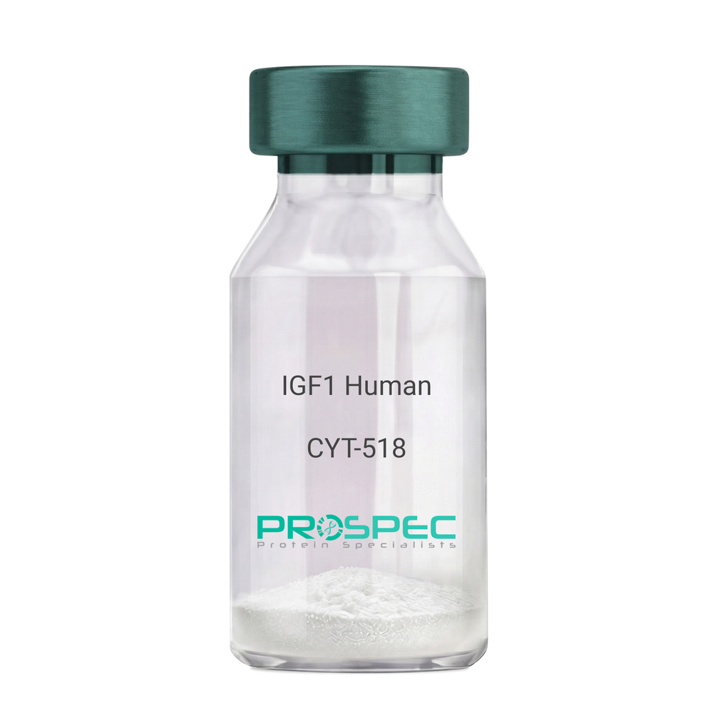
Somatomedin C, IGF-I, IGFI, IGF1, IGF-IA, Mechano growth factor, MGF, Des(1-3), Des1-3, Des 1-3, Des (1-3), IGF-1 (4-70).
Product # :
CYT-518Price :
Quantity :
Shipping Method :
Shipped at Room temp
More Info
- description
- source
- formulation
- purity
- biological activity
- More Info
Description
IGF-I Des(1-3) Human Recombinant produced in E.Coli is a single, non-glycosylated, polypeptide chain containing 67 amino acids (aa 4-70) and having a molecular mass of 7368.5 Dalton. IGF-1 Des1-3 is purified by proprietary chromatographic techniques.
Source
Escherichia Coli.
Formulation
The protein (1mg/ml) was lyophilized was lyophilized from a 0.2µm filtered concentrated solution in 20mM PBS, pH 7.0.
Purity
Greater than 97% as determined by:
(a) Analysis by RP-HPLC.
(b) Analysis by SDS-PAGE.Biological Activity
The ED50 as determined by a cell proliferation assay using FDC-P1 cells is less than 2.0 ng/ml, corresponding to a specific activity of > 5×10⁵ units/mg.More Info
-
Introduction
The somatomedins, or insulin-like growth factors (IGFs), comprise a family of peptides that play important roles in mammalian growth and development. IGF1 mediates many of the growth-promoting effects of growth hormone (GH; MIM 139250). Early studies showed that growth hormone did not directly stimulate the incorporation of sulfate into cartilage, but rather acted through a serum factor, termed 'sulfation factor,' which later became known as 'somatomedin' (Daughaday et al., 1972). Three main somatomedins have been characterized: somatomedin C (IGF1), somatomedin A (IGF2; MIM 147470), and somatomedin B (MIM 193190) (Rotwein, 1986; Rosenfeld, 2003).
-
Synonyms
Somatomedin C, IGF-I, IGFI, IGF1, IGF-IA, Mechano growth factor, MGF, Des(1-3), Des1-3, Des 1-3, Des (1-3), IGF-1 (4-70).
-
Physical Appearance
Sterile Filtered White lyophilized (freeze-dried) powder.
-
Stability
Lyophilized IGF-I des(1-3) although stable at room temperature for 3 weeks, should be stored desiccated below -18°C. Upon reconstitution IGF1 des-1-3 should be stored at 4°C between 2-7 days and for future use below -18°C.For long term storage it is recommended to add a carrier protein (0.1% HSA or BSA).Please prevent freeze-thaw cycles.
-
Solubility
It is recommended to reconstitute the IGF-I des(1-3) in sterile 18M-cm H2O not less than 100µg/ml, which can then be further diluted to other aqueous solutions.
-
Amino Acid Sequence
TLCGAELVDA LQFVCGDRGF YFNKPTGYGS SSRRAPQTGI VDECCFRSCD LRRLEMYCAP LKPAKSA.
-
Background
What is the molecular weight/Mw of IGF1 HUMAN DES1-3 Protein?
IGF1 HUMAN DES1-3 Protein has a total Mw of 7.3kDa.
What is the source or expression system of IGF1 HUMAN DES1-3 Protein?
Escherichia Coli.
What is the Purity of IGF1 HUMAN DES1-3 Protein?
IGF1 HUMAN DES1-3 Protein is >97% pure as determined by SDS-PAGE.
What is the Biological Activity of IGF1 HUMAN DES1-3 Protein?
The ED50 as determined by a cell proliferation assay using FDC-P1 cells is less than 2.0 ng/ml, corresponding to a specific activity of > 5×10⁵ units/mg.
What is the amino acid sequence of IGF1 HUMAN DES1-3 Protein?
TLCGAELVDA LQFVCGDRGF YFNKPTGYGS SSRRAPQTGI VDECCFRSCD LRRLEMYCAP LKPAKSA.
What applications can IGF1 HUMAN DES1-3 Protein be used in?
IGF1 HUMAN DES1-3 Protein can probably be used in western blot, ELISA and Lateral Flow.
What is the endotoxin level for IGF1 HUMAN DES1-3 Protein?
The endotoxin level is minimal, IGF1 HUMAN DES1-3 Protein was purified using conventional chromatography techniques.
ProSpec's products are furnished for LABORATORY RESEARCH USE ONLY. The product may not be used as drugs, agricultural or pesticidal products, food additives or household chemicals.
- View Data Sheet
Name :
SCF MouseDescription:
Stem Cell Factor Mouse Recombinant
Kit ligand Precursor, C-kit ligand, SCF, Mast cell growth factor, MGF, SF, KL-1, Kitl, DKFZp686F2250, Hematopoietic growth factor KL, Steel factor.
Product # :
CYT-275Price :
Quantity :
Shipping Method :
Shipped at Room temp
More Info
- description
- source
- formulation
- purity
- biological activity
- More Info
Description
Stem Cell Factor Mouse Recombinant produced in E.Coli is a single, non-glycosylated polypeptide chain containing 165 amino acids and having a molecular mass of 18309 Dalton. The SCF is purified by proprietary chromatographic techniques.
Source
Escherichia Coli.
Formulation
Lyophilized from a concentrated (1mg/ml) solution in water containing 0.02% NaHCO3.
Purity
Greater than 98.0% as determined by:
(a) Analysis by SEC-HPLC.
(b) Analysis by SDS-PAGE.Biological Activity
The ED50 as determined by the dose-dependant stimulation of Human TF-1 cell line is < 10 ng/ml, corresponding to a Specific Activity of 1x10⁵ IU/mg.More Info
-
Introduction
Stem cell factor / KIT ligand (SCF) is a cytokine which binds CD117(c-Kit). SCF is also known as "steel factor" or "c-kit ligand". SCF exists in two forms, cell surface bound SCF and soluble (or free) SCF. Soluble SCF is produced by the cleavage of surface bound SCF by metalloproteases.
SCF is a growth factor important for the survival, proliferation, and differentiation of hematopoietic stem cells and other hematopoietic progenitor cells. One of its roles is to change the BFU-E (burst-forming unit-erythroid) cells, which are the earliest erythrocyte precursors in the erythrocytic series, into the CFU-E (colony-forming unit-erythroid). -
Synonyms
Kit ligand Precursor, C-kit ligand, SCF, Mast cell growth factor, MGF, SF, KL-1, Kitl, DKFZp686F2250, Hematopoietic growth factor KL, Steel factor.
-
Physical Appearance
Sterile Filtered White lyophilized (freeze-dried) powder.
-
Stability
Lyophilized KIT ligand although stable at room temperature for 3 weeks, should be stored desiccated below -18°C. Upon reconstitution SCF should be stored at 4°C between 2-7 days and for future use below -18°C.For long term storage it is recommended to add a carrier protein (0.1% HSA or BSA).Please prevent freeze-thaw cycles.
-
Solubility
It is recommended to reconstitute the lyophilized Stem Cell Factor in sterile 18MΩ-cm H2O not less than 100µg/ml, which can then be further diluted to other aqueous solutions.
-
Amino Acid Sequence
MKEICGNPVT DNVKDITKLV ANLPNDYMIT LNYVAGMDVL PSHCWLRDMV IQLSLSLTTL LDKFSNISEG LSNYSIIDKL GKIVDDLVLC MEENAPKNIK ESPKRPETRS FTPEEFFSIF NRSIDAFKDF MVASDTSDCV LSSTLGPEKD SRVSVTKPFM LPPVA.
-
Protein content
Protein quantitation was carried out by two independent methods:1. UV spectroscopy at 280 nm using the absorbency value of 0.52 as the extinction coefficient for a 0.1% (1mg/ml) solution. This value is calculated by the PC GENE computer analysis program of protein sequences (IntelliGenetics). 2. Analysis by RP-HPLC, using a standard solution of Stem Cell Factor as a Reference Standard.
ProSpec's products are furnished for LABORATORY RESEARCH USE ONLY. The product may not be used as drugs, agricultural or pesticidal products, food additives or household chemicals.