Search results
542 results found for “cell division cycle”
Name
Description
Product #
Price
Quantity
Shipping Method
- View Data Sheet
Name :
CD105 (27-581) MouseDescription:
Endoglin (27-581) Mouse Recombinant
Endoglin, Cell surface MJ7/18 antigen, CD105, Eng, Edg.
Product # :
CYT-972Price :
Quantity :
Shipping Method :
Shipped with Ice Packs
More Info
- description
- source
- formulation
- purity
- More Info
- Citations
Description
Endoglin Mouse Recombinant produced in Sf9 Baculovirus cells is a single, glycosylated polypeptide chain containing 563 amino acids (27-581 a.a.) and having a molecular mass of 60.9kDa (Migrates at 50-70kDa on SDS-PAGE under reducing conditions).Endoglin is expressed with an 8 amino acid His tag at C-Terminus and purified by proprietary chromatographic techniques.
Source
Sf9, Baculovirus cells.
Formulation
Endoglin protein solution (0.5mg/ml) containing Phosphate Buffered Saline (pH 7.4) and 10% glycerol.
Purity
Greater than 95.0% as determined by SDS-PAGE.
Citations
More Info
-
Introduction
Endoglin is a type I membrane glycoprotein located on cell surfaces and is part of the TGF beta receptor complex.
The Endoglin protein consists of a homodimer of 180 kDA with disulfide links. Endoglin has been found on endothelial cells, activated macrophages, fibroblasts, and smooth muscle cells. Furthermore, Endoglin has been found to be part of the TGF-beta1 receptor complex. Endoglin thus may be involved in the binding of TGF-beta1, TGF-beta3, activin-A, BMP-2, and BMP-7. Beside TGF-beta signaling endoglin may have other functions. It has been postulated that endoglin is involved in the cytoskeletal organization affecting cell morphology and migration. Endoglin has a role in the development of the cardiovascular system and in vascular remodeling. Endoglin expression is regulated during heart development . Experimental mice without the endoglin gene die due to cardiovascular abnormalities. -
Synonyms
Endoglin, Cell surface MJ7/18 antigen, CD105, Eng, Edg.
-
Physical Appearance
Sterile Filtered colorless solution.
-
Stability
Store at 4°C if entire vial will be used within 2-4 weeks. Store, frozen at -20°C for longer periods of time. For long term storage it is recommended to add a carrier protein (0.1% HSA or BSA). Avoid multiple freeze-thaw cycles.
-
Amino Acid Sequence
ERVGCDLQPV DPTRGEVTFT TSQVSEGCVA QAANAVREVH VLFLDFPGML SHLELTLQAS KQNGTETQEV FLVLVSNKNV FVKFQAPEIP LHLAYDSSLV IFQGQPRVNI TVLPSLTSRK QILDWAATKG AITSIAALDD PQSIVLQLGQ DPKAPFLCLP EAHKDMGATL EWQPRAQTPV QSCRLEGVSG HKEAYILRIL PGSEAGPRTV TVMMELSCTS GDAILILHGP PYVSWFIDIN HSMQILTTGE YSVKIFPGSK VKGVELPDTP QGLIAEARKL NASIVTSFVE LPLVSNVSLR ASSCGGVFQT TPAPVVTTPP KDTCSPVLLM SLIQPKCGNQ VMTLALNKKH VQTLQCTITG LTFWDSSCQA EDTDDHLVLS SAYSSCGMKV TAHVVSNEVI ISFPSGSPPL RKKVQCIDMD SLSFQLGLYL SPHFLQASNT IELGQQAFVQ VSVSPLTSEV TVQLDSCHLD LGPEGDMVEL IQSRTAKGSC VTLLSPSPEG DPRFSFLLRV YMVPTPTAGT LSCNLALRPS TLSQEVYKTV SMRLNIVSPD LSGKGLEHHH HHH
-
Background
What is the molecular weight/Mw of CD105 Protein?
CD105 Protein has a total Mw of 60.9kDa.
What is the source or expression system of CD105 Protein?
Sf9, Baculovirus cells.
What is the Purity of CD105 Protein?
CD105 Protein is >95% pure as determined by SDS-PAGE.
What is the Biological Activity of CD105 Protein?
The biological functionality of CD105 Protein will be determined in the future.
What is the amino acid sequence of CD105 Protein?
ERVGCDLQPV DPTRGEVTFT TSQVSEGCVA QAANAVREVH VLFLDFPGML SHLELTLQAS KQNGTETQEV FLVLVSNKNV FVKFQAPEIP LHLAYDSSLV IFQGQPRVNI TVLPSLTSRK QILDWAATKG AITSIAALDD PQSIVLQLGQ DPKAPFLCLP EAHKDMGATL EWQPRAQTPV QSCRLEGVSG HKEAYILRIL PGSEAGPRTV TVMMELSCTS GDAILILHGP PYVSWFIDIN HSMQILTTGE YSVKIFPGSK VKGVELPDTP QGLIAEARKL NASIVTSFVE LPLVSNVSLR ASSCGGVFQT TPAPVVTTPP KDTCSPVLLM SLIQPKCGNQ VMTLALNKKH VQTLQCTITG LTFWDSSCQA EDTDDHLVLS SAYSSCGMKV TAHVVSNEVI ISFPSGSPPL RKKVQCIDMD SLSFQLGLYL SPHFLQASNT IELGQQAFVQ VSVSPLTSEV TVQLDSCHLD LGPEGDMVEL IQSRTAKGSC VTLLSPSPEG DPRFSFLLRV YMVPTPTAGT LSCNLALRPS TLSQEVYKTV SMRLNIVSPD LSGKGLEHHH HHH
What applications can CD105 Protein be used in?
CD105 Protein can probably be used in western blot, ELISA and Lateral Flow.
What is the endotoxin level for CD105 Protein?
The endotoxin level is minimal, CD105 Protein was purified using conventional chromatography techniques.
ProSpec's products are furnished for LABORATORY RESEARCH USE ONLY. The product may not be used as drugs, agricultural or pesticidal products, food additives or household chemicals.
- View Data Sheet
Description:
B-Cell Leukemia/Lymphoma 2 Human Recombinant (–BH2)
Apoptosis regulator Bcl-2, BCL2, B-cell CLL/lymphoma 2, Bcl-2.
Product # :
PRO-632Price :
Quantity :
Shipping Method :
Shipped at Room temp
More Info
- description
- source
- formulation
- purity
- More Info
- Citations
Description
Bcl-2 Des BH2 domain (187-202 residues) Human Recombinant produced in E.Coli is a single, non-glycosylated polypeptide chain containing 202 amino acids 1-186 and 203-218.The Bcl-2 is expressed as His-Tag fusion protein and purified by proprietary chromatographic techniques.
Source
Escherichia Coli.
Formulation
The protein contains 10mM Tris-HCL pH-8, 1mM EDTA and 250mM NaCl.
Purity
Greater than 95.0% as determined by(a) Analysis by RP-HPLC.
(b) Analysis by SDS-PAGE.Citations
More Info
-
Introduction
BCL2 gene encodes an integral outer mitochondrial membrane protein that blocks the apoptotic death of some cells such as lymphocytes. Constitutive expression of BCL2, such as in the case of translocation of BCL2 to Ig heavy chain locus, is thought to be the cause of follicular lymphoma. Two transcript variants, produced by alternate splicing, differ in their C-terminal ends.
-
Synonyms
Apoptosis regulator Bcl-2, BCL2, B-cell CLL/lymphoma 2, Bcl-2.
-
Physical Appearance
Sterile Filtered White lyophilized (freeze-dried) powder.
-
Stability
Lyophilized Bcl-2 although stable at room temperature for 3 weeks, should be stored desiccated below -18°C. Upon reconstitution Bcl-2 should be stored at 4°C between 2-7 days and for future use below -18°C.For long term storage it is recommended to add a carrier protein (0.1% HSA or BSA).Please prevent freeze-thaw cycles.
-
Solubility
Suspend Bcl-2 in 100?l of 0.5M Acetic acid, over night at 4°C.
Dilute 10 fold into selected buffer system.
BCL-2 has tendency to form intramolecular disulfide bond, 5mM DTT is recommended in assay buffer. When running SDS-PAGE gel, 10mM DTT is recommended. -
Applications
Input marker or positive control (Western Blotting).
Function study.
ProSpec's products are furnished for LABORATORY RESEARCH USE ONLY. The product may not be used as drugs, agricultural or pesticidal products, food additives or household chemicals.
- View Data Sheet
Name :
CD72 HumanDescription:
CD72 Human Recombinant
B-cell differentiation antigen CD72, Lyb-2, CD72, LYB2, CD72b.
Product # :
PRO-486Price :
Quantity :
Shipping Method :
Shipped with Ice Packs
More Info
- description
- source
- formulation
- More Info
- Citations
Description
CD72 Human Recombinant (aa 1-98) expressed in E.coli, shows a 38 kDa band on SDS-PAGE. The CD72 is purified by proprietary chromatographic techniques.
Source
Escherichia Coli.
Formulation
CD72 at 100µg/ml in 50mM Tris-HCl, pH7.5, 10mM L-glutathione (reduced).
Citations
More Info
-
Introduction
CD72 antigen is a member of the type II integral membrane glycoproteins which includes other related cell surface moleculaes such as the asialoglycoprotein receptors, CD23 and the Kupffer cell recpeotr. The function of CD72 is unknown but the exposure of B cells to CD72 antibodies activates a variety of signaling pathways and can induce MHC class II expression and B cell proliferation. CD72 antigen is expressed on all cells of B cell lineage with the exception of plasma cells and weakly on human tissue macrophages.
-
Synonyms
B-cell differentiation antigen CD72, Lyb-2, CD72, LYB2, CD72b.
-
Physical Appearance
Sterile Filtered clear solution.
-
Stability
Store vial at -20°C to -80°C. When stored at the recommended temperature, this protein is stable for 12 months.Please prevent freeze-thaw cycles.
-
Applications
• ELISA.
• Inhibition Assays.
• Western Blotting.
ProSpec's products are furnished for LABORATORY RESEARCH USE ONLY. The product may not be used as drugs, agricultural or pesticidal products, food additives or household chemicals.
- View Data Sheet
Name :
CD3G Human, Sf9Description:
CD3G Human Recombinant, Sf9
T-cell surface glycoprotein CD3 gamma chain, CD3-GAMMA, T3G, CD3G
Product # :
PRO-2412Price :
Quantity :
Shipping Method :
Shipped with Ice Packs
More Info
- description
- source
- formulation
- purity
- More Info
- Citations
Description
CD3G Human Recombinant produced in Sf9 Insect cells is a single, glycosylated polypeptide chain containing 103 amino acids (23-116 a.a.) and having a molecular mass of 11.8kDa.CD3G is expressed with a 6 amino acids His tag at C-Terminus and purified by proprietary chromatographic techniques.
Source
Sf9, Insect cells.
Formulation
CD3G protein solution (0.5mg/ml) contains phosphate buffered saline (pH7.4) and 10% glycerol.
Purity
Greater than 85.0% as determined by SDS-PAGE.
Citations
More Info
-
Introduction
CD3-gamma polypeptide is encoded by CD3 Gamma (CD3G) which together with CD3-epsilon, -delta and -zeta, and the T-cell receptor alpha/beta and gamma/delta heterodimers, configures the T-cell receptor-CD3 complex. The T-cell receptor-CD3 complex takes part in coupling antigen recognition to numerous intracellular signal-transduction pathways. The genes encoding the epsilon, gamma and delta polypeptides are located in the same cluster on chromosome 11. Deficiencies in this gene are associated with T cell immunodeficiency.
-
Synonyms
T-cell surface glycoprotein CD3 gamma chain, CD3-GAMMA, T3G, CD3G
-
Physical Appearance
Sterile Filtered clear solution.
-
Stability
Store at 4°C if entire vial will be used within 2-4 weeks. Store, frozen at -20°C for longer periods of time. For long term storage it is recommended to add a carrier protein (0.1% HSA or BSA).Avoid multiple freeze-thaw cycles.
-
Amino Acid Sequence
ADPQSIKGNH LVKVYDYQED GSVLLTCDAE AKNITWFKDG KMIGFLTEDK KKWNLGSNAK DPRGMYQCKG SQNKSKPLQV YYRMCQNCIE LNAATISHHH HHH.
ProSpec's products are furnished for LABORATORY RESEARCH USE ONLY. The product may not be used as drugs, agricultural or pesticidal products, food additives or household chemicals.
- View Data Sheet
Name :
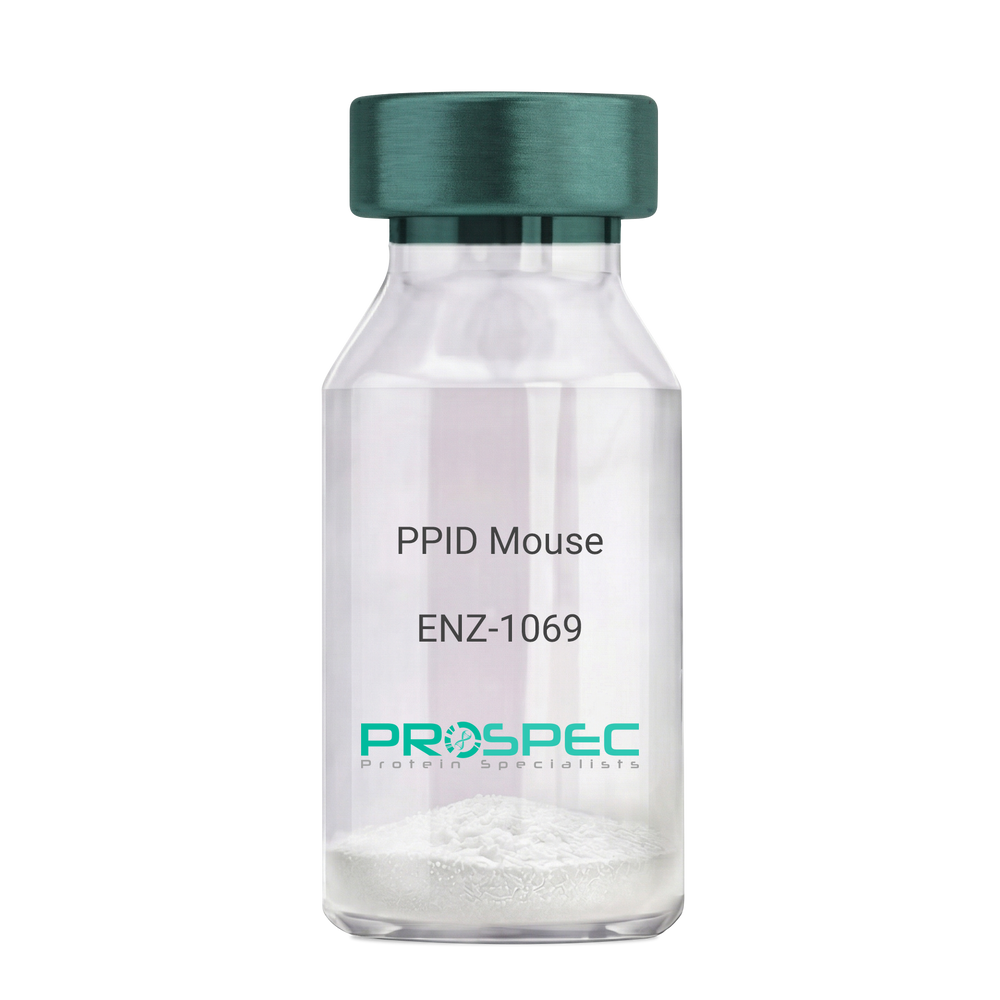
PPID MouseDescription:
Peptidylprolyl Isomerase D Mouse Recombinant
Peptidyl-prolyl cis-trans isomerase D, PPIase D, 40 kDa peptidyl-prolyl cis-trans isomerase, Cyclophilin-40, CYP-40, Cyclophilin-related protein, CYP40, CYPD, PPID, Peptidylprolyl Isomerase D.
Product # :
ENZ-1069Price :
Quantity :
Shipping Method :
Shipped with Ice Packs
More Info
- description
- source
- formulation
- purity
- biological activity
- More Info
- Citations
Description
PPID Mouse Recombinant produced in E.Coli is a single, non-glycosylated polypeptide chain containing 395 amino acids (1-370a.a.) and having a molecular mass of 43.4kDa. PPID is fused to a 25 amino acid His-tag at N-terminus & purified by proprietary chromatographic techniques.
Source
Escherichia Coli.
Formulation
PPID protein solution (1mg/ml) containing 20mM Tris-Hcl buffer (pH8.0), 10% glycerol and 1mM DTT.
Purity
Greater than 90.0% as determined by SDS-PAGE.
Biological Activity
Specific activity is > 700nmol/min/mg, and is defined as the amount of enzyme that cleaves 1umole of suc-AAFP-PNA per minute at 37°C in Tris–HCl pH 8.0 using chymotrypsin.
Citations
More Info
-
Introduction
Cyclophilin-D is a member of the peptidyl-prolyl cis-trans isomerase (PPIase) family. PPIases catalyze the cis-trans isomerization of proline imidic peptide bonds in oligopeptides and speeds up the protein folding. Cyclophilin-D possess PPIase activity and binds to the immunosuppressant cyclosporin-A. Cyclophilin-D is very well known that its overexpression suppresses the apoptosis in cancer cell. Cyclophilin-D suppresses apoptotic cell death by the use of mitochondrial hexokinase-2 dependent mechanism in cancer cells.
-
Synonyms
Peptidyl-prolyl cis-trans isomerase D, PPIase D, 40 kDa peptidyl-prolyl cis-trans isomerase, Cyclophilin-40, CYP-40, Cyclophilin-related protein, CYP40, CYPD, PPID, Peptidylprolyl Isomerase D.
-
Physical Appearance
Sterile Filtered colorless solution.
-
Stability
Store at 4°C if entire vial will be used within 2-4 weeks. Store, frozen at -20°C for longer periods of time. For long term storage it is recommended to add a carrier protein (0.1% HSA or BSA). Please prevent freeze-thaw cycles.
-
Amino Acid Sequence
MGSSHHHHHH SSGLVPRGSH MGSEFMSHAS PAAKPSNSKN PRVFFDVDIG GERVGRIVLE LFADIVPKTA ENFRALCTGE KGTGSTTGKP LHFKGCPFHR IIKKFMIQGG DFSNQNGTGG ESIYGEKFED ENFHYKHDRE GLLSMANAGP NTNGSQFFIT TVPTPHLDGK HVVFGQVIKG LGVARTLENV EVNGEKPAKL CVIAECGELK EGDDWGIFPK DGSGDSHPDF PEDADIDLKD VDKILLISED LKNIGNTFFK SQNWEMAIKK YAKVLRYVDS SKAVIEKADR SRLQPIALSC VLNIGACKLK MSNWQGAIDS CLEALEMDPS NTKALYRKAQ GWQGLKEYDQ ALADLKKAQE IAPGDKAIQA ELLKVKQMIK AQKDKEKAVY AKMFA.
ProSpec's products are furnished for LABORATORY RESEARCH USE ONLY. The product may not be used as drugs, agricultural or pesticidal products, food additives or household chemicals.
- View Data Sheet
Name :
UBA3 HumanDescription:
Ubiquitin-Like Modifier Activating Enzyme 3 Human Recombinant
NEDD8-activating enzyme E1 catalytic subunit, NEDD8-activating enzyme E1C, Ubiquitin-activating enzyme E1C, Ubiquitin-like modifier-activating enzyme 3, Ubiquitin-activating enzyme 3, UBA3, UBE1C, hUBA3.
Product # :
ENZ-576Price :
Quantity :
Shipping Method :
Shipped with Ice Packs
More Info
- description
- source
- formulation
- purity
- More Info
- Citations
Description
UBA3 Human Recombinant produced in E.coli is a single, non-glycosylated polypeptide chain containing 487 amino acids (1-463) and having a molecular mass of 54.4kDa.UBA3 is fused to a 24 amino acid His-tag at N-terminus & purified by proprietary chromatographic techniques.
Source
Escherichia Coli.
Formulation
The UBA3 solution (1mg/ml) contains 20mM Tris-HCl buffer (pH8.0), 20% glycerol and 1mM DTT.
Purity
Greater than 90.0% as determined by SDS-PAGE.
Citations
More Info
-
Introduction
NEDD8-activating enzyme E1 catalytic subunit (UBA3) is the catalytic subunit of the dimeric UBA3-NAE1 E1 enzyme, which belongs to the E1 ubiquitin-activating enzyme family. E1 activates NEDD8 by initially adenylating its C-terminal glycine residue with ATP, afterwards linking this residue to the side chain of the catalytic cysteine, generating a NEDD8-UBA3 thioester and free AMP. E1 at last transfers NEDD8 to the catalytic cysteine of UBE2M. The UBA3 enzyme connects with AppBp1, an amyloid beta precursor protein binding protein, to form a heterodimer, and at that point the enzyme complex activates NEDD8, a ubiquitin-like protein, which controls cell division, signaling and embryogenesis.
-
Synonyms
NEDD8-activating enzyme E1 catalytic subunit, NEDD8-activating enzyme E1C, Ubiquitin-activating enzyme E1C, Ubiquitin-like modifier-activating enzyme 3, Ubiquitin-activating enzyme 3, UBA3, UBE1C, hUBA3.
-
Physical Appearance
Sterile filtered colorless solution.
-
Stability
Store at 4°C if entire vial will be used within 2-4 weeks. Store, frozen at -20°C for longer periods of time. For long term storage it is recommended to add a carrier protein (0.1% HSA or BSA).Avoid multiple freeze-thaw cycles.
-
Amino Acid Sequence
MGSSHHHHHH SSGLVPRGSH MGSHMADGEE PERKRRRIEE LLAEKMAVDG GCGDTGDWEG RWNHVKKFLE RSGPFTHPDF EPSTESLQFL LDTCKVLVIG AGGLGCELLK NLALSGFRQI HVIDMDTIDV SNLNRQFLFR PKDIGRPKAE VAAEFLNDRV PNCNVVPHFN KIQDFNDTFY RQFHIIVCGL DSIIARRWIN GMLISLLNYE DGVLDPSSIV PLIDGGTEGF KGNARVILPG MTACIECTLE LYPPQVNFPM CTIASMPRLP EHCIEYVRML QWPKEQPFGE GVPLDGDDPE HIQWIFQKSL ERASQYNIRG VTYRLTQGVV KRIIPAVAST NAVIAAVCAT EVFKIATSAY IPLNNYLVFN DVDGLYTYTF EAERKENCPA CSQLPQNIQF SPSAKLQEVL DYLTNSASLQ MKSPAITATL EGKNRTLYLQ SVTSIEERTR PNLSKTLKEL GLVDGQELAV ADVTTPQTVL FKLHFTS.
ProSpec's products are furnished for LABORATORY RESEARCH USE ONLY. The product may not be used as drugs, agricultural or pesticidal products, food additives or household chemicals.
- View Data Sheet
Name :
CD7 Human, Sf9Description:
CD7 Human Recombinant, Sf9
T-cell antigen CD7, GP40, LEU-9, Tp40, TP41, CD7, T-cell leukemia antigen, T-cell surface antigen Leu-9.
Product # :
PRO-2420Price :
Quantity :
Shipping Method :
Shipped with Ice Packs
More Info
- description
- source
- formulation
- purity
- More Info
- Citations
Description
CD7 Human Recombinant produced in Sf9 Baculovirus cells is a single, glycosylated polypeptide chain containing 164 amino acids (26-180a.a.) and having a molecular mass of 17.5kDa (Molecular size on SDS-PAGE will appear at approximately 18-28kDa). CD7 is expressed with a 6 amino acids His tag at C-Terminus and purified by proprietary chromatographic techniques.
Source
Sf9, Baculovirus cells.
Formulation
CD7 protein solution (1mg/ml) contains Phosphate Buffered Saline (pH 7.4) and 10% glycerol.
Purity
Greater than 95.0% as determined by SDS-PAGE.
Citations
More Info
-
Introduction
T-cell antigen CD7 (CD7) is a type I transmembrane glycoprotein which is expressed on pluripotential hemapoietic cells, nearly all human thymocytes and some peripheral blood T cells. The CD7 molecule is missing in a subset of human T cells under some physiologic conditions. An increase in CD7 negative T cells can be observed in a number of inflammatory skin diseases.
-
Synonyms
T-cell antigen CD7, GP40, LEU-9, Tp40, TP41, CD7, T-cell leukemia antigen, T-cell surface antigen Leu-9.
-
Physical Appearance
Sterile filtered colorless solution.
-
Stability
Store at 4°C if entire vial will be used within 2-4 weeks. Store, frozen at -20°C for longer periods of time. For long term storage it is recommended to add a carrier protein (0.1% HSA or BSA).Avoid multiple freeze-thaw cycles.
-
Amino Acid Sequence
ADLAQEVQQS PHCTTVPVGA SVNITCSTSG GLRGIYLRQL GPQPQDIIYY EDGVVPTTDR RFRGRIDFSG SQDNLTITMH RLQLSDTGTY TCQAITEVNV YGSGTLVLVT EEQSQGWHRC SDAPPRASAL PAPPTGSALP DPQTASALPD PPAASALPHH HHHH.
ProSpec's products are furnished for LABORATORY RESEARCH USE ONLY. The product may not be used as drugs, agricultural or pesticidal products, food additives or household chemicals.
- View Data Sheet
Name :
CD4 FITC AntibodyDescription:
CD4-FITC, Mouse Anti-Human
gp55, HLA-2, L3 / T4, Ly-4, T cell antigen T4/LEU3, T4, sCD4, CD4mut.
Product # :
ANT-132Price :
Quantity :
Shipping Method :
Shipped at Room temp
More Info
- formulation
- More Info
- Citations
Formulation
1mg/ml in PBS (after reconstitution).
Citations
More Info
-
Introduction
CD4 is a cell-surface glycoprotein found on the mature helper T cells and immature thymocytes, as well as on monocytes and macrophages. (Some cytotoxic T cells have CD4 protein as well.) Normally, about 65% of T cells in the blood are CD4+ (have CD4 protein protruding from their membrane). A mature T cell with either have CD4 or CD8, but not both. During one stage of development T cells develop CD4 and CD8 receptors, but they eventually are differentiated in the thymus and become more specialized.
-
Synonyms
gp55, HLA-2, L3 / T4, Ly-4, T cell antigen T4/LEU3, T4, sCD4, CD4mut.
-
Solubility
Reconstitute with of H20. Mix gently, wash the sides of the vial and wait 30-60 seconds before use.
-
Immunogen
Purified human PBL CD4+ T cells.
-
Ig Subclass
Mouse IgG2b.
-
Clone
hCD4.
-
Applications
Blocking and staining antibody. Blocks blinding of HIV to CD4. For staining, use 10 ml/1,000,000 cells. Titer for blocking T cell activation should be determined by the investigator.
-
Available Conjugates
This antibody is also available conjugated to biotin.
-
Type
Mouse Anti Human Monoclonal.
-
Storage Procedures
Lyophilized: store at 4oC. After reconstitution, if not intended for use within a month, aliquot and store at -20oC.
-
Purification Method
Ion exchange column.
ProSpec's products are furnished for LABORATORY RESEARCH USE ONLY. The product may not be used as drugs, agricultural or pesticidal products, food additives or household chemicals.
- View Data Sheet
Name :
FTSJ2 HumanDescription:
FtsJ RNA Methyltransferase Homolog 2 Human Recombinant
FtsJ RNA methyltransferase homolog 2 (E. coli), putative ribosomal RNA methyltransferase 2, MRM2 RNA methyltransferase homolog, rRNA (uridine-2'-O-)-methyltransferase, cell division protein FtsJ, Protein ftsJ homolog 2, MRM2, FJH1, EC 2.1.1.-.
Product # :
PRO-1207Price :
Quantity :
Shipping Method :
Shipped with Ice Packs
More Info
- description
- source
- formulation
- purity
- More Info
- Citations
Description
FTSJ2 Human Recombinant produced in E. coli is a single polypeptide chain containing 219 amino acids (51-246) and having a molecular mass of 24.1 kDa.FTSJ2 is fused to a 23 amino acid His-tag at N-terminus & purified by proprietary chromatographic techniques.
Source
E.coli.
Formulation
The FTSJ2 solution (1mg/1ml) contains 20mM Tris-HCl buffer (pH 8.0), 0.15M NaCl, 1mM DTT and 30% glycerol.
Purity
Greater than 95% as determined by SDS-PAGE.
Citations
More Info
-
Introduction
FtsJ RNA Methyltransferase Homolog 2 (FTSJ2) belongs to the S-adenosylmethionine-binding protein family. FTSJ2 is a nucleolar protein and which is involved in cell cycle control and DNA repair and the processing and modification of rRNA. FTSJ2 is extensively expressed, with the highest expression in the muscle, placenta, and heart.
-
Synonyms
FtsJ RNA methyltransferase homolog 2 (E. coli), putative ribosomal RNA methyltransferase 2, MRM2 RNA methyltransferase homolog, rRNA (uridine-2'-O-)-methyltransferase, cell division protein FtsJ, Protein ftsJ homolog 2, MRM2, FJH1, EC 2.1.1.-.
-
Physical Appearance
Sterile Filtered colorless solution.
-
Stability
Store at 4°C if entire vial will be used within 2-4 weeks. Store, frozen at -20°C for longer periods of time. For long term storage it is recommended to add a carrier protein (0.1% HSA or BSA).Avoid multiple freeze-thaw cycles.
-
Amino Acid Sequence
MGSSHHHHHH SSGLVPRGSH MGSSYRCRSA FKLLEVNERH QILRPGLRVL DCGAAPGAWS QVAVQKVNAA GTDPSSPVGF VLGVDLLHIF PLEGATFLCP ADVTDPRTSQ RILEVLPGRR ADVILSDMAP NATGFRDLDH DRLISLCLTL LSVTPDILQP GGTFLCKTWA GSQSRRLQRR LTEEFQNVRI IKPEASRKES SEVYFLATQY HGRKGTVKQ
ProSpec's products are furnished for LABORATORY RESEARCH USE ONLY. The product may not be used as drugs, agricultural or pesticidal products, food additives or household chemicals.
- View Data Sheet
Name :
PDCD1 Human, ActiveDescription:
Programmed Cell Death 1 Human Recombinant, Active
Programmed cell death protein 1, CD279, hPD-1, SLEB2, hPD-l, hSLE1, PD-1, PD1, PDCD1.
Product # :
PRO-2724Price :
Quantity :
Shipping Method :
Shipped with Ice Packs
More Info
- description
- source
- formulation
- purity
- biological activity
- More Info
- Citations
Description
PDCD1 Human Recombinant produced in HEK293 Cells is a single, glycosylated polypeptide chain containing 383 amino acids (21-170 a.a) and having a molecular mass of 42.9kDa.PDCD1 is fused to a 233 amino acid hIgG-His-Tag at C-terminus & purified by proprietary chromatographic techniques.
Source
HEK293 Cells.
Formulation
The PDCD1 solution (0.5mg/1ml) contains phosphate buffered saline (pH7.4) and 10% glycerol.
Purity
Greater than 95.0% as determined by SDS-PAGE.
Biological Activity
Measured by its binding ability in a functional ELISA with Human PD-L1/B7-H1
Citations
More Info
-
Introduction
PDCD1 is a cell surface membrane protein of the immunoglobulin superfamily. PDCD1 is expressed in pro-B-cells and is thought to have a role in their differentiation. In mice, expression of PDCD1 gene is induced in the thymus when anti-CD3 antibodies are injected and large numbers of thymocytes undergo apoptosis. Mice deficient for the PDCD1 gene bred on a BALB/c background developed dilated cardiomyopathy and died from congestive heart failure. These studies suggest that the PDCD1 gene product may also be important in T cell function and contribute to the prevention of autoimmune diseases.
-
Synonyms
Programmed cell death protein 1, CD279, hPD-1, SLEB2, hPD-l, hSLE1, PD-1, PD1, PDCD1.
-
Physical Appearance
Sterile filtered colorless solution.
-
Stability
Store at 4°C if entire vial will be used within 2-4 weeks.Store, frozen at -20°C for longer periods of time.For long term storage it is recommended to add a carrier protein (0.1% HSA or BSA).Avoid multiple freeze-thaw cycles.
-
Amino Acid Sequence
PGWFLDSPDR PWNPPTFSPA LLVVTEGDNA TFTCSFSNTS ESFVLNWYRM SPSNQTDKLA AFPEDRSQPG QDCRFRVTQL PNGRDFHMSV VRARRNDSGT YLCGAISLAP KAQIKESLRA ELRVTERRAE VPTAHPSPSP RPAGQFQTLV LEPKSCDKTH TCPPCPAPEL LGGPSVFLFP PKPKDTLMIS RTPEVTCVVV DVSHEDPEVK FNWYVDGVEV HNAKTKPREE QYNSTYRVVS VLTVLHQDWL NGKEYKCKVS NKALPAPIEK TISKAKGQPR EPQVYTLPPS RDELTKNQVS LTCLVKGFYP SDIAVEWESN GQPENNYKTT PPVLDSDGSF FLYSKLTVDK SRWQQGNVFS CSVMHEALHN HYTQKSLSLS PGK
ProSpec's products are furnished for LABORATORY RESEARCH USE ONLY. The product may not be used as drugs, agricultural or pesticidal products, food additives or household chemicals.
- View Data Sheet
Name :
TYMP HumanDescription:
Thymidine Phosphorylase Human Recombinant
Thymidine phosphorylase, Gliostatin, Platelet-derived endothelial cell growth factor, PD-ECGF, TdRPase, TYMP, ECGF1, TP, MNGIE, MEDPS1, MTDPS1, PDECGF, hPD-ECGF.
Product # :
ENZ-005Price :
Quantity :
Shipping Method :
Shipped with Ice Packs
More Info
- description
- source
- formulation
- purity
- More Info
- Citations
Description
TYMP Human Recombinant fused with a 21 amino acid His tag at N-terminus produced in E.Coli is a single, non-glycosylated, polypeptide chain containing 493 amino acids (11-482 a.a.) and having a molecular mass of 51.3kDa. The TYMP is purified by proprietary chromatographic techniques.
Source
Escherichia Coli.
Formulation
The TYMP solution (1mg/ml) contains 20mM Tris-HCl buffer (pH 8.0), 1mM DTT and 10% glycerol.
Purity
Greater than 90.0% as determined by SDS-PAGE.
Citations
More Info
-
Introduction
Thymidine phosphorylase precursor (TYMP) is a platelet-derived endothelial cell growth factor that catalyzes the formation of thymine and 2-deoxy-D-ribose-1-phosphate from thymidine and orthophosphate. TYMP is an angiogenic inducer that potently stimulates the growth of endothelial cells and induces chemotaxis. TYMP has a highly restricted target cell specificity acting only on endothelial cells. An increased expression of TYMP is found in a broad array of different solid tumors and inflammatory diseases and is frequently associated with poor prognosis. Mutations in the TYMP gene are linked to mitochondrial neurogastrointestinal encephalomyopathy.
-
Synonyms
Thymidine phosphorylase, Gliostatin, Platelet-derived endothelial cell growth factor, PD-ECGF, TdRPase, TYMP, ECGF1, TP, MNGIE, MEDPS1, MTDPS1, PDECGF, hPD-ECGF.
-
Physical Appearance
Sterile Filtered colorless solution.
-
Stability
Store at 4°C if entire vial will be used within 2-4 weeks. Store, frozen at -20°C for longer periods of time. For long term storage it is recommended to add a carrier protein (0.1% HSA or BSA).Avoid multiple freeze-thaw cycles.
-
Amino Acid Sequence
MGSSHHHHHH SSGLVPRGSH MAPPAPGDFS GEGSQGLPDP SPEPKQLPEL IRMKRDGGRL SEADIRGFVA AVVNGSAQGA QIGAMLMAIR LRGMDLEETS VLTQALAQSG QQLEWPEAWR QQLVDKHSTG GVGDKVSLVL APALAACGCK VPMISGRGLG HTGGTLDKLE SIPGFNVIQS PEQMQVLLDQ AGCCIVGQSE QLVPADGILY AARDVTATVD SLPLITASIL SKKLVEGLSA LVVDVKFGGA AVFPNQEQAR ELAKTLVGVG ASLGLRVAAA LTAMDKPLGR CVGHALEVEE ALLCMDGAGP PDLRDLVTTL GGALLWLSGH AGTQAQGAAR VAAALDDGSA LGRFERMLAA QGVDPGLARA LCSGSPAERR QLLPRAREQE ELLAPADGTV ELVRALPLAL VLHELGAGRS RAGEPLRLGV GAELLVDVGQ RLRRGTPWLR VHRDGPALSG PQSRALQEAL VLSDRAPFAA PSPFAELVLP PQQ.
ProSpec's products are furnished for LABORATORY RESEARCH USE ONLY. The product may not be used as drugs, agricultural or pesticidal products, food additives or household chemicals.
- View Data Sheet
Name :
SDC1 HumanDescription:
Syndecan-1 Human Recombinant
Heparan Sulfate proteoglycan fibroblast growth factor receptor, syndecan proteoglycan 1, Syndecan 1, SYND1, CD138 antigen, SDC.
Product # :
PRO-1065Price :
Quantity :
Shipping Method :
Shipped with Ice Packs
More Info
- description
- source
- formulation
- purity
- More Info
- Citations
Description
SDC1 Human Recombinant produced in E. coli is a single polypeptide chain containing 262 amino acids (18-254) and having a molecular mass of 27kDa (molecular size on SDS-PAGE will appear higher).SDC1 is fused to a 25 amino acid His-tag at N-terminus & purified by proprietary chromatographic techniques.
Source
E.coli.
Formulation
The SDC1 solution (1mg/1ml) contains 20mM Tris-HCl buffer (pH 8.0), 100mM Nacl and 10% glycerol.
Purity
Greater than 90% as determined by SDS-PAGE.
Citations
More Info
-
Introduction
CD138 takes a part in cell adhesion. CD138 is expressed on the surface of pre-B cells and plasma cells but is absent from mature B cells. SDC1 is a selective marker for B cell lymphoblastic leukemia and lymphoplasmacytoid leukemia. SDC1 is missing from the apoptotic myeloma cells; therefore is a valuable marker for plasma cell and myeloma cells.
-
Synonyms
Heparan Sulfate proteoglycan fibroblast growth factor receptor, syndecan proteoglycan 1, Syndecan 1, SYND1, CD138 antigen, SDC.
-
Physical Appearance
Sterile Filtered colorless solution.
-
Stability
Store at 4°C if entire vial will be used within 2-4 weeks. Store, frozen at -20°C for longer periods of time. For long term storage it is recommended to add a carrier protein (0.1% HSA or BSA).Avoid multiple freeze-thaw cycles.
-
Amino Acid Sequence
MGSSHHHHHH SSGLVPRGSH MGSHMQPALP QIVATNLPPE DQDGSGDDSD NFSGSGAGAL QDITLSQQTP STWKDTQLLT AIPTSPEPTG LEATAASTST LPAGEGPKEG EAVVLPEVEP GLTAREQEAT PRPRETTQLP TTHQASTTTA TTAQEPATSH PHRDMQPGHH ETSTPAGPSQ ADLHTPHTED GGPSATERAA EDGASSQLPA AEGSGEQDFT FETSGENTAV VAVEPDRRNQ SPVDQGATGA SQGLLDRKEV LG.
ProSpec's products are furnished for LABORATORY RESEARCH USE ONLY. The product may not be used as drugs, agricultural or pesticidal products, food additives or household chemicals.
- View Data Sheet
Description:
B-Cell Leukemia/Lymphoma 2 Human Recombinant (–BH1)
Apoptosis regulator Bcl-2, BCL2, B-cell CLL/lymphoma 2, Bcl-2.
Product # :
PRO-631Price :
Quantity :
Shipping Method :
Shipped at Room temp
More Info
- description
- source
- formulation
- purity
- More Info
- Citations
Description
Bcl-2 Des BH1 domain (136-155 residues) Human Recombinant produced in E.Coli is a single, non-glycosylated polypeptide chain containing 198 amino acids 1-135 and 156-218.The Bcl-2 is expressed as His-Tag fusion protein and purified by proprietary chromatographic techniques.
Source
Escherichia Coli.
Formulation
The protein contains 10mM Tris-HCL pH-8, 1mM EDTA and 250mM NaCl.
Purity
Greater than 95.0% as determined by(a) Analysis by RP-HPLC.
(b) Analysis by SDS-PAGE.Citations
More Info
-
Introduction
BCL2 gene encodes an integral outer mitochondrial membrane protein that blocks the apoptotic death of some cells such as lymphocytes. Constitutive expression of BCL2, such as in the case of translocation of BCL2 to Ig heavy chain locus, is thought to be the cause of follicular lymphoma. Two transcript variants, produced by alternate splicing, differ in their C-terminal ends.
-
Synonyms
Apoptosis regulator Bcl-2, BCL2, B-cell CLL/lymphoma 2, Bcl-2.
-
Physical Appearance
Sterile Filtered White lyophilized (freeze-dried) powder.
-
Stability
Lyophilized Bcl-2 although stable at room temperature for 3 weeks, should be stored desiccated below -18°C. Upon reconstitution Bcl-2 should be stored at 4°C between 2-7 days and for future use below -18°C.For long term storage it is recommended to add a carrier protein (0.1% HSA or BSA).Please prevent freeze-thaw cycles.
-
Solubility
Suspend Bcl-2 in 100?l of 0.5M Acetic acid, over night at 4°C.
Dilute 10 fold into selected buffer system.
BCL-2 has tendency to form intramolecular disulfide bond, 5mM DTT is recommended in assay buffer. When running SDS-PAGE gel, 10mM DTT is recommended. -
Applications
Input marker or positive control (Western Blotting).
Function study.
ProSpec's products are furnished for LABORATORY RESEARCH USE ONLY. The product may not be used as drugs, agricultural or pesticidal products, food additives or household chemicals.
- View Data Sheet
Name :
CD274 Human, Sf9Description:
CD274 Human Recombinant, Sf9
CD274 molecule, CD274 antigen, programmed cell death 1 ligand 1, PD-L1, PDCD1L1, PDCD1LG1, PDCD1 ligand 1, B7 homolog 1, B7H1.
Product # :
PRO-2453Price :
Quantity :
Shipping Method :
Shipped with Ice Packs
More Info
- description
- source
- formulation
- purity
- More Info
- Citations
Description
CD274 Human Recombinant produced in Sf9 Baculovirus cells is a single, glycosylated polypeptide chain containing 462 amino acids (19-238a.a.) and having a molecular mass of 52.5kDa (Molecular size on SDS-PAGE will appear at approximately 50-70kDa). CD274 is expressed with a 239 amino acids hIgG-His tag at C-Terminus and purified by proprietary chromatographic techniques.
Source
Sf9, Baculovirus cells.
Formulation
CD274 protein solution (1mg/ml) contains Phosphate Buffered Saline (pH 7.4) and 10% glycerol.
Purity
Greater than 95% as determined by SDS-PAGE.
Citations
More Info
-
Introduction
CD274 takes part in the costimulatory signal, vital for T-cell proliferation and creation of IL10 and IFNG, in a PDCD1-independent and an IL2-dependent manner. CD274 collaboration with PDCD1 inhibits cytokine production and T-cell proliferation.
-
Synonyms
CD274 molecule, CD274 antigen, programmed cell death 1 ligand 1, PD-L1, PDCD1L1, PDCD1LG1, PDCD1 ligand 1, B7 homolog 1, B7H1.
-
Physical Appearance
Sterile Filtered colorless solution.
-
Stability
Store at 4°C if entire vial will be used within 2-4 weeks. Store, frozen at -20°C for longer periods of time. For long term storage it is recommended to add a carrier protein (0.1% HSA or BSA).Avoid multiple freeze-thaw cycles.
-
Amino Acid Sequence
ADLFTVTVPK DLYVVEYGSN MTIECKFPVE KQLDLAALIV YWEMEDKNII QFVHGEEDLK VQHSSYRQRA RLLKDQLSLG NAALQITDVK LQDAGVYRCM ISYGGADYKR ITVKVNAPYN KINQRILVVD PVTSEHELTC QAEGYPKAEV IWTSSDHQVL SGKTTTTNSK REEKLFNVTS TLRINTTTNE IFYCTFRRLD PEENHTAELV IPELPLAHPP NERLEPKSCD KTHTCPPCPA PELLGGPSVF LFPPKPKDTL MISRTPEVTC VVVDVSHEDP EVKFNWYVDG VEVHNAKTKP REEQYNSTYR VVSVLTVLHQ DWLNGKEYKC KVSNKALPAP IEKTISKAKG QPREPQVYTL PPSRDELTKN QVSLTCLVKG FYPSDIAVEW ESNGQPENNY KTTPPVLDSD GSFFLYSKLT VDKSRWQQGN VFSCSVMHEA LHNHYTQKSL SLSPGKHHHH HH.
ProSpec's products are furnished for LABORATORY RESEARCH USE ONLY. The product may not be used as drugs, agricultural or pesticidal products, food additives or household chemicals.
- View Data Sheet
Name :
CD72 Human, Sf9Description:
CD72 Human Recombinant, Sf9
CD72, CD72b, LYB2, B-cell differentiation antigen CD72, Lyb-2.
Product # :
PRO-2608Price :
Quantity :
Shipping Method :
Shipped with Ice Packs
More Info
- description
- source
- formulation
- purity
- More Info
- Citations
Description
CD72 Human Recombinant produced in Sf9 Baculovirus cells is a single, glycosylated polypeptide chain containing 485 amino acids (117-359a.a.) and having a molecular mass of 55.3kDa (Molecular size on SDS-PAGE will appear at approximately 50-70kDa).CD72 is expressed with a 242 amino acid hIgG-His tag at C-Terminus and purified by proprietary chromatographic techniques.
Source
Sf9, Baculovirus cells.
Formulation
CD72 protein solution (0.5mg/ml) contains 50mM Tris-HCl buffer (pH6.8), 0.2M NaCl, 2mM DTT and 50% glycerol.
Purity
Greater than 90% as determined by SDS-PAGE.
Citations
More Info
-
Introduction
CD72 antigen is a member of the type II integral membrane glycoproteins which includes other related cell surface moleculaes such as the asialoglycoprotein receptors, CD23 and the Kupffer cell recpeotr. The exposure of B cells to CD72 antibodies activates a variety of signalling pathways and can induce MHC class II expression and B cell proliferation. CD72 antigen is expressed on all cells of B cell lineage with the exception of plasma cells and weakly on human tissue macrophages.
-
Synonyms
CD72, CD72b, LYB2, B-cell differentiation antigen CD72, Lyb-2.
-
Physical Appearance
Sterile Filtered colorless solution.
-
Stability
Store at 4°C if entire vial will be used within 2-4 weeks. Store, frozen at -20°C for longer periods of time. For long term storage it is recommended to add a carrier protein (0.1% HSA or BSA).Avoid multiple freeze-thaw cycles.
-
Amino Acid Sequence
ADPRYLQVSQ QLQQTNRVLE VTNSSLRQQL RLKITQLGQS AEDLQGSRRE LAQSQEALQV
EQRAHQAAEG QLQACQADRQ KTKETLQSEE QQRRALEQKL SNMENRLKPF FTCGSADTCC
PSGWIMHQKS CFYISLTSKN WQESQKQCET LSSKLATFSE IYPQSHSYYF LNSLLPNGGS
GNSYWTGLSS NKDWKLTDDT QRTRTYAQSS KCNKVHKTWS WWTLESESCR SSLPYICEMT
AFRFPDLEPK SCDKTHTCPP CPAPELLGGP SVFLFPPKPK DTLMISRTPE VTCVVVDVSH
EDPEVKFNWY VDGVEVHNAK TKPREEQYNS TYRVVSVLTV LHQDWLNGKE YKCKVSNKAL
PAPIEKTISK AKGQPREPQV YTLPPSRDEL TKNQVSLTCL VKGFYPSDIA VEWESNGQPE
NNYKTTPPVL DSDGSFFLYS KLTVDKSRWQ QGNVFSCSVM HEALHNHYTQ KSLSLSPGKH HHHHH.
ProSpec's products are furnished for LABORATORY RESEARCH USE ONLY. The product may not be used as drugs, agricultural or pesticidal products, food additives or household chemicals.
- View Data Sheet
Name :
MNDA HumanDescription:
Myeloid Cell Nuclear Differentiation Antigen Human Recombinant
PYHIN3, Myeloid cell nuclear differentiation antigen, MNDA.
Product # :
PRO-763Price :
Quantity :
Shipping Method :
Shipped with Ice Packs
More Info
- description
- source
- formulation
- purity
- More Info
- Citations
Description
MNDA Human Recombinant produced in E.Coli is a single, non-glycosylated, polypeptide chain containing 427 amino acids (1-407 a.a.) and having a molecular mass of 47.9 kDa. The MNDA is fused to a 20 amino acid His Tag at N-terminus and purified by proprietary chromatographic techniques.
Source
Escherichia Coli.
Formulation
The MNDA solution (1mg/ml) contains 20mM Tris-HCl buffer (pH 8.0), 0.1M NaCl and 10% glycerol.
Purity
Greater than 90.0% as determined by SDS-PAGE.
Citations
More Info
-
Introduction
MNDA plays a role as a transcriptional activator/repressor in the myeloid lineage and in the granulocyte/monocyte cell-specific response. MNDA Stimulates the DNA binding of the transcriptional repressor protein YY1. MNDA is identified only in nuclei of cells of the granulocyte-monocyte lineage.
-
Synonyms
PYHIN3, Myeloid cell nuclear differentiation antigen, MNDA.
-
Physical Appearance
Sterile Filtered colorless solution.
-
Stability
Store at 4°C if entire vial will be used within 2-4 weeks. Store, frozen at -20°C for longer periods of time. For long term storage it is recommended to add a carrier protein (0.1% HSA or BSA).Avoid multiple freeze-thaw cycles.
-
Amino Acid Sequence
MGSSHHHHHH SSGLVPRGSH MVNEYKKILL LKGFELMDDY HFTSIKSLLA YDLGLTTKMQ EEYNRIKITD LMEKKFQGVA CLDKLIELAK DMPSLKNLVN NLRKEKSKVA KKIKTQEKAP VKKINQEEVG LAAPAPTARN KLTSEARGRI PVAQKRKTPN KEKTEAKRNK VSQEQSKPPG PSGASTSAAV DHPPLPQTSS STPSNTSFTP NQETQAQRQV DARRNVPQND PVTVVVLKAT APFKYESPEN GKSTMFHATV ASKTQYFHVK VFDINLKEKF VRKKVITISD YSECKGVMEI KEASSVSDFN QNFEVPNRII EIANKTPKIS QLYKQASGTM VYGLFMLQKK SVHKKNTIYE IQDNTGSMDV VGSGKWHNIK CEKGDKLRLF CLQLRTVDRK LKLVCGSHSF IKVIKAKKNK EGPMNVN.
ProSpec's products are furnished for LABORATORY RESEARCH USE ONLY. The product may not be used as drugs, agricultural or pesticidal products, food additives or household chemicals.
- View Data Sheet
Name :
MEMO1 AntibodyDescription:
Mediator of Cell Motility 1, Mouse Anti Human
Protein MEMO1, C21orf19-like protein, Hepatitis C virus NS5A-transactivated protein 7, HCV NS5A-transactivated protein 7, Mediator of ErbB2-driven cell motility 1, Mediator of cell motility 1, Memo-1, MEMO1, C2orf4, MEMO, NS5ATP7, CGI-27.
Product # :
ANT-090Price :
Quantity :
Shipping Method :
Shipped with Ice Packs
More Info
- formulation
- More Info
- Citations
Formulation
1mg/ml containing PBS, pH-7.4, & 0.1% Sodium Azide.
Citations
More Info
-
Introduction
Mediator of ErbB2-driven cell motility 1 (MEMO1) is a member of the UPF0103 family. MEMO1 regulates cell migration by transmitting extracellular chemotactic signals to the microtubule cytoskeleton. Furthermore, MEMO1 controls the localization of APC and CLASP2 to the cell membrane, using the regulation of GSK3B activity. MEMO1 is essential for breast carcinoma cell migration, suggesting a key role in tumorigenesis. MEMO1 is also a mediator of ERBB2 signaling.
-
Synonyms
Protein MEMO1, C21orf19-like protein, Hepatitis C virus NS5A-transactivated protein 7, HCV NS5A-transactivated protein 7, Mediator of ErbB2-driven cell motility 1, Mediator of cell motility 1, Memo-1, MEMO1, C2orf4, MEMO, NS5ATP7, CGI-27.
-
Immunogen
Anti-human MEMO1 mAb, is derived from hybridization of mouse FO myeloma cells with spleen cells from BALB/c mice immunized with recombinant human MEMO1 amino acids 1-297 purified from E. coli.
-
Ig Subclass
Mouse IgG1 heavy chain and kappa light chain.
-
Clone
P1E9AT.
-
Type
Mouse Anti Human Monoclonal.
-
Storage Procedures
For periods up to 1 month store at 4°C, for longer periods of time, store at -20°C. Prevent freeze thaw cycles.
-
Purification Method
MEMO1 antibody was purified from mouse ascitic fluids by protein-G affinity chromatography.
ProSpec's products are furnished for LABORATORY RESEARCH USE ONLY. The product may not be used as drugs, agricultural or pesticidal products, food additives or household chemicals.
- View Data Sheet
Name :
IGFBP1 Human, HEKDescription:
Insulin-Like Growth Factor Binding Protein-1 Human Recombinant, HEK
IBP-1, IGF-Binding Protein 1, AFBP, PP12, IGF-BP25, hIGFBP-1, IGFBP-1.
Product # :
CYT-1214Price :
Quantity :
Shipping Method :
Shipped with Ice Packs
More Info
- description
- source
- formulation
- purity
- biological activity
- More Info
- Citations
Description
IGFBP1 Human Recombinant is a single, glycosylated, polypeptide chain (26-259 a.a) containing a total of 234 amino acids, having a molecular mass of 25.2 kDa. IGFBP1 is purified by proprietary chromatographic techniques.
Source
HEK293 Cells.
Formulation
The IGFBP1 solution (0.5mg/ml) contains 10% Glycerol and Phosphate-Buffered Saline (pH 7.4).
Purity
Greater than 95.0% as determined by SDS-PAGE.
Biological Activity
The ED50 is ≤3 ug/ml, measured by its ability to inhibit proliferation using MCF-7 human breast cancer cells in the presence of Human IGF-1.
Citations
More Info
-
Synonyms
IBP-1, IGF-Binding Protein 1, AFBP, PP12, IGF-BP25, hIGFBP-1, IGFBP-1.
-
Physical Appearance
Sterile Filtered colorless solution.
-
Stability
Store at 4°C if entire vial will be used within 2-4 weeks. Store, frozen at -20°C for longer periods of time. For long term storage it is recommended to add a carrier protein (0.1% HSA or BSA).Avoid multiple freeze-thaw cycles.
-
Amino Acid Sequence
APWQCAPCSA EKLALCPPVS ASCSEVTRSA GCGCCPMCAL PLGAACGVAT ARCARGLSCR ALPGEQQPLH ALTRGQGACV QESDASAPHA AEAGSPESPE STEITEEELL DNFHLMAPSE EDHSILWDAI STYDGSKALH VTNIKKWKEP CRIELYRVVE SLAKAQETSG EEISKFYLPN CNKNGFYHSR QCETSMDGEA GLCWCVYPWN GKRIPGSPEI RGDPNCQIYF NVQN.
-
Background
IGFBP-1 (Insulin-like Growth Factor Binding Protein-1) is a vital protein that regulates the actions of insulin-like growth factors (IGFs) in various physiological processes. This research paper aims to investigate the structure, function, and potential therapeutic applications of IGFBP-1, shedding light on its diverse roles in growth regulation and its therapeutic potential.
IGFBP-1 belongs to the IGFBP family and is primarily synthesized and secreted by the liver. It acts as a carrier protein, binding to IGFs in the bloodstream and modulating their availability and distribution to target tissues. By binding to IGFs, IGFBP-1 regulates IGF signaling pathways, influencing cellular growth, differentiation, and metabolism.
The structure of IGFBP-1 comprises an N-terminal domain responsible for IGF binding, followed by linker regions and a C-terminal domain involved in protein-protein interactions. Post-translational modifications, including phosphorylation and glycosylation, further regulate the activity and stability of IGFBP-1.
IGFBP-1 plays a pivotal role in modulating IGF actions in various tissues and physiological contexts. It is involved in fetal development, skeletal growth, and tissue repair. Additionally, IGFBP-1 has been implicated in metabolic regulation, insulin sensitivity, and the pathogenesis of metabolic disorders such as diabetes and obesity.
Therapeutically, IGFBP-1 holds significant promise. Its ability to modulate IGF activity opens avenues for targeted therapies in conditions associated with dysregulated IGF signaling, including cancer. The dysregulation of the IGF pathway is frequently observed in cancer, making IGFBP-1 an attractive candidate for novel therapeutic approaches. Manipulating IGFBP-1 levels or developing IGFBP-1-derived peptides may offer innovative strategies for inhibiting tumor growth or enhancing the effectiveness of existing cancer therapies.
The availability of IGFBP-1 human recombinant proteins has greatly facilitated research and development endeavors. Recombinant IGFBP-1 proteins provide invaluable tools for investigating the interactions between IGFBP-1, IGFs, and other regulatory molecules. They enable detailed exploration of the molecular mechanisms underlying IGFBP-1 function and offer opportunities to unlock its full therapeutic potential.
What is the molecular weight/Mw of IGFBP1 HUMAN, HEK Protein?
IGFBP1 HUMAN, HEK Protein has a total Mw of 25.2kDa.
What is the source or expression system of IGFBP1 HUMAN, HEK Protein?
HEK293 Cells.
What is the Purity of IGFBP1 HUMAN, HEK Protein?
IGFBP1 HUMAN, HEK Protein is >95% pure as determined by SDS-PAGE.
What is the Biological Activity of IGFBP1 HUMAN, HEK Protein?
The ED50 is ≤3 ug/ml, measured by its ability to inhibit proliferation using MCF-7 human breast cancer cells in the presence of Human IGF-1.
What is the amino acid sequence of IGFBP1 HUMAN, HEK Protein?
APWQCAPCSA EKLALCPPVS ASCSEVTRSA GCGCCPMCAL PLGAACGVAT ARCARGLSCR ALPGEQQPLH ALTRGQGACV QESDASAPHA AEAGSPESPE STEITEEELL DNFHLMAPSE EDHSILWDAI STYDGSKALH VTNIKKWKEP CRIELYRVVE SLAKAQETSG EEISKFYLPN CNKNGFYHSR QCETSMDGEA GLCWCVYPWN GKRIPGSPEI RGDPNCQIYF NVQN.
What applications can IGFBP1 HUMAN, HEK Protein be used in?
IGFBP1 HUMAN, HEK Protein can probably be used in western blot, ELISA and Lateral Flow.
What is the endotoxin level for IGFBP1 HUMAN, HEK Protein?
The endotoxin level is minimal, IGFBP1 HUMAN, HEK Protein was purified using conventional chromatography techniques.
ProSpec's products are furnished for LABORATORY RESEARCH USE ONLY. The product may not be used as drugs, agricultural or pesticidal products, food additives or household chemicals.
- View Data Sheet
Name :
UBE2G2 HumanDescription:
Ubiquitin-Conjugating Enzyme E2G2 Human Recombinant
Ubiquitin-conjugating enzyme E2 G2, E2 ubiquitin-conjugating enzyme G2, Ubiquitin carrier protein G2, Ubiquitin-protein ligase G2, UBE2G2, Ubiquitin-Conjugating Enzyme E2G2, Ubiquitin-conjugating enzyme E2 G2 isoform 1, UBC7.
Product # :
ENZ-884Price :
Quantity :
Shipping Method :
Shipped with Ice Packs
More Info
- description
- source
- formulation
- purity
- More Info
- Citations
Description
UBE2G2 Human Recombinant produced in E.Coli is a single, non-glycosylated polypeptide chain containing 188 amino acids (1-165a.a.) and having a molecular mass of 21kDa.UBE2G2 is fused to a 23 amino acid His-tag at N-terminus & purified by proprietary chromatographic techniques.
Source
Escherichia Coli.
Formulation
UBE2G2 protein solution (1mg/ml) containing Phosphate buffered saline (pH7.4), 10% glycerol and 1mM DTT.
Purity
Greater than 95.0% as determined by SDS-PAGE.
Citations
More Info
-
Introduction
Ubiquitin-Conjugating Enzyme E2G2 (UBE2G2) is a protein coding gene which is a part of the E2 ubiquitin-conjugating enzyme family. UBE2G2 accepts ubiquitin from the E1 complex and catalyzes its covalent attachment to various proteins. UBE2G2 is ubiquitously expressed mainly in adult muscle. UBE2G2 also takes part in endoplasmic reticulum-associated degradation (ERAD).
-
Synonyms
Ubiquitin-conjugating enzyme E2 G2, E2 ubiquitin-conjugating enzyme G2, Ubiquitin carrier protein G2, Ubiquitin-protein ligase G2, UBE2G2, Ubiquitin-Conjugating Enzyme E2G2, Ubiquitin-conjugating enzyme E2 G2 isoform 1, UBC7.
-
Physical Appearance
Sterile Filtered clear solution.
-
Stability
Store at 4°C if entire vial will be used within 2-4 weeks. Store, frozen at -20°C for longer periods of time. For long term storage it is recommended to add a carrier protein (0.1% HSA or BSA).Avoid multiple freeze-thaw cycles.
-
Amino Acid Sequence
MGSSHHHHHH SSGLVPRGSH MGSMAGTALK RLMAEYKQLT LNPPEGIVAG PMNEENFFEW EALIMGPEDT CFEFGVFPAI LSFPLDYPLS PPKMRFTCEM FHPNIYPDGR VCISILHAPG DDPMGYESSA ERWSPVQSVE KILLSVVSML AEPNDESGAN VDASKMWRDD REQFYKIAKQ IVQKSLGL.
ProSpec's products are furnished for LABORATORY RESEARCH USE ONLY. The product may not be used as drugs, agricultural or pesticidal products, food additives or household chemicals.
- View Data Sheet
Name :
CCN1 HumanDescription:
Cysteine-Rich Angiogenic Inducer 61 Human Recombinant
CYR61, Protein CYR61, Cysteine-rich angiogenic inducer 61, IGF-binding protein 10, IGFBP-10, IBP-10, Protein GIG1, CCN family member 1, CCN1, GIG1, IGFBP10.
Product # :
CYT-164Price :
Quantity :
Shipping Method :
Shipped at Room temp
More Info
- description
- source
- formulation
- purity
- biological activity
- More Info
- Citations
Description
CYR61 Human Recombinant produced in E.Coli is a single, non-glycosylated, polypeptide chain containing 357 amino acids and having a molecular mass of 39.5kDa.The CYR61 is purified by proprietary chromatographic techniques.
Source
Escherichia Coli.
Formulation
Lyophilized from a 0.2m filtered concentrated solution in PBS, pH 7.4.
Purity
Greater than 95.0% as determined by SDS-PAGE.
Biological Activity
The ED50 was determined by the proliferation of mouse 3T3 cells is < 2.0 ug/ml, corresponding to a specific activity of > 500 units/mg.Citations
More Info
-
Introduction
CYR61 is a growth factor-inducible, immediate-early gene that has multifaceted activities in various cancers. CYR61 is a secreted, cysteine-rich, binding protein which is encoded by a growth factor-inducible immediate-early gene. Acting as an extracellular, matrix-associated signaling molecule, CYR61 promotes the adhesion of endothelial cells through interaction with integrin and enhances growth factor-induced DNA synthesis in the same cell type.
-
Synonyms
CYR61, Protein CYR61, Cysteine-rich angiogenic inducer 61, IGF-binding protein 10, IGFBP-10, IBP-10, Protein GIG1, CCN family member 1, CCN1, GIG1, IGFBP10.
-
Physical Appearance
Sterile Filtered White lyophilized (freeze-dried) powder.
-
Stability
Lyophilized CYR61 Human although stable at room temperature for 3 weeks, should be stored desiccated below -18°C. Upon reconstitution CYR61 should be stored at 4°C between 2-7 days and for future use below -18°C.Please prevent freeze-thaw cycles.
-
Solubility
It is recommended to reconstitute the lyophilized CYR61 in sterile 18M-cm H2O not less than 100µg/ml, which can then be further diluted to other aqueous solutions.
-
Amino Acid Sequence
TCPAACHCPL EAPKCAPGVG LVRDGCGCCK VCAKQLNEDC SKTQPCDHTK GLECNFGASS TALKGICRAQ SEGRPCEYNS RIYQNGESFQ PNCKHQCTCI DGAVGCIPLC PQELSLPNLG CPNPRLVKVT GQCCEEWVCD EDSIKDPMED QDGLLGKELG FDASEVELTR NNELIAVGKG SSLKRLPVFG MEPRILYNPL QGQKCIVQTT SWSQCSKTCG TGISTRVTND NPECRLVKET RICEVRPCGQ PVYSSLKKGK KCSKTKKSPE PVRFTYAGCL SVKKYRPKYC GSCVDGRCCT
PQLTRTVKMR FRCEDGETFS KNVMMIQSCK CNYNCPHANE AAFPFYRLFN DIHKFRD -
Background
Title: Cysteine-Rich Angiogenic Inducer 61 Human Recombinant: A Potential Regulator of Angiogenesis
Abstract:
Cysteine-rich angiogenic inducer 61 (CYR61) is an important extracellular matrix-associated protein that plays a significant role in angiogenesis and cell adhesion. This research paper provides a comprehensive analysis of human recombinant CYR61, focusing on its production, characterization, and potential applications in regulating angiogenesis. The paper discusses the significance of CYR61 in physiological and pathological angiogenesis, including wound healing, tumor development, and cardiovascular diseases. Furthermore, it explores ongoing research and clinical trials investigating the therapeutic potential of recombinant CYR61 in various pathological conditions. The information presented in this paper aims to enhance our understanding of human recombinant CYR61 and its utility as a research tool and a potential regulator of angiogenesis.Introduction:
Cysteine-rich angiogenic inducer 61 (CYR61) is an extracellular matrix-associated protein that plays a crucial role in angiogenesis, the formation of new blood vessels from pre-existing ones. Human recombinant CYR61, produced through genetic engineering techniques, provides researchers with a valuable tool to study its biological functions and explore its therapeutic potential.Production and Characterization:
Recombinant CYR61 is typically generated using expression systems such as mammalian cells or bacteria. The protein is then purified and characterized to ensure its structural integrity and functional activity. Quality control measures are implemented to confirm the specificity and bioactivity of the recombinant CYR61.Role in Angiogenesis:
CYR61 is involved in various aspects of angiogenesis, including endothelial cell proliferation, migration, and tube formation. It interacts with integrins and other cell surface receptors to modulate signaling pathways involved in angiogenic processes. Recombinant CYR61 serves as a valuable tool for studying the mechanisms underlying angiogenesis and exploring its potential as a therapeutic target.Therapeutic Implications:
The dysregulation of angiogenesis is associated with several pathological conditions, including cancer, cardiovascular diseases, and chronic wounds. Recombinant CYR61 has shown promise as a potential regulator of angiogenesis and a therapeutic agent. It can be used to promote or inhibit angiogenesis, depending on the specific context. Ongoing research and clinical trials are investigating the therapeutic applications of recombinant CYR61 in various diseases, including cancer and ischemic disorders.Conclusion:
Human recombinant CYR61 is a valuable research tool and a potential regulator of angiogenesis. Its production, characterization, and applications in modulating angiogenic processes contribute to our understanding of angiogenesis and the development of novel therapeutic interventions. Continued research and clinical trials exploring the therapeutic potential of recombinant CYR61 offer promising prospects for improving outcomes in cancer, cardiovascular diseases, and wound healing.What is the molecular weight/Mw of CCN1 Protein?
CCN1 Protein has a total Mw of 39.5kDa.
What is the source or expression system of CCN1 Protein?
Escherichia Coli.
What is the Purity of CCN1 Protein?
CCN1 Protein is >95% pure as determined by SDS-PAGE.
What is the Biological Activity of CCN1 Protein?
The ED50 was determined by the proliferation of mouse 3T3 cells is < 2.0 ug/ml, corresponding to a specific activity of > 500 units/mg.
What is the amino acid sequence of CCN1 Protein?
TCPAACHCPL EAPKCAPGVG LVRDGCGCCK VCAKQLNEDC SKTQPCDHTK GLECNFGASS TALKGICRAQ SEGRPCEYNS RIYQNGESFQ PNCKHQCTCI DGAVGCIPLC PQELSLPNLG CPNPRLVKVT GQCCEEWVCD EDSIKDPMED QDGLLGKELG FDASEVELTR NNELIAVGKG SSLKRLPVFG MEPRILYNPL QGQKCIVQTT SWSQCSKTCG TGISTRVTND NPECRLVKET RICEVRPCGQ PVYSSLKKGK KCSKTKKSPE PVRFTYAGCL SVKKYRPKYC GSCVDGRCCT
PQLTRTVKMR FRCEDGETFS KNVMMIQSCK CNYNCPHANE AAFPFYRLFN DIHKFRD
What applications can CCN1 Protein be used in?
CCN1 Protein can probably be used in western blot, ELISA and Lateral Flow.
What is the endotoxin level for CCN1 Protein?
The endotoxin level is minimal, CCN1 Protein was purified using conventional chromatography techniques.
ProSpec's products are furnished for LABORATORY RESEARCH USE ONLY. The product may not be used as drugs, agricultural or pesticidal products, food additives or household chemicals.
- View Data Sheet
Name :
BCL2 AntibodyDescription:
B-Cell Leukemia/Lymphoma 2, Mouse Anti Human
Apoptosis regulator Bcl-2, BCL2, B-cell CLL/lymphoma 2, Bcl-2.
Product # :
ANT-022Price :
Quantity :
Shipping Method :
Shipped with Ice Packs
More Info
- formulation
- More Info
- Citations
Formulation
1mg/ml containing PBS, pH-7.4, & 0.1% Sodium Azide.
Citations
More Info
-
Introduction
BCL2 gene encodes an integral outer mitochondrial membrane protein that blocks the apoptotic death of some cells such as lymphocytes. Constitutive expression of BCL2, such as in the case of translocation of BCL2 to Ig heavy chain locus, is thought to be the cause of follicular lymphoma. Two transcript variants, produced by alternate splicing, differ in their C-terminal ends.
-
Synonyms
Apoptosis regulator Bcl-2, BCL2, B-cell CLL/lymphoma 2, Bcl-2.
-
Immunogen
Anti-human BCL2 mAb, is derived from hybridization of mouse FO myeloma cells with spleen cells from BALB/c mice immunized with recombinant human BCL2 amino acids 1-211 purified from E. coli.
-
Ig Subclass
Mouse IgG2b heavy chain and k light chain.
-
Clone
PAT1B5AT.
-
Applications
BCL2 antibody has been tested by ELISA, Western blot, and Immunofluorescence analyses to assure specificity and reactivity. Since applications vary, however, each investigation should be titrated by a reagent to obtain optimal results. Recommended dilution range for Western blot analysis and Immunofluorescence is 1:250 ~ 500.
Recommended starting dilution is 1:500. -
Type
Mouse Anti Human Monoclonal.
-
Storage Procedures
For periods up to 1 month store at 4°C, for longer periods of time, store at -20°C. Prevent freeze thaw cycles.
-
Purification Method
BCL2 antibody was purified from mouse ascitic fluids by protein-G affinity chromatography.
ProSpec's products are furnished for LABORATORY RESEARCH USE ONLY. The product may not be used as drugs, agricultural or pesticidal products, food additives or household chemicals.
- View Data Sheet
Name :
FTCD HumanDescription:
Formiminotransferase Cyclodeaminase Human Recombinant
Formiminotransferase-cyclodeaminase, FTCD, LCHC1, LC-1.
Product # :
ENZ-302Price :
Quantity :
Shipping Method :
Shipped with Ice Packs
More Info
- description
- source
- formulation
- purity
- More Info
- Citations
Description
Formiminotransferase Cyclodeaminase Human Recombinant (also called liver cytosol type 1) produced in SF9, is a glycosylated, polypeptide chain having a molecular mass of 59,749 Dalton.The FTCD is expressed with a -6xHis tag and purified by proprietary chromatographic techniques.
Source
Sf9 insect cells.
Formulation
FTCD is supplied in 16mM HEPES buffer pH-7.6, 240mM sodium chloride, and 20% glycerol.
Purity
Greater than 95% as determined by SDS-PAGE.
Citations
More Info
-
Introduction
Formiminotransferase cyclodeaminase is an enzyme which catalyzes the conversion of formiminoglutamateand tetrahydrofolateinto formiminotetrahydrofolateand glutamate.
Formiminotransferase cyclodeaminase, a bifunctional enzyme of tetrahydrofolate synthesis, is the target antigen of anti-LC1 (liver cytosol antigen type 1) autoantibodies. Presence of LC1 autoantibodies is a marker for type 2 autoimmune hepatitis (for which anti-LKM 1/cytochrome P450 2D6 autoantibodies are a further marker).
Serum LC1 autoantibody concentrations appear to fluctuate in parallel with aminotransferase levels, a particularly intriguing observation that suggests a possible role of LC1 autoreactivity in the pathogenic mechanism leading to hepatocyte injury. -
Synonyms
Formiminotransferase-cyclodeaminase, FTCD, LCHC1, LC-1.
-
Stability
Store at 4°C if entire vial will be used within 2-4 weeks. Store, frozen at -20°Cfor longer periods of time. Avoid multiple freeze-thaw cycles.
-
Immunological Functions
1. Binds IgG-type human auto-antibodies.2. Standard ELISA test (checker-board analysis of positive/negative sera panels, immuno-dot test).
-
coating concentration
0.35-0.7 µg/ml (depending on the type of ELISA plate and coating buffer).Suitable for biotinylation and iodination.
ProSpec's products are furnished for LABORATORY RESEARCH USE ONLY. The product may not be used as drugs, agricultural or pesticidal products, food additives or household chemicals.
- View Data Sheet
Name :
CD14 Human HEKDescription:
CD14 Human Recombinant HEK
Monocyte differentiation antigen CD14, Myeloid cell-specific leucine-rich glycoprotein, CD14.
Product # :
PRO-1642Price :
Quantity :
Shipping Method :
Shipped at Room temp
More Info
- description
- source
- formulation
- purity
- More Info
- Citations
Description
CD14 Human Recombinant produced by mammalian expression system in human cells is a single polypeptide chain containing 341 amino acids (20-352). CD14 is fused to an 8 amino acid His-tag at C-terminus & purified by proprietary chromatographic techniques.
Source
HEK293 cells.
Formulation
CD14 was lyophilized from a 0.2 µM filtered solution of 20mM PB and 150mM NaCl, PH 7.4.
Purity
Greater than 95% as determined by SDS-PAGE.
Citations
More Info
-
Introduction
CD14 (also known lipopolysaccharide (LPS) receptor) is expressed strongly on monocytes and macrophage and weakly on the surface of neutrophils. CD14 is anchored to cells by linkage to glycosylphosphatidylinositol (GPI) and functions as a high affinity receptor for complexes of LPS and LPS binding protein (LBP). Soluble CD14, also binding to LPS, acts at physiological concentration as an LPS agonist and has, at higher concentrations, an LPS antagonizing effect in cell activation. CD14 has been shown to bind apoptotic cells.
-
Synonyms
Monocyte differentiation antigen CD14, Myeloid cell-specific leucine-rich glycoprotein, CD14.
-
Physical Appearance
Sterile Filtered White lyophilized (freeze-dried) powder.
-
Stability
Lyophilized CD14 although stable at room temperature for 3 weeks, should be stored desiccated below -18°C. Upon reconstitution CD14 should be stored at 4°C between 2-7 days and for future use below -18°C. Please prevent freeze-thaw cycles.
-
Solubility
It is recommended to reconstitute the lyophilized CD14 in 1xPBS to a concentration no less than 100 µg/ml, which can then be further diluted to other aqueous solutions.
-
Amino Acid Sequence
TTPEPCELDDEDFRCVCNFSEPQPDWSEAFQCVSAVEVEIHAGGLNLEPFLKRVD
ADADPRQYADTVKALRVRRLTVGAAQVPAQLLVGALRVLAYSRLKELTLEDLKIT
GTMPPLPLEATGLALSSLRLRNVSWATGRSWLAELQQWLKPGLKVLSIAQAHSPAF
SCEQVRAFPALTSLDLSDNPGLGERGLMAALCPHKFPAIQNLALRNTGMETPTGVCA
ALAAAGVQPHSLDLSHNSLRATVNPSAPRCMWSSALNSLNLSFAGLEQVPKGLPAKL
RVLDLSCNRLNRAPQPDELPEVDNLTLDGNPFLVPGTALPHEGSMNSGVVPACVDHHHHHH
ProSpec's products are furnished for LABORATORY RESEARCH USE ONLY. The product may not be used as drugs, agricultural or pesticidal products, food additives or household chemicals.
- View Data Sheet
Name :
HAVCR2 MouseDescription:
Hepatitis A Virus Cellular Receptor 2 Mouse Recombinant
Hepatitis A virus cellular receptor 2, HAVcr-2, T-cell immunoglobulin and mucin domain-containing protein 3, TIMD-3, T-cell membrane protein 3, TIM-3, HAVCR2, TIM3, TIMD3, TIM3, KIM-3
Product # :
HAV-233Price :
Quantity :
Shipping Method :
Shipped with Ice Packs
More Info
- description
- source
- formulation
- purity
- More Info
- Citations
Description
HAVCR2 Mouse Recombinant produced in HEK293 cells is a single, glycosylated polypeptide chain (20-193 a.a) containing 413 amino acids and having a molecular mass of 46.3kDa.HAVCR2 is fused to a 239 amino acid hIgG-His-Tag at C-terminus & purified by proprietary chromatographic techniques.
Source
HEK293 cells.
Formulation
The HAVCR2 solution (0.5mg/ml) contains 10% Glycerol and Phosphate-Buffered Saline (pH 7.4).
Purity
Greater than 95.0% as determined by SDS-PAGE.
Citations
More Info
-
Introduction
Hepatitis A Virus Cellular Receptor 2 (HAVCR2) is a member of the immunoglobulin superfamily. HACVR2 controls macrophage activation. HACVR2 inhibits T-helper type 1 lymphocyte (Th1)-mediated auto- and alloimmune responses and stimulates immunological tolerance.
-
Synonyms
Hepatitis A virus cellular receptor 2, HAVcr-2, T-cell immunoglobulin and mucin domain-containing protein 3, TIMD-3, T-cell membrane protein 3, TIM-3, HAVCR2, TIM3, TIMD3, TIM3, KIM-3
-
Physical Appearance
Sterile filtered colorless solution.
-
Stability
Store at 4°C if entire vial will be used within 2-4 weeks.Store, frozen at -20°C for longer periods of time.For long term storage it is recommended to add a carrier protein (0.1% HSA or BSA).Avoid multiple freeze-thaw cycles.
-
Amino Acid Sequence
RSLENAYVFE VGKNAYLPCS YTLSTPGALV PMCWGKGFCP WSQCTNELLR TDERNVTYQK SSRYQLKGDL NKGDVSLIIK NVTLDDHGTY CCRIQFPGLM NDKKLELKLD IKAAKVTPAQ TAHGDSTTAS PRTLTTERNG SETQTLVTLH NNNGTKISTW ADEIKDSGET IRTALEPKSC DKTHTCPPCP APELLGGPSV FLFPPKPKDT LMISRTPEVT CVVVDVSHED PEVKFNWYVD GVEVHNAKTK PREEQYNSTY RVVSVLTVLH QDWLNGKEYK CKVSNKALPA PIEKTISKAK GQPREPQVYT LPPSRDELTK NQVSLTCLVK GFYPSDIAVE WESNGQPENN YKTTPPVLDS DGSFFLYSKL TVDKSRWQQG NVFSCSVMHE ALHNHYTQKS LSLSPGKHHH HHH
ProSpec's products are furnished for LABORATORY RESEARCH USE ONLY. The product may not be used as drugs, agricultural or pesticidal products, food additives or household chemicals.