Search results
1000 results found for “Sclerostin”
Name
Description
Product #
Price
Quantity
Shipping Method
- View Data Sheet
Name :
SLAMF6 HumanDescription:
SLAMF6 Human Recombinant
CD352, KALI, KALIb, Ly108, NTB-A, NTBA, Activating NK receptor, SLAM family member 6.
Product # :
PRO-1286Price :
Quantity :
Shipping Method :
Shipped with Ice Packs
More Info
- description
- source
- formulation
- purity
- More Info
Description
SLAMF6 Human Recombinant produced in E.Coli is a single, non-glycosylated polypeptide chain containing 228 amino acids (22-226 a.a.) and having a molecular mass of 25.5kDa. SLAMF6 is fused to a 23 amino acid His-tag at N-terminus & purified by proprietary chromatographic techniques.
Source
Escherichia Coli.
Formulation
SLAMF6 protein solution (1mg/ml) contains 20mM Tris-HCl buffer, (pH 8.0), 0.4M Urea and 10% glycerol.
Purity
Greater than 85.0% as determined by SDS-PAGE.
More Info
-
Introduction
SLAM family member 6 (SLAMF6) is a member of the SLAM family of immune cell receptors. SLAMF6 is a unique receptor on T cells which, when triggered potentiates T cell expansion in a CD28-independent manner. SLAMF6 has a high expression on NK-, T-, and B cells. SLAMF6 exhibits homotypic interactions and can connect with adaptor molecules such as SAP to alter immune cell function.
-
Synonyms
CD352, KALI, KALIb, Ly108, NTB-A, NTBA, Activating NK receptor, SLAM family member 6.
-
Physical Appearance
Sterile Filtered clear solution.
-
Stability
Store at 4°C if entire vial will be used within 2-4 weeks. Store, frozen at -20°C for longer periods of time. For long term storage it is recommended to add a carrier protein (0.1% HSA or BSA).Avoid multiple freeze-thaw cycles.
-
Amino Acid Sequence
MGSSHHHHHH SSGLVPRGSH MGSQSSLTPL MVNGILGESV TLPLEFPAGE KVNFITWLFN ETSLAFIVPH ETKSPEIHVT NPKQGKRLNF TQSYSLQLSN LKMEDTGSYR AQISTKTSAK LSSYTLRILR QLRNIQVTNH SQLFQNMTCE LHLTCSVEDA DDNVSFRWEA LGNTLSSQPN LTVSWDPRIS SEQDYTCIAE NAVSNLSFSV SAQKLCEDVK IQYTDTKM.
ProSpec's products are furnished for LABORATORY RESEARCH USE ONLY. The product may not be used as drugs, agricultural or pesticidal products, food additives or household chemicals.
- View Data Sheet
Name :
Collagen-I MouseDescription:
Mouse Collagen-I
Product # :
PRO-2681Price :
Quantity :
Shipping Method :
Shipped at Room temp
More Info
- description
- source
- formulation
- purity
- More Info
Description
Mouse Collagen-I is a natural protein purified from Mouse tail tendon. Collagen-I is purified by proprietary chromatographic techniques.
Source
Mouse tail tendon.
Formulation
Collagen-I was lyophilized without additives.
Purity
> 90.0%.
More Info
-
Introduction
Collagen, a major component of the extracellular matrix, is a fibrous protein that provides tensile strength to tissues giving them structural integrity. Collagen and its derivative, gelatin, have been widely used in medical, pharmaceutical and consumer products for more than 100 years. The supply of these materials, created from animal remains, is both abundant and inexpensive. However, most formulations are not highly purified and have the potential to cause an inflammatory reaction in some product users. In addition, concerns have been raised over the last several years about the potential for contamination of bovine products with the agent that causes mad cow disease and its human variant, Creutzfeldt-Jakob Disease. Animal collagens are subject to extensive modifications that continue over the life of the molecule in the extracellular space. These differences influence both the extractability of collagens from tissue and the biophysical characteristics of these collagens. As a result, collagens isolated from tissues exhibit significant lot-to-lot variability and, as bulk materials, are often analytically intractable. Products that contain animal-derived collagen can induce potentially harmful inflammatory or immune responses in humans and pose risk of contamination with viruses or prions, potentially life-threatening pathogens. Recombinant collagens are essentially identical to the native collagen protein thereby reducing the risk of inflammation, immune response, and disease as compared to animal-sourced collagen.
-
Physical Appearance
Filtered White lyophilized (freeze-dried) powder.
-
Stability
Lyophilized Collagen-I although stable at room temperature for 3 weeks, should be stored desiccated below -18C. Upon reconstitution Collagen-I should be stored at 4C between 2-7 days and for future use below -18C.For long term storage it is recommended to add a carrier protein (0.1% HSA or BSA).Please prevent freeze-thaw cycles.
-
Solubility
It is recommended to Add 0.5 M acetic acid, pH 3 at 4°C to prepare a working stock solution not less than 100µg/ml, which can then be further diluted to other aqueous solutions.
ProSpec's products are furnished for LABORATORY RESEARCH USE ONLY. The product may not be used as drugs, agricultural or pesticidal products, food additives or household chemicals.
- View Data Sheet
Name :
GDF6 HumanDescription:
Bone Morphogenetic protein-13 Human Recombinant
Growth Differentiation Factor 6, Growth/Differentiation Factor 16, Bone Morphogenetic Protein 13, BMP-13, BMP13, GDF-6, Klippel-Feil Malformation, Segmentation Syndrome 1, Klip-Feil Malformation, Klippel-Feil Syndrome, MCOPCB6, SCDO4, CDMP2, LCA17, MCOP4, GDF16, KFS1, KFSL, SGM1, KFM, KFS, GDF6.
Product # :
CYT-938Price :
Quantity :
Shipping Method :
Shipped at Room temp
More Info
- description
- source
- formulation
- purity
- biological activity
- More Info
Description
BMP13 Human Recombinant produced in E.coli is a non-glycosylated disulfide linked homodimer containing 2 chains of 120 amino acids and having a molecular mass of 27.1kDa.The BMP-13 is purified by proprietary chromatographic techniques.
Source
Escherichia Coli.
Formulation
BMP-13 protein was lyophilized from a 0.2µm filtered concentrated solution in 30% Acetonitrile and 0.1% TFA.
Purity
Greater than 95.0% as determined by: (a) Analysis by HPLC. (b) Analysis by SDS-PAGE.
Biological Activity
The ED50 as determined by inducing alkaline phosphatase production of murine ATDC5 cells is less than 2.0µg/ml, corresponding to a specific activity of > 500IU/mg.More Info
-
Introduction
Growth/differentiation factors (GDF1-GDF15) belong to the BMP family of TGF-beta superfamily proteins. These factors are produced as inactive preproproteins which are subsequently cleaved and assembled into active secreted homodimers. BMP13 is a growth factor which controls proliferation and cellular differentiation in the retina and bone formation. BMP13 has a central role in regulating apoptosis during retinal development. GDF proteins are vital during embryonic development, particularly in the skeletal, nervous, and muscular systems. BMP13 gene mutations result in colobomata, which are congenital abnormalities in ocular development, and in Klippel-Feil syndrome (KFS), which is a congenital disorder of spinal segmentation.
-
Synonyms
Growth Differentiation Factor 6, Growth/Differentiation Factor 16, Bone Morphogenetic Protein 13, BMP-13, BMP13, GDF-6, Klippel-Feil Malformation, Segmentation Syndrome 1, Klip-Feil Malformation, Klippel-Feil Syndrome, MCOPCB6, SCDO4, CDMP2, LCA17, MCOP4, GDF16, KFS1, KFSL, SGM1, KFM, KFS, GDF6.
-
Physical Appearance
Sterile Filtered White lyophilized (freeze-dried) powder.
-
Stability
Lyophilized BMP13 although stable at room temperature for 3 weeks, should be stored desiccated below -18°C. Upon reconstitution BMP-13 should be stored at 4°C between 2-7 days and for future use below -18°C. For long term storage it is recommended to add a carrier protein (0.1% HSA or BSA). Please prevent freeze-thaw cycles.
-
Solubility
It is recommended to reconstitute the lyophilized BMP13 in sterile 18M-cm H2O not less than 100µg/ml, which can then be further diluted to other aqueous solutions.
-
Amino Acid Sequence
TAFASRHGKR HGKKSRLRCS KKPLHVNFKE LGWDDWIIAP LEYEAYHCEG VCDFPLRSHL EPTNHAIIQT LMNSMDPGST PPSCCVPTKL TPISILYIDA GNNVVYKQYE DMVVESCGCR.
-
Background
Bone Morphogenetic Protein-13 Human Recombinant: Unraveling its Potential in Tissue Engineering and Regenerative Medicine
Abstract:
Bone Morphogenetic Protein-13 (BMP-13) human recombinant is a pivotal member of the bone morphogenetic protein family, known for its crucial role in tissue development, regeneration, and repair. This research paper aims to provide a comprehensive analysis of BMP-13, including its characteristics, signaling pathways, and potential therapeutic applications. Furthermore, innovative methodologies for the production and optimization of BMP-13 human recombinant are proposed, shedding light on its future implications in the field of tissue engineering and regenerative medicine.
Introduction:
Tissue engineering and regenerative medicine hold great promise in addressing tissue repair and regeneration challenges. BMP-13, a prominent member of the BMP family, plays a vital role in orchestrating cellular responses during tissue development and healing. This paper explores the distinctive features of BMP-13 and presents novel approaches for the production and optimization of BMP-13 human recombinant, aiming to unlock its therapeutic potential in various regenerative contexts.
Characteristics and Signaling Pathways:
BMP-13 is a secreted growth factor belonging to the transforming growth factor-beta (TGF-β) superfamily. It exerts its biological effects by binding to specific cell surface receptors, initiating intricate intracellular signaling cascades. BMP-13 signaling pathways, including Smad-dependent and Smad-independent pathways, regulate critical processes such as cell differentiation, proliferation, and extracellular matrix synthesis, influencing tissue development and repair.
Production of BMP-13 Human Recombinant:
Efficient production methodologies are crucial for harnessing the therapeutic potential of BMP-13 human recombinant. Various recombinant protein expression systems, such as mammalian cells or baculovirus-insect cell systems, have been utilized for the production of functional BMP-13. Optimization strategies, including codon optimization, signal peptide engineering, and protein folding enhancement, have been employed to improve the yield and bioactivity of BMP-13 recombinant protein.
Potential Therapeutic Applications:
BMP-13 human recombinant holds immense promise in the field of tissue engineering and regenerative medicine. Its involvement in cartilage formation, osteogenesis, and tissue repair makes it a potential candidate for the treatment of musculoskeletal disorders, joint injuries, and cartilage defects. Furthermore, the ability of BMP-13 to modulate cell behavior and tissue remodeling indicates its wider therapeutic applications in diverse regenerative processes.
Conclusion:
BMP-13 human recombinant emerges as a crucial regulator in tissue engineering and regenerative medicine, offering significant potential for tissue repair and regeneration. Optimizing production methodologies and further unraveling its signaling mechanisms will undoubtedly enhance its therapeutic applications. Given its involvement in cartilage and bone formation, as well as tissue repair, BMP-13 human recombinant represents a valuable tool for promoting tissue regeneration and addressing the unmet clinical needs in regenerative medicine.
What is the molecular weight/Mw of GDF6 Protein?
GDF6 Protein has a total Mw of 27.1kDa.
What is the source or expression system of GDF6 Protein?
Escherichia Coli.
What is the Purity of GDF6 Protein?
GDF6 Protein is >95% pure as determined by SDS-PAGE.
What is the Biological Activity of GDF6 Protein?
The ED50 as determined by inducing alkaline phosphatase production of murine ATDC5 cells is less than 2.0µg/ml, corresponding to a specific activity of > 500IU/mg.
What is the amino acid sequence of GDF6 Protein?
TAFASRHGKR HGKKSRLRCS KKPLHVNFKE LGWDDWIIAP LEYEAYHCEG VCDFPLRSHL EPTNHAIIQT LMNSMDPGST PPSCCVPTKL TPISILYIDA GNNVVYKQYE DMVVESCGCR.
What applications can GDF6 Protein be used in?
GDF6 Protein can probably be used in western blot, ELISA and Lateral Flow.
What is the endotoxin level for GDF6 Protein?
The endotoxin level is minimal, GDF6 Protein was purified using conventional chromatography techniques.
ProSpec's products are furnished for LABORATORY RESEARCH USE ONLY. The product may not be used as drugs, agricultural or pesticidal products, food additives or household chemicals.
- View Data Sheet
Name :
KLK7 Human, sf9Description:
Kallikrein-7 Human Recombinant, sf9
Kallikrein Related Peptidase 7, Kallikrein 7 (Chymotryptic, Stratum Corneum), Stratum Corneum Chymotryptic Enzyme, Serine Protease 6, PRSS6, SCCE, HK7, Kallikrein-Related Peptidase 7, Signal Protein, EC 3.4.21.117, EC 3.4.21, HSCCE.
Product # :
ENZ-962Price :
Quantity :
Shipping Method :
Shipped with Ice Packs
More Info
- description
- source
- formulation
- purity
- More Info
Description
KLK7 produced in Sf9 Baculovirus cells is a single, glycosylated polypeptide chain containing 190 amino acids (1-181 a.a.) and having a molecular mass of 20.9kDa (Migrates at 28-40kDa on SDS-PAGE under reducing conditions). KLK7 is expressed with a 6 amino acid His tag at C-Terminus and purified by proprietary chromatographic techniques.
Source
Sf9, Baculovirus cells.
Formulation
KLK7 protein solution (0.5mg/ml) contains Phosphate Buffered Saline (pH 7.4) and 10% glycerol.
Purity
Greater than 95.0% as determined by SDS-PAGE.
More Info
-
Introduction
KLK7 catalyzes the degradation of intercellular cohesive structures in the cornified layer of the skin in the continuous shedding of cells from the skin surface. Specific for amino acid residues with aromatic side chains in the P1 position. KLK7 cleaves insulin B chain at ''6-Leu- -Cys-7'', ''16-Tyr- -Leu-17'', ''25-Phe- -Tyr-26'' and ''26-Tyr--Thr-27''. KLK7 is involved in the activation of precursors to inflammatory cytokines.
-
Synonyms
Kallikrein Related Peptidase 7, Kallikrein 7 (Chymotryptic, Stratum Corneum), Stratum Corneum Chymotryptic Enzyme, Serine Protease 6, PRSS6, SCCE, HK7, Kallikrein-Related Peptidase 7, Signal Protein, EC 3.4.21.117, EC 3.4.21, HSCCE.
-
Physical Appearance
Sterile Filtered colorless solution.
-
Stability
Store at 4°C if entire vial will be used within 2-4 weeks. Store, frozen at -20°C for longer periods of time. For long term storage it is recommended to add a carrier protein (0.1% HSA or BSA). Avoid multiple freeze-thaw cycles.
-
Amino Acid Sequence
ADPMNEYTVH LGSDTLGDRR AQRIKASKSF RHPGYSTQTH VNDLMLVKLN SQARLSSMVK KVRLPSRCEP PGTTCTVSGW GTTTSPDVTF PSDLMCVDVK LISPQDCTKV YKDLLENSML CAGIPDSKKN ACNGDSGGPL VCRGTLQGLV SWGTFPCGQP NDPGVYTQVC KFTKWINDTM KKHRHHHHHH.
ProSpec's products are furnished for LABORATORY RESEARCH USE ONLY. The product may not be used as drugs, agricultural or pesticidal products, food additives or household chemicals.
- View Data Sheet
Name :
SDF 1g HumanDescription:
Stromal Cell-Derived Factor-1 gamma Human Recombinant (CXCL12)
Stromal cell-derived factor 1, SDF-1, hSDF-1, C-X-C motif chemokine 12, Intercrine reduced in hepatomas, IRH, hIRH, Pre-B cell growth-stimulating factor, PBSF, CXCL12, SDF1, hSDF-1gamma, SDF-1g, TLSF, TPAR1, SCYB12.
Product # :
CHM-020Price :
Quantity :
Shipping Method :
Shipped at Room temp
More Info
- description
- source
- formulation
- purity
- biological activity
- More Info
Description
SDF-1 gamma Human Recombinant produced in E.coli is a single, non-glycosylated polypeptide chain containing 99 amino acids and having a molecular mass of 11.6kDa.The SDF-1g is purified by proprietary chromatographic techniques.
Source
Escherichia Coli.
Formulation
SDF-1g protein was lyophilized from a 0.2µm filtered concentrated solution in PBS, pH 7.4.
Purity
Greater than 96.0% as determined by:
(a) Analysis by RP-HPLC.
(b) Analysis by SDS-PAGE.Biological Activity
The biological activity determined by a chemotaxis bioassay using PHA and rHuIL-2 activated human peripheral blood T-lymphocytes is in a concentration range of 30-100 ng/ml.More Info
-
Introduction
SDF-1 (stromal cell-derived factor-1) is small cytokine belonging to the chemokine family that is officially designated Chemokine (C-X-C motif) ligand 12 (CXCL12). It is produced in two forms, SDF-1a/CXCL12a and SDF-1b/CXCL12b, by alternate splicing of the same gene. Chemokines are characterized by the presence of four conserved cysteines, which form two disulfide bonds. The CXCL12 proteins belong to the group of CXC chemokines, whose initial pair of cysteines are separated by one intervening amino acid. CXCL12 is strongly chemotactic for lymphocytes and has been implicated as an important cell co-ordinator during development. During embryogenesis it directs the migration of hematopoietic cells from foetal liver to bone marrow. Mice which were knocked-out for CXCL12 gene were lethal before the birth or within just 1 hour of life. As another role, CXCL12a alters also the electrophysiology of neurons. CXCL12 was shown to be expressend in many tissues in mice (including brain, thymus, heart, lung, liver, kidney, spleen and bone marrow).
The receptor for this chemokine is CXCR4, which was previously called fusin. This CXCL12-CXCR4 interaction used to be considered exclusive (unlike for other chemokines and their receptors), but recently it was suggested that CXCL12 is also bound by CXCR7 receptor.
The gene for CXCL12 is located on human chromosome 10. In human and mouse both CXCL12 and CXCR4 show high identity of sequence: 99% and 90%, respectively. -
Synonyms
Stromal cell-derived factor 1, SDF-1, hSDF-1, C-X-C motif chemokine 12, Intercrine reduced in hepatomas, IRH, hIRH, Pre-B cell growth-stimulating factor, PBSF, CXCL12, SDF1, hSDF-1gamma, SDF-1g, TLSF, TPAR1, SCYB12.
-
Physical Appearance
Sterile Filtered White lyophilized (freeze-dried) powder.
-
Stability
Lyophilized SDF-1g although stable at room temperature for 3 weeks, should be stored desiccated below -18°C. Upon reconstitution SDF-1 gamma should be stored at 4°C between 2-7 days and for future use below -18°C. For long term storage it is recommended to add a carrier protein (0.1% HSA or BSA). Please prevent freeze-thaw cycles.
-
Solubility
It is recommended to reconstitute the lyophilized SDF-1g in sterile 18M-cm H2O not less than 100µg/ml, which can then be further diluted to other aqueous solutions.
-
Amino Acid Sequence
GKPVSLSYRC PCRFFESHVA RANVKHLKIL NTPNCALQIV ARLKNNNRQV CIDPKLKWIQ EYLEKALNKG RREEKVGKKE KIGKKKRQKK RKAAQKRKN.
ProSpec's products are furnished for LABORATORY RESEARCH USE ONLY. The product may not be used as drugs, agricultural or pesticidal products, food additives or household chemicals.
- View Data Sheet
Name :
SELE Human, Sf9Description:
E-Selectin Human Recombinant, Sf9
E-selectin, Endothelial leukocyte adhesion molecule 1, ELAM-1, Leukocyte-endothelial cell adhesion molecule 2, LECAM2, CD62E antigen, SELE, ELAM1, ELAM, ESEL, CD62E
Product # :
PRO-2712Price :
Quantity :
Shipping Method :
Shipped with Ice Packs
More Info
- description
- source
- formulation
- purity
- biological activity
- More Info
Description
SELE Human Recombinant produced in Sf9 Baculovirus cells is a single, glycosylated polypeptide chain containing 541 amino acids (22-556 a.a) and having a molecular mass of 59.4kDa.SELE is fused to a 6 amino acid His-tag at C-terminus & purified by proprietary chromatographic techniques.
Source
Sf9, Baculovirus cells.
Formulation
The SELE solution (0.5mg/1ml) contains Phosphate-Buffered Saline (pH 7.4) and 10% glycerol.
Purity
Greater than 95.0% as determined by SDS-PAGE.
Biological Activity
Measured by the ability of the immobilized protein to support the adhesion of U937 human histiocytic lymphoma cells, which are added to human E-Seletin/CD62E coated plates 2ug/ml. This effect is > 40%.
More Info
-
Introduction
E-selectin (SELE) is a part of a family of divalent cation-dependent carbohydrate-binding glycoproteins or adhesion molecules. E-selectin is expressed on the surface of endothelial cells and mediates the interaction of leukocytes and platelets with endothelial cells during an inflammatory response. E-selectin is present in single copy in the human genome and contains 14 exons spanning about 13 kb of DNA.
-
Synonyms
E-selectin, Endothelial leukocyte adhesion molecule 1, ELAM-1, Leukocyte-endothelial cell adhesion molecule 2, LECAM2, CD62E antigen, SELE, ELAM1, ELAM, ESEL, CD62E
-
Physical Appearance
Sterile filtered colorless solution.
-
Stability
Store at 4°C if entire vial will be used within 2-4 weeks.Store, frozen at -20°C for longer periods of time.For long term storage it is recommended to add a carrier protein (0.1% HSA or BSA).Avoid multiple freeze-thaw cycles.
-
Amino Acid Sequence
WSYNTSTEAM TYDEASAYCQ QRYTHLVAIQ NKEEIEYLNS ILSYSPSYYW IGIRKVNNVW VWVGTQKPLT EEAKNWAPGE PNNRQKDEDC VEIYIKREKD VGMWNDERCS KKKLALCYTA ACTNTSCSGH GECVETINNY TCKCDPGFSG LKCEQIVNCT ALESPEHGSL VCSHPLGNFS YNSSCSISCD RGYLPSSMET MQCMSSGEWS APIPACNVVE CDAVTNPANG FVECFQNPGS FPWNTTCTFD CEEGFELMGA QSLQCTSSGN WDNEKPTCKA VTCRAVRQPQ NGSVRCSHSP AGEFTFKSSC NFTCEEGFML QGPAQVECTT QGQWTQQIPV CEAFQCTALS NPERGYMNCL PSASGSFRYG SSCEFSCEQG FVLKGSKRLQ CGPTGEWDNE KPTCEAVRCD AVHQPPKGLV RCAHSPIGEF TYKSSCAFSC EEGFELHGST QLECTSQGQW TEEVPSCQVV KCSSLAVPGK INMSCSGEPV FGTVCKFACP EGWTLNGSAA RTCGATGHWS GLLPTCEAPT ESNIPHHHHH H
ProSpec's products are furnished for LABORATORY RESEARCH USE ONLY. The product may not be used as drugs, agricultural or pesticidal products, food additives or household chemicals.
- View Data Sheet
Name :
NCALD HumanDescription:
Neurocalcin Delta Recombinant Human
Rocalcin Delta, MGC33870, MGC74858, Neurocalcin-Delta.
Product # :
PRO-093Price :
Quantity :
Shipping Method :
Shipped with Ice Packs
More Info
- description
- source
- formulation
- purity
- More Info
Description
NCALD produced in E.Coli is a single, non-glycosylated polypeptide chain containing 213 amino acids (1-193.a.a) and having a molecular mass of 24.4kDa. NCALD is fused to a 20 amino acid His-tag at N-terminus & purified by proprietary chromatographic techniques.
Source
Escherichia Coli.
Formulation
The NCALD protein solution (0.5mg/1ml) is formulated in 20 mM Tris-HCl buffer (pH8.0), 100mM NaCl, 1mM DTT and 40% glycerol.
Purity
Greater than 90% as determined by SDS-PAGE.
More Info
-
Introduction
NCALD belongs to the neuronal calcium sensor (NCS) family. NCALD is cytosolic at inactive calcium levels; but, high intracellular calcium levels induce a conformational transformation which exposes the myristoyl group, causing protein association with membranes and partial co-localization with the perinuclear trans-golgi network. NCALD protein is a regulator of signal transduction by G protein-coupled receptor.
-
Synonyms
Rocalcin Delta, MGC33870, MGC74858, Neurocalcin-Delta.
-
Physical Appearance
Sterile Filtered clear solution.
-
Stability
Store at 4°C if entire vial will be used within 2-4 weeks. Store, frozen at -20°C for longer periods of time.For long term storage it is recommended to add a carrier protein (0.1% HSA or BSA).Please avoid freeze thaw cycles.
-
Amino Acid Sequence
MGSSHHHHHH SSGLVPRGSH MGKQNSKLRP EVMQDLLEST DFTEHEIQEW YKGFLRDCPS GHLSMEEFKK IYGNFFPYGD ASKFAEHVFR TFDANGDGTI DFREFIIALS VTSRGKLEQK LKWAFSMYDL DGNGYISKAE MLEIVQAIYK MVSSVMKMPE DESTPEKRTE KIFRQMDTNR DGKLSLEEFI RGAKSDPSIV RLLQCDPSSA GQF.
ProSpec's products are furnished for LABORATORY RESEARCH USE ONLY. The product may not be used as drugs, agricultural or pesticidal products, food additives or household chemicals.
- View Data Sheet
Name :
CLTA HumanDescription:
Clathrin, Light Chain A Human Recombinant
Clathrin light chain A, Lca, CLTA.
Product # :
PRO-1144Price :
Quantity :
Shipping Method :
Shipped with Ice Packs
More Info
- description
- source
- formulation
- purity
- More Info
Description
CLTA Human Recombinant produced in E.Coli is a single, non-glycosylated polypeptide chain containing 242 amino acids (1-218 a.a) and having a molecular mass of 26.2kDa (Molecular weight on SDS-PAGE will appear higher).CLTA is fused to a 24 amino acid His-tag at N-terminus & purified by proprietary chromatographic techniques.
Source
Escherichia Coli.
Formulation
CLTA protein solution (1mg/ml) containing 20mM Tris-HCl buffer (pH 8.0), 0.1M NaCl and 10% glycerol.
Purity
Greater than 90.0% as determined by SDS-PAGE.
More Info
-
Introduction
Clathrin, light chain A (CLTA) is a member of the clathrin family. Clathrin, which is a large, soluble protein, is comprised of heavy and light chains. CLTA is one of 2 clathrin light chain proteins which are supposed to function as regulatory elements. CLTA is a key cytosolic coat protein in pits and vesicles originating from the plasma membrane and the trans-Golgi network. In receptor-mediated endocytosis, receptor proteins are encapsulated by Clathrin-coated vesicles.
-
Synonyms
Clathrin light chain A, Lca, CLTA.
-
Physical Appearance
Sterile Filtered colorless solution.
-
Stability
Store at 4°C if entire vial will be used within 2-4 weeks. Store, frozen at -20°C for longer periods of time. For long term storage it is recommended to add a carrier protein (0.1% HSA or BSA).Avoid multiple freeze-thaw cycles.
-
Amino Acid Sequence
MGSSHHHHHH SSGLVPRGSH MGSHMAELDP FGAPAGAPGG PALGNGVAGA GEEDPAAAFL AQQESEIAGI ENDEAFAILD GGAPGPQPHG EPPGGPDAVD GVMNGEYYQE SNGPTDSYAA ISQVDRLQSE PESIRKWREE QMERLEALDA NSRKQEAEWK EKAIKELEEW YARQDEQLQK
TKANNRAAEE AFVNDIDESS PGTEWERVAR LCDFNPKSSK QAKDVSRMRS VLISLKQAPL VH.
ProSpec's products are furnished for LABORATORY RESEARCH USE ONLY. The product may not be used as drugs, agricultural or pesticidal products, food additives or household chemicals.
- View Data Sheet
Name :
SSTDescription:
Somatostatin
Growth hormone release-inhibiting factor, SST, SMS, SMST, GHIH.
Product # :
HOR-299Price :
Quantity :
Shipping Method :
Shipped at Room temp
More Info
- description
- formulation
- purity
- More Info
Description
Somatostatin Synthetic is a single, non-glycosylated polypeptide chain containing 14 amino acids, having a molecular mass of 1637.9 Dalton and a Molecular formula of C76H104N18O19S2. The CAS# is 38916-34-6.
Formulation
The protein was lyophilized with no additives.
Purity
Greater than 98.0% as determined by:
(a) Analysis by RP-HPLC.
(b) Analysis by SDS-PAGE.More Info
-
Introduction
Somatostatin (also known as growth hormone inhibiting hormone (GHIH) or somatotropin release-inhibiting hormone (SRIF) is a peptide hormone that regulates the endocrine systemand affects neurotransmission and cell proliferation via interaction with G-protein-coupled somatostatin receptors and inhibition of the release of numerous secondary hormones. Somatostatin has two active forms produced by alternative cleavage of a single preproprotein: one of 14 amino acids, the other of 28 amino acids.
-
Synonyms
Growth hormone release-inhibiting factor, SST, SMS, SMST, GHIH.
-
Physical Appearance
Sterile Filtered White lyophilized (freeze-dried) powder.
-
Stability
Lyophilized SST although stable at room temperature for 3 weeks, should be stored desiccated below -18°C. Upon reconstitution Somatostatin should be stored at 4°C between 2-7 days and for future use below -18°C.For long term storage it is recommended to add a carrier protein (0.1% HSA or BSA).Please prevent freeze-thaw cycles.
-
Solubility
It is recommended to reconstitute the lyophilized Somatostatin in sterile 18MΩ-cm H2O not less than 100 µg/ml, which can then be further diluted to other aqueous solutions.
-
Amino Acid Sequence
Ala-Gly-Cys-Lys-Asn-Phe- Phe-Trp-Lys-Thr-Phe-Thr-Ser-Cys-OH.
ProSpec's products are furnished for LABORATORY RESEARCH USE ONLY. The product may not be used as drugs, agricultural or pesticidal products, food additives or household chemicals.
- View Data Sheet
Name :
ACTN1 HumanDescription:
Actinin Alpha 1 Human Recombinant
ACTN1, Actinin, Alpha 1, Alpha-Actinin Cytoskeletal Isoform, F-Actin Cross-Linking Protein, Non-Muscle Alpha-Actinin-1, BDPLT15, Actinin 1 Smooth Muscle, Alpha-Actinin-1.
Product # :
PRO-2227Price :
Quantity :
Shipping Method :
Shipped with Ice Packs
More Info
- description
- source
- formulation
- purity
- More Info
Description
ACTN1 Human Recombinant produced in E.Coli is a single, non-glycosylated polypeptide chain containing 274 amino acids (1-249 a.a) and having a molecular mass of 31.4kDa. ACTN1 is fused to a 25 amino acid His-tag at N-terminus & purified by proprietary chromatographic techniques.
Source
Escherichia Coli.
Formulation
ACTN1 protein solution (1mg/ml) containing Phosphate Buffered Saline (pH7.4) and 10% glycerol.
Purity
Greater than 95.0% as determined by SDS-PAGE.
More Info
-
Introduction
ACTN1 encodes a nonmuscle, cytoskeletal, alpha actinin isoform and maps to the same site as the structurally similar erythroid beta spectrin gene. Alpha actinins belong to the spectrin gene superfamily which represents a diverse group of cytoskeletal proteins, including the alpha and beta spectrins and dystrophins. Alpha actinin is an actin-binding protein with multiple roles in different cell types. In nonmuscle cells, the cytoskeletal isoform is found along microfilament bundles and adherens-type junctions, where it is involved in binding actin to the membrane. In contrast, skeletal, cardiac, and smooth muscle isoforms are localized to the Z-disc and analogous dense bodies, where they help anchor the myofibrillar actin filaments.
-
Synonyms
ACTN1, Actinin, Alpha 1, Alpha-Actinin Cytoskeletal Isoform, F-Actin Cross-Linking Protein, Non-Muscle Alpha-Actinin-1, BDPLT15, Actinin 1 Smooth Muscle, Alpha-Actinin-1.
-
Physical Appearance
Sterile Filtered colorless solution.
-
Stability
Store at 4°C if entire vial will be used within 2-4 weeks. Store, frozen at -20°C for longer periods of time. For long term storage it is recommended to add a carrier protein (0.1% HSA or BSA).Avoid multiple freeze-thaw cycles.
-
Amino Acid Sequence
MGSSHHHHHH SSGLVPRGSH MGSEFMDHYD SQQTNDYMQP EEDWDRDLLL DPAWEKQQRK TFTAWCNSHL RKAGTQIENI EEDFRDGLKL MLLLEVISGE RLAKPERGKM RVHKISNVNK ALDFIASKGV KLVSIGAEEI VDGNVKMTLG MIWTIILRFA IQDISVEETS AKEGLLLWCQ RKTAPYKNVN IQNFHISWKD GLGFCALIHR HRPELIDYGK LRKDDPLTNL NTAFDVAEKY LDIPKMLDAE DIVGTARPDE KAIMTYVSSF YHAF.
ProSpec's products are furnished for LABORATORY RESEARCH USE ONLY. The product may not be used as drugs, agricultural or pesticidal products, food additives or household chemicals.
- View Data Sheet
Name :
M CSF MouseDescription:
Macrophage-Colony Stimulating Factor Mouse Recombinant
CSF-1, Lanimostim, MCSF, M-CSF.
Product # :
CYT-439Price :
Quantity :
Shipping Method :
Shipped at Room temp
More Info
- description
- source
- formulation
- purity
- biological activity
- More Info
Description
Macrophage Colony Stimulating Factor Mouse Recombinant produced in E.coli is a disulfide linked homodimer, non-glycosylated, polypeptide chain containing 2 x 156 amino acids and having a total molecular mass of 36.4 KD.MCSF is purified by proprietary chromatographic techniques.
Source
Escherichia Coli.
Formulation
The protein was lyophilized from a sterile (0.2µm) filtered solution containing 10mM sodium phosphate, 50mM sodium chloride, pH 7.5.
Purity
Greater than 95.0% as determined by SDS-PAGE.
Biological Activity
The ED50, as calculated by the dose-dependant stimulation of the proliferation of murine M-NFS-60 indicator cells is 1.33ng/ml corresponding to a specific activity of 7.5x10⁵ units/mg.
More Info
-
Introduction
Granulocyte/Macrophage Colony-Stimulating Factors are cytokines that act in hematopoiesis by controlling the production, differentiation, and function of 2 related white cell populations of the blood, the granulocytes and the monocytes-macrophages. CSF-1 induces cells of the monocyte/macrophage lineage. It plays a role in immunological defenses, bone metabolism, lipoproteins clearance, fertility and pregnancy.
-
Synonyms
CSF-1, Lanimostim, MCSF, M-CSF.
-
Physical Appearance
Sterile Filtered White lyophilized (freeze-dried) powder.
-
Stability
Lyophilized M-CSF although stable at room temperature for 3 weeks, should be stored desiccated below -18°C. Upon reconstitution MCSF should be stored at 4°C between 2-7 days and for future use below -18°C.Please prevent freeze-thaw cycles.
-
Solubility
It is recommended to reconstitute the lyophilized M-CSF in sterile 18MΩ-cm H2O not less than 100µg/ml, which can then be further diluted to other aqueous solutions.
-
Amino Acid Sequence
MKEVSEHCSH MIGNGHLKVL QQLIDSQMET SCQIAFEFVD QEQLDDPVCY LKKAFFLVQD IIDETMRFKD NTPNANATER LQELSNNLNS CFTKDYEEQN KACVRTFHET PLQLLEKIKN FFNETKNLLE KDWNIFTKNC NNSFAKCSSR DVVTKP.
-
Background
Macrophage-Colony Stimulating Factor Mouse Recombinant: An In-Depth Analysis
Abstract:
Macrophage-Colony Stimulating Factor (M-CSF) is a crucial cytokine involved in the regulation of macrophage biology, including their differentiation, survival, and function. This research paper provides an in-depth analysis of M-CSF Mouse Recombinant, focusing on its structure, signaling pathways, and diverse functions in the context of human research. Additionally, the paper explores the therapeutic potential of M-CSF modulation in various diseases.
Introduction:
M-CSF plays a vital role in the development and maintenance of macrophages, key immune cells involved in innate immunity and tissue homeostasis. This paper aims to provide a comprehensive analysis of M-CSF Mouse Recombinant, highlighting its importance in human macrophage biology and its potential therapeutic applications.
Structure and Function of M-CSF:
M-CSF is a homodimeric protein that binds to its receptor, CSF-1R, leading to the activation of downstream signaling pathways. It regulates the proliferation, survival, and activation of macrophages, influencing immune responses and tissue remodeling processes.
Signaling Pathways:
Upon binding to CSF-1R, M-CSF triggers various intracellular signaling pathways, including the MAPK pathway, PI3K/Akt pathway, and JAK/STAT pathway. These pathways regulate gene expression and mediate cellular responses, impacting macrophage functions.
Role in Macrophage Development and Function:
M-CSF is essential for the differentiation and maturation of macrophages from hematopoietic progenitor cells. It promotes the survival, proliferation, and activation of macrophages, enhancing their phagocytic activity, cytokine production, and antigen presentation capabilities.
Therapeutic Potential:
Given its crucial role in macrophage biology, M-CSF modulation has emerged as a potential therapeutic strategy. M-CSF inhibitors and CSF-1R antagonists have shown promise in the treatment of inflammatory and autoimmune diseases, as well as certain cancers. Targeting M-CSF signaling can modulate immune responses and affect disease progression.
Clinical Applications and Future Directions:
The therapeutic potential of M-CSF modulation is being explored in various clinical settings. Clinical trials investigating M-CSF inhibitors as monotherapy or combination therapy are underway in diseases such as rheumatoid arthritis and cancer. Future research should focus on understanding the intricate mechanisms of M-CSF signaling, optimizing therapeutic strategies, and developing personalized treatment approaches.
ProSpec's products are furnished for LABORATORY RESEARCH USE ONLY. The product may not be used as drugs, agricultural or pesticidal products, food additives or household chemicals.
- View Data Sheet
Name :
Resistin Human, AntagonistDescription:
Resistin Antagonist Human Recombinant
Cysteine-rich secreted protein FIZZ3, Adipose tissue-specific secretory factor, ADSF, C/EBP-epsilon-regulated myeloid-specific secreted cysteine-rich protein, Cysteine-rich secreted protein A12-alpha-like 2, RSTN, XCP1, RETN1, MGC126603, MGC126609.
Product # :
CYT-1255Price :
Quantity :
Shipping Method :
Shipped at Room temp
More Info
- description
- source
- formulation
- purity
- biological activity
- More Info
Description
Resistin Human antagonist is a monomeric C7A mutant that does not form covalent dimers. Resistin Human antagonist is purified by proprietary chromatographic techniques.
Source
Escherichia coli.
Formulation
Resistin was lyophilized from a concentrated (1mg/ml) solution with 0.03% NaHCO3.
Purity
Greater than 98.0% as determined by:
(a) Analysis by Gel Filtration.
(b) Analysis by SDS-PAGE.
(c) Analysis by RP-HPLC.
Biological Activity
The biological activity was evidenced by resistin antagonist activity to inhibit resistin-induced Akt phosphorylation in two cell lines. It also reduced the weight (mainly the visceral fat) and normalized GTT and ITT inHFD-fed mice.
More Info
-
Synonyms
Cysteine-rich secreted protein FIZZ3, Adipose tissue-specific secretory factor, ADSF, C/EBP-epsilon-regulated myeloid-specific secreted cysteine-rich protein, Cysteine-rich secreted protein A12-alpha-like 2, RSTN, XCP1, RETN1, MGC126603, MGC126609.
-
Physical Appearance
Sterile Filtered White lyophilized (freeze-dried) powder.
-
Stability
Lyophilized Resistin although stable at room temperature for 3 weeks, should be stored desiccated below -18°C. Upon reconstitution Resistin Mouse should be stored at 4°C between 2-7 days and for future use below -18°C. For long term storage it is recommended to add a carrier protein (0.1% HSA or BSA). Please prevent freeze-thaw cycles.
-
Solubility
It is recommended to reconstitute the lyophilized Resistin in sterile 18MΩ-cm H2O at a concentration of 100µg/ml, which can then be further diluted to other aqueous solutions.
-
Amino Acid Sequence
The sequence of the first seven N-terminal amino acids was determined and was found to be Ala-Ser-Ser-Lys-Thr-Leu-Ala.
-
Background
Resistin, also known as adipose tissue-specific secretory factor (ADSF) is a cysteine-rich peptide derived from adipose tissue. Resistin takes part in the inflammatory response, glucose metabolism, and angiogenesis. Resistin blocks insulin stimulated uptake of glucose by adipocytes and promote glucose release by hepatocytes. As such,Resistin considered to participate in diet‑induced insulin-sensitivity. Resistin causes high levels of low-density lipoprotein (LDL), increasing the risk of heart disease.
ProSpec's products are furnished for LABORATORY RESEARCH USE ONLY. The product may not be used as drugs, agricultural or pesticidal products, food additives or household chemicals.
- View Data Sheet
Name :
SPP1 HumanDescription:
Osteopontin Human Recombinant
Secreted Phosphoprotein-1, OPN, BNSP, BSPI, ETA-1, MGC110940, SPP-1, Osteopontin, Bone sialoprotein 1, Urinary stone protein, Nephropontin, Uropontin, SPP1.
Product # :
CYT-635Price :
Quantity :
Shipping Method :
Shipped with Ice Packs
More Info
- description
- source
- formulation
- purity
- More Info
Description
Secreted Phosphoprotein-1 Human Recombinant produced in E.Coli is single, a non-glycosylated, Polypeptide chain containing 321 amino acids fragment (17-314) and having a total molecular mass of 36.2 kDa(molecular weight on SDS-PAGE will shift up). The SPP1 protein is fused to a 20 amino acid His-Tag at N-terminus and purified by proprietary chromatographic techniques.
Source
Escherichia Coli.
Formulation
Osteopontin is supplied in 20mM Tris-HCl buffer pH-7.5, 1mM DTT, 2mM EDTA and 10% glycerol.
Purity
Greater than 90.0% as determined by SDS-PAGE.
More Info
-
Introduction
Osteopontin is a glycoprotein that was first identified in osteoblasts and is involved in bone remodeling, immune functions in fibroblasts, macrophages, and lymphocytes during inflammation and wound healing. SPP1 binds tightly to hydroxyapatite. SPP1 forms an integral part of the mineralized matrix. SPP1 is vital to cell-matrix interaction.
Secreted Phosphoprotein-1 protects against cardiac ischemia-reperfusion injury via late preconditioning. Expression of both Ostepontin and CD44 in hepatocellular carcinoma is linked with advanced tumor stage and contributes to prognosis information. SPP1 is the most over-expressed gene in intrahepatic cholangiocarcinoma. Secreted Phosphoprotein-1 overexpression is related with interstitial lung diseases. -
Synonyms
Secreted Phosphoprotein-1, OPN, BNSP, BSPI, ETA-1, MGC110940, SPP-1, Osteopontin, Bone sialoprotein 1, Urinary stone protein, Nephropontin, Uropontin, SPP1.
-
Physical Appearance
Sterile Filtered clear colorless solution.
-
Stability
Store at 4°C if entire vial will be used within 2-4 weeks. Store, frozen at -20°C for longer periods of time. For long term storage it is recommended to add a carrier protein (0.1% HSA or BSA).Avoid multiple freeze-thaw cycles.
-
Amino Acid Sequence
MGSSHHHHHH SSGLVPRGSH RSMIPVKQAD SGSSEEKQLY NKYPDAVATW LNPDPSQKQN LLAPQNAVSS EETNDFKQET LPSKSNESHD HMDDMDDEDD DDHVDSQDSI DSNDSDDVDD TDDSHQSDES HHSDESDELV TDFPTDLPAT EVFTPVVPTV DTYDGRGDSV VYGLRSKSKK FRRPDIQYPD ATDEDITSHM ESEELNGAYK AIPVAQDLNA PSDWDSRGKD SYETSQLDDQ SAETHSHKQS RLYKRKANDE SNEHSDVIDS QELSKVSREF HSHEFHSHED MLVVDPKSKE EDKHLKFRIS HELDSASSEV N.
ProSpec's products are furnished for LABORATORY RESEARCH USE ONLY. The product may not be used as drugs, agricultural or pesticidal products, food additives or household chemicals.
- View Data Sheet
Name :
L Selectin HumanDescription:
L-selectin Human Recombinant
L-selectin, Lymph node homing receptor, Leukocyte adhesion molecule 1, LAM-1, Leukocyte surface antigen Leu-8, TQ1, gp90-MEL, Leukocyte-endothelial cell adhesion molecule 1, LECAM1, CD62 antigen-like family member L, CD62L antigen, LAM1, LNHR, LSEL, CD62L, LYAM1, Leu-8, PLNHR, hLHRc, Lyam-1, L-Sel.
Product # :
PRO-381Price :
Quantity :
Shipping Method :
Shipped with Ice Packs
More Info
- description
- source
- formulation
- purity
- More Info
Description
L-Selectin Human Recombinant is expressed in E. coli containing 294 amino acids 39-332 fused to an amino terminal hexahistidine tag, having a total molecular weight of 37.55kDa.
Source
Escherichia Coli.
Formulation
L-Sel is supplied in 1x PBS and 50% Glycerol.
Purity
Greater than 95% as determined by SDS-PAGE.
Single band on Western Blot.More Info
-
Introduction
L-Selectin belongs to a family of divalent cation-dependent carbohydrate-binding glycoproteins or adhesion molecules. The L-Selectin molecule is composed of various domains: one homologous to lectins, one to epidermal growth factor, and two to the consensus repeat units found in C3/C4 binding proteins.
L-selectin is expressed constitutively on lymphocytes, monocytes and granulocytes and interacts specifically with carbohydrate groups on activated endothelial cells. L-Selectin may be shed by proteolytic cleavage and circulating levels in biological fluids may be used as an indicator of various pathological conditions. L-Selectin is cleaved by ADAM17.
L-selectin works as a "homing receptor" for leukocytes to enter secondary lymphoid tissues via the high endothelial venules. Ligands present on endothelial cells will attach to leukocytes expressing L-selectin, which causes the leukocytes to become localized at that juncture. The receptor is also located on the cell surfaces of "naive" T cells, which have not yet encountered their particular antigen. This surface expression is lost following the cells activation. -
Synonyms
L-selectin, Lymph node homing receptor, Leukocyte adhesion molecule 1, LAM-1, Leukocyte surface antigen Leu-8, TQ1, gp90-MEL, Leukocyte-endothelial cell adhesion molecule 1, LECAM1, CD62 antigen-like family member L, CD62L antigen, LAM1, LNHR, LSEL, CD62L, LYAM1, Leu-8, PLNHR, hLHRc, Lyam-1, L-Sel.
-
Physical Appearance
Sterile filtered colorless solution.
-
Stability
Store at 4°C if entire vial will be used within 1-2 weeks. Store, frozen at -20°C for longer periods of time. Avoid multiple freeze-thaw cycles.
-
Applications
L-Selectin can be used directly as a positive control in Western blotting, ELISA, immunoprecipitation and other immunological experiments.
The biological activity of this product has not yet been tested.
ProSpec's products are furnished for LABORATORY RESEARCH USE ONLY. The product may not be used as drugs, agricultural or pesticidal products, food additives or household chemicals.
- View Data Sheet
Name :
Leptin Mouse, PEGDescription:
Pegylated Mouse Leptin Recombinant
OB Protein, Obesity Protein, OBS, Obesity factor.
Product # :
CYT-591Price :
Quantity :
Shipping Method :
Shipped at Room temp
More Info
- description
- source
- formulation
- purity
- biological activity
- More Info
Description
Leptin Mono-Pegylated Mouse Recombinant produced in E.Coli is a single, non-glycosylated, polypeptide chain containing 146 amino acids and an additional Ala at N-terminus. Pegylated Mouse Leptin contains PEG 20 kDa at its N-terminus and having a molecular mass of 35.6 kDa as determined by mass spectrometry. Since its enlarged hydrodymanic volume Pegylated Leptin runs on SDS-PAGE as A 48 kDa protein and in gel-filtration on Superdex 200 as over 100 kDa protein. Pegylated Mouse Leptin half-life in circulation after SC injection was over 20 hours. Mouse Leptin was purified by proprietary chromatographic techniques according to Salomon et al (2006) Protein Expression and Purification 47, 128–136 and then pegylated.
Source
Escherichia Coli.
Formulation
The mouse Leptin was lyophilized from a concentrated (0.65mg/ml) solution containing 0.003mM NaHCO3.
Purity
Greater than 99.0% as determined by:
(a) Analysis by Gel-Filtration.
(b) Analysis by SDS-PAGE.Biological Activity
Pegylated mouse Leptin is capable of stimulatng proliferation of BAF/3 cells stably transfected with the long form of human leptin receptor. Pegylated mouse Leptin in vitro activity is only slightly lower than the non-pegylated antagonist but in vivo it has profound weight reducing effect (as compared to the non-pegylated leptin), resulting mainly from reduced food intake.More Info
-
Introduction
A 16-kDa peptide hormone secreted from white adipocytes and implicated in the regulation of food intake and energy balance. Leptin provides the key afferent signal from fat cells in the feedback system that controls body fat stores.
-
Synonyms
OB Protein, Obesity Protein, OBS, Obesity factor.
-
Physical Appearance
Sterile Filtered White lyophilized (freeze-dried) powder.
-
Stability
Lyophilized Leptin although stable at room temperature for 3 weeks, should be stored desiccated below -18°C. Upon reconstitution Leptin should be stored at 4°C between 2-7 days and for future use below -18°C.For long term storage it is recommended to add a carrier protein (0.1% HSA or BSA).Please prevent freeze-thaw cycles.
-
Solubility
It is recommended to reconstitute the lyophilized Leptin in sterile 0.4% NaHCO3 adjusted to pH-8.5 not less than 100µg/ml, which can then be further diluted to other aqueous solutions.
ProSpec's products are furnished for LABORATORY RESEARCH USE ONLY. The product may not be used as drugs, agricultural or pesticidal products, food additives or household chemicals.
- View Data Sheet
Name :
Ferritin HumanDescription:
Human Liver Ferritin
Product # :
PRO-564Price :
Quantity :
Shipping Method :
Shipped with Ice Packs
More Info
- description
- source
- formulation
- purity
- More Info
Description
Ferritin is a glycoprotein produced in Human Liver having a molecular mass of 440- 450kDa and pI of 5.5, which stores iron atoms in the ferric state. It is predominantly intracellular, where it forms an exchangeable pool of iron acting as an iron store. Ferritin level in serum is directly proportional to body iron stores and serum levels are an excellent indicator in monitoring iron status in anemia. It can be used as a marker for inflammation and also used for monitoring and prediction of future events in coronary artery disease.
Source
Human Liver.
Formulation
The protein solution is in 0.05M TRIS buffer pH 7.5 containing 1.0M NaCl and 0.09% NaN3.
Purity
Greater than 96.0%.
More Info
-
Introduction
Ferritin is the main intracellular iron storage protein in prokaryotes and eukaryotes. Ferritin’s major functions are the storage of iron in a soluble and nontoxic state and its release in a controlled fashion. An iron-containing protein complex is found mostly in the intestinal mucosa, spleen, and liver. Ferritin is composed of 24 subunits of the heavy and light chains. Variation in ferritin subunit composition may influence the rates of iron uptake and release in different tissues. Defects in the light chain ferritin gene are linked to a number of neurodegenerative diseases and hyperferritinemia-cataract syndrome. The genes that encode the light and heavy chains are on located different chromosomes. The light chain genes are in chromosome region 19q13.3-q13.4 whilst those for the heavy chain are in chromosome region 11q12-q13. Ferritin is shaped like a hollow sphere, inside which the iron is stored in the Fe(III) oxidation state. The iron is integrated in the mineral ferrihydrite, [FeO(OH)]8[FeO(H2PO4)], which is attached to the inner wall of the sphere. To release iron once the body needs it, the iron must be altered from the Fe(III) to the Fe(II) oxidation state. Subsequently, the iron leaves through channels in the spherical structure. Therefore, the structure of ferritin is tremendously important for the protein's ability to store and release iron in a controlled mode.
The amount of ferritin in the blood (serum ferritin level) is directly related to the amount of iron stored in the body. The body has a "buffer" against iron deficiency (if the blood has too little iron, ferritin can release more) and, to a lesser extent, iron overload (if the blood and tissues of the body have too much iron, ferritin can help store the excess iron). -
Physical Appearance
Sterile Filtered brownish solution.
-
Stability
Human Ferritin should be stored at 2-8°C.
-
Human Virus Test
Tissue sample tested and found negative for HIV-1 & 2 antibodies, Hepatatis B surface antigen, Syphilis RPR and Hepatatis C antibodies.
ProSpec's products are furnished for LABORATORY RESEARCH USE ONLY. The product may not be used as drugs, agricultural or pesticidal products, food additives or household chemicals.
- View Data Sheet
Name :
CLEC1B Human, Sf9Description:
C-type Lectin Domain Family 1, Member B, Human Recombinant, Sf9
C-Type Lectin Domain Family 1 Member B, C-Type Lectin-Like Receptor 2, CLEC-2, CLEC2, C-Type Lectin Domain Family 1, Member B, 1810061I13Rik, PRO1384, QDED721, CLEC2B, C-type lectin domain family 1 member B, C-type lectin-like receptor 2.
Product # :
PRO-2351Price :
Quantity :
Shipping Method :
Shipped with Ice Packs
More Info
- description
- source
- formulation
- purity
- More Info
Description
CLEC1B Human Recombinant produced in Sf9 Baculovirus cells is a single, non-glycosylated polypeptide chain containing 184 amino acids (55-229a.a) and having a molecular mass of 21.8kD (Molecular size on SDS-PAGE will appear at approximately 28-57kDa). CLEC1B is fused to a 6 amino acid His-tag at C-terminus & purified by proprietary chromatographic techniques.
Source
Sf9, Baculovirus cells.
Formulation
CLEC1B protein solution (0.5mg/ml) containing Phosphate Buffered Saline (pH 7.4) and 10% glycerol.
Purity
Greater than 90.0% as determined by SDS-PAGE.
More Info
-
Introduction
C-type Lectin Domain Family 1 Member B, also known as CLEC1B. NK-Natural killer cells express multiple calcium-dependent (C-type) lectin-like receptors, for instance CD94 (KLRD1; MIM 602894) and NKG2D (KLRC4; MIM 602893), which interact with major histocompatibility complex class I molecules and either inhibit or else activate cytotoxicity and cytokine secretion. CLEC1B is a C-type lectin-like receptor which is expressed in myeloid cells as well as in NK cells.
-
Synonyms
C-Type Lectin Domain Family 1 Member B, C-Type Lectin-Like Receptor 2, CLEC-2, CLEC2, C-Type Lectin Domain Family 1, Member B, 1810061I13Rik, PRO1384, QDED721, CLEC2B, C-type lectin domain family 1 member B, C-type lectin-like receptor 2.
-
Physical Appearance
Sterile Filtered colorless solution.
-
Stability
Store at 4°C if entire vial will be used within 2-4 weeks.Store, frozen at -20°C for longer periods of time. For long term storage it is recommended to add a carrier protein (0.1% HSA or BSA).Avoid multiple freeze-thaw cycles.
-
Amino Acid Sequence
ADPSVMQRNY LQGENENRTG TLQQLAKRFC QYVVKQSELK GTFKGHKCSP CDTNWRYYGD SCYGFFRHNL TWEESKQYCT DMNATLLKID NRNIVEYIKA RTHLIRWVGL SRQKSNEVWK WEDGSVISEN MFEFLEDGKG NMNCAYFHNG KMHPTFCENK HYLMCERKAG MTKVDQLPHH HHHH.
ProSpec's products are furnished for LABORATORY RESEARCH USE ONLY. The product may not be used as drugs, agricultural or pesticidal products, food additives or household chemicals.
- View Data Sheet
Name :
KLRK1 Human, Sf9Description:
Killer Cell lectin-Like Receptor Subfamily K, Member 1 Human Recombinant, Sf9
Killer Cell Lectin Like Receptor K1, Killer Cell Lectin-Like Receptor Subfamily K, Member 1, NKG2-D-Activating NK Receptor, NK Cell Receptor D, D12S2489E, NKG2D, DNA Segment On Chromosome 12 (Unique) 2489 Expressed Sequence, Killer Cell Lectin-Like Receptor Subfamily K Member 1, NKG2-D Type II Integral Membrane Protein, CD314 Antigen, NKG2-D, CD314, KLR.
Product # :
PRO-2474Price :
Quantity :
Shipping Method :
Shipped with Ice Packs
More Info
- description
- source
- formulation
- purity
- More Info
Description
KLRK1 Human Recombinant produced in Sf9 Baculovirus cells is a single, glycosylated polypeptide chain containing 386 amino acids (73-216a.a.) and having a molecular mass of 43.9kDa (Molecular size on SDS-PAGE will appear at approximately 40-57kDa). KLRK1 is expressed with a 242 amino acids hIgG-His tag at C-Terminus and purified by proprietary chromatographic techniques.
Source
Sf9, Baculovirus cells.
Formulation
KLRK1 protein solution (0.5mg/ml) contains Phosphate Buffered Saline (pH 7.4) and 10% glycerol.
Purity
Greater than 95.0% as determined by SDS-PAGE.
More Info
-
Introduction
Killer Cell lectin-Like Receptor Subfamily K, Member 1 (KLRK1) is an activating receptor which has recently generated considerable interest. The most fascinating of these, are a couple of closely related proteins known as MICA and MICB. These are cell-surface molecules distantly related to MHC class I proteins, and their genes have elements of heat shock promoters. Thus, MICA and MICB are expressed in the course of cell stress and are up-regulated in tumor cells and during viral infections. This receptor-ligand combination has a crucial role in the immune response to various pathologies.
-
Synonyms
Killer Cell Lectin Like Receptor K1, Killer Cell Lectin-Like Receptor Subfamily K, Member 1, NKG2-D-Activating NK Receptor, NK Cell Receptor D, D12S2489E, NKG2D, DNA Segment On Chromosome 12 (Unique) 2489 Expressed Sequence, Killer Cell Lectin-Like Receptor Subfamily K Member 1, NKG2-D Type II Integral Membrane Protein, CD314 Antigen, NKG2-D, CD314, KLR.
-
Physical Appearance
Sterile Filtered colorless solution.
-
Stability
Store at 4°C if entire vial will be used within 2-4 weeks. Store, frozen at -20°C for longer periods of time. For long term storage it is recommended to add a carrier protein (0.1% HSA or BSA).Avoid multiple freeze-thaw cycles.
-
Amino Acid Sequence
ADPIWSAVFL NSLFNQEVQI PLTESYCGPC PKNWICYKNN CYQFFDESKN WYESQASCMS QNASLLKVYS KEDQDLLKLV KSYHWMGLVH IPTNGSWQWE DGSILSPNLL TIIEMQKGDC ALYASSFKGY IENCSTPNTY ICMQRTVVEP KSCDKTHTCP PCPAPELLGG PSVFLFPPKP KDTLMISRTP EVTCVVVDVS HEDPEVKFNW YVDGVEVHNA KTKPREEQYN STYRVVSVLT VLHQDWLNGK EYKCKVSNKA LPAPIEKTIS KAKGQPREPQ VYTLPPSRDE LTKNQVSLTC LVKGFYPSDI AVEWESNGQP ENNYKTTPPV LDSDGSFFLY SKLTVDKSRW QQGNVFSCSV MHEALHNHYT QKSLSLSPGK HHHHHH.
ProSpec's products are furnished for LABORATORY RESEARCH USE ONLY. The product may not be used as drugs, agricultural or pesticidal products, food additives or household chemicals.
- View Data Sheet
Name :
NCEH1 HumanDescription:
Neutral Cholesterol Ester Hydrolase 1 Human Recombinant
AADACL1, NCEH, Neutral cholesterol ester hydrolase 1, Arylacetamide deacetylase-like, KIAA1363, NCEH1.
Product # :
PRO-1393Price :
Quantity :
Shipping Method :
Shipped with Ice Packs
More Info
- description
- source
- formulation
- purity
- More Info
Description
NCEH1 Human Recombinant produced in E.Coli is a single, non-glycosylated polypeptide chain containing 298 amino acids (1-275a.a) and having a molecular mass of 33.6kDa. NCEH1 is fused to a 23 amino acid His-tag at N-terminus & purified by proprietary chromatographic techniques.
Source
E.coli.
Formulation
NCEH1 protein solution (1mg/ml) containing 20mM Tris-HCl buffer (pH 8.0), 0.4M urea and 10% glycerol.
Purity
Greater than 90% as determined by SDS-PAGE.
More Info
-
Introduction
Neutral cholesterol ester hydrolase 1 (NCEH1), hydrolyzes 2-acetyl monoalkylglycerol ether, the penultimate precursor of the pathway for de novo synthesis of platelet-activating factor. NCEH1 is responsible for cholesterol ester hydrolysis in macrophages, by this means contributing to the development of atherosclerosis. NCEH1 contributes also to cancer pathogenesis by promoting tumor cell migration. NCEH1 is involved in organ detoxification by hydrolyzing exogenous organophosphorus compounds.
-
Synonyms
AADACL1, NCEH, Neutral cholesterol ester hydrolase 1, Arylacetamide deacetylase-like, KIAA1363, NCEH1.
-
Physical Appearance
Sterile Filtered colorless solution.
-
Stability
Store at 4°C if entire vial will be used within 2-4 weeks. Store, frozen at -20°C for longer periods of time. For long term storage it is recommended to add a carrier protein (0.1% HSA or BSA).Avoid multiple freeze-thaw cycles.
-
Amino Acid Sequence
MGSSHHHHHH SSGLVPRGSH MGSMAEELNA VIVSIEYRLV PKVYFPEQIH DVVRATKYFL KPEVLQKYMV DPGRICISGD SAGGNLAAAL GQQFTQDASL KNKLKLQALI YPVLQALDFN TPSYQQNVNT PILPRYVMVK YWVDYFKGNY DFVQAMIVNN HTSLDVEEAA AVRARLNWTS LLPASFTKNY KPVVQTTGNA RIVQELPQLL DARSAPLIAD QAVLQLLPKT YILTCEHDVL RDDGIMYAKR LESAGVEVTL DHFEDGFHGC MIFTSWPTNF SVGIRTRNSY IKWLDQNL.
ProSpec's products are furnished for LABORATORY RESEARCH USE ONLY. The product may not be used as drugs, agricultural or pesticidal products, food additives or household chemicals.
- View Data Sheet
Name :
SKP E. ColiDescription:
Chaperone Protein SKP E.Coli Recombinant
hlpA, ompH, Chaperone protein skp, skp.
Product # :
HSP-034Price :
Quantity :
Shipping Method :
Shipped with Ice Packs
More Info
- description
- source
- formulation
- purity
- More Info
Description
SKP Recombinant E.coli produced in E.Coli is a single, non-glycosylated polypeptide chain containing 162 amino acids (21-161 a.a.) and having a molecular mass of 17.9 kDa. The SKP is fused to a 21 amino acid His-Tag at N-terminus and purified by proprietary chromatographic techniques.
Source
Escherichia Coli.
Formulation
SKP E.Coli solution containing 20mM Tris-HCl pH-8 & 20% glycerol.
Purity
Greater than 95.0% as determined by SDS-PAGE.
More Info
-
Introduction
SKP is a 17kDa trimeric periplasmic chaperone that supports outer membrane proteins in their folding and insertion into membranes. SKP protein is necessary for the normal release of ompA from the inner membrane, the maintenance of its solubility in the periplasm, and, in association with lipopolysaccharide (LPS), for the efficient folding and insertion of ompA into the outer membrane.
-
Synonyms
hlpA, ompH, Chaperone protein skp, skp.
-
Physical Appearance
Sterile filtered colorless solution.
-
Stability
Store at 4°C if entire vial will be used within 2-4 weeks. Store, frozen at -20°C for longer periods of time. For long term storage it is recommended to add a carrier protein (0.1% HSA or BSA).Avoid multiple freeze-thaw cycles.
-
Amino Acid Sequence
MGSSHHHHHH SSGLVPRGSH MADKIAIVNM GSLFQQVAQK TGVSNTLENE FKGRASELQR METDLQAKMK KLQSMKAGSD RTKLEKDVMA QRQTFAQKAQ AFEQDRARRS NEERGKLVTR IQTAVKSVAN SQDIDLVVDA NAVAYNSSDV KDITADVLKQ VK.
ProSpec's products are furnished for LABORATORY RESEARCH USE ONLY. The product may not be used as drugs, agricultural or pesticidal products, food additives or household chemicals.
- View Data Sheet
Name :
RLN2 HumanDescription:
Relaxin-2 Human Recombinant
Prorelaxin H2, RLN2, H2, RLXH2, H2-RLX, bA12D24.1.1, bA12D24.1.2.
Product # :
PRO-1327Price :
Quantity :
Shipping Method :
Shipped with Ice Packs
More Info
- description
- source
- formulation
- purity
- More Info
Description
RLN2 Human Recombinant produced in E.Coli is a single, non-glycosylated polypeptide chain containing 184 amino acids (25-185 a.a) and having a molecular mass of 20.7kDa.RLN2 is fused to a 23 amino acid His-tag at N-terminus & purified by proprietary chromatographic techniques.
Source
Escherichia Coli.
Formulation
RLN2 protein solution (1mg/ml) containing 20mM Tris-HCl buffer (pH 8.0), 0.4M Urea and 10% glycerol.
Purity
Greater than 85.0% as determined by SDS-PAGE.
More Info
-
Introduction
Prorelaxin H2 (RLN2) is a member of a family which is produced by the ovary, targets the mammalian reproductive system to ripen the cervix, elongate the pubic symphysis and inhibit uterine contraction. It may also have other roles in boosting sperm motility, regulating blood pressure, controlling heart rate and releasing vasopressin. RLN2 is a peptide hormone linked to several therapeutically relevant physiological effects, including regulation of collagen metabolism and multiple vascular control pathways. The active form of the RLN2 protein consists of an A chain and a B chain linked by disulfide bonds.
-
Synonyms
Prorelaxin H2, RLN2, H2, RLXH2, H2-RLX, bA12D24.1.1, bA12D24.1.2.
-
Physical Appearance
Sterile Filtered colorless solution.
-
Stability
Store at 4°C if entire vial will be used within 2-4 weeks. Store, frozen at -20°C for longer periods of time. For long term storage it is recommended to add a carrier protein (0.1% HSA or BSA).Avoid multiple freeze-thaw cycles.
-
Amino Acid Sequence
MGSSHHHHHH SSGLVPRGSH MGSDSWMEEV IKLCGRELVR AQIAICGMST WSKRSLSQED APQTPRPVAE IVPSFINKDT ETINMMSEFV ANLPQELKLT LSEMQPALPQ LQQHVPVLKD SSLLFEEFKK LIRNRQSEAA DSSPSELKYL GLDTHSRKKR QLYSALANKC CHVGCTKRSL ARFC.
ProSpec's products are furnished for LABORATORY RESEARCH USE ONLY. The product may not be used as drugs, agricultural or pesticidal products, food additives or household chemicals.
- View Data Sheet
Name :
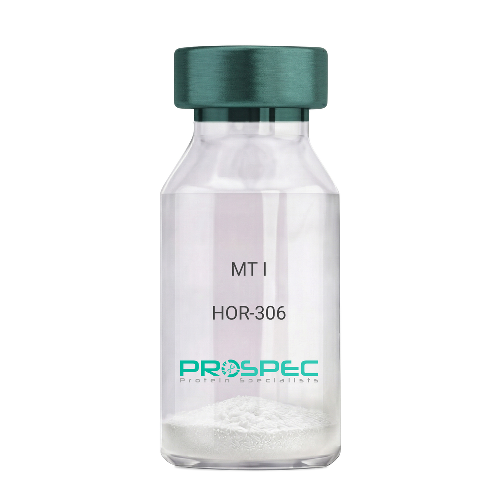
MT IDescription:
Melanotan-I
Melanotan-I, MT-I, Melanotan-1.
Product # :
HOR-306Price :
Quantity :
Shipping Method :
Shipped at Room temp
More Info
- description
- formulation
- purity
- More Info
Description
Melanotan-I has the amino acid sequence of Ser-Tyr-Ser-Nle-Glu-His-D-Phe-Arg-Trp-Gly-Lys-Pro-Val and a molecular weight of 1647.4 Dalton.
Formulation
The protein (1mg/ml) was lyophilized with no additives.
Purity
Greater than 98.0% as determined by RP-HPLC.
More Info
-
Synonyms
Melanotan-I, MT-I, Melanotan-1.
-
Physical Appearance
Sterile Filtered off-White lyophilized (freeze-dried) powder.
-
Stability
Lyophilized Melanotan-I although stable at room temperature for 3 weeks, should be stored desiccated below -18°C. Upon reconstitution MT-I should be stored at 4°C between 2-7 days and for future use below -18°C.For long term storage it is recommended to add a carrier protein (0.1% HSA or BSA).Please prevent freeze-thaw cycles.
-
Solubility
It is recommended to reconstitute the lyophilized Melanotan-I in sterile 1% acetic acid not less than 100 µg/ml, which can then be further diluted to other aqueous solutions.
ProSpec's products are furnished for LABORATORY RESEARCH USE ONLY. The product may not be used as drugs, agricultural or pesticidal products, food additives or household chemicals.
- View Data Sheet
Name :
Lymphotactin Human, HisDescription:
Lymphotactin Human Recombinant (XCL1), His Tag
XCL1, Cytokine SCM-1, ATAC, Lymphotaxin, SCM-1-alpha, Small inducible cytokine C1, XC chemokine ligand 1, LTN, LPTN, SCM1, SCM-1, SCYC1, SCM-1a.
Product # :
CHM-357Price :
Quantity :
Shipping Method :
Shipped with Ice Packs
More Info
- description
- source
- formulation
- purity
- More Info
Description
XCL1 Human Recombinant produced in E.Coli is a single,non-glycosylated, polypeptide chain containing 114 amino acids (22-114 a.a.) and having a molecular mass of 12.5 kDa. The XCL1 is fused to a 20 amino acid His Tag at N-terminus and purified by proprietary chromatographic techniques.
Source
Escherichia Coli.
Formulation
Recombinant XCL1 contains 20mM Tris buffer pH-8, 200mM NaCl, 2mM DTT and 30% glycerol.
Purity
Greater than 95.0% as determined by SDS-PAGE.
More Info
-
Introduction
XCL1 is a small cytokine belonging to the XC chemokine family that is also known as lymphotactin. It is located in high levels in spleen, thymus, intestine and peripheral blood leukocytes, and at lower levels in lung, prostate gland and ovary. Cellular sources for XCL1 include activated thymic and peripheral blood CD8+ T cells. This chemokine attracts T cells. In humans, XCL1 is closely related to another chemokine called XCL2, whose gene is found at the same locus on chromosome 1. XCL1 induces it chemotactic function by binding to a chemokine receptor called XCR1.
-
Synonyms
XCL1, Cytokine SCM-1, ATAC, Lymphotaxin, SCM-1-alpha, Small inducible cytokine C1, XC chemokine ligand 1, LTN, LPTN, SCM1, SCM-1, SCYC1, SCM-1a.
-
Physical Appearance
Sterile Filtered colorless solution.
-
Stability
Store at 4°C if entire vial will be used within 2-4 weeks. Store, frozen at -20°C for longer periods of time. For long term storage it is recommended to add a carrier protein (0.1% HSA or BSA).Avoid multiple freeze-thaw cycles.
-
Amino Acid Sequence
MGSSHHHHHH SSGLVPRGSH MVGSEVSDKR TCVSLTTQRL PVSRIKTYTI TEGSLRAVIF ITKRGLKVCA DPQATWVRDV VRSMDRKSNT RNNMIQTKPT GTQQSTNTAV TLTG.
ProSpec's products are furnished for LABORATORY RESEARCH USE ONLY. The product may not be used as drugs, agricultural or pesticidal products, food additives or household chemicals.
- View Data Sheet
Name :
P Selectin HumanDescription:
P-selectin Human Recombinant
P-selectin, Granule membrane protein 140, GMP-140, PADGEM, Leukocyte-endothelial cell adhesion molecule 3, LECAM3, CD62 antigen-like family member P, CD62P antigen, SELP, GMRP, GRMP, CD62, PSEL, CD62P, GMP140, FLJ45155.
Product # :
PRO-382Price :
Quantity :
Shipping Method :
Shipped with Ice Packs
More Info
- description
- source
- formulation
- purity
- More Info
Description
P-Selectin Human Recombinant is expressed in E. coli containing 566 amino acids 197-761 fused to an amino terminal hexahistidine tag.
Source
Escherichia Coli.
Formulation
P-Selectin is supplied in 50% Glycerol.
Purity
Greater than 95% as determined by SDS-PAGE.
Single band on Western Blot.More Info
-
Introduction
P-Selectin also called Platelet Alpha-Granule Membrane Protein, CD62, and Granulocyte Membrane Protein GRMP belongs to a family of divalent cation-dependent carbohydrate-binding glycoproteins or adhesion molecules. Pselectin is expressed transiently on the surface of activated platelets and endothelial cells. P-Selectin is a 140 kDa protein which is stored in the alpha-granules of platelets and Weibel-Palade bodies of endothelial cells. Secreted P-selectin is thought to play a key role in the adhesion of platelets to monocytes and neutrophils during an inflammatory response. P-Selectin is a calcium-dependent receptor that binds to sialylated forms of Lewis blood group carbohydrate antigens on neutrophils and monocytes. Levels of P-Selectin may be elevated in a number of pathological conditions.
-
Synonyms
P-selectin, Granule membrane protein 140, GMP-140, PADGEM, Leukocyte-endothelial cell adhesion molecule 3, LECAM3, CD62 antigen-like family member P, CD62P antigen, SELP, GMRP, GRMP, CD62, PSEL, CD62P, GMP140, FLJ45155.
-
Physical Appearance
Sterile Filtered liquid formulation.
-
Stability
Store at 4°C if entire vial will be used within 1-2 weeks. Store, frozen at -20°C for longer periods of time. Avoid multiple freeze-thaw cycles.
-
Applications
P-Selectin can be used directly as a positive control in Western blotting, ELISA, immunoprecipitation and other immunological experiments.
The biological activity of this product has not yet been tested.
ProSpec's products are furnished for LABORATORY RESEARCH USE ONLY. The product may not be used as drugs, agricultural or pesticidal products, food additives or household chemicals.