Skip to content

High-quality research proteins.

  • 1-800-805-6531

  • Mon - Sat 8.00-18.00, Sun - Closed

  • info@prospecbio.com

  • PO Box 6591 East Brunswick NJ 08816

  • Products
    • Cytokines
    • Growth factors
    • Chemokines
    • Cd antigens
    • Neurotrophins
    • Hormones
    • Enzymes
    • Viral antigens
    • Recombinant proteins
    • Natural proteins
    • Monoclonal antibodies
    • Polyclonal antibodies
    • Thermal Packaging
  • About
    • Company
    • Founder
    • Contribution
  • Customers
    • Eastern Asia
    • Eastern Europe
    • North America
    • South America
    • South East Asia
    • Southern Asia
    • Western Europe
  • Ordering
    • Ordering Method
    • Ordering - Credit Card
    • Ordering - PO
    • Shipping Fees
    • FAQs
  • Services
    • DNA Cloning
    • Protein Expression
    • Monoclonal Antibody
    • Fermentation
    • Technology License
  • Contact us
Log in

Country/region

  • USD $ | Albania
  • USD $ | Andorra
  • USD $ | Argentina
  • USD $ | Armenia
  • USD $ | Australia
  • USD $ | Austria
  • USD $ | Azerbaijan
  • USD $ | Belarus
  • USD $ | Belgium
  • USD $ | Bolivia
  • USD $ | Brazil
  • USD $ | Bulgaria
  • USD $ | Cambodia
  • USD $ | Canada
  • USD $ | Chile
  • USD $ | China
  • USD $ | Colombia
  • USD $ | Costa Rica
  • USD $ | Croatia
  • USD $ | Cyprus
  • USD $ | Czechia
  • USD $ | Denmark
  • USD $ | Estonia
  • USD $ | Finland
  • USD $ | France
  • USD $ | Georgia
  • USD $ | Germany
  • USD $ | Greece
  • USD $ | Hong Kong SAR
  • USD $ | Hungary
  • USD $ | Iceland
  • USD $ | India
  • USD $ | Ireland
  • USD $ | Israel
  • USD $ | Italy
  • USD $ | Japan
  • USD $ | Kazakhstan
  • USD $ | Latvia
  • USD $ | Liechtenstein
  • USD $ | Lithuania
  • USD $ | Madagascar
  • USD $ | Malta
  • USD $ | Mexico
  • USD $ | Moldova
  • USD $ | Monaco
  • USD $ | Netherlands
  • USD $ | New Zealand
  • USD $ | North Macedonia
  • USD $ | Norway
  • USD $ | Panama
  • USD $ | Paraguay
  • USD $ | Peru
  • USD $ | Philippines
  • USD $ | Poland
  • USD $ | Portugal
  • USD $ | Romania
  • USD $ | Singapore
  • USD $ | Slovakia
  • USD $ | Slovenia
  • USD $ | South Africa
  • USD $ | South Korea
  • USD $ | Spain
  • USD $ | Sri Lanka
  • USD $ | Sweden
  • USD $ | Switzerland
  • USD $ | Taiwan
  • USD $ | Thailand
  • USD $ | Türkiye
  • USD $ | Ukraine
  • USD $ | United Kingdom
  • USD $ | United States
  • USD $ | Uruguay
  • USD $ | Vatican City
  • USD $ | Venezuela
  • USD $ | Vietnam
  • Facebook
  • Instagram
logoProSpec - Recombinant Protein Manufacturer for academic and R&D industry
Become a distributor
Account Log in
Wishlist
Cart Cart(0)
  • Products
    +
    • Cytokines
    • Growth factors
    • Chemokines
    • Cd antigens
    • Neurotrophins
    • Hormones
    • Enzymes
    • Viral antigens
    • Recombinant proteins
    • Natural proteins
    • Monoclonal antibodies
    • Polyclonal antibodies
    • Thermal Packaging
  • About
    +
    • Company
    • Founder
    • Contribution
  • Customers
    +
    • Eastern Asia
    • Eastern Europe
    • North America
    • South America
    • South East Asia
    • Southern Asia
    • Western Europe
  • Ordering
    +
    • Ordering Method
    • Ordering - Credit Card
    • Ordering - PO
    • Shipping Fees
    • FAQs
  • Services
    +
    • DNA Cloning
    • Protein Expression
    • Monoclonal Antibody
    • Fermentation
    • Technology License
  • Contact us

Item added to your cart

View cart
View All
  • Cytokines
  • Growth factors
  • Chemokines
  • Cd antigens
  • Neurotrophins
  • Hormones
  • Enzymes
  • Viral antigens
  • Recombinant proteins
  • Natural proteins
  • Monoclonal antibodies
  • Polyclonal antibodies
  • Thermal Packaging
  • Tumor Necrosis Factor

    Tumor Necrosis Factor

    View All
  • 4-1BB

    4-1BB

    View All
  • Adiponectin

    Adiponectin

    View All
  • AIF1

    AIF1

    View All
  • Other Cytokines

    Other Cytokines

    View All
  • AITRL

    AITRL

    View All
  • Angiopoietin

    Angiopoietin

    View All
  • Apolipoprotein

    Apolipoprotein

    View All
  • B-Cell Activating Factor

    B-Cell Activating Factor

    View All
  • Beta Defensin

    Beta Defensin

    View All
  • Bone Morphogenetic Protein

    Bone Morphogenetic Protein

    View All
  • B type Natriuretic Peptide

    B type Natriuretic Peptide

    View All
  • BST

    BST

    View All
  • Betacellulin

    Betacellulin

    View All
  • Cardiotrophin

    Cardiotrophin

    View All
  • Activin

    Activin

    View All
  • Fibroblast Growth Factor

    Fibroblast Growth Factor

    View All
  • Other Growth Factors

    Other Growth Factors

    View All
  • CSF

    CSF

    View All
  • CTGF

    CTGF

    View All
  • IGFBP

    IGFBP

    View All
  • VEGF Protein

    VEGF Protein

    View All
  • EGF

    EGF

    View All
  • Epigen

    Epigen

    View All
  • Erythropoietin

    Erythropoietin

    View All
  • Insulin-Like Growth Factor

    Insulin-Like Growth Factor

    View All
  • Growth Hormone

    Growth Hormone

    View All
  • HDGF

    HDGF

    View All
  • Hepatocyte Growth Factor

    Hepatocyte Growth Factor

    View All
  • Insulin

    Insulin

    View All
  • BCA-1/ BLC (CXCL13)

    BCA-1/ BLC (CXCL13)

    View All
  • BRAK (CXCL14)

    BRAK (CXCL14)

    View All
  • C-10 (CCL6)

    C-10 (CCL6)

    View All
  • Other Chemokines

    Other Chemokines

    View All
  • MEC (CCL28)

    MEC (CCL28)

    View All
  • LD78-beta (CCL3L1)

    LD78-beta (CCL3L1)

    View All
  • CTACK (CCL27)

    CTACK (CCL27)

    View All
  • CXCL16

    CXCL16

    View All
  • CXCL17

    CXCL17

    View All
  • Platelet Factor-4 (CXCL4)

    Platelet Factor-4 (CXCL4)

    View All
  • ENA-78 (CXCL5)

    ENA-78 (CXCL5)

    View All
  • Eotaxin (CCL11,24,26)

    Eotaxin (CCL11,24,26)

    View All
  • Exodus-2 (CCL21)

    Exodus-2 (CCL21)

    View All
  • Fractalkine (CX3CL1)

    Fractalkine (CX3CL1)

    View All
  • CXCL6

    CXCL6

    View All
  • Other CD Antigens

    Other CD Antigens

    View All
  • CD14

    CD14

    View All
  • CD164

    CD164

    View All
  • CD1

    CD1

    View All
  • CD2

    CD2

    View All
  • CD200

    CD200

    View All
  • CD207

    CD207

    View All
  • CD226

    CD226

    View All
  • CD23

    CD23

    View All
  • CD244

    CD244

    View All
  • CD247

    CD247

    View All
  • CD27

    CD27

    View All
  • CD274

    CD274

    View All
  • CD300

    CD300

    View All
  • CD33

    CD33

    View All
  • Other Neurotrophins

    Other Neurotrophins

    View All
  • Beta-NGF

    Beta-NGF

    View All
  • BDNF

    BDNF

    View All
  • CDNF

    CDNF

    View All
  • CNTF

    CNTF

    View All
  • GDNF

    GDNF

    View All
  • Glia Maturation Factor

    Glia Maturation Factor

    View All
  • MANF

    MANF

    View All
  • Midkine

    Midkine

    View All
  • Neuroglobin

    Neuroglobin

    View All
  • Neurotrophic factors

    Neurotrophic factors

    View All
  • Neuregulin

    Neuregulin

    View All
  • Neuropilin

    Neuropilin

    View All
  • Pigment Epithelium-Derived Factor

    Pigment Epithelium-Derived Factor

    View All
  • Pleiotrophin

    Pleiotrophin

    View All
  • Peptide Hormones

    Peptide Hormones

    View All
  • Other Hormones

    Other Hormones

    View All
  • HCG

    HCG

    View All
  • Endothelin

    Endothelin

    View All
  • Exendin

    Exendin

    View All
  • FSH

    FSH

    View All
  • Glucagon

    Glucagon

    View All
  • GHRP

    GHRP

    View All
  • GLP

    GLP

    View All
  • Thyrostimulin

    Thyrostimulin

    View All
  • Inhibin A

    Inhibin A

    View All
  • LHRH

    LHRH

    View All
  • Melanotan

    Melanotan

    View All
  • Procalcitonin

    Procalcitonin

    View All
  • PTH

    PTH

    View All
  • Peroxiredoxin

    Peroxiredoxin

    View All
  • Peptidase

    Peptidase

    View All
  • Synthetase

    Synthetase

    View All
  • Transferase

    Transferase

    View All
  • Hydrolase

    Hydrolase

    View All
  • Dehydrogenase

    Dehydrogenase

    View All
  • ACE2

    ACE2

    View All
  • Esterase

    Esterase

    View All
  • Protein Kinases

    Protein Kinases

    View All
  • Other Enzymes

    Other Enzymes

    View All
  • Phosphatase

    Phosphatase

    View All
  • Deaminase

    Deaminase

    View All
  • Oxygenase

    Oxygenase

    View All
  • Adenylate Kinase

    Adenylate Kinase

    View All
  • Lyase

    Lyase

    View All
  • Chagas

    Chagas

    View All
  • SARS

    SARS

    View All
  • Influenza

    Influenza

    View All
  • ASFV

    ASFV

    View All
  • Astrovirus

    Astrovirus

    View All
  • Borrelia

    Borrelia

    View All
  • Helicobacter Pylori

    Helicobacter Pylori

    View All
  • Chikungunya

    Chikungunya

    View All
  • Chlamydia

    Chlamydia

    View All
  • Cytomegalo

    Cytomegalo

    View All
  • Dengue

    Dengue

    View All
  • EBV

    EBV

    View All
  • Ebola

    Ebola

    View All
  • Feline Leukemia Virus

    Feline Leukemia Virus

    View All
  • Hepatitis A

    Hepatitis A

    View All
  • Other

    Other

    View All
  • Actin

    Actin

    View All
  • ADAM

    ADAM

    View All
  • Complement Component

    Complement Component

    View All
  • Ag85

    Ag85

    View All
  • Eukaryotic Translation Initiation Factor

    Eukaryotic Translation Initiation Factor

    View All
  • Anterior Gradient Protein

    Anterior Gradient Protein

    View All
  • Heat Shock Protein

    Heat Shock Protein

    View All
  • Alpha-2-HS-Glycoprotein

    Alpha-2-HS-Glycoprotein

    View All
  • Hemoglobin

    Hemoglobin

    View All
  • Allergy

    Allergy

    View All
  • Anaplasma

    Anaplasma

    View All
  • Angiogenin

    Angiogenin

    View All
  • Ankyrin Repeat Domain

    Ankyrin Repeat Domain

    View All
  • Annexin

    Annexin

    View All
  • Other Natural Proteins

    Other Natural Proteins

    View All
  • Aprotinin

    Aprotinin

    View All
  • Transferrin

    Transferrin

    View All
  • Avidin

    Avidin

    View All
  • Anti Coagulation Factors

    Anti Coagulation Factors

    View All
  • Natural Albumin

    Natural Albumin

    View All
  • Natural Coagulation Factors

    Natural Coagulation Factors

    View All
  • Fibronectin

    Fibronectin

    View All
  • Thrombin

    Thrombin

    View All
  • Anti Human Enzyme

    Anti Human Enzyme

    View All
  • Other Monoclonal Antibodies

    Other Monoclonal Antibodies

    View All
  • Anti Human Cytokine

    Anti Human Cytokine

    View All
  • Anti Human Heat Shock Protein

    Anti Human Heat Shock Protein

    View All
  • Anti Mouse Lymphocyte

    Anti Mouse Lymphocyte

    View All
  • Anti Human Chemokine

    Anti Human Chemokine

    View All
  • Anti Human Lymphocyte

    Anti Human Lymphocyte

    View All
  • Anti Viral Monoclonal

    Anti Viral Monoclonal

    View All
  • Anti Mouse Cytokine

    Anti Mouse Cytokine

    View All
  • Anti-GST Monoclonal

    Anti-GST Monoclonal

    View All
  • Other Polyclonal Antibodies

    Other Polyclonal Antibodies

    View All
  • Anti Viral

    Anti Viral

    View All
  • Anti-GST Monoclonal

    Anti-GST Monoclonal

    View All
Back

Search results

1000 results found for “PDGF”

Name

Description

Product #

Price

Quantity

Shipping Method

  • View Data Sheet

    Name :

    BMP 4 Human

    Description:

    Bone Morphogenetic Protein-4 Human Recombinant

    BMP4, ZYME, BMP2B, BMP2B1.

    Product # :

    CYT-361

    Price :

    Quantity :

    Shipping Method :

    Room Temp Icon

    Shipped at Room temp

    Add To Cart

    More Info

    • description
    • source
    • formulation
    • purity
    • More Info

    Description

    Bone Morphogenetic Protein-4 Human Recombinant produced in E.Coli is a monomeric, non-glycosylated, Polypeptide chain containing 116 amino acids and having a molecular mass of 13kDa. The BMP-4 is purified by proprietary chromatographic techniques.

    Source

    Escherichia Coli.

    Formulation

    BMP-4 was lyophilized from a 0.2µm filtered concentrated (1mg/ml) solution in 20mM Na2CO3 buffer, pH 9.0.

    Purity

    Greater than 95.0% as determined by:
    (a) Analysis by RP-HPLC.
    (b) Analysis by SDS-PAGE.

    More Info

    • Introduction

      The protein encoded by this gene is a member of the bone morphogenetic protein family which is part of the transforming growth factor-beta superfamily. The superfamily includes large families of growth and differentiation factors. Bone morphogenetic proteins were originally identified by an ability of demineralized bone extract to induce endochondral osteogenesis in vivo in an extraskeletal site. This particular family member plays an important role in the onset of endochondral bone formation in humans, and a reduction in expression has been associated with a variety of bone diseases, including the heritable disorder Fibrodysplasia Ossificans Progressiva. Alternative splicing in the 5' untranslated region of this gene has been described and three variants are described, all encoding an identical protein.

    • Synonyms

      BMP4, ZYME, BMP2B, BMP2B1.

    • Physical Appearance

      Sterile Filtered White lyophilized (freeze-dried) powder.

    • Stability

      Lyophilized Bone Morphogenetic Protein-4 although stable at room temperature for 3 weeks, should be stored desiccated below -18°C. Upon reconstitution BMP4 should be stored at 4°C between 2-7 days and for future use below -18°C.For long term storage it is recommended to add a carrier protein (0.1% HSA or BSA).Please prevent freeze-thaw cycles.

    • Solubility

      It is recommended to reconstitute the lyophilized Bone Morphogenetic Protein-4 in sterile 18M-cm H2O not less than 100µg/ml, which can then be further diluted to other aqueous solutions.

    • Amino Acid Sequence

      SPKHHSQRAR KKNKNCRRHS LYVDFSDVGW NDWIVAPPGY QAFYCHGDCP FPLADHLNST NHAIVQTLVN SVNSSIPKAC CVPTELSAIS MLYLDEYDKV VLKNYQEMVV EGCGCR.

    • Background

      What You Should Know About Bone Morphogenetic Protein-4 (BMP-4) Human Recombinant

      As part of the transforming growth factor-beta (TGF-β) superfamily, Bone morphogenetic protein-4 (BMP-4) participates in multiple developmental processes, from embryogenesis to bone and cartilage formation.

      Since this signaling protein is involved in many physiological processes, its laboratory-produced version has been studied for different medical applications. Additionally, a reduction in BMP-4 expression has been associated with multiple diseases, leading to further research into its potential therapeutic benefits.

      Are you interested in learning more about Bone Morphogenetic Protein-4 (BMP-4) human recombinant? Read on to find more information!

      How Does Bone Morphogenetic Protein-4 (BMP-4) Work?

      Bone Morphogenetic Protein-4 (BMP-4) regulates microRNAs miR-494 and miR-126-5p expression, controlling endothelial cells' involvement and function in angiogenesis. As such, it has diverse effects on cell growth, differentiation, and survival.

      The Role of BMP-4

      This protein emits signals that promote the formation of different tissues and organs, including the bones and cartilage, kidneys, teeth, and the neural tube. In other words, it's essential for the development of the heart, skeleton, and central nervous system.

      However, the role of BMP-4 goes beyond these processes. It participates in different physiological activities, such as:

      • Embryonic development
      • Wound healing
      • Bone remodeling
      • Immune response modulation
      • Tissue repair
      • Cardiac development and function

      What Is Bone Morphogenetic Protein-4 (BMP-4) Human Recombinant?

      To replicate the effects of the BMP4 found in humans and explore its possible therapeutic applications, many laboratories have started producing this protein in Chinese hamster ovary (CHO) cells.

      As mentioned, decreased BMP-4 expression has been associated with different diseases, including bone disorders, fibrosis, and cancer, which can cause other conditions, such as organ dysfunction.

      More research is needed, but BMP-4 human recombinant (rhBMP4) produced in CHO has the potential to address these diseases and could be used for other medical applications. These are some examples:

      • Cancer therapy
      • Development of engineered tissues and organs
      • Bone regeneration for the treatment of osteoporosis and nonunion fractures
      • Bone growth and fusion in spinal fusion surgeries (the U.S. Food and Drug Administration approved some bone morphogenetic proteins for these procedures)
      • Promotion of tissue repair and regeneration

      Final Thoughts BMP-4

      Although BMP-4 human recombinant produced in CHO offers potential benefits, several challenges remain, including possible side effects, as high doses can cause inflammation, bone overgrowth, and other issues.

      However, the long-term effects of rhBMP4 are still under investigation. Further research will provide solutions to address these challenges and allow experts to explore this laboratory-produced protein's power in different medical fields.

      What is the molecular weight/Mw of BMP4 Protein?
      BMP4 Protein has a total Mw of 13kDa.

      What is the source or expression system of BMP4 Protein?
      Escherichia Coli.

      What is the Purity of BMP4 Protein?
      BMP4 Protein is >95% pure as determined by SDS-PAGE.

      What is the Biological Activity of BMP4 Protein?
      The biological functionality of BMP4 Protein will be determined in the future.

      What is the amino acid sequence of BMP4 Protein?
      SPKHHSQRAR KKNKNCRRHS LYVDFSDVGW NDWIVAPPGY QAFYCHGDCP FPLADHLNST NHAIVQTLVN SVNSSIPKAC CVPTELSAIS MLYLDEYDKV VLKNYQEMVV EGCGCR.

      What applications can BMP4 Protein be used in?
      BMP4 Protein can probably be used in western blot, ELISA and Lateral Flow.

      What is the endotoxin level for BMP4 Protein?
      The endotoxin level is minimal, BMP4 Protein was purified using conventional chromatography techniques.

    ProSpec's products are furnished for LABORATORY RESEARCH USE ONLY. The product may not be used as drugs, agricultural or pesticidal products, food additives or household chemicals.

    Bmp4 Human
  • View Data Sheet

    Name :

    FGF 2 Mouse

    Description:

    Fibroblast Growth Factor-Basic Mouse Recombinant

    HBGF-2, Prostatropin, FGF-2, FGB-b.

    Product # :

    CYT-386

    Price :

    Quantity :

    Shipping Method :

    Room Temp Icon

    Shipped at Room temp

    Add To Cart

    More Info

    • description
    • source
    • formulation
    • purity
    • biological activity
    • More Info

    Description

    Fibroblast Growth Factor-basic Mouse Recombinant (FGF-2) produced in E.Coli is a single, non-glycosylated, polypeptide chain containing 146 amino acids and having a molecular mass of 16.3kDa. The FGF-2 is purified by proprietary chromatographic techniques.

    Source

    Escherichia Coli.

    Formulation

    FGF-b was lyophilized from 5mM Na2PO4, pH7.5 and 50mM NaCl.

    Purity

    Greater than 95.0% as determined by SDS-PAGE.

    Biological Activity

    The activity as calculated by the dose-dependant proliferation of BALB/3T3 cells was found to be less than 1ng/ml corresponding to a specific activity of 1,000,000 units/mg.

    More Info

    • Introduction

      FGF-basic is a member of the fibroblast growth factor (FGF) family. FGF family members bind and possess broad mitogenic and angiogenic activities. This protein has been implicated in diverse biological processes, such as limb and nervous system development, wound healing, and tumor growth. The mRNA for this gene contains multiple polyadenylation sites, and is alternatively translated from AUG and non-AUG (CUG) initiation codons resulting in five different isoforms with distinct properties. The CUG-initiated isoforms are localized in the nucleus and are responsible for the intracrine effect, whereas, the AUG-initiated form is mostly cytosolic and is responsible for the paracrine and autocrine effects of this FGF.
      The binding growth factors are angiogenic agents in vivo and are potent mitogens for a variety of cell types in vitro. there are differences in the tissue distribution and concentration of these 2 growth factors.

    • Synonyms

      HBGF-2, Prostatropin, FGF-2, FGB-b.

    • Physical Appearance

      Sterile Filtered White lyophilized (freeze-dried) powder.

    • Stability

      Lyophilized Fibroblast Growth Factor-2 although stable at room temperature for 3 weeks, should be stored desiccated below -18°C. Upon reconstitution FGF-basic should be stored at 4°C between 2-7 days and for future use below -18°C.For long term storage it is recommended to add a carrier protein (0.1% HSA or BSA).Please prevent freeze-thaw cycles.

    • Solubility

      It is recommended to reconstitute the lyophilized Fibroblast Growth Factor b in sterile 18M-cm H2O not less than 100µg/ml, which can then be further diluted to other aqueous solutions.

    • Amino Acid Sequence

      MPALPEDGGA AFPPGHFKDP KRLYCKNGGF FLRIHPDGRV DGVREKSDPH VKLQLQAEER GVVSIKGVCA NRYLAMKEDG RLLASKCVTE ECFFFERLES NNYNTYRSRK YSSWYVALKR TGQYKLGSKT GPGQKAILFL PMSAKS.

    • Background

      What is the molecular weight/Mw of FGF 2 MOUSE Protein?
      FGF 2 MOUSE Protein has a total Mw of 16.3kDa.

      What is the source or expression system of FGF 2 MOUSE Protein?
      Escherichia Coli.

      What is the Purity of FGF 2 MOUSE Protein?
      FGF 2 MOUSE Protein is >95% pure as determined by SDS-PAGE.

      What is the Biological Activity of FGF 2 MOUSE Protein?
      The activity as calculated by the dose-dependant proliferation of BALB/3T3 cells was found to be less than 1ng/ml corresponding to a specific activity of 1,000,000 units/mg.
      What is the amino acid sequence of FGF 2 MOUSE Protein?
      MPALPEDGGA AFPPGHFKDP KRLYCKNGGF FLRIHPDGRV DGVREKSDPH VKLQLQAEER GVVSIKGVCA NRYLAMKEDG RLLASKCVTE ECFFFERLES NNYNTYRSRK YSSWYVALKR TGQYKLGSKT GPGQKAILFL PMSAKS.

      What applications can FGF 2 MOUSE Protein be used in?
      FGF 2 MOUSE Protein can probably be used in western blot, ELISA and Lateral Flow.

      What is the endotoxin level for FGF 2 MOUSE Protein?
      The endotoxin level is minimal, FGF 2 MOUSE Protein was purified using conventional chromatography techniques.

    • Protein content

      Protein quantitation was carried out by two independent methods:1. UV spectroscopy at 280 nm using the absorbency value of 0.885 as the extinction coefficient for a 0.1% (1mg/ml) solution. This value is calculated by the PC GENE computer analysis program of protein sequences (IntelliGenetics). 2. Analysis by RP-HPLC, using a calibrated solution of FGF2 as a Reference Standard.

    ProSpec's products are furnished for LABORATORY RESEARCH USE ONLY. The product may not be used as drugs, agricultural or pesticidal products, food additives or household chemicals.

    Fgf 2 Mouse
  • View Data Sheet

    Name :

    GDF15 Human

    Description:

    Growth and Differentiation Factor 15 Human Recombinant

    GDF-15, MIC1, MIC-1, NAG-1, PDF, PLAB, PTGFB, Growth/differentiation factor 15, Placental bone morphogenetic protein, Placental TGF-beta, Macrophage inhibitory cytokine 1, Prostate differentiation factor, NSAID-activated gene 1 protein, NSAID-regulated gene 1 protein, NRG-1, GDF15.

    Product # :

    CYT-335

    Price :

    Quantity :

    Shipping Method :

    Room Temp Icon

    Shipped at Room temp

    Add To Cart

    More Info

    • description
    • source
    • formulation
    • purity
    • More Info

    Description

    GDF15 Human Recombinant produced in E.Coli is a homodimeric, non-glycosylated, Polypeptide chain containing 2x113 amino acids and having a molecular mass of 24.8 kDa. The GDF15 is purified by proprietary chromatographic techniques.

    Source

    Escherichia Coli.

    Formulation

    GDF15 is lyophilized from a sterile filtered solution containing 0.1% Trifluoroacetic Acid (TFA).

    Purity

    Greater than 95.0% as determined by SDS-PAGE.

    More Info

    • Introduction

      GDF15 is part of the TGF-Beta superfamily that is involved in regulating inflammatory and apoptotic pathways in injured tissues and throughout disease processes. GDF15 is most abundant in the liver. Its expression in liver can be considerably up-regulated in during injury of organs such as liver, kidney, heart and lung. GDF-15 promotes proliferation or growth arrest and differentiation due to differences in cellular differentiation. GDF15 prevents apoptosis in cerebellar granule neurons by activating Akt and inhibiting endogenously active ERK. GDF15 is a novel autocrine/endocrine factor that antagonizes the hypertrophic response and loss of ventricular performance.

    • Synonyms

      GDF-15, MIC1, MIC-1, NAG-1, PDF, PLAB, PTGFB, Growth/differentiation factor 15, Placental bone morphogenetic protein, Placental TGF-beta, Macrophage inhibitory cytokine 1, Prostate differentiation factor, NSAID-activated gene 1 protein, NSAID-regulated gene 1 protein, NRG-1, GDF15.

    • Physical Appearance

      Sterile Filtered White lyophilized (freeze-dried) powder.

    • Stability

      Lyophilized GDF15 although stable at room temperature for 3 weeks, should be stored desiccated below -18°C. Upon reconstitution GDF15 should be stored at 4°C between 2-7 days and for future use below -18°C.For long term storage it is recommended to add a carrier protein (0.1% HSA or BSA).Please prevent freeze-thaw cycles.

    • Solubility

      It is recommended to reconstitute the lyophilized GDF15 in sterile 18M-cm H2O at a concentration of 100µg/ml, which can then be further diluted to other aqueous solutions.

    • Amino Acid Sequence

      MARNGDHCPL GPGRCCRLHT VRASLEDLGW ADWVLSPREV QVTMCIGACP SQFRAANMHA QIKTSLHRLK PDTVPAPCCV PASYNPMVLI QKTDTGVSLQ TYDDLLAKDC HCI.

    • Background

      What is the molecular weight/Mw of GDF15 HUMAN Protein?
      GDF15 HUMAN Protein has a total Mw of 24.8kDa.

      What is the source or expression system of GDF15 HUMAN Protein?
      Escherichia Coli.

      What is the Purity of GDF15 HUMAN Protein?
      GDF15 HUMAN Protein is >95% pure as determined by SDS-PAGE.

      What is the Biological Activity of GDF15 HUMAN Protein?
      The biological functionality of GDF15 HUMAN Protein will be determined in the future.

      What is the amino acid sequence of GDF15 HUMAN Protein?
      MARNGDHCPL GPGRCCRLHT VRASLEDLGW ADWVLSPREV QVTMCIGACP SQFRAANMHA QIKTSLHRLK PDTVPAPCCV PASYNPMVLI QKTDTGVSLQ TYDDLLAKDC HCI.

      What applications can GDF15 HUMAN Protein be used in?
      GDF15 HUMAN Protein can probably be used in western blot, ELISA and Lateral Flow.

      What is the endotoxin level for GDF15 HUMAN Protein?
      The endotoxin level is minimal, GDF15 HUMAN Protein was purified using conventional chromatography techniques.


    ProSpec's products are furnished for LABORATORY RESEARCH USE ONLY. The product may not be used as drugs, agricultural or pesticidal products, food additives or household chemicals.

    Gdf15 H Human
  • View Data Sheet

    Name :

    DKK3 Antibody

    Description:

    Mouse Anti Human Dickkopf-Related Protein 3

    Dickkopf 3 homolog (Xenopus laevis), dickkopf-related protein 3, regulated in glioma, RIG, RIG-like 7-1, RIG-like 5-6, Dkk-3, REIC.

    Product # :

    ANT-707

    Price :

    Quantity :

    Shipping Method :

    Ice Icon

    Shipped with Ice Packs

    Add To Cart

    More Info

    • formulation
    • More Info

    Formulation

    1mg/ml containing PBS, pH-7.4, 10% Glycerol and 0.02% Sodium Azide.

    More Info

    • Introduction

      Dickkopf-related protein 3 (DKK3) belongs to the DKK protein family including Dkk-1, 2, 3 and -4. DKK3 is a 350 amino acid secreted glycoprotein which is comprised of an N-terminal signal peptide and 2 conserved cysteine-rich domains that are separated by a 12 amino acid linker region. DKK3 is involved in embryonic development through its inhibition of the WNT signaling pathway. DKK3 gene expression is decreased in a variety of cancer cell lines and it may act as a tumor suppressor gene.

    • Synonyms

      Dickkopf 3 homolog (Xenopus laevis), dickkopf-related protein 3, regulated in glioma, RIG, RIG-like 7-1, RIG-like 5-6, Dkk-3, REIC.

    • Physical Appearance

      Sterile filtered colorless solution.

    • Immunogen

      Anti-human DKK3 mAb, is derived from hybridization of mouse F0 myeloma cells with spleen cells from BALB/c mice immunized with a recombinant human DKK3 protein (22-350 amino acids purified from E. coli.

    • Ig Subclass

      Mouse IgG1 heavy chain and k light chain.

    • Clone

      PAT103B5AT.

    • Applications

      DKK3 antibody has been tested by ELISA and Western blot analysis to assure specificity and reactivity. Since application varies, however, each investigation should be titrated by the reagent to obtain optimal results.

    • Type

      Mouse Anti Human Monoclonal.

    • Storage Procedures

      For periods up to 1 month store at 4°C, for longer periods of time, store at -20°C. Prevent freeze thaw cycles.

    • Purification Method

      DKK3 antibody was purified from mouse ascitic fluids by protein-A affinity chromatography.

    ProSpec's products are furnished for LABORATORY RESEARCH USE ONLY. The product may not be used as drugs, agricultural or pesticidal products, food additives or household chemicals.

    Dkk3 Antibody
  • View Data Sheet

    Name :

    TGFB1 (113 a.a.) Human

    Description:

    Transforming Growth Factor-Beta 1 (113 a.a.) Human Recombinant

    Transforming growth factor beta-1, TGF-beta-1, CED, DPD1, TGFB, TGF-b 1.

    Product # :

    CYT-679

    Price :

    Quantity :

    Shipping Method :

    Ice Icon

    Shipped with Ice Packs

    Add To Cart

    More Info

    • description
    • source
    • formulation
    • purity
    • More Info

    Description

    TGF-b 1 Human Recombinant produced in E.Coli is a single, non-glycosylated, polypeptide chain containing 113 amino acids (279-390 a.a.) and having a total molecular mass of 12.9 kDa. TGF-b 1 (113 a.a.) is purified by proprietary chromatographic techniques.

    Source

    Escherichia Coli.

    Formulation

    The TGF-b 1 solution contains 10mM Sodium Citrate (pH3.5) and 10% glycerol.

    Purity

    Greater than 95.0% as determined by SDS-PAGE.

    More Info

    • Introduction

      Transforming growth factor betas (TGFBetas) mediate many cell-cell interactions that occur during embryonic development. Three TGFBetas have been identified in mammals. TGFBeta1, TGFBeta2 and TGFBeta3 are each synthesized as precursor proteins that are very similar in that each is cleaved to yield a 112 amino acid polypeptide that remains associated with the latent portion of the molecule.

    • Synonyms

      Transforming growth factor beta-1, TGF-beta-1, CED, DPD1, TGFB, TGF-b 1.

    • Physical Appearance

      Sterile Filtered colorless solution.

    • Stability

      Store at 4°C if entire vial will be used within 2-4 weeks. Store, frozen at -20°C for longer periods of time. For long term storage it is recommended to add a carrier protein (0.1% HSA or BSA).Avoid multiple freeze-thaw cycles.

    • Amino Acid Sequence

      MALDTNYCFS STEKNCCVRQ LYIDFRKDLG WKWIHEPKGY HANFCLGPCP YIWSLDTQYS KVLALYNQHN PGASAAPCCV PQALEPLPIVYYVGRKPKVE QLSNMIVRSC KCS.

    • Background

      Title: Transforming Growth Factor-Beta 1 (113 a.a.) Human Recombinant: A Key Regulator of Cellular Processes with Therapeutic Potential

      Abstract:


      Transforming Growth Factor-Beta 1 (TGF-β1) is a multifunctional cytokine that plays a crucial role in various cellular processes, including cell growth, differentiation, and immune modulation. The development of TGF-β1 human recombinant proteins has provided valuable tools for studying its biological functions and therapeutic applications. This research paper explores the production process, characteristics, and potential therapeutic uses of TGF-β1 human recombinant, highlighting its importance and clinical significance.

      Introduction:


      TGF-β1 is a pivotal cytokine involved in numerous physiological and pathological processes, such as embryonic development, tissue repair, and immune regulation. Harnessing the therapeutic potential of TGF-β1 has been facilitated by the development of TGF-β1 human recombinant proteins using recombinant DNA technology. These recombinant proteins have become valuable tools for investigating the biological functions of TGF-β1 and exploring its therapeutic applications.

      Production Process and Characteristics:


      TGF-β1 human recombinant proteins are produced using recombinant DNA technology, allowing for the expression of the TGF-β1 gene in different host systems. The resulting recombinant proteins possess similar structural and functional characteristics to native TGF-β1. They exhibit the ability to bind to the TGF-β receptor, initiate intracellular signaling pathways, and modulate various cellular responses.

      Therapeutic Applications:


      TGF-β1 human recombinant proteins have shown promise in a wide range of therapeutic applications. They have been investigated for their potential in tissue regeneration and wound healing, as TGF-β1 plays a crucial role in promoting cell proliferation and extracellular matrix production. Additionally, TGF-β1 has been studied in the context of fibrotic diseases, such as pulmonary fibrosis and liver fibrosis, where it is implicated in the fibrotic cascade. Furthermore, TGF-β1 has been explored as a potential target for antitumor therapies due to its involvement in tumor progression and immune evasion.

      Advantages and Challenges:


      The use of TGF-β1 human recombinant proteins offers several advantages, including the ability to study and manipulate its biological functions in a controlled manner. Recombinant proteins also provide a consistent and reproducible source of TGF-β1, overcoming the challenges associated with sourcing native TGF-β1 from biological samples. However, challenges remain in optimizing production processes, ensuring correct protein folding, and maintaining protein stability.

      Conclusion:


      TGF-β1 human recombinant proteins have emerged as valuable tools for studying the biological functions of TGF-β1 and exploring its therapeutic applications. The production of TGF-β1 recombinant proteins using recombinant DNA technology allows for the investigation of its diverse roles in cellular processes. The therapeutic potential of TGF-β1 human recombinant proteins extends to tissue regeneration, fibrotic diseases, and cancer research. Continued research and development efforts are essential to further optimize production processes, address challenges, and fully exploit the clinical benefits of TGF-β1 human recombinant proteins.

    ProSpec's products are furnished for LABORATORY RESEARCH USE ONLY. The product may not be used as drugs, agricultural or pesticidal products, food additives or household chemicals.

    Tgf Beta 1 Human 113 Aa
  • View Data Sheet

    Name :

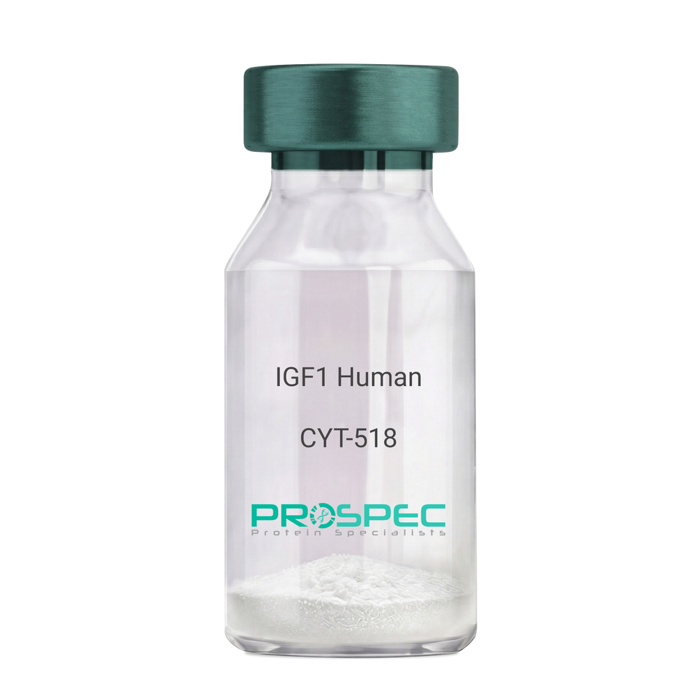
    IGF1 Human Des1-3

    Description:

    Insulin-Like Growth Factor 1 Des (1-3) Human Recombinant

    Somatomedin C, IGF-I, IGFI, IGF1, IGF-IA, Mechano growth factor, MGF, Des(1-3), Des1-3, Des 1-3, Des (1-3), IGF-1 (4-70).

    Product # :

    CYT-518

    Price :

    Quantity :

    Shipping Method :

    Room Temp Icon

    Shipped at Room temp

    Add To Cart

    More Info

    • description
    • source
    • formulation
    • purity
    • biological activity
    • More Info

    Description

    IGF-I Des(1-3) Human Recombinant produced in E.Coli is a single, non-glycosylated, polypeptide chain containing 67 amino acids (aa 4-70) and having a molecular mass of 7368.5 Dalton. IGF-1 Des1-3 is purified by proprietary chromatographic techniques.

    Source

    Escherichia Coli.

    Formulation

    The protein (1mg/ml) was lyophilized was lyophilized from a 0.2µm filtered concentrated solution in 20mM PBS, pH 7.0.

    Purity

    Greater than 97.0% as determined by:
    (a) Analysis by RP-HPLC.
    (b) Analysis by SDS-PAGE.

    Biological Activity

    The ED50 as determined by a cell proliferation assay using FDC-P1 cells is less than 2.0 ng/ml, corresponding to a specific activity of > 5×10⁵ units/mg.

    More Info

    • Introduction

      The somatomedins, or insulin-like growth factors (IGFs), comprise a family of peptides that play important roles in mammalian growth and development. IGF1 mediates many of the growth-promoting effects of growth hormone (GH; MIM 139250). Early studies showed that growth hormone did not directly stimulate the incorporation of sulfate into cartilage, but rather acted through a serum factor, termed 'sulfation factor,' which later became known as 'somatomedin' (Daughaday et al., 1972). Three main somatomedins have been characterized: somatomedin C (IGF1), somatomedin A (IGF2; MIM 147470), and somatomedin B (MIM 193190) (Rotwein, 1986; Rosenfeld, 2003).

    • Synonyms

      Somatomedin C, IGF-I, IGFI, IGF1, IGF-IA, Mechano growth factor, MGF, Des(1-3), Des1-3, Des 1-3, Des (1-3), IGF-1 (4-70).

    • Physical Appearance

      Sterile Filtered White lyophilized (freeze-dried) powder.

    • Stability

      Lyophilized IGF-I des(1-3) although stable at room temperature for 3 weeks, should be stored desiccated below -18°C. Upon reconstitution IGF1 des-1-3 should be stored at 4°C between 2-7 days and for future use below -18°C.For long term storage it is recommended to add a carrier protein (0.1% HSA or BSA).Please prevent freeze-thaw cycles.

    • Solubility

      It is recommended to reconstitute the IGF-I des(1-3) in sterile 18M-cm H2O not less than 100µg/ml, which can then be further diluted to other aqueous solutions.

    • Amino Acid Sequence

      TLCGAELVDA LQFVCGDRGF YFNKPTGYGS SSRRAPQTGI VDECCFRSCD LRRLEMYCAP LKPAKSA.

    ProSpec's products are furnished for LABORATORY RESEARCH USE ONLY. The product may not be used as drugs, agricultural or pesticidal products, food additives or household chemicals.

    Igf I Des1 3 Human
  • View Data Sheet

    Name :

    PDPN P56F7AT Antibody

    Description:

    Podoplanin clone P56F7AT, Mouse Anti Human

    Podoplanin, Glycoprotein 36, PA2.26 antigen, T1A, GP36, GP40, Gp38, OTS8, T1A2, HT1A-1, PA2.26, T1-alpha, PDPN.

    Product # :

    ANT-641

    Price :

    Quantity :

    Shipping Method :

    Ice Icon

    Shipped with Ice Packs

    Add To Cart

    More Info

    • formulation
    • More Info

    Formulation

    1mg/ml containing PBS, pH-7.4, 10% Glycerol and 0.02% Sodium Azide.

    More Info

    • Introduction

      Podoplanin is a small mucin-like type-1 transmembrane protein, typically expressed in various specialized cell types throughout the body. Podoplanin is a type-I integral membrane glycoprotein with diverse distribution in human tissues. PDPN physiological function is related to its mucin-type character. The homologous protein in other species has been described as a differentiation antigen and influenza-virus receptor.
      PDPN is expressed in lymphatic progenitor cells and afterwards during mouse development in lymphatic endothelial cells. Podoplanin is a specific marker for lymph vessel endothelial cells. Over-expression of podoplanin significantly elevates endothelial cell adhesion, migration, and tube formation. Inhibition of Podoplanin expression decreases cell adhesion in human dermal lymphatic endothelial cells. Podoplanin is used as a specific marker for lymphatic endothelium in histopathology.
      Podoplanin expression is increased in nearly all human colon, rectum, and small intestine tumors. AGGRUS may serve as a diagnostic marker that distinguishes seminomas, the majority of which over express the protein, from embryonal carcinoma in testicular germ cell tumors.

    • Synonyms

      Podoplanin, Glycoprotein 36, PA2.26 antigen, T1A, GP36, GP40, Gp38, OTS8, T1A2, HT1A-1, PA2.26, T1-alpha, PDPN.

    • Physical Appearance

      Sterile filtered colorless solution.

    • Immunogen

      Anti-human PDPN mAb, is derived from hybridization of mouse F0 myeloma cells with spleen cells from BALB/c mice immunized with recombinant human PDPN amino acids 99-207 purified from E. coli.

    • Ig Subclass

      Mouse IgG1 heavy chain and k light chain.

    • Clone

      P56F7AT.

    • Applications

      PDPN antibody has been tested by ELISA, Western blot analysis, Flow cytometry and ICC/IF to assure specificity and reactivity. Since application varies, however, each investigation should be titrated by the reagent to obtain optimal results.

    • Type

      Mouse Anti Human Monoclonal.

    • Storage Procedures

      For periods up to 1 month store at 4°C, for longer periods of time, store at -20°C. Prevent freeze thaw cycles.

    • Purification Method

      PDPN antibody was purified from mouse ascitic fluids by protein-A affinity chromatography.

    ProSpec's products are furnished for LABORATORY RESEARCH USE ONLY. The product may not be used as drugs, agricultural or pesticidal products, food additives or household chemicals.

    Pdpn P56F7At Antibody
  • View Data Sheet

    Name :

    PGK1 Antibody

    Description:

    Phosphoglycerate Kinase 1, Mouse Anti Human

    Phosphoglycerate kinase 1, Primer recognition protein 2, Cell migration-inducing gene 10 protein, PRP 2, PGKA, MIG10, MGC8947, MGC117307, MGC142128, PGK1, Pgk-1.

    Product # :

    ANT-604

    Price :

    Quantity :

    Shipping Method :

    Ice Icon

    Shipped with Ice Packs

    Add To Cart

    More Info

    • formulation
    • More Info

    Formulation

    1mg/ml containing PBS, pH-7.4, 10% Glycerol and 0.02% Sodium Azide.

    More Info

    • Introduction

      PGK1 is an X-linked enzyme that has a major role in the glycolytic pathway. PGK1 is a glycolytic enzyme which catalyzes the conversion of 1,3-diphosphoglycerate to 3-phosphoglycerate, generating an ATP molecule. PGK1 may also act as a cofactor for polymerase alpha. Defects in the PGK1 gene are usually associated with chronic hemolytic anemia, though it can be accompanied by either mental retardation or muscular disease (rhabdomyolysis). Overexpression of PGK1 and its signalling targets are possibly an expression-pathway in diffuse primary gastric carcinomas promoting peritoneal dissemination. It was shown that PGK1 is differentially expressed in the dorsolateral prefrontal cortex from patients with schizophrenia.

    • Synonyms

      Phosphoglycerate kinase 1, Primer recognition protein 2, Cell migration-inducing gene 10 protein, PRP 2, PGKA, MIG10, MGC8947, MGC117307, MGC142128, PGK1, Pgk-1.

    • Physical Appearance

      Sterile filtered colorless solution.

    • Immunogen

      Anti-human PGK1 mAb, is derived from hybridization of mouse F0 myeloma cells with spleen cells from BALB/c mice immunized with a recombinant human PGK1 protein 1-417 amino acids purified from E. coli.

    • Ig Subclass

      Mouse IgG1 heavy chain and k light chain.

    • Clone

      PAT2F4AT.

    • Applications

      PGK1 antibody has been tested by ELISA, Western blot analysis, ICC/IF and Flow cytometry to assure specificity and reactivity. Since application varies, however, each investigation should be titrated by the reagent to obtain optimal results.

    • Type

      Mouse Anti Human Monoclonal.

    • Storage Procedures

      For periods up to 1 month store at 4°C, for longer periods of time, store at -20°C. Prevent freeze thaw cycles.

    • Purification Method

      PGK1 antibody was purified from mouse ascitic fluids by protein-A affinity chromatography.

    ProSpec's products are furnished for LABORATORY RESEARCH USE ONLY. The product may not be used as drugs, agricultural or pesticidal products, food additives or household chemicals.

    Pgk1 Antibody
  • View Data Sheet

    Name :

    PGRN Human

    Description:

    Progranulin Human Recombinant

    GRN, PGRN, granulin, Acrogranin, propithelin, PC cell derived growth Factor, GEP, GP88, PEPI, PCDGF.

    Product # :

    CYT-524

    Price :

    Quantity :

    Shipping Method :

    Room Temp Icon

    Shipped at Room temp

    Add To Cart

    More Info

    • description
    • source
    • formulation
    • purity
    • biological activity
    • More Info

    Description

    Progranulin Human Recombinant produced in HEK is a single, glycosylated, polypeptide chain containing 1-593 amino acids and having a molecular mass of 74kDa. The Progranulin is purified by standard chromatographic techniques.

    Source

    HEK 293 cells.

    Formulation

    The protein contains 1xPBS.

    Purity

    Greater than 98.0% as determined by SDS-PAGE.

    Biological Activity

    Activates phospho-ERK1/2 in neuronal mouse P19 cells and regulates food intake and body weight.

    More Info

    • Introduction

      A 88-kDa progranulin, also called proepithelin and PC cell-derived growth factor, is a single precursor protein of granulins which are a family of secreted, glycosylated peptides that are cleaved from a single precursor protein with 7.5 repeats of a highly conserved 12-cysteine granulin/epithelin motif. Granulins are a variety of active, 6 kDa peptides and named granulin A (epithelin 1), granulin B (epithelin 2), granulin C, etc. Both the peptides and intact progranulin protein regulate cell growth. However, different members of the granulin protein family may act as inhibitors, stimulators, or have dual actions on cell growth. Granulin family members are important in normal development, wound healing, and tumorigenesis.

    • Synonyms

      GRN, PGRN, granulin, Acrogranin, propithelin, PC cell derived growth Factor, GEP, GP88, PEPI, PCDGF.

    • Physical Appearance

      Sterile Filtered White lyophilized (freeze-dried) powder.

    • Stability

      Lyophilized Progranulin although stable at room temperature for 3 weeks, should be stored desiccated below -18C. Upon reconstitution PGRN should be stored at 4C between 2-7 days and for future use below -18C. For long term storage it is recommended to add a carrier protein (0.1% HSA or BSA). Please prevent freeze-thaw cycles.

    • Solubility

      It is recommended to reconstitute the lyophilized Progranulin in sterile 18MΩ-cm H2O not less than 100µg/ml, which can then be further diluted to other aqueous solutions.

    ProSpec's products are furnished for LABORATORY RESEARCH USE ONLY. The product may not be used as drugs, agricultural or pesticidal products, food additives or household chemicals.

    Progranulin Human
  • View Data Sheet

    Name :

    GDF11 Human

    Description:

    Growth and Differentiation factor 11 Human Recombinant

    Growth Differentiation Factor 11, GDF-11, Bone Morphogenetic Protein 11, BMP11.

    Product # :

    CYT-402

    Price :

    Quantity :

    Shipping Method :

    Room Temp Icon

    Shipped at Room temp

    Add To Cart

    More Info

    • description
    • source
    • formulation
    • purity
    • biological activity
    • More Info

    Description

    GDF11 Human Recombinant produced in E.Coli is a non-glycosylated homodimer containing 2x10⁹ amino acids and having a total molecular mass of 25kDa.

    Source

    Escherichia Coli.

    Formulation

    Lyophilized from a concentrated (1mg/ml) solution containing 0.1% Trifluoroacetic Acid (TFA).

    Purity

    Greater than 95.0% as determined by SDS-PAGE.

    Biological Activity

    The ED50, determined by the ability to inhibit alkaline phosphatase activity in ATDC5 cells, is typically less than 1 ng/mL. This corresponds to a specific activity of 1x10⁶ units/mg.

    More Info

    • Introduction

      GDF-11 belongs to the bone morphogenetic protein (BMP) family and the TGF-beta superfamily. GDF-11 is a central developmental factor which controls muscular and neural development. In adults, GDF-11 encourages cardiac hypertrophy reverse by the revival of cardiomyocytes.

    • Synonyms

      Growth Differentiation Factor 11, GDF-11, Bone Morphogenetic Protein 11, BMP11.

    • Physical Appearance

      Sterile Filtered White lyophilized (freeze-dried) powder.

    • Stability

      Lyophilized GDF11 although stable at room temperature for 3 weeks, should be stored desiccated below -18°C. Upon reconstitution GDF11 should be stored at 4°C between 2-7 days and for future use below -18°C.For long term storage it is recommended to add a carrier protein (0.1% HSA or BSA).Please prevent freeze-thaw cycles.

    • Solubility

      It is recommended to reconstitute the lyophilized GDF11 in sterile 18M-cm H2O not less than 100µg/ml, which can then be further diluted to other aqueous solutions.

    • Amino Acid Sequence

      NLGLDCDEHS SESRCCRYPL TVDFEAFGWD WIIAPKRYKA NYCSGQCEYM FMQKYPHTHLVQQANPRGSA GPCCTPTKMS PINMLYFNDK QQIIYGKIPG MVVDRCGCS

    • Background

      What is the molecular weight/Mw of GDF11 HUMAN Protein?
      GDF11 HUMAN Protein has a total Mw of 25kDa.

      What is the source or expression system of GDF11 HUMAN Protein?
      Escherichia Coli.

      What is the Purity of GDF11 HUMAN Protein?
      GDF11 HUMAN Protein is >95% pure as determined by SDS-PAGE.

      What is the Biological Activity of GDF11 HUMAN Protein?
      The ED50, determined by the ability to inhibit alkaline phosphatase activity in ATDC5 cells, is typically less than 1 ng/mL. This corresponds to a specific activity of 1x10⁶ units/mg.
      What is the amino acid sequence of GDF11 HUMAN Protein?
      NLGLDCDEHS SESRCCRYPL TVDFEAFGWD WIIAPKRYKA NYCSGQCEYM FMQKYPHTHL
      VQQANPRGSA GPCCTPTKMS PINMLYFNDK QQIIYGKIPG MVVDRCGCS.

      What applications can GDF11 HUMAN Protein be used in?
      GDF11 HUMAN Protein can probably be used in western blot, ELISA and Lateral Flow.

      What is the endotoxin level for GDF11 HUMAN Protein?
      The endotoxin level is minimal, GDF11 HUMAN Protein was purified using conventional chromatography techniques.


    ProSpec's products are furnished for LABORATORY RESEARCH USE ONLY. The product may not be used as drugs, agricultural or pesticidal products, food additives or household chemicals.

    Gdf11 Human
  • View Data Sheet

    Name :

    PGD Human

    Description:

    Phosphogluconate Dehydrogenase Human Recombinant

    EC 1.1.1.44, 6PGD, PGDH, 6-phosphogluconate dehydrogenase decarboxylating, PGD.

    Product # :

    ENZ-501

    Price :

    Quantity :

    Shipping Method :

    Ice Icon

    Shipped with Ice Packs

    Add To Cart

    More Info

    • description
    • source
    • formulation
    • purity
    • More Info

    Description

    PGD Human Recombinant produced in E.Coli is a single, non-glycosylated, polypeptide chain containing 503 amino acids (1-483 a.a.) and having a molecular mass of 55.3 kDa. The PGD is fused to a 20 amino acid His Tag at N-terminus and purified by proprietary chromatographic techniques.

    Source

    Escherichia Coli.

    Formulation

    The PGD solution contains 20mM Tris-HCl pH-8, 1mM DTT, 0.1M NaCl and 10% glycerol.

    Purity

    Greater than 95% as determined by SDS-PAGE.

    More Info

    • Introduction

      6PGD is the second dehydrogenase in the pentose phosphate shunt. Pentose is neccesary for nucleic acid biosynthesis. The pentose phosphate cycle is a major source of NADPH 6PGD deficiency is usually asymptomatic, and the inheritance of this disorder is autosomal dominant. PGD deficiency raises the erythrocyte pyruvate kinase levels of activity and decreases glutathione synthetase, resulting in hemolysis.

    • Synonyms

      EC 1.1.1.44, 6PGD, PGDH, 6-phosphogluconate dehydrogenase decarboxylating, PGD.

    • Physical Appearance

      Sterile Filtered clear colorless solution.

    • Stability

      Store at 4°C if entire vial will be used within 2-4 weeks. Store, frozen at -20°C for longer periods of time. For long term storage it is recommended to add a carrier protein (0.1% HSA or BSA).Avoid multiple freeze-thaw cycles.

    • Amino Acid Sequence

      MGSSHHHHHH SSGLVPRGSH MAQADIALIG LAVMGQNLIL NMNDHGFVVC AFNRTVSKVD DFLANEAKGT KVVGAQSLKE MVSKLKKPRR IILLVKAGQA VDDFIEKLVP LLDTGDIIID GGNSEYRDTT RRCRDLKAKG ILFVGSGVSG GEEGARYGPS LMPGGNKEAW PHIKTIFQGI AAKVGTGEPC CDWVGDEGAG HFVKMVHNGI EYGDMQLICE AYHLMKDVLG MAQDEMAQAF EDWNKTELDS FLIEITANIL KFQDTDGKHL LPKIRDSAGQ KGTGKWTAIS ALEYGVPVTL IGEAVFARCL SSLKDERIQA SKKLKGPQKF QFDGDKKSFL EDIRKALYAS KIISYAQGFM LLRQAATEFG WTLNYGGIAL MWRGGCIIRS VFLGKIKDAF DRNPELQNLL LDDFFKSAVE NCQDSWRRAV STGVQAGIPM PCFTTALSFY DGYRHEMLPA SLIQAQRDYF GAHTYELLAK PGQFIHTNWT GHGGTVSSSS YNA.

    ProSpec's products are furnished for LABORATORY RESEARCH USE ONLY. The product may not be used as drugs, agricultural or pesticidal products, food additives or household chemicals.

    Pgd Human
  • View Data Sheet

    Name :

    VEGF Mouse

    Description:

    Vascular Endothelial Growth Factor Mouse Recombinant

    Vascular endothelial growth factor A, VEGF-A, Vascular permeability factor, VPF, VEGF, MGC70609.

    Product # :

    CYT-336

    Price :

    Quantity :

    Shipping Method :

    Room Temp Icon

    Shipped at Room temp

    Add To Cart

    More Info

    • description
    • source
    • formulation
    • purity
    • biological activity
    • More Info

    Description

    Vascular Endothelial Growth Factor Mouse Recombinant produced in E.Coli is a disulfide-linked homodimeric, double polypeptide chains containing 165 amino acids and having a molecular mass of 38.8kDa.

    Source

    Escherichia Coli.

    Formulation

    The protein was lyophilized from a 0.2µm filtered solution with PBS, pH 7.4.

    Purity

    Greater than 95.0% as determined by:
    (a) Analysis by RP-HPLC.
    (b) Analysis by SDS-PAGE.

    Biological Activity

    The biological activity is determined by the dose-dependent stimulation of the proliferation of human umbilical vein endothelial cells (HUVEC) using a concentration range of 5.0 ng/ml corresponding to a specific activity of 200,000IU/mg.

    More Info

    • Introduction

      Vascular endothelial growth factor is an important signaling protein involved in both vasculogenesis and angiogenesis. As its name implies, VEGF activity has been mostly studied on cells of the vascular endothelium, although it does have effects on a number of other cell types (e.g. stimulation monocyte/ macrophagemigration, neurons, cancer cells, kidney epithelial cells ). VEGF mediates increased vascular permeability, induces angiogenesis, vasculogenesis and endothelial cell growth, promotes cell migration, and inhibits apoptosis. In vitro, VEGF has been shown to stimulate endothelial cell mitogenesisand cell migration. VEGF is also a vasodilator and increases microvascular permeability and was originally referred to as vascular permeability factor.
      Elevated levels of this protein are linked to POEMS syndrome, also known as Crow-Fukase syndrome. Mutations in this gene have been associated with proliferative and nonproliferative diabetic retinopathy.

    • Synonyms

      Vascular endothelial growth factor A, VEGF-A, Vascular permeability factor, VPF, VEGF, MGC70609.

    • Physical Appearance

      Sterile Filtered White lyophilized (freeze-dried) powder.

    • Stability

      Lyophilized Vascular Endothelial Growth Factor although stable at room temperature for 3 weeks, should be stored desiccated below -18°C. Upon reconstitution VEGF should be stored at 4°C between 2-7 days and for future use below -18°C.For long term storage it is recommended to add a carrier protein (0.1% HSA or BSA).Please prevent freeze-thaw cycles.

    • Solubility

      It is recommended to reconstitute the lyophilized Vascular Endothelial Growth Factor in sterile 18MΩ-cm H2O not less than 100µg/ml, which can then be further diluted to other aqueous solutions.

    • Amino Acid Sequence

      MAPTTEGEQK SHEVIKFMDV YQRSYCRPIE TLVDIFQEYP DEIEYIFKPS CVPLMRCAGC CNDEALECVP TSESNITMQI MRIKPHQSQH IGEMSFLQHS RCECRPKKDR TKPEKHCEPC SERRKHLFVQ DPQTCKCSCK NTDSRCKARQ LELNERTCRC DKPRR.

    ProSpec's products are furnished for LABORATORY RESEARCH USE ONLY. The product may not be used as drugs, agricultural or pesticidal products, food additives or household chemicals.

    Vegf Mouse
  • View Data Sheet

    Name :

    DPPA3 Human

    Description:

    Developmental Pluripotency Associated 3 Human Recombinant

    Developmental pluripotency-associated protein 3, Stella-related protein, DPPA3, STELLAR, STELLA.

    Product # :

    PRO-1291

    Price :

    Quantity :

    Shipping Method :

    Ice Icon

    Shipped with Ice Packs

    Add To Cart

    More Info

    • description
    • source
    • formulation
    • purity
    • More Info

    Description

    DPPA3 Human Recombinant produced in E.Coli is a single, non-glycosylated polypeptide chain containing 182 amino acids (1-159 a.a) and having a molecular mass of 20.2kDa.DPPA3 is fused to a 24 amino acid His-tag at N-terminus & purified by proprietary chromatographic techniques.

    Source

    Escherichia Coli.

    Formulation

    DPPA3 protein solution (1mg/ml) containing 20mM Tris-HCl buffer (pH 8.0), 0.4M Urea and 10% glycerol.

    Purity

    Greater than 90.0% as determined by SDS-PAGE.

    More Info

    • Introduction

      Developmental Pluripotency Associated 3 (DPPA3) is a protein which in mice may serve as a maternal factor during the preimplantation stage of development. In mice, the DPPA3 protein has a role in transcriptional repression, cell division, and maintenance of cell pluripotentiality. In humans, related intronless loci are found on chromosomes 14 and X. DPPA3 is expressed in embryonic stem and carcinoma cells. DPPA3 has a low expression in the testis, ovary and thymus. DPPA3 is highly expressed in testicular germ cell tumors.

    • Synonyms

      Developmental pluripotency-associated protein 3, Stella-related protein, DPPA3, STELLAR, STELLA.

    • Physical Appearance

      Sterile Filtered colorless solution.

    • Stability

      Store at 4°C if entire vial will be used within 2-4 weeks. Store, frozen at -20°C for longer periods of time. For long term storage it is recommended to add a carrier protein (0.1% HSA or BSA).Avoid multiple freeze-thaw cycles.

    • Amino Acid Sequence

      MGSSHHHHHH SSGLVPRGSH MGSMDPSQFN PTYIPGSPQM LTEENSRDDS GASQISSETL IKNLSNLTIN ASSESVSPLS EALLRRESVG AAVLREIEDE WLYSRRGVRT LLSVQREKMA RLRYMLLGGV RTHERRPTNK EPKGVKKESR PFKCPCSFCV SNGWDPSENA RIGNQDTKPL QP.

    ProSpec's products are furnished for LABORATORY RESEARCH USE ONLY. The product may not be used as drugs, agricultural or pesticidal products, food additives or household chemicals.

    Dppa3 Human
  • View Data Sheet

    Name :

    PGK1 Human

    Description:

    Phosphoglycerate Kinase 1 Human Recombinant

    Phosphoglycerate kinase 1, Primer recognition protein 2, Cell migration-inducing gene 10 protein, PRP 2, PGKA, MIG10, MGC8947, MGC117307, MGC142128, PGK1.

    Product # :

    PKA-351

    Price :

    Quantity :

    Shipping Method :

    Ice Icon

    Shipped with Ice Packs

    Add To Cart

    More Info

    • description
    • source
    • formulation
    • purity
    • More Info

    Description

    PGK1 Human Recombinant produced in E.Coli is a single, non-glycosylated, polypeptide chain containing 437 amino acids (1-417 a.a.) and having a molecular mass of 46.8kDa. PGK1 is fused to 20 a.a. His-Tag at N-terminus and purified by proprietary chromatographic techniques.

    Source

    Escherichia Coli.

    Formulation

    The PGK1 solution containing 20mM Tris (pH 8.0), 10% Glycerol and 1mM DTT.

    Purity

    Greater than 95.0% as determined by SDS-PAGE.

    More Info

    • Introduction

      PGK1 is an X-linked enzyme that has a major role in the glycolytic pathway. PGK1 is a glycolytic enzyme which catalyzes the conversion of 1,3-diphosphoglycerate to 3-phosphoglycerate, generating an ATP molecule. PGK1 may also act as a cofactor for polymerase alpha. Defects in the PGK1 gene are usually associated with chronic hemolytic anemia, though it can be accompanied by either mental retardation or muscular disease (rhabdomyolysis). Overexpression of PGK1 and its signalling targets are possibly an expression-pathway in diffuse primary gastric carcinomas promoting peritoneal dissemination. It was shown that PGK1 is differentially expressed in the dorsolateral prefrontal cortex from patients with schizophrenia.

    • Synonyms

      Phosphoglycerate kinase 1, Primer recognition protein 2, Cell migration-inducing gene 10 protein, PRP 2, PGKA, MIG10, MGC8947, MGC117307, MGC142128, PGK1.

    • Physical Appearance

      Sterile filtered colorless solution.

    • Stability

      PGK1 although stable at 4°C for 1 week, should be stored below -18°C. For long term storage it is recommended to add a carrier protein (0.1% HSA or BSA).Please prevent freeze thaw cycles.

    • Amino Acid Sequence

      MGSSHHHHHH SSGLVPRGSH MSLSNKLTLD KLDVKGKRVV MRVDFNVPMK NNQITNNQRI KAAVPSIKFC LDNGAKSVVL MSHLGRPDGV PMPDKYSLEP VAVELKSLLG KDVLFLKDCV GPEVEKACAN PAAGSVILLE NLRFHVEEEG KGKDASGNKV KAEPAKIEAF RASLSKLGDV YVNDAFGTAH RAHSSMVGVN LPQKAGGFLM KKELNYFAKA LESPERPFLA ILGGAKVADK IQLINNMLDK VNEMIIGGGM AFTFLKVLNN MEIGTSLFDE EGAKIVKDLM SKAEKNGVKI TLPVDFVTAD KFDENAKTGQ ATVASGIPAG WMGLDCGPES SKKYAEAVTR AKQIVWNGPV GVFEWEAFAR GTKALMDEVV KATSRGCITI IGGGDTATCC AKWNTEDKVS HVSTGGGASL ELLEGKVLPG VDALSNI.

    ProSpec's products are furnished for LABORATORY RESEARCH USE ONLY. The product may not be used as drugs, agricultural or pesticidal products, food additives or household chemicals.

    Pgk1 Human
  • View Data Sheet

    Name :

    TGFB1 Mouse

    Description:

    Transforming Growth Factor-Beta 1 Mouse Recombinant

    Transforming growth factor beta-1, TGF-beta-1, Tgfb, Tgfb-1, TGFbeta1.

    Product # :

    CYT-858

    Price :

    Quantity :

    Shipping Method :

    Ice Icon

    Shipped with Ice Packs

    Add To Cart

    More Info

    • description
    • source
    • formulation
    • purity
    • More Info

    Description

    TGFB1 Mouse Recombinant produced in E.Coli is a single, non-glycosylated polypeptide chain containing 135 amino acids (279-390 a.a) and having a molecular mass of 15.2kDa. TGFB1 is fused to a 23 amino acid His-tag at N-terminus & purified by proprietary chromatographic techniques.

    Source

    Escherichia Coli.

    Formulation

    TGFB1 protein solution (0.25mg/ml) containing 20mM Tris-HCl (pH 8.0) and 10% glycerol.

    Purity

    Greater than 85.0% as determined by SDS-PAGE.

    More Info

    • Introduction

      Transforming growth factor betas (TGFBetas) mediate many cell-cell interactions which occur during embryonic development. Three TGFBetas have been identified in mammals. TGFBeta1, TGFBeta2 and TGFBeta3 are each synthesized as precursor proteins which are very similar in that each is cleaved to yield a 112 amino acid polypeptide which remains associated with the latent portion of the molecule.

    • Synonyms

      Transforming growth factor beta-1, TGF-beta-1, Tgfb, Tgfb-1, TGFbeta1.

    • Physical Appearance

      Sterile Filtered colorless solution.

    • Stability

      Store at 4°C if entire vial will be used within 2-4 weeks. Store, frozen at -20°C for longer periods of time. For long term storage it is recommended to add a carrier protein (0.1% HSA or BSA).Avoid multiple freeze-thaw cycles.

    • Amino Acid Sequence

      MGSSHHHHHH SSGLVPRGSH MGSALDTNYC FSSTEKNCCV RQLYIDFRKD LGWKWIHEPK GYHANFCLGP CPYIWSLDTQ YSKVLALYNQ HNPGASASPC CVPQALEPLP IVYYVGRKPK VEQLSNMIVR SCKCS.

    ProSpec's products are furnished for LABORATORY RESEARCH USE ONLY. The product may not be used as drugs, agricultural or pesticidal products, food additives or household chemicals.

    Tgfb1 Mouse
  • View Data Sheet

    Name :

    VEGF Rat

    Description:

    Vascular Endothelial Growth Factor Rat Recombinant

    Vascular endothelial growth factor A, VEGF-A, Vascular permeability factor, VPF, VEGF, MGC70609.

    Product # :

    CYT-392

    Price :

    Quantity :

    Shipping Method :

    Room Temp Icon

    Shipped at Room temp

    Add To Cart

    More Info

    • description
    • source
    • formulation
    • purity
    • biological activity
    • More Info

    Description

    Vascular Endothelial Growth Factor Rat Recombinant produced in E.Coli is a double, non-glycosylated, polypeptide chain containing 165 amino acids and having a molecular mass of 38,750 Dalton. The VEGF is purified by proprietary chromatographic techniques.

    Source

    Escherichia Coli.

    Formulation

    The protein was lyophilized from 10mM NaP, pH 7.5.

    Purity

    Greater than 97.0% as determined by:
    (a) Analysis by RP-HPLC.
    (b) Analysis by SDS-PAGE.

    Biological Activity

    Determined by the dose-dependent stimulation of the proliferation of human umbilical vein endothelial cells (HUVEC) using a concentration range of 4.0-8.0 ng/ml.

    More Info

    • Introduction

      Vascular endothelial growth factor is an important signaling protein involved in both vasculogenesis and angiogenesis. As its name implies, VEGF activity has been mostly studied on cells of the vascular endothelium, although it does have effects on a number of other cell types (e.g. stimulation monocyte/ macrophagemigration, neurons, cancer cells, kidney epithelial cells ).VEGF mediates increased vascular permeability, induces angiogenesis, vasculogenesis and endothelial cell growth, promotes cell migration, and inhibits apoptosis. In vitro, VEGF has been shown to stimulate endothelial cell mitogenesisand cell migration. VEGF is also a vasodilator and increases microvascular permeability and was originally referred to as vascular permeability factor.
      Elevated levels of this protein are linked to POEMS syndrome, also known as Crow-Fukase syndrome. Mutations in this gene have been associated with proliferative and nonproliferative diabetic retinopathy.

    • Synonyms

      Vascular endothelial growth factor A, VEGF-A, Vascular permeability factor, VPF, VEGF, MGC70609.

    • Physical Appearance

      Sterile Filtered White lyophilized (freeze-dried) powder.

    • Stability

      Lyophilized Vascular Endothelial Growth Factor although stable at room temperature for 3 weeks, should be stored desiccated below -18°C. Upon reconstitution VEGF should be stored at 4°C between 2-7 days and for future use below -18°C.For long term storage it is recommended to add a carrier protein (0.1% HSA or BSA).Please prevent freeze-thaw cycles.

    • Solubility

      It is recommended to reconstitute the lyophilized Vascular Endothelial Growth Factor in sterile 18MΩ-cm H2O not less than 100µg/ml, which can then be further diluted to other aqueous solutions.

    • Amino Acid Sequence

      MAPTTEGEQK AHEVVKFMDV YQRSYCRPIE TLVDIFQEYP DEIEYIFKPS CVPLMRCAGC CNDEALECVP TSESNVTMQI MRIKPHQSQH IGEMSFLQHS RCECRPKKDR TKPEKHCEPC SERRKHLFVQ DPQTCKCSCK NTDSRCKARQ LELNERTCRC DKPRR.

    ProSpec's products are furnished for LABORATORY RESEARCH USE ONLY. The product may not be used as drugs, agricultural or pesticidal products, food additives or household chemicals.

    Vegf Rat
  • View Data Sheet

    Name :

    PGK2 Human, Active

    Description:

    Phosphoglycerate Kinase 2 Human Recombinant, BioActive

    Phosphoglycerate kinase 2, dJ417L20.2, PGKB, PGKPSS, Phosphoglycerate kinase, testis specific

    Product # :

    PKA-125

    Price :

    Quantity :

    Shipping Method :

    Ice Icon

    Shipped with Ice Packs

    Add To Cart

    More Info

    • description
    • source
    • formulation
    • purity
    • biological activity
    • More Info

    Description

    PGK2 Human Recombinant produced in E.Coli is a single, non-glycosylated polypeptide chain containing 437 amino acids (1-417a.a.) and having a molecular mass of 46.9kDa.PGK2 is fused to a 20 amino acid His tag at N-Terminus and purified by proprietary chromatographic techniques.

    Source

    Escherichia Coli.

    Formulation

    The PGK2 solution (0.5mg/ml) contains 20% glycerol, 20mM Tris-HCl buffer (pH 8.0), 1mM DTT and 0.1M NaCl.

    Purity

    Greater than 95.0% as determined by SDS-PAGE.

    Biological Activity

    Specific activity > 500unit/mg. One unit will convert 1 umole of 1,3-Bisphosphoglycerate to 3-PGA per minute at pH 8.0 at 37˚C.

    More Info

    • Introduction

      PGK2, also known as Phosphoglycerate kinase 2, is a testis-specific form of phosphoglycerate kinase.Originally, it was assumed that PGK2was a pseudogene. However, nowadays it is known that this protein is a functional phosphoglycerate kinase. During the Embden-Meyerhof-Parnas pathway of glycolysis, in thelater stages of spermatogenesis, the protein catalyses the reversible conversion of 1, 3- bisphosphoglycerate to 3-phosphoglycerate.

    • Synonyms

      Phosphoglycerate kinase 2, dJ417L20.2, PGKB, PGKPSS, Phosphoglycerate kinase, testis specific

    • Physical Appearance

      Sterile Filtered colorless solution.

    • Stability

      Store at 4°C if entire vial will be used within 2-4 weeks. Store, frozen at -20°C for longer periods of time. For long term storage it is recommended to add a carrier protein (0.1% HSA or BSA).Avoid multiple freeze-thaw cycles.

    • Amino Acid Sequence

      MGSSHHHHHH SSGLVPRGSH MSLSKKLTLD KLDVRGKRVI MRVDFNVPMK KNQITNNQRI KASIPSIKYC LDNGAKAVVL MSHLGRPDGV PMPDKYSLAP VAVELKSLLG KDVLFLKDCV GAEVEKACAN PAPGSVILLE NLRFHVEEEG KGQDPSGKKI KAEPDKIEAF RASLSKLGDV YVNDAFGTAH RAHSSMVGVN LPHKASGFLM KKELDYFAKA LENPVRPFLA ILGGAKVADK IQLIKNMLDK VNEMIIGGGM AYTFLKVLNN MEIGASLFDE EGAKIVKDIM AKAQKNGVRI TFPVDFVTGD KFDENAQVGK ATVASGISPG WMGLDCGPES NKNHAQVVAQ ARLIVWNGPL GVFEWDAFAK GTKALMDEIV KATSKGCITV IGGGDTATCC AKWNTEDKVS HVSTGGGASL ELLEGKILPG VEALSNM

    ProSpec's products are furnished for LABORATORY RESEARCH USE ONLY. The product may not be used as drugs, agricultural or pesticidal products, food additives or household chemicals.

    Pgk2 Enzyme
  • View Data Sheet

    Name :

    DKK3 Human

    Description:

    Dickkopf-Related Protein 3 Human Recombinant

    Dickkopf 3 homolog (Xenopus laevis), dickkopf-related protein 3, regulated in glioma, RIG, RIG-like 7-1, RIG-like 5-6, Dkk-3, REIC.

    Product # :

    PRO-1175

    Price :

    Quantity :

    Shipping Method :

    Ice Icon

    Shipped with Ice Packs

    Add To Cart

    More Info

    • description
    • source
    • formulation
    • purity
    • More Info

    Description

    DKK3 Human Recombinant produced in E. coli is a single polypeptide chain containing 353 amino acids (22-350) and having a molecular mass of 38.8 kDa.DKK3 is fused to a 24 amino acid His-tag at N-terminus & purified by proprietary chromatographic techniques.

    Source

    E.coli.

    Formulation

    The DKK3 solution (0.5mg/1ml) contains 20mM Tris-HCl buffer (pH 8.0), 1M Urea, 1mM DTT and 10% glycerol.

    Purity

    Greater than 85% as determined by SDS-PAGE.

    More Info

    • Introduction

      Dickkopf-related protein 3 (DKK3) belongs to the DKK protein family including Dkk-1, 2, 3 and -4. DKK3 is a 350 amino acid secreted glycoprotein which is comprised of an N-terminal signal peptide and 2 conserved cysteine-rich domains that are separated by a 12 amino acid linker region. DKK3 is involved in embryonic development through its inhibition of the WNT signaling pathway. DKK3 gene expression is decreased in a variety of cancer cell lines and it may act as a tumor suppressor gene.

    • Synonyms

      Dickkopf 3 homolog (Xenopus laevis), dickkopf-related protein 3, regulated in glioma, RIG, RIG-like 7-1, RIG-like 5-6, Dkk-3, REIC.

    • Physical Appearance

      Sterile Filtered colorless solution.

    • Stability

      Store at 4°C if entire vial will be used within 2-4 weeks. Store, frozen at -20°C for longer periods of time. For long term storage it is recommended to add a carrier protein (0.1% HSA or BSA).Avoid multiple freeze-thaw cycles.

    • Amino Acid Sequence

      MGSSHHHYHH SSGLVPRGSH MGSMAPAPTA TSAPVKPGPA LSYPQEEATL NEMFREVEEL MEDTQHKLRS AVEEMEAEEA AAKASSEVNL ANLPPSYHNE TNTDTKVGNN TIHVHREIHK ITNNQTGQMV FSETVITSVG DEEGRRSHEC IIDEDCGPSM YCQFASFQYT CQPCRGQRML CTRDSECCGD QLCVWGHCTK MATRGSNGTI CDNQRDCQPG LCCAFQRGLL FPVCTPLPVE GELCHDPASR LLDLITWELE PDGALDRCPC ASGLLCQPHS HSLVYVCKPT FVGSRDQDGE ILLPREVPDE YEVGSFMEEV RQELEDLERS LTEEMALREP AAAAAALLGG EEI

    ProSpec's products are furnished for LABORATORY RESEARCH USE ONLY. The product may not be used as drugs, agricultural or pesticidal products, food additives or household chemicals.

    Dkk3 Human
  • View Data Sheet

    Name :

    Noggin Mouse

    Description:

    Noggin Mouse Recombinant

    Noggin, SYM1, SYNS1, NOG.

    Product # :

    CYT-600

    Price :

    Quantity :

    Shipping Method :

    Room Temp Icon

    Shipped at Room temp

    Add To Cart

    More Info

    • description
    • source
    • formulation
    • purity
    • biological activity
    • More Info

    Description

    Noggin Mouse Recombinant produced in E.Coli is a non-glycosylated, disulfide-linked protein consisting of two 206 amino acid polypeptide chains, having a total molecular mass of approximately 46.4 kDa (each chain 23.2 kDa).

    Source

    Escherichia Coli.

    Formulation

    Lyophilized from a 0.2μm filtered solution in 30% acetonitrile, 0.1% TFA.

    Purity

    Greater than 95.0% as determined by SDS-PAGE.

    Biological Activity

    The ED50 as determined by inhibiting BMP-4-induced alkaline phosphatase production of murine ATDC5 cells is less than 2ng/ml, corresponding to a specific activity of > 5.0 × 10⁵ IU/mg in the presence of 5ng/ml BMP-4.

    More Info

    • Introduction

      The secreted polypeptide noggin, encoded by the NOG gene, binds and inactivates members of the transforming growth factor-beta (TGF-beta) superfamily signaling proteins, such as bone morphogenetic protein-4 (BMP4). By diffusing through extracellular matrices more efficiently than members of the TGF-beta superfamily, noggin may have a principal role in creating morphogenic gradients. Noggin appears to have pleiotropic effect, both early in development as well as in later stages. It was originally isolated from Xenopus based on its ability to restore normal dorsal-ventral body axis in embryos that had been artificially ventralized by UV treatment. The results of the mouse knockout of noggin suggest that it is involved in numerous developmental processes, such as neural tube fusion and joint formation. Recently, several dominant human NOG mutations in unrelated families with proximal symphalangism (SYM1) and multiple synostoses syndrome (SYNS1) were identified; both SYM1 and SYNS1 have multiple joint fusion as their principal feature, and map to the same region (17q22) as NOG. All NOG mutations altered evolutionarily conserved amino acid residues. The amino acid sequence of human noggin is highly homologous to that of Xenopus, rat and mouse.

    • Synonyms

      Noggin, SYM1, SYNS1, NOG.

    • Physical Appearance

      Sterile Filtered White lyophilized (freeze-dried) powder.

    • Stability

      Lyophilized Mouse Noggin although stable at room temperature for 3 weeks, should be stored desiccated below -18°C. Upon reconstitution Mouse Noggin should be stored at 4°C between 2-7 days and for future use below -18°C.For long term storage it is recommended to add a carrier protein (0.1% HSA or BSA).Please prevent freeze-thaw cycles.

    • Solubility

      It is recommended to be briefly centrifuged prior to opening to bring the contents to the bottom. Reconstitute in 10mM HAc to a concentration of 0.1-1.0 mg/ml. Further dilutions should be made in appropriate buffered solutions.

    • Amino Acid Sequence

      MQHYLHIRPAPSDNLPLVDLIEHPDPIFDPKEKDLNETLLRSLLGGHYD
      PGFMATSPPEDRPGGGGGPAGGAEDLAELDQLLRQRPSGAMPSEIKG
      LEFSEGLAQGKKQRLSKKLRRKLQMWLWSQTFCPVLYAWNDLGSRF
      WPRYVKVGSCFSKRSCSVPEGMVCKPSKSVHLTVLRWRCQRRGQR
      CGWIPIQYPIISECKCSC.

    ProSpec's products are furnished for LABORATORY RESEARCH USE ONLY. The product may not be used as drugs, agricultural or pesticidal products, food additives or household chemicals.

    Noggin Mouse
  • View Data Sheet

    Name :

    EG VEGF Mouse

    Description:

    Endocrine Gland Vascular Endothelial Growth Factor Mouse Recombinant

    PK1, Prokineticin 1, EG-VEGF, Prok1, Endocrine-gland-derived vascular endothelial growth factor.

    Product # :

    CYT-825

    Price :

    Quantity :

    Shipping Method :

    Room Temp Icon

    Shipped at Room temp

    Add To Cart

    More Info

    • description
    • source
    • formulation
    • purity
    • More Info

    Description

    EG-VEGF Mouse Recombinant produced in E.Coli is a single, non-glycosylated, polypeptide chain containing 86 amino acids and having a molecular mass of 9.6kDa.

    Source

    Escherichia Coli.

    Formulation

    Lyophilized from a 0.2µm filtered concentrated solution in PBS pH7.4 and 3% Trehalose.

    Purity

    Greater than 95.0% as determined by:
    (a) Analysis by RP-HPLC.
    (b) Analysis by SDS-PAGE.

    More Info

    • Introduction

      Endocrine gland-derived vascular endothelial growth factor (EG-VEGF) induces proliferation, migration, and fenestration in capillary endothelial cells derived from endocrine glands. Its expression is induced by hypoxia and is restricted to the steroidogenic glands (ovary, testis, adrenal, and placenta). Its expression is often complementary to the expression of VEGF (MIM 192240), suggesting that these molecules function in a coordinated manner. EG-VEGF potently contracts gastrointestinal (gi) smooth muscle. Induces proliferation, migration and fenestration (the formation of membrane discontinuities) in capillary endothelial cells derived from endocrine glands. Has little or no effect on a variety of other endothelial and non-endothelial cell types.

    • Synonyms

      PK1, Prokineticin 1, EG-VEGF, Prok1, Endocrine-gland-derived vascular endothelial growth factor.

    • Physical Appearance

      Sterile Filtered White lyophilized (freeze-dried) powder.

    • Stability

      Lyophilized EG-VEGF Human Recombinant although stable at room temperature for 3 weeks, should be stored desiccated below -18°C. Upon reconstitution EG-VEGF should be stored at 4°C between 2-7 days and for future use below -18°C.Please prevent freeze-thaw cycles.

    • Solubility

      It is recommended to reconstitute the lyophilized Endocrine Gland Vascular Endothelial Growth Factor in sterile 18MΩ-cm H2O not less than 100µg/ml, which can then be further diluted to other aqueous solutions.

    • Amino Acid Sequence

      AVITGACERD IQCGAGTCCA ISLWLRGLRL CTPLGREGEE CHPGSHKIPF LRKRQHHTCP CSPSLLCSRF PDGRYRCFRD LKNANF.

    • Background

      What is the molecular weight/Mw of EG-VEGF Protein?
      EG-VEGF Protein has a total Mw of 9.6kDa.

      What is the source or expression system of EG-VEGF Protein?
      Escherichia Coli.

      What is the Purity of EG-VEGF Protein?
      EG-VEGF Protein is >95% pure as determined by SDS-PAGE.

      What is the Biological Activity of EG-VEGF Protein?
      The biological functionality of EG-VEGF Protein will be determined in the future.

      What is the amino acid sequence of EG-VEGF Protein?
      AVITGACERD IQCGAGTCCA ISLWLRGLRL CTPLGREGEE CHPGSHKIPF LRKRQHHTCP CSPSLLCSRF PDGRYRCFRD LKNANF.

      What applications can EG-VEGF Protein be used in?
      EG-VEGF Protein can probably be used in western blot, ELISA and Lateral Flow.

      What is the endotoxin level for EG-VEGF Protein?
      The endotoxin level is minimal, EG-VEGF Protein was purified using conventional chromatography techniques.

    ProSpec's products are furnished for LABORATORY RESEARCH USE ONLY. The product may not be used as drugs, agricultural or pesticidal products, food additives or household chemicals.

    Eg Vegf Mouse
  • View Data Sheet

    Name :

    PEDF Human, His

    Description:

    Pigment Epithelium-Derived Factor Human Recombinant, His Tag

    Pigment epithelium-derived factor, PEDF, Serpin-F1, SerpinF1, EPC-1, EPC1, PIG35.

    Product # :

    CYT-552

    Price :

    Quantity :

    Shipping Method :

    Ice Icon

    Shipped with Ice Packs

    Add To Cart

    More Info

    • description
    • source
    • formulation
    • purity
    • More Info

    Description

    PEDF Human Recombinant produced in E.Coli containing a natural variant M72T is a single, non-glycosylated, polypeptide chain containing 420 amino acids (20-418 a.a.) and having a total molecular mass of 46.7 kDa. PEDF is fused to a 20 amino acid His Tag at N-terminus and purified by proprietary chromatographic techniques.

    Source

    Escherichia Coli.

    Formulation

    The PEDF solution contains 20mM Tris-HCl buffer(pH 8.0), 0.1M NaCl , and 20% glycerol.

    Purity

    Greater than 90% as determined by SDS-PAGE.

    More Info

    • Introduction

      PEDF is a neurotrophic protein that induces extensive neuronal differentiation in retinoblastoma cells. SerpinF1 is a potent inhibitor of angiogenesis. EPC1 doesn’t undergo the stressed to relaxed conformation transition characteristic as of the active serpins since it exhibits no serine protease inhibitory activity.
      Aqueous humour level of asymmetric dimethylarginine is correlated with PEDF in humans. ADMA and PEDF levels are increased in response to inflammation in uveitis.
      Lack of PEDF expression is a potent factor for the enhancement of tumor growth and angiogenesis in breast cancer.
      PEDF & VEGF genes contribute to the development of diabetic retinopathy.
      PEDF and VEGF structural changes in blood vessel wall play an important role in the pathophysiology of PD patients.
      PEDF-overexpressing tumors exhibited reduced intratumoral angiogenesis.
      SerpinF1 is a new promising approach for the treatment of osteosarcoma.
      Levels of the natural ocular anti-angiogenic factor SentrinF1 (PEDF) is associated with proliferative retinopathy.
      VEGF secreted by retinal pigment epithelial cells upregulates PEDF expression via VEGFR-1 in an autocrine manner.
      Sentrin-F1 concentration in the aqueous humor of diabetic patients predicts who will develop progression of retinopathy.
      PEDF blocks angiogenic effects of leptin through its anti-oxidative properties.

    • Synonyms

      Pigment epithelium-derived factor, PEDF, Serpin-F1, SerpinF1, EPC-1, EPC1, PIG35.

    • Physical Appearance

      Sterile Filtered colorless solution.

    • Stability

      Store at 4°C if entire vial will be used within 2-4 weeks. Store, frozen at -20°C for longer periods of time. For long term storage it is recommended to add a carrier protein (0.1% HSA or BSA). Please prevent freeze-thaw cycles.

    • Amino Acid Sequence

      MGSSHHHHHH SSGLVPRGSH MQNPASPPEE GSPDPDSTGA LVEEEDPFFK VPVNKLAAAV SNFGYDLYRV RSSMSPTTNV LLSPLSVATA LSALSLGAEQ RTESIIHRAL YYDLISSPDI HGTYKELLDT VTAPQKNLKS ASRIVFEKKL RIKSSFVAPL EKSYGTRPRV LTGNPRLDLQ EINNWVQAQM KGKLARSTKE IPDEISILLL GVAHFKGQWV TKFDSRKTSL EDFYLDEERT VRVPMMSDPK AVLRYGLDSD LSCKIAQLPL TGSMSIIFFL PLKVTQNLTL IEESLTSEFI HDIDRELKTV QAVLTVPKLK LSYEGEVTKS LQEMKLQSLF DSPDFSKITG KPIKLTQVEH RAGFEWNEDG AGTTPSPGLQ PAHLTFPLDY HLNQPFIFVL RDTDTGALLF IGKILDPRGP.

    ProSpec's products are furnished for LABORATORY RESEARCH USE ONLY. The product may not be used as drugs, agricultural or pesticidal products, food additives or household chemicals.

    Serpinf1 Human His
  • View Data Sheet

    Name :

    FGF 8 Human

    Description:

    Fibroblast Growth Factor-8 Human Recombinant

    FGF8B, FGF-8B, FGF8-B, KAL6, HBGF-8, HBGF8, AIGF, HBGF-8, MGC149376, fibroblast growth factor 8.

    Product # :

    CYT-839

    Price :

    Quantity :

    Shipping Method :

    Room Temp Icon

    Shipped at Room temp

    Add To Cart

    More Info

    • description
    • source
    • formulation
    • purity
    • biological activity
    • More Info

    Description

    FGF 8 Human Recombinant produced in E.Coli is a non-glycosylated polypeptide chain containing 194 amino acids and having a total molecular mass of 22.5kDa.

    Source

    Escherichia Coli.

    Formulation

    Lyophilized from a sterile 0.2 micron filtered aqueous solution containing 0.1% TFA.

    Purity

    Greater than 95.0% as determined by analysis by SDS-PAGE.

    Biological Activity

    The ED50, as determined by its ability to induce proliferation of NR6-R 3T3, is 0.915ng/ml, corresponding to a specific activity of 1.1x10⁶units/mg.

    More Info

    • Introduction

      FGF8 is part of the fibroblast growth factor family. FGF family members have wide mitogenic and cell survival activities, and participate in a variety of biological processes, including embryonic development, cell growth, morphogenesis, tissue repair, tumor growth and invasion. FGF8 supports androgen and anchorage independent growth of mammary tumor cells. FGF8 over expression increases tumor growth and angiogensis. The adult expression of FGF-8 gene is restricted to testes and ovaries. FGF8 functions as an embryonic epithelial factor. FGF8 takes part in midbrain and limb development, organogenesis, embryo gastrulation and left-right axis determination.

    • Synonyms

      FGF8B, FGF-8B, FGF8-B, KAL6, HBGF-8, HBGF8, AIGF, HBGF-8, MGC149376, fibroblast growth factor 8.

    • Physical Appearance

      Sterile Filtered White lyophilized (freeze-dried) powder.

    • Stability

      Lyophilized FGF-8 although stable at room temperature for 3 weeks, should be stored desiccated below -18°C. Upon reconstitution FGF-8 should be stored at 4°C between 2-7 days and for future use below -18°C. For long term storage it is recommended to add a carrier protein (0.1% HSA or BSA). Please prevent freeze-thaw cycles.

    • Solubility

      It is recommended to reconstitute the lyophilized FGF-8 in sterile 18MΩ-cm H2O not less than 100µg/ml, which can then be further diluted to other aqueous solutions.

    • Amino Acid Sequence

      MQVTVQSSPN FTQHVREQSL VTDQLSRRLI RTYQLYSRTS GKHVQVLANK RINAMAEDGDPFAKLIVETD TFGSRVRVRG AETGLYICMN KKGKLIAKSN GKGKDCVFTE IVLENNYTAL QNAKYEGWYM AFTRKGRPRK GSKTRQHQRE VHFMKRLPRG HHTTEQSLRF EFLNYPPFTR SLRGSQRTWA PEPR.

    • Background

      What is the molecular weight/Mw of FGF8 Protein?
      FGF8 Protein has a total Mw of 22.5kDa.

      What is the source or expression system of FGF8 Protein?
      Escherichia Coli.

      What is the Purity of FGF8 Protein?
      FGF8 Protein is >95% pure as determined by SDS-PAGE.

      What is the Biological Activity of FGF8 Protein?
      The ED50, as determined by its ability to induce proliferation of NR6-R 3T3, is 0.915ng/ml, corresponding to a specific activity of 1.1x10⁶units/mg.

      What is the amino acid sequence of FGF8 Protein?
      MQVTVQSSPN FTQHVREQSL VTDQLSRRLI RTYQLYSRTS GKHVQVLANK RINAMAEDGDPFAKLIVETD TFGSRVRVRG AETGLYICMN KKGKLIAKSN GKGKDCVFTE IVLENNYTAL QNAKYEGWYM AFTRKGRPRK GSKTRQHQRE VHFMKRLPRG HHTTEQSLRF EFLNYPPFTR SLRGSQRTWA PEPR.

      What applications can FGF8 Protein be used in?
      FGF8 Protein can probably be used in western blot, ELISA and Lateral Flow.

      What is the endotoxin level for FGF8 Protein?
      The endotoxin level is minimal, FGF8 Protein was purified using conventional chromatography techniques.

    ProSpec's products are furnished for LABORATORY RESEARCH USE ONLY. The product may not be used as drugs, agricultural or pesticidal products, food additives or household chemicals.

    Fgf 8 Human
  • View Data Sheet

    Name :

    FGF12 Human

    Description:

    Fibroblast Growth Factor 12 Human Recombinant

    FGF-12, FGF12, FGF12B, FHF1, Fibroblast growth factor 12, Fibroblast growth factor homologous factor 1, FHF-1, Myocyte-activating factor.

    Product # :

    CYT-1113

    Price :

    Quantity :

    Shipping Method :

    Room Temp Icon

    Shipped at Room temp

    Add To Cart

    More Info

    • description
    • source
    • formulation
    • purity
    • biological activity
    • More Info

    Description

    Fibroblast Growth Factor 12 Human Recombinant produced in E.Coli is a single, non-glycosylated polypeptide chain containing 181 amino acids and having a molecular mass of 20.5kDa. The FGF12 is purified by proprietary chromatographic techniques.

    Source

    Escherichia Coli.

    Formulation

    Lyophilized from a 0.2μm filtered concentrated solution in PBS, pH7.4 and 1mM DTT.

    Purity

    Greater than 98.0% as determined by:
    (a) Analysis by RP-HPLC.
    (b) Analysis by SDS-PAGE.

    Biological Activity

    Determined by FGF12 binding ability in a functional ELISA. Immobilized FGFR4/Fc Chimera at 5 µg/mL (100 µL/well) can bind FGF12 with a linear range of 1.6100 ng/mL.

    More Info

    • Introduction

      FGF12 is part of the Fibroblast Growth Factor (FGF) family which has a vast mitogenic and cell survival functions, and play a role in a range of biological activities, among them are embryonic development, cell growth, morphogenesis, tissue repair, tumor growth, and invasion. FGF-12 doesn’t obtain the N-terminal signal sequence present in the majority of the FGF family members, but it contains clusters of basic residues that act as a nuclear localization signal. When transfected into mammalian cells, FGF12 accumulated in the nucleus, but was not secreted. FGF12 is involved in nervous system development and function. FGF12 binds to IB2 (islet brain-2), a cellular kinase scaffold, and voltage gated sodium channels and is also involved in intracellular signalling and ion exchange.

    • Synonyms

      FGF-12, FGF12, FGF12B, FHF1, Fibroblast growth factor 12, Fibroblast growth factor homologous factor 1, FHF-1, Myocyte-activating factor.

    • Physical Appearance

      Sterile Filtered White lyophilized (freeze-dried) powder.

    • Stability

      Lyophilized FGF12 although stable at room temperature for 3 weeks, should be stored desiccated below -18°C. Upon reconstitution Fibroblast Growth Factor 12 should be stored at 4°C between 2-7 days and for future use below -18°C. Please prevent freeze-thaw cycles.

    • Solubility

      It is recommended to reconstitute the lyophilized Fibroblast Growth Factor 12 in sterile 18MΩ-cm H2O not less than 100µg/ml, which can then be further diluted to other aqueous solutions.

    • Amino Acid Sequence

      MESKEPQLKG IVTRLFSQQG YFLQMHPDGT IDGTKDENSD YTLFNLIPVG LRVVAIQGVK ASLYVAMNGE GYLYSSDVFT PECKFKESVF ENYYVIYSST LYRQQESGRA WFLGLNKEGQ IMKGNRVKKT KPSSHFVPKP IEVCMYREQS LHEIGEKQGR SRKSSGTPTM NGGKVVNQDS T.

    • Background

      What is the molecular weight/Mw of FGF12 Protein?
      FGF12 Protein has a total Mw of 20.5kDa.

      What is the source or expression system of FGF12 Protein?
      Escherichia Coli.

      What is the Purity of FGF12 Protein?
      FGF12 Protein is >98% pure as determined by SDS-PAGE.

      What is the Biological Activity of FGF12 Protein?
      Determined by FGF12 binding ability in a functional ELISA. Immobilized FGFR4/Fc Chimera at 5 µg/mL (100 µL/well) can bind FGF12 with a linear range of 1.6100 ng/mL.

      What is the amino acid sequence of FGF12 Protein?
      MESKEPQLKG IVTRLFSQQG YFLQMHPDGT IDGTKDENSD YTLFNLIPVG LRVVAIQGVK ASLYVAMNGE GYLYSSDVFT PECKFKESVF ENYYVIYSST LYRQQESGRA WFLGLNKEGQ IMKGNRVKKT KPSSHFVPKP IEVCMYREQS LHEIGEKQGR SRKSSGTPTM NGGKVVNQDS T.

      What applications can FGF12 Protein be used in?
      FGF12 Protein can probably be used in western blot, ELISA and Lateral Flow.

      What is the endotoxin level for FGF12 Protein?
      The endotoxin level is minimal, FGF12 Protein was purified using conventional chromatography techniques.

    ProSpec's products are furnished for LABORATORY RESEARCH USE ONLY. The product may not be used as drugs, agricultural or pesticidal products, food additives or household chemicals.

    Fgf 12 Protein
  • View Data Sheet

    Name :

    PGAM1 Mouse, Active

    Description:

    Phosphoglycerate Mutase 1 Mouse Recombinant, Active

    Phosphoglycerate mutase 1, BPG-dependent PGAM 1, Phosphoglycerate mutase isozyme B, PGAM-B, Pgam1, Pgam-1, 2310050F24Rik.

    Product # :

    ENZ-980

    Price :

    Quantity :

    Shipping Method :

    Ice Icon

    Shipped with Ice Packs

    Add To Cart

    More Info

    • description
    • source
    • formulation
    • purity
    • biological activity
    • More Info

    Description

    PGAM1 Mouse Recombinant produced in E.coli is a single, non-glycosylated polypeptide chain containing 278 amino acids (1-254) and having a molecular mass of 31.4kDa.PGAM1 is fused to a 24 amino acid His-tag at N-terminus & purified by proprietary chromatographic techniques.

    Source

    Escherichia Coli.

    Formulation

    The PGAM1 solution (1mg/ml) contains 20mM Tris-HCl buffer (pH8.0), 20% glycerol, 0.1M NaCl and 1mM DTT.

    Purity

    Greater than 95% as determined by SDS-PAGE.

    Biological Activity

    Specific activity is >150units/mg, in which One unit will convert 1.0 umole of 3-phosphoglycerate to 2-phosphoglcerate per minute at pH 7.6 at 37C.

    More Info

    • Introduction

      PGAM1 is part of the phosphoglycerate mutase family. PGAM1 is an essential component of glucose and 2,3-BPGA (2,3-bisphosphoglycerate) metabolism and catalyzes the reversible reaction of 3-phosphoglycerate (3-PGA) to 2-phosphoglycerate (2-PGA) in the glycolytic pathway. PGAM1 is a dimeric enzyme containing, in different tissues, different proportions of a slow-migrating muscle (MM) isozyme, a fast-migrating brain (BB) isozyme, and a hybrid form (MB). PGAM1 mutations lead to muscle phosphoglycerate mutase deficiency, a.k.a. glycogen storage disease X.

    • Synonyms

      Phosphoglycerate mutase 1, BPG-dependent PGAM 1, Phosphoglycerate mutase isozyme B, PGAM-B, Pgam1, Pgam-1, 2310050F24Rik.

    • Physical Appearance

      Sterile Filtered clear colorless solution.

    • Stability

      Store at 4°C if entire vial will be used within 2-4 weeks. Store, frozen at -20°C for longer periods of time. For long term storage it is recommended to add a carrier protein (0.1% HSA or BSA).Avoid multiple freeze-thaw cycles.

    • Amino Acid Sequence

      MGSSHHHHHH SSGLVPRGSH MGSHMAAYKL VLIRHGESAW NLENRFSGWY DADLSPAGHE EAKRGGQALR DAGYEFDICF TSVQKRAIRT LWTVLDAIDQ MWLPVVRTWR LNERHYGGLT GLNKAETAAK HGEAQVKIWR RSYDVPPPPM EPDHPFYSNI SKDRRYADLT EDQLPSCESL KDTIARALPF WNEEIVPQIK EGKRVLIAAH GNSLRGIVKH LEGLSEEAIM ELNLPTGIPI VYELDKNLKP IKPMQFLGDE ETVRKAMEAV AAQGKVKK.

    ProSpec's products are furnished for LABORATORY RESEARCH USE ONLY. The product may not be used as drugs, agricultural or pesticidal products, food additives or household chemicals.

    Pgam1 Mouse Active
  • 1
  • …
  • 3
  • 4
  • 5
  • 6
  • 7
  • …
  • 42
Your browser does not support the video tag.

Products

  • Cytokines
  • Growth Factors
  • Chemokines
  • CD Antigens
  • Neurotrophins
  • Hormones
  • Enzymes
  • Viral Antigens
  • Recombinant Proteins
  • Natural Proteins
  • Monoclonal Antibodies
  • Polyclonal Antibodies
  • Thermal Packaging

About

  • Company
  • Founder
  • Contribution
  • Contact Us
  • My Account

Ordering

  • Ordering Method
  • Ordering - Credit Card
  • Ordering PO
  • Shipping Fees
  • FAQ's

Legal

  • Terms & Conditions
  • Privacy
  • Site Map
  • info@prospecbio.com
  • Facebook
  • Instagram
  • Linkedin Profile

© 2026 ProSpec-Tany TechnoGene Ltd. All Rights Reserved.

  • Choosing a selection results in a full page refresh.
  • Opens in a new window.