Skip to content

High-quality research proteins.

  • 1-800-805-6531

  • Mon - Sat 8.00-18.00, Sun - Closed

  • info@prospecbio.com

  • PO Box 6591 East Brunswick NJ 08816

  • Products
    • Cytokines
    • Growth factors
    • Chemokines
    • Cd antigens
    • Neurotrophins
    • Hormones
    • Enzymes
    • Viral antigens
    • Recombinant proteins
    • Natural proteins
    • Monoclonal antibodies
    • Polyclonal antibodies
    • Thermal Packaging
  • About
    • Company
    • Founder
    • Contribution
  • Customers
    • Eastern Asia
    • Eastern Europe
    • North America
    • South America
    • South East Asia
    • Southern Asia
    • Western Europe
  • Ordering
    • Ordering Method
    • Ordering - Credit Card
    • Ordering - PO
    • Shipping Fees
    • FAQs
  • Services
    • DNA Cloning
    • Protein Expression
    • Monoclonal Antibody
    • Fermentation
    • Technology License
  • Contact us
Log in

Country/region

  • USD $ | Albania
  • USD $ | Andorra
  • USD $ | Argentina
  • USD $ | Armenia
  • USD $ | Australia
  • USD $ | Austria
  • USD $ | Azerbaijan
  • USD $ | Belarus
  • USD $ | Belgium
  • USD $ | Bolivia
  • USD $ | Brazil
  • USD $ | Bulgaria
  • USD $ | Cambodia
  • USD $ | Canada
  • USD $ | Chile
  • USD $ | China
  • USD $ | Colombia
  • USD $ | Costa Rica
  • USD $ | Croatia
  • USD $ | Cyprus
  • USD $ | Czechia
  • USD $ | Denmark
  • USD $ | Estonia
  • USD $ | Finland
  • USD $ | France
  • USD $ | Georgia
  • USD $ | Germany
  • USD $ | Greece
  • USD $ | Hong Kong SAR
  • USD $ | Hungary
  • USD $ | Iceland
  • USD $ | India
  • USD $ | Ireland
  • USD $ | Israel
  • USD $ | Italy
  • USD $ | Japan
  • USD $ | Kazakhstan
  • USD $ | Latvia
  • USD $ | Liechtenstein
  • USD $ | Lithuania
  • USD $ | Madagascar
  • USD $ | Malta
  • USD $ | Mexico
  • USD $ | Moldova
  • USD $ | Monaco
  • USD $ | Netherlands
  • USD $ | New Zealand
  • USD $ | North Macedonia
  • USD $ | Norway
  • USD $ | Panama
  • USD $ | Paraguay
  • USD $ | Peru
  • USD $ | Philippines
  • USD $ | Poland
  • USD $ | Portugal
  • USD $ | Romania
  • USD $ | Singapore
  • USD $ | Slovakia
  • USD $ | Slovenia
  • USD $ | South Africa
  • USD $ | South Korea
  • USD $ | Spain
  • USD $ | Sri Lanka
  • USD $ | Sweden
  • USD $ | Switzerland
  • USD $ | Taiwan
  • USD $ | Thailand
  • USD $ | Türkiye
  • USD $ | Ukraine
  • USD $ | United Kingdom
  • USD $ | United States
  • USD $ | Uruguay
  • USD $ | Vatican City
  • USD $ | Venezuela
  • USD $ | Vietnam
  • Facebook
  • Instagram
logoProSpec - Recombinant Protein Manufacturer for academic and R&D industry
Become a distributor
Account Log in
Wishlist
Cart Cart(0)
  • Products
    +
    • Cytokines
    • Growth factors
    • Chemokines
    • Cd antigens
    • Neurotrophins
    • Hormones
    • Enzymes
    • Viral antigens
    • Recombinant proteins
    • Natural proteins
    • Monoclonal antibodies
    • Polyclonal antibodies
    • Thermal Packaging
  • About
    +
    • Company
    • Founder
    • Contribution
  • Customers
    +
    • Eastern Asia
    • Eastern Europe
    • North America
    • South America
    • South East Asia
    • Southern Asia
    • Western Europe
  • Ordering
    +
    • Ordering Method
    • Ordering - Credit Card
    • Ordering - PO
    • Shipping Fees
    • FAQs
  • Services
    +
    • DNA Cloning
    • Protein Expression
    • Monoclonal Antibody
    • Fermentation
    • Technology License
  • Contact us

Item added to your cart

View cart
View All
  • Cytokines
  • Growth factors
  • Chemokines
  • Cd antigens
  • Neurotrophins
  • Hormones
  • Enzymes
  • Viral antigens
  • Recombinant proteins
  • Natural proteins
  • Monoclonal antibodies
  • Polyclonal antibodies
  • Thermal Packaging
  • Tumor Necrosis Factor

    Tumor Necrosis Factor

    View All
  • 4-1BB

    4-1BB

    View All
  • Adiponectin

    Adiponectin

    View All
  • AIF1

    AIF1

    View All
  • Other Cytokines

    Other Cytokines

    View All
  • AITRL

    AITRL

    View All
  • Angiopoietin

    Angiopoietin

    View All
  • Apolipoprotein

    Apolipoprotein

    View All
  • B-Cell Activating Factor

    B-Cell Activating Factor

    View All
  • Beta Defensin

    Beta Defensin

    View All
  • Bone Morphogenetic Protein

    Bone Morphogenetic Protein

    View All
  • B type Natriuretic Peptide

    B type Natriuretic Peptide

    View All
  • BST

    BST

    View All
  • Betacellulin

    Betacellulin

    View All
  • Cardiotrophin

    Cardiotrophin

    View All
  • Activin

    Activin

    View All
  • Fibroblast Growth Factor

    Fibroblast Growth Factor

    View All
  • Other Growth Factors

    Other Growth Factors

    View All
  • CSF

    CSF

    View All
  • CTGF

    CTGF

    View All
  • IGFBP

    IGFBP

    View All
  • VEGF Protein

    VEGF Protein

    View All
  • EGF

    EGF

    View All
  • Epigen

    Epigen

    View All
  • Erythropoietin

    Erythropoietin

    View All
  • Insulin-Like Growth Factor

    Insulin-Like Growth Factor

    View All
  • Growth Hormone

    Growth Hormone

    View All
  • HDGF

    HDGF

    View All
  • Hepatocyte Growth Factor

    Hepatocyte Growth Factor

    View All
  • Insulin

    Insulin

    View All
  • BCA-1/ BLC (CXCL13)

    BCA-1/ BLC (CXCL13)

    View All
  • BRAK (CXCL14)

    BRAK (CXCL14)

    View All
  • C-10 (CCL6)

    C-10 (CCL6)

    View All
  • Other Chemokines

    Other Chemokines

    View All
  • MEC (CCL28)

    MEC (CCL28)

    View All
  • LD78-beta (CCL3L1)

    LD78-beta (CCL3L1)

    View All
  • CTACK (CCL27)

    CTACK (CCL27)

    View All
  • CXCL16

    CXCL16

    View All
  • CXCL17

    CXCL17

    View All
  • Platelet Factor-4 (CXCL4)

    Platelet Factor-4 (CXCL4)

    View All
  • ENA-78 (CXCL5)

    ENA-78 (CXCL5)

    View All
  • Eotaxin (CCL11,24,26)

    Eotaxin (CCL11,24,26)

    View All
  • Exodus-2 (CCL21)

    Exodus-2 (CCL21)

    View All
  • Fractalkine (CX3CL1)

    Fractalkine (CX3CL1)

    View All
  • CXCL6

    CXCL6

    View All
  • Other CD Antigens

    Other CD Antigens

    View All
  • CD14

    CD14

    View All
  • CD164

    CD164

    View All
  • CD1

    CD1

    View All
  • CD2

    CD2

    View All
  • CD200

    CD200

    View All
  • CD207

    CD207

    View All
  • CD226

    CD226

    View All
  • CD23

    CD23

    View All
  • CD244

    CD244

    View All
  • CD247

    CD247

    View All
  • CD27

    CD27

    View All
  • CD274

    CD274

    View All
  • CD300

    CD300

    View All
  • CD33

    CD33

    View All
  • Other Neurotrophins

    Other Neurotrophins

    View All
  • Beta-NGF

    Beta-NGF

    View All
  • BDNF

    BDNF

    View All
  • CDNF

    CDNF

    View All
  • CNTF

    CNTF

    View All
  • GDNF

    GDNF

    View All
  • Glia Maturation Factor

    Glia Maturation Factor

    View All
  • MANF

    MANF

    View All
  • Midkine

    Midkine

    View All
  • Neuroglobin

    Neuroglobin

    View All
  • Neurotrophic factors

    Neurotrophic factors

    View All
  • Neuregulin

    Neuregulin

    View All
  • Neuropilin

    Neuropilin

    View All
  • Pigment Epithelium-Derived Factor

    Pigment Epithelium-Derived Factor

    View All
  • Pleiotrophin

    Pleiotrophin

    View All
  • Peptide Hormones

    Peptide Hormones

    View All
  • Other Hormones

    Other Hormones

    View All
  • HCG

    HCG

    View All
  • Endothelin

    Endothelin

    View All
  • Exendin

    Exendin

    View All
  • FSH

    FSH

    View All
  • Glucagon

    Glucagon

    View All
  • GHRP

    GHRP

    View All
  • GLP

    GLP

    View All
  • Thyrostimulin

    Thyrostimulin

    View All
  • Inhibin A

    Inhibin A

    View All
  • LHRH

    LHRH

    View All
  • Melanotan

    Melanotan

    View All
  • Procalcitonin

    Procalcitonin

    View All
  • PTH

    PTH

    View All
  • Peroxiredoxin

    Peroxiredoxin

    View All
  • Peptidase

    Peptidase

    View All
  • Synthetase

    Synthetase

    View All
  • Transferase

    Transferase

    View All
  • Hydrolase

    Hydrolase

    View All
  • Dehydrogenase

    Dehydrogenase

    View All
  • ACE2

    ACE2

    View All
  • Esterase

    Esterase

    View All
  • Protein Kinases

    Protein Kinases

    View All
  • Other Enzymes

    Other Enzymes

    View All
  • Phosphatase

    Phosphatase

    View All
  • Deaminase

    Deaminase

    View All
  • Oxygenase

    Oxygenase

    View All
  • Adenylate Kinase

    Adenylate Kinase

    View All
  • Lyase

    Lyase

    View All
  • Chagas

    Chagas

    View All
  • SARS

    SARS

    View All
  • Influenza

    Influenza

    View All
  • ASFV

    ASFV

    View All
  • Astrovirus

    Astrovirus

    View All
  • Borrelia

    Borrelia

    View All
  • Helicobacter Pylori

    Helicobacter Pylori

    View All
  • Chikungunya

    Chikungunya

    View All
  • Chlamydia

    Chlamydia

    View All
  • Cytomegalo

    Cytomegalo

    View All
  • Dengue

    Dengue

    View All
  • EBV

    EBV

    View All
  • Ebola

    Ebola

    View All
  • Feline Leukemia Virus

    Feline Leukemia Virus

    View All
  • Hepatitis A

    Hepatitis A

    View All
  • Other

    Other

    View All
  • Actin

    Actin

    View All
  • ADAM

    ADAM

    View All
  • Complement Component

    Complement Component

    View All
  • Ag85

    Ag85

    View All
  • Eukaryotic Translation Initiation Factor

    Eukaryotic Translation Initiation Factor

    View All
  • Anterior Gradient Protein

    Anterior Gradient Protein

    View All
  • Heat Shock Protein

    Heat Shock Protein

    View All
  • Alpha-2-HS-Glycoprotein

    Alpha-2-HS-Glycoprotein

    View All
  • Hemoglobin

    Hemoglobin

    View All
  • Allergy

    Allergy

    View All
  • Anaplasma

    Anaplasma

    View All
  • Angiogenin

    Angiogenin

    View All
  • Ankyrin Repeat Domain

    Ankyrin Repeat Domain

    View All
  • Annexin

    Annexin

    View All
  • Other Natural Proteins

    Other Natural Proteins

    View All
  • Aprotinin

    Aprotinin

    View All
  • Transferrin

    Transferrin

    View All
  • Avidin

    Avidin

    View All
  • Anti Coagulation Factors

    Anti Coagulation Factors

    View All
  • Natural Albumin

    Natural Albumin

    View All
  • Natural Coagulation Factors

    Natural Coagulation Factors

    View All
  • Fibronectin

    Fibronectin

    View All
  • Thrombin

    Thrombin

    View All
  • Anti Human Enzyme

    Anti Human Enzyme

    View All
  • Other Monoclonal Antibodies

    Other Monoclonal Antibodies

    View All
  • Anti Human Cytokine

    Anti Human Cytokine

    View All
  • Anti Human Heat Shock Protein

    Anti Human Heat Shock Protein

    View All
  • Anti Mouse Lymphocyte

    Anti Mouse Lymphocyte

    View All
  • Anti Human Chemokine

    Anti Human Chemokine

    View All
  • Anti Human Lymphocyte

    Anti Human Lymphocyte

    View All
  • Anti Viral Monoclonal

    Anti Viral Monoclonal

    View All
  • Anti Mouse Cytokine

    Anti Mouse Cytokine

    View All
  • Anti-GST Monoclonal

    Anti-GST Monoclonal

    View All
  • Other Polyclonal Antibodies

    Other Polyclonal Antibodies

    View All
  • Anti Viral

    Anti Viral

    View All
  • Anti-GST Monoclonal

    Anti-GST Monoclonal

    View All
Back

Search results

1000 results found for “Galectin”

Name

Description

Product #

Price

Quantity

Shipping Method

  • View Data Sheet

    Name :

    GLB1 E.Coli

    Description:

    Galactosidase-Beta 1 E.coli Recombinant

    lacZ, beta-gal, β-gal.

    Product # :

    ENZ-041

    Price :

    Quantity :

    Shipping Method :

    Ice Icon

    Shipped with Ice Packs

    Add To Cart

    More Info

    • description
    • source
    • formulation
    • purity
    • More Info
    • Citations

    Description

    The E.Coli derived recombinant protein Beta-galactosidase (114 kDa) is enzymatically inactive and Non-reactive with human serum.

    Source

    Escherichia Coli.

    Formulation

    Beta-Galactosidase (1mg/1ml) is formulated in 8M urea, 20mM Tris-HCl pH 8.0, and 10mM beta-mercaptoethanol

    Purity

    Protein is >95% pure as determined by SDS-PAGE, by measuring optical density at 280 nm and by method of Bradford et al.

    Citations

    More Info

    • Introduction

      Beta-galactosidase is a hydrolase enzyme that catalyzes the hydrolysis of Beta-galactosides into monosaccharides. Substrates of different Beta-galactosidases include ganglioside GM1, lactosylceramides, lactose, and various glycoproteins. Beta-galactosidase is produced In E. coli by activation of the lac operon as the lacZ gene.

    • Synonyms

      lacZ, beta-gal, β-gal.

    • Physical Appearance

      Sterile filtered colorless solution.

    • Stability

      Protein should be stored for Short Term at 4°C and for long term at -20°C.

    • Purification Method

      Purified by proprietary chromatographic technique.

    ProSpec's products are furnished for LABORATORY RESEARCH USE ONLY. The product may not be used as drugs, agricultural or pesticidal products, food additives or household chemicals.

    Glb1 Ecoli Recombinant
  • View Data Sheet

    Name :

    Adiponectin Human, HMW

    Description:

    Adiponectin glycosylated Human Recombinant, HMW Rich

    Acrp30, AdipoQ, GBP-28, APM-1, ACDC.

    Product # :

    CYT-764

    Price :

    Quantity :

    Shipping Method :

    Room Temp Icon

    Shipped at Room temp

    Add To Cart

    More Info

    • description
    • source
    • formulation
    • purity
    • More Info
    • Citations

    Description

    Adiponectin Human Recombinant HMW Rich produced in HEK cells is a single, glycosylated, polypeptide chain (19-244) containing a total of 226 amino acids, having a molecular mass of 24.6kDa (calculated). Human Acrp30 HMW Rich migrates on SDS-PAGE under non-reducing conditions at ~ 884 kDa.

    Source

    HEK293.

    Formulation

    Acrp30 was filtered (0.4µm) and lyophilized from 0.5mg/ml in 20mM Tris, 50mM NaCl, pH 7.5 and 1mM CaCl2.

    Purity

    Greater than 90% as determined by SDS-PAGE.

    Citations

    More Info

    • Introduction

      Adiponectin is a recently discovered 244 amino acid protein, the product of the apM1 gene, which is physiologically active and specifically and highly expressed in adipose cells (Adipokine). The protein belongs to the soluble defense collagen super family; it has a collagen-like domain structurally homologous with collagen VIII and X and complement factor C1q-like globular domain. APM-1 forms homotrimers, which are the building blocks for higher order complexes found circulating in serum.

    • Synonyms

      Acrp30, AdipoQ, GBP-28, APM-1, ACDC.

    • Physical Appearance

      Filtered White lyophilized (freeze-dried) powder.

    • Stability

      Store lyophilized protein at -20°C. Aliquot the product after reconstitution to avoid repeated freezing/thawing cycles. Reconstituted protein can be stored at 4°C for a limited period of time.

    • Solubility

      It is recommended to add deionized water to a working concentration of 0.5mg/ml and let the lyophilized pellet dissolve completely. Acrp30 is not sterile! Please filter the product by an appropriate sterile filter before using it in the cell culture.

    • Amino Acid Sequence

      ETTTQGPGVL LPLPKGACTG WMAGIPGHPG HNGAPGRDGR DGTPGEKGEK GDPGLIGPKG DIGETGVPGA EGPRGFPGIQ GRKGEPGEGA YVYRSAFSVG LETYVTIPNM PIRFTKIFYN QQNHYDGSTG KFHCNIPGLY YFAYHITVYM KDVKVSLFKK DKAMLFTYDQ YQENNVDQAS GSVLLHLEVG DQVWLQVYGE GERNGLYADN DNDSTFTGFL LYHDTN.

    • Background

      What is the molecular weight/Mw of ADIPONECTIN Protein?
      ADIPONECTIN Protein has a total Mw of 24.6 kDa.

      What is the source or expression system of ADIPONECTIN Protein?
      HEK293.


      What is the Purity of ADIPONECTIN Protein?
      ADIPONECTIN Protein is >90% pure as determined by SDS-PAGE.

      What is the Biological Activity of ADIPONECTIN Protein?
      The biological functionality of ADIPONECTIN Protein will be determined in the future.

      What is the amino acid sequence of ADIPONECTIN Protein?
      ETTTQGPGVL LPLPKGACTG WMAGIPGHPG HNGAPGRDGR DGTPGEKGEK GDPGLIGPKG DIGETGVPGA EGPRGFPGIQ GRKGEPGEGA YVYRSAFSVG LETYVTIPNM PIRFTKIFYN QQNHYDGSTG KFHCNIPGLY YFAYHITVYM KDVKVSLFKK DKAMLFTYDQ YQENNVDQAS GSVLLHLEVG DQVWLQVYGE GERNGLYADN DNDSTFTGFL LYHDTN.

      What applications can ADIPONECTIN Protein be used in?
      ADIPONECTIN Protein can probably be used in western blot, ELISA and Lateral Flow.

      What is the endotoxin level for ADIPONECTIN Protein?
      The endotoxin level is minimal, ADIPONECTIN Protein was purified using conventional chromatography techniques.

    ProSpec's products are furnished for LABORATORY RESEARCH USE ONLY. The product may not be used as drugs, agricultural or pesticidal products, food additives or household chemicals.

    Adiponectin Human Hmw
  • View Data Sheet

    Name :

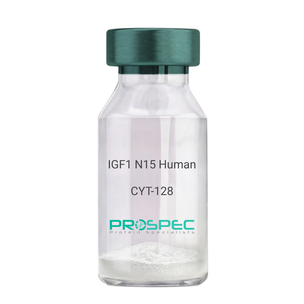
    IGF1 N15 Human

    Description:

    Insulin Like Growth Factor-1 N15 Labeled Human Recombinant

    Somatomedin C, IGF-I, IGFI, IGF1, IGF-IA, Mechano growth factor, MGF.

    Product # :

    CYT-128

    Price :

    Quantity :

    Shipping Method :

    Room Temp Icon

    Shipped at Room temp

    Add To Cart

    More Info

    • description
    • source
    • formulation
    • purity
    • biological activity
    • More Info
    • sds-page
    • Citations

    Description

    IGF1 N15 Human Recombinant produced in E.coli is a single, non-glycosylated polypeptide chain containing 70 amino acids and having a molecular mass of 7.74kDa. The N15 is stable isotope labeled. The IGF1 N15 is purified by proprietary chromatographic techniques.

    Source

    Escherichia Coli.

    Formulation

    IGF1 N15 protein was lyophilized from a 0.2µm filtered concentrated solution in PBS, pH 7.2.

    Purity

    Greater than 97.0% as determined by:
    (a) Analysis by RP-HPLC.
    (b) Analysis by SDS-PAGE.

    Biological Activity

    The ED50 as determined by a cell proliferation assay using serum free human MCF-7 cells is less than 2ng/ml, corresponding to a specific activity of > 5.0 × 10⁵ IU/mg.

    sds-page

    IGF-1 15N Human sds-page - Product image 1

    Citations

    More Info

    • Introduction

      The somatomedins, or insulin-like growth factors (IGFs), comprise a family of peptides that play important roles in mammalian growth and development. IGF1 mediates many of the growth-promoting effects of growth hormone (GH; MIM 139250). Early studies showed that growth hormone did not directly stimulate the incorporation of sulfate into cartilage, but rather acted through a serum factor, termed 'sulfation factor,' which later became known as 'somatomedin' (Daughaday et al., 1972). Three main somatomedins have been characterized: somatomedin C (IGF1), somatomedin A (IGF2; MIM 147470), and somatomedin B (MIM 193190) (Rotwein, 1986; Rosenfeld, 2003).

    • Synonyms

      Somatomedin C, IGF-I, IGFI, IGF1, IGF-IA, Mechano growth factor, MGF.

    • Physical Appearance

      Sterile Filtered White lyophilized (freeze-dried) powder.

    • Stability

      Lyophilized IGF1 N15 although stable at room temperature for 3 weeks, should be stored desiccated below -18°C. Upon reconstitution IGF1 N15 should be stored at 4°C between 2-7 days and for future use below -18°C. For long term storage it is recommended to add a carrier protein (0.1% HSA or BSA). Please prevent freeze-thaw cycles.

    • Solubility

      It is recommended to reconstitute the lyophilized IGF1 N15 in sterile 18M-cm H2O not less than 100µg/ml, which can then be further diluted to other aqueous solutions.

    • Amino Acid Sequence

      GPETLCGAEL VDALQFVCGD RGFYFNKPTG YGSSSRRAPQ TGIVDECCFR SCDLRRLEMY CAPLKPAKSA.

    • Background

      What is the molecular weight/Mw of IGF1 N15 HUMAN Protein?
      IGF1 N15 HUMAN Protein has a total Mw of 7.74kDa.

      What is the source or expression system of IGF1 N15 HUMAN Protein?
      Escherichia Coli.

      What is the Purity of IGF1 N15 HUMAN Protein?
      IGF1 N15 HUMAN Protein is >97% pure as determined by SDS-PAGE.

      What is the Biological Activity of IGF1 N15 HUMAN Protein?
      The ED50 as determined by a cell proliferation assay using serum free human MCF-7 cells is less than 2ng/ml, corresponding to a specific activity of > 5.0 × 10⁵ IU/mg.

      What is the amino acid sequence of IGF1 N15 HUMAN Protein?
      GPETLCGAEL VDALQFVCGD RGFYFNKPTG YGSSSRRAPQ TGIVDECCFR SCDLRRLEMY CAPLKPAKSA.

      What applications can IGF1 N15 HUMAN Protein be used in?
      IGF1 N15 HUMAN Protein can probably be used in western blot, ELISA and Lateral Flow.

      What is the endotoxin level for IGF1 N15 HUMAN Protein?
      The endotoxin level is minimal, IGF1 N15 HUMAN Protein was purified using conventional chromatography techniques.

    ProSpec's products are furnished for LABORATORY RESEARCH USE ONLY. The product may not be used as drugs, agricultural or pesticidal products, food additives or household chemicals.

    Igf1 N15 Human
  • View Data Sheet

    Name :

    NECTIN2 Human

    Description:

    Nectin Cell Adhesion Molecule 2 Human Recombinant

    Nectin-2, Herpes virus entry mediator B, Herpesvirus entry mediator B, HveB, Nectin cell adhesion molecule 2, Poliovirus receptor-related protein 2, CD112, HVEB, PRR2, PVRL2.

    Product # :

    PRO-2461

    Price :

    Quantity :

    Shipping Method :

    Ice Icon

    Shipped with Ice Packs

    Add To Cart

    More Info

    • description
    • source
    • formulation
    • purity
    • More Info
    • Citations

    Description

    NECTIN2 Human Recombinant produced in Sf9 Insect cells is a single, glycosylated polypeptide chain containing 337 amino acids (32-360a.a.) and having a molecular mass of 36.3kDa (Molecular size on SDS-PAGE will appear at approximately 40-57kDa).NECTIN2 is expressed with an 8 amino acids His tag at C-Terminus and purified by proprietary chromatographic techniques.

    Source

    Sf9, Insect cells.

    Formulation

    NECTIN2 protein solution (0.25mg/ml) contains Phosphate Buffered Saline (pH 7.4) and 10% glycerol.

    Purity

    Greater than 90.0% as determined by SDS-PAGE.

    Citations

    More Info

    • Introduction

      Nectin Cell Adhesion Molecule 2, also known as NECTIN2, is a Ca(2+)-independent cell-cell adhesion molecule which is one of the plasma membrane components of adherens junctions. NECTIN2 takes a vital part in the establishment of homotypic as well as heterotypic cell to cell contacts. NECTIN2 is essential for resisting herpes simplex virus type 2 infection in transfected cells. NECTIN2 is also a possible target for antibody therapy of breast & ovarian cancers. NECTIN2 might be related with human longevity. Two diseases which have been associated with NECTIN2 are Herpes Simplex and Ovarian Cystic Teratoma.

    • Synonyms

      Nectin-2, Herpes virus entry mediator B, Herpesvirus entry mediator B, HveB, Nectin cell adhesion molecule 2, Poliovirus receptor-related protein 2, CD112, HVEB, PRR2, PVRL2.

    • Stability

      Store at 4°C if entire vial will be used within 2-4 weeks. Store, frozen at -20°C for longer periods of time. For long term storage it is recommended to add a carrier protein (0.1% HSA or BSA).Avoid multiple freeze-thaw cycles.

    • Amino Acid Sequence

      QDVRVQVLPE VRGQLGGTVE LPCHLLPPVP GLYISLVTWQ RPDAPANHQN VAAFHPKMGP SFPSPKPGSE RLSFVSAKQS TGQDTEAELQ DATLALHGLT VEDEGNYTCE FATFPKGSVR GMTWLRVIAK PKNQAEAQKV TFSQDPTTVA LCISKEGRPP ARISWLSSLD WEAKETQVSG TLAGTVTVTS RFTLVPSGRA DGVTVTCKVE HESFEEPALI PVTLSVRYPP EVSISGYDDN WYLGRTDATL SCDVRSNPEP TGYDWSTTSG TFPTSAVAQG SQLVIHAVDS LFNTTFVCTV TNAVGMGRAE QVIFVRETPN TAGAGATGGL EHHHHHH.

    ProSpec's products are furnished for LABORATORY RESEARCH USE ONLY. The product may not be used as drugs, agricultural or pesticidal products, food additives or household chemicals.

    Nectin2 Human
  • View Data Sheet

    Name :

    CD33 Human

    Description:

    CD33 Human Recombinant

    CD33Molecule, CD33Antigen (Gp67), Sialic Acid Binding Ig-Like Lectin 3, Sialic Acid-Binding Ig-Like Lectin 3, SIGLEC3, gp67, SIGLEC-3, Myeloid Cell Surface AntigenCD33, p67, Siglec-3, CD33Antigen.

    Product # :

    PRO-1887

    Price :

    Quantity :

    Shipping Method :

    Ice Icon

    Shipped with Ice Packs

    Add To Cart

    More Info

    • description
    • source
    • formulation
    • purity
    • More Info
    • Citations

    Description

    CD33 Human Recombinant produced in E.Coli is a single, non-glycosylated polypeptide chain containing 265 amino acids (18-259 a.a) and having a molecular mass of 29.1kDa.CD33 is fused to a 23 amino acid His-tag at N-terminus & purified by proprietary chromatographic techniques.

    Source

    Escherichia Coli.

    Formulation

    CD33 protein solution (1mg/ml) containing 20mM Tris-HCl buffer (pH 8.0), 0.4M Urea, and 10% glycerol.

    Purity

    Greater than 90% as determined by SDS-PAGE.

    Citations

    More Info

    • Introduction

      CD33 is putative adhesion molecule of myelomonocytic-derived cells which mediates sialic-acid dependent binding to cells. CD33 prefers to bind to alpha-2,6-linked sialic acid. The sialic acid recognition site is masked by cis interactions with sialic acids on the same cell surface. In the immune response, CD33 perform as an inhibitory receptor upon ligand induced tyrosine phosphorylation by recruiting cytoplasmic phosphatases through their SH2 domains which block signal transduction through dephosphorylation of signaling molecules. In addition, CD33 induces apoptosis in acute myeloid leukemia.

    • Synonyms

      CD33Molecule, CD33Antigen (Gp67), Sialic Acid Binding Ig-Like Lectin 3, Sialic Acid-Binding Ig-Like Lectin 3, SIGLEC3, gp67, SIGLEC-3, Myeloid Cell Surface AntigenCD33, p67, Siglec-3, CD33Antigen.

    • Physical Appearance

      Sterile Filtered clear solution.

    • Stability

      Store at 4°C if entire vial will be used within 2-4 weeks. Store, frozen at -20°C for longer periods of time.For long term storage it is recommended to add a carrier protein (0.1% HSA or BSA).Please avoid freeze thaw cycles.

    • Amino Acid Sequence

      MGSSHHHHHH SSGLVPRGSH MGSDPNFWLQ VQESVTVQEG LCVLVPCTFF HPIPYYDKNS PVHGYWFREG AIISGDSPVA TNKLDQEVQE ETQGRFRLLG DPSRNNCSLS IVDARRRDNG SYFFRMERGS TKYSYKSPQL SVHVTDLTHR PKILIPGTLE PGHSKNLTCS VSWACEQGTP PIFSWLSAAP TSLGPRTTHS SVLIITPRPQ DHGTNLTCQV KFAGAGVTTE RTIQLNVTYV PQNPTTGIFP GDGSGKQETR AGVVH.

    ProSpec's products are furnished for LABORATORY RESEARCH USE ONLY. The product may not be used as drugs, agricultural or pesticidal products, food additives or household chemicals.

    Cd33 Human
  • View Data Sheet

    Name :

    MGP Human

    Description:

    Matrix Gla Protein Human Recombinant

    Matrix Gla protein, Cell growth-inhibiting gene 36 protein, MGLAP, NTI.

    Product # :

    PRO-922

    Price :

    Quantity :

    Shipping Method :

    Ice Icon

    Shipped with Ice Packs

    Add To Cart

    More Info

    • description
    • source
    • formulation
    • purity
    • More Info
    • Citations

    Description

    MGP Human Recombinant produced in E.coli is a single, non-glycosylated polypeptide chain containing 98 amino acids (20-96) and having a molecular mass of 11.8 kDa.The MGP is fused to a 21 amino acid His-Tag at N-terminus and purified by proprietary chromatographic techniques.

    Source

    E.coli.

    Formulation

    The MGP solution (1mg/ml) contains 20mM Tris-HCl buffer (pH 8.0) and 10% glycerol.

    Purity

    Greater than 90% as determined by SDS-PAGE.

    Citations

    More Info

    • Introduction

      MGP is vital for the regulation of calcification in the extracellular matrix, particularly in cartilage and arteries. The K-dependent vitamin MGP has five to six residues of Gla, a Ca2+ binding amino acid crucial for vitamin K-dependent gamma carboxylase for its formation. MGP is formed by COOH-terminal processing by carboxypeptidase B-like enzymatic activity and localized in the human bone.

    • Synonyms

      Matrix Gla protein, Cell growth-inhibiting gene 36 protein, MGLAP, NTI.

    • Physical Appearance

      Sterile Filtered colorless solution.

    • Stability

      Store at 4°C if entire vial will be used within 2-4 weeks. Store, frozen at -20°C for longer periods of time. For long term storage it is recommended to add a carrier protein (0.1% HSA or BSA).Avoid multiple freeze-thaw cycles.

    • Amino Acid Sequence

      MGSSHHHHHH SSGLVPRGSH MYESHESMES YELNPFINRR NANTFISPQQ RWRAKVQERI RERSKPVHEL NREACDDYRL CERYAMVYGY NAAYNRYF

    ProSpec's products are furnished for LABORATORY RESEARCH USE ONLY. The product may not be used as drugs, agricultural or pesticidal products, food additives or household chemicals.

    Mgp Human
  • View Data Sheet

    Name :

    G CSF Antibody, Biotin

    Description:

    Granulocyte Colony Stimulating Factor, Mouse Anti-Human, Biotin

    CSF-3, MGI-1G, GM-CSF beta, Pluripoietin, Filgrastim, Lenograstim, G-CSF, MGC45931, GCSF.

    Product # :

    ANT-240

    Price :

    Quantity :

    Shipping Method :

    Room Temp Icon

    Shipped at Room temp

    Add To Cart

    More Info

    • formulation
    • More Info
    • Citations

    Formulation

    1 mg/ml in PBS (after reconstitution).

    Citations

    More Info

    • Introduction

      GCSF is a cytokine that controls the production, differentiation, and function of granulocytes. The active protein is found extracellularly. Three transcript variants encoding three different isoforms have been found for this gene.
      Granulocyte/macrophage colony-stimulating factors are cytokines that act in hematopoiesis by controlling the production, differentiation, and function of 2 related white cell populations of the blood, the granulocytes and the monocytes-macrophages. This csf induces granulocytes.

    • Synonyms

      CSF-3, MGI-1G, GM-CSF beta, Pluripoietin, Filgrastim, Lenograstim, G-CSF, MGC45931, GCSF.

    • Solubility

      Reconstitute with sterile H20. Mix gently, wash the sides of the vial and wait 30-60 seconds before use.

    • Immunogen

      r.Human G-CSF.

    • Ig Subclass

      Mouse IgG.

    • Clone

      NYRhGCSF.

    • Applications

      Direct ELISA, Western Blot, Intra-cellular staining.

    • Titer

      For intra-cellular staining use 10µl per 1,000,000 cells.

    • Shipping Conditions

      Antibody is shipped lyophilized at ambient temperature.

    • Type

      Mouse Anti Human Monoclonal.

    • Storage Procedures

      In lyophilized form, for long periods, store at 4°C in a dry environment. After reconstitution, if not intended for use within a month, aliquot and store at -20°C.

    • Purification Method

      Protein A.

    ProSpec's products are furnished for LABORATORY RESEARCH USE ONLY. The product may not be used as drugs, agricultural or pesticidal products, food additives or household chemicals.

    G Csf Antibody Biotin
  • View Data Sheet

    Name :

    Leptin Receptor Chicken

    Description:

    Leptin Receptor Chicken Recombinant

    OB Protein, Obesity Protein, OBS, Obesity factor, Leptin Receptor.

    Product # :

    CYT-509

    Price :

    Quantity :

    Shipping Method :

    Ice Icon

    Shipped with Ice Packs

    Add To Cart

    More Info

    • description
    • source
    • formulation
    • purity
    • More Info
    • Citations

    Description

    Leptin Binding Domain Chicken Recombinant also called Leptin Receptor produced in E.Coli is a single, non-glycosylated, polypeptide chain containing 208 amino acids and having a molecular mass of 24.5 kDa. Chicken Leptin Receptor consists of the cytokine binding domain of leptin receptor amino acids 420-626 of chicken leptin receptor.The Leptin Binding Domain is purified by proprietary chromatographic techniques.

    Source

    Escherichia Coli.

    Formulation

    The protein was filter sterilized and stored at 4°C (0.2 to 0.5 mg/ml) solution of Tris-HCl buffer, pH 9.0 with 150mM NaCl.

    Purity

    Greater than 99.0% as determined by:
    (a) Analysis by SEC-HPLC.
    (b) Analysis by SDS-PAGE.

    Citations

    More Info

    • Introduction

      Leptin Receptor is a part of the gp130 family of cytokine receptors that stimulate gene transcription by activating cytosolic STAT proteins. Leptin Receptor plays a role in the regulation of fat metabolism and in novel hematopoietic pathway that is obligatory for normal lymphopoiesis. Leptin Receptorparticipates in the regulation of counter-regulatory response to hypoglycemia by inhibiting neurons of the parabrachial nucleus.Leptin Receptoraffectsspecifically on T lymphocyte responses.

    • Synonyms

      OB Protein, Obesity Protein, OBS, Obesity factor, Leptin Receptor.

    • Physical Appearance

      Sterile Filtered colorless solution at a concentration of 0.4 mg/ml.

    • Stability

      Sterile solutions at 0.5mg/ml or less are stable at 4°C for several months.

    • Amino Acid Sequence

      The sequence of the first six N-terminal amino acids was determined and was found to be Ala-Ile-Asp-Val-Asn-Ile Biological ActivityBiological Activity is evidenced by high affinity binding of mammalian leptins at 1:1 molar ratio.

    • Protein content

      Protein quantitation was carried out by two independent methods1. UV spectroscopy at 280 nm using the absorbency value of 2.45 as the extinction coefficient for a 0.1% (1mg/ml) solution. This value is calculated by the PC GENE computer analysis program of protein sequences (IntelliGenetics). 2. Analysis by RP-HPLC, using a calibrated solution of Leptin Binding Domain as a Reference Standard.

    ProSpec's products are furnished for LABORATORY RESEARCH USE ONLY. The product may not be used as drugs, agricultural or pesticidal products, food additives or household chemicals.

    Leptin Receptor Chicken
  • View Data Sheet

    Name :

    CTGF Human, HEK

    Description:

    Connective Tissue Growth Factor Human Recombinant , HEK

    CCN2, NOV2, HCS24, IGFBP8, MGC102839, CTGF.

    Product # :

    CYT-687

    Price :

    Quantity :

    Shipping Method :

    Ice Icon

    Shipped with Ice Packs

    Add To Cart

    More Info

    • description
    • source
    • formulation
    • purity
    • More Info
    • Citations

    Description

    The CTGF Human Recombinant produced in HEK293 cells, is 36kDa protein containing a total of 329 amino acid residues (aa 27-349) including a C-terminal 6×His tag.

    Source

    HEK293 cells.

    Formulation

    CTGF filtered (0.2µm) solution in 0.1M Citrate buffer pH 4.7 and 20% (w/v) glycerol.

    Purity

    Greater than 90.0% as determined by SDS-PAGE.

    Citations

    More Info

    • Introduction

      Connective Tissue Growth Factor belongs to the CCN family of proteins. The CCN family presently consists of six members in human also known as: Cyr61 (Cystein rich 61), CTGF (Connective Tissue Growth Factor), Nov (Nephroblastoma Overexpressed gene), WISP-1, 2 and 3 (Wnt-1 Induced Secreted Proteins). The CCN genes encode secreted proteins associated with the Extracellular Matrix (ECM) and cell membrane.
      CCN proteins are matricellular proteins which are involved in the regulation of various cellular functions including: proliferation, differentiation, survival, adhesion and migration. They are expressed in derivatives of the three embryonic sheets and are implicated in the development of kidney, nervous system, muscle, bone marrow, cartilage and bone. During adulthood, they are implicated in wound healing, bone fracture repair, and pathologies such as: fibrosis, vascular ailments and tumorigenesis.
      Full length secreted CCN proteins can show an antiproliferative activity, whereas truncated isoforms are likely to stimulate proliferation and behave as oncogenes.
      The full length protein consists of four modules: Module I shares partial identity with the N-terminal part of the Insulin-like Growth Factor Binding Proteins (IGFBPs).
      Module II includes a stretch of 70amino acid residues – which shares sequence identity with the Von Willebrand Factor Type C repeat (VWC).
      Module III contains sequences sharing identity with the Thrombospondin type 1 repeat (TSP1) (WSXCSXXCG), which is thought to be implicated in the binding of sulfated glycoconjugates and to be important for cell adhesion.
      Module IV, also designated CT, is encoded by exon5. It is the leasts conserved one of the four domains at the level of nucleotide sequence, but it appears to be critical for several of the biological functions attributed to the CCN proteins. Module IV resembles the CT domain of several extracellular protein including, Von Willebrand's factor and mucins. Sequence similarities to heparin-binding motifs are also found within this domain.
      Proteolysis of the secreted full-length CCN proteins that has been reported in the case of CCN2 and CCN3 might result in the production of CCN-derived peptides with high affinity for ligands that full-length CNN proteins bind only poorly. Amino-truncated CCN2 isoforms were biologically active whereas no specific biological activity has been attributed to the truncated CCN3. Although the molecular processes underlying the production of these secreted isoforms is presently unknown, it is important to note that proteolysis occur at the same amino acid residues in both CCN2 and CCN3. An elevated expression of CCN2 has also been detected by Northern blotting in human invasive mammary ductal carcinomas, dermatofibromas, pyogenic granuloma, endothelial cells of angiolipomas and angioleiomyomas, and in pancreatic tumors. A study performed with chondrosarcomas representative of various histological grades established that CCN2 expression was closely correlated with increasing levels of malignancy.
      In agreement with CCN2 playing a role in brain tumor angiogenesis, immunocytochemistry studies indicated that both glioblastoma tumor cells and proliferating endothelial cells stained positive for CCN2. In astrocytomas, CCN2 expression was particularly elevated in high grade tumors, with a marked effect of CCN2 on cell proliferation. Downregulation of CCN2 expression in these cells was associated with a growth arrest at the G1/S transition while over-expression of CCN2 induced a two-fold increase of the number of cells in the G1 phase. Gene profiling analysis allowed to identify a set of about 50 genes whose expression might account for the proliferative activity of CCN2 in these cells.
      CCN2 was seen in a higher proportion of mononuclear cells of patients with acute lymphoblastic leukemia.

    • Synonyms

      CCN2, NOV2, HCS24, IGFBP8, MGC102839, CTGF.

    • Physical Appearance

      Filtered colorless solution.

    • Stability

      Store at 4°C if entire vial will be used within 2-4 weeks. Store, frozen at -20°C for longer periods of time. For long term storage it is recommended to add a carrier protein (0.1% HSA or BSA).Avoid multiple freeze-thaw cycles.

    • Amino Acid Sequence

      QNCSGPCRCP DEPAPRCPAG VSLVLDGCGC CRVCAKQLGE LCTERDPCDP HKGLFCHFGS PANRKIGVCT AKDGAPCIFG GTVYRSGESF QSSCKYQCTC LDGAVGCMPL CSMDVRLPSP DCPFPRRVKL PGKCCEEWVC DEPKDQTVVG PALAAYRLED TFGPDPTMIR ANCLVQTTEW SACSKTCGMG ISTRVTNDNA SCRLEKQSRL CMVRPCEADL EENIKKGKKC IRTPKISKPI KFELSGCTSM KTYRAKFCGV CTDGRCCTPH RTTTLPVEFK CPDGEVMKKN MMFIKTCACH YNCPGDNDIF ESLYYRKMYG DMA HHHHHH.

    • Background

      What is the molecular weight/Mw of CTGF Protein?
      CTGF Protein has a total Mw of 36kDa.

      What is the source or expression system of CTGF Protein?
      HEK293 cells.

      What is the Purity of CTGF Protein?
      CTGF Protein is >90% pure as determined by SDS-PAGE.

      What is the Biological Activity of CTGF Protein?
      The biological functionality of CTGF Protein will be determined in the future.

      What is the amino acid sequence of CTGF Protein?
      QNCSGPCRCP DEPAPRCPAG VSLVLDGCGC CRVCAKQLGE LCTERDPCDP HKGLFCHFGS PANRKIGVCT AKDGAPCIFG GTVYRSGESF QSSCKYQCTC LDGAVGCMPL CSMDVRLPSP DCPFPRRVKL PGKCCEEWVC DEPKDQTVVG PALAAYRLED TFGPDPTMIR ANCLVQTTEW SACSKTCGMG ISTRVTNDNA SCRLEKQSRL CMVRPCEADL EENIKKGKKC IRTPKISKPI KFELSGCTSM KTYRAKFCGV CTDGRCCTPH RTTTLPVEFK CPDGEVMKKN MMFIKTCACH YNCPGDNDIF ESLYYRKMYG DMA HHHHHH.

      What applications can CTGF Protein be used in?
      CTGF Protein can probably be used in western blot, ELISA and Lateral Flow.

      What is the endotoxin level for CTGF Protein?
      The endotoxin level is minimal, CTGF Protein was purified using conventional chromatography techniques.

    ProSpec's products are furnished for LABORATORY RESEARCH USE ONLY. The product may not be used as drugs, agricultural or pesticidal products, food additives or household chemicals.

    Ctgf Humam Hek
  • View Data Sheet

    Name :

    CANX Antibody

    Description:

    Mouse Anti Human Calnexin

    Calnexin, Major histocompatibility complex class I antigen-binding protein p88, p90, IP90, CANX, CNX, FLJ26570. 

    Product # :

    ANT-713

    Price :

    Quantity :

    Shipping Method :

    Ice Icon

    Shipped with Ice Packs

    Add To Cart

    More Info

    • formulation
    • More Info
    • Citations

    Formulation

    1mg/ml containing PBS, pH-7.4, 10% Glycerol and 0.02% Sodium Azide.

    Citations

    More Info

    • Introduction

      Calnexin (CANX) belongs to the calnexin family of molecular chaperones. Calnexin is a calcium-binding, ER-associated protein that interacts briefly with newly synthesized N-linked glycoproteins, facilitating protein folding and assembly. Calnexin may also have a key role in the quality control of protein folding by retaining incorrectly folded protein subunits within the ER for degradation. Calnexin grants long-term protection of wild-type Shaker protein from ER-associated degradation. Polypeptide substrate recognition by Calnexin requires specific conformations of the Calnexin protein. Calnexin dwindles with aging and might contribute to a cytoprotection in an array of human age-related diseases.

    • Synonyms

      Calnexin, Major histocompatibility complex class I antigen-binding protein p88, p90, IP90, CANX, CNX, FLJ26570.

    • Physical Appearance

      Sterile filtered colorless solution.

    • Immunogen

      Anti-human CANX mAb, is derived from hybridization of mouse F0 myeloma cells with spleen cells from BALB/c mice immunized with a recombinant human CANX protein 21-481 amino acids purified from E. coli.

    • Ig Subclass

      Mouse IgG1 heavy chain and k light chain.

    • Clone

      PAT18B9AT.

    • Applications

      CANX antibody has been tested by ELISA and Western blot analysis to assure specificity and reactivity. Since application varies, however, each investigation should be titrated by the reagent to obtain optimal results.

    • Type

      Mouse Anti Human Monoclonal.

    • Storage Procedures

      For periods up to 1 month store at 4°C, for longer periods of time, store at -20°C. Prevent freeze thaw cycles.

    • Purification Method

      CANX antibody was purified from mouse ascitic fluids by protein-A affinity chromatography.

    ProSpec's products are furnished for LABORATORY RESEARCH USE ONLY. The product may not be used as drugs, agricultural or pesticidal products, food additives or household chemicals.

    Canx Antibody
  • View Data Sheet

    Name :

    GLB1 Human

    Description:

    Galactosidase-Beta 1 Human Recombinant

    lacZ, beta-gal, b-gal, Acid beta-galactosidase, Lactase, Elastin receptor 1

    Product # :

    ENZ-1136

    Price :

    Quantity :

    Shipping Method :

    Ice Icon

    Shipped with Ice Packs

    Add To Cart

    More Info

    • description
    • source
    • formulation
    • purity
    • More Info
    • Citations

    Description

    GLB1 Human produced in Sf9 Baculovirus cells is a single, glycosylated polypeptide chain containing 662 amino acids (24-677 a.a.) and having a molecular mass of 74.6 kDa.GLB1 is fused to an 8 amino acid His tag at C-Terminus and purified by proprietary chromatographic techniques.

    Source

    Sf9, Baculovirus cells.

    Formulation

    GLB1 Human protein (0.25mg/ml) is formulated in 10% glycerol and Phosphate-Buffered Saline (pH 7.4).

    Purity

    Greater than 90.0% as determined by SDS-PAGE.

    Citations

    More Info

    • Introduction

      Beta-galactosidase or GLB1 is a B-galactosidase found in the lysosome. This enzyme hydrolyzes the finale B-galactose from the precursors ganglioside & keratin sulfate. GLB1 has a crucial part in the creation of elastogenesis (extracellular elastic fibers) and connective tissue development. This enzyme is similar to elastin-binding protein which is a key part in non-integrin cell surface receptor. In cells that extort elastin GLB1 is linked to tropoelastin intracellularly and act as recycling molecular chaperone.

    • Synonyms

      lacZ, beta-gal, b-gal, Acid beta-galactosidase, Lactase, Elastin receptor 1

    • Physical Appearance

      Sterile Filtered colorless solution.

    • Stability

      Store at 4°C if entire vial will be used within 2-4 weeks. Store, frozen at -20°C for longer periods of time. For long term storage it is recommended to add a carrier protein (0.1% HSA or BSA).Avoid multiple freeze-thaw cycles.

    • Amino Acid Sequence

      LRNATQRMFE IDYSRDSFLK DGQPFRYISG SIHYSRVPRF YWKDRLLKMK MAGLNAIQTY VPWNFHEPWP GQYQFSEDHD VEYFLRLAHE LGLLVILRPG PYICAEWEMG GLPAWLLEKE SILLRSSDPD YLAAVDKWLG VLLPKMKPLL YQNGGPVITV QVENEYGSYF ACDFDYLRFL QKRFRHHLGD DVVLFTTDGA HKTFLKCGAL QGLYTTVDFG TGSNITDAFL SQRKCEPKGP LINSEFYTGW LDHWGQPHST IKTEAVASSL YDILARGASV NLYMFIGGTN FAYWNGANSP YAAQPTSYDY DAPLSEAGDL TEKYFALRNI IQKFEKVPEG PIPPSTPKFA YGKVTLEKLK TVGAALDILC PSGPIKSLYP LTFIQVKQHY GFVLYRTTLP QDCSNPAPLS SPLNGVHDRA YVAVDGIPQG VLERNNVITL NITGKAGATL DLLVENMGRV NYGAYINDFK GLVSNLTLSS NILTDWTIFP LDTEDAVRSH LGGWGHRDSG HHDEAWAHNS SNYTLPAFYM GNFSIPSGIP DLPQDTFIQF PGWTKGQVWI NGFNLGRYWP ARGPQLTLFV PQHILMTSAP NTITVLELEW APCSSDDPEL CAVTFVDRPV IGSSVTYDHP SKPVEKRLMP PPPQKNKDSW LDHVLEHHHH HH

    ProSpec's products are furnished for LABORATORY RESEARCH USE ONLY. The product may not be used as drugs, agricultural or pesticidal products, food additives or household chemicals.

    Glb1 Human
  • View Data Sheet

    Name :

    CALB1 Human

    Description:

    Calbindin-1 Human Recombinant

    Calbindin, Vitamin D-dependent calcium-binding protein, avian-type, Calbindin D28, D-28K, CALB1, CAB27, CALB, calbindin 1 28kDa.

    Product # :

    PRO-721

    Price :

    Quantity :

    Shipping Method :

    Ice Icon

    Shipped with Ice Packs

    Add To Cart

    More Info

    • description
    • source
    • formulation
    • purity
    • More Info
    • Citations

    Description

    CALB1 Human Recombinant produced in E.Coli is a single, non-glycosylated, polypeptide chain containing 261 amino acids (1-261 a.a.) and having a molecular mass of 30kDa.The CALB1 is purified by proprietary chromatographic techniques.

    Source

    Escherichia Coli.

    Formulation

    The CALB1 protein solution contains 20mM Tris-HCl buffer (pH8.0), 1mM DTT, 10% glycerol and 2mM EDTA.

    Purity

    Greater than 95.0% as determined by SDS-PAGE.

    Citations

    More Info

    • Introduction

      Calbindin 1 (CALB1) is a calcium binding protein that is a member of the troponin C superfamily. CALB1 plays a vital role in calcium regulation (including calcium transport and uptake, calcification of bone and teeth) and calcium associated signaling in neurons and transiently in embryological development. CALB1 also has a role in protecting neurons from apoptotic cell death. CALB1 buffers cytosolic calcium and may stimulate a membrane Ca2+-ATPase and a 3',5'-cyclic nucleotide phosphodiesterase. The biological function of CALB1 seems to be tied to the redox state of its five cysteine residues.
      CALB1 has 4 active calcium-binding domains, and 2 modified domains that seemingly have lost their calcium-binding ability. CALB1 is expressed in neural tissues. In the brain, the CALB1 synthesis is independent of vitamin-D-derived hormones.
      Disregulation of the CALB1 is associated with epilepsy, amyotrophic lateral sclerosis, Huntington's disease. The neurons in brains of Huntington disease patients are calbindin-depleted.

    • Synonyms

      Calbindin, Vitamin D-dependent calcium-binding protein, avian-type, Calbindin D28, D-28K, CALB1, CAB27, CALB, calbindin 1 28kDa.

    • Physical Appearance

      Sterile Filtered colorless solution.

    • Stability

      Store at 4°C if entire vial will be used within 2-4 weeks. Store, frozen at -20°C for longer periods of time. For long term storage it is recommended to add a carrier protein (0.1% HSA or BSA).Avoid multiple freeze-thaw cycles.

    • Amino Acid Sequence

      MAESHLQSSL ITASQFFEIW LHFDADGSGY LEGKELQNLI QELQQARKKA GLELSPEMKT FVDQYGQRDD GKIGIVELAH VLPTEENFLL LFRCQQLKSC EEFMKTWRKY DTDHSGFIET EELKNFLKDL LEKANKTVDD TKLAEYTDLM LKLFDSNNDG KLELTEMARL LPVQENFLLK FQGIKMCGKE FNKAFELYDQ DGNGYIDENE LDALLKDLCE KNKQDLDINN ITTYKKNIMA LSDGGKLYRT DLALILCAGD N.

    ProSpec's products are furnished for LABORATORY RESEARCH USE ONLY. The product may not be used as drugs, agricultural or pesticidal products, food additives or household chemicals.

    Calb1 Human
  • View Data Sheet

    Name :

    Aprotinin Protein

    Description:

    Aprotinin

    Pancreatic trypsin inhibitor, Basic protease inhibitor, BPI, BPTI, Aprotinin, AP.

    Product # :

    PRO-285

    Price :

    Quantity :

    Shipping Method :

    Room Temp Icon

    Shipped at Room temp

    Add To Cart

    More Info

    • description
    • source
    • formulation
    • More Info
    • Citations

    Description

    Aprotinin is a natural proteinase inhibitor polypeptide consisting of fifty-eight amino acids {C284H432N84O79S7} arranged in a single polypeptide chain, cross-linked by three disulfide bridges and having a molecular mass of 6512.

    Source

    Bovine Lung.

    Formulation

    The protein (1mg/ml) was lyophilized with no additives.

    Citations

    More Info

    • Introduction

      Aprotinin inhibits the activity of several proteolytic enzymes such as chymotrypsin, kallikrein, plasmin and trypsin. Aprotinin is present in blood and in most tissues, with a high concentration in lung. Aprotinin inhibits pro-inflammatory cytokine release and maintains glycoprotein homeostasis. In platelets, aprotinin reduces glycoprotein loss (e.g., GpIb, GpIIb/IIIa), while in granulocytes it prevents the expression of pro-inflammatory adhesive glycoproteins (e.g., CD11b).

    • Synonyms

      Pancreatic trypsin inhibitor, Basic protease inhibitor, BPI, BPTI, Aprotinin, AP.

    • Physical Appearance

      Sterile Filtered White lyophilized (freeze-dried) powder.

    • Stability

      Lyophilized Aprotinin although stable at room temperature for 3 weeks, should be stored desiccated below -18°C. Upon reconstitution Aprotinin should be stored at 4°C between 2-7 days and for future use below -18°C.For long term storage it is recommended to add a carrier protein (0.1% HSA or BSA).Please prevent freeze-thaw cycles.

    • Solubility

      It is recommended to reconstitute the lyophilized Aprotinin in sterile 18MΩ-cm H2O not less than 100µg/ml, which can then be further diluted to other aqueous solutions.

    • Specific Activity

      5,940 KIU (Kallikrein Inactivator Units) per mg, 3.3 pH.Eur.U/mg.

    ProSpec's products are furnished for LABORATORY RESEARCH USE ONLY. The product may not be used as drugs, agricultural or pesticidal products, food additives or household chemicals.

    Bpti
  • View Data Sheet

    Name :

    Adiponectin Mouse, Trimeric

    Description:

    Adiponectin Mouse Recombinant, Trimeric form

    Acrp30, AdipoQ, GBP-28, APM-1, ACDC.

    Product # :

    CYT-247

    Price :

    Quantity :

    Shipping Method :

    Room Temp Icon

    Shipped at Room temp

    Add To Cart

    More Info

    • description
    • source
    • formulation
    • purity
    • More Info
    • Citations

    Description

    Trimeric form of Acrp30 Mouse was expressed in HEK293 cells.The cysteine 39 was replaced with alanine (C39A) 9. mAd-C39A can only form trimer, but not hexamer or HMW form.

    Source

    HEK293 (Human embryonic kidney cell line).

    Formulation

    Mouse Acrp30 filtered (0.4µm) and lyophilized from 0.5 mg/ml in 0.05M phosphate buffer, 0.05M NaCl, pH 7.2.

    Purity

    Acrp30 Mouse purity is greater than 95% as determined by SDS-PAGE.

    Citations

    More Info

    • Introduction

      Adiponectin is a hormone exclusively expressed from adipose tissue. Many studies demonstrate that adiponectin has direct anti-diabetic, anti-atherogenic and anti-inflammatory functions. APM-1 can increase insulin sensitivity of skeletal muscle, attenuate hepatic lipogenesis and luconeogenesis, regulate NO production in endothelial cells, inhibit proliferation of smooth muscle cells and prevent lipid accumulation of macrophage cells.
      In the circulation, adiponectin is present as three different oligomeric complexes, including the high molecular weight (HMW), the middle molecular weight (MMW, also called hexamer) and low molecular weigh (MMW, also called trimer) forms 8. Different oligomeric complex of adiponectin activates different signaling pathways and exerts distinct functions.

    • Synonyms

      Acrp30, AdipoQ, GBP-28, APM-1, ACDC.

    • Physical Appearance

      Filtered white lyophilized (freeze-dried) powder.

    • Stability

      Store lyophilized Acrp30 Mouse at -20°C. Aliquot the product after reconstitution to avoid repeated freezing/thawing cycles. Reconstituted Acrp30 Mouse can be stored at 4°C for a limited period of time; it does not show any change after two weeks at 4°C.

    • Solubility

      Add deionized water to prepare a working stock solution of approximately 0.5 mg/mL and let the lyophilized pellet dissolve completely. Product is not sterile! Please filter the product by an appropriate sterile filter before using it in the cell culture.

    • Background

      What is the molecular weight/Mw of ADIPONECTIN Protein?
      ADIPONECTIN Protein has a total Mw of 26kDa.

      What is the source or expression system of ADIPONECTIN Protein?
      HEK293

      What is the Purity of ADIPONECTIN Protein?
      ADIPONECTIN Protein is >95% pure as determined by SDS-PAGE.

      What is the Biological Activity of ADIPONECTIN Protein?
      The biological functionality of ADIPONECTIN Protein will be determined in the future.

      What applications can ADIPONECTIN Protein be used in?
      ADIPONECTIN Protein can probably be used in western blot, ELISA and Lateral Flow.

      What is the endotoxin level for ADIPONECTIN Protein?
      The endotoxin level is minimal, ADIPONECTIN Protein was purified using conventional chromatography techniques.

    ProSpec's products are furnished for LABORATORY RESEARCH USE ONLY. The product may not be used as drugs, agricultural or pesticidal products, food additives or household chemicals.

    Adiponectin Mouse Trimeric
  • View Data Sheet

    Name :

    Leptin qA Ovine, PEG

    Description:

    Leptin Quadruple Antagonist Pegylated Ovine Recombinant

    Product # :

    CYT-1246

    Price :

    Quantity :

    Shipping Method :

    Room Temp Icon

    Shipped at Room temp

    Add To Cart

    More Info

    • description
    • source
    • formulation
    • purity
    • biological activity
    • More Info
    • Citations

    Description

    Leptin Antagonist Quadruple Mutant Ovine Recombinant is a single non-glycosilated polypeptide chain containing 146 amino acids and additional Ala at N-terminus. The Ovine Leptin antagonist was mutated, resulting in D23L/L39A/D40A/F41A mutant. The Ovine Leptin antagonist is bound to 20 kDa mono-PEG at N-terminus, resulting in 35.6 kDa. The Ovine Leptin Quadruple anatagonist Pegylated runs as a 48 kDa due to enlarged hydrodymanic volume. Leptin Antagonist Quadruple Mutant Ovine Recombinant was purified by proprietary chromatographic techniques.

    Source

    Escherichia coli.

    Formulation

    The Ovine Leptin Quadruple anatagonist was lyophilized from a concentrated (0.65mg/ml) solution with 0.003mM NaHCO3.

    Purity

    Greater than 95.0% as determined by:

    (a) Gel filtration analysis.

    (b) Analysis by SDS-PAGE.

    Biological Activity

    Pegylated Ovine Leptin Quadruple Antagonist inhibits leptin-induced proliferation of BAF/3 cells stably transfected with the long form of human leptin receptor. Pegylated recombinant Ovine leptin antagonist in vitro activity is 6-8 fold lower than the non-pegylated recombinant super Ovine leptin antagonist but is 15 fold higher as compared to pegylated recombinant super active ovine leptin antagonist.

    Citations

    More Info

    • Physical Appearance

      White lyophilized (freeze-dried) powder.

    • Stability

      Lyophilized Leptin Antagonist Quadruple Mutant Ovine Recombinant although stable at room temperature for several weeks, should be stored desiccated below -18°C. Upon reconstitution at > 0.1 mg/ml and up to 2mM and filter sterilization LEP mutant can be stored at 4°C or even room temperature for several weeks making it suitable for long term infusion studies using osmotic pumps. At lower concentration addition of a carrier protein (0.1% HSA or BSA) is suggested. Please prevent freeze-thaw cycles.

    • Solubility

      It is recommended to reconstitute the lyophilized Leptin Antagonist Quadruple Mutant Ovine Recombinant in sterile water or sterile 0.4% NaHCO3adjusted to pH 8-9, not less than 100µg/ml, which can then be further diluted with other aqueous solutions.

    • Background

      Leptin is mainly produced by adipocytes. Leptin’s main part is to regulate long-term energy balance. Leptin is encoded by the LEP gene. Leptin effects mostly on leptin receptors in the cell mambrane of various cells in the human body. The leptin receptor can be found on a various cell types. The leptin receptor is a single-transmembrane-domain type 1 cytokine receptor. leptin levels influence satiety, appetite and triggers behaviours which save energy. High leptin levels are interpreted by the brain that energy reserves are high, whereas low leptin levels means that energy reserves are low, in the process adapting the organism to starvation through a variety of metabolic, neurobiochemical, endocrine and behavioral change.

    • Protein content

      Protein quantization was carried out by UV spectroscopy at 280 nm using the absorbency value of 0.2 as the extinction coefficient for a 0.1% (1mg/ml) solution at pH 8.0. This value is calculated by the PC GENE computer analysis program of protein sequences (IntelliGenetics).

    ProSpec's products are furnished for LABORATORY RESEARCH USE ONLY. The product may not be used as drugs, agricultural or pesticidal products, food additives or household chemicals.

    Leptin Antagonist Peg Ovine
  • View Data Sheet

    Name :

    SRGN Human, HEK

    Description:

    Serglycin Human Recombinant, HEK

    Serglycin, PRG, PRG1, PPG, Proteoglycan 1 secretory granule, Hematopoetic proteoglycan core protein, Platelet proteoglycan core protein, proteoglycan protein core for mast cell secretory granule.

    Product # :

    PRO-2046

    Price :

    Quantity :

    Shipping Method :

    Ice Icon

    Shipped with Ice Packs

    Add To Cart

    More Info

    • description
    • source
    • formulation
    • purity
    • More Info
    • Citations

    Description

    Serglycin Human Recombinant produced in HEK cells is a single, glycosylated, polypeptide chain (Tyr28-Leu158) containing a total of 137 amino acids, having a calculated molecular mass of 15.5kDa and fused to a 6 aa His tag at C-Terminus.

    Source

    HEK 293.

    Formulation

    SRGN filtered (0.4µm) solution in phosphate buffered saline and 20% (w/v) glycerol.

    Purity

    Greater than 95.0% as determined by SDS-PAGE.

    Citations

    More Info

    • Introduction

      SRGN is identified as a hematopoietic cell granule proteoglycan. Proteoglycans stored in the secretory granules of various hematopoietic cells also hold a protease-resistant peptide core, and is vital for neutralizing hydrolytic enzymes. SRGN is related to the macromolecular complex of granzymes and perforin that acts as a intermediary of granule-mediated apoptosis.

    • Synonyms

      Serglycin, PRG, PRG1, PPG, Proteoglycan 1 secretory granule, Hematopoetic proteoglycan core protein, Platelet proteoglycan core protein, proteoglycan protein core for mast cell secretory granule.

    • Physical Appearance

      Filtered colorless solution.

    • Stability

      Store at 4°C if entire vial will be used within 2-4 weeks. Store, frozen at -20°C for longer periods of time. For long term storage it is recommended to add a carrier protein (0.1% HSA or BSA).Avoid multiple freeze-thaw cycles.

    • Amino Acid Sequence

      YPTRRARYQW VRCNPDSNSA NCLEEKGPMF ELLPGESNKI PRLRTDLFPK TRIQDLNRIF PLSEDYSGSG FGSGSGSGSG SGSGFLTEME QDYQLVDESD AFHDNLRSLD RNLPSDSQDL GQHGLEEDFM L HHHHHH.

    ProSpec's products are furnished for LABORATORY RESEARCH USE ONLY. The product may not be used as drugs, agricultural or pesticidal products, food additives or household chemicals.

    Srgn Human Hek
  • View Data Sheet

    Name :

    CLEC5A Human

    Description:

    C-Type Lectin Domain Family 5, Member A Human Recombinant

    C-type lectin domain family 5 member A , CLECSF5, MDL-1, MDL1, C-type lectin superfamily member 5, Myeloid DAP12-associating lectin 1.

    Product # :

    PRO-2165

    Price :

    Quantity :

    Shipping Method :

    Ice Icon

    Shipped with Ice Packs

    Add To Cart

    More Info

    • description
    • source
    • formulation
    • purity
    • More Info
    • Citations

    Description

    CLEC5A Human Recombinant produced in E.Coli is a single, non-glycosylated polypeptide chain containing 184 amino acids (28-188 a.a) and having a molecular mass of 20.8kDa.CLEC5A is fused to a 23 amino acid His-tag at N-terminus & purified by proprietary chromatographic techniques.

    Source

    Escherichia Coli.

    Formulation

    CLEC5A protein solution (0.25mg/ml) containing 20mM Tris-HCl buffer, (pH8.0) and 10% glycerol.

    Purity

    Greater than 80.0% as determined by SDS-PAGE.

    Citations

    More Info

    • Introduction

      C-type lectin domain family 5-member A isoform 1 (CLEC5A) is part of the CTL/CTLD superfamily which carry various functions, for instance cell-cell signaling, cell adhesion, glycoprotein turnover, in addition to their inflammation & immune response abilities. CLEC5A operates as a cell attachment receptor for all 4 serotypes of Dengue virus in addition to Japanese encephalitis virus. CLEC5A binds to the dengue virus and it triggers signaling through the phosphylation of TYROBP, as a result no viral entrance occurs, however this interaction does stimulate proinflammatory cytokine release. CLEC5A preforms as a positive regulator of osteoclastogenesis and also a main regulator of synovial injury & bone erosion for the period of autoimmune joint inflammation.

    • Synonyms

      C-type lectin domain family 5 member A , CLECSF5, MDL-1, MDL1, C-type lectin superfamily member 5, Myeloid DAP12-associating lectin 1.

    • Physical Appearance

      Sterile Filtered clear solution.

    • Stability

      Store at 4°C if entire vial will be used within 2-4 weeks. Store, frozen at -20°C for longer periods of time. For long term storage it is recommended to add a carrier protein (0.1% HSA or BSA).Avoid multiple freeze-thaw cycles.

    • Amino Acid Sequence

      MGSSHHHHHH SSGLVPRGSH MGSPQIFNKS NDGFTTTRSY GTVSQIFGSS SPSPNGFITT RSYGTVCPKD WEFYQARCFF LSTSESSWNE SRDFCKGKGS TLAIVNTPEK LKFLQDITDA EKYFIGLIYH REEKRWRWIN NSVFNGNVTN QNQNFNCATI GLTKTFDAAS CDISYRRICE KNAK.

    ProSpec's products are furnished for LABORATORY RESEARCH USE ONLY. The product may not be used as drugs, agricultural or pesticidal products, food additives or household chemicals.

    Clec5A Human
  • View Data Sheet

    Name :

    Adiponectin Human (72-244)

    Description:

    Adiponectin (72-244) Human Recombinant

    Acrp30, AdipoQ, GBP-28, APM-1, ACDC.

    Product # :

    CYT-1231

    Price :

    Quantity :

    Shipping Method :

    Room Temp Icon

    Shipped at Room temp

    Add To Cart

    More Info

    • description
    • source
    • formulation
    • purity
    • More Info
    • Citations

    Description

    The Adiponectin Human is created as a recombinant protein with a 4kda N-terminal fusion of His Tag. The Adiponectin His-Tagged Fusion Protein, produced in E. coli, is a 24kDa protein containing 173 amino acid residues of the Acrp30 Human, 72-244 amino acids.

    Source

    Escherichia Coli.

    Formulation

    Each mg was lyophilized with 1xPBS, 0.4% SDS and 4mM DTT.

    Purity

    Greater than 90% as determined by SDS-PAGE.

    Citations

    More Info

    • Synonyms

      Acrp30, AdipoQ, GBP-28, APM-1, ACDC.

    • Physical Appearance

      Filtered White lyophilized (freeze-dried) powder.

    • Stability

      Store lyophilized Adiponectin at -20°C. Aliquot the product after reconstitution to avoid repeated freezing/thawing cycles. Reconstituted protein can be stored at 4°C for a limited period of time; it does not show any change after two weeks at 4°C.

    • Solubility

      It is recommended to add deionized water to prepare a working stock solution of approximately 0.5mg/ml and let the lyophilized pellet dissolve completely. Product is not sterile! Please filter the product by an appropriate sterile filter before using it on cell culture.

    • Amino Acid Sequence

      IGPKGDI GETGVPGAEG PRGFPGIQGR KGEPGEGAYV YRSAFSVGLE TYVTIPNMPI RFTKIFYNQQ NHYDGSTGKF HCNIPGLYYF AYHITVYMKD VKVSLFKKDK AMLFTYDQYQ ENNVDQASGS VLLHLEVGDQ VWLQVYGEGE RNGLYADNDN DSTFTGFLLY HDTN

    • Background

      Adiponectin is a protein produced and secreted by adipose tissue. Adiponectin takes part in regulating glucose levels as well as fatty acid breakdown.

      Adiponectin ‘s Functions:

      Anti-Inflammatory Effects - Adiponectin has anti-inflammatory properties that helps mitigate chronic inflammation.

      Regulation of Glucose and Lipid Metabolism - Adiponectin Enhances insulin sensitivity, helping in regulation of blood sugar levels and also promotes fatty acid oxidation, which helps reduce fat accumulation.

      Cardiovascular Health - It may influence vascular health and is associated with a lower risk of cardiovascular diseases.

      Levels and Health Implications:

      Normal Levels - usually, higher levels of adiponectin are associated with a lower risk of metabolic syndrome, cardiovascular diseases and type 2 diabetes.

      Low Levels - Reduced adiponectin levels are often linked with obesity, insulin resistance, and other metabolic disorders.

      Factors Influencing on the Adiponectin Levels:

      Weight - High body fat (especially visceral fat) can lower adiponectin levels.

      Diet and Exercise - Regular physical activity and a healthy diet can increase adiponectin levels.

      Genetics - Genetic factors might also be an influence on an individual adiponectin level.

      Adiponectin is an important component in metabolic health, therefore continuing the research of its functions and regulation keeps advance our understanding of its role in diseases like diabetes and cardiovascular conditions.

      What is the molecular weight / Mw of ADIPONECTIN Protein?
      ADIPONECTIN Protein has a total Mw of 24kDa.

      What is the source or expression system of ADIPONECTIN Protein?
      Escherichia Coli.

      What is the Purity of ADIPONECTIN Protein?
      ADIPONECTINProtein is >90% pure as determined by SDS-PAGE.

      What is the Biological Activity of ADIPONECTIN Protein?
      The biological functionality of ADIPONECTIN Protein will be determined in the future.

      What is the amino acid sequence of ADIPONECTIN Protein?
      IGPKGDI GETGVPGAEG PRGFPGIQGR KGEPGEGAYV YRSAFSVGLE TYVTIPNMPI RFTKIFYNQQ NHYDGSTGKF HCNIPGLYYF AYHITVYMKD VKVSLFKKDK AMLFTYDQYQ ENNVDQASGS VLLHLEVGDQ VWLQVYGEGE RNGLYADNDN DSTFTGFLLY HDTN.

      What applications can ADIPONECTIN Protein be used in ?
      ADIPONECTIN Protein can probably be used in western blot, ELISA and Lateral Flow.

      What is the endotoxin level for ADIPONECTIN Protein?
      The endotoxin level is minimal, ADIPONECTIN Protein was purified using conventional chromatography techniques.


    ProSpec's products are furnished for LABORATORY RESEARCH USE ONLY. The product may not be used as drugs, agricultural or pesticidal products, food additives or household chemicals.

    Adiponectin Human Protein
  • View Data Sheet

    Name :

    GABARAPL2 Human

    Description:

    GABA(A) Receptor-Associated Protein Like 2 Human Recombinant

    GABA(A) receptor-associated protein-like 2, GATE-16, GEF2, ATG8, ATG8C, Ganglioside expression factor 2, General protein transport factor p16, Golgi-associated ATPase enhancer of 16 kDa, MAP1 light chain 3-related protein, gamma-aminobutyric acid receptor-associated protein-like 2, MAP1 light chain 3 related protein, FLC3A.

    Product # :

    PRO-079

    Price :

    Quantity :

    Shipping Method :

    Ice Icon

    Shipped with Ice Packs

    Add To Cart

    More Info

    • description
    • source
    • formulation
    • purity
    • More Info
    • Citations

    Description

    GABARAPL2 produced in E.Coli is a single, non-glycosylated polypeptide chain containing 137 amino acids (1-117a.a.) and having a molecular mass of 15.8kDa. GABARAPL2 is fused to a 20 amino acid His-tag at N-terminus & purified by proprietary chromatographic techniques.

    Source

    Escherichia Coli.

    Formulation

    The GABARAPL2 protein solution (0.5mg/1ml) is formulated in 20mM Tris-HCl buffer (pH8.0), 0.2M NaCl, 5mM DTT and 20% glycerol.

    Purity

    Greater than 90% as determined by SDS-PAGE.

    Citations

    More Info

    • Introduction

      GABARAPL2 takes part in intra Golgi traffic and modulates intra Golgi transport by coupling between NSF activity and SNAREs activation. GABARAPL2 first stimulates the ATPase activity of NSF which in turn stimulates the association with GOSR1.

    • Synonyms

      GABA(A) receptor-associated protein-like 2, GATE-16, GEF2, ATG8, ATG8C, Ganglioside expression factor 2, General protein transport factor p16, Golgi-associated ATPase enhancer of 16 kDa, MAP1 light chain 3-related protein, gamma-aminobutyric acid receptor-associated protein-like 2, MAP1 light chain 3 related protein, FLC3A.

    • Physical Appearance

      Sterile Filtered clear solution.

    • Stability

      Store at 4°C if entire vial will be used within 2-4 weeks. Store, frozen at -20°C for longer periods of time.Please avoid freeze thaw cycles.

    • Amino Acid Sequence

      MGSSHHHHHH SSGLVPRGSH MKWMFKEDHS LEHRCVESAK IRAKYPDRVP VIVEKVSGSQ IVDIDKRKYL VPSDITVAQF MWIIRKRIQL PSEKAIFLFV DKTVPQSSLT MGQLYEKEKD EDGFLYVAYS GENTFGF

    ProSpec's products are furnished for LABORATORY RESEARCH USE ONLY. The product may not be used as drugs, agricultural or pesticidal products, food additives or household chemicals.

    Gabarapl2 Human
  • View Data Sheet

    Name :

    Streptavidin

    Description:

    Streptavidin Recombinant

    Product # :

    PRO-791

    Price :

    Quantity :

    Shipping Method :

    Room Temp Icon

    Shipped at Room temp

    Add To Cart

    More Info

    • description
    • source
    • formulation
    • purity
    • More Info
    • Citations

    Description

    Streptavidin Streptomyces Avidinii Recombinant produced in E.Coli. The molecular weight per tetramer is approximately 52kDa.

    Source

    Escherichia Coli.

    Formulation

    Lyophilized in 10mM potassium phosphate buffer pH 6.5.

    Purity

    Greater than 98.0% as determined by SDS-PAGE and HPLC.

    Citations

    More Info

    • Introduction

      Streptavidin is a tetrameric protein secreted by Streptomyces avidinii which binds firmly to biotin. Streptavidin is widely used in molecular biology through its unique high affinity for the vitamin biotin. The dissociation constant (Kd) of the biotin-streptavidin complex is about ~10-15 mol/L. The strong affinity recognition of biotin and biotinylated molecules has made streptavidin one of the most important components in diagnostics and laboratory kits. The streptavidin/biotin system has one of the biggest free energies of association of yet observed for noncovalent binding of a protein and small ligand in aqueous solution (K_assoc = 10**14). The complexes are also extremely stable over a wide range of temperature and pH.

    • Physical Appearance

      Sterile Filtered White lyophilized (freeze-dried) powder.

    • Stability

      Streptavidin is shipped at ambient temperature, upon arrival store at -20°C.

    • Solubility

      It is recommended to reconstitute the lyophilized Streptavidin in sterile 18MΩ-cm H2O not less than 0.5mg/ml, which can then be further diluted to other aqueous solutions.

    • Amino Acid Sequence

      MAEAGITGTWYNQLGSTFIVTAGADGALTGTYESAVGNAESRYVLT
      GRYDSAPATDGSGTALGWTVAWKNNYRNAHSATTWSGQYVGGA
      EARINTQWLLTSGTTEANAWKSTLVGHDTFTKVKPSAAS.

    • Proteolytic Activity

      < 10-3 U/mg protein (Azocoll, 25 °C, 24 h, pH 8.0).

    • Specific Activity

      > 17U/mg (one unit binds 1 μg D-biotin).

    ProSpec's products are furnished for LABORATORY RESEARCH USE ONLY. The product may not be used as drugs, agricultural or pesticidal products, food additives or household chemicals.

    Streptavidin Recombinant
  • View Data Sheet

    Name :

    Periostin Human, HEK

    Description:

    Periostin Human Recombinant, HEK

    OSF-2, Periostin, Osteoblast Specific Factor 2, PN OSF-2, PDLPOSTN, POSTN, MGC119510, MGC119511, PN, RP11-412K4.1.

    Product # :

    CYT-835

    Price :

    Quantity :

    Shipping Method :

    Room Temp Icon

    Shipped at Room temp

    Add To Cart

    More Info

    • description
    • source
    • formulation
    • purity
    • More Info
    • Citations

    Description

    Periostin Human Recombinant produced in HEK cells is a single, glycosylated, polypeptide chain (Asn22-Gln836) containing a total of 821 amino acids, having a calculated molecular mass of 91.8kDa and fused to a 6 aa His tag at C-Terminus.

    Source

    HEK 293.

    Formulation

    Periostin was filtered (0.4 µm) and lyophilized from 0.5mg/ml solution in phosphate buffered saline and 5% trehalose.

    Purity

    Greater than 38.0% as determined by SDS-PAGE.

    Citations

    More Info

    • Introduction

      Periostin is a disulfide linked 90 kDa, 811 amino acid protein originally isolated as a osteoblast-specific factor that functions as a cell adhesion molecule for preosteoblasts and is thought to be involved in osteoblast recruitment, attachment and spreading. Additionally, periostin expression has previously been shown to be significantly increased by both transforming growth factor beta-1(TGFbeta1) and bone morphogenetic protein (BMP-2). OSF-2 has a typical signal sequence, followed by a cysteine-rich domain, a fourfold repeated domain and a C-terminal domain. The fourfold repeated domain of OSF-2 shows homology with the insect protein fasciclin
      Periostin mRNA is expressed in the developing mouse embryonic and fetal heart, and that it is localized to the endocardial cushions that ultimately divide the primitive heart tube into a four-chambered heart.

    • Synonyms

      OSF-2, Periostin, Osteoblast Specific Factor 2, PN OSF-2, PDLPOSTN, POSTN, MGC119510, MGC119511, PN, RP11-412K4.1.

    • Physical Appearance

      Filtered White lyophilized (freeze-dried) powder.

    • Stability

      Store lyophilized protein at -20°C. Aliquot the product after reconstitution to avoid repeated freezing/thawing cycles. Reconstituted protein can be stored at 4°C for a limited period of time.

    • Solubility

      It is recommended to add deionized water to prepare a working stock solution of approximately 0.5 mg/ml and let the lyophilized pellet dissolve completely. Periostin is not sterile! Please filter the product by an appropriate sterile filter before using it in the cell culture.

    • Amino Acid Sequence

      NNHYDKILAH SRIRGRDQGP NVCALQQILG TKKKYFSTCK NWYKKSICGQ KTTVLYECCP GYMRMEGMKG CPAVLPIDHV YGTLGIVGAT TTQRYSDASK LREEIEGKGS FTYFAPSNEA WDNLDSDIRR GLESNVNVEL LNALHSHMIN KRMLTKDLKN GMIIPSMYNN LGLFINHYPN GVVTVNCARI IHGNQIATNG VVHVIDRVLT QIGTSIQDFI EAEDDLSSFR AAAITSDILE ALGRDGHFTL FAPTNEAFEK LPRGVLERIM GDKVASEALM KYHILNTLQC SESIMGGAVF ETLEGNTIEI GCDGDSITVN GIKMVNKKDI VTNNGVIHLI DQVLIPDSAK QVIELAGKQQ TTFTDLVAQL GLASALRPDG EYTLLAPVNN AFSDDTLSMD QRLLKLILQN HILKVKVGLN ELYNGQILET IGGKQLRVFV YRTAVCIENS CMEKGSKQGR NGAIHIFREI IKPAEKSLHE KLKQDKRFST FLSLLEAADL KELLTQPGDW TLFVPTNDAF KGMTSEEKEI LIRDKNALQN IILYHLTPGV FIGKGFEPGV TNILKTTQGS KIFLKEVNDT LLVNELKSKE SDIMTTNGVI HVVDKLLYPA DTPVGNDQLL EILNKLIKYI QIKFVRGSTF KEIPVTVYTT KIITKVVEPK IKVIEGSLQP IIKTEGPTLT KVKIEGEPEF RLIKEGETIT EVIHGEPIIK KYTKIIDGVP VEITEKETRE ERIITGPEIK YTRISTGGGE TEETLKKLLQ EEVTKVTKFI EGGDGHLFED EEIKRLLQGD TPVRKLQANK KVQGSRRRLR EGRSQHHHHH H.

    ProSpec's products are furnished for LABORATORY RESEARCH USE ONLY. The product may not be used as drugs, agricultural or pesticidal products, food additives or household chemicals.

    Periostin Human Hek
  • View Data Sheet

    Name :

    GYPA Human

    Description:

    Glycophorin A Human Recombinant

    Glycophorin A (MNS Blood Group), Glycophorin A (MN Blood Group), Sialoglycoprotein Alpha, MN Sialoglycoprotein, PAS-2, GPA, Erythroid-Lineage-Specific Membrane Sialoglycoprotein, Recombinant Glycophorin A-B Miltenberger-DR, Glycophorin A (Includes MN Blood Group),  Mi.V Glycoprotein (24 AA), Glycophorin Sta Type C, Glycophorin A, GPA, Glycophorin Erik, Glycophorin MiV, Glycophorin SAT, CD235a Antigen, Glycophorin-A, HGpSta(C),  HGpMiXI, CD235a, GPErik, HGpMiV, GPSAT, MNS, MN.

    Product # :

    PRO-2426

    Price :

    Quantity :

    Shipping Method :

    Ice Icon

    Shipped with Ice Packs

    Add To Cart

    More Info

    • description
    • source
    • formulation
    • purity
    • More Info
    • Citations

    Description

    GYPA Human Recombinant produced in Sf9 Insect cells is a single, glycosylated polypeptide chain containing 81 amino acids (20-91a.a.) and having a molecular mass of 9.1kDa. (Molecular size on SDS-PAGE under reducing conditions 18-28kDa).GYPA is expressed with a 9 amino acids His tag at C-Terminus and purified by proprietary chromatographic techniques.

    Source

    Sf9, Insect cells.

    Formulation

    GYPA protein solution (1mg/ml) containing Phosphate Buffered Saline (pH 7.4) and 10% glycerol.

    Purity

    Greater than 85.0% as determined by SDS-PAGE.

    Citations

    More Info

    • Introduction

      Glycophorins A & B (GYPA &GYPB) are the main sialoglycoproteins of the human erythrocyte membrane which carry the antigenic determinants for the MN and Ss blood groups. Along with the M or N and S or s antigens which normally occur in all populations, approximately 40 related variant phenotypes were identified. These variants comprise all the variants of the Miltenberger complex and some isoforms of Sta, as well as Dantu, Sat, He, Mg, and deletion variants Ena, S-s-U- and Mk. GYPA is significant for the function of SLC4A1 and is necessary for high activity of SLC4A1. GYPA is involved in translocation of SLC4A1 to the plasma membrane. GYPA is also a receptor for: the influenza virus, Plasmodium falciparum erythrocyte-binding antigen 175 (EBA-175); binding of EBA-175 is dependent on sialic acid residues of the O-linked glycans and is also a receptor for Hepatitis A virus (HAV).

    • Synonyms

      Glycophorin A (MNS Blood Group), Glycophorin A (MN Blood Group), Sialoglycoprotein Alpha, MN Sialoglycoprotein, PAS-2, GPA, Erythroid-Lineage-Specific Membrane Sialoglycoprotein, Recombinant Glycophorin A-B Miltenberger-DR, Glycophorin A (Includes MN Blood Group), Mi.V Glycoprotein (24 AA), Glycophorin Sta Type C, Glycophorin A, GPA, Glycophorin Erik, Glycophorin MiV, Glycophorin SAT, CD235a Antigen, Glycophorin-A, HGpSta(C), HGpMiXI, CD235a, GPErik, HGpMiV, GPSAT, MNS, MN.

    • Physical Appearance

      Sterile Filtered colorless solution.

    • Stability

      Store at 4°C if entire vial will be used within 2-4 weeks. Store, frozen at -20°C for longer periods of time. For long term storage it is recommended to add a carrier protein (0.1% HSA or BSA).Avoid multiple freeze-thaw cycles.

    • Amino Acid Sequence

      ADPLSTTEVA MHTSTSSSVT KSYISSQTND THKRDTYAAT PRAHEVSEIS VRTVYPPEEE TGERVQLAHH FSEPEHHHHH H

    ProSpec's products are furnished for LABORATORY RESEARCH USE ONLY. The product may not be used as drugs, agricultural or pesticidal products, food additives or household chemicals.

    Gypa Human
  • View Data Sheet

    Name :

    GAGA-POZ

    Description:

    GAGA-POZ Drosophila Melanogaster Recombinant

    Transcription factor GAGA, Trithorax-like protein, GAGA factor, GAF, Adh transcription factor 2, Neural conserved at 70F, Trl, Adf-2, GAGA, Nc70F, TFGAGA, CG33261, GAGA-POZ.

    Product # :

    PRO-435

    Price :

    Quantity :

    Shipping Method :

    Ice Icon

    Shipped with Ice Packs

    Add To Cart

    More Info

    • description
    • source
    • formulation
    • purity
    • More Info
    • Citations

    Description

    GAGA-POZ Drosophila Melanogaster Recombinant produced in E.Coli is a single, non-glycosylated polypeptide chain containing 130 amino acids & having a molecular mass of 14 kDa.

    Source

    Escherichia Coli.

    Formulation

    The protein containing 10mM HEPES (pH-7.4) and 25mM NaCl.

    Purity

    Greater than 95% as determined by SDS-PAGE.

    Citations

    More Info

    • Introduction

      The GAGA factor is a sequence-specific DNA-binding protein, which participates in the regulation of the expression of a variety of different classes of genes in Drosophila such as many developmentally regulated genes, stress induced genes, and cell cycle regulated genes, as well as housekeeping genes. GAGA contains a C-terminal glutamine-rich domain and a highly conserved N-terminal POZ domain which reported to be involved in self-oligomerization in a number of other POZ domain containing proteins. In case of GAGA protein, the N-terminal POZ domain mediates the formation of oligomers both in vitro and in vivo.

    • Synonyms

      Transcription factor GAGA, Trithorax-like protein, GAGA factor, GAF, Adh transcription factor 2, Neural conserved at 70F, Trl, Adf-2, GAGA, Nc70F, TFGAGA, CG33261, GAGA-POZ.

    • Physical Appearance

      Sterile filtered colorless solution.

    • Stability

      Store at 4°C if entire vial will be used within 2-4 weeks. Store, frozen at -20°C for longer periods of time. For long term storage it is recommended to add a carrier protein (0.1% HSA or BSA). Avoid multiple freeze-thaw cycles.

    • Amino Acid Sequence

      MSLPMNSLYS LTWGDYGTSL VSAIQLLRCH GDLVDCTLAA GGRSFPAHKI VLCAASPFLLDLLKNTPCKH PVVMLAGVNA NDLEALLEFV YRGEVSVDHA QLPSLLQAAQ CLNIQGLAPQTVTKDDYTTH.

    ProSpec's products are furnished for LABORATORY RESEARCH USE ONLY. The product may not be used as drugs, agricultural or pesticidal products, food additives or household chemicals.

    Gaga Factor
  • View Data Sheet

    Name :

    AMBP

    Description:

    Alpha-1 Microglobulin Human Recombinant

    Alpha-1-microglobulin/bikunin precursor, HCP, ITIL, ITI, EDC1, HI30, IATIL, ITILC, UTI, A1M, bikunin, complex-forming glycoprotein heterogeneous in charge, growth-inhibiting protein 19, inter-alpha-trypsin inhibitor light chain, protein AMBP, protein HC, trypstatin, uristatin, uronic-acid-rich protein.

    Product # :

    PRO-957

    Price :

    Quantity :

    Shipping Method :

    Ice Icon

    Shipped with Ice Packs

    Add To Cart

    More Info

    • description
    • source
    • formulation
    • purity
    • More Info
    • Citations

    Description

    AMBP Human Recombinant produced in E.coli is a single, non-glycosylated polypeptide chain containing 205 amino acids (20-203) and having a molecular mass of 23.1 kDa.AMBP is fused to a 21 amino acid His-tag at N-terminus & purified by proprietary chromatographic techniques.

    Source

    E.coli.

    Formulation

    The AMBP solution (0.5mg/ml) contains 20mM Tris-HCl buffer (pH 8.0), 1mM DTT, 0.1M NaCl and 10% glycerol.

    Purity

    Greater than 90% as determined by SDS-PAGE.

    Citations

    More Info

    • Introduction

      Alpha 1-microglobulin (A1M) is a lipocalin superfamily member (kernal lipocalins). A1M is a low molecular weight protein component of plasma. A1M is distributed in plasma and extravascular compartments of all organs. Alpha-1 Microglobulin is found in mammals, birds, amphibians and fish. The primary sites of A1M synthesis are the liver and the kidney. Around the opening of the lipocalin pocket three lysyl residues are situated; those residues carry yellow-brown modification derived from the binding and degradation of heme and kynurenin (a tryptophan metabolite). A1-Microglobulin’s reductase and dehydrogenase have broad biological substrate specificity properties due to its’ free cysteine side-chain which is located in a flexible loop. Alpha-1-microglobulin is glycosylated by three separate carbohydrate chains: two complex carbohydrates which are N-linked to asparagines at residues 17 and 96, and the other simple carbohydrate which is O-linked to threonine at position 5. The carbohydrates comprise 22% of the total molecular mass of the protein. The glycosylation varies between species.
      A1M exists in two forms- a free form and complexed to other macromolecules: in humans- complexed to immunoglobulin A (IgA), in rat- complexed to alpha-1-inhibitor-3. Free A1M is exceptionally heterogeneous in charge (therefore also known as protein HC), and is found tightly linked to a chromophore. The free Alpha-1-microglobulin is a monomeric protein composed of one 188 residue polypeptide and contains three cysteines, two of which (residues 75 and 173) form a conserved intra-molecular disulphide link. The chromophoric group is covalently bound to the free cysteine residue at position 34. A1M binds retinol as a major ligand, but this is probably distinct from its covalent chromophore.
      Half of all human plasma A1M (approximately 0.03mg/ml) forms a 1:1 complex with about 5% of plasma immunoglobulin A. The resulting macromolecular complexes’ molecular weight is 200000, and a plasma concentration of 0.3mg/ml. The complex can exhibit both antibody activity and affect many of the biological actions of free Alpha-1-microglobulin.
      Alpha-1-microglobulin was first discovered in pathological human urine.
      It was suggested that A1M might be involved in tissue defense against reactive oxygen species, oxidation by heme and kynurenin. Evidence also suggests that A1M functions in the regulation of the immune system. Other functions include inhibition of stimulation of cultured lymphocytes by protein antigens; induction of cell division of lymphocytes, a mitogenic effect that can either be enhanced or inhibited by the action of other plasma components; inhibition of neutrophil granulocyte migration in vitro; and inhibition of chemotaxis.

    • Synonyms

      Alpha-1-microglobulin/bikunin precursor, HCP, ITIL, ITI, EDC1, HI30, IATIL, ITILC, UTI, A1M, bikunin, complex-forming glycoprotein heterogeneous in charge, growth-inhibiting protein 19, inter-alpha-trypsin inhibitor light chain, protein AMBP, protein HC, trypstatin, uristatin,
      uronic-acid-rich protein.

    • Physical Appearance

      Sterile Filtered colorless solution.

    • Stability

      Store at 4°C if entire vial will be used within 2-4 weeks. Store, frozen at -20°C for longer periods of time. For long term storage it is recommended to add a carrier protein (0.1% HSA or BSA).Avoid multiple freeze-thaw cycles.

    • Amino Acid Sequence

      MGSSHHHHHH SSGLVPRGSH MGPVPTPPDN IQVQENFNIS RIYGKWYNLA IGSTCPWLKK IMDRMTVSTL VLGEGATEAE ISMTSTRWRK GVCEETSGAY EKTDTDGKFL YHKSKWNITM ESYVVHTNYD EYAIFLTKKF SRHHGPTITA KLYGRAPQLR ETLLQDFRVV AQGVGIPEDS IFTMADRGEC VPGEQEPEPI LIPRV.

    ProSpec's products are furnished for LABORATORY RESEARCH USE ONLY. The product may not be used as drugs, agricultural or pesticidal products, food additives or household chemicals.

    Ambp Human
  • 1
  • …
  • 3
  • 4
  • 5
  • 6
  • 7
  • …
  • 42
Your browser does not support the video tag.

Products

  • Cytokines
  • Growth Factors
  • Chemokines
  • CD Antigens
  • Neurotrophins
  • Hormones
  • Enzymes
  • Viral Antigens
  • Recombinant Proteins
  • Natural Proteins
  • Monoclonal Antibodies
  • Polyclonal Antibodies
  • Thermal Packaging

About

  • Company
  • Founder
  • Contribution
  • Contact Us
  • My Account

Ordering

  • Ordering Method
  • Ordering - Credit Card
  • Ordering PO
  • Shipping Fees
  • FAQ's

Legal

  • Terms & Conditions
  • Privacy
  • Site Map
  • info@prospecbio.com
  • Facebook
  • Instagram
  • Linkedin Profile

© 2026 ProSpec-Tany TechnoGene Ltd. All Rights Reserved.

  • Choosing a selection results in a full page refresh.
  • Opens in a new window.