Search results
174 results found for “Caspase Recruitment Domain Family”
Name
Description
Product #
Price
Quantity
Shipping Method
- View Data Sheet
Name :
CHMP5 HumanDescription:
Charged Multivesicular Body Protein 5 Human Recombinant
Charged Multivesicular Body Protein 5, SNF7 Domain-Containing Protein 2, Chromosome 9 Open Reading Frame 83, Chromatin-Modifying Protein 5, Apoptosis-Related Protein PNAS-2, Vacuolar Protein Sorting-Associated Protein 60, Chromatin Modifying Protein 5, HSPC177, PNAS-2, HVps60, SNF7DC2, CGI-34, C9orf83.
Product # :
PRO-1232Price :
Quantity :
Shipping Method :
Shipped with Ice Packs
More Info
- description
- source
- formulation
- purity
- More Info
- Citations
Description
CHMP5 Human Recombinant produced in E. coli is a single polypeptide chain containing 243 amino acids (1-219) and having a molecular mass of 27.0 kDa.CHMP5 is fused to a 24 amino acid His-tag at N-terminus & purified by proprietary chromatographic techniques.
Source
E.coli.
Formulation
The CHMP5 solution (0.25mg/1ml) contains 20mM Tris-HCl buffer (pH 8.0), 0.15M NaCl and 30% glycerol.
Purity
Greater than 80% as determined by SDS-PAGE.
Citations
More Info
-
Introduction
Charged Multivesicular Body Protein 5 (CHMP5) is a member of the chromatin-modifying protein/charged multivesicular body protein (CHMP) family. These proteins comprise the ESCRT-III (endosomal sorting complex required for transport III), a complex involved in degradation of surface receptor proteins and development of endocytic multivesicular bodies (MVBs). Certain CHMPs have both nuclear and cytoplasmic/vesicular distributions, and one such CHMP the CHMP1A, is essential for both MVB formation and regulation of cell cycle progression.
-
Synonyms
Charged Multivesicular Body Protein 5, SNF7 Domain-Containing Protein 2, Chromosome 9 Open Reading Frame 83, Chromatin-Modifying Protein 5, Apoptosis-Related Protein PNAS-2, Vacuolar Protein Sorting-Associated Protein 60, Chromatin Modifying Protein 5, HSPC177, PNAS-2, HVps60, SNF7DC2, CGI-34, C9orf83.
-
Physical Appearance
Sterile Filtered colorless solution.
-
Stability
Store at 4°C if entire vial will be used within 2-4 weeks. Store, frozen at -20°C for longer periods of time. For long term storage it is recommended to add a carrier protein (0.1% HSA or BSA).Avoid multiple freeze-thaw cycles.
-
Amino Acid Sequence
MGSSHHHHHH SSGLVPRGSH MGSH MNRLFG KAKPKAPPPS LTDCIGTVDS RAESIDKKIS RLDAELVKYK DQIKKMREGP AKNMVKQKAL RVLKQKRMYE QQRDNLAQQS FNMEQANYTI QSLKDTKTTV DAMKLGVKEM KKAYKQVKID QIEDLQDQLE DMMEDANEIQ EALSRSYGTP ELDEDDLEAE LDALGDELLA DEDSSYLDEA ASAPAIPEGV PTDTKNKDGV LVDEFGLPQI PAS
ProSpec's products are furnished for LABORATORY RESEARCH USE ONLY. The product may not be used as drugs, agricultural or pesticidal products, food additives or household chemicals.
- View Data Sheet
Name :
FAHD1 HumanDescription:
Fumarylacetoacetate Hydrolase Domain Containing 1 Human Recombinant
Fumarylacetoacetate hydrolase domain-containing protein 1, YisK-like protein, FAHD1, C16orf36, YISKL, MGC74876, DKFZp566J2046.
Product # :
ENZ-067Price :
Quantity :
Shipping Method :
Shipped with Ice Packs
More Info
- description
- source
- formulation
- purity
- More Info
- Citations
Description
FAHD1 Human Recombinant fused with a 20 amino acid His tag at N-terminus produced in E.Coli is a single, non-glycosylated, polypeptide chain containing 244 amino acids (1-224 a.a.) and having a molecular mass of 27kDa. The FAHD1 is purified by proprietary chromatographic techniques.
Source
Escherichia Coli.
Formulation
The FAHD1 solution (1 mg/ml) contains 20mM Tris-HCl buffer (pH 8.0), 1mM DTT, 10% glycerol and 0.1M NaCl.
Purity
Greater than 95.0% as determined by SDS-PAGE.
Citations
More Info
-
Introduction
Fumarylacetoacetate hydrolase domain-containing protein 1 (FAHD1) is a member of the FAH family. FAHD1 is considered to have hydrolase activity and it uses Magnesium and Calcium as cofactors. It seems quite likely that the metal binding sites are involved in an enzymatic activity connected to the catabolism of aromatic amino acids.
-
Synonyms
Fumarylacetoacetate hydrolase domain-containing protein 1, YisK-like protein, FAHD1, C16orf36, YISKL, MGC74876, DKFZp566J2046.
-
Physical Appearance
Sterile Filtered colorless solution.
-
Stability
Store at 4°C if entire vial will be used within 2-4 weeks. Store, frozen at -20°C for longer periods of time. For long term storage it is recommended to add a carrier protein (0.1% HSA or BSA).Avoid multiple freeze-thaw cycles.
-
Amino Acid Sequence
MGSSHHHHHH SSGLVPRGSH MGIMAASRPL SRFWEWGKNI VCVGRNYADH VREMRSAVLS EPVLFLKPST AYAPEGSPIL MPAYTRNLHH ELELGVVMGK RCRAVPEAAA MDYVGGYALC LDMTARDVQD ECKKKGLPWT LAKSFTASCP VSAFVPKEKI PDPHKLKLWL KVNGELRQEG ETSSMIFSIP YIISYVSKII TLEEGDIILT GTPKGVGPVK ENDEIEAGIH GLVSMTFKVE KPEY.
ProSpec's products are furnished for LABORATORY RESEARCH USE ONLY. The product may not be used as drugs, agricultural or pesticidal products, food additives or household chemicals.
- View Data Sheet
Name :
NOL3 HumanDescription:
Nucleolar Protein 3 Human Recombinant
Nucleolar protein 3 (apoptosis repressor with CARD domain), Muscle-enriched cytoplasmic protein, Nucleolar protein of 30 kDa, Apoptosis repressor with CARD, nucleolar protein 3, NOP30, CARD2, ARC, NOP, MYP, MYC.
Product # :
PRO-1178Price :
Quantity :
Shipping Method :
Shipped with Ice Packs
More Info
- description
- source
- formulation
- purity
- More Info
- Citations
Description
NOL3 Human Recombinant produced in E. coli is a single polypeptide chain containing 231 amino acids (1-208) and having a molecular mass of 25.0 kDa.NOL3 is fused to a 23 amino acid His-tag at N-terminus & purified by proprietary chromatographic techniques.
Source
E.coli.
Formulation
The NOL3 solution (1mg/1ml) contains 20mM Tris-HCl buffer (pH 8.0), 100mM NaCl, 1mM DTT, 1mM EDTA and 10% glycerol.
Purity
Greater than 90% as determined by SDS-PAGE.
Citations
More Info
-
Introduction
Nucleolar protein 3 (NOL3) is primarily expressed in the muscle and is appears in the nucleoplasm and concentrated in nucleoli. NOL3 is an anti-apoptotic protein which down-regulates the enzymatic activities of caspase 2, caspase 8 and tumor protein p53. NOL3 is highly expressed in the heart and skeletal muscle. NOL3 is identified at low levels in the placenta, liver, kidney and pancreas. The NOL3 mRNA and protein levels are stimulated by neuronal activity, which is vital in order to promote neuroplasticity, thus indicating a potential role for NOL3 in activity-dependent changes in dendrite function. NOL3 localizes to the cytoskeleton of neuronal cells and seems to colocalize with F-Actin, though it may connect with an actin-associated protein instead of directly with F-Actin.
-
Synonyms
Nucleolar protein 3 (apoptosis repressor with CARD domain), Muscle-enriched cytoplasmic protein, Nucleolar protein of 30 kDa, Apoptosis repressor with CARD, nucleolar protein 3, NOP30, CARD2, ARC, NOP, MYP, MYC.
-
Physical Appearance
Sterile Filtered colorless solution.
-
Stability
Store at 4°C if entire vial will be used within 2-4 weeks. Store, frozen at -20°C for longer periods of time. For long term storage it is recommended to add a carrier protein (0.1% HSA or BSA).Avoid multiple freeze-thaw cycles.
-
Amino Acid Sequence
MGSSHHHHHH SSGLVPRGSH MGSMGNAQER PSETIDRERK RLVETLQADS GLLLDALLAR GVLTGPEYEA LDALPDAERR VRRLLLLVQG KGEAACQELL RCAQRTAGAP DPAWDWQHVG PGYRDRSYDP PCPGHWTPEA PGSGTTCPGL PRASDPDEAG GPEGSEAVQS GTPEEPEPEL EAEASKEAEP EPEPEPELEP EAEAEPEPEL EPEPDPEPEP DFEERDESED S.
ProSpec's products are furnished for LABORATORY RESEARCH USE ONLY. The product may not be used as drugs, agricultural or pesticidal products, food additives or household chemicals.
- View Data Sheet
Name :
BCL2 Human, HisDescription:
B-Cell Lymphoma Protein 2 Alpha Human Recombinant, His Tag
Apoptosis regulator Bcl-2, BCL2, B-cell CLL/lymphoma 2, Bcl-2.
Product # :
PRO-683Price :
Quantity :
Shipping Method :
Shipped with Ice Packs
More Info
- description
- source
- formulation
- purity
- More Info
- Citations
Description
BCL2 Human Recombinant produced in E.Coli is a single, non-glycosylated polypeptide chain containing amino acids 1-211 and having a molecular mass of 25.4 kDa. The BCL2 is fused to a 20 a.a. His-Tag at N-terminus and purified by proprietary chromatographic techniques.
Source
Escherichia Coli.
Formulation
The BCL2 protein solution contains 20mM Tris-HCl, pH-8, 2mM DTT and 20% glycerol.
Purity
Greater than 90.0% as determined by SDS-PAGE.
Citations
More Info
-
Introduction
BCL2 gene encodes an integral outer mitochondrial membrane protein that blocks the apoptotic death of some cells such as lymphocytes. Constitutive expression of BCL2, such as in the case of translocation of BCL2 to Ig heavy chain locus, is thought to be the cause of follicular lymphoma. Two transcript variants, produced by alternate splicing, differ in their C-terminal ends.
-
Synonyms
Apoptosis regulator Bcl-2, BCL2, B-cell CLL/lymphoma 2, Bcl-2.
-
Physical Appearance
Sterile filtered colorless solution.
-
Stability
Store at 4°C if entire vial will be used within 2-4 weeks. Store, frozen at -20°C for longer periods of time. For long term storage it is recommended to add a carrier protein (0.1% HSA or BSA).Avoid multiple freeze-thaw cycles.
-
Amino Acid Sequence
MGSSHHHHHH SSGLVPRGSH MAHAGRTGYD NREIVMKYIH YKLSQRGYEW DAGDVGAAPP GAAPAPGIFS SQPGHTPHPA ASRDPVARTS PLQTPAAPGA AAGPALSPVP PVVHLTLRQA GDDFSRRYRR DFAEMSSQLH LTPFTARGRF ATVVEELFRD GVNWGRIVAF FEFGGVMCVE SVNREMSPLV DNIALWMTEY LNRHLHTWIQ DNGGWDAFVE LYGPSMRPLF D.
ProSpec's products are furnished for LABORATORY RESEARCH USE ONLY. The product may not be used as drugs, agricultural or pesticidal products, food additives or household chemicals.
- View Data Sheet
Name :
UBTD2 HumanDescription:
Ubiquitin Domain Containing 2 Human Recombinant
Ubiquitin domain containing protein 2, Dendritic cell-derived ubiquitin-like protein, Ubiquitin-like protein SB72, DCUBP, MGC30022.
Product # :
PRO-1203Price :
Quantity :
Shipping Method :
Shipped with Ice Packs
More Info
- description
- source
- formulation
- purity
- More Info
- Citations
Description
UBTD2 Human Recombinant produced in E. coli is a single polypeptide chain containing 257 amino acids (1-234) and having a molecular mass of 28.6 kDa.UBTD2 is fused to a 23 amino acid His-tag at N-terminus & purified by proprietary chromatographic techniques.
Source
E.coli.
Formulation
The UBTD2 solution (0.5mg/1ml) contains 20mM Tris-HCl buffer (pH 8.0), 0.1M NaCl, 1mM DTT and 20% glycerol.
Purity
Greater than 95% as determined by SDS-PAGE.
Citations
More Info
-
Introduction
UBTD2 is an ubiquitin (Ub) domain-containing protein, originally recognized in dendritic cells, that takes part in ubiquitination pathway. Ubiquitin is the best understood post-translation modifier; however, there is a growing family of ubiquitin-like proteins (UBLs) who change cellular targets in a pathway. UbL proteins take part in a variation of cellular courses, like DNA repair, protein sorting, protein degradation, cell division, apoptosis and autophagy.
-
Synonyms
Ubiquitin domain containing protein 2, Dendritic cell-derived ubiquitin-like protein, Ubiquitin-like protein SB72, DCUBP, MGC30022.
-
Physical Appearance
Sterile Filtered colorless solution.
-
Stability
Store at 4°C if entire vial will be used within 2-4 weeks. Store, frozen at -20°C for longer periods of time. For long term storage it is recommended to add a carrier protein (0.1% HSA or BSA).Avoid multiple freeze-thaw cycles.
-
Amino Acid Sequence
MGSSHHHHHH SSGLVPRGSH MGSMGGCVGA QHDSSGSLNE NSEGTGVALG RNQPLKKEKP KWKSDYPMTD GQLRSKRDEF WDTAPAFEGR KEIWDALKAA AHAFESNDHE LAQAIIDGAN ITLPHGALTE CYDELGNRYQ LPVYCLAPPI NMIEEKSDIE TLDIPEPPPN SGYECQLRLR LSTGKDLKLV VRSTDTVFHM KRRLHAAEGV EPGSQRWFFS GRPLTDKMKF EELKIPKDYV VQVIVSQPVQ NPTPVEN
ProSpec's products are furnished for LABORATORY RESEARCH USE ONLY. The product may not be used as drugs, agricultural or pesticidal products, food additives or household chemicals.
- View Data Sheet
Name :
CLEC2D HumanDescription:
C-type Lectin Domain Family 2, Member D Human Recombinant
C-type lectin domain family 2 member D, CLAX1, LLT11, OCIL.
Product # :
PRO-2650Price :
Quantity :
Shipping Method :
Shipped with Ice Packs
More Info
- description
- source
- formulation
- purity
- More Info
- Citations
Description
CLEC2D Human Recombinant produced in E.Coli is a single, non-glycosylated polypeptide chain containing 155 amino acids (60-191 a.a.) and having a molecular mass of 17.7 kDa. CLEC2D is fused to a 23 amino acid His tag at N-Terminus and purified by proprietary chromatographic techniques.
Source
Escherichia Coli.
Formulation
The CLEC2D solution (1mg/ml) contains 20mM Tris-HCl buffer (pH 8.0), 10% glycerol and 0.4M urea.
Purity
Greater than 85.0% as determined by SDS-PAGE.
Citations
More Info
-
Introduction
C-type lectin domain family 2, also referred to CLEC2D, is a receptor for KLRB1 which protects target cells against natural killer cell-mediated lysis.It has been shown that CLEC2Dinhibits osteoclast formation and bone resorption. The protein controls the release of interferon-gamma and contains a transmembrane domain near the N-terminus likewisethe C-type lectin-like extracellular domain.
-
Synonyms
C-type lectin domain family 2 member D, CLAX1, LLT11, OCIL.
-
Physical Appearance
Sterile Filtered colorless solution.
-
Stability
Store at 4°C if entire vial will be used within 2-4 weeks. Store, frozen at -20°C for longer periods of time. For long term storage it is recommended to add a carrier protein (0.1% HSA or BSA).Avoid multiple freeze-thaw cycles.
-
Amino Acid Sequence
MGSSHHHHHH SSGLVPRGSH MGSRANCHQE PSVCLQAACP ESWIGFQRKC FYFSDDTKNW TSSQRFCDSQ DADLAQVESF QELNFLLRYK GPSDHWIGLS REQGQPWKWI NGTEWTRQFP ILGAGECAYL NDKGASSARH YTERKWICSK SDIHV
ProSpec's products are furnished for LABORATORY RESEARCH USE ONLY. The product may not be used as drugs, agricultural or pesticidal products, food additives or household chemicals.
- View Data Sheet
Name :
TNFRSF14 HumanDescription:
HVEM-Fc Human Recombinant
Tumor Necrosis Factor Receptor Superfamily Member 14, HVEM, TR2, Herpes Virus Entry Mediator A, Tumor Necrosis Factor Receptor-Like 2, Herpesvirus Entry Mediator, HVEA, ATAR, CD270, LIGHTR, CD40-Like Protein, Tumor Necrosis Factor Receptor-Like Gene2.
Product # :
CYT-821Price :
Quantity :
Shipping Method :
Shipped at Room temp
More Info
- description
- source
- formulation
- purity
- biological activity
- More Info
- Citations
Description
TNFRSF14 Human Recombinant produced in Pichia Pastoris is a single, glycosylated, polypeptide chain containing 396 amino acids and having a molecular mass of 58.0kDa. However, TNFRSF14 migrates with an apparent molecular mass of 70 kDa in SDS-PAGE under reducing conditions.
Source
Pichia Pastoris.
Formulation
TNFRSF14 protein was lyophilized from a 0.2 µm filtered concentrated solution in PBS, pH7.4, with 3 % Trehalose.
Purity
Greater than 95.0% as determined by: (a) Analysis by RP-HPLC.(b) Analysis by SDS-PAGE.
Biological Activity
Fully biologically active when compared to standard. The biologically active as determined by its ability to inhibit TNF-beta -mediated cytotoxicity using murine L929 cells.Citations
More Info
-
Introduction
TNFRSF14, a member of the TNF receptor superfamily, is a type I transmembrane protein. TNFRSF14 is expressed in peripheral blood T cells, B cells, monocytes and in various tissues enriched in lymphoid cells. TNFRSF14 operates as a co-stimulatory factor for the activation of lymphoid cells and as a deterrent to infection by herpesvirus. Additionally, TNFRSF14 encourages the proliferation of T cells, and triggers apoptosis of various tumor cells.
-
Synonyms
Tumor Necrosis Factor Receptor Superfamily Member 14, HVEM, TR2, Herpes Virus Entry Mediator A, Tumor Necrosis Factor Receptor-Like 2, Herpesvirus Entry Mediator, HVEA, ATAR, CD270, LIGHTR, CD40-Like Protein, Tumor Necrosis Factor Receptor-Like Gene2.
-
Physical Appearance
Sterile Filtered White lyophilized (freeze-dried) powder.
-
Stability
Lyophilized TNFRSF14 although stable at room temperature for 3 weeks, should be stored desiccated below -18°C. Upon reconstitution TNFRSF14 should be stored at 4°C between 2-7 days and for future use below -18°C. For long term storage it is recommended to add a carrier protein (0.1% HSA or BSA). Please prevent freeze-thaw cycles.
-
Solubility
It is recommended to reconstitute the lyophilized TNFRSF14 in sterile 18MΩ-cm H2O not less than 100µg/ml, which can then be further diluted to other aqueous solutions.
-
Amino Acid Sequence
LPSCKEDEYP VGSECCPKCS PGYRVKEACG ELTGTVCEPC PPGTYIAHLN GLSKCLQCQM CDPAMGLRAS RNCSRTENAV CGCSPGHFCI VQDGDHCAAC RAYATSSPGQ RVQKGGTESQ DTLCQNCPPG TFSPNGTLEE CQHQTKCSWL VTKAGAGTSS SHWVEPKSSD KTHTCPPCPA PEFEGAPSVF LFPPKPKDTL MISRTPEVTC VVVDVSHEDP EVKFNWYVDG VEVHNAKTKP REEQYNSTYR VVSVLTVLHQ DWLNGKEYKC KVSNKALPTP IEKTISKAKG QPREPQVYTL PPSRDELTKN QVSLTCLVKG FYPSDIAVEW ESNGQPENNY KTTPPVLDSD GSFFLYSKLT VDKSRWQQGN VFSCSVMHEA LHNHYTQKSL SLSPGK
ProSpec's products are furnished for LABORATORY RESEARCH USE ONLY. The product may not be used as drugs, agricultural or pesticidal products, food additives or household chemicals.
- View Data Sheet
Name :
TNFRSF21 HumanDescription:
TNF Ligand Receptor Superfamily Member 21 Human Recombinant
Tumor necrosis factor receptor superfamily member 21, BM-018, CD358, DR6, Death receptor 6.
Product # :
CYT-1073Price :
Quantity :
Shipping Method :
Shipped with Ice Packs
More Info
- description
- source
- formulation
- purity
- More Info
- Citations
Description
TNFRSF21 produced in Sf9 Baculovirus cells is a single, glycosylated polypeptide chain containing 547 amino acids (42-349a.a.) and having a molecular mass of 60.4kDa. (Molecular size on SDS-PAGE will appear at approximately 50-70kDa).TNFRSF21 is expressed with a 239 amino acid His tag at C-Terminus and purified by proprietary chromatographic techniques.
Source
Sf9, Baculovirus cells.
Formulation
TNFRSF21 protein solution (1mg/ml) contains phosphate buffered saline (pH7.4),10% glycerol and 0.1 mM PMSF.
Purity
Greater than 85.0% as determined by SDS-PAGE.
Citations
More Info
-
Introduction
Tumor necrosis factor receptor superfamily member 21 or TNFRSF21, is a protein that located in the cell membrane from the TNF receptor superfamily. By activating the NF-kappaB pathway, TNFRSF21 promotes cell apoptosis. The degeneration of cells caused by activating caspase 3 and caspase 6, the TNFRSF21 binds to the N-terminal APP in neuronal cell bodies and axons which leads to apoptosis. TNFRSF21 takes part in signaling cascades activated by stimulation of T-cell receptor.
-
Synonyms
Tumor necrosis factor receptor superfamily member 21, BM-018, CD358, DR6, Death receptor 6.
-
Physical Appearance
Sterile filtered colorless solution.
-
Stability
Store at 4°C if entire vial will be used within 2-4 weeks. Store, frozen at -20°C for longer periods of time. For long term storage it is recommended to add a carrier protein (0.1% HSA or BSA). Avoid multiple freeze-thaw cycles.
-
Amino Acid Sequence
QPEQKASNLI GTYRHVDRAT GQVLTCDKCP AGTYVSEHCT NTSLRVCSSC PVGTFTRHEN GIEKCHDCSQ PCPWPMIEKL PCAALTDREC TCPPGMFQSN ATCAPHTVCP VGWGVRKKGT ETEDVRCKQC ARGTFSDVPS SVMKCKAYTD CLSQNLVVIK PGTKETDNVC GTLPSFSSST SPSPGTAIFP RPEHMETHEV PSSTYVPKGM NSTESNSSAS VRPKVLSSIQ EGTVPDNTSS ARGKEDVNKT LPNLQVVNHQ QGPHHRHILK LLPSMEATGG EKSSTPIKGP KRGHPRQNLH KHFDINEHLE PKSCDKTHTC PPCPAPELLG GPSVFLFPPK PKDTLMISRT PEVTCVVVDV SHEDPEVKFN WYVDGVEVHN AKTKPREEQY NSTYRVVSVL TVLHQDWLNG KEYKCKVSNK ALPAPIEKTI SKAKGQPREP QVYTLPPSRD ELTKNQVSLT CLVKGFYPSD IAVEWESNGQ PENNYKTTPP VLDSDGSFFL YSKLTVDKSR WQQGNVFSCS VMHEALHNHY TQKSLSLSPG KHHHHHH.
ProSpec's products are furnished for LABORATORY RESEARCH USE ONLY. The product may not be used as drugs, agricultural or pesticidal products, food additives or household chemicals.
- View Data Sheet
Name :
BCL10 HumanDescription:
B-cell CLL/Lymphoma 10 Human Recombinant
B-cell CLL/lymphoma 10, B-cell lymphoma/leukemia 10, Mammalian CARD-containing adapter molecule E10, CARD-containing molecule enhancing NF-kappa-B, CED-3/ICH-1 prodomain homologous E10-like regulator, CARD-containing apoptotic signaling protein, caspase-recruiting domain-containing protein, Cellular homolog of vCARMEN, cellular-E10, c-E10, CARD-like apoptotic protein, cCARMEN, mE10, CLAP, CIPER, Bcl-10.
Product # :
PRO-1058Price :
Quantity :
Shipping Method :
Shipped with Ice Packs
More Info
- description
- source
- formulation
- purity
- More Info
- Citations
Description
BCL10 Human Recombinant produced in E. coli is a single polypeptide chain containing 257 amino acids (1-233) and having a molecular mass of 28.8kDa (molecular weight on SDS-PAGE will appear higher).BCL10 is fused to a 24 amino acid His-tag at N-terminus & purified by proprietary chromatographic techniques.
Source
E.coli.
Formulation
The BCL10 solution (0.5mg/ml) contains 20mM Tris-HCl buffer (pH 8.0), 50mM NaCl, 1mM DTT and 10% glycerol.
Purity
Greater than 85% as determined by SDS-PAGE.
Citations
More Info
-
Introduction
BCL10 holds a caspase recruitment domain (CARD), and is known as an inducer of apoptosis and as NF-kappaB activator. BCL10 cooperates with other CARD domain containing proteins such as CARD9, 10, 11 and 14, which operates as upstream regulators in NF-kappaB signaling. BCL10 is known to create a complex with MALT1, a protein encoded by another gene and is translocated in MALT lymphoma. MALT1 and BCL10 protein synergize the activation of NF-kappaB, and the deregulation of either of them facilitates the same pathogenetic process which leads to the malignancy.
-
Synonyms
B-cell CLL/lymphoma 10, B-cell lymphoma/leukemia 10, Mammalian CARD-containing adapter molecule E10, CARD-containing molecule enhancing NF-kappa-B, CED-3/ICH-1 prodomain homologous E10-like regulator, CARD-containing apoptotic signaling protein, caspase-recruiting domain-containing protein, Cellular homolog of vCARMEN, cellular-E10, c-E10, CARD-like apoptotic protein, cCARMEN, mE10, CLAP, CIPER, Bcl-10.
-
Physical Appearance
Sterile Filtered colorless solution.
-
Stability
Store at 4°C if entire vial will be used within 2-4 weeks. Store, frozen at -20°C for longer periods of time. For long term storage it is recommended to add a carrier protein (0.1% HSA or BSA).Avoid multiple freeze-thaw cycles.
-
Amino Acid Sequence
MGSSHHHHHH SSGLVPRGSH MGSHMEPTAP SLTEEDLTEV KKDALENLRV YLCEKIIAER HFDHLRAKKI LSREDTEEIS CRTSSRKRAG KLLDYLQENP KGLDTLVESI RREKTQNFLI QKITDEVLKL RNIKLEHLKG LKCSSCEPFP DGATNNLSRS NSDESNFSEK LRASTVMYHP EGESSTTPFF STNSSLNLPV LEVGRTENTI FSSTTLPRPG DPGAPPLPPD LQLEEEGTCA NSSEMFLPLR SRTVSRQ.
ProSpec's products are furnished for LABORATORY RESEARCH USE ONLY. The product may not be used as drugs, agricultural or pesticidal products, food additives or household chemicals.
- View Data Sheet
Name :
SARS MERS RBD, ActiveDescription:
SARS MERS Spike Receptor Binding Domain Recombinant, Active
Middle East respiratory syndrome coronavirus, Human betacoronavirus 2c EMC/2012, MERS-CoV, MERS, MERSCoV RBD, MERS RBD, receptor binding domain, RBD, Spike RBD protein, Spike glycoprotein, S glycoprotein, E2, Peplomer protein
Product # :
SARS-060Price :
Quantity :
Shipping Method :
Shipped with Ice Packs
More Info
- description
- source
- formulation
- purity
- biological activity
- More Info
- Citations
Description
SARS MERS RBD Recombinant produced in Sf9 Baculovirus cells is a single, glycosylated polypeptide chain containing 258 amino acids (358-606 aa) and having a molecular mass of 28.2kDa. SARS MERS RBD is fused to a 6 amino acid His-tag at C-terminus & purified by proprietary chromatographic techniques.
Source
Sf9, Baculovirus cells.
Formulation
The SARS MERS RBD solution (0.5mg/ml) contains 10% glycerol and Phosphate-Buffered Saline (pH 7.4).
Purity
Greater than 90.0% as determined by SDS-PAGE.
Biological Activity
Measured by its binding ability in a functional ELISA with Human DPPIV/CD26 (CAT# enz-1187).
Citations
More Info
-
Synonyms
Middle East respiratory syndrome coronavirus, Human betacoronavirus 2c EMC/2012, MERS-CoV, MERS, MERSCoV RBD, MERS RBD, receptor binding domain, RBD, Spike RBD protein, Spike glycoprotein, S glycoprotein, E2, Peplomer protein
-
Physical Appearance
Sterile filtered colorless solution.
-
Stability
Store at 4°C if entire vial will be used within 2-4 weeks.Store, frozen at -20°C for longer periods of time.For long term storage it is recommended to add a carrier protein (0.1% HSA or BSA).Avoid multiple freeze-thaw cycles.
-
Amino Acid Sequence
ADPSGVYSVS SFEAKPSGSV VEQAEGVECD FSPLLSGTPP QVYNFKRLVF TNCNYNLTKL LSLFSVNDFT CSQISPAAIA SNCYSSLILD YFSYPLSMKS DLSVSSAGPI SQFNYKQSFS NPTCLILATV PHNLTTITKP LKYSYINKCS RLLSDDRTEV PQLVNANQYS PCVSIVPSTV WEDGDYYRKQ LSPLEGGGWL VASGSTVAMT EQLQMGFGIT VQYGTDTNSV CPKLEFANDT KIASQLGNCV EYHHHHHH.
-
Background
The severe acute respiratory syndrome coronavirus (SARS-CoV) and Middle East respiratory syndrome coronavirus (MERS-CoV) have posed significant global health threats in recent years. Central to their pathogenesis is the interaction between the viral spike proteins and host cell receptors. This research aims to investigate the receptor binding domain (RBD) of SARS and MERS spike proteins and its implications for viral entry and the development of therapeutic interventions. Understanding the molecular mechanisms underlying viral-host interactions can pave the way for targeted therapeutic strategies against these deadly coronaviruses.
Structure and Function of SARS and MERS Spike RBD:
The spike proteins of SARS-CoV and MERS-CoV are critical for viral entry into host cells. These proteins consist of two subunits: S1, responsible for receptor binding, and S2, involved in membrane fusion. The receptor binding domain (RBD) within the S1 subunit specifically interacts with host cell receptors, enabling viral attachment and entry. The RBDs of SARS and MERS spike proteins exhibit unique structural features and binding affinities for their respective receptors.
Interaction with ACE2 and DPP4 Receptors:
The SARS-CoV spike protein RBD interacts with the angiotensin-converting enzyme 2 (ACE2) receptor, which is abundantly expressed in the respiratory tract. The binding of SARS-CoV RBD to ACE2 facilitates viral entry into host cells. On the other hand, the MERS-CoV spike protein RBD interacts with the dipeptidyl peptidase 4 (DPP4) receptor, predominantly expressed in the lungs and other tissues. The ACE2 and DPP4 receptors play crucial roles in determining the host range and tissue tropism of SARS and MERS coronaviruses.
Implications for Viral Pathogenesis:
The binding of SARS and MERS spike RBDs to their respective receptors triggers conformational changes in the spike protein, leading to membrane fusion and subsequent viral entry. This process is crucial for viral replication and the spread of infection within the host. The specificity and affinity of the RBD-receptor interaction influence viral tropism, tissue damage, and disease severity. Understanding the determinants of RBD-receptor binding can provide insights into viral pathogenesis and potential therapeutic targets.
Development of Therapeutic Interventions:
The RBD of SARS and MERS spike proteins represents a promising target for the development of antiviral therapeutics. Several strategies have been explored, including monoclonal antibodies and small molecule inhibitors, to disrupt the RBD-receptor interaction and inhibit viral entry. These approaches aim to block the binding interface between the spike RBD and the host receptor, thereby preventing viral attachment and entry. Additionally, vaccine development efforts have focused on generating neutralizing antibodies against the RBD to elicit protective immune responses.
Conclusion:
The investigation of the receptor binding domain (RBD) of SARS and MERS spike proteins sheds light on the molecular mechanisms underlying viral entry and pathogenesis. The specific interactions between the spike RBD and host cell receptors play a crucial role in determining viral tropism and tissue damage. Targeting the RBD-receptor interaction holds promise for the development of effective therapeutics against SARS-CoV, MERS-CoV, and potentially other related coronaviruses. Further research and development efforts are needed to exploit the potential of the spike RBD as a therapeutic target and to combat future coronavirus outbreaks.
ProSpec's products are furnished for LABORATORY RESEARCH USE ONLY. The product may not be used as drugs, agricultural or pesticidal products, food additives or household chemicals.
- View Data Sheet
Name :
BLNK HumanDescription:
B-Cell Linker Human Recombinant
B-cell linker protein, B-cell adapter containing a SH2 domain protein, B-cell adapter containing a Src homology 2 domain protein, Cytoplasmic adapter protein, Src homology 2 domain-containing leukocyte protein of 65 kDa, SLP-65, BLNK, BASH, SLP65, AGM4, LY57, BLNK-S, MGC111051.
Product # :
PRO-102Price :
Quantity :
Shipping Method :
Shipped with Ice Packs
More Info
- description
- source
- formulation
- purity
- More Info
- Citations
Description
BLNK Human Recombinant fused with a 20 amino acid His tag at N-terminus produced in E.Coli is a single, non-glycosylated, polypeptide chain containing 476 amino acids (1-456 a.a.) and having a molecular mass of 52.6kDa (Molecular weight on SDS-PAGE will appear higher). The BLNK is purified by proprietary chromatographic techniques.
Source
Escherichia Coli.
Formulation
The BLNK solution (1 mg/ml) contains 20mM Tris-HCl buffer (pH8.0), 20% glycerol, 0.1M NaCl, 1mM DTT and 0.1mM PMSF.
Purity
Greater than 90.0% as determined by SDS-PAGE.
Citations
More Info
-
Introduction
BLNK is a cytoplasmic linker or adaptor protein which has a significant role in B cell development. B-cell linker (BLNK) is essential for normal B-cell development. BLNK bridges B cell receptor-associated kinase activation with downstream signaling pathways, thus affecting different biological functions. BLNK associates with the effector proteins GRB2, Vav, NCK and PLC-g following activation of the B cell receptor. BLNK is phosphorylated by the Syk tyrosine kinase, which in turn permits activation of downstream effector proteins including GRB2 and PLC-g. Mutations in the BLNK gene cause hypoglobulinemia and absent B cells, a disease in which the pro- to pre-B-cell transition is developmentally blocked. Deficiency in the BLNK protein is seen in some cases of pre-B acute lymphoblastic leukemia.
-
Synonyms
B-cell linker protein, B-cell adapter containing a SH2 domain protein, B-cell adapter containing a Src homology 2 domain protein, Cytoplasmic adapter protein, Src homology 2 domain-containing leukocyte protein of 65 kDa, SLP-65, BLNK, BASH, SLP65, AGM4, LY57, BLNK-S, MGC111051.
-
Physical Appearance
Sterile Filtered colorless solution.
-
Stability
Store at 4°C if entire vial will be used within 2-4 weeks. Store, frozen at -20°C for longer periods of time. For long term storage it is recommended to add a carrier protein (0.1% HSA or BSA).Avoid multiple freeze-thaw cycles.
-
Amino Acid Sequence
MGSSHHHHHH SSGLVPRGSH MDKLNKITVP ASQKLRQLQK MVHDIKNNEG GIMNKIKKLK VKAPPSVPRR DYASESPADE EQQWSDDFDS DYENPDEHSD SEMYVMPAEE NADDSYEPPP VEQETRPVHP ALPFARGEYI DNRSSQRHSP PFSKTLPSKP SWPSEKARLT STLPALTALQ KPQVPPKPKG LLEDEADYVV PVEDNDENYI HPTESSSPPP EKAPMVNRST KPNSSTPASP PGTASGRNSG AWETKSPPPA APSPLPRAGK KPTTPLKTTP VASQQNASSV CEEKPIPAER HRGSSHRQEA VQSPVFPPAQ KQIHQKPIPL PRFTEGGNPT VDGPLPSFSS NSTISEQEAG VLCKPWYAGA CDRKSAEEAL HRSNKDGSFL IRKSSGHDSK QPYTLVVFFN KRVYNIPVRF IEATKQYALG RKKNGEEYFG SVAEIIRNHQ HSPLVLIDSQ NNTKDSTRLK YAVKVS.
ProSpec's products are furnished for LABORATORY RESEARCH USE ONLY. The product may not be used as drugs, agricultural or pesticidal products, food additives or household chemicals.
- View Data Sheet
Name :
KCTD5 HumanDescription:
Potassium Channel Tetramerisation Domain Containing 5 Human Recombinant
BTB/POZ domain-containing protein KCTD5, KCTD5.
Product # :
PRO-1276Price :
Quantity :
Shipping Method :
Shipped with Ice Packs
More Info
- description
- source
- formulation
- purity
- More Info
- Citations
Description
KCTD5 Human Recombinant produced in E. coli is a single polypeptide chain containing 257 amino acids (1-234) and having a molecular mass of 28.5 kDa. KCTD5 is fused to a 23 amino acid His-tag at N-terminus & purified by proprietary chromatographic techniques.
Source
E.coli.
Formulation
The KCTD5 solution (0.25mg/ml) contains 20mM Tris-HCl buffer (pH 8.0), 0.2M NaCl, 40% glycerol and 1mM DTT.
Purity
Greater than 90% as determined by SDS-PAGE.
Citations
More Info
-
Introduction
BTB/POZ domain-containing protein KCTD5 (KCTD5), is a 234 amino acid protein which localizes mainly in the cytoplasm however translocates to the nucleus on interaction with REP proteins. The expression of KCTD5 is up regulated post-transcriptionally in peripheral blood lymphocytes stimulated via the T-cell receptor. KCTD5 interacts particularly with cullin3, attaches ubiquitinated proteins, and created oligomers through its BTB domain.
-
Synonyms
BTB/POZ domain-containing protein KCTD5, KCTD5.
-
Physical Appearance
Sterile Filtered colorless solution.
-
Stability
Store at 4°C if entire vial will be used within 2-4 weeks. Store, frozen at -20°C for longer periods of time. For long term storage it is recommended to add a carrier protein (0.1% HSA or BSA).Avoid multiple freeze-thaw cycles.
-
Amino Acid Sequence
MGSSHHHHHH SSGLVPRGSH MGSMAENHCE LLSPARGGIG AGLGGGLCRR CSAGLGALAQ RPGSVSKWVR LNVGGTYFLT TRQTLCRDPK SFLYRLCQAD PDLDSDKDET GAYLIDRDPT YFGPVLNYLR HGKLVINKDL AEEGVLEEAE FYNITSLIKL VKDKIRERDS KTSQVPVKHV YRVLQCQEEE LTQMVSTMSD GWKFEQLVSI GSSYNYGNED QAEFLCVVSK ELHNTPYGTA SEPSEKAKIL QERGSRM
ProSpec's products are furnished for LABORATORY RESEARCH USE ONLY. The product may not be used as drugs, agricultural or pesticidal products, food additives or household chemicals.
- View Data Sheet
Name :
RWDD1 HumanDescription:
RWD Domain Containing 1 Human Recombinant
RWD domain-containing protein 1, DRG family-regulatory protein 2, RWDD1, DFRP2, CGI-24, PTD013.
Product # :
PRO-1309Price :
Quantity :
Shipping Method :
Shipped with Ice Packs
More Info
- description
- source
- formulation
- purity
- More Info
- Citations
Description
RWDD1 Human Recombinant produced in E.Coli is a single, non-glycosylated polypeptide chain containing 266 amino acids (1-243 a.a.) and having a molecular mass of 30.3kDa (Molecular size on SDS-PAGE will appear higher).RWDD1 is fused to a 23 amino acid His-tag at N-terminus & purified by proprietary chromatographic techniques.
Source
Escherichia Coli.
Formulation
RWDD1 protein solution (0.5mg/ml) containing 20mM Tris-HCl buffer (pH 8.0), 0.1M NaCl, 10% glycerol and 2mM DTT.
Purity
Greater than 85.0% as determined by SDS-PAGE.
Citations
More Info
-
Introduction
RWD Domain Containing 1 (RWDD1) is a member of the RWDD1/GIR2 family. The RWDD1 protein protects DRG2 (developmentally regulated GTP binding protein 2), from proteolytic degradation. RWD domain refers to 3 signature motifs in proteins: RING finger, WD-repeats, and yeast DEAD (DEXD)-like motif. Current evidence proposes that RWD domain may be required for protein-protein interaction.
-
Synonyms
RWD domain-containing protein 1, DRG family-regulatory protein 2, RWDD1, DFRP2, CGI-24, PTD013.
-
Physical Appearance
Sterile Filtered colorless solution.
-
Stability
Store at 4°C if entire vial will be used within 2-4 weeks. Store, frozen at -20°C for longer periods of time. For long term storage it is recommended to add a carrier protein (0.1% HSA or BSA).Avoid multiple freeze-thaw cycles.
-
Amino Acid Sequence
MGSSHHHHHH SSGLVPRGSH MGSMTDYGEE QRNELEALES IYPDSFTVLS ENPPSFTITV TSEAGENDET VQTTLKFTYS EKYPDEAPLY EIFSQENLED NDVSDILKLL ALQAEENLGM VMIFTLVTAV QEKLNEIVDQ IKTRREEEKK QKEKEAEEAE KQLFHGTPVT IENFLNWKAK FDAELLEIKK KRMKEEEQAG KNKLSGKQLF ETDHNLDTSD IQFLEDAGNN VEVDESLFQE MDDLELEDDE DDPDYNPADP ESDSAD.
ProSpec's products are furnished for LABORATORY RESEARCH USE ONLY. The product may not be used as drugs, agricultural or pesticidal products, food additives or household chemicals.
- View Data Sheet
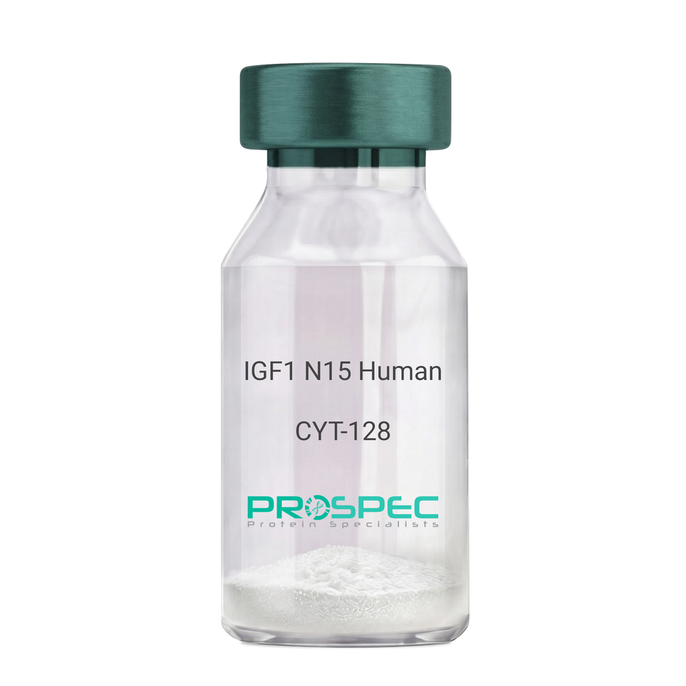
Name :
IGF1 N15 HumanDescription:
Insulin Like Growth Factor-1 N15 Labeled Human Recombinant
Somatomedin C, IGF-I, IGFI, IGF1, IGF-IA, Mechano growth factor, MGF.
Product # :
CYT-128Price :
Quantity :
Shipping Method :
Shipped at Room temp
More Info
- description
- source
- formulation
- purity
- biological activity
- More Info
- sds-page
- Citations
Description
IGF1 N15 Human Recombinant produced in E.coli is a single, non-glycosylated polypeptide chain containing 70 amino acids and having a molecular mass of 7.74kDa. The N15 is stable isotope labeled. The IGF1 N15 is purified by proprietary chromatographic techniques.
Source
Escherichia Coli.
Formulation
IGF1 N15 protein was lyophilized from a 0.2µm filtered concentrated solution in PBS, pH 7.2.
Purity
Greater than 97.0% as determined by:
(a) Analysis by RP-HPLC.
(b) Analysis by SDS-PAGE.Biological Activity
The ED50 as determined by a cell proliferation assay using serum free human MCF-7 cells is less than 2ng/ml, corresponding to a specific activity of > 5.0 × 10⁵ IU/mg.sds-page
Citations
More Info
-
Introduction
The somatomedins, or insulin-like growth factors (IGFs), comprise a family of peptides that play important roles in mammalian growth and development. IGF1 mediates many of the growth-promoting effects of growth hormone (GH; MIM 139250). Early studies showed that growth hormone did not directly stimulate the incorporation of sulfate into cartilage, but rather acted through a serum factor, termed 'sulfation factor,' which later became known as 'somatomedin' (Daughaday et al., 1972). Three main somatomedins have been characterized: somatomedin C (IGF1), somatomedin A (IGF2; MIM 147470), and somatomedin B (MIM 193190) (Rotwein, 1986; Rosenfeld, 2003).
-
Synonyms
Somatomedin C, IGF-I, IGFI, IGF1, IGF-IA, Mechano growth factor, MGF.
-
Physical Appearance
Sterile Filtered White lyophilized (freeze-dried) powder.
-
Stability
Lyophilized IGF1 N15 although stable at room temperature for 3 weeks, should be stored desiccated below -18°C. Upon reconstitution IGF1 N15 should be stored at 4°C between 2-7 days and for future use below -18°C. For long term storage it is recommended to add a carrier protein (0.1% HSA or BSA). Please prevent freeze-thaw cycles.
-
Solubility
It is recommended to reconstitute the lyophilized IGF1 N15 in sterile 18M-cm H2O not less than 100µg/ml, which can then be further diluted to other aqueous solutions.
-
Amino Acid Sequence
GPETLCGAEL VDALQFVCGD RGFYFNKPTG YGSSSRRAPQ TGIVDECCFR SCDLRRLEMY CAPLKPAKSA.
-
Background
What is the molecular weight/Mw of IGF1 N15 HUMAN Protein?
IGF1 N15 HUMAN Protein has a total Mw of 7.74kDa.
What is the source or expression system of IGF1 N15 HUMAN Protein?
Escherichia Coli.
What is the Purity of IGF1 N15 HUMAN Protein?
IGF1 N15 HUMAN Protein is >97% pure as determined by SDS-PAGE.
What is the Biological Activity of IGF1 N15 HUMAN Protein?
The ED50 as determined by a cell proliferation assay using serum free human MCF-7 cells is less than 2ng/ml, corresponding to a specific activity of > 5.0 × 10⁵ IU/mg.
What is the amino acid sequence of IGF1 N15 HUMAN Protein?
GPETLCGAEL VDALQFVCGD RGFYFNKPTG YGSSSRRAPQ TGIVDECCFR SCDLRRLEMY CAPLKPAKSA.
What applications can IGF1 N15 HUMAN Protein be used in?
IGF1 N15 HUMAN Protein can probably be used in western blot, ELISA and Lateral Flow.
What is the endotoxin level for IGF1 N15 HUMAN Protein?
The endotoxin level is minimal, IGF1 N15 HUMAN Protein was purified using conventional chromatography techniques.
ProSpec's products are furnished for LABORATORY RESEARCH USE ONLY. The product may not be used as drugs, agricultural or pesticidal products, food additives or household chemicals.
- View Data Sheet
Name :
TNFR2 Human, HisDescription:
Tumor Necrosis Factor Receptor Type 2 Human Recombinant, His Tag
Tumor necrosis factor receptor superfamily member 1B, Tumor necrosis factor receptor 2, Tumor necrosis factor receptor type II, p75, p80 TNF-alpha receptor, CD120b, Etanercept, TNF-R2, TNF-RII, TNFR-II, TNFRSF1B, TNFBR, TNFR2, TBPII, TNFR2, TNFR1B, TNFR80, TNF-R75, p75TNFR, TNF-R-II.
Product # :
CYT-674Price :
Quantity :
Shipping Method :
Shipped with Ice Packs
More Info
- description
- source
- formulation
- purity
- More Info
- Citations
Description
TNFR2 Human Recombinant produced in E.Coli is a single, non-glycosylated, Polypeptide chain containing 184 amino acids fragment (23-206) having a molecular weight of 24.45kDa and fused with a 4.5kDa amino-terminal hexahistidine tag. The TNFR2 is purified by proprietary chromatographic techniques.
Source
Escherichia Coli.
Formulation
TNFR2 protein is supplied in 20mM Tris HCl pH-8, 5mM EDTA and 50% glycerol.
Purity
Greater than 95.0% as determined by SDS-PAGE.
Citations
More Info
-
Introduction
TNFR2 belongs to the TNF-receptor superfamily. TNFR2 is receptor with high affinity for TNFSF2/TNF-alpha and approximately 5-fold lower affinity for homotrimeric TNFSF1/lymphotoxin-alpha. TNFR2 mediates the majority of the metabolic effects of TNF-alpha. In addition, knockout studies in mice propose a role for TNFR2 in protecting neurons from apoptosis by stimulating antioxidative pathways. TNFR2 expression might have a significant role in the angiogenesis, tumor cell proliferation and metastasis of Invasive micropapillary carcinoma of the breast.
There are 2 types of soluble TNF receptors: sTNFR-I and sTNFR-II, which act to neutralize the biological activities of TNF alpha and TNF beta. The levels of these soluble receptors seem to increase as a result of shedding of the extracellular domains of the membrane bound receptors. High levels of soluble TNF receptors are found in the amniotic fluid of pregnant women. TNFR2 and TNFR1 form a heterocomplex which mediates the recruitment of 2 anti-apoptotic proteins, c-IAP1 and c-IAP2, which possess E3 ubiquitin ligase activity. IAPs’ function in TNF-receptor signaling is unknown; nevertheless, c-IAP1 is believed to potentiate TNF-induced apoptosis by the ubiquitination and degradation of TNF-receptor-associated factor 2, which mediates anti-apoptotic signals. Oxidative stress promotes TNFR1 and TNFR2 self-interaction, ligand-independent and enhanced ligand-dependent TNF signaling. TNF-a, TNFR1 and TNFR2 have roles in cellular differentiation. TNFR1 and TNFR2 function in cell type-specific renal injury. -
Synonyms
Tumor necrosis factor receptor superfamily member 1B, Tumor necrosis factor receptor 2, Tumor necrosis factor receptor type II, p75, p80 TNF-alpha receptor, CD120b, Etanercept, TNF-R2, TNF-RII, TNFR-II, TNFRSF1B, TNFBR, TNFR2, TBPII, TNFR2, TNFR1B, TNFR80, TNF-R75, p75TNFR, TNF-R-II.
-
Physical Appearance
Sterile Filtered clear solution.
-
Stability
Store at 4°C if entire vial will be used within 2-4 weeks. Store, frozen at -20°C for longer periods of time. Please avoid freeze thaw cycles.
-
Amino Acid Sequence
LPAQVAFTPYAPEPGSTCRLREYYDQTAQMCCSKCSPGQHAKVFCTKTSDTVCDSCEDSTYTQLWNWV
PECLSCGSRCSSDQVETQACTREQNRICTCRPGWYCALSKQEGCRLCAPLRKCRPGFGVARPGTETSD
VVCKPCAPGTFSNTTSSTDICRPHQICNVVAIPGNASMDAVCTSTSPT.
ProSpec's products are furnished for LABORATORY RESEARCH USE ONLY. The product may not be used as drugs, agricultural or pesticidal products, food additives or household chemicals.
- View Data Sheet
Name :
SH2D1B HumanDescription:
SH2 domain containing 1B Human Recombinant
SH2 Domain Containing 1B, EWS/FLI1-Activated Transcript 2, EAT-2, EAT2, SH2 Domain-Containing Molecule EAT2, SH2 Domain-Containing Protein 1B, SH2 domain-containing protein 1B, EWS/FLI1-activated transcript 2, EAT-2.
Product # :
PRO-2470Price :
Quantity :
Shipping Method :
Shipped with Ice Packs
More Info
- description
- source
- formulation
- purity
- More Info
- Citations
Description
SH2D1B Human Recombinant produced in Sf9 Baculovirus cells is a single, glycosylated polypeptide chain containing 141 amino acids (1-132a.a.) and having a molecular mass of 16.4kDa (Molecular size on SDS-PAGE will appear at approximately 18-28kDa). SH2D1B is expressed with a 6 amino acid His tag at C-Terminus and purified by proprietary chromatographic techniques.
Source
Sf9, Baculovirus cells.
Formulation
SH2D1B protein solution (0.25mg/ml) contains Phosphate Buffered Saline (pH 7.4) and 20% glycerol.
Purity
Greater than 90.0% as determined by SDS-PAGE.
Citations
More Info
-
Introduction
SH2 domain-containing protein 1B, also known as SH2D1B activates NK cells. SH2D1B participates in controlling signal transduction through at least 4 receptors, CD84, SLAMF1, LY9 and CD244, expressed on the surface of professional antigen-presenting cells. Two isoforms of the human SH2D1B are produced by alternative splicing. Superior Mesenteric Artery Syndrome is one of the diseases which is associated with SH2D1B.
-
Synonyms
SH2 Domain Containing 1B, EWS/FLI1-Activated Transcript 2, EAT-2, EAT2, SH2 Domain-Containing Molecule EAT2, SH2 Domain-Containing Protein 1B, SH2 domain-containing protein 1B, EWS/FLI1-activated transcript 2, EAT-2.
-
Physical Appearance
Sterile filtered colorless solution.
-
Stability
Store at 4°C if entire vial will be used within 2-4 weeks.Store, frozen at -20°C for longer periods of time. For long term storage it is recommended to add a carrier protein (0.1% HSA or BSA).Avoid multiple freeze-thaw cycles.
-
Amino Acid Sequence
ADPMDLPYYH GRLTKQDCET LLLKEGVDGN FLLRDSESIP GVLCLCVSFK NIVYTYRIFR EKHGYYRIQT AEGSPKQVFP SLKELISKFE KPNQGMVVHL LKPIKRTSPS LRWRGLKLEL ETFVNSNSDY VDVLPHHHHH H.
ProSpec's products are furnished for LABORATORY RESEARCH USE ONLY. The product may not be used as drugs, agricultural or pesticidal products, food additives or household chemicals.
- View Data Sheet
Name :
HSPA5 Human, Hi-5Description:
Heat shock 70kDa protein 5 Human Recombinant, Hi-5
78 kDa glucose-regulated protein, GRP-78, Endoplasmic reticulum lumenal Ca(2+)-binding protein grp78, Heat shock 70 kDa protein 5, Immunoglobulin heavy chain-binding protein, BiP, HSPA5, GRP78, MIF2, FLJ26106.
Product # :
HSP-037Price :
Quantity :
Shipping Method :
Shipped with Ice Packs
More Info
- description
- source
- formulation
- purity
- More Info
- Citations
Description
HSPA5 produced in Hi-5 cells is a single, glycosylated polypeptide chain containing 640 amino acids (20-650 a.a.) and having a molecular mass of 71kDa. HSPA5 is fused to an 8 amino acid His Tag at C-Terminus and purified by proprietary chromatographic techniques.
Source
Hi-5 Cells.
Formulation
The HSPA5 protein solution (1mg/ml) 20mM Tris-HCl buffer (pH8.0), 10% glycerol, 2mM DTT and 200mM NaCl.
Purity
Greater than 90.0% as determined by SDS-PAGE.
Citations
More Info
-
Introduction
Binding immunoglobulin protein (BiP or HSPA5) is a member of the family of ~70kDa heat shock proteins (HSP 70). HSPA5 is a stress response protein which is induced by agents or conditions that adversely affect endoplasmic reticulum (ER) function. HSPA5 is crucial for the proper glycosylation, folding as well as for the maintenance of cell homeostasis and the prevention of apoptosis.
-
Synonyms
78 kDa glucose-regulated protein, GRP-78, Endoplasmic reticulum lumenal Ca(2+)-binding protein grp78, Heat shock 70 kDa protein 5, Immunoglobulin heavy chain-binding protein, BiP, HSPA5, GRP78, MIF2, FLJ26106.
-
Physical Appearance
Sterile filtered colorless solution.
-
Stability
Store at 4°C if entire vial will be used within 2-4 weeks. Store, frozen at -20°C for longer periods of time.For long term storage it is recommended to add a carrier protein (0.1% HSA or BSA).Avoid multiple freeze-thaw cycles.
-
Amino Acid Sequence
MEEDKKEDVG TVVGIDLGTT YSCVGVFKNG RVEIIANDQG NRITPSYVAF TPEGERLIGD AAKNQLTSNP ENTVFDAKRL IGRTWNDPSV QQDIKFLPFK VVEKKTKPYI QVDIGGGQTK TFAPEEISAM VLTKMKETAE AYLGKKVTHA VVTVPAYFND AQRQATKDAG TIAGLNVMRI INEPTAAAIA YGLDKREGEK NILVFDLGGG TFDVSLLTID NGVFEVVATN GDTHLGGEDF DQRVMEHFIK LYKKKTGKDV RKDNRAVQKL RREVEKAKRA LSSQHQARIE IESFYEGEDF SETLTRAKFE ELNMDLFRST MKPVQKVLED SDLKKSDIDE IVLVGGSTRI PKIQQLVKEF FNGKEPSRGI NPDEAVAYGA AVQAGVLSGD QDTGDLVLLD VCPLTLGIET VGGVMTKLIP RNTVVPTKKS QIFSTASDNQ PTVTIKVYEG ERPLTKDNHL LGTFDLTGIP PAPRGVPQIE VTFEIDVNGI LRVTAEDKGT GNKNKITITN DQNRLTPEEI ERMVNDAEKF AEEDKKLKER IDTRNELESY AYSLKNQIGD KEKLGGKLSS EDKETMEKAV EEKIEWLESH QDADIEDFKA KKKELEEIVQ PIISKLYGSA GPPPTGEEDT AELEHHHHHH.
ProSpec's products are furnished for LABORATORY RESEARCH USE ONLY. The product may not be used as drugs, agricultural or pesticidal products, food additives or household chemicals.
- View Data Sheet
Name :
KIR2DL3 AntibodyDescription:
Killer Cell Immunoglobulin-Like Receptor 2 Domains Long Cytoplasmic Tail 3, Mouse Anti Human
Killer cell immunoglobulin-like receptor 2DL3, MHC class I NK cell receptor, Natural killer-associated transcript 2, NKAT-2, NKAT2a, NKAT2b, p58 natural killer cell receptor clone CL-6, p58 NK receptor, p58.2 MHC class-I-specific NK receptor, Killer inhibitory receptor cl 2-3, KIR-023GB, CD158 antigen-like family member B2, CD158b2 antigen, KIR2DL3, CD158B2, KIRCL23, NKAT2, p58, NKAT, GL183, CD158b, KIR-K7b, KIR-K7c, MGC129943.
Product # :
ANT-300Price :
Quantity :
Shipping Method :
Shipped with Ice Packs
More Info
- formulation
- More Info
- Citations
Formulation
1mg/ml containing PBS, pH-7.4, & 0.1% Sodium Azide.
Citations
More Info
-
Introduction
Killer-cell immunoglobulin-like receptors (KIRs), are a family of cell surface glycoproteins found on Natural Killer (NK) Cells, which are important cells of the immune system. They control the killing function of these cells by interacting with MHC class I molecules, which are expressed on all cell types. This interaction allows them to identify virally infected cells or tumor cells that have a distinctive low level of Class I MHC on their surface. The majority of KIRs are inhibitory, which means that their recognition of MHC suppresses the cytotoxic activity of their NK cell. Only a limited number of KIRs have the capacity to activate cells. The KIR genes are found in a cluster on chromosome 19q13.4 within the 1 Mb leukocyte receptor complex (LRC). KIR molecules are extremely polymorphic, meaning their gene sequences differ significantly between individuals, so that different individuals have different arrays/repertoires of KIR genes. The KIR proteins are categorized by the number of extracellular immunoglobulin domains (2D or 3D) and by whether they have a long (L) or short (S) cytoplasmic domain. KIR proteins with the long cytoplasmic domain transduce inhibitory signals upon ligand binding via an immune tyrosine-based inhibitory motif (ITIM). Whereas KIR proteins with the short cytoplasmic domain lack the ITIM motif and instead associate with the TYRO protein tyrosine kinase binding protein to transduce activating signals. KIR2DL3 is an inhibitory Killer Cell Ig-like Receptor (KIR, previously called p58 KIR, cl-6, NKAT2 or KIR-K7), which recognizes class I MHC molecules (HLA-Cw1, -Cw3, -Cw7, and Cw8). KIR2DL3 inhibits the activity of NK cells thus preventing cell lysis.
-
Synonyms
Killer cell immunoglobulin-like receptor 2DL3, MHC class I NK cell receptor, Natural killer-associated transcript 2, NKAT-2, NKAT2a, NKAT2b, p58 natural killer cell receptor clone CL-6, p58 NK receptor, p58.2 MHC class-I-specific NK receptor, Killer inhibitory receptor cl 2-3, KIR-023GB, CD158 antigen-like family member B2, CD158b2 antigen, KIR2DL3, CD158B2, KIRCL23, NKAT2, p58, NKAT, GL183, CD158b, KIR-K7b, KIR-K7c, MGC129943.
-
Physical Appearance
Sterile Filtered colorless solution.
-
Immunogen
Anti-human KIR2DL3 mAb, is derived from hybridization of mouse P3-x63-Ag8.653 myeloma cells with spleen cells from BALB/c mice immunized with recombinant human KIR2DL3 amino acids 19-161 purified from E. coli.
-
Ig Subclass
Mouse IgG2a heavy chains and κ light chain.
-
Clone
P190IIC311AT.
-
Applications
KIR2DL3 antibody has been tested by ELISA and Western blot immunofluorescent staining with flow cytometric analysis to assure specificity. Since application varies, however, each investigation should be titrated by the reagent to obtain optimal results. Applications include flow cytometry (5-10g/1×1000000), ELISA (1:1,000 when tested against the immunized protein) and western blot analysis (1μg/ml).
-
Type
Mouse Anti Human Monoclonal.
-
Storage Procedures
For periods up to 1 month store at 4°C, for longer periods of time, store at -20°C. Prevent freeze thaw cycles.
-
Purification Method
KIR2DL3 antibody was purified from mouse ascitic fluids by protein-G affinity chromatography.
ProSpec's products are furnished for LABORATORY RESEARCH USE ONLY. The product may not be used as drugs, agricultural or pesticidal products, food additives or household chemicals.
- View Data Sheet
Name :
IL36RN HumanDescription:
Interleukin-36 Receptor Antagonist Human Recombinant
Interleukin-36 receptor antagonist protein, FIL1 delta, IL-1-related protein 3, IL-1RP3, Interleukin-1 HY1, IL-1HY1, Interleukin-1 delta, IL-1 delta, Interleukin-1 family member 5, IL-1F5, Interleukin-1 receptor antagonist homolog 1, IL-1ra homolog 1, Interleukin-1-like protein 1, IL-1L1, IL36RN, FIL1D, IL1F5, IL1HY1, IL1L1, IL1RP3, FIL1, PSORP, IL36RA, FIL1(DELTA).
Product # :
CYT-009Price :
Quantity :
Shipping Method :
Shipped at Room temp
More Info
- description
- source
- formulation
- purity
- biological activity
- More Info
- Citations
Description
IL1F5 Human Recombinant produced in E.Coli is a single, non-glycosylated, Polypeptide chain containing 155 amino acids and having a molecular mass of 17kDa.The IL1F5 is purified by proprietary chromatographic techniques.
Source
Escherichia Coli.
Formulation
IL1F5 was lyophilized after extensive dialysis against 20mM Phosphate buffer, pH7.4.
Purity
Greater than 95.0% as determined by:
(a) Analysis by RP-HPLC.
(b) Analysis by SDS-PAGE.Biological Activity
As measured by its binding ability in a functional ELISA, immobilized IL1F5 at 1 µg/ml (100 µl/well) can bind rHuIL-1 Rrp2/Fc Chimera with a linear range of 0.15- 5 µg/ml.Citations
More Info
-
Introduction
Human interleukin family 1, member 5 (IL-1F5 / FIL1-delta) belongs to the interleukin 1 cytokine family. IL1F5 is expressed by a variety of cells including monocytes, Bcells, dendritic cells/Langerhans cells, keratinocytes,and gastric fundus Parietal and Chief cells. IL1F5 is an antagonist of IL1F9; however IL1F5 activity related to receptor binding remains unclear. Human and mouse IL1F5 share 90% amino acid sequence identity.
-
Synonyms
Interleukin-36 receptor antagonist protein, FIL1 delta, IL-1-related protein 3, IL-1RP3, Interleukin-1 HY1, IL-1HY1, Interleukin-1 delta, IL-1 delta, Interleukin-1 family member 5, IL-1F5, Interleukin-1 receptor antagonist homolog 1, IL-1ra homolog 1, Interleukin-1-like protein 1, IL-1L1, IL36RN, FIL1D, IL1F5, IL1HY1, IL1L1, IL1RP3, FIL1, PSORP, IL36RA, FIL1(DELTA).
-
Physical Appearance
Sterile Filtered White lyophilized (freeze-dried) powder.
-
Stability
Lyophilized IL1F5 although stable at room temperature for 3 weeks, should be stored desiccated below -18°C. Upon reconstitution IL1F5 should be stored at 4°C between 2-7 days and for future use below -18°C.Please prevent freeze-thaw cycles.
-
Solubility
It is recommended to quick spin followed by reconstitution of IL1F5 in PBS to a concentration no less than 100 µg/ml, which can then be further diluted to other aqueous solutions.
-
Amino Acid Sequence
The sequence of the first five N-terminal amino acids was determined and was found to be Met-Val-Leu-Ser-Gly.
ProSpec's products are furnished for LABORATORY RESEARCH USE ONLY. The product may not be used as drugs, agricultural or pesticidal products, food additives or household chemicals.
- View Data Sheet
Name :
KLRB1 HumanDescription:
Killer Cell Lectin-Like Receptor Subfamily B, Member 1 Human Recombinant
Killer cell lectin-like receptor subfamily B member 1, Natural killer cell surface protein P1A, C-type lectin domain family 5 member B, CD161 antigen, HNKR-P1a, CLEC5B, NKR, NKR-P1.
Product # :
PRO-1189Price :
Quantity :
Shipping Method :
Shipped with Ice Packs
More Info
- description
- source
- formulation
- purity
- More Info
- Citations
Description
KLRB1 Human Recombinant produced in E. coli is a single polypeptide chain containing 183 amino acids (67-225) and having a molecular mass of 21.0 kDa.KLRB1 is fused to a 24 amino acid His-tag at N-terminus & purified by proprietary chromatographic techniques.
Source
E.coli.
Formulation
The KLRB1 solution (1mg/1ml) contains 20mM Tris-HCl buffer (pH 8.0), 4M urea and 10% glycerol.
Purity
Greater than 85% as determined by SDS-PAGE.
Citations
More Info
-
Introduction
KLRB1 has an inhibitory role on natural killer (NK) cells cytotoxicity which are lymphocytes who facilitate cytotoxicity and secrete cytokines subsequent to immune stimulation. Some genes of the C-type lectin superfamily, like the rodent NKRP1 family of glycoproteins, are expressed by NK cells and take part in NK cell function regulation. KLRB1 holds an extracellular domain with a few characteristic motifs of C-type lectins, a transmembrane domain, and a cytoplasmic domain. Due to its external C terminus KLRB1 is considered to be a type II membrane protein.
-
Synonyms
Killer cell lectin-like receptor subfamily B member 1, Natural killer cell surface protein P1A, C-type lectin domain family 5 member B, CD161 antigen, HNKR-P1a, CLEC5B, NKR, NKR-P1.
-
Physical Appearance
Sterile Filtered colorless solution.
-
Stability
Store at 4°C if entire vial will be used within 2-4 weeks. Store, frozen at -20°C for longer periods of time. For long term storage it is recommended to add a carrier protein (0.1% HSA or BSA).Avoid multiple freeze-thaw cycles.
-
Amino Acid Sequence
MGSSHHHHHH SSGLVPRGSH MGSMQKSSIE KCSVDIQQSR NKTTERPGLL NCPIYWQQLR EKCLLFSHTV NPWNNSLADC STKESSLLLI RDKDELIHTQ NLIRDKAILF WIGLNFSLSE KNWKWINGSF LNSNDLEIRG DAKENSCISI SQTSVYSEYC STEIRWICQK ELTPVRNKVY PDS
ProSpec's products are furnished for LABORATORY RESEARCH USE ONLY. The product may not be used as drugs, agricultural or pesticidal products, food additives or household chemicals.
- View Data Sheet
Name :
TNFR (22-211) HumanDescription:
Tumor Necrosis Factor Receptor (22-211 a.a.) Human Recombinant
CD120a, FPF, MS5, p55, p55-R, p60, TBP1, TNF-R, TNF-R-I, TNF-R55, TNFAR, TNFR1, TNFR1-d2,TNFR55, TNFR60, Tumor necrosis factor receptor superfamily member 1A, Tumor necrosis factor receptor superfamily member 1A.
Product # :
CYT-851Price :
Quantity :
Shipping Method :
Shipped with Ice Packs
More Info
- description
- source
- formulation
- purity
- More Info
- Citations
Description
TNFR Human Recombinant produced in E.Coli is a single, non-glycosylated polypeptide chain containing 213 amino acids (22-211 a.a) and having a molecular mass of 23.6kDa.TNFR is fused to a 23 amino acid His-tag at N-terminus & purified by proprietary chromatographic techniques.
Source
Escherichia Coli.
Formulation
TNFR protein solution (1mg/ml) containing 20mM Tirs-HCl (pH8.0) and 10% glycerol.
Purity
Greater than 85% as determined by SDS-PAGE.
Citations
More Info
-
Introduction
TNFR1 belongs to the TNF-receptor superfamily. TNFR1 is a receptor for TNFSF2/TNF-alpha and homotrimeric TNFSF1/lymphotoxin-alpha.
There are 2 types of soluble TNF receptors: sTNFR-I and sTNFR-II, which act to neutralize the biological activities of TNF alpha and TNF beta. The levels of these soluble receptors seem to increase as a result of shedding of the extracellular domains of the membrane bound receptors. TNF-a, TNFR1 and TNFR2 have roles in cellular differentiation. TNFR1 and TNFR2 function in cell type-specific renal injury.
TNFR1 is capable of signaling both cell survival and apoptosis. TNFR1-induced apoptosis requires 2 sequential signaling complexes. TNFR1 is capable of activating NF-kappaB, mediate apoptosis, and function as a regulator of inflammation. Oxidative stress promotes TNFR1 and TNFR2 self-interaction, ligand-independent and enhanced ligand-dependent TNF signaling. TNFR1 contributes to the induction of non-cytocidal TNF effects including anti-viral state and activation of the acid sphingomyelinase. Human TNFR1 has a major region which controls cell surface expression. High levels of soluble TNF receptors are found in the amniotic fluid of pregnant women.
Germline mutations of the extracellular domains of TNFR1 are linked to the autosomal dominant periodic fever syndrome. The impaired receptor clearance is believed to be a mechanism of the disease. Familial hibernian fever (FHF) is caused by defects in TNFRSF1A gene. -
Synonyms
CD120a, FPF, MS5, p55, p55-R, p60, TBP1, TNF-R, TNF-R-I, TNF-R55, TNFAR, TNFR1, TNFR1-d2,TNFR55, TNFR60, Tumor necrosis factor receptor superfamily member 1A, Tumor necrosis factor receptor superfamily member 1A.
-
Physical Appearance
Sterile filtered colorless solution.
-
Stability
Store at 4°C if entire vial will be used within 2-4 weeks. Store, frozen at -20°C for longer periods of time. For long term storage it is recommended to add a carrier protein (0.1% HSA or BSA).Avoid multiple freeze-thaw cycles.
-
Amino Acid Sequence
MGSSHHHHHH SSGLVPRGSH MGSIYPSGVI GLVPHLGDRE KRDSVCPQGK YIHPQNNSIC CTKCHKGTYL YNDCPGPGQD TDCRECESGS FTASENHLRH CLSCSKCRKE MGQVEISSCT VDRDTVCGCR KNQYRHYWSE NLFQCFNCSL CLNGTVHLSC QEKQNTVCTC HAGFFLRENE CVSCSNCKKS LECTKLCLPQ IENVKGTEDS GTT.
ProSpec's products are furnished for LABORATORY RESEARCH USE ONLY. The product may not be used as drugs, agricultural or pesticidal products, food additives or household chemicals.
- View Data Sheet
Name :
BCL2L10 HumanDescription:
BCL2 Like 10 Human Recombinant
Bcl-2-like protein 10, Bcl2-L-10, Anti-apoptotic protein NrH, Apoptosis regulator Bcl-B, BCL2L10, BCLB, Boo, Diva, BCL-B.
Product # :
PRO-1321Price :
Quantity :
Shipping Method :
Shipped with Ice Packs
More Info
- description
- source
- formulation
- purity
- More Info
- Citations
Description
BCL2L10 Human Recombinant produced in E.Coli is a single, non-glycosylated polypeptide chain containing 195 amino acids (1-172 a.a.) and having a molecular mass of 21.8kDa.BCL2L10 is fused to a 23 amino acid His-tag at N-terminus & purified by proprietary chromatographic techniques.
Source
E.coli.
Formulation
BCL2L10 protein solution (0.5mg/ml) containing 20mM Tris-HCl buffer (pH 8.0), 0.15M NaCl, 20% glycerol and 1mM DTT.
Purity
Greater than 90% as determined by SDS-PAGE.
Citations
More Info
-
Introduction
BCL2 Like 10 (BCL2L10) is a member of the BCL-2 protein family. BCL2 family members form hetero or homodimers and serve as anti or pro-apoptotic regulators which are involved in a wide variety of cellular activities. BCL2L10 includes the conserved BH4, BH1 and BH2 domains. BCL2L10 interacts with other members of BCL2 protein family including BCL2, BCL2L1/BCL-X(L), and BAX. BCL2L10 overexpression suppresses cell apoptosis possibly through the prevention of cytochrome C release from the mitochondria, and consequently activating caspase-3 activation.
-
Synonyms
Bcl-2-like protein 10, Bcl2-L-10, Anti-apoptotic protein NrH, Apoptosis regulator Bcl-B, BCL2L10, BCLB, Boo, Diva, BCL-B.
-
Physical Appearance
Sterile Filtered colorless solution.
-
Stability
Store at 4°C if entire vial will be used within 2-4 weeks. Store, frozen at -20°C for longer periods of time. For long term storage it is recommended to add a carrier protein (0.1% HSA or BSA).Avoid multiple freeze-thaw cycles.
-
Amino Acid Sequence
MGSSHHHHHH SSGLVPRGSH MGSMVDQLRE RTTMADPLRE RTELLLADYL GYCAREPGTP EPAPSTPEAA VLRSAAARLR QIHRSFFSAY LGYPGNRFEL VALMADSVLS DSPGPTWGRV VTLVTFAGTL LERGPLVTAR WKKWGFQPRL KEQEGDVARD CQRLVALLSS RLMGQHRAWL QAQGGWDGFC HFFRT.
ProSpec's products are furnished for LABORATORY RESEARCH USE ONLY. The product may not be used as drugs, agricultural or pesticidal products, food additives or household chemicals.
- View Data Sheet
Name :
SH3GLB1 HumanDescription:
SH3-domain GRB2-like endophilin B1 Human Recombinant
Bif-1, CGI-61, dJ612B15.2, PPP1R70, Endophilin-B1, Bax-interacting factor 1, SH3 domain-containing GRB2-like protein B1, KIAA0491 .
Product # :
PRO-1278Price :
Quantity :
Shipping Method :
Shipped with Ice Packs
More Info
- description
- source
- formulation
- purity
- More Info
- Citations
Description
SH3GLB1 Human Recombinant produced in E. coli is a single polypeptide chain containing 373 amino acids (1-365) and having a molecular mass of 41.9 kDa. SH3GLB1 is fused to 8 amino acid His-tag at C-terminus & purified by proprietary chromatographic techniques.
Source
E.coli.
Formulation
The SH3GLB1 solution (1mg/1ml) contains 20mM Tris-HCl buffer (pH 8.0), 0.1M NaCl, 10% glycerol and 2mM DTT.
Purity
Greater than 90% as determined by SDS-PAGE.
Citations
More Info
-
Introduction
Endophilin-B1 (SH3GLB1) is a part of the endophilin family and highly expresses in heart, skeletal muscle, kidney and placenta. Endophilin B1 belongs to the B subgroup of the endophilin family which is needed for preservation of mitochondrial morphology and for the regulation of the outer mitochondrial membrane dynamics. SH3GLB1 is required for normal outer mitochondrial membrane dynamics. Furthermore, SH3GLB1 is required for coatomer-mediated retrograde transport in certain cells. SH3GLB1 interacts with SH3GLB2 and Bcl-2-associated X protein and involved in regulating apoptotic signaling pathways.
-
Synonyms
Bif-1, CGI-61, dJ612B15.2, PPP1R70, Endophilin-B1, Bax-interacting factor 1, SH3 domain-containing GRB2-like protein B1, KIAA0491 .
-
Physical Appearance
Sterile Filtered colorless solution.
-
Stability
Store at 4°C if entire vial will be used within 2-4 weeks. Store, frozen at -20°C for longer periods of time. For long term storage it is recommended to add a carrier protein (0.1% HSA or BSA).Avoid multiple freeze-thaw cycles.
-
Amino Acid Sequence
MNIMDFNVKK LAADAGTFLS RAVQFTEEKL GQAEKTELDA HLENLLSKAE CTKIWTEKIM KQTEVLLQPN PNARIEEFVY EKLDRKAPSR INNPELLGQY MIDAGTEFGP GTAYGNALIK CGETQKRIGT ADRELIQTSA LNFLTPLRNF IEGDYKTIAK ERKLLQNKRL DLDAAKTRLK KAKAAETRNS SEQELRITQS EFDRQAEITR LLLEGISSTH AHHLRCLNDF VEAQMTYYAQ CYQYMLDLQK QLGSFPSNYL SNNNQTSVTP VPSVLPNAIG SSAMASTSGL VITSPSNLSD LKECSGSRKA RVLYDYDAAN STELSLLADE VITVFSVVGM DSDWLMGERG NQKGKVPITY LELLNLEHHH HHH.
ProSpec's products are furnished for LABORATORY RESEARCH USE ONLY. The product may not be used as drugs, agricultural or pesticidal products, food additives or household chemicals.
- View Data Sheet
Name :
SLAMF7 HumanDescription:
SLAMF7 Human Recombinant
SLAMF7, SLAM Family Member 7, CD2-Like Receptor-Activating Cytotoxic Cells, Membrane Protein FOAP-12, CD2 Subset 1, Protein 19A, CRACC, CS1, Novel LY9 (Lymphocyte Antigen 9) Like Protein, CD2-Like Receptor Activating Cytotoxic Cells, 19A24 Protein, CD319 Antigen, Novel Ly9, CD319, 19A.
Product # :
PRO-2261Price :
Quantity :
Shipping Method :
Shipped with Ice Packs
More Info
- description
- source
- formulation
- purity
- More Info
- Citations
Description
SLAMF7 produced in Sf9 Baculovirus cells is a single, glycosylated polypeptide chain containing 212 amino acids (23-226a.a.) and having a molecular mass of 23.4kDa (Molecular size on SDS-PAGE will appear at approximately 28-40kDa). SLAMF7 is expressed with an 8 amino acid His tag at C-Terminus and purified by proprietary chromatographic techniques.
Source
Sf9, Baculovirus cells.
Formulation
SLAMF7 protein solution (0.5mg/ml) contains Phosphate Buffered Saline (pH 7.4) and 10% glycerol.
Purity
Greater than 95.0% as determined by SDS-PAGE.
Citations
More Info
-
Introduction
SLAMF7 belongs to the CS2 family of cell surface receptors. SLAMF7 is a single-pass type 1 membrane protein, SLAMF7 Isoform 1 mediates NK cell activation via aSH2D1A-independent extracellular signal-regulated ERK-mediated pathway. SLAMF7 functions in lymphocyteadhesion. Furthermore, SLAMF7 can exert activating of inhibitory influences on cells of the immune system depending on cellular context as well as the availability of effector proteins.
-
Synonyms
SLAMF7, SLAM Family Member 7, CD2-Like Receptor-Activating Cytotoxic Cells, Membrane Protein FOAP-12, CD2 Subset 1, Protein 19A, CRACC, CS1, Novel LY9 (Lymphocyte Antigen 9) Like Protein, CD2-Like Receptor Activating Cytotoxic Cells, 19A24 Protein, CD319 Antigen, Novel Ly9, CD319, 19A.
-
Physical Appearance
Sterile Filtered clear solution.
-
Stability
Store at 4°C if entire vial will be used within 2-4 weeks. Store, frozen at -20°C for longer periods of time. For long term storage it is recommended to add a carrier protein (0.1% HSA or BSA).Avoid multiple freeze-thaw cycles.
-
Amino Acid Sequence
SGPVKELVGS VGGAVTFPLK SKVKQVDSIV WTFNTTPLVT IQPEGGTIIV TQNRNRERVD FPDGGYSLKL SKLKKNDSGI YYVGIYSSSL QQPSTQEYVL HVYEHLSKPK VTMGLQSNKN GTCVTNLTCC MEHGEEDVIY TWKALGQAAN ESHNGSILPI SWRWGESDMT FICVARNPVS RNFSSPILAR KLCEGAADDP DSSMLEHHHH HH.
ProSpec's products are furnished for LABORATORY RESEARCH USE ONLY. The product may not be used as drugs, agricultural or pesticidal products, food additives or household chemicals.