Skip to content

High-quality research proteins.

  • 1-800-805-6531

  • Mon - Sat 8.00-18.00, Sun - Closed

  • info@prospecbio.com

  • PO Box 6591 East Brunswick NJ 08816

  • Products
    • Cytokines
    • Growth factors
    • Chemokines
    • Cd antigens
    • Neurotrophins
    • Hormones
    • Enzymes
    • Viral antigens
    • Recombinant proteins
    • Natural proteins
    • Monoclonal antibodies
    • Polyclonal antibodies
    • Thermal Packaging
  • About
    • Company
    • Founder
    • Contribution
  • Customers
    • Eastern Asia
    • Eastern Europe
    • North America
    • South America
    • South East Asia
    • Southern Asia
    • Western Europe
  • Ordering
    • Ordering Method
    • Ordering - Credit Card
    • Ordering - PO
    • Shipping Fees
    • FAQs
  • Services
    • DNA Cloning
    • Protein Expression
    • Monoclonal Antibody
    • Fermentation
    • Technology License
  • Contact us
Log in

Country/region

  • USD $ | Albania
  • USD $ | Andorra
  • USD $ | Argentina
  • USD $ | Armenia
  • USD $ | Australia
  • USD $ | Austria
  • USD $ | Azerbaijan
  • USD $ | Belarus
  • USD $ | Belgium
  • USD $ | Bolivia
  • USD $ | Brazil
  • USD $ | Bulgaria
  • USD $ | Cambodia
  • USD $ | Canada
  • USD $ | Chile
  • USD $ | China
  • USD $ | Colombia
  • USD $ | Costa Rica
  • USD $ | Croatia
  • USD $ | Cyprus
  • USD $ | Czechia
  • USD $ | Denmark
  • USD $ | Estonia
  • USD $ | Finland
  • USD $ | France
  • USD $ | Georgia
  • USD $ | Germany
  • USD $ | Greece
  • USD $ | Hong Kong SAR
  • USD $ | Hungary
  • USD $ | Iceland
  • USD $ | India
  • USD $ | Ireland
  • USD $ | Israel
  • USD $ | Italy
  • USD $ | Japan
  • USD $ | Kazakhstan
  • USD $ | Latvia
  • USD $ | Liechtenstein
  • USD $ | Lithuania
  • USD $ | Madagascar
  • USD $ | Malta
  • USD $ | Mexico
  • USD $ | Moldova
  • USD $ | Monaco
  • USD $ | Netherlands
  • USD $ | New Zealand
  • USD $ | North Macedonia
  • USD $ | Norway
  • USD $ | Panama
  • USD $ | Paraguay
  • USD $ | Peru
  • USD $ | Philippines
  • USD $ | Poland
  • USD $ | Portugal
  • USD $ | Romania
  • USD $ | Singapore
  • USD $ | Slovakia
  • USD $ | Slovenia
  • USD $ | South Africa
  • USD $ | South Korea
  • USD $ | Spain
  • USD $ | Sri Lanka
  • USD $ | Sweden
  • USD $ | Switzerland
  • USD $ | Taiwan
  • USD $ | Thailand
  • USD $ | Türkiye
  • USD $ | Ukraine
  • USD $ | United Kingdom
  • USD $ | United States
  • USD $ | Uruguay
  • USD $ | Vatican City
  • USD $ | Venezuela
  • USD $ | Vietnam
  • Facebook
  • Instagram
logoProSpec - Recombinant Protein Manufacturer for academic and R&D industry
Become a distributor
Account Log in
Wishlist
Cart Cart(0)
  • Products
    +
    • Cytokines
    • Growth factors
    • Chemokines
    • Cd antigens
    • Neurotrophins
    • Hormones
    • Enzymes
    • Viral antigens
    • Recombinant proteins
    • Natural proteins
    • Monoclonal antibodies
    • Polyclonal antibodies
    • Thermal Packaging
  • About
    +
    • Company
    • Founder
    • Contribution
  • Customers
    +
    • Eastern Asia
    • Eastern Europe
    • North America
    • South America
    • South East Asia
    • Southern Asia
    • Western Europe
  • Ordering
    +
    • Ordering Method
    • Ordering - Credit Card
    • Ordering - PO
    • Shipping Fees
    • FAQs
  • Services
    +
    • DNA Cloning
    • Protein Expression
    • Monoclonal Antibody
    • Fermentation
    • Technology License
  • Contact us

Item added to your cart

View cart
View All
  • Cytokines
  • Growth factors
  • Chemokines
  • Cd antigens
  • Neurotrophins
  • Hormones
  • Enzymes
  • Viral antigens
  • Recombinant proteins
  • Natural proteins
  • Monoclonal antibodies
  • Polyclonal antibodies
  • Thermal Packaging
  • Tumor Necrosis Factor

    Tumor Necrosis Factor

    View All
  • 4-1BB

    4-1BB

    View All
  • Adiponectin

    Adiponectin

    View All
  • AIF1

    AIF1

    View All
  • Other Cytokines

    Other Cytokines

    View All
  • AITRL

    AITRL

    View All
  • Angiopoietin

    Angiopoietin

    View All
  • Apolipoprotein

    Apolipoprotein

    View All
  • B-Cell Activating Factor

    B-Cell Activating Factor

    View All
  • Beta Defensin

    Beta Defensin

    View All
  • Bone Morphogenetic Protein

    Bone Morphogenetic Protein

    View All
  • B type Natriuretic Peptide

    B type Natriuretic Peptide

    View All
  • BST

    BST

    View All
  • Betacellulin

    Betacellulin

    View All
  • Cardiotrophin

    Cardiotrophin

    View All
  • Activin

    Activin

    View All
  • Fibroblast Growth Factor

    Fibroblast Growth Factor

    View All
  • Other Growth Factors

    Other Growth Factors

    View All
  • CSF

    CSF

    View All
  • CTGF

    CTGF

    View All
  • IGFBP

    IGFBP

    View All
  • VEGF Protein

    VEGF Protein

    View All
  • EGF

    EGF

    View All
  • Epigen

    Epigen

    View All
  • Erythropoietin

    Erythropoietin

    View All
  • Insulin-Like Growth Factor

    Insulin-Like Growth Factor

    View All
  • Growth Hormone

    Growth Hormone

    View All
  • HDGF

    HDGF

    View All
  • Hepatocyte Growth Factor

    Hepatocyte Growth Factor

    View All
  • Insulin

    Insulin

    View All
  • BCA-1/ BLC (CXCL13)

    BCA-1/ BLC (CXCL13)

    View All
  • BRAK (CXCL14)

    BRAK (CXCL14)

    View All
  • C-10 (CCL6)

    C-10 (CCL6)

    View All
  • Other Chemokines

    Other Chemokines

    View All
  • MEC (CCL28)

    MEC (CCL28)

    View All
  • LD78-beta (CCL3L1)

    LD78-beta (CCL3L1)

    View All
  • CTACK (CCL27)

    CTACK (CCL27)

    View All
  • CXCL16

    CXCL16

    View All
  • CXCL17

    CXCL17

    View All
  • Platelet Factor-4 (CXCL4)

    Platelet Factor-4 (CXCL4)

    View All
  • ENA-78 (CXCL5)

    ENA-78 (CXCL5)

    View All
  • Eotaxin (CCL11,24,26)

    Eotaxin (CCL11,24,26)

    View All
  • Exodus-2 (CCL21)

    Exodus-2 (CCL21)

    View All
  • Fractalkine (CX3CL1)

    Fractalkine (CX3CL1)

    View All
  • CXCL6

    CXCL6

    View All
  • Other CD Antigens

    Other CD Antigens

    View All
  • CD14

    CD14

    View All
  • CD164

    CD164

    View All
  • CD1

    CD1

    View All
  • CD2

    CD2

    View All
  • CD200

    CD200

    View All
  • CD207

    CD207

    View All
  • CD226

    CD226

    View All
  • CD23

    CD23

    View All
  • CD244

    CD244

    View All
  • CD247

    CD247

    View All
  • CD27

    CD27

    View All
  • CD274

    CD274

    View All
  • CD300

    CD300

    View All
  • CD33

    CD33

    View All
  • Other Neurotrophins

    Other Neurotrophins

    View All
  • Beta-NGF

    Beta-NGF

    View All
  • BDNF

    BDNF

    View All
  • CDNF

    CDNF

    View All
  • CNTF

    CNTF

    View All
  • GDNF

    GDNF

    View All
  • Glia Maturation Factor

    Glia Maturation Factor

    View All
  • MANF

    MANF

    View All
  • Midkine

    Midkine

    View All
  • Neuroglobin

    Neuroglobin

    View All
  • Neurotrophic factors

    Neurotrophic factors

    View All
  • Neuregulin

    Neuregulin

    View All
  • Neuropilin

    Neuropilin

    View All
  • Pigment Epithelium-Derived Factor

    Pigment Epithelium-Derived Factor

    View All
  • Pleiotrophin

    Pleiotrophin

    View All
  • Peptide Hormones

    Peptide Hormones

    View All
  • Other Hormones

    Other Hormones

    View All
  • HCG

    HCG

    View All
  • Endothelin

    Endothelin

    View All
  • Exendin

    Exendin

    View All
  • FSH

    FSH

    View All
  • Glucagon

    Glucagon

    View All
  • GHRP

    GHRP

    View All
  • GLP

    GLP

    View All
  • Thyrostimulin

    Thyrostimulin

    View All
  • Inhibin A

    Inhibin A

    View All
  • LHRH

    LHRH

    View All
  • Melanotan

    Melanotan

    View All
  • Procalcitonin

    Procalcitonin

    View All
  • PTH

    PTH

    View All
  • Peroxiredoxin

    Peroxiredoxin

    View All
  • Peptidase

    Peptidase

    View All
  • Synthetase

    Synthetase

    View All
  • Transferase

    Transferase

    View All
  • Hydrolase

    Hydrolase

    View All
  • Dehydrogenase

    Dehydrogenase

    View All
  • ACE2

    ACE2

    View All
  • Esterase

    Esterase

    View All
  • Protein Kinases

    Protein Kinases

    View All
  • Other Enzymes

    Other Enzymes

    View All
  • Phosphatase

    Phosphatase

    View All
  • Deaminase

    Deaminase

    View All
  • Oxygenase

    Oxygenase

    View All
  • Adenylate Kinase

    Adenylate Kinase

    View All
  • Lyase

    Lyase

    View All
  • Chagas

    Chagas

    View All
  • SARS

    SARS

    View All
  • Influenza

    Influenza

    View All
  • ASFV

    ASFV

    View All
  • Astrovirus

    Astrovirus

    View All
  • Borrelia

    Borrelia

    View All
  • Helicobacter Pylori

    Helicobacter Pylori

    View All
  • Chikungunya

    Chikungunya

    View All
  • Chlamydia

    Chlamydia

    View All
  • Cytomegalo

    Cytomegalo

    View All
  • Dengue

    Dengue

    View All
  • EBV

    EBV

    View All
  • Ebola

    Ebola

    View All
  • Feline Leukemia Virus

    Feline Leukemia Virus

    View All
  • Hepatitis A

    Hepatitis A

    View All
  • Other

    Other

    View All
  • Actin

    Actin

    View All
  • ADAM

    ADAM

    View All
  • Complement Component

    Complement Component

    View All
  • Ag85

    Ag85

    View All
  • Eukaryotic Translation Initiation Factor

    Eukaryotic Translation Initiation Factor

    View All
  • Anterior Gradient Protein

    Anterior Gradient Protein

    View All
  • Heat Shock Protein

    Heat Shock Protein

    View All
  • Alpha-2-HS-Glycoprotein

    Alpha-2-HS-Glycoprotein

    View All
  • Hemoglobin

    Hemoglobin

    View All
  • Allergy

    Allergy

    View All
  • Anaplasma

    Anaplasma

    View All
  • Angiogenin

    Angiogenin

    View All
  • Ankyrin Repeat Domain

    Ankyrin Repeat Domain

    View All
  • Annexin

    Annexin

    View All
  • Other Natural Proteins

    Other Natural Proteins

    View All
  • Aprotinin

    Aprotinin

    View All
  • Transferrin

    Transferrin

    View All
  • Avidin

    Avidin

    View All
  • Anti Coagulation Factors

    Anti Coagulation Factors

    View All
  • Natural Albumin

    Natural Albumin

    View All
  • Natural Coagulation Factors

    Natural Coagulation Factors

    View All
  • Fibronectin

    Fibronectin

    View All
  • Thrombin

    Thrombin

    View All
  • Anti Human Enzyme

    Anti Human Enzyme

    View All
  • Other Monoclonal Antibodies

    Other Monoclonal Antibodies

    View All
  • Anti Human Cytokine

    Anti Human Cytokine

    View All
  • Anti Human Heat Shock Protein

    Anti Human Heat Shock Protein

    View All
  • Anti Mouse Lymphocyte

    Anti Mouse Lymphocyte

    View All
  • Anti Human Chemokine

    Anti Human Chemokine

    View All
  • Anti Human Lymphocyte

    Anti Human Lymphocyte

    View All
  • Anti Viral Monoclonal

    Anti Viral Monoclonal

    View All
  • Anti Mouse Cytokine

    Anti Mouse Cytokine

    View All
  • Anti-GST Monoclonal

    Anti-GST Monoclonal

    View All
  • Other Polyclonal Antibodies

    Other Polyclonal Antibodies

    View All
  • Anti Viral

    Anti Viral

    View All
  • Anti-GST Monoclonal

    Anti-GST Monoclonal

    View All
Back

Search results

1000 results found for “other cd antigens”

Name

Description

Product #

Price

Quantity

Shipping Method

  • View Data Sheet

    Name :

    S100A8 Mouse

    Description:

    S100 Calcium Binding Protein A8 Mouse Recombinant

    Calgranulin A, MRP8, CAGA, CGLA, CFAG, Protein S100-A8, S100 calcium-binding protein A8, Migration inhibitory factor-related protein 8, MRP-8, p8, Cystic fibrosis antigen, Leukocyte L1 complex light chain, Calprotectin L1L subunit, Urinary stone protein band A, S100A8, MIF, NIF, L1Ag, CP-10, MA387, 60B8AG.

    Product # :

    PRO-233

    Price :

    Quantity :

    Shipping Method :

    Ice Icon

    Shipped with Ice Packs

    Add To Cart

    More Info

    • description
    • source
    • formulation
    • purity
    • More Info
    • Citations

    Description

    S100A8 Mouse Recombinant produced in E.Coli is a single, non-glycosylated polypeptide chain containing 109 amino acids (1-89a.a) and having a molecular mass of 12.4kDa.S100A8 is fused to a 20 amino acid His-tag at N-terminus & purified by proprietary chromatographic techniques.

    Source

    Escherichia Coli.

    Formulation

    S100A8 protein solution (1mg/1ml) containing 20mM Tris-HCl buffer (pH8.0), 30% glycerol, 0.1M NaCl and 1mM DTT.

    Purity

    Greater than 95.0% as determined by SDS-PAGE.

    Citations

    More Info

    • Introduction

      S100A8 is a part of the S100 family of proteins containing 2 EF-hand calcium-binding motifs. S100 proteins are localized in the cytoplasm and/or nucleus of a broad range of cells, and participate in the regulation of cellular processes such as cell cycle progression and differentiation. S100A8 plays a role in the inhibition of casein kinase and as a cytokine. S100A8 altered expression is related with cystic fibrosis disease. S100A8 is a calcium-binding protein that has antimicrobial activity against bacteria and fungi.S100A8 is crucial for resistance towards invasion by pathogenic bacteria. S100A8 up-regulates transcription of genes that are under the control of NF-kappa-B. S100A8 plays a role in the development of endotoxic shock in response to bacterial lipopolysaccharide. S100A8 endorses tubulin polymerization and promotes phagocyte migration and infiltration of granulocytes at sites of wounding. S100A8 takes part as a pro-inflammatory mediator in acute and chronic inflammation and up-regulates the release of IL8 and cell-surface expression of ICAM1.

    • Synonyms

      Calgranulin A, MRP8, CAGA, CGLA, CFAG, Protein S100-A8, S100 calcium-binding protein A8, Migration inhibitory factor-related protein 8, MRP-8, p8, Cystic fibrosis antigen, Leukocyte L1 complex light chain, Calprotectin L1L subunit, Urinary stone protein band A, S100A8, MIF, NIF, L1Ag, CP-10, MA387, 60B8AG.

    • Physical Appearance

      Sterile Filtered clear solution.

    • Stability

      Store at 4°C if entire vial will be used within 2-4 weeks. Store, frozen at -20°C for longer periods of time. For long term storage it is recommended to add a carrier protein (0.1% HSA or BSA).Avoid multiple freeze-thaw cycles.

    • Amino Acid Sequence

      MGSSHHHHHH SSGLVPRGSH MPSELEKALS NLIDVYHNYS NIQGNHHALY KNDFKKMVTT ECPQFVQNIN IENLFRELDI NSDNAINFEE FLAMVIKVGV ASHKDSHKE

    ProSpec's products are furnished for LABORATORY RESEARCH USE ONLY. The product may not be used as drugs, agricultural or pesticidal products, food additives or household chemicals.

    S100A8 Mouse
  • View Data Sheet

    Name :

    FCAR Human

    Description:

    Fc Fragment Of IgA Receptor Human Recombinant

    Fc Fragment Of IgA Receptor, Fc Fragment Of IgA, Receptor For, CD89, Immunoglobulin Alpha Fc Receptor, Fc Alpha Receptor, FCAR Variant 14, IgA Fc Receptor, CD89 Antigen, CTB-61M7.2, FcalphaRI, Immunoglobulin alpha Fc receptor, IgA Fc receptor.

    Product # :

    PRO-2471

    Price :

    Quantity :

    Shipping Method :

    Ice Icon

    Shipped with Ice Packs

    Add To Cart

    More Info

    • description
    • source
    • formulation
    • purity
    • More Info
    • Citations

    Description

    FCAR produced in Sf9 Baculovirus cells is a single, glycosylated polypeptide chain containing 215 amino acids (22-227a.a.) and having a molecular mass of 24.5kDa. (Molecular size on SDS-PAGE will appear at approximately 28-40kDa).FCAR is expressed with a 9 amino acid His tag at C-Terminus and purified by proprietary chromatographic techniques.

    Source

    Sf9, Baculovirus cells.

    Formulation

    FCAR protein solution (0.25mg/ml) contains Phosphate Buffered Saline (pH 7.4) and 10% glycerol.

    Purity

    Greater than 85.0% as determined by SDS-PAGE.

    Citations

    More Info

    • Introduction

      Fc Fragment Of IgA Receptor, also known as FCAR, is part of the multichain immune recognition receptor family which is the most abundant immunoglobulin in mucosal areas however it is only the second most common antibody isotype in serum. FCAR participates in both pro-and anti-inflammatory responses depending on the state of IgA bound. FCAR is also a vital Fc receptor for neutrophil killing of tumor cells. Once FCAR expressing neutrophils interact with IgA-opsonized tumor cells, the neutrophils not only show antibody-dependent cell-mediated cytotoxicity, but also release the cytokines TNF-α and IL-1β which cause increased neutrophil migration to the site.

    • Synonyms

      Fc Fragment Of IgA Receptor, Fc Fragment Of IgA, Receptor For, CD89, Immunoglobulin Alpha Fc Receptor, Fc Alpha Receptor, FCAR Variant 14, IgA Fc Receptor, CD89 Antigen, CTB-61M7.2, FcalphaRI, Immunoglobulin alpha Fc receptor, IgA Fc receptor.

    • Physical Appearance

      Sterile Filtered colorless solution.

    • Stability

      Store at 4°C if entire vial will be used within 2-4 weeks. Store, frozen at -20°C for longer periods of time. For long term storage it is recommended to add a carrier protein (0.1% HSA or BSA).Avoid multiple freeze-thaw cycles.

    • Amino Acid Sequence

      ADPQEGDFPM PFISAKSSPV IPLDGSVKIQ CQAIREAYLT QLMIIKNSTY REIGRRLKFW NETDPEFVID HMDANKAGRY QCQYRIGHYR FRYSDTLELV VTGLYGKPFL SADRGLVLMP GENISLTCSS AHIPFDRFSL AKEGELSLPQ HQSGEHPANF SLGPVDLNVS GIYRCYGWYN RSPYLWSFPS NALELVVTDS IHQDYTTQNH HHHHH.

    ProSpec's products are furnished for LABORATORY RESEARCH USE ONLY. The product may not be used as drugs, agricultural or pesticidal products, food additives or household chemicals.

    Fcar Human
  • View Data Sheet

    Name :

    TBEV NS3

    Description:

    Tick-Borne Encephalitis Virus NS3 Recombinant

    Product # :

    TBE-286

    Price :

    Quantity :

    Shipping Method :

    Ice Icon

    Shipped with Ice Packs

    Add To Cart

    More Info

    • description
    • source
    • formulation
    • purity
    • More Info
    • Citations

    Description

    The E.coli derived recombinant protein contains the Tick-borne Encephalitis Virus NS3 protein epitopes.

    Source

    Escherichia Coli.

    Formulation

    20mM MES pH 6.5, 8M urea, 200mM NaCl and 0.05% Tween-20.

    Purity

    Encephalitis protein is >95% pure as determined by 10% PAGE (coomassie staining).

    Citations

    More Info

    • Introduction

      TBE is caused by tick-borne encephalitis virus (TBEV), a member of the family Flaviviridae.
      A closely related virus in Far Eastern Eurasia, Russian spring-summer encephalitis virus (RSSEV).
      The family Flaviviridae includes other tick-borne viruses are closely related to TBEV and RSSEV, such as Omsk hemorrhagic fever virus & Kyasanur Forest virus.
      Louping ill virus is also a member of this family.

    • Stability

      Encephalitis protein although stable at 4°C for 1 week, should be stored below -18°C. Please prevent freeze thaw cycles.

    • Applications

      Encephalitis antigen is suitable for ELISA and Western blots, excellent antigen for detection of Tick-borne encephalitis virus with minimal specificity problems.

    • Specificity

      Immunoreactive with sera of encephalitis virus infected individuals.

    • Purification Method

      Encephalitis protein was purified by proprietary chromatographic technique.

    ProSpec's products are furnished for LABORATORY RESEARCH USE ONLY. The product may not be used as drugs, agricultural or pesticidal products, food additives or household chemicals.

    Tbev Ns3
  • View Data Sheet

    Name :

    ARF1 Antibody

    Description:

    ADP-Ribosylation Factor 1, Mouse Anti Human

    ARF-1, ADP-ribosylation factor 1, ARF1.

    Product # :

    ANT-058

    Price :

    Quantity :

    Shipping Method :

    Ice Icon

    Shipped with Ice Packs

    Add To Cart

    More Info

    • formulation
    • More Info
    • Citations

    Formulation

    1mg/ml containing PBS, pH-7.4, & 0.1% Sodium Azide.

    Citations

    More Info

    • Introduction

      ARF1 is a small guanine nucleotide-binding protein that increases the enzymatic activities of cholera toxin. ARF1 is necessary and ubiquitous in eukaryotes. ARF1 takes part in vesicular transport and functioning via phospholipase D activation. ARF1 is involved in membrane traffic and organelle integrity which are closely tied to their reversible association with membranes and distinct interactions with membrane phospholipids.

    • Synonyms

      ARF-1, ADP-ribosylation factor 1, ARF1.

    • Immunogen

      Anti-human ARF1 mAb, is derived from hybridization of mouse FO myeloma cells with spleen cells from BALB/c mice immunized with recombinant human ARF1 amino acids 1-181 purified from E. coli.

    • Ig Subclass

      Mouse IgG1 heavy chain and k light chain.

    • Clone

      P1B3AT.

    • Applications

      ARF1 antibody has been tested by ELISA, Western blot analysis to assure specificity and reactivity. Since application varies, however, each investigation should be titrated by the reagent to obtain optimal results. Recommended dilution range for Western blot analysis is 1:1,000.

    • Type

      Mouse Anti Human Monoclonal.

    • Storage Procedures

      For periods up to 1 month store at 4°C, for longer periods of time, store at -20°C. Prevent freeze thaw cycles.

    ProSpec's products are furnished for LABORATORY RESEARCH USE ONLY. The product may not be used as drugs, agricultural or pesticidal products, food additives or household chemicals.

    Arf1 Antibody
  • View Data Sheet

    Name :

    SNCA Antibody

    Description:

    Alpha-Synuclein, Mouse Anti Human

    Alpha-synuclein, Non-A beta component of AD amyloid, Non-A4 component of amyloid precursor, NACP, PD1, PARK1, PARK4, MGC110988, α-Synuclein, SNCA.

    Product # :

    ANT-315

    Price :

    Quantity :

    Shipping Method :

    Ice Icon

    Shipped with Ice Packs

    Add To Cart

    More Info

    • formulation
    • More Info
    • Citations

    Formulation

    1mg/ml containing PBS, pH-7.4, & 0.1% Sodium Azide.

    Citations

    More Info

    • Introduction

      α-Synuclein (amino acids 1-140), an acidic neuronal protein of 140 amino acids, is extremely heat-resistant and is natively unfolded with an extended structure primarily composed of random coils. α-synuclein has been suggested to be implicated in the pathogenesis of Parkinson’s disease and related neurodegenerative disorders, and more recently, to be an important regulatory component of vesicular transport in neuronal cells. Moreover, recent studies have shown that α-synuclein has chaperone activity and that this activity is lost upon removing its C-terminal acidic tail (amino acids 96-140).

    • Synonyms

      Alpha-synuclein, Non-A beta component of AD amyloid, Non-A4 component of amyloid precursor, NACP, PD1, PARK1, PARK4, MGC110988, α-Synuclein, SNCA.

    • Physical Appearance

      Sterile Filtered colorless solution.

    • Immunogen

      Anti-human α-Synuclein mAb is derived from hybridization of mouse SP2/0 myeloma cells with spleen cells from BALB/c mice immunized with recombinant human α -Synuclein amino acids 61-95 purified from E. coli.

    • Ig Subclass

      Mouse IgG1 heavy chain and κ light chain.

    • Clone

      P5C2AT.

    • Applications

      α-Synuclein antibody has been tested by ELISA and Western blot analysis to assure specificity and reactivity. Since application varies, however, each investigation should be titrated by the reagent to obtain optimal results. Recommended dilution range for Western blot analysis is 1:500 ~ 1:2,000. The -Synuclein antibody has the specificity against the NAC domain (61-95aa) of α-synuclein. Recommended starting dilution is 1:1,000.

    • Type

      Mouse Anti Human Monoclonal.

    • Storage Procedures

      For periods up to 1 month store at 4°C, for longer periods of time, store at -20°C. Prevent freeze thaw cycles.

    • Purification Method

      α-Synuclein antibody was purified from mouse ascitic fluids by protein-G affinity chromatography.

    ProSpec's products are furnished for LABORATORY RESEARCH USE ONLY. The product may not be used as drugs, agricultural or pesticidal products, food additives or household chemicals.

    Snca Antibody
  • View Data Sheet

    Name :

    MAFK Antibody

    Description:

    V-maf Musculoaponeurotic Fibrosarcoma Oncogene K, Mouse Anti Human

    NFE2U, P18, Erythroid transcription factor NF-E2 p18 subunit, MAFK, Transcription factor MafK.

    Product # :

    ANT-463

    Price :

    Quantity :

    Shipping Method :

    Ice Icon

    Shipped with Ice Packs

    Add To Cart

    More Info

    • formulation
    • More Info
    • Citations

    Formulation

    1mg/ml containing PBS, pH-7.4, 10% Glycerol and 0.02% Sodium Azide.

    Citations

    More Info

    • Introduction

      MAFK is a member of the bZIP family and Maf subfamily. Members from Maf family of basic region/leucine zipper (bZIP) transcription factors influence transcription in a positive or negative way, depending on their protein associate and the context of the target promoter. Small Mafs act as transcriptional repressors when they dimerize among themselves. Yet, they seem to function as transcriptional activators by dimerizing with other basic-zipper proteins and employing them to specific DNA-binding sites.

    • Synonyms

      NFE2U, P18, Erythroid transcription factor NF-E2 p18 subunit, MAFK, Transcription factor MafK.

    • Physical Appearance

      Sterile Filtered colorless solution.

    • Immunogen

      Anti-human MAFK mAb, clone PAT2F7A, is derived from hybridization of mouse F0 myeloma cells with spleen cells from BALB/c mice immunized with a recombinant human MAFK protein 1-156 amino acids purified from E. coli.

    • Ig Subclass

      Mouse IgG2a heavy chain and Kappa light chain.

    • Clone

      PAT2F7A.

    • Applications

      The antibody has been tested by ELISA, Western blot analysis to assure specificity and reactivity. Since application varies, however, each investigation should be titrated by the reagent to obtain optimal results. Recommended starting dilution is 1:1000.

    • Type

      Mouse Anti Human Monoclonal.

    • Storage Procedures

      For periods up to 1 month store at 4°C, for longer periods of time, store at -20°C. Prevent freeze thaw cycles.

    • Purification Method

      MAFK antibody was purified from mouse ascitic fluids by protein-G affinity chromatography.

    ProSpec's products are furnished for LABORATORY RESEARCH USE ONLY. The product may not be used as drugs, agricultural or pesticidal products, food additives or household chemicals.

    Mafk Antibody
  • View Data Sheet

    Name :

    CNBP Antibody

    Description:

    Cellular Nucleic Acid Binding Protein, Mouse Anti Human

    CNBP1, DM2, PROMM, RNF163, ZCCHC22, ZNF9, Cellular nucleic acid-binding protein, CNBP, Zinc finger protein 9.

    Product # :

    ANT-483

    Price :

    Quantity :

    Shipping Method :

    Ice Icon

    Shipped with Ice Packs

    Add To Cart

    More Info

    • formulation
    • More Info
    • Citations

    Formulation

    1mg/ml containing PBS, pH-7.4, 10% Glycerol and 0.02% Sodium Azide.

    Citations

    More Info

    • Introduction

      Cellular nucleic acid-binding protein (CNBP) is a nucleic-acid binding protein with seven zinc-finger domains, which has a preference for binding single stranded DNA and RNA. CNBP serves in cap-independent translation of ornithine decarboxylase mRNA, and also in sterol-mediated transcriptional regulation. The expansion of A CCTG in CNBP protein’s first intron results in myotonic dystrophy type 2.

    • Synonyms

      CNBP1, DM2, PROMM, RNF163, ZCCHC22, ZNF9, Cellular nucleic acid-binding protein, CNBP, Zinc finger protein 9.

    • Physical Appearance

      Sterile filtered colorless solution.

    • Immunogen

      Anti-human CNBP mAb, clone PAT38F10A, is derived from hybridization of mouse F0 myeloma cells with spleen cells from BALB/c mice immunized with a recombinant human CNBP protein 1-170 amino acids purified from E. coli.

    • Ig Subclass

      Mouse IgG2b heavy chain and Kappa light chain.

    • Clone

      PAT38F10A.

    • Applications

      The antibody has been tested by ELISA, Western blot analysis to assure specificity and reactivity. Since application varies, however, each investigation should be titrated by the reagent to obtain optimal results. Recommended dilution range for Western blot analysis is 1:500 ~ 1:1000. Recommended starting dilution is 1:1000.

    • Type

      Mouse Anti Human Monoclonal.

    • Storage Procedures

      For periods up to 1 month store at 4°C, for longer periods of time, store at -20°C. Prevent freeze thaw cycles.

    • Purification Method

      CNBP antibody was purified from mouse ascitic fluids by protein-A affinity chromatography.

    ProSpec's products are furnished for LABORATORY RESEARCH USE ONLY. The product may not be used as drugs, agricultural or pesticidal products, food additives or household chemicals.

    Cnbp Antibody
  • View Data Sheet

    Name :

    HTLV-1 gp46 mosaic

    Description:

    HTLV-I gp46 Mosaic Recombinant

    Product # :

    HIV-142

    Price :

    Quantity :

    Shipping Method :

    Ice Icon

    Shipped with Ice Packs

    Add To Cart

    More Info

    • description
    • formulation
    • purity
    • More Info
    • Citations

    Description

    The E.Coli derived recombinant mosaic protein contains the gp46 immunodominant region, 21-313 amino acids; Mw on SDA-PAGE is 34.2kDa and fuse to 6 histidines at C-terminus.

    Formulation

    The protein solution contains 20mM Phosphate buffer pH 7.5.

    Purity

    Protein is >90% pure as determined by SDS-PAGE.

    Citations

    More Info

    • Introduction

      Human T-lymphotropic virus (HTLV) is a human, single-stranded RNA retrovirus that causes T-cell leukemia and T-cell lymphoma. The virus activates a subset of T-helper cellscalled Th1cells. The result is a proliferation of Th1 cells and overproduction of Th1 related cytokines (mainly IFN-gamma and TNF-alpha). Feedback mechanisms of these cytokines cause a suppression of the Th2 lymphocytes and a reduction of Th2 cytokine production (mainly IL-4, IL-5, IL-10 and IL-13). The end result is a reduction in the ability of the infected host to mount an adequate immune response to invading organisms that require a predominantly Th2 dependant response (these include parasitic infections and production of mucosal and humoral antibodies).

    • Stability

      HTLV-1 gp46 although stable at 4°C for 1 week, should be stored below -18°C. Please prevent freeze thaw cycles.

    • Amino Acid Sequence

      PSCCTLTVGV SSYHSKPCNP AQPVCSWTLD LLALSADRAL QPPCPNLVSY SSYHATYSLY LFPHWIKKPN RNGGGYYSAS YSDPCSLKCP YLGCQSWTCP YTGAVSSPYW KFQQDVNFTQ EVSHLNINLH FSKCGFPFSL LVDAPGYDPI WFLNTEPSQL PPTAPPLLSH SNLDHILEPS IPWKSKLLTL VQLTLQSTNY TCIVCIDRAS LSTWHVLYSP NVSVPSPSST PLLYPSLALP APHLTLPFNW THCFDPQIQA IVSSPCHNSL ILPPFSLSPV PTLGSRSRRA.

    • Purification Method

      HTLV-1 gp46 was purified by proprietary chromatographic technique.

    ProSpec's products are furnished for LABORATORY RESEARCH USE ONLY. The product may not be used as drugs, agricultural or pesticidal products, food additives or household chemicals.

    Htlv 1 Gp46
  • View Data Sheet

    Name :

    CYP2D6

    Description:

    Cytochrome P450 2D6 Human Recombinant

    Cytochrome P450 2D6, CYPIID6, P450-DB1, Debrisoquine 4-hydroxylase, CYP2D6, CPD6, CYP2D, CYP2D@, CYP2DL1, P450C2D, MGC120389, MGC120390, LKM1, liver/kidney microsomal antigen 1.

    Product # :

    ENZ-316

    Price :

    Quantity :

    Shipping Method :

    Ice Icon

    Shipped with Ice Packs

    Add To Cart

    More Info

    • description
    • source
    • formulation
    • purity
    • More Info
    • Citations

    Description

    Cytochrome P450 2D6 Human Recombinant (also called liver/kidney microsomal antigen 1) produced in SF9, is a glycosylated, polypeptide chain having a molecular mass of 60 kDa. The LKM1 is purified by proprietary chromatographic techniques.

    Source

    Sf9 insect cells.

    Formulation

    LKM-1 is supplied in 17mM HEPES pH-7.5, 425mM NaCl, 1mM DTT, 1% Triton X-100 and 15% glycerol.

    Purity

    Greater than 95% as determined by SDS-PAGE.

    Citations

    More Info

    • Introduction

      Cytochrome P450 2D6 is a member of a complex family of microsomal enzymes (mono-oxygenases) present in the endoplasmic reticulum membrane, which perform detoxification reaction on xenobiotic compounds. Cytochrome P450 2D6 is the molecular target of autoantibodies against the "liver kidney microsomal antigen 1" (LKM 1) which has been classically defined by immunofluorescence microscopy. The presence of these autoantibodies is considered indicative of Autoimmune Hepatitis Type 2; LKM 1 antibodies have also been detected in patients with hepatitis C viral infection. The International Autoimmune Hepatitis Group therefore has subdivided the AIH type 2 into two subgroups: 2a with HCV and 2b without HCV. AIH 2a patients are often over 40 and predominantly male.
      The use of a purified recombinant Cytochrome P450 2D6 antigen allows the differentiation of autoimmune hepatitis from drug-induced hepatitis where transient autoantibodies to other P450 family members occur which cannot be differentiated by immunofluorescence techniques.

    • Synonyms

      Cytochrome P450 2D6, CYPIID6, P450-DB1, Debrisoquine 4-hydroxylase, CYP2D6, CPD6, CYP2D, CYP2D@, CYP2DL1, P450C2D, MGC120389, MGC120390, LKM1, liver/kidney microsomal antigen 1.

    • Physical Appearance

      Sterile filtered liquid formulation.

    • Stability

      Store at 4°C if entire vial will be used within 2-4 weeks. Store, frozen at -20°C for longer periods of time. Avoid multiple freeze-thaw cycles.

    • Immunological Functions

      1. Binds IgG-type human auto-antibodies.2. Standard ELISA test (checker-board analysis of positive/negative samples). 3. Immunodot analysis with positive/negative samples.

    • coating concentration

      0.35-0.6 µg/ml (depending on the type of ELISA plate and coating buffer). Suitable for labeling of functional groups.

    • Applications

      Western-Blot with monoclonal anti-LKM-1 antibody and anti-LKM-1 autoantibody- positive sample.

    ProSpec's products are furnished for LABORATORY RESEARCH USE ONLY. The product may not be used as drugs, agricultural or pesticidal products, food additives or household chemicals.

    Cyp2D6 Human
  • View Data Sheet

    Name :

    MIF Antibody

    Description:

    Macrophage Migration Inhibitory Factor, Mouse Anti Human

    Phenylpyruvate tautomerase, Glycosylation- inhibiting factor, GIF, MMIF, MIF.

    Product # :

    ANT-311

    Price :

    Quantity :

    Shipping Method :

    Ice Icon

    Shipped with Ice Packs

    Add To Cart

    More Info

    • formulation
    • More Info
    • Citations

    Formulation

    1mg/ml containing PBS, pH-7.4, 0.02% Sodium Azide and 10% Glycerol.

    Citations

    More Info

    • Introduction

      The cytokine Macrophage migration inhibitory factor (MIF) has been identified to be secreted by the pituitary gland and the monocyte/macrophage and to play an important role in endotoxic shock. MIF has the unique property of being released from macrophages and T cells in response to physiological concentrations of glucocorticoids. The secretion of MIF is tightly regulated and decreases at high, anti-inflammatory steroid concentration.

    • Synonyms

      Phenylpyruvate tautomerase, Glycosylation- inhibiting factor, GIF, MMIF, MIF.

    • Immunogen

      Anti-human MIF mAb, is derived from hybridization of mouse SP2/0 myeloma cells with spleen cells from BALB/c mice immunized with Recombinant human MIF amino acids 1-114 purified from E. coli.

    • Ig Subclass

      Mouse IgG1 heavy chain and κ light chain.

    • Clone

      P4E4AT.

    • Applications

      MIF antibody has been tested by ELISA and Western blot analysis to assure specificity and reactivity. Since application varies, however, each investigation should be titrated by the reagent to obtain optimal results.

    • Type

      Mouse Anti Human Monoclonal.

    • Storage Procedures

      For periods up to 1 month store at 4°C, for longer periods of time, store at -20°C. Prevent freeze thaw cycles.

    • Purification Method

      MIF antibody was purified from mouse ascitic fluids by protein-G affinity chromatography.

    ProSpec's products are furnished for LABORATORY RESEARCH USE ONLY. The product may not be used as drugs, agricultural or pesticidal products, food additives or household chemicals.

    Mif Antibody
  • View Data Sheet

    Name :

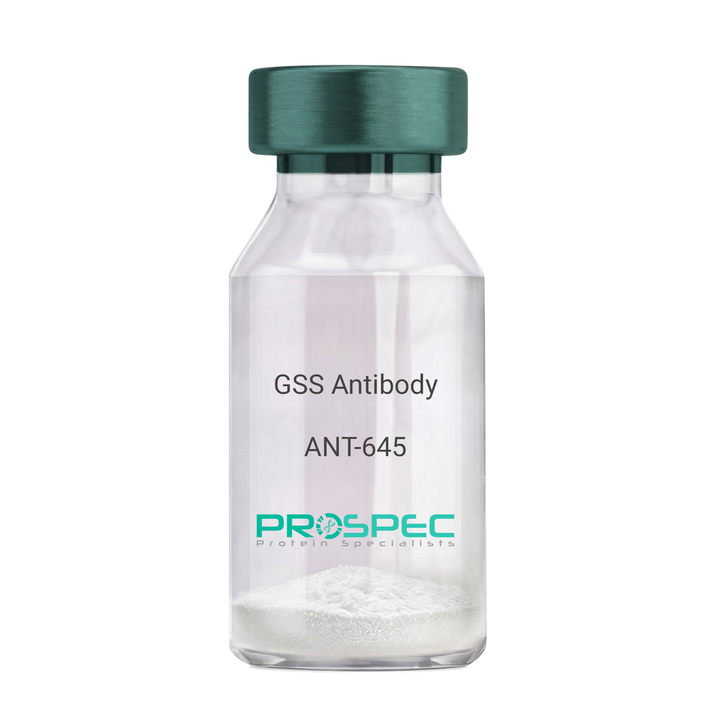
    GSS Antibody

    Description:

    Glutathione Synthetase, Mouse Anti Human

    Glutathione synthetase, GSH synthetase, GSH-S, Glutathione synthase, GSHS, MGC14098, GSS.

    Product # :

    ANT-645

    Price :

    Quantity :

    Shipping Method :

    Ice Icon

    Shipped with Ice Packs

    Add To Cart

    More Info

    • formulation
    • More Info
    • Citations

    Formulation

    1mg/ml containing PBS, pH-7.4, 10% Glycerol and 0.02% Sodium Azide.

    Citations

    More Info

    • Introduction

      Glutathione synthetase (GSS) is the second enzyme in the glutathione biosynthesis pathway. GSS catalyses the condensation of gamma-glutamylcysteine and glycine to form glutathione. Defects in the GSS are the cause of glutathione synthetase deficiency aka GSS deficiency or 5-oxoprolinuria or pyroglutamic aciduria, which is a severe form characterized by an increased rate of hemolysis and defective function of the central nervous system.

    • Synonyms

      Glutathione synthetase, GSH synthetase, GSH-S, Glutathione synthase, GSHS, MGC14098, GSS.

    • Physical Appearance

      Sterile filtered colorless solution.

    • Immunogen

      Anti-human GSS mAb, is derived from hybridization of mouse F0 myeloma cells with spleen cells from BALB/c mice immunized with recombinant human GSS amino acids 1-474 purified from E. coli.

    • Ig Subclass

      Mouse IgG1 heavy chain and k light chain.

    • Clone

      PAT12F2AT.

    • Applications

      GSS antibody has been tested by ELISA, Western blot analysis and ICC/IF to assure specificity and reactivity. Since application varies, however, each investigation should be titrated by the reagent to obtain optimal results.

    • Type

      Mouse Anti Human Monoclonal.

    • Storage Procedures

      For periods up to 1 month store at 4°C, for longer periods of time, store at -20°C. Prevent freeze thaw cycles.

    • Purification Method

      GSS antibody was purified from mouse ascitic fluids by protein-A affinity chromatography.

    ProSpec's products are furnished for LABORATORY RESEARCH USE ONLY. The product may not be used as drugs, agricultural or pesticidal products, food additives or household chemicals.

    Gss Antibody
  • View Data Sheet

    Name :

    H1N1 Antibody

    Description:

    Influenza-A Hemagglutinin H1N1, Mouse Anti Human

    Protein atonal homolog 1, ATH1, bHLHa14, HATH1, MATH-1.

    Product # :

    ANT-754

    Price :

    Quantity :

    Shipping Method :

    Ice Icon

    Shipped with Ice Packs

    Add To Cart

    More Info

    • formulation
    • More Info
    • Citations

    Formulation

    1mg/ml containing PBS, pH-7.4, 10% Glycerol and 0.02% Sodium Azide.

    Citations

    More Info

    • Introduction

      H1N1 is a sub specie of the virus Influenza A. H1N1 Influenza Virus has evolved into many strains for example: the Spanish Flustrain, mild humanflu strains, endemic pigstrains & different strains found in birds.
      The Virusis is a globular particle is sheathed in a lipid bilayer derived from the plasma membrane of its host and is about 100nm in diameter. In the lipid bilayer are two integral membrane antigens some 500 proteins of hemagglutinin ("H") and some 100 proteins of neuraminidase ("N").

    • Synonyms

      Protein atonal homolog 1, ATH1, bHLHa14, HATH1, MATH-1.

    • Physical Appearance

      Sterile filtered colorless solution.

    • Immunogen

      Recombinant human H1N1/HA1 (18-344aa) purified from Baculovirus.

    • Ig Subclass

      Mouse IgG1 heavy chain and κ light chain.

    • Clone

      PAT1G7AT.

    • Applications

      H1N1 antibody has been tested by ELISA, Western blot analysis to assure specificity and reactivity. Since application varies, however, each investigation should be titrated by the reagent to obtain optimal results.

    • Type

      Mouse Anti Human Monoclonal.

    • Storage Procedures

      For periods up to 1 month store at 4°C, for longer periods of time, store at -20°C. Prevent freeze thaw cycles.

    • Purification Method

      H1N1 antibody was purified from mouse ascitic fluids by protein-G affinity chromatography.

    ProSpec's products are furnished for LABORATORY RESEARCH USE ONLY. The product may not be used as drugs, agricultural or pesticidal products, food additives or household chemicals.

    H1N1 Antibody
  • View Data Sheet

    Name :

    TNNI1 Paired Antibody

    Description:

    Mouse Anti Human Troponin I Type 1 Paired

    DKFZp451O223, SSTNI, TNN1, Troponin I, slow skeletal muscle ,Troponin I, slow-twitch isoform.

    Product # :

    ANT-724

    Price :

    Quantity :

    Shipping Method :

    Ice Icon

    Shipped with Ice Packs

    Add To Cart

    More Info

    • description
    • formulation
    • purity
    • More Info
    • Citations

    Description

    TNNI1 conjugation antibody and TNNI1 coating antibody are used to develop rapid test for TNNI1 rapid test.

    Please note that when ordering for example: 100µg antibody we ship 50µg from each of the antibodies (100µg in total).

    Formulation

    * TNNI1 conjugation in 50mM Na-citrate, pH 6.0, 0.9% NaCl and 0.095 % NaN3.

    * TNNI1 coating antibody in 50mM Na-citrate, pH 6.0 0.9% NaCl and 0.095 % NaN3.

    Purity

    Greater than 95%.

    Citations

    More Info

    • Introduction

      Troponin I, (TNNI1) is a member of the troponin I family. Troponin complex has 3 subunits, TNNI1 known as the inhibitory Subunit which prevents the actin-myosin interactions and thus mediating striated muscle relaxation. TNNI1 combines with tropomyosin and regulates calcium sensitivity of striated muscles by structural modifications in actin-myosin complexes.

    • Synonyms

      DKFZp451O223, SSTNI, TNN1, Troponin I, slow skeletal muscle ,Troponin I, slow-twitch isoform.

    • Physical Appearance

      2 vials of sterile Filtered clear colorless solution.

    • Applications

      Lateral flow immunoassay.

    • Type

      Mouse Anti Human Monoclonal.

    • Purification Method

      Purified monoclonal IgG by protein A chromatography.

    ProSpec's products are furnished for LABORATORY RESEARCH USE ONLY. The product may not be used as drugs, agricultural or pesticidal products, food additives or household chemicals.

    Tnni1 Paired Antibody
  • View Data Sheet

    Name :

    LDHB Human, His

    Description:

    Lactate Dehydrogenase B Human Recombinant, His Tag

    LDH-H, TRG-5, L-lactate dehydrogenase B chain, LDH-B, EC=1.1.1.27, Renal carcinoma antigen NY-REN-46, LDHB.

    Product # :

    ENZ-539

    Price :

    Quantity :

    Shipping Method :

    Ice Icon

    Shipped with Ice Packs

    Add To Cart

    More Info

    • description
    • source
    • formulation
    • purity
    • biological activity
    • More Info
    • Citations

    Description

    LDHB Recombinant Human produced in E.Coli is a single, non-glycosylated polypeptide chain containing 354 amino acids (1-334 a.a.) and having a molecular mass of 38.8 kDa. The LDHB is fused to a 20 amino acids His-Tag at N-terminus and purified by proprietary chromatographic techniques.

    Source

    Escherichia Coli.

    Formulation

    LDHB Human solution containing 20mM Tris-HCl pH-8, 1mM DTT & 10% glycerol.

    Purity

    Greater than 95.0% as determined by SDS-PAGE.

    Biological Activity

    Specific activity is > 6 units/mg. in which one unit will convert 1.0 umole of pyruvate to L-lactate and beta-NAD per minute at pH 7.5 at 37°C.

    Citations

    More Info

    • Introduction

      LDHB is part of the lactate dehydrogenase family. LDHB is an oxidoreductase which catalyses the interconversion of pyruvate and lactate with concomitant interconversion of NADH and NAD+. LDHB catalyzes the oxidation of hydroxybutyrate, and frequently called Hydroxybutyrate Dehydrogenase (HBD). The LDH family consists of three members, LDH-A, LDH-B and LDH-C. LDHs function as powerful markers for germ cell tumors.

    • Synonyms

      LDH-H, TRG-5, L-lactate dehydrogenase B chain, LDH-B, EC=1.1.1.27, Renal carcinoma antigen NY-REN-46, LDHB.

    • Physical Appearance

      Sterile filtered colorless solution.

    • Stability

      Store at 4°C if entire vial will be used within 2-4 weeks. Store, frozen at -20°C for longer periods of time. For long term storage it is recommended to add a carrier protein (0.1% HSA or BSA).Avoid multiple freeze-thaw cycles.

    • Amino Acid Sequence

      MGSSHHHHHH SSGLVPRGSH MATLKEKLIA PVAEEEATVP NNKITVVGVG QVGMACAISI LGKSLADELA LVDVLEDKLK GEMMDLQHGS LFLQTPKIVA DKDYSVTANS KIVVVTAGVR QQEGESRLNL VQRNVNVFKF IIPQIVKYSP DCIIIVVSNP VDILTYVTWK LSGLPKHRVI GSGCNLDSAR FRYLMAEKLG IHPSSCHGWI LGEHGDSSVA VWSGVNVAGV SLQELNPEMG TDNDSENWKE VHKMVVESAY EVIKLKGYTN WAIGLSVADL IESMLKNLSR IHPVSTMVKG MYGIENEVFL SLPCILNARG LTSVINQKLK DDEVAQLKKS ADTLWDIQKD LKDL.

    ProSpec's products are furnished for LABORATORY RESEARCH USE ONLY. The product may not be used as drugs, agricultural or pesticidal products, food additives or household chemicals.

    Ldhb Human
  • View Data Sheet

    Name :

    PSG1 Human, Sf9

    Description:

    Pregnancy Specific Beta-1-Glycoprotein 1 Human Recombinant, Sf9

    Pregnancy Specific Beta-1-Glycoprotein 1, PSBG1, B1G1, Fetal Liver Non-Specific Cross-Reactive Antigen 1/2, Pregnancy-Specific Beta-1 Glycoprotein C/D, CD66 Antigen-Like Family Member F, PS-Beta-C/D, PS-Beta-G-1, FL-NCA-1/2, PSBG-1, PSGGA, PSG95, SP1, Pregnancy-Specific Beta-1-Glycoprotein 1, Pregnancy-Specific B-1 Glycoprotein, Pregnancy-Specific Glycoprotein 1, CD66f Antigen, DHFRP2, PSGIIA, CD66f, PBG1, PSG1.

    Product # :

    PRO-2452

    Price :

    Quantity :

    Shipping Method :

    Ice Icon

    Shipped with Ice Packs

    Add To Cart

    More Info

    • description
    • source
    • formulation
    • purity
    • More Info
    • Citations

    Description

    PSG1 Human Recombinant produced in Sf9 Baculovirus cells is a single, glycosylated polypeptide chain containing 394 amino acids (35-419a.a.) and having a molecular mass of 44.6kDa (Molecular size on SDS-PAGE will appear at approximately 40-57kDa). PSG1 is expressed with a 6 amino acids His tag at C-Terminus and purified by proprietary chromatographic techniques.

    Source

    Sf9, Baculovirus cells.

    Formulation

    PSG1 protein solution (0.25mg/ml) contains Phosphate Buffered Saline (pH 7.4) and 20% glycerol.

    Purity

    Greater than 90% as determined by SDS-PAGE.

    Citations

    More Info

    • Introduction

      Pregnancy Specific Beta-1-Glycoprotein 1 (PSG1) is a member of a subgroup of transcription factors which are phosphorylated upon binding to promoter sequences. PSGs belong to the carcinoembryonic antigen (CEA) family and function as early biochemical markers of syncytiotrophoblast formation. Furthermore, PSG1 is assumed to mediate placental vascular morphogenesis by enhancing VEGF-A production and endothelial tube formation. PSG1 is a major product of the syncytiotrophoblast, reaching concentrations of 100- 290 mg/l at term in the serum of pregnant women.

    • Synonyms

      Pregnancy Specific Beta-1-Glycoprotein 1, PSBG1, B1G1, Fetal Liver Non-Specific Cross-Reactive Antigen 1/2, Pregnancy-Specific Beta-1 Glycoprotein C/D, CD66 Antigen-Like Family Member F, PS-Beta-C/D, PS-Beta-G-1, FL-NCA-1/2, PSBG-1, PSGGA, PSG95, SP1, Pregnancy-Specific Beta-1-Glycoprotein 1, Pregnancy-Specific B-1 Glycoprotein, Pregnancy-Specific Glycoprotein 1, CD66f Antigen, DHFRP2, PSGIIA, CD66f, PBG1, PSG1.

    • Physical Appearance

      Sterile Filtered colorless solution.

    • Stability

      Store at 4°C if entire vial will be used within 2-4 weeks. Store, frozen at -20°C for longer periods of time. For long term storage it is recommended to add a carrier protein (0.1% HSA or BSA).Avoid multiple freeze-thaw cycles.

    • Amino Acid Sequence

      ADLQVTIEAE PTKVSEGKDV LLLVHNLPQN LTGYIWYKGQ MRDLYHYITS YVVDGEIIIY GPAYSGRETA YSNASLLIQN VTREDAGSYT LHIIKGDDGT RGVTGRFTFT LHLETPKPSI SSSNLNPRET MEAVSLTCDP ETPDASYLWW MNGQSLPMTH SLKLSETNRT LFLLGVTKYT AGPYECEIRN PVSASRSDPV TLNLLPKLPK PYITINNLNP RENKDVLNFT CEPKSENYTY IWWLNGQSLP VSPRVKRPIE NRILILPSVT RNETGPYQCE IRDRYGGIRS DPVTLNVLYG PDLPRIYPSF TYYRSGEVLY LSCSADSNPP AQYSWTINEK FQLPGQKLFI RHITTKHSGL YVCSVRNSAT GKESSKSMTV EVSGKWIPHH HHHH.

    ProSpec's products are furnished for LABORATORY RESEARCH USE ONLY. The product may not be used as drugs, agricultural or pesticidal products, food additives or household chemicals.

    Psg1 Protein
  • View Data Sheet

    Name :

    HEL antibody

    Description:

    Hen Egg Lysozyme, Mouse antibody

    Product # :

    ANT-149

    Price :

    Quantity :

    Shipping Method :

    Room Temp Icon

    Shipped at Room temp

    Add To Cart

    More Info

    • formulation
    • More Info
    • Citations

    Formulation

    1mg/ml in PBS (after reconstitution).

    Citations

    More Info

    • Solubility

      Reconstitute with H20 to get a 1 mg/ml concentration. Mix gently, wash the sides of the vial and wait 30-60 seconds before use.

    • Immunogen

      Purified HEL.

    • Ig Subclass

      Mouse IgG2b.

    • Clone

      NYRHEL.

    • Titer

      By direct ELISA, 1:5,000 dilution will yield 0.2 O.D using alkaline phosphatase conjugated rabbit anti-mouse Ig (Jackson Laboratories).

    • Shipping Conditions

      Antibody is shipped lyophilized at ambient temperature.

    • Type

      Mouse Antibody Monoclonal.

    • Storage Procedures

      In lyophilized form, for long periods, store at 4°C in a dry environment. After reconstitution, if not intended for use within a month, aliquot and store at -20°C.

    • Purification Method

      Ion exchange column.

    ProSpec's products are furnished for LABORATORY RESEARCH USE ONLY. The product may not be used as drugs, agricultural or pesticidal products, food additives or household chemicals.

    Hel Antibody
  • View Data Sheet

    Name :

    MAGEA3 Human

    Description:

    Melanoma Antigen Family A, 3 Human Recombinant

    CT1.3, MAGE3, HYPD, Melanoma Antigen family A, 3, MAGE-3 antigen, MAGEA6, Antigen MZ2-D, Melanoma-Associated antigen 3.

    Product # :

    PRO-502

    Price :

    Quantity :

    Shipping Method :

    Ice Icon

    Shipped with Ice Packs

    Add To Cart

    More Info

    • description
    • source
    • formulation
    • purity
    • More Info
    • Citations

    Description

    MAGEA3 produced in E.Coli is a single, non-glycosylated polypeptide chain containing 337 amino acids (1-314 a.a.) and having a molecular mass of 37.1kDa.MAGEA3 is fused to a 23 amino acid His-tag at N-terminus & purified by proprietary chromatographic techniques.

    Source

    Escherichia Coli.

    Formulation

    The MAGEA3 protein solution (1mg/1ml) is formulated in 20mM Tris-HCl buffer (pH 8.0) 1mM DTT, 100mM NaCl and 10% glycerol.

    Purity

    Greater than 90% as determined by SDS-PAGE.

    Citations

    More Info

    • Introduction

      MAGE belongs to the MAGE gene family, that comprises 12 known genes, of which 6 are expressed in tumors. The Melanoma-associated antigen 3 genes were originally isolated from different kinds of tumors, and based on their virtually limited tumor-specific expression in adult tissues, they were used as targets for cancer immunotherapy. MAGEA3 is a tumor-specific antigen extensively expressed in solid and hematologic malignancies, but not in normal tissues, with the exclusion of testis and placenta. Consequently, MAGEA3 is an outstanding candidate tumor antigen.

    • Synonyms

      CT1.3, MAGE3, HYPD, Melanoma Antigen family A, 3, MAGE-3 antigen, MAGEA6, Antigen MZ2-D, Melanoma-Associated antigen 3.

    • Physical Appearance

      Sterile Filtered clear solution.

    • Stability

      Store at 4°C if entire vial will be used within 2-4 weeks.Store, frozen at -20°C for longer periods of time. For long term storage it is recommended to add a carrier protein (0.1% HSA or BSA).Avoid multiple freeze-thaw cycles.

    • Amino Acid Sequence

      MGSSHHHHHH SSGLVPRGSH MGSMPLEQRS QHCKPEEGLE ARGEALGLVG AQAPATEEQE AASSSSTLVE VTLGEVPAAE SPDPPQSPQG ASSLPTTMNY PLWSQSYEDS SNQEEEGPST FPDLESEFQA ALSRKVAELV HFLLLKYRAR EPVTKAEMLG SVVGNWQYFF PVIFSKASSS LQLVFGIELM EVDPIGHLYI FATCLGLSYD GLLGDNQIMP KAGLLIIVLA IIAREGDCAP EEKIWEELSV LEVFEGREDS ILGDPKKLLT QHFVQENYLE YRQVPGSDPA CYEFLWGPRA LVETSYVKVL HHMVKISGGP HISYPPLHEW VLREGEE

    ProSpec's products are furnished for LABORATORY RESEARCH USE ONLY. The product may not be used as drugs, agricultural or pesticidal products, food additives or household chemicals.

    Magea3 Human
  • View Data Sheet

    Name :

    S.Typhi OMP 52kDa

    Description:

    Salmonella Typhi Outer Membrane Protein 52kDa Recombinant

    Product # :

    STY-003

    Price :

    Quantity :

    Shipping Method :

    Room Temp Icon

    Shipped at Room temp

    Add To Cart

    More Info

    • description
    • source
    • formulation
    • purity
    • More Info
    • Citations

    Description

    Recombinant S.Typhi OMP produced in E.coli is a non-glycosylated polypeptide chain having a molecular mass of 52 kDa and fused to a His tag at C-terminus.

    Source

    Escherichia Coli.

    Formulation

    Lyophilized from 1mg/ml in 20mM sodium carbonate pH-10.

    Purity

    Greater than 95.0% as determined by SDS-PAGE.

    Citations

    More Info

    • Introduction

      Salmonella Typhi is a pathogen causing typhoid fever, affecting over 17 million people with approximately 600,000 deaths annually worldwide. If untreated, typhoid fever cases result in mortality rates ranging from 12-30%.

    • Physical Appearance

      Sterile Filtered White lyophilized (freeze-dried) powder.

    • Stability

      Store the lyophilized S.Typhi OMP between 2-8°C, do not freeze. Upon reconstitution S.Typhi OMP should be stored at 4°C between 2-7 days and for future use below -18°C. Please prevent freeze-thaw cycles.

    • Solubility

      It is recommended to reconstitute the lyophilized S.Typhi OMP in sterile 18M-cm H2O not less than 100µg/ml, which can then be further diluted to other aqueous solutions.

    ProSpec's products are furnished for LABORATORY RESEARCH USE ONLY. The product may not be used as drugs, agricultural or pesticidal products, food additives or household chemicals.

    Styphi Omp 52Kda
  • View Data Sheet

    Name :

    ADRM1 Antibody

    Description:

    Adhesion Regulating Molecule 1, Mouse Anti Human

    Proteasomal ubiquitin receptor ADRM1, 110 kDa cell membrane glycoprotein, Gp110, Adhesion-regulating molecule 1, ARM-1, Proteasome regulatory particle non-ATPase 13, hRpn13, Rpn13 homolog, ADRM1, ARM1, Rpn13.

    Product # :

    ANT-409

    Price :

    Quantity :

    Shipping Method :

    Ice Icon

    Shipped with Ice Packs

    Add To Cart

    More Info

    • formulation
    • More Info
    • Citations

    Formulation

    1mg/ml containing PBS, pH-7.4, 10% Glycerol and 0.02% Sodium Azide.

    Citations

    More Info

    • Introduction

      Proteasomal ubiquitin receptor ADRM1 (ADRM1) functions as a proteasomal ubiquitin receptor. ADRM1 is an integral plasma membrane protein which promotes cell adhesion. ADRM1 is assumed to undergo O-linked glycosylation. ADRM1 gene expression is induced by gamma interferon in some cancer cells. ADRM1 employs the deubiquitinating enzyme UCHL5 at the 26S proteasome and promotes its activity.

    • Synonyms

      Proteasomal ubiquitin receptor ADRM1, 110 kDa cell membrane glycoprotein, Gp110, Adhesion-regulating molecule 1, ARM-1, Proteasome regulatory particle non-ATPase 13, hRpn13, Rpn13 homolog, ADRM1, ARM1, Rpn13.

    • Physical Appearance

      Sterile filtered colorless solution.

    • Immunogen

      Anti-human ADRM1 mAb, is derived from hybridization of mouse F0 myeloma cells with spleen cells from BALB/c mice immunized with a recombinant human ADRM1 protein 1-407 amino acids purified from E. coli.

    • Ig Subclass

      Mouse IgG2b heavy chain and k light chain.

    • Clone

      PAT34C2AT.

    • Applications

      The antibody has been tested by ELISA, Western blot analysis, Flow cytometry and ICC/IF to assure specificity and reactivity. Since application varies, however, each investigation should be titrated by the reagent to obtain optimal results.

    • Type

      Mouse Anti Human Monoclonal.

    • Storage Procedures

      For periods up to 1 month store at 4°C, for longer periods of time, store at -20°C. Prevent freeze thaw cycles.

    • Purification Method

      ADRM1 antibody was purified from mouse ascitic fluids by protein-G affinity chromatography.

    ProSpec's products are furnished for LABORATORY RESEARCH USE ONLY. The product may not be used as drugs, agricultural or pesticidal products, food additives or household chemicals.

    Adrm1 Antibody
  • View Data Sheet

    Name :

    HTNV (1-200 a.a.)

    Description:

    Hantavirus Recombinant (1-200 a.a.)

    HTNV, Hantaan Virus, Hantanvirus.

    Product # :

    HTV-002

    Price :

    Quantity :

    Shipping Method :

    Ice Icon

    Shipped with Ice Packs

    Add To Cart

    More Info

    • description
    • source
    • formulation
    • purity
    • More Info
    • Citations

    Description

    The Hantavirus Recombinant (1-200 a.a.) protein is expressed in E. coli and is fused to a Six histidine tag at C-terminus and has a MW of 23.8kDa (pI 7.96).

    Source

    E.Coli

    Formulation

    HTNV is formulated in 1XPBS.

    Purity

    HTNV protein is >90% pure as determined by SDS PAGE.

    Citations

    More Info

    • Synonyms

      HTNV, Hantaan Virus, Hantanvirus.

    • Physical Appearance

      Sterile filtered colorless solution.

    • Stability

      Upon arrival, Store at -20°C. Please avoid multiple freeze/thaw cycles.

    • Background

      What is the source or expression system of Hantavirus- Nucleocapsid Protein?
      Escherichia Coli.

      What is the Purity of Hantavirus- Nucleocapsid Protein?
      Hantavirus- Nucleocapsid Protein is >90% pure as determined by SDS-PAGE.

      What is the molecular weight / Mw of Hantavirus- Nucleocapsid Protein?
      Hantavirus- Nucleocapsid Protein having a total Mw of 23.8kDa.

      Is Hantavirus- Nucleocapsid conjugated to a Tag?
      Hantavirus- Nucleocapsid is fused to 6xHis Tag at N-terminal.

      What is the Biological Activity of Hantavirus- Nucleocapsid Protein?
      The biological functionality of Hantavirus- Nucleocapsid Protein will be determined in the future.

      What is the endotoxin level for Hantavirus- Nucleocapsid Protein?
      The endotoxin level is minimal, Hantavirus- Nucleocapsid Protein was purified using conventional chromatography techniques.

      What is the amino acid sequence of Hantavirus- Nucleocapsid Protein?
      Hantavirus- Nucleocapsid Protein is composed from 1-200 amino acids.

      What applications can Hantavirus- Nucleocapsid Protein be used in?
      Hantavirus-Nucleocapsid Protein can probably be used in western blot, ELISA and Lateral Flow.

      What is the function of the hantavirus nucleocapsid (N) protein?
      The hantavirus nucleocapsid (N) protein surrounds the viral RNA binding site and protects it during viral replication and assembly.

      Why is hantavirus nucleocapsid protein commonly used in ELISA assays?
      The hantavirus N protein is highly immunogenic, conserved and widely expressed and induces a strong antibody response in infected individuals.

      Does hantavirus N protein bind RNA?
      Hantavirus N protein contains RNA-binding domains that specifically interact with viral genomic

      Is the hantavirus nucleocapsid protein suitable for antibody production?
      Resulting from high immunogenicity, recombinant hantavirus N protein is highly used as an immunogen for generating both polyclonal and monoclonal antibodies.

    • Purification Method

      Purified by proprietary chromatographic technique.

    ProSpec's products are furnished for LABORATORY RESEARCH USE ONLY. The product may not be used as drugs, agricultural or pesticidal products, food additives or household chemicals.

    hantavirus nucleocapsid
  • View Data Sheet

    Name :

    KIR2DL3 Human

    Description:

    Killer Cell Immunoglobulin-Like Receptor, 2 Domains Long Cytoplasmic Tail 3 Human Recombinant

    Killer cell immunoglobulin-like receptor 2DL3, MHC class I NK cell receptor, Natural killer-associated transcript 2, NKAT-2, NKAT2a, NKAT2b, p58 natural killer cell receptor clone CL-6, p58 NK receptor, p58.2 MHC class-I-specific NK receptor, Killer inhibitory receptor cl 2-3, KIR-023GB, CD158 antigen-like family member B2, CD158b2 antigen, KIR2DL3, CD158B2, KIRCL23, NKAT2, p58, NKAT, GL183, CD158b, KIR-K7b, KIR-K7c, MGC129943.

    Product # :

    PRO-429

    Price :

    Quantity :

    Shipping Method :

    Ice Icon

    Shipped with Ice Packs

    Add To Cart

    More Info

    • description
    • source
    • formulation
    • purity
    • More Info
    • Citations

    Description

    Recombinant KIR2DL3 produced in E.Coli is a single, non-glycosylated polypeptide chain containing amino acids 23-223 and having a molecular mass of 22.2kDa.The KIR2DL3 is purified by proprietary chromatographic techniques.

    Source

    Escherichia Coli.

    Formulation

    The protein (1mg/ml) contains 25mM Tris-HCl (pH-7.5).

    Purity

    Greater than 95.0% as determined by SDS-PAGE.

    Citations

    More Info

    • Introduction

      Killer-cell immunoglobulin-like receptors (KIRs), are a family of cell surface glycoproteins found on Natural Killer (NK) Cells, which are important cells of the immune system. They control the killing function of these cells by interacting with MHC class I molecules, which are expressed on all cell types. This interaction allows them to identify virally infected cells or tumor cells that have a distinctive low level of Class I MHC on their surface. The majority of KIRs are inhibitory, which means that their recognition of MHC suppresses the cytotoxic activity of their NK cell. Only a limited number of KIRs have the capacity to activate cells.
      The KIR genes are found in a cluster on chromosome 19q13.4 within the 1 Mb leukocyte receptor complex (LRC). KIR molecules are extremely polymorphic, meaning their gene sequences differ significantly between individuals, so that different individuals have different arrays/repertoires of KIR genes.
      The KIR proteins are categorized by the number of extracellular immunoglobulin domains (2D or 3D) and by whether they have a long (L) or short (S) cytoplasmic domain. KIR proteins with the long cytoplasmic domain transduce inhibitory signals upon ligand binding via an immune tyrosine-based inhibitory motif (ITIM). Whereas KIR proteins with the short cytoplasmic domain lack the ITIM motif and instead associate with the TYRO protein tyrosine kinase binding protein to transduce activating signals.
      KIR2DL3 is an inhibitory Killer Cell Ig-like Receptor (KIR, previously called p58 KIR, cl-6, NKAT2 or KIR-K7), which recognizes class I MHC molecules (HLA-Cw1, -Cw3, -Cw7, and Cw8). KIR2DL3 inhibits the activity of NK cells thus preventing cell lysis.

    • Synonyms

      Killer cell immunoglobulin-like receptor 2DL3, MHC class I NK cell receptor, Natural killer-associated transcript 2, NKAT-2, NKAT2a, NKAT2b, p58 natural killer cell receptor clone CL-6, p58 NK receptor, p58.2 MHC class-I-specific NK receptor, Killer inhibitory receptor cl 2-3, KIR-023GB, CD158 antigen-like family member B2, CD158b2 antigen, KIR2DL3, CD158B2, KIRCL23, NKAT2, p58, NKAT, GL183, CD158b, KIR-K7b, KIR-K7c, MGC129943.

    • Physical Appearance

      Sterile filtered colorless solution.

    • Stability

      Store at 4°C if entire vial will be used within 2-4 weeks. Store, frozen at -20°C for longer periods of time. For long term storage it is recommended to add a carrier protein (0.1% HSA or BSA).Avoid multiple freeze-thaw cycles.

    • Amino Acid Sequence

      MEGVHRKPSL LAHPGPLVKS EETVILQCWS DVRFQHFLLH REGKFKDTLH LIGEHHDGIS KANFSIGPMM QDLAGTYRCY GSVTHSPYQL SAPSDPLDIV ITGLYEKPSL SAQPGPTVLA GESVTLSCSS RSSYDMYHLS REGEAHERRF SAGPKVNGTF QADFPLGPAT HGGTYRCFGS FRDSPYEWSN SSDPLLVSVT GN.

    ProSpec's products are furnished for LABORATORY RESEARCH USE ONLY. The product may not be used as drugs, agricultural or pesticidal products, food additives or household chemicals.

    Kir2Dl3 Human
  • View Data Sheet

    Name :

    STOM Antibody

    Description:

    Stomatin, Mouse Anti Human

    BND7, EPB7, EPB72, Erythrocyte band 7 integral membrane protein, Protein 7.2b, Stomatin, STOM.

    Product # :

    ANT-514

    Price :

    Quantity :

    Shipping Method :

    Ice Icon

    Shipped with Ice Packs

    Add To Cart

    More Info

    • formulation
    • More Info
    • Citations

    Formulation

    1mg/ml containing PBS, pH-7.4, 10% Glycerol and 0.02% Sodium Azide.

    Citations

    More Info

    • Introduction

      Stomatin (STOM) is a part of a highly conserved family of integral membrane proteins. STOM is a membrane protein involved in regulation of monovalent cation transport through lipid membranes which regulates ACCN1 and ACCN3 gating. STOM is a major lipid-raft component of erythrocytes and epithelial cells, and an abundant platelet protein which acts as a cytoskeletal anchor.

    • Synonyms

      BND7, EPB7, EPB72, Erythrocyte band 7 integral membrane protein, Protein 7.2b, Stomatin, STOM.

    • Physical Appearance

      Sterile filtered colorless solution.

    • Immunogen

      Anti-human STOM mAb, clone PAT33F5AT, is derived from hybridization of mouse F0 myeloma cells with spleen cells from BALB/c mice immunized with a recombinant human STOM protein 55-288 amino acids purified from E. coli.

    • Ig Subclass

      Mouse IgG2b heavy chain and k light chain.

    • Clone

      PAT33F5AT.

    • Applications

      The antibody has been tested by ELISA, Western blot analysis to assure specificity and reactivity. Since application varies, however, each investigation should be titrated by the reagent to obtain optimal results. Recommended starting dilution is 1:1000.

    • Type

      Mouse Anti Human Monoclonal.

    • Storage Procedures

      For periods up to 1 month store at 4°C, for longer periods of time, store at -20°C. Prevent freeze thaw cycles.

    • Purification Method

      STOM antibody was purified from mouse ascitic fluids by protein-A affinity chromatography.

    ProSpec's products are furnished for LABORATORY RESEARCH USE ONLY. The product may not be used as drugs, agricultural or pesticidal products, food additives or household chemicals.

    Stom Antibody
  • View Data Sheet

    Name :

    FIS1 Antibody

    Description:

    MFission-1, Mouse Anti Human

    TTC11, Tetratricopeptide repeat domain 11, Fission 1 (mitochondrial outer membrane) homolog (S. cerevisiae). 

    Product # :

    ANT-720

    Price :

    Quantity :

    Shipping Method :

    Ice Icon

    Shipped with Ice Packs

    Add To Cart

    More Info

    • formulation
    • More Info
    • Citations

    Formulation

    1mg/ml containing PBS, pH-7.4, & 0.1% Sodium Azide.

    Citations

    More Info

    • Introduction

      FIS1 is part of the mitochondrial complex that promotes mitochondrial fission. FIS1 induces cytochrome C discharge from the mitochondrion to the cytosol, eventually leading to apoptosis. FIS1 participates in peroxisomal growth and division. The C terminus is required for mitochondrial localisation, while the N teminus is necessary for mitochondrial fission.

    • Synonyms

      TTC11, Tetratricopeptide repeat domain 11, Fission 1 (mitochondrial outer membrane) homolog (S. cerevisiae).

    • Immunogen

      Anti-human FIS1 mAb, is derived from hybridization of mouse F0 myeloma cells with spleen cells from BALB/c mice immunized with a recombinant human FIS1 protein 1-122 amino acids purified from E. coli.

    • Ig Subclass

      Mouse IgG1 heavy chain and k light chain.

    • Clone

      PAT3B7AT.

    • Applications

      FIS1 antibody has been tested by ELISA and Western blot analysis to assure specificity and reactivity. Since application varies, however, each investigation should be titrated by the reagent to obtain optimal results.

    • Type

      Mouse Anti Human Monoclonal.

    • Storage Procedures

      For periods up to 1 month store at 4°C, for longer periods of time, store at -20°C. Prevent freeze thaw cycles.

    • Purification Method

      FIS1 antibody was purified from mouse ascitic fluids by protein-G affinity chromatography.

    ProSpec's products are furnished for LABORATORY RESEARCH USE ONLY. The product may not be used as drugs, agricultural or pesticidal products, food additives or household chemicals.

    Fis1 Antibody
  • View Data Sheet

    Name :

    HIV 1 gp41 Antibody

    Description:

    HIV-1 gp41, Rabbit Polyclonal Antibody

    Product # :

    ANT-160

    Price :

    Quantity :

    Shipping Method :

    Ice Icon

    Shipped with Ice Packs

    Add To Cart

    More Info

    • description
    • More Info
    • Citations

    Description

    Rabbit serum against the E. coli derived recombinant HIV-I protein, DEV-I (contains the C- terminus of gp120 and most of gp41).

    Citations

    More Info

    • Introduction

      Human immunodeficiency virus (HIV) is a retrovirusthat can lead to a condition in which the immune systembegins to fail, leading to opportunistic infections. HIV primarily infects vital cells in the humanimmune systemsuch as helper T cells(specifically CD4+ T cells), macrophagesand dendritic cells. HIV infection leads to low levels of CD4+ T cells through three main mechanisms: firstly, direct viral killing of infected cells; secondly, increased rates of apoptosisin infected cells; and thirdly, killing of infected CD4+ T cells by CD8 cytotoxic lymphocytesthat recognize infected cells. When CD4+ T cell numbers decline below a critical level, cell-mediated immunityis lost, and the body becomes progressively more susceptible to opportunistic infections. HIV was classified as a member of the genus Lentivirus, part of the family of Retroviridae. Lentiviruses have many common morphologies and biological properties. Many species are infected by lentiviruses, which are characteristically responsible for long-duration illnesses with a long incubation period. Lentiviruses are transmitted as single-stranded, positive-sense, enveloped RNA viruses. Upon entry of the target cell, the viral RNA genomeis converted to double-stranded DNAby a virally encoded reverse transcriptasethat is present in the virus particle. This viral DNA is then integrated into the cellular DNA by a virally encoded integraseso that the genome can be transcribed. Once the virus has infected the cell, two pathways are possible: either the virus becomes latentand the infected cell continues to function, or the virus becomes active and replicates, and a large number of virus particles are liberated that can then infect other cells.

    • Physical Appearance

      Sterile Filtered solution.

    • Specificity

      Immunoreactive with HIV-I gp41. Generates a Strong positive control spot on Immuno-blot tests. Generates 1 OD (410nm) at a dilution of 1: 250 on AS 1001 (DEV-1) ELISA.

    • Type

      Polyclonal Rabbit Antibody.

    • Storage Procedures

      Store at -20°C.

    ProSpec's products are furnished for LABORATORY RESEARCH USE ONLY. The product may not be used as drugs, agricultural or pesticidal products, food additives or household chemicals.

    Hiv 1 Gp41 Polyclonal Antibody
  • 1
  • …
  • 40
  • 41
  • 42
Your browser does not support the video tag.

Products

  • Cytokines
  • Growth Factors
  • Chemokines
  • CD Antigens
  • Neurotrophins
  • Hormones
  • Enzymes
  • Viral Antigens
  • Recombinant Proteins
  • Natural Proteins
  • Monoclonal Antibodies
  • Polyclonal Antibodies
  • Thermal Packaging

About

  • Company
  • Founder
  • Contribution
  • Contact Us
  • My Account

Ordering

  • Ordering Method
  • Ordering - Credit Card
  • Ordering PO
  • Shipping Fees
  • FAQ's

Legal

  • Terms & Conditions
  • Privacy
  • Site Map
  • info@prospecbio.com
  • Facebook
  • Instagram
  • Linkedin Profile

© 2026 ProSpec-Tany TechnoGene Ltd. All Rights Reserved.

  • Choosing a selection results in a full page refresh.
  • Opens in a new window.