Search results
1000 results found for “myxovirus”
Name
Description
Product #
Price
Quantity
Shipping Method
- View Data Sheet
Name :
NMI HumanDescription:
N-Myc Interactor Human Recombinant
N-myc (and STAT) Interactor.
Product # :
PRO-070Price :
Quantity :
Shipping Method :
Shipped with Ice Packs
More Info
- description
- source
- formulation
- purity
- More Info
Description
NMI produced in E.Coli is a single, non-glycosylated polypeptide chain containing 327 amino acids (1-307a.a.) and having a molecular mass of 37.2kDa.NMI is fused to a 20 amino acid His-tag at N-terminus & purified by proprietary chromatographic techniques.
Source
Escherichia Coli.
Formulation
The NMI protein solution (0.5mg/1ml) is formulated in 20mM Tris-HCl buffer (pH8.0)
1mM DTT and 20% glycerol.Purity
Greater than 90% as determined by SDS-PAGE.
More Info
-
Introduction
N-myc-interactor belongs to the oncogene Myc family which has a significant part in cell proliferation, differentiation, and neoplastic transformation. In Addition, NMI cooperates with all STATs except STAT2 and augments STAT-mediated transcription in response to cytokines IL2 and IFN-gamma. The NMI mRNA is expressed in small quantities in all human fetal and adult tissues tested except brain and but in large quantities in cancer cell line-myeloid leukemias.
-
Synonyms
N-myc (and STAT) Interactor.
-
Physical Appearance
Sterile Filtered clear solution.
-
Stability
Store at 4°C if entire vial will be used within 2-4 weeks. Store, frozen at -20°C for longer periods of time.Please avoid freeze thaw cycles.
-
Amino Acid Sequence
MGSSHHHHHH SSGLVPRGSH MEADKDDTQQ ILKEHSPDEF IKDEQNKGLI DEITKKNIQLK KEIQKLETE LQEATKEFQI KEDIPETKMK FLSVETPEND SQLSNISCSF QVSSKVPYEI QKGQALITFE KEEVAQNVVS MSKHHVQIKDV NLEVTAKPV PLNSGVRFQV YVEVSKMKIN VTEIPDTLRE DQMRDKLELS FSKSRNGGGE VDRVDYDRQS GSAVITFVEI GVADKILKKKEYPLYINQTC HRVTVSPYTE IHLKKYQIFS GTSKRTVLLT GMEGIQMDEE IVEDLINIHF QRAKNGGGEV DVVKCSLGQP HIAYFEE
ProSpec's products are furnished for LABORATORY RESEARCH USE ONLY. The product may not be used as drugs, agricultural or pesticidal products, food additives or household chemicals.
- View Data Sheet
Name :
IFN b Mouse, HisDescription:
IFN Beta Mouse Recombinant, His Tag
Leukocyte IFN, B cell IFN, Type I IFN, IFNB1, IFB, IFF, IFNB, IFN-b 1b, MGC96956.
Product # :
CYT-651Price :
Quantity :
Shipping Method :
Shipped with Ice Packs
More Info
- description
- source
- formulation
- purity
- More Info
- SDS-PAGE
Description
IFN beta Mouse Recombinant produced in E.Coli is a single, non-glycosylated polypeptide chain containing 182 amino acids (22-182 a.a.) and having a molecular mass of 22 kDa. Mouse IFN beta is fused to 21 amino acid at N-terminus and purified by proprietary chromatographic techniques.
Source
Escherichia Coli.
Formulation
The IFN-Beta (0.25mg/ml) contains 20mM HEPES (pH6.0), 0.5M NaCl, 10% glycerol.
Purity
Greater than 95% as determined by Analysis by SDS-PAGE.
SDS-PAGE
More Info
-
Introduction
IFN-beta 1b has antiviral, antibacterial and anticancer activities.
Influenza A viruses not only inhibit IFN-beta gene induction but also supress Type-I IFN signaling via mechanism involving induction of the SOCS-3 protein.
Intracellular bacteria and cytosolic poly (dA-dT) trigger IFN-beta responses in different human cells without requiring human ZBP1. -
Synonyms
Leukocyte IFN, B cell IFN, Type I IFN, IFNB1, IFB, IFF, IFNB, IFN-b 1b, MGC96956.
-
Physical Appearance
Sterile filtered colorless solution.
-
Stability
Store at 4°C if entire vial will be used within 2-4 weeks. Store, frozen at -20°C for longer periods of time. For long term storage it is recommended to add a carrier protein (0.1% HSA or BSA). Avoid multiple freeze-thaw cycles.
-
Amino Acid Sequence
MGSSHHHHHH SSGLVPRGSH MINYKQLQLQ ERTNIRKCQE LLEQLNGKIN LTYRADFKIP MEMTEKMQKS YTAFAIQEML QNVFLVFRNN FSSTGWNETI VVRLLDELHQ QTVFLKTVLE EKQEERLTWE MSSTALHLKS YYWRVQRYLK LMKYNSYAWM VVRAEIFRNF LIIRRLTRNF QN.
-
Background
What is the molecular weight/Mw of IFN B MOUSE, HIS Protein?
IFN B MOUSE, HIS Protein has a total Mw of 22kDa.
What is the source or expression system of IFN B MOUSE, HIS Protein?
Escherichia Coli.
What is the Purity of IFN B MOUSE, HIS Protein?
IFN B MOUSE, HIS Protein is >95% pure as determined by SDS-PAGE.
What is the Biological Activity of IFN B MOUSE, HIS Protein?
The biological functionality of IFN B MOUSE, HIS Protein will be determined in the future.
What is the amino acid sequence of IFN B MOUSE, HIS Protein?
MGSSHHHHHH SSGLVPRGSH MINYKQLQLQ ERTNIRKCQE LLEQLNGKIN LTYRADFKIP MEMTEKMQKS YTAFAIQEML QNVFLVFRNN FSSTGWNETI VVRLLDELHQ QTVFLKTVLE EKQEERLTWE MSSTALHLKS YYWRVQRYLK LMKYNSYAWM VVRAEIFRNF LIIRRLTRNF QN.
What applications can IFN B MOUSE, HIS Protein be used in?
IFN B MOUSE, HIS Protein can probably be used in western blot, ELISA and Lateral Flow.
What is the endotoxin level for IFN B MOUSE, HIS Protein?
The endotoxin level is minimal, IFN B MOUSE, HIS Protein was purified using conventional chromatography techniques.
ProSpec's products are furnished for LABORATORY RESEARCH USE ONLY. The product may not be used as drugs, agricultural or pesticidal products, food additives or household chemicals.
- View Data Sheet
Name :
CCL21 MouseDescription:
Exodus-2 Mouse Recombinant (CCL21)
Small inducible cytokine A21, CCL21, Beta chemokine exodus-2, 6Ckine, Secondary lymphoid-tissue chemokine, SLC, chemokine (C-C motif) ligand 21, ECL, CKb9, TCA4, SCYA21, MGC34555.
Product # :
CHM-371Price :
Quantity :
Shipping Method :
Shipped at Room temp
More Info
- description
- source
- formulation
- purity
- biological activity
- More Info
Description
Exodus-2 Mouse Recombinant produced in E.Coli is a single, non-glycosylated, polypeptide chain containing 110 amino acids and having a molecular mass of 12kDa. The CCL21 is purified by proprietary chromatographic techniques.
Source
Escherichia Coli.
Formulation
Lyophilized from a concentrated (1mg/ml) solution in water containing 20mM Phosphate buffer and 0.15M NaCl pH-7.4.
Purity
Greater than 97.0% as determined by SDS-PAGE & RP-HPLC.
Biological Activity
Determined by its ability to chemoattract total murine T cell population using a concentration range of 10-100ng/ml corresponding to a Specific Activity of 10,000-100,000IU/mg.More Info
-
Introduction
Chemokine (C-C motif) ligand 21 (CCL21) is a small cytokine belonging to the CC chemokine family. This chemokine is also known as 6Ckine (because it has six conserved cysteine residues instead of the four cysteines typical to chemokines), exodus-2, and secondary lymphoid-tissue chemokine (SLC). CCL21 is expressed predominantly in the lymph nodes and, in contrast to other CC chemokines, is chemotactic for lymphocytes. The gene for CCL21 is located on human chromosome 9. CCL21 elicits its effects by binding to a cell surface chemokine receptor known as CCR7.
-
Synonyms
Small inducible cytokine A21, CCL21, Beta chemokine exodus-2, 6Ckine, Secondary lymphoid-tissue chemokine, SLC, chemokine (C-C motif) ligand 21, ECL, CKb9, TCA4, SCYA21, MGC34555.
-
Physical Appearance
Sterile Filtered White lyophilized (freeze-dried) powder.
-
Stability
Lyophilized Exodus-2 although stable at room temperature for 3 weeks, should be stored desiccated below -18°C. Upon reconstitution CCL21 should be stored at 4°C between 2-7 days and for future use below -18°C. For long term storage it is recommended to add a carrier protein (0.1% HSA or BSA). Please prevent freeze-thaw cycles.
-
Solubility
It is recommended to reconstitute the lyophilized Exodus2 in sterile 18MΩ-cm H2O not less than 100µg/ml, which can then be further diluted to other aqueous solutions
-
Amino Acid Sequence
SDGGGQDCCL KYSQKKIPYS IVRGYRKQEP SLGCPIPAIL FLPRKHSKPE LCANPEEGWV QNLMRRLDQP PAPGKQSPGC RKNRGTSKSG KKGKGSKGCK RTEQTQPSRG.
-
Background
What is the molecular weight/Mw of CCL21 MOUSE Protein?
CCL21 MOUSE Protein has a total Mw of 12kDa.
What is the source or expression system of CCL21 MOUSE Protein?
Escherichia Coli.
What is the Purity of CCL21 MOUSE Protein?
CCL21 MOUSE Protein is >97% pure as determined by SDS-PAGE.
What is the Biological Activity of CCL21 MOUSE Protein?
Determined by its ability to chemoattract total murine T cell population using a concentration range of 10-100ng/ml corresponding to a Specific Activity of 10,000-100,000IU/mg.
What is the amino acid sequence of CCL21 MOUSE Protein?
SDGGGQDCCL KYSQKKIPYS IVRGYRKQEP SLGCPIPAIL FLPRKHSKPE LCANPEEGWV QNLMRRLDQP PAPGKQSPGC RKNRGTSKSG KKGKGSKGCK RTEQTQPSRG.
What applications can CCL21 MOUSE Protein be used in?
CCL21 MOUSE Protein can probably be used in western blot, ELISA and Lateral Flow.
What is the endotoxin level for CCL21 MOUSE Protein?
The endotoxin level is minimal, CCL21 MOUSE Protein was purified using conventional chromatography techniques.
ProSpec's products are furnished for LABORATORY RESEARCH USE ONLY. The product may not be used as drugs, agricultural or pesticidal products, food additives or household chemicals.
- View Data Sheet
Name :
FBXO6 HumanDescription:
F-Box Protein 6 Human Recombinant
FBG2, FBS2, FBX6, Fbx6b, F-box only protein 6, F-box protein that recognizes sugar chains 2, F-box/G-domain protein 2, FBXO6.
Product # :
PRO-1522Price :
Quantity :
Shipping Method :
Shipped with Ice Packs
More Info
- description
- source
- formulation
- purity
- More Info
Description
FBXO6 Human Recombinant produced in E. coli is a single polypeptide chain containing 316 amino acids (1-293) and having a molecular mass of 36.3kDa. FBXO6 is fused to a 23 amino acid His-tag at N-terminus & purified by proprietary chromatographic techniques.
Source
Escherichia Coli.
Formulation
The FBXO6 solution (1mg/ml) contains 20mM Tris-HCl buffer (pH 8.0), 0.4M Urea and 10% glycerol.
Purity
Greater than 90% as determined by SDS-PAGE.
More Info
-
Introduction
F-Box Protein 6, also known as FBXO6, is a part of the F-box protein family which is characterized by a roughly forty amino acid motif, the F-box. The F-box proteins are one of the 4 subunits of the ubiquitin protein ligase complex called SCFs (SKP1-cullin-F-box) that operates in phosphorylation-dependent ubiquitination. The F-box proteins are divided into three categories: Fbws containing WD-40 domains, Fbls containing leucine-rich repeats, and Fbxs containing different protein-protein interaction modules or no recognizable motifs. FBXO6 is a part of the Fbxs category, and its C-terminal area is very similar to that of rat NFB42 (neural F Box 42 kDa) which is involved in the control of the cell cycle.
-
Synonyms
FBG2, FBS2, FBX6, Fbx6b, F-box only protein 6, F-box protein that recognizes sugar chains 2, F-box/G-domain protein 2, FBXO6.
-
Physical Appearance
Sterile Filtered clear solution.
-
Stability
Store at 4°C if entire vial will be used within 2-4 weeks. Store, frozen at -20°C for longer periods of time. For long term storage it is recommended to add a carrier protein (0.1% HSA or BSA).Avoid multiple freeze-thaw cycles.
-
Amino Acid Sequence
MGSSHHHHHH SSGLVPRGSH MGSMDAPHSK AALDSINELP ENILLELFTH VPARQLLLNC RLVCSLWRDL IDLMTLWKRK CLREGFITKD WDQPVADWKI FYFLRSLHRN LLRNPCAEED MFAWQIDFNG GDRWKVESLP GAHGTDFPDP KVKKYFVTSY EMCLKSQLVD LVAEGYWEEL
LDTFRPDIVV KDWFAARADC GCTYQLKVQL ASADYFVLAS FEPPPVTIQQ WNNATWTEVS YTFSDYPRGV RYILFQHGGR DTQYWAGWYG PRVTNSSIVV SPKMTRNQAS SEAQPGQKHG QEEAAQSPYR AVVQIF.
ProSpec's products are furnished for LABORATORY RESEARCH USE ONLY. The product may not be used as drugs, agricultural or pesticidal products, food additives or household chemicals.
- View Data Sheet
Name :
CCL28 HumanDescription:
Mucosae-Associated Epithelial Chemokine Human Recombinant (CCL28)
MEC, CCK1, SCYA28, MGC71902, CCL28, C-C motif chemokine 28, Small-inducible cytokine A28, Mucosae-associated epithelial chemokine, Protein CCK1.
Product # :
CHM-353Price :
Quantity :
Shipping Method :
Shipped at Room temp
More Info
- description
- source
- formulation
- purity
- biological activity
- More Info
Description
CCL28 Human Recombinant produced in E.Coli is a single, non-glycosylated, polypeptide chain containing 108 amino acids and having a molecular mass of 12.3 kDa. The CCL28 is purified by proprietary chromatographic techniques.
Source
Escherichia Coli.
Formulation
Lyophilized from a concentrated (1mg/ml) solution in water containing 20mM PBS and 150mM NaCl.
Purity
Greater than 97.0% as determined by:
(a) Analysis by RP-HPLC.
(b) Analysis by SDS-PAGE.Biological Activity
Determined by its ability to chemoattract human lymphocytes using a concentration range of 1-10ng/ml corresponding to a Specific Activity of 100,000-1,000,000IU/mg.More Info
-
Introduction
CCL28 is part of the subfamily of small cytokine CC genes. CCL28 shows chemotactic activity for resting CD4 or CD8 T cells and eosinophils. CCL28 binds to chemokine receptors CCR3 and CCR10. CCL28 is involved in the physiology of extracutaneous epithelial tissues, including diverse mucosal organs. CCL28 mediates mucosal immunity in HIV exposure and infection. CCL28 is involved in the pathogenesis of inflammatory skin diseases.
Human CCL28 cDNA encodes a 127 amino acid residue precursor protein with a putative 22 amino acid residue signal peptide that is cleaved to produce the 105 amino acid residue mature protein. Human and mouse CCL28 are highly conserved, sharing 83% amino acid identity in their mature regions. CCL28 shares the most homology with CCL27/CTACK. Human and mouse CCL28 RNA expression was found to be highest in normal and pathologic colon with the protein being expressed by epithelial cells. Human CCL28 RNA was also present in normal and asthmatic lung tissues. -
Synonyms
MEC, CCK1, SCYA28, MGC71902, CCL28, C-C motif chemokine 28, Small-inducible cytokine A28, Mucosae-associated epithelial chemokine, Protein CCK1.
-
Physical Appearance
Sterile Filtered White lyophilized (freeze-dried) powder.
-
Stability
Lyophilized CCL28 although stable at room temperature for 3 weeks, should be stored desiccated below -18°C. Upon reconstitution CCL28 should be stored at 4°C between 2-7 days and for future use below -18°C.For long term storage it is recommended to add a carrier protein (0.1% HSA or BSA).Please prevent freeze-thaw cycles.
-
Solubility
It is recommended to reconstitute the lyophilized CCL28 in sterile 18MΩ-cm H2O not less than 100µg/ml, which can then be further diluted to other aqueous solutions.
-
Amino Acid Sequence
SEAILPIASS CCTEVSHHIS RRLLERVNMC RIQRADGDCD LAAVILHVKR RRICVSPHNH TVKQWMKVQA AKKNGKGNVC HRKKHHGKRN SNRAHQGKHE TYGHKTPY.
-
Background
What is the molecular weight/Mw of CCL28 HUMAN Protein?
CCL28 HUMAN Protein has a total Mw of 12.3kDa.
What is the source or expression system of CCL28 HUMAN Protein?
Escherichia Coli.
What is the Purity of CCL28 HUMAN Protein?
CCL28 HUMAN Protein is > 97% pure as determined by SDS-PAGE.
What is the Biological Activity of CCL28 HUMAN Protein?
Determined by its ability to chemoattract human lymphocytes using a concentration range of 1-10ng/ml corresponding to a Specific Activity of 100,000-1,000,000IU/mg.
What is the amino acid sequence of CCL28 HUMAN Protein?
SEAILPIASS CCTEVSHHIS RRLLERVNMC RIQRADGDCD LAAVILHVKR RRICVSPHNH TVKQWMKVQA AKKNGKGNVC HRKKHHGKRN SNRAHQGKHE TYGHKTPY.
What applications can CCL28 HUMAN Protein be used in?
CCL28 HUMAN Protein can probably be used in western blot, ELISA and Lateral Flow.
What is the endotoxin level for CCL28 HUMAN Protein?
The endotoxin level is minimal, CCL28 HUMAN Protein was purified using conventional chromatography techniques.
ProSpec's products are furnished for LABORATORY RESEARCH USE ONLY. The product may not be used as drugs, agricultural or pesticidal products, food additives or household chemicals.
- View Data Sheet
Name :
CCL28 MouseDescription:
Mucosae-Associated Epithelial Chemokine Mouse Recombinant (CCL28)
MEC, CCK1, SCYA28, MGC71902, CCL28, C-C motif chemokine 28, Small-inducible cytokine A28, Mucosae-associated epithelial chemokine, Protein CCK1.
Product # :
CHM-369Price :
Quantity :
Shipping Method :
Shipped at Room temp
More Info
- description
- source
- formulation
- purity
- biological activity
- More Info
Description
CCL28 Mouse Recombinant produced in E.Coli is a single, non-glycosylated, polypeptide chain containing 111 amino acids and having a molecular mass of 12.6 kDa. The CCL28 is purified by proprietary chromatographic techniques.
Source
Escherichia Coli.
Formulation
Lyophilized from a concentrated (1mg/ml) solution in water containing 20mM Phosphate buffer pH-7.4 and 150mM NaCl.
Purity
Greater than 97.0% as determined by:
(a) Analysis by RP-HPLC.
(b) Analysis by SDS-PAGE.Biological Activity
Determined by its ability to chemoattract mouse lymphocytes using a concentration range of 1-10ng/ml corresponding to a Specific Activity of 100,000-1,000,000IU/mg.More Info
-
Introduction
CCL28 is part of the subfamily of small cytokine CC genes. CCL28 shows chemotactic activity for resting CD4 or CD8 T cells and eosinophils. CCL28 binds to chemokine receptors CCR3 and CCR10. CCL28 is involved in the physiology of extracutaneous epithelial tissues, including diverse mucosal organs. CCL28 mediates mucosal immunity in HIV exposure and infection. CCL28 is involved in the pathogenesis of inflammatory skin diseases.
Human CCL28 cDNA encodes a 127 amino acid residue precursor protein with a putative 22 amino acid residue signal peptide that is cleaved to produce the 105 amino acid residue mature protein. Human and mouse CCL28 are highly conserved, sharing 83% amino acid identity in their mature regions. CCL28 shares the most homology with CCL27/CTACK. Human and mouse CCL28 RNA expression was found to be highest in normal and pathologic colon with the protein being expressed by epithelial cells. Human CCL28 RNA was also present in normal and asthmatic lung tissues. -
Synonyms
MEC, CCK1, SCYA28, MGC71902, CCL28, C-C motif chemokine 28, Small-inducible cytokine A28, Mucosae-associated epithelial chemokine, Protein CCK1.
-
Physical Appearance
Sterile Filtered White lyophilized (freeze-dried) powder.
-
Stability
Lyophilized CCL28 although stable at room temperature for 3 weeks, should be stored desiccated below -18°C. Upon reconstitution CCL28 should be stored at 4°C between 2-7 days and for future use below -18°C.For long term storage it is recommended to add a carrier protein (0.1% HSA or BSA).Please prevent freeze-thaw cycles.
-
Solubility
It is recommended to reconstitute the lyophilized CCL28 in sterile 18MΩ-cm H2O not less than 100µg/ml, which can then be further diluted to other aqueous solutions.
-
Amino Acid Sequence
SEAILPMASS CCTEVSHHVS GRLLERVSSC SIQRADGDCD LAAVILHVKR RRICISPHNR TLKQWMRASE VKKNGRENVC SGKKQPSRKD RKGHTTRKHR TRGTHRHEAS R.
-
Background
What is the molecular weight/Mw of CCL28 MOUSE Protein?
CCL28 MOUSE Protein has a total Mw of 12.6kDa.
What is the source or expression system of CCL28 MOUSE Protein?
Escherichia Coli.
What is the Purity of CCL28 MOUSE Protein?
CCL28 MOUSE Protein is > 97% pure as determined by SDS-PAGE.
What is the Biological Activity of CCL28 MOUSE Protein?
Determined by its ability to chemoattract mouse lymphocytes using a concentration range of 1-10ng/ml corresponding to a Specific Activity of 100,000-1,000,000IU/mg.
What is the amino acid sequence of CCL28 MOUSE Protein?
SEAILPMASS CCTEVSHHVS GRLLERVSSC SIQRADGDCD LAAVILHVKR RRICISPHNR TLKQWMRASE VKKNGRENVC SGKKQPSRKD RKGHTTRKHR TRGTHRHEAS R.
What applications can CCL28 MOUSE Protein be used in?
CCL28 MOUSE Protein can probably be used in western blot, ELISA and Lateral Flow.
What is the endotoxin level for CCL28 MOUSE Protein?
The endotoxin level is minimal, CCL28 MOUSE Protein was purified using conventional chromatography techniques.
ProSpec's products are furnished for LABORATORY RESEARCH USE ONLY. The product may not be used as drugs, agricultural or pesticidal products, food additives or household chemicals.
- View Data Sheet
Name :
CX3CL1 MouseDescription:
Fractalkine Mouse Recombinant (CX3CL1)
Fractalkine, C-X3-C motif chemokine 1, CX3C membrane-anchored chemokine, Neurotactin, Small-inducible cytokine D1, Cx3cl1, Cx3c, Fkn, Scyd1, CX3C, ABCD-3, AB030188, AI848747, D8Bwg0439e.
Product # :
CHM-017Price :
Quantity :
Shipping Method :
Shipped at Room temp
More Info
- description
- source
- formulation
- purity
- biological activity
- More Info
Description
Fractalkine Mouse Recombinant produced in E.Coli is a single, non-glycosylated, polypeptide chain containing 76 amino acids and having a molecular mass of 8.7kDa. The CX3CL1 is purified by proprietary chromatographic techniques.
Source
Escherichia Coli.
Formulation
Lyophilized from a 0.2µm filtered concentrated solution in PBS, pH 7.4.
Purity
Greater than 97.0% as determined by:
(a) Analysis by RP-HPLC.
(b) Analysis by SDS-PAGE.Biological Activity
The ED50as determined by a cell proliferation assay using human peripheral blood lymphocytes (PBL) is less than 0.5 µg/ml, corresponding to a specific activity of > 2000IU/mg.More Info
-
Introduction
Fractalkine soluble form is chemotactic for t-cells and monocytes, but not for neutrophils. Fractalkine membrane-bound form promotes adhesion of those leukocytes to endothelial cells. Fractalkine regulates leukocyte adhesion and migration processes at the endothelium and binds to CX3CR1. Natural Human Fractalkine is produced as a long protein (373-amino acid) with an extended mucin-like stalk and a chemokine domain on top. The mucin-like stalk permits it to bind to the cell surface. Fractalkine gene is located on human chromosome 16 along with some CC chemokines known as CCL17 and CCL22.
-
Synonyms
Fractalkine, C-X3-C motif chemokine 1, CX3C membrane-anchored chemokine, Neurotactin, Small-inducible cytokine D1, Cx3cl1, Cx3c, Fkn, Scyd1, CX3C, ABCD-3, AB030188, AI848747, D8Bwg0439e.
-
Physical Appearance
Sterile Filtered White lyophilized (freeze-dried) powder.
-
Stability
Lyophilized CX3CL1 although stable at room temperature for 3 weeks, should be stored desiccated below -18°C. Upon reconstitution CX3CL1 should be stored at 4°C between 2-7 days and for future use below -18°C.For long term storage it is recommended to add a carrier protein (0.1% HSA or BSA).Please prevent freeze-thaw cycles.
-
Solubility
It is recommended to reconstitute the lyophilized CX3CL1 in sterile 18MΩ-cm H2O not less than 100µg/ml, which can then be further diluted to other aqueous solutions.
-
Amino Acid Sequence
QHLGMTKCEI MCGKMTSRIP VALLIRYQLN QESCGKRAIV LETTQHRRFC ADPKEKWVQD AMKHLDHQAA ALTKNG.
-
Background
What is the molecular weight/Mw of CX3CL1 MOUSE Protein?
CX3CL1 MOUSE Protein has a total Mw of 8.7kDa.
What is the source or expression system of CX3CL1 MOUSE Protein?
Escherichia Coli.
What is the Purity of CX3CL1 MOUSE Protein?
CX3CL1 MOUSE Protein is >97% pure as determined by SDS-PAGE.
What is the Biological Activity of CX3CL1 MOUSE Protein?
The ED50as determined by a cell proliferation assay using human peripheral blood lymphocytes (PBL) is less than 0.5 µg/ml, corresponding to a specific activity of > 2000IU/mg.
What is the amino acid sequence of CX3CL1 MOUSE Protein?
QHLGMTKCEI MCGKMTSRIP VALLIRYQLN QESCGKRAIV LETTQHRRFC ADPKEKWVQD AMKHLDHQAA ALTKNG.
What applications can CX3CL1 MOUSE Protein be used in?
CX3CL1 MOUSE Protein can probably be used in western blot, ELISA and Lateral Flow.
What is the endotoxin level for CX3CL1 MOUSE Protein?
The endotoxin level is minimal, CX3CL1 MOUSE Protein was purified using conventional chromatography techniques.
ProSpec's products are furnished for LABORATORY RESEARCH USE ONLY. The product may not be used as drugs, agricultural or pesticidal products, food additives or household chemicals.
- View Data Sheet
Name :
PRDX1 HumanDescription:
Peroxiredoxin-1 Human Recombinant
Peroxiredoxin-1, EC 1.11.1.15, Thioredoxin peroxidase 2, Thioredoxin-dependent peroxide reductase 2, Proliferation-associated gene protein, Natural killer cell-enhancing factor A, NKEF-A, PRDX1, TDPX2, PRDX-1, PAG, PAGA, PRX1, PAGB, PRXI, MSP23, NKEFA.
Product # :
ENZ-372Price :
Quantity :
Shipping Method :
Shipped with Ice Packs
More Info
- description
- source
- formulation
- purity
- biological activity
- More Info
Description
Peroxiredoxin Human Recombinant fused with a 20 amino acid His tag at N-terminus produced in E.Coli is a single, non-glycosylated, polypeptide chain (1-199) containing 219 amino acids and having a molecular mass of 24 kDa. The Peroxiredoxin is purified by proprietary chromatographic techniques.
Source
Escherichia Coli.
Formulation
The Peroxiredoxin solution (1mg/ml) contains 20mM Tris-HCl pH-7.5 & 20% glycerol.
Purity
Greater than 90.0% as determined by SDS-PAGE.
Biological Activity
Enzymatic activity is defined as the amount of hydroperoxide that 1ug of enzyme can reduce at 25C for minute. Specific activity is >2,000pmol/min/ug.
More Info
-
Introduction
PRDX1 is part of the peroxiredoxin family of antioxidant enzymes, which reduce hydrogen peroxide and alkyl hydroperoxides. PRDX1 is an important protector of red blood cells against reactive oxygen species and in tumor prevention.
PRDX1 is antioxidant protective in cells, and contributes to the antiviral activity of CD8(+) T-cells. PRDX1 has a proliferative effect and is involved in cancer development or progression.
Peroxiredoxin-1 is plays a role in redox regulation of the cell. Peroxiredoxin decreases peroxides with reducing equivalents provided through the thioredoxin system but not from glutaredoxin. Peroxiredoxin is involved in eliminating peroxides generated during metabolism. Peroxiredoxin participates in the signaling cascades of growth factors and TNF-alpha by regulating the intracellular concentrations of h(2)o(2). -
Synonyms
Peroxiredoxin-1, EC 1.11.1.15, Thioredoxin peroxidase 2, Thioredoxin-dependent peroxide reductase 2, Proliferation-associated gene protein, Natural killer cell-enhancing factor A, NKEF-A, PRDX1, TDPX2, PRDX-1, PAG, PAGA, PRX1, PAGB, PRXI, MSP23, NKEFA.
-
Physical Appearance
Sterile Filtered colorless solution.
-
Stability
Store at 4°C if entire vial will be used within 2-4 weeks. Store, frozen at -20°C for longer periods of time. For long term storage it is recommended to add a carrier protein (0.1% HSA or BSA).Avoid multiple freeze-thaw cycles.
-
Amino Acid Sequence
MGSSHHHHHH SSGLVPRGSH MSSGNAKIGH PAPNFKATAV MPDGQFKDIS LSDYKGKYVV FFFYPLDFTF VCPTEIIAFS DRAEEFKKLN CQVIGASVDS HFCHLAWVNT PKKQGGLGPM NIPLVSDPKR TIAQDYGVLK ADEGISFRGL FIIDDKGILR QITVNDLPVG RSVDETLRLV QAFQFTDKHG EVCPAGWKPG SDTIKPDVQK SKEYFSKQK.
ProSpec's products are furnished for LABORATORY RESEARCH USE ONLY. The product may not be used as drugs, agricultural or pesticidal products, food additives or household chemicals.
- View Data Sheet
Name :
TXNL1 HumanDescription:
Thioredoxin-Like 1 Human Recombinant
Thioredoxin-like protein 1, TRP32, Txl, 32 kDa thioredoxin-related protein, TXNL, Thioredoxin-like 32kDa, TXL-1.
Product # :
PRO-084Price :
Quantity :
Shipping Method :
Shipped with Ice Packs
More Info
- description
- source
- formulation
- purity
- More Info
Description
TXNL1 produced in E.Coli is a single, non-glycosylated polypeptide chain containing 309 amino acids (1-289.a.a) and having a molecular mass of 34.4kDa. TXNL1 is fused to a 20 amino acid His-tag at N-terminus & purified by proprietary chromatographic techniques.
Source
Escherichia Coli.
Formulation
The TXNL1 protein solution (0.5mg/1ml) is formulated in 20 mM Tris-HCl buffer (pH8.0), 0.2M NaCl, 5mM DTT and 20% glycerol.
Purity
Greater than 95% as determined by SDS-PAGE.
More Info
-
Introduction
TXNL1 is a cytoplasmic protein which takes part in endocytotic signaling pathways and acts as a redox sensor. TXNL1 is universally expressed in the body and its goal is to couple oxidative stress to endocytosis, thereby regulating the GDI. Moreover, overexpression of TXNL1 inhibits cell proliferation by predisposing the cell to G0/G1 arrest, signifying that TXNL1 also functions as a transcriptional repressor.
-
Synonyms
Thioredoxin-like protein 1, TRP32, Txl, 32 kDa thioredoxin-related protein, TXNL, Thioredoxin-like 32kDa, TXL-1.
-
Physical Appearance
Sterile Filtered clear solution.
-
Stability
Store at 4°C if entire vial will be used within 2-4 weeks. Store, frozen at -20°C for longer periods of time.Please avoid freeze thaw cycles.
-
Amino Acid Sequence
MGSSHHHHHH SSGLVPRGSH MVGVKPVGSD PDFQPELSGA GSRLAVVKFT MRGCGPCLRI APAFSSMSNK YPQAVFLEVD VHQCQGTAAT NNISATPTFL FFRNKVRIDQ YQGADAVGLE EKIKQHLEND PGSNEDTDIP KGYMDLMPFI NKAGCECLNE SDEHGFDNCL RKDTTFLESD CDEQLLITVA FNQPVKLYSM KFQGPDNGQG PKYVKIFINL PRSMDFEEAE RSEPTQALEL TEDDIKEDGI VPLRYVKFQN VNSVTIFVQS NQGEEETTRI SYFTFIGTPV QATNMNDFKR VVGKKGESH
ProSpec's products are furnished for LABORATORY RESEARCH USE ONLY. The product may not be used as drugs, agricultural or pesticidal products, food additives or household chemicals.
- View Data Sheet
Name :
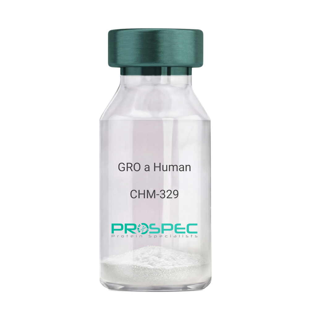
CXCL1 HumanDescription:
GRO-Alpha Human Recombinant (CXCL1)
Growth-regulated protein alpha, CXCL1, Melanoma growth stimulatory activity, MGSA, Neutrophil-activating protein 3, NAP-3, GRO-alpha (1-73), chemokine (C-X-C motif) ligand 1, GRO1, GROa, SCYB1, MGSA-a, MGSA alpha.
Product # :
CHM-329Price :
Quantity :
Shipping Method :
Shipped at Room temp
More Info
- description
- source
- formulation
- purity
- biological activity
- More Info
Description
GRO Alpha Human Recombinant produced in E.Coli is a single,non-glycosylated, polypeptide chain containing 73 amino acids and having a molecular mass of 7811 Dalton. The GRO-alpha is purified by proprietary chromatographic techniques.
Source
Escherichia Coli.
Formulation
Lyophilized from a 0.2µm filtered concentrated (1mg/ml) solution in 20mM PB, pH 7.4, 50mM NaCl.
Purity
Greater than 97.0% as determined by:
(a) Analysis by RP-HPLC.
(b) Analysis by SDS-PAGE.Biological Activity
Determined by its ability to chemoattract human peripheral blood neutrophils using a concentration range of 10.0-100.0 ng/ml.
More Info
-
Introduction
Chemokine (C-X-C motif) ligand 1 (CXCL1) is a small cytokine belonging to the CXC chemokine family that was previously called GRO1 oncogene, Neutrophil-activating protein 3 (NAP-3) and melanoma growth stimulating activity, alpha (MSGA-a). It is secreted by human melanoma cells, has mitogenic properties and is implicated in melanoma pathogenesis. CXCL1 is expressed by macrophages, neutrophilsand epithelial cells, and has neutrophil chemoattractant activity. CXCL1 plays a role in spinal cord development by inhibiting the migration of oligodendrocyte precursors and is involved in the processes of angiogenesis, inflammation, wound healing, and tumorigenesis. This chemokine elicits its effects by signaling through the chemokine receptor CXCR2. The gene for CXCL1 is located on human chromosome 4 amongst genes for other CXC chemokines.
-
Synonyms
Growth-regulated protein alpha, CXCL1, Melanoma growth stimulatory activity, MGSA, Neutrophil-activating protein 3, NAP-3, GRO-alpha (1-73), chemokine (C-X-C motif) ligand 1, GRO1, GROa, SCYB1, MGSA-a, MGSA alpha.
-
Physical Appearance
Sterile Filtered White lyophilized (freeze-dried) powder.
-
Stability
Lyophilized GRO-alpha although stable at room temperature for 3 weeks, should be stored desiccated below -18°C. Upon reconstitution CXCL1 should be stored at 4°C between 2-7 days and for future use below -18°C. For long term storage it is recommended to add a carrier protein (0.1% HSA or BSA).Please prevent freeze-thaw cycles.
-
Solubility
It is recommended to reconstitute the lyophilized CXCL1 in sterile 18MΩ-cm H2O not less than 100µg/ml, which can then be further diluted to other aqueous solutions.
-
Amino Acid Sequence
The sequence of the first five N-terminal amino acids was determined and was found to be Ala-Ser-Val-Ala-Thr.
-
Background
What is the molecular weight/Mw of CXCL1 HUMAN Protein?
CXCL1 HUMAN Protein has a total Mw of 7.81kDa.
What is the source or expression system of CXCL1 HUMAN Protein?
Escherichia Coli.
What is the Purity of CXCL1 HUMAN Protein?
CXCL1 HUMAN Protein is >97% pure as determined by SDS-PAGE.
What is the Biological Activity of CXCL1 HUMAN Protein?
Determined by its ability to chemoattract human peripheral blood neutrophils using a concentration range of 10.0-100.0 ng/ml.
What is the amino acid sequence of CXCL1 HUMAN Protein?
The sequence of the first five N-terminal amino acids was determined and was found to be Ala-Ser-Val-Ala-Thr.
What applications can CXCL1 HUMAN Protein be used in?
CXCL1 HUMAN Protein can probably be used in western blot, ELISA and Lateral Flow.
What is the endotoxin level for CXCL1 HUMAN Protein?
The endotoxin level is minimal, CXCL1 HUMAN Protein was purified using conventional chromatography techniques.
ProSpec's products are furnished for LABORATORY RESEARCH USE ONLY. The product may not be used as drugs, agricultural or pesticidal products, food additives or household chemicals.
- View Data Sheet
Name :
VAMP3 HumanDescription:
Synaptobrevin-3 Human Recombinant
VAMP3, VAMP-3, Cellubrevin, Vesicle-Associated Membrane Protein 3, Synaptobrevin-3, CEB, SYB3.
Product # :
PRO-652Price :
Quantity :
Shipping Method :
Shipped with Ice Packs
More Info
- description
- source
- formulation
- purity
- More Info
Description
VAMP3 Human Recombinant produced in E.Coli is a single, non-glycosylated polypeptide chain containing 77 amino acids and having a molecular mass of 8.7 kDa.
Source
Escherichia Coli.
Formulation
The VAMP3 protein solution contains 20mM Tris pH-7.5 and 10% glycerol.
Purity
Greater than 95.0% as determined by:
(a) Analysis by RP-HPLC.
(b) Analysis by SDS-PAGE.More Info
-
Introduction
VAMP3 is present in recycling endosomes and endosome-derived vesicles. VAMP3 has been implicated in recycling of transferrin receptors to the plasma membrane, secretion of alpha-granules in platelets, recycling of T-cell receptors to the immunological synapses, and membrane trafficking during cell migration. VAMP-3 is present in human platelets and necessary for granule secretion. Synaptobrevins are the main components of a protein complex involved in the docking and/or fusion of synaptic vesicles with the presynaptic membrane. VAMP3 high homology to other VAMPs in its broad tissue distribution and subcellular localization is shown to be the human equivalent of the rodent cellubrevin. In platelets the protein resides on a compartment that is not mobilized to the plasma membrane on calcium or thrombin stimulation.
-
Synonyms
VAMP3, VAMP-3, Cellubrevin, Vesicle-Associated Membrane Protein 3, Synaptobrevin-3, CEB, SYB3.
-
Physical Appearance
Sterile Filtered colorless solution.
-
Stability
Store at 4°C if entire vial will be used within 2-4 weeks. Store, frozen at -20°C for longer periods of time. For long term storage it is recommended to add a carrier protein (0.1% HSA or BSA).Avoid multiple freeze-thaw cycles.
-
Amino Acid Sequence
MSTGPTAATG SNRRLQQTQN QVDEVVDIMR VNVDKVLERD QKLSELDDRA DALQAGASQF ETSAAKLKRK YWWKNCK.
ProSpec's products are furnished for LABORATORY RESEARCH USE ONLY. The product may not be used as drugs, agricultural or pesticidal products, food additives or household chemicals.
- View Data Sheet
Name :
VAMP7 HumanDescription:
Vesicle-Associated Membrane Protein 7 Human Recombinant
Vesicle-associated membrane protein 7, Tetanus-insensitive VAMP, tetanus neurotoxin-insensitive VAMP, Synaptobrevin-like protein 1, TI-VAMP, VAMP-7, TIVAMP, SYBL1.
Product # :
PRO-1194Price :
Quantity :
Shipping Method :
Shipped with Ice Packs
More Info
- description
- source
- formulation
- purity
- More Info
Description
VAMP7 Human Recombinant produced in E. coli is a single polypeptide chain containing 211 amino acids (1-188) and having a molecular mass of 23.0 kDa.VAMP7 is fused to a 23 amino acid His-tag at N-terminus & purified by proprietary chromatographic techniques.
Source
E.coli.
Formulation
The VAMP7 solution (0.5mg/1ml) contains 20mM Tris-HCl buffer (pH 8.0), 0.15M NaCl and 20% glycerol.
Purity
Greater than 90% as determined by SDS-PAGE.
More Info
-
Introduction
Vesicle-Associated Membrane Protein 7 (VAMP7) is a member of the synaptobrevin family. VAMP7 is a transmembrane protein, which is a member of the soluble N-ethylmaleimide-sensitive factor attachment protein receptor (SNARE) family. VAMP7 is involved in the targeting and/or fusion of transport vesicles to their target membrane during transport of proteins from the early endosome to the lysosome. VAMP7 localizes to late endosomes and lysosomes and is involved in the fusion of transport vesicles to their target membranes. VAMP7 is essential for heterotypic fusion of late endosomes with lysosomes and homotypic lysosomal fusion. It is also necessary for calcium regulated lysosomal exocytosis. In addition, VAMP7 is involved in the export of chylomicrons from the endoplasmic reticulum to the cis Golgi. Furthermore, VAMP7 is needed for exocytosis of mediators during eosinophil and neutrophil degranulation, and target cell killing by natural killer cells.
-
Synonyms
Vesicle-associated membrane protein 7, Tetanus-insensitive VAMP, tetanus neurotoxin-insensitive VAMP, Synaptobrevin-like protein 1, TI-VAMP, VAMP-7, TIVAMP, SYBL1.
-
Physical Appearance
Sterile Filtered colorless solution.
-
Stability
Store at 4°C if entire vial will be used within 2-4 weeks. Store, frozen at -20°C for longer periods of time. For long term storage it is recommended to add a carrier protein (0.1% HSA or BSA).Avoid multiple freeze-thaw cycles.
-
Amino Acid Sequence
MGSSHHHHHH SSGLVPRGSH MGSMAILFAV VARGTTILAK HAWCGGNFLE VTEQILAKIP SENNKLTYSH GNYLFHYICQ DRIVYLCITD DDFERSRAFN FLNEIKKRFQ TTYGSRAQTA LPYAMNSEFS SVLAAQLKHH SENKGLDKVM ETQAQVDELK GIMVRNIDLV AQRGERLELL IDKTENLVDS SVTFKTTSRN LARAMCMKNL K
ProSpec's products are furnished for LABORATORY RESEARCH USE ONLY. The product may not be used as drugs, agricultural or pesticidal products, food additives or household chemicals.
- View Data Sheet
Name :
RAC1 Human, HisDescription:
Ras-Related C3 Botulinum Toxin substrate 1 Human Recombinant, His Tag
P21-RAC1, RAC-1, RAC1, RAS-like protein TC25, MIG5, Cell-migration-inducing gene 5 protein,Ras-related C3 botulinum toxin substrate 1, rho family small GTP binding protein Rac1, TC-25, MGC111543.
Product # :
PRO-731Price :
Quantity :
Shipping Method :
Shipped with Ice Packs
More Info
- description
- source
- formulation
- purity
- More Info
Description
Ras-Related C3 Botulinum Toxin substrate 1 Human Recombinant produced in E.Coli is a single, non-glycosylated polypeptide chain containing 212 amino acids (1-192 a.a.) and having a molecular mass of 23.6 kDa. RAC1 contains a 20 amino acid His Tag at N-terminus and is purified by proprietary chromatographic techniques.
Source
Escherichia Coli.
Formulation
The RAC1 His Tag protein solution contains 20mM Tris-HCl pH7.5, 1mM EDTA, 10% glycerol and 1mM DTT.
Purity
Greater than 95.0% as determined by SDS-PAGE.
More Info
-
Introduction
RAC1 is a GTPase which belongs to the RAS superfamily of small GTP-binding proteins. Members of this superfamily appear to regulate a diverse array of cellular events, including the control of cell growth, cytoskeletal reorganization, and the activation of protein kinases. RAC-1 regulates the actin cytoskeleton but also other cellular processes. RAC1 have been shown to be involved in the regulation of cell-cell adhesion.
-
Synonyms
P21-RAC1, RAC-1, RAC1, RAS-like protein TC25, MIG5, Cell-migration-inducing gene 5 protein,Ras-related C3 botulinum toxin substrate 1, rho family small GTP binding protein Rac1, TC-25, MGC111543.
-
Physical Appearance
Sterile Filtered colorless solution.
-
Stability
Store at 4°C if entire vial will be used within 2-4 weeks. Store, frozen at -20°C for longer periods of time. For long term storage it is recommended to add a carrier protein (0.1% HSA or BSA).Please prevent freeze-thaw cycles.
-
Amino Acid Sequence
MGSSHHHHHH SSGLVPRGSH MQAIKCVVVG DGAVGKTCLL ISYTTNAFPG EYIPTVFDNY SANVMVDGKP VNLGLWDTAG QEDYDRLRPLSYPQTDVFLI CFSLVSPASF ENVRAKWYPE VRHHCPNTPI ILVGTKLDLR DDKDTIEKLK EKKLTPITYP QGLAMAKEIG AVKYLECSALTQRGLKTVFD EAIRAVLCPP PVKKRKRKCL LL.
ProSpec's products are furnished for LABORATORY RESEARCH USE ONLY. The product may not be used as drugs, agricultural or pesticidal products, food additives or household chemicals.
- View Data Sheet
Name :
RAC2 HumanDescription:
RAC2 Human Recombinant
p21-Rac2, Small G protein, GX, RAC2, HSPC022, EN-7.
Product # :
PRO-619Price :
Quantity :
Shipping Method :
Shipped with Ice Packs
More Info
- description
- source
- formulation
- purity
- More Info
Description
Recombinant RAC2 produced in E.Coli is a single, non-glycosylated polypeptide chain containing 419 amino acids and having a molecular mass of 47 kDa. RAC2 is fused to GST and purified by conventional chromatography techniques.
Source
Escherichia Coli.
Formulation
The RAC2 protein solution (1mg/ml) contains 1x PBS pH-7.4.
Purity
Greater than 90.0% as determined by SDS-PAGE.
More Info
-
Introduction
RAC2 is a small signaling G protein/GTPase which is part of the RAS superfamily of small GTP-binding proteins which regulate cell growth, actin cytoskeletal organization, cell proliferation, survival, cell cycle progression, gene transcription rgulation and the activation of protein kinases. RAC2 cycles between an active gtp-bound and inactive gdp-bound state. In active state, RAC2 binds to a variety of effector proteins to regulate cellular responses, such as secretory processes, phagocytose of apoptotic cells and epithelial cell polarization. RAC2 is involved in the regulation of the nadph oxidase.
-
Synonyms
p21-Rac2, Small G protein, GX, RAC2, HSPC022, EN-7.
-
Physical Appearance
Sterile filtered colorless solution.
-
Stability
Store at 4°C if entire vial will be used within 2-4 weeks. Store, frozen at -20°C for longer periods of time. For long term storage it is recommended to add a carrier protein (0.1% HSA or BSA). Avoid multiple freeze-thaw cycles.
-
Amino Acid Sequence
MSPILGYWKI KGLVQPTRLL LEYLEEKYEE HLYERDEGDK WRNKKFELGL EFPNLPYYID GDVKLTQSMA IIRYIADKHN MLGGCPKERA EISMLEGAVL DIRYGVSRIA YSKDFETLKV DFLSKLPEML KMFEDRLCHK TYLNGDHVTH PDFMLYDALD VVLYMDPMCL DAFPKLVCFK KRIEAIPQID KYLKSSKYIA WPLQGWQATF GGGDHPPKSD LVPRGSHMQA IKCVVVGDGA VGKTCLLISY TTNAFPGEYI PTVFDNYSAN VMVDSKPVNL GLWDTAGQED YDRLRPLSYP QTDVFLICFS LVSPASYENV RAKWFPEVRH HCPSTPIILV GTKLDLRDDK DTIEKLKEKK
LAPITYPQGL ALAKEIDSVK YLECSALTQR GLKTVFDEAI RAV
LCPQPTR QQKRACSLL.
ProSpec's products are furnished for LABORATORY RESEARCH USE ONLY. The product may not be used as drugs, agricultural or pesticidal products, food additives or household chemicals.
- View Data Sheet
Name :
MCP 1 HumanDescription:
Monocyte Chemotactic Protein-1 Human Recombinant (CCL2)
Small inducible cytokine A2, CCL2, Monocyte chemotactic protein 1, MCP-1, Monocyte chemoattractant protein 1, Monocyte chemotactic and activating factor, MCAF, Monocyte secretory protein JE, HC11, chemokine (C-C motif) ligand 2, MCP1, SCYA2, GDCF-2, SMC-CF, HSMCR30, MGC9434, GDCF-2 HC11.
Product # :
CHM-271Price :
Quantity :
Shipping Method :
Shipped at Room temp
More Info
- description
- source
- formulation
- purity
- biological activity
- More Info
Description
Monocyte Chemotactic Protein-1 Human Recombinant also known as Monocyte Chemotactic and Activating Factor (MCAF) produced in E.Coli is a non-glycosylated, Polypeptide chain containing 76 amino acids and having a molecular mass of 8.6kDa. The MCP-1 is purified by proprietary chromatographic techniques.
Source
Escherichia Coli.
Formulation
CCL2 was lyophilized from a concentrated (1mg/ml) sterile solution without any additives.
Purity
Greater than 98.0% as determined by RP-HPLC and SDS-PAGE.
Biological Activity
The specific activity as determined by the ability of MCP-1 to chemoattract human THP-1 cells is detectable starting at 1ng/ml.More Info
-
Introduction
Chemokine (C-C motif) ligand 2 (CCL2) is a small cytokine belonging to the CC chemokine family that is also known as monocyte chemotactic protein-1 (MCP-1). It is found at the site of tooth eruption and bone degradation. In the bone, CCL2 is expressed by mature osteoclasts and osteoblasts and is under the control of nuclear factor ?B (NF?B). CCL2 recruits immune cells, such as monocytes, to sites of tissue injury and infection. This chemokine is produced as a protein precursor containing signal peptide of 23 amino acids and a mature peptide of 76 amino acids. It is a monomeric polypeptide, with a molecular weight of approximately 13kDa. As with many other CC chemokines, CCL2 is located on chromosome 17 in humans. The cell surface receptors that bind CCL2 are CCR2 and CCR5.
-
Synonyms
Small inducible cytokine A2, CCL2, Monocyte chemotactic protein 1, MCP-1, Monocyte chemoattractant protein 1, Monocyte chemotactic and activating factor, MCAF, Monocyte secretory protein JE, HC11, chemokine (C-C motif) ligand 2, MCP1, SCYA2, GDCF-2, SMC-CF, HSMCR30, MGC9434, GDCF-2 HC11.
-
Physical Appearance
Sterile Filtered White lyophilized (freeze-dried) powder.
-
Stability
Lyophilized MCP-1 although stable at room temperature for 3 weeks, should be stored desiccated below -18°C. Upon reconstitution CCL2 should be stored at 4°C between 2-7 days and for future use below -18°C. For long term storage it is recommended to add a carrier protein (0.1% HSA or BSA).Please prevent freeze-thaw cycles.
-
Solubility
It is recommended to reconstitute the lyophilized Monocyte Chemotactic Protein-1in sterile 18MΩ-cm H2O not less than 100µg/ml, which can then be further diluted to other aqueous solutions.
-
Amino Acid Sequence
QPDAINAPVT CCYNFTNRKI SVQRLASYRR ITSSKCPKEA VIFKTIVAKE ICADPKQKWV QDSMDHLDKQ TQTPKT.
ProSpec's products are furnished for LABORATORY RESEARCH USE ONLY. The product may not be used as drugs, agricultural or pesticidal products, food additives or household chemicals.
- View Data Sheet
Name :
YWHAH HumanDescription:
Tyr-3/Trp-5 Monooxygenase Activation Protein ETA Human Recombinant
14-3-3 ETA, YWHAH, YWHA1, Protein AS1, Tyr-3/Trp-5 Monooxygenase Activation Protein ETA.
Product # :
PKA-087Price :
Quantity :
Shipping Method :
Shipped with Ice Packs
More Info
- description
- source
- formulation
- purity
- More Info
Description
YWHAH Human Recombinant produced in E.Coli is a single, non-glycosylated, polypeptide chain containing 246 amino acids and having a molecular mass of 28.2kDa. The YWHAH is purified by proprietary chromatographic techniques.
Source
Escherichia Coli.
Formulation
The YWHAH protein solution (1mg/ml) containing Phosphate Buffered Saline (pH7.4) 10% glycerol.
Purity
Greater than 90.0% as determined by SDS-PAGE.
More Info
-
Introduction
YWHAH belongs to the 14-3-3 family of proteins which mediate signal transduction by binding to phosphoserine-containing proteins. 14-3-3 ETA is found in plants and mammals, and there is 99% identity to the mouse, rat and bovine orthologs. YWHAH gene contains a 7 base pair repeat sequence in its 5' UTR, and changes in the number of this repeat has been associated with early-onset schizophrenia.
14-3-3 eta is specific to the site of joint inflammation.
14-3-3 proteins are colocalized with Lewy bodies in Parkinson disease, though there is no specific staining for the 14-3-3 eta subunit.
There are 3 different isoforms types of 14-3-3: Beta, Gamma and ETA that are DAL-1/Protein 4.1B-binding proteins. -
Synonyms
14-3-3 ETA, YWHAH, YWHA1, Protein AS1, Tyr-3/Trp-5 Monooxygenase Activation Protein ETA.
-
Physical Appearance
Sterile filtered colorless solution.
-
Stability
Store at 4°C if entire vial will be used within 2-4 weeks. Store, frozen at -20°C for longer periods of time. For long term storage it is recommended to add a carrier protein (0.1% HSA or BSA).Avoid multiple freeze-thaw cycles.
-
Amino Acid Sequence
MGDREQLLQR ARLAEQAERY DDMASAMKAV TELNEPLSNE DRNLLSVAYK NVVGARRSSW RVISSIEQKT MADGNEKKLE KVKAYREKIE KELETVCNDV LSLLDKFLIK NCNDFQYESK VFYLKMKGDY YRYLAEVASG EKKNSVVEAS EAAYKEAFEI SKEQMQPTHP IRLGLALNFS VFYYEIQNAP EQACLLAKQA FDDAIAELDT LNEDSYKDST LIMQLLRDNL TLWTSDQQDE EAGEGN.
ProSpec's products are furnished for LABORATORY RESEARCH USE ONLY. The product may not be used as drugs, agricultural or pesticidal products, food additives or household chemicals.
- View Data Sheet
Name :
HK1/2/3 AntibodyDescription:
Hexokinase-1/2/3, Mouse Anti Human
Hexokinase-3, EC 2.7.1.1, Hexokinase type III, HK III, HXK3, HK3.
Product # :
ANT-726Price :
Quantity :
Shipping Method :
Shipped with Ice Packs
More Info
- formulation
- More Info
Formulation
1mg/ml containing PBS, pH-7.4, 10% Glycerol and 0.02% Sodium Azide.
More Info
-
Introduction
Hexokinases phosphorylate glucose to produce glucose-6-phosphate, thus committing glucose to the glycolytic pathway. HK3 encodes hexokinase 3. Similar to hexokinases 1 and 2, this allosteric enzyme is inhibited by its product glucose-6-phosphate. Hexokinase3 lacks the hydrophobic N-terminal sequence critical for targeting to mitochondria. Hexpkinase3 may have anabolic functions, providing H6P for glycogen or lipid synthesis.
-
Synonyms
Hexokinase-3, EC 2.7.1.1, Hexokinase type III, HK III, HXK3, HK3.
-
Physical Appearance
Sterile filtered colorless solution.
-
Immunogen
Anti-human HK1/2/3mAb, is derived from hybridization of mouse F0 myeloma cells with spleen cells from BALB/c mice immunized with recombinant human HK1/2/3amino acids 1-923 purified from E. coli.
-
Ig Subclass
Mouse IgG1 heavy chain and κ light chain.
-
Clone
PAT4C12AT.
-
Applications
HK1/2/3antibody has been tested by ELISA and Western blot analysis to assure specificity and reactivity. Since application varies, however, each investigation should be titrated by the reagent to obtain optimal results.
-
Type
Mouse Anti Human Monoclonal.
-
Storage Procedures
For periods up to 1 month store at 4°C, for longer periods of time, store at -20°C. Prevent freeze thaw cycles.
-
Purification Method
HK1/2/3 antibody was purified from mouse ascitic fluids by protein-A affinity chromatography.
ProSpec's products are furnished for LABORATORY RESEARCH USE ONLY. The product may not be used as drugs, agricultural or pesticidal products, food additives or household chemicals.
- View Data Sheet
Name :
KIR2DL3 AntibodyDescription:
Killer Cell Immunoglobulin-Like Receptor 2 Domains Long Cytoplasmic Tail 3, Mouse Anti Human
Killer cell immunoglobulin-like receptor 2DL3, MHC class I NK cell receptor, Natural killer-associated transcript 2, NKAT-2, NKAT2a, NKAT2b, p58 natural killer cell receptor clone CL-6, p58 NK receptor, p58.2 MHC class-I-specific NK receptor, Killer inhibitory receptor cl 2-3, KIR-023GB, CD158 antigen-like family member B2, CD158b2 antigen, KIR2DL3, CD158B2, KIRCL23, NKAT2, p58, NKAT, GL183, CD158b, KIR-K7b, KIR-K7c, MGC129943.
Product # :
ANT-300Price :
Quantity :
Shipping Method :
Shipped with Ice Packs
More Info
- formulation
- More Info
Formulation
1mg/ml containing PBS, pH-7.4, & 0.1% Sodium Azide.
More Info
-
Introduction
Killer-cell immunoglobulin-like receptors (KIRs), are a family of cell surface glycoproteins found on Natural Killer (NK) Cells, which are important cells of the immune system. They control the killing function of these cells by interacting with MHC class I molecules, which are expressed on all cell types. This interaction allows them to identify virally infected cells or tumor cells that have a distinctive low level of Class I MHC on their surface. The majority of KIRs are inhibitory, which means that their recognition of MHC suppresses the cytotoxic activity of their NK cell. Only a limited number of KIRs have the capacity to activate cells. The KIR genes are found in a cluster on chromosome 19q13.4 within the 1 Mb leukocyte receptor complex (LRC). KIR molecules are extremely polymorphic, meaning their gene sequences differ significantly between individuals, so that different individuals have different arrays/repertoires of KIR genes. The KIR proteins are categorized by the number of extracellular immunoglobulin domains (2D or 3D) and by whether they have a long (L) or short (S) cytoplasmic domain. KIR proteins with the long cytoplasmic domain transduce inhibitory signals upon ligand binding via an immune tyrosine-based inhibitory motif (ITIM). Whereas KIR proteins with the short cytoplasmic domain lack the ITIM motif and instead associate with the TYRO protein tyrosine kinase binding protein to transduce activating signals. KIR2DL3 is an inhibitory Killer Cell Ig-like Receptor (KIR, previously called p58 KIR, cl-6, NKAT2 or KIR-K7), which recognizes class I MHC molecules (HLA-Cw1, -Cw3, -Cw7, and Cw8). KIR2DL3 inhibits the activity of NK cells thus preventing cell lysis.
-
Synonyms
Killer cell immunoglobulin-like receptor 2DL3, MHC class I NK cell receptor, Natural killer-associated transcript 2, NKAT-2, NKAT2a, NKAT2b, p58 natural killer cell receptor clone CL-6, p58 NK receptor, p58.2 MHC class-I-specific NK receptor, Killer inhibitory receptor cl 2-3, KIR-023GB, CD158 antigen-like family member B2, CD158b2 antigen, KIR2DL3, CD158B2, KIRCL23, NKAT2, p58, NKAT, GL183, CD158b, KIR-K7b, KIR-K7c, MGC129943.
-
Physical Appearance
Sterile Filtered colorless solution.
-
Immunogen
Anti-human KIR2DL3 mAb, is derived from hybridization of mouse P3-x63-Ag8.653 myeloma cells with spleen cells from BALB/c mice immunized with recombinant human KIR2DL3 amino acids 19-161 purified from E. coli.
-
Ig Subclass
Mouse IgG2a heavy chains and κ light chain.
-
Clone
P190IIC311AT.
-
Applications
KIR2DL3 antibody has been tested by ELISA and Western blot immunofluorescent staining with flow cytometric analysis to assure specificity. Since application varies, however, each investigation should be titrated by the reagent to obtain optimal results. Applications include flow cytometry (5-10g/1×1000000), ELISA (1:1,000 when tested against the immunized protein) and western blot analysis (1μg/ml).
-
Type
Mouse Anti Human Monoclonal.
-
Storage Procedures
For periods up to 1 month store at 4°C, for longer periods of time, store at -20°C. Prevent freeze thaw cycles.
-
Purification Method
KIR2DL3 antibody was purified from mouse ascitic fluids by protein-G affinity chromatography.
ProSpec's products are furnished for LABORATORY RESEARCH USE ONLY. The product may not be used as drugs, agricultural or pesticidal products, food additives or household chemicals.
- View Data Sheet
Name :
MFGE8 MouseDescription:
Milk Fat Globule-EGF Factor 8 Protein Mouse Recombinant
Milk fat globule-EGF factor 8 protein, isoform CRA_a, Putative uncharacterized protein, Mfge8, mCG_6301.
Product # :
CYT-1000Price :
Quantity :
Shipping Method :
Shipped with Ice Packs
More Info
- description
- source
- formulation
- purity
- More Info
Description
MFGE8 Mouse Recombinant produced in Sf9 Baculovirus cells is a single, glycosylated polypeptide chain containing 413 amino acids (23-426a.a.) and having a molecular mass of 46kDa (Molecular size on SDS-PAGE will appear at approximately 40-57kDa).MFGE8 is expressed with a 6 amino acid His-tag at C-Terminus and purified by proprietary chromatographic techniques.
Source
Sf9, Baculovirus cells.
Formulation
MFGE8 protein solution (0.25mg/ml) contains Phosphate Buffered Saline (pH 7.4) and 10% glycerol.
Purity
Greater than 90.0% as determined by SDS-PAGE.
More Info
-
Introduction
Milk fat globule-EGF factor 8 protein (Mfge8) is pleiotropic secreted glycoprotein which promotes mammary gland morphogenesis, angiogenesis, and tumor progression. Mfge8 has also an imperative role in tissue homeostasis and the prevention of inflammation. Mfge8 functions as a bridge between phosphatidylserine on apoptotic cells and Integrin alpha V beta 3 on phagocytes, leading to the clearance of apoptotic debris.
-
Synonyms
Milk fat globule-EGF factor 8 protein, isoform CRA_a, Putative uncharacterized protein, Mfge8, mCG_6301.
-
Physical Appearance
Sterile filtered colorless solution.
-
Stability
Store at 4°C if entire vial will be used within 2-4 weeks. Store, frozen at -20°C for longer periods of time. For long term storage it is recommended to add a carrier protein (0.1% HSA or BSA).Avoid multiple freeze-thaw cycles.
-
Amino Acid Sequence
ADLASGDFCD SSLCLNGGTC LTGQDNDIYC LCPEGFTGLV CNETERGPCS PNPCYNDAKC LVTLDTQRGD IFTEYICQCP VGYSGIHCET GCSTQLGMEG GAIADSQISA SSVYMGFMGL QRWGPELARL YRTGIVNAWT ASNYDSKPWI QVNLLRKMRV SGVMTQGASR AGRAEYLKTF KVAYSLDGRK FEFIQDESGG DKEFLGNLDN NSLKVNMFNP TLEAQYIKLY PVSCHRGCTL RFELLGCELH GCSEPLGLKN NTIPDSQMSA SSSYKTWNLR AFGWYPHLGR LDNQGKINAW TAQSNSAKEW LQVDLGTQRQ VTGIITQGAR DFGHIQYVAS YKVAHSDDGV QWTVYEEQGS SKVFQGNLDN NSHKKNIFEK PFMARYVRVL PVSWHNRITL RLELLGCHHH HHH.
-
Background
An Insight into Milk Fat Globule-EGF Factor 8 Protein (Mouse Recombinant): Characteristics, Applications, and Future Directions
Abstract
The Milk Fat Globule-EGF Factor 8 (MFG-E8) protein, particularly in its mouse recombinant form, has emerged as a pivotal player in multiple physiological processes. This paper aims to elucidate its unique characteristics, delve into current methodologies employed in its study, and chart the trajectory for future research directions.
Introduction
Milk Fat Globule-EGF Factor 8 (MFG-E8) is a glycoprotein known for its vital role in several cellular processes, including cell signaling, apoptosis, and phagocytosis. The recombinant form of MFG-E8 derived from mouse models offers a valuable tool for researchers in deciphering its biological relevance.
Characteristics of MFG-E8 (Mouse Recombinant)
1. Structural Profile: The MFG-E8 protein harbors EGF-like domains, which enable its participation in numerous cellular signaling events.
2. Expression Spectrum: While originally identified in mammary epithelial cells, its expression spectrum extends to macrophages, dendritic cells, and other tissues.
3. Biochemical Activity: It plays a key role in facilitating the phagocytic clearance of apoptotic cells by bridging these cells to phagocytes.
Methodologies Employed in MFG-E8 Research
1. Production of Recombinant MFG-E8: Using bacterial expression systems, such as E. coli, mouse MFG-E8 DNA is introduced, followed by protein purification techniques like gel filtration chromatography.
2. Assays: The phagocytosis assays employing fluorescently tagged apoptotic cells and phagocytes enable researchers to quantify MFG-E8's effectiveness in apoptotic cell clearance.
3. Immunoblotting: Through SDS-PAGE and Western blotting, the expression and purification of MFG-E8 can be monitored and validated.
4. Knockout Models: MFG-E8 knockout mice models help decipher its in vivo significance, especially concerning its immune-regulatory roles.
Original Ideas & Implications
The potential for MFG-E8, particularly in its mouse recombinant form, to serve as a therapeutic agent in autoimmune disorders remains a tantalizing prospect. Considering its role in apoptotic cell clearance, dysregulation in MFG-E8 might be implicated in the etiology of autoimmune disorders. Thus, targeting this protein therapeutically may pave the way for innovative treatments.
Conclusions & Future Directions
While the recombinant MFG-E8 protein has elucidated much about the biological implications of this protein, the horizon is rife with potential. Future research might focus on its therapeutic potential, particularly in autoimmunity and inflammation.
ProSpec's products are furnished for LABORATORY RESEARCH USE ONLY. The product may not be used as drugs, agricultural or pesticidal products, food additives or household chemicals.
- View Data Sheet
Name :
HHEX HumanDescription:
Hematopoietically Expressed Homeobox Human Recombinant
Hematopoietically Expressed Homeobox, Hematopoietically-Expressed Homeobox Protein HHEX, Homeobox Hematopoietically Expressed, Proline-Rich Homeodomain-Containing Transcription Factor, Homeobox Protein PRH, Homeobox Protein HEX, HOX11L-PEN, PRHX, HMPH, PRH, HEX.
Product # :
PRO-1624Price :
Quantity :
Shipping Method :
Shipped with Ice Packs
More Info
- description
- source
- formulation
- purity
- More Info
Description
HHEX Human Recombinant produced in E.coli is a single, non-glycosylated polypeptide chain containing 293 amino acids (1-270) and having a molecular mass of 32.4kDa. HHEX is fused to a 23 amino acid His-tag at N-terminus & purified by proprietary chromatographic techniques.
Source
Escherichia Coli.
Formulation
The HHEX solution contains 20mM Tris-HCl buffer (pH 8.0), 0.15M NaCl, 1mM DTT and 40% glycerol.
Purity
Greater than 85% as determined by SDS-PAGE.
More Info
-
Introduction
The homeobox protein family of transcription factors has many members that take part in developmental processes such as HHEX. HHEX is known to interact with Promyelocytic leukemia protein and expression in specific hematopoietic lines indicates that this protein is involved in hematopoietic differentiation.
-
Synonyms
Hematopoietically Expressed Homeobox, Hematopoietically-Expressed Homeobox Protein HHEX, Homeobox Hematopoietically Expressed, Proline-Rich Homeodomain-Containing Transcription Factor, Homeobox Protein PRH, Homeobox Protein HEX, HOX11L-PEN, PRHX, HMPH, PRH, HEX.
-
Physical Appearance
Sterile filtered colorless solution.
-
Stability
Store at 4°C if entire vial will be used within 2-4 weeks. Store, frozen at -20°C for longer periods of time. For long term storage it is recommended to add a carrier protein (0.1% HSA or BSA).Avoid multiple freeze-thaw cycles.
-
Amino Acid Sequence
MGSSHHHHHH SSGLVPRGSH MGSMQYPHPG PAAGAVGVPL YAPTPLLQPA HPTPFYIEDI LGRGPAAPTP APTLPSPNSS FTSLVSPYRT PVYEPTPIHP AFSHHSAAAL AAAYGPGGFG GPLYPFPRTV NDYTHALLRH DPLGKPLLWS PFLQRPLHKR KGGQVRFSND QTIELEKKFE TQKYLSPPER KRLAKMLQLS ERQVKTWFQN RRAKWRRLKQ ENPQSNKKEE LESLDSSCDQ RQDLPSEQNK GASLDSSQCS PSPASQEDLE SEISEDSDQE VDIEGDKSYF NAG
ProSpec's products are furnished for LABORATORY RESEARCH USE ONLY. The product may not be used as drugs, agricultural or pesticidal products, food additives or household chemicals.
- View Data Sheet
Name :
HIV-1 TAT Clade-ADescription:
HIV-1 TAT Clade-A Recombinant
Product # :
HIV-105Price :
Quantity :
Shipping Method :
Shipped with Ice Packs
More Info
- description
- source
- formulation
- purity
- More Info
Description
HIV-1 TAT Clade-A Recombinant produced in E.coli is a single, non-glycosylated, polypeptide chain having the Accession number: AAL06113.1
Source
Escherichia Coli.
Formulation
Lyophilized with 10% glycerol.
Purity
Greater than 90.0% as determined by SDS-PAGE.
More Info
-
Introduction
Human immunodeficiency virus type-1 (HIV-1) regulatory Tat protein plays an essential role in viral replication (Jones KA, 1994) and infectivity (Arya SK, 1985; Fisher AG, 1986). In addition, during acute infection, Tat is released extracellularly by infected cells (Chang HC, 1997; Ensoli B, 1990) and is taken up by neighboring cells where it transactivates viral replication (Ensoli B, 1993) and increases virus infectivity.
HIV-1 Tat activates transcription of HIV-1 viral genes by inducing phosphorylation of the C-terminal domain (CTD) of RNA polymerase II (RNAPII). Tat can also disturb cellular metabolism by inhibiting proliferation of antigen-specific T lymphocytes and by inducing cellular apoptosis. Tat-induced apoptosis of T-cells is attributed, in part, to the distortion of microtubules polymerization. LIS1 is a microtubule-associated protein that facilitates microtubule polymerization. -
Physical Appearance
Sterile Filtered and lyophilized, though might appear as a solution as a result of the glycerol content.
-
Stability
Store at 4°C if entire vial will be used within 2-4 weeks. Store, frozen at -20°C for longer periods of time. Avoid multiple freeze-thaw cycles.
-
Solubility
It is recommended to reconstitute the lyophilized HIV-1 TAT in sterile 18MΩ-cm H2O not less than 100µg/ml, which can then be further diluted to other aqueous solutions.
ProSpec's products are furnished for LABORATORY RESEARCH USE ONLY. The product may not be used as drugs, agricultural or pesticidal products, food additives or household chemicals.
- View Data Sheet
Name :
Myoglobin HumanDescription:
Myoglobin Human Recombinant
Myoglobin, MB, PVALB, MGC13548.
Product # :
PRO-336Price :
Quantity :
Shipping Method :
Shipped with Ice Packs
More Info
- description
- source
- formulation
- purity
- More Info
Description
Myoglobin Human Recombinant produced in E.Coli is a non-glycosylated polypeptide chain having a molecular mass of 17.67 kDa. The Myoglobin is purified by proprietary chromatographic techniques.
Source
Escherichia Coli.
Formulation
The sterile solution contains phosphate-buffered saline (pH 7.4) and 0.05% NaN3.
Purity
Greater than 95.0% as determined by SDS-PAGE.
More Info
-
Introduction
Myoglobin is a member of the globin superfamily and can be found in skeletal and cardiac muscles. It is a haemoprotein that contributs to intracellular oxygen storage and transcellular facilitated diffusion of oxygen. Myoglobin has a single-chain globular structure of 153 amino acids, containing a heme prosthetic group (iron-containing porphyrin) in the core around which the remaining apoprotein folds. Myoglobin has 8 alpha helices and a hydrophobic core. Myoglobin’s molecular weight is 16.7 kDa, and it is the primary oxygen-carrying pigment of muscle tissues. The binding of oxygen in myoglobin is different from the cooperative oxygen binding in hemoglobin, since positive collaboration is a property of multimeric/oligomeric proteins only. Instead, the binding of oxygen by myoglobin is uninfluenced by the oxygen pressure in the surrounding tissue. Myoglobin is frequently referred to as having an "instant binding tenacity" to oxygen given its hyperbolic oxygen dissociation curve. Different organisms are able to hold their breaths longer due to high concentrations of myoglobin in their muscle cells. Myoglobin is responsible for the pigments that make meat red. The color of the meat is partly determined by the charge of the iron atom in myoglobin and the oxygen attached to it. Myoglobin is found in Type I muscle, Type II A and Type II B, but it is mostly deemed that myoglobin is not found in smooth muscle. Myoglobin is discharged from damaged muscle tissue (rhabdomyolysis), which contains very high concentrations of myoglobin. Even though the released myoglobin is filtered by the kidneys, it is toxic to the renal tubular epithelium and thus may cause acute renal failure.
-
Synonyms
Myoglobin, MB, PVALB, MGC13548.
-
Physical Appearance
Sterile Filtered brownish solution.
-
Stability
Myoglobin should be stored at 4°C. For long term storage it is recommended to add a carrier protein (0.1% HSA or BSA).Please do not freeze.
ProSpec's products are furnished for LABORATORY RESEARCH USE ONLY. The product may not be used as drugs, agricultural or pesticidal products, food additives or household chemicals.
- View Data Sheet
Name :
AsnRSDescription:
Asparagine tRNA Synthetase Brugia Malayi Recombinant
Asparagine--tRNA ligase, cytoplasmic (EC:6.1.1.22), Asparaginyl-tRNA synthetase, AsnRS, Potentially protective 63 kDa antigen.
Product # :
ENZ-941Price :
Quantity :
Shipping Method :
Shipped with Ice Packs
More Info
- description
- source
- formulation
- purity
- More Info
Description
AsnRS Brugia Malayi Recombinant produced in E.Coli is a single, non-glycosylated, polypeptide chain containing 568 amino acids (including a 6xHis Tag at N-terminus) and having a molecular mass of 64.5kDa.The AsnRS is purified by proprietary chromatographic techniques.
Source
Escherichia Coli.
Formulation
AsnRS 0.2µm filtered solution containing 20mM HEPES, pH7.4, 100mM NaCl, 5mM MgCl2, 5mM b-Mercaptoethanol and 10 % Glycerol.
Purity
Greater than 95.0% as determined by SDS-PAGE.
More Info
-
Introduction
The AsnRS enzyme is a member of the ligases family, specifically those forming carbon-oxygen bonds in aminoacyl-tRNA and related compounds. An asparagine-tRNA ligase is an enzyme which catalyzes the chemical reaction: ATP + L-asparagine + tRNAAsn AMP + diphosphate + L-asparaginyl-tRNAAsn. The three substrates of the AsnRS enzyme are ATP, L-asparagine, and tRNA(Asn), whereas its three products are AMP, diphosphate, and L-asparaginyl-tRNA(Asn).
-
Synonyms
Asparagine--tRNA ligase, cytoplasmic (EC:6.1.1.22), Asparaginyl-tRNA synthetase, AsnRS, Potentially protective 63 kDa antigen.
-
Physical Appearance
Sterile filtered colorless solution.
-
Stability
Store at 4°C if entire vial will be used within 2-4 weeks. Store, frozen at -20°C for longer periods of time. For long term storage it is recommended to add a carrier protein (0.1% HSA or BSA).Avoid multiple freeze-thaw cycles.
-
Amino Acid Sequence
MGSSHHHHHH SSGLVPRGSH MTVYICPETG DDGNDGSELK PLRTLYQAMI ITKSSKGDFL IRTKKDGKQV WEAASKTALK KSWKRYEQEM LKNEKVAAKM LEKDATEVGV KAALEEAKKV QIELDTSLSY ITGVKIRDLV KHRNERVCIK GWIHRMRRQG KSLMFFILRD GTGFLQVLLM DKLCQTYDAL TVNTECTVEI YGAIKEVPEG KEAPNGHELI ADFWKIIGNA PSGGIDNVLN EEASVDKMLD NRHLVIRGEN AAALLRLRAA ATRAMREHFY NAGYVEVAPP TLVQTQVEGG STLFNLDYFG EQSFLTQSSQ LYLETCIPTL GDVFLHCSVL QGGKISHSST LAEYAHVEAE CPFITLDDLM EKIEELVCDT VDRLLADEEA KKLLEHINPK FQPPERPFLR MEYKDAIKWL QEHNVENEFG NTFTYGEDIA EAAERFMTDT INKPILLNRF PSEIKAFYMQ RDAKDNTLTE SVDLLMPGVG EIVGGSMRIW KFDELSKAFK NVEIDPKPYY WYLDQRLYGT CPHGGYGLGL ERFICWLTNT NHIRDVCLYP RFVGRCVP.
ProSpec's products are furnished for LABORATORY RESEARCH USE ONLY. The product may not be used as drugs, agricultural or pesticidal products, food additives or household chemicals.
- View Data Sheet
Name :
SNCG MouseDescription:
Gamma-Synuclein Mouse Recombinant
Gamma-synuclein, Persyn.
Product # :
PRO-2490Price :
Quantity :
Shipping Method :
Shipped with Ice Packs
More Info
- description
- source
- formulation
- purity
- More Info
Description
SNCG Mouse Recombinant produced in E.Coli is a single, non-glycosylated polypeptide chain containing 146 amino acids (1-123 a.a) and having a molecular mass of 15.5kDa.SNCG is fused to a 23 amino acid His-tag at N-terminus & purified by proprietary chromatographic techniques.
Source
Escherichia Coli.
Formulation
SNCG protein solution (1mg/ml) containing 20mM Tris-HCl(pH8.0) and 0.1M NaCl.
Purity
Greater than 95.0% as determined by SDS-PAGE.
More Info
-
Introduction
g-synuclein(Originally known as a breast cancer specific gene product, BCSG1) is an acidic neuronal protein of 127 amino acids. Gamma-Synuclein is a member of the Synuclein protein family, which is believed to be involved in the pathogenesis of neurodegenerative diseases. High levels of Gamma-Synuclein have been found in advanced breast carcinomas suggesting a correlation between overexpression of SNCG and breast tumor development. Synuclein-Gamma is found mostly in the peripheral nervous system (in primary sensory neurons, sympathetic neurons, and motor neurons) and retina. SNCG is also identified in the brain, ovarian tumors, and in the olfactory epithelium. SNCG expression in breast tumors is a marker for tumor progression. A modification in the expression of gamma-synuclein has been detected in the retina of Alzheimer's patients.
-
Synonyms
Gamma-synuclein, Persyn.
-
Physical Appearance
Sterile filtered colorless solution.
-
Stability
Store at 4°C if entire vial will be used within 2-4 weeks. Store, frozen at -20°C for longer periods of time.For long term storage it is recommended to add a carrier protein (0.1% HSA or BSA). Avoid multiple freeze-thaw cycles.
-
Amino Acid Sequence
MGSSHHHHHH SSGLVPRGSH MGSMDVFKKG FSIAKEGVVG AVEKTKQGVT EAAEKTKEGV MYVGTKTKEN VVQSVTSVAE KTKEQANAVS AVVSSVNTV ANKTVEEAEN IVVTTGVVRK EDLEPPAQDQ EAKEQEENEE AKSGED.
ProSpec's products are furnished for LABORATORY RESEARCH USE ONLY. The product may not be used as drugs, agricultural or pesticidal products, food additives or household chemicals.