Skip to content

High-quality research proteins.

  • 1-800-805-6531

  • Mon - Sat 8.00-18.00, Sun - Closed

  • info@prospecbio.com

  • PO Box 6591 East Brunswick NJ 08816

  • Products
    • Cytokines
    • Growth factors
    • Chemokines
    • Cd antigens
    • Neurotrophins
    • Hormones
    • Enzymes
    • Viral antigens
    • Recombinant proteins
    • Natural proteins
    • Monoclonal antibodies
    • Polyclonal antibodies
    • Thermal Packaging
  • About
    • Company
    • Founder
    • Contribution
  • Customers
    • Eastern Asia
    • Eastern Europe
    • North America
    • South America
    • South East Asia
    • Southern Asia
    • Western Europe
  • Ordering
    • Ordering Method
    • Ordering - Credit Card
    • Ordering - PO
    • Shipping Fees
    • FAQs
  • Services
    • DNA Cloning
    • Protein Expression
    • Monoclonal Antibody
    • Fermentation
    • Technology License
  • Contact us
Log in

Country/region

  • USD $ | Albania
  • USD $ | Andorra
  • USD $ | Argentina
  • USD $ | Armenia
  • USD $ | Australia
  • USD $ | Austria
  • USD $ | Azerbaijan
  • USD $ | Belarus
  • USD $ | Belgium
  • USD $ | Bolivia
  • USD $ | Brazil
  • USD $ | Bulgaria
  • USD $ | Cambodia
  • USD $ | Canada
  • USD $ | Chile
  • USD $ | China
  • USD $ | Colombia
  • USD $ | Costa Rica
  • USD $ | Croatia
  • USD $ | Cyprus
  • USD $ | Czechia
  • USD $ | Denmark
  • USD $ | Estonia
  • USD $ | Finland
  • USD $ | France
  • USD $ | Georgia
  • USD $ | Germany
  • USD $ | Greece
  • USD $ | Hong Kong SAR
  • USD $ | Hungary
  • USD $ | Iceland
  • USD $ | India
  • USD $ | Ireland
  • USD $ | Israel
  • USD $ | Italy
  • USD $ | Japan
  • USD $ | Kazakhstan
  • USD $ | Latvia
  • USD $ | Liechtenstein
  • USD $ | Lithuania
  • USD $ | Madagascar
  • USD $ | Malta
  • USD $ | Mexico
  • USD $ | Moldova
  • USD $ | Monaco
  • USD $ | Netherlands
  • USD $ | New Zealand
  • USD $ | North Macedonia
  • USD $ | Norway
  • USD $ | Panama
  • USD $ | Paraguay
  • USD $ | Peru
  • USD $ | Philippines
  • USD $ | Poland
  • USD $ | Portugal
  • USD $ | Romania
  • USD $ | Singapore
  • USD $ | Slovakia
  • USD $ | Slovenia
  • USD $ | South Africa
  • USD $ | South Korea
  • USD $ | Spain
  • USD $ | Sri Lanka
  • USD $ | Sweden
  • USD $ | Switzerland
  • USD $ | Taiwan
  • USD $ | Thailand
  • USD $ | Türkiye
  • USD $ | Ukraine
  • USD $ | United Kingdom
  • USD $ | United States
  • USD $ | Uruguay
  • USD $ | Vatican City
  • USD $ | Venezuela
  • USD $ | Vietnam
  • Facebook
  • Instagram
logoProSpec - Recombinant Protein Manufacturer for academic and R&D industry
Become a distributor
Account Log in
Wishlist
Cart Cart(0)
  • Products
    +
    • Cytokines
    • Growth factors
    • Chemokines
    • Cd antigens
    • Neurotrophins
    • Hormones
    • Enzymes
    • Viral antigens
    • Recombinant proteins
    • Natural proteins
    • Monoclonal antibodies
    • Polyclonal antibodies
    • Thermal Packaging
  • About
    +
    • Company
    • Founder
    • Contribution
  • Customers
    +
    • Eastern Asia
    • Eastern Europe
    • North America
    • South America
    • South East Asia
    • Southern Asia
    • Western Europe
  • Ordering
    +
    • Ordering Method
    • Ordering - Credit Card
    • Ordering - PO
    • Shipping Fees
    • FAQs
  • Services
    +
    • DNA Cloning
    • Protein Expression
    • Monoclonal Antibody
    • Fermentation
    • Technology License
  • Contact us

Item added to your cart

View cart
View All
  • Cytokines
  • Growth factors
  • Chemokines
  • Cd antigens
  • Neurotrophins
  • Hormones
  • Enzymes
  • Viral antigens
  • Recombinant proteins
  • Natural proteins
  • Monoclonal antibodies
  • Polyclonal antibodies
  • Thermal Packaging
  • Tumor Necrosis Factor

    Tumor Necrosis Factor

    View All
  • 4-1BB

    4-1BB

    View All
  • Adiponectin

    Adiponectin

    View All
  • AIF1

    AIF1

    View All
  • Other Cytokines

    Other Cytokines

    View All
  • AITRL

    AITRL

    View All
  • Angiopoietin

    Angiopoietin

    View All
  • Apolipoprotein

    Apolipoprotein

    View All
  • B-Cell Activating Factor

    B-Cell Activating Factor

    View All
  • Beta Defensin

    Beta Defensin

    View All
  • Bone Morphogenetic Protein

    Bone Morphogenetic Protein

    View All
  • B type Natriuretic Peptide

    B type Natriuretic Peptide

    View All
  • BST

    BST

    View All
  • Betacellulin

    Betacellulin

    View All
  • Cardiotrophin

    Cardiotrophin

    View All
  • Activin

    Activin

    View All
  • Fibroblast Growth Factor

    Fibroblast Growth Factor

    View All
  • Other Growth Factors

    Other Growth Factors

    View All
  • CSF

    CSF

    View All
  • CTGF

    CTGF

    View All
  • IGFBP

    IGFBP

    View All
  • VEGF Protein

    VEGF Protein

    View All
  • EGF

    EGF

    View All
  • Epigen

    Epigen

    View All
  • Erythropoietin

    Erythropoietin

    View All
  • Insulin-Like Growth Factor

    Insulin-Like Growth Factor

    View All
  • Growth Hormone

    Growth Hormone

    View All
  • HDGF

    HDGF

    View All
  • Hepatocyte Growth Factor

    Hepatocyte Growth Factor

    View All
  • Insulin

    Insulin

    View All
  • BCA-1/ BLC (CXCL13)

    BCA-1/ BLC (CXCL13)

    View All
  • BRAK (CXCL14)

    BRAK (CXCL14)

    View All
  • C-10 (CCL6)

    C-10 (CCL6)

    View All
  • Other Chemokines

    Other Chemokines

    View All
  • MEC (CCL28)

    MEC (CCL28)

    View All
  • LD78-beta (CCL3L1)

    LD78-beta (CCL3L1)

    View All
  • CTACK (CCL27)

    CTACK (CCL27)

    View All
  • CXCL16

    CXCL16

    View All
  • CXCL17

    CXCL17

    View All
  • Platelet Factor-4 (CXCL4)

    Platelet Factor-4 (CXCL4)

    View All
  • ENA-78 (CXCL5)

    ENA-78 (CXCL5)

    View All
  • Eotaxin (CCL11,24,26)

    Eotaxin (CCL11,24,26)

    View All
  • Exodus-2 (CCL21)

    Exodus-2 (CCL21)

    View All
  • Fractalkine (CX3CL1)

    Fractalkine (CX3CL1)

    View All
  • CXCL6

    CXCL6

    View All
  • Other CD Antigens

    Other CD Antigens

    View All
  • CD14

    CD14

    View All
  • CD164

    CD164

    View All
  • CD1

    CD1

    View All
  • CD2

    CD2

    View All
  • CD200

    CD200

    View All
  • CD207

    CD207

    View All
  • CD226

    CD226

    View All
  • CD23

    CD23

    View All
  • CD244

    CD244

    View All
  • CD247

    CD247

    View All
  • CD27

    CD27

    View All
  • CD274

    CD274

    View All
  • CD300

    CD300

    View All
  • CD33

    CD33

    View All
  • Other Neurotrophins

    Other Neurotrophins

    View All
  • Beta-NGF

    Beta-NGF

    View All
  • BDNF

    BDNF

    View All
  • CDNF

    CDNF

    View All
  • CNTF

    CNTF

    View All
  • GDNF

    GDNF

    View All
  • Glia Maturation Factor

    Glia Maturation Factor

    View All
  • MANF

    MANF

    View All
  • Midkine

    Midkine

    View All
  • Neuroglobin

    Neuroglobin

    View All
  • Neurotrophic factors

    Neurotrophic factors

    View All
  • Neuregulin

    Neuregulin

    View All
  • Neuropilin

    Neuropilin

    View All
  • Pigment Epithelium-Derived Factor

    Pigment Epithelium-Derived Factor

    View All
  • Pleiotrophin

    Pleiotrophin

    View All
  • Peptide Hormones

    Peptide Hormones

    View All
  • Other Hormones

    Other Hormones

    View All
  • HCG

    HCG

    View All
  • Endothelin

    Endothelin

    View All
  • Exendin

    Exendin

    View All
  • FSH

    FSH

    View All
  • Glucagon

    Glucagon

    View All
  • GHRP

    GHRP

    View All
  • GLP

    GLP

    View All
  • Thyrostimulin

    Thyrostimulin

    View All
  • Inhibin A

    Inhibin A

    View All
  • LHRH

    LHRH

    View All
  • Melanotan

    Melanotan

    View All
  • Procalcitonin

    Procalcitonin

    View All
  • PTH

    PTH

    View All
  • Peroxiredoxin

    Peroxiredoxin

    View All
  • Peptidase

    Peptidase

    View All
  • Synthetase

    Synthetase

    View All
  • Transferase

    Transferase

    View All
  • Hydrolase

    Hydrolase

    View All
  • Dehydrogenase

    Dehydrogenase

    View All
  • ACE2

    ACE2

    View All
  • Esterase

    Esterase

    View All
  • Protein Kinases

    Protein Kinases

    View All
  • Other Enzymes

    Other Enzymes

    View All
  • Phosphatase

    Phosphatase

    View All
  • Deaminase

    Deaminase

    View All
  • Oxygenase

    Oxygenase

    View All
  • Adenylate Kinase

    Adenylate Kinase

    View All
  • Lyase

    Lyase

    View All
  • Chagas

    Chagas

    View All
  • SARS

    SARS

    View All
  • Influenza

    Influenza

    View All
  • ASFV

    ASFV

    View All
  • Astrovirus

    Astrovirus

    View All
  • Borrelia

    Borrelia

    View All
  • Helicobacter Pylori

    Helicobacter Pylori

    View All
  • Chikungunya

    Chikungunya

    View All
  • Chlamydia

    Chlamydia

    View All
  • Cytomegalo

    Cytomegalo

    View All
  • Dengue

    Dengue

    View All
  • EBV

    EBV

    View All
  • Ebola

    Ebola

    View All
  • Feline Leukemia Virus

    Feline Leukemia Virus

    View All
  • Hepatitis A

    Hepatitis A

    View All
  • Other

    Other

    View All
  • Actin

    Actin

    View All
  • ADAM

    ADAM

    View All
  • Complement Component

    Complement Component

    View All
  • Ag85

    Ag85

    View All
  • Eukaryotic Translation Initiation Factor

    Eukaryotic Translation Initiation Factor

    View All
  • Anterior Gradient Protein

    Anterior Gradient Protein

    View All
  • Heat Shock Protein

    Heat Shock Protein

    View All
  • Alpha-2-HS-Glycoprotein

    Alpha-2-HS-Glycoprotein

    View All
  • Hemoglobin

    Hemoglobin

    View All
  • Allergy

    Allergy

    View All
  • Anaplasma

    Anaplasma

    View All
  • Angiogenin

    Angiogenin

    View All
  • Ankyrin Repeat Domain

    Ankyrin Repeat Domain

    View All
  • Annexin

    Annexin

    View All
  • Other Natural Proteins

    Other Natural Proteins

    View All
  • Aprotinin

    Aprotinin

    View All
  • Transferrin

    Transferrin

    View All
  • Avidin

    Avidin

    View All
  • Anti Coagulation Factors

    Anti Coagulation Factors

    View All
  • Natural Albumin

    Natural Albumin

    View All
  • Natural Coagulation Factors

    Natural Coagulation Factors

    View All
  • Fibronectin

    Fibronectin

    View All
  • Thrombin

    Thrombin

    View All
  • Anti Human Enzyme

    Anti Human Enzyme

    View All
  • Other Monoclonal Antibodies

    Other Monoclonal Antibodies

    View All
  • Anti Human Cytokine

    Anti Human Cytokine

    View All
  • Anti Human Heat Shock Protein

    Anti Human Heat Shock Protein

    View All
  • Anti Mouse Lymphocyte

    Anti Mouse Lymphocyte

    View All
  • Anti Human Chemokine

    Anti Human Chemokine

    View All
  • Anti Human Lymphocyte

    Anti Human Lymphocyte

    View All
  • Anti Viral Monoclonal

    Anti Viral Monoclonal

    View All
  • Anti Mouse Cytokine

    Anti Mouse Cytokine

    View All
  • Anti-GST Monoclonal

    Anti-GST Monoclonal

    View All
  • Other Polyclonal Antibodies

    Other Polyclonal Antibodies

    View All
  • Anti Viral

    Anti Viral

    View All
  • Anti-GST Monoclonal

    Anti-GST Monoclonal

    View All
Back

Search results

1000 results found for “gluteradoxin”

Name

Description

Product #

Price

Quantity

Shipping Method

  • View Data Sheet

    Name :

    CD105 Human, Sf9

    Description:

    Endoglin Human Recombinant, Sf9

    CD105, ENG, END, ORW, HHT1, ORW1, FLJ41744, Endoglin.

    Product # :

    CYT-389

    Price :

    Quantity :

    Shipping Method :

    Room Temp Icon

    Shipped at Room temp

    Add To Cart

    More Info

    • description
    • source
    • formulation
    • purity
    • biological activity
    • More Info
    • Citations

    Description

    CD105 Human Recombinant extracellular domain produced in baculovirus is a homodimeric, glycosylated, Polypeptide containing 586 amino acids and having a molecular mass of 61 kDa but as a result of glycosylation, migrates at 90 kDa under reducing conditions in SDS-PAGE. The CD105 is fused to a C-terminal His-tag (6xHis) and purified by proprietary chromatographic techniques.

    Source

    Sf9 Insect Cells.

    Formulation

    Endoglin was lyophilized from a concentrated (1mg/ml) sterile solution containing 1xPBS.

    Purity

    Greater than 95.0% as determined by:
    (a) Analysis by RP-HPLC.
    (b) Analysis by SDS-PAGE.

    Biological Activity

    Measured by its ability to bind with rhTGF-beta RII/Fc in a functional ELISA. Optimal dilutions should be determined by each laboratory for each application.

    Citations

    More Info

    • Introduction

      Endoglin is a type I membrane glycoprotein located on cell surfaces and is part of the TGF beta receptor complex.
      The protein consists of a homodimer of 180 kDA with disulfide links. It has been found on endothelial cells, activated macrophages, fibroblasts, and smooth muscle cells. Endoglin has been found to be part of the TGF-beta1 receptor complex. It thus may be involved in the binding of TGF-beta1, TGF-beta3, activin-A, BMP-2, and BMP-7. Beside TGF-beta signaling endoglin may have other functions. It has been postulated that endoglin is involved in the cytoskeletal organization affecting cell morphology and migration. Endoglin has a role in the development of the cardiovascular system and in vascular remodeling. Its expression is regulated during heart development . Experimental mice without the endoglin gene die due to cardiovascular abnormalities.

    • Synonyms

      CD105, ENG, END, ORW, HHT1, ORW1, FLJ41744, Endoglin.

    • Physical Appearance

      Sterile Filtered White lyophilized (freeze-dried) powder.

    • Stability

      Lyophilized Endoglin although stable at room temperature for 3 weeks, should be stored desiccated below -18°C. Upon reconstitution CD105 should be stored at 4°C between 2-7 days and for future use below -18°C.For long term storage it is recommended to add a carrier protein (0.1% HSA or BSA).Please prevent freeze-thaw cycles.

    • Solubility

      It is recommended to reconstitute the lyophilized CD-105 in sterile PBS not less than 100µg/ml, which can then be further diluted to other aqueous solutions.

    • Amino Acid Sequence

      MDRGTLPLAVALLLASCSLSPTSLAETVHCDLQPVGPERGEVTY TTSQVSKGCVAQAPNAILEVHVLFLEFPTGPSQLELTLQASKQNGTWPREVLLVL SVNSSVFLHLQALGIPLHLAYNSSLVTFQEPPGVNTTELPSFPKTQILEWAAERGPI TSAAELNDPQSILLRLGQAQGSLSFCMLEASQDMGRTLEWRPRTPALVRGCHLE GVAGHKEAHILRVLPGHSAGPRTVTVKVELSCAPGDLDAVLILQGPPYVSWLID ANHNMQIWTTGEYSFKIFPEKNIRGFKLPDTPQGLLGEARMLNASIVASFVELPL ASIVSLHASSCGGRLQTSPAPIQTTPPKDTCSPELLMSLIQTKCADDAMTLVLKKE LVAHLKCTITGLTFWDPSCEAEDRGDKFVLRSAYSSCGMQVSASMISNEAVVNI LSSSSPQRKKVHCLNMDSLSFQLGLYLSPHFLQASNTIEPGQQSFVQVRVSPSVSE FLLQLDSCHLDLGPEGGTVELIQGRAAKGNCVSLLSPSPEGDPRFSFLLHFYTVPI PKTGTLSCTVALRPKTGS.

    • Background

      What is the molecular weight/Mw of CD105 Protein?
      CD105 Protein has a total Mw of 90kDa.

      What is the source or expression system of CD105 Protein?
      Sf9 Insect Cells.

      What is the Purity of CD105 Protein?
      CD105 Protein is >95% pure as determined by SDS-PAGE.

      What is the Biological Activity of CD105 Protein?
      Measured by its ability to bind with rhTGF-beta RII/Fc in a functional ELISA. Optimal dilutions should be determined by each laboratory for each application.

      What is the amino acid sequence of CD105 Protein?
      MDRGTLPLAVALLLASCSLSPTSLAETVHCDLQPVGPERGEVTY TTSQVSKGCVAQAPNAILEVHVLFLEFPTGPSQLELTLQASKQNGTWPREVLLVL SVNSSVFLHLQALGIPLHLAYNSSLVTFQEPPGVNTTELPSFPKTQILEWAAERGPI TSAAELNDPQSILLRLGQAQGSLSFCMLEASQDMGRTLEWRPRTPALVRGCHLE GVAGHKEAHILRVLPGHSAGPRTVTVKVELSCAPGDLDAVLILQGPPYVSWLID ANHNMQIWTTGEYSFKIFPEKNIRGFKLPDTPQGLLGEARMLNASIVASFVELPL ASIVSLHASSCGGRLQTSPAPIQTTPPKDTCSPELLMSLIQTKCADDAMTLVLKKE LVAHLKCTITGLTFWDPSCEAEDRGDKFVLRSAYSSCGMQVSASMISNEAVVNI LSSSSPQRKKVHCLNMDSLSFQLGLYLSPHFLQASNTIEPGQQSFVQVRVSPSVSE FLLQLDSCHLDLGPEGGTVELIQGRAAKGNCVSLLSPSPEGDPRFSFLLHFYTVPI PKTGTLSCTVALRPKTGS.

      What applications can CD105 Protein be used in?
      CD105 Protein can probably be used in western blot, ELISA and Lateral Flow.

      What is the endotoxin level for CD105 Protein?
      The endotoxin level is minimal, CD105 Protein was purified using conventional chromatography techniques.

    ProSpec's products are furnished for LABORATORY RESEARCH USE ONLY. The product may not be used as drugs, agricultural or pesticidal products, food additives or household chemicals.

    Endoglin Human Sf9
  • View Data Sheet

    Name :

    PROK Tritirachium album

    Description:

    Tritirachium album Proteinase-K Recombinant

    Proteinase K (EC:3.4.21.64), Endopeptidase K, Tritirachium alkaline proteinase, PROK.

    Product # :

    ENZ-1015

    Price :

    Quantity :

    Shipping Method :

    Room Temp Icon

    Shipped at Room temp

    Add To Cart

    More Info

    • description
    • source
    • formulation
    • purity
    • biological activity
    • More Info
    • Citations

    Description

    Recombinant Tritirachium album Proteinase-K expressed in yeast containing 285 amino acids having a Mw of 29.3 kDa is purified by standard chromatography techniques.

    Source

    Yeast

    Formulation

    The Proteinase-K was lyophilized without any additives.

    Purity

    Greater than 95% as determined by SDS-PAGE.

    Biological Activity

    36 Units/mg.
    One unit is defined as the amount of enzyme that will hydrolyze urea-denatured hemoglobin to produce color equivalent to 1.0 mol tyrosine per min at 37°C, pH 7.5 (color by Folin-Ciocalteu reagent).

    Citations

    More Info

    • Introduction

      The Proteinase K enzyme is a member of the Peptidase family S8. Proteinase K is a broad-spectrum serine protease. Proteinase K is capable of digesting hair (keratin), henceforth, the name "Proteinase K". Proteinase K is activated by calcium, the enzyme digests proteins especially after hydrophobic amino acids (aliphatic, aromatic and other hydrophobic amino acids). Proteinase K is frequently utilized in molecular biology to digest protein and remove contamination from preparations of nucleic acid. Addition of Proteinase K to nucleic acid preparations rapidly inactivates nucleases which may otherwise degrade the DNA or RNA during purification. Proteinase K is greatly fitting to this application as the enzyme is active in the presence of chemicals which denature proteins, such as SDS and urea, chelating agents such as EDTA, sulfhydryl reagents, as well as trypsin or chymotrypsin inhibitors. Proteinase K is utilized for the destruction of proteins in cell lysates (tissue, cell culture cells) and for the release of nucleic acids, given that it quite effectively inactivates DNases and RNases.

    • Synonyms

      Proteinase K (EC:3.4.21.64), Endopeptidase K, Tritirachium alkaline proteinase, PROK.

    • Physical Appearance

      Sterile Filtered lyophilized powder.

    • Stability

      Recombinant Proteinase-K although stable at room temperature, should be stored between 2-8°C. Do not freeze!

    • Solubility

      It is recommended to reconstitute the lyophilized Proteinase-K in 20mM Tris-HCl (pH 7.4~8.0), 1mM CaCl2, 50% glycerol not less than 100µg/ml, which can then be further diluted to other aqueous solutions.

    • Note

      Bulk Proteinase-K recombinant is available 1,000 grams price is $100 per gram

    ProSpec's products are furnished for LABORATORY RESEARCH USE ONLY. The product may not be used as drugs, agricultural or pesticidal products, food additives or household chemicals.

    Prok Tritirachium Album
  • View Data Sheet

    Name :

    POGLUT1 Human

    Description:

    Protein O-Glucosyltransferase 1 Human Recombinant

    POGLUT1, C3orf9, CLP46, hCLP46, KDELCL1, KTELC1, Protein O-glucosyltransferase 1, CAP10-like 46 kDa protein, KTEL motif-containing protein 1, Myelodysplastic syndromes relative protein, O-glucosyltransferase Rumi homolog, hRumi, Protein O-xylosyltransferase, MDSRP.

    Product # :

    ENZ-956

    Price :

    Quantity :

    Shipping Method :

    Ice Icon

    Shipped with Ice Packs

    Add To Cart

    More Info

    • description
    • source
    • formulation
    • purity
    • More Info
    • Citations

    Description

    POGLUT1 Human Recombinant produced in in Sf9 Baculovirus cells is a single, non-glycosylated polypeptide chain containing 377 amino acids (24-392a.a) and having a molecular mass of 44.5kDa (Migrates at 40-57kDa on SDS-PAGE under reducing conditions). POGLUT1 is fused to a 8 amino acid His-tag at C-Terminus and purified by proprietary chromatographic techniques.

    Source

    Sf9, Baculovirus cells.

    Formulation

    The POGLUT1 solution (0.25mg/ml) contains Phosphate Buffered Saline (pH 7.4) and 10% glycerol.

    Purity

    Greater than 90% as determined by SDS-PAGE.

    Citations

    More Info

    • Introduction

      POGLUT1 is a homologue of Rumi from Drosophila, an endoplasmic reticulum (ER)-retaining glucosyltransferase which catalyzes the transfer of glucose and xylose from UDP-glucose and UDP-xylose, respectively, to EGF repeats on the consensus sequence C-X-S-X-P-C. POGLUT1 positively regulates Notch signaling without affecting Notch ligand binding.

    • Synonyms

      POGLUT1, C3orf9, CLP46, hCLP46, KDELCL1, KTELC1, Protein O-glucosyltransferase 1, CAP10-like 46 kDa protein, KTEL motif-containing protein 1, Myelodysplastic syndromes relative protein, O-glucosyltransferase Rumi homolog, hRumi, Protein O-xylosyltransferase, MDSRP.

    • Physical Appearance

      Sterile Filtered colorless solution.

    • Stability

      Store at 4°C if entire vial will be used within 2-4 weeks. Store, frozen at -20°C for longer periods of time. For long term storage it is recommended to add a carrier protein (0.1% HSA or BSA). Avoid multiple freeze-thaw cycles.

    • Amino Acid Sequence

      RQKESGSKWK VFIDQINRSL ENYEPCSSQN CSCYHGVIEE DLTPFRGGIS RKMMAEVVRR KLGTHYQITK NRLYRENDCM FPSRCSGVEH FILEVIGRLP DMEMVINVRD YPQVPKWMEP AIPVFSFSKT SEYHDIMYPA WTFWEGGPAV WPIYPTGLGR WDLFREDLVR SAAQWPWKKK NSTAYFRGSR TSPERDPLIL LSRKNPKLVD AEYTKNQAWK SMKDTLGKPA AKDVHLVDHC KYKYLFNFRG VAASFRFKHL FLCGSLVFHV GDEWLEFFYP QLKPWVHYIP VKTDLSNVQE LLQFVKANDD VAQEIAERGS QFIRNHLQMD DITCYWENLL SEYSKFLSYN VTRRKGYDQI IPKMLKTELL EHHHHHH.

    ProSpec's products are furnished for LABORATORY RESEARCH USE ONLY. The product may not be used as drugs, agricultural or pesticidal products, food additives or household chemicals.

    Poglut1 Human
  • View Data Sheet

    Name :

    BETV4

    Description:

    Polcalcin Bet v 4 Recombinant

    Polcalcin Bet v 4, Calcium-binding pollen allergen Bet v 4, Bet v 4, BETV4.

    Product # :

    ALR-017

    Price :

    Quantity :

    Shipping Method :

    Ice Icon

    Shipped with Ice Packs

    Add To Cart

    More Info

    • description
    • source
    • formulation
    • purity
    • More Info
    • Citations

    Description

    Recombinant BETV4 produced in SF9 is a glycosylated, polypeptide chain having a calculated molecular mass of 10,473 Dalton. BETV4 is expressed with a 6xHis tag at N-terminus and purified by proprietary chromatographic techniques.

    Source

    Sf9 insect cells.

    Formulation

    BETV4 is supplied in 20mM HEPES buffer pH-7.9 and 6M Urea.

    Purity

    Greater than 80.0% as determined by SDS-PAGE.

    Citations

    More Info

    • Introduction

      BETV4 is primarily expressed in mature birch (Betula verrucosa) pollen and Causes an allergic reaction in human. BETV4 is a calcium-binding protein of 2-EF-hand type, which is exists in pollen of various plant species. This cross-reactivity can serve as a marker allergen for plant polysensitization.

    • Synonyms

      Polcalcin Bet v 4, Calcium-binding pollen allergen Bet v 4, Bet v 4, BETV4.

    • Physical Appearance

      Sterile Filtered clear solution.

    • Stability

      Store at 4°C if entire vial will be used within 2-4 weeks. Store, frozen at -20°C for longer periods of time. Avoid multiple freeze-thaw cycles.

    • Immunological Functions

      1. Binds IgE type human antibodies. 2. Immunodot test with positive/negative sera panels.

    ProSpec's products are furnished for LABORATORY RESEARCH USE ONLY. The product may not be used as drugs, agricultural or pesticidal products, food additives or household chemicals.

    Betv4
  • View Data Sheet

    Name :

    Leptin Protein

    Description:

    Leptin Human Recombinant

    OB Protein, Obesity Protein, OBS, Obesity factor.

    Product # :

    CYT-228

    Price :

    Quantity :

    Shipping Method :

    Room Temp Icon

    Shipped at Room temp

    Add To Cart

    More Info

    • description
    • source
    • formulation
    • purity
    • biological activity
    • More Info
    • Citations

    Description

    Leptin Human Recombinant produced in E.Coli is a single, non-glycosylated, polypeptide chain containing 146 amino acids and having a molecular mass of 16 kDa.The Leptin is purified by proprietary chromatographic techniques.

    Source

    Escherichia Coli.

    Formulation

    The protein was lyophilized from a concentrated (1mg/ml) solution with 0.0045mM NaHCO3.

    Purity

    Greater than 95.0% as determined by:
    (a) Analysis by Gel filtration.
    (b) Analysis by SDS-PAGE.

    Biological Activity

    Biological Activity is evidenced by inducing proliferation of BAF/3 cells stably transfected with the long form of human leptin receptor.

    Citations

    More Info
    • Introduction

      A 16-kDa peptide hormone secreted from white adipocytes and implicated in the regulation of food intake and energy balance. Leptin provides the key afferent signal from fat cells in the feedback system that controls body fat stores.

    • Synonyms

      OB Protein, Obesity Protein, OBS, Obesity factor.

    • Physical Appearance

      Sterile Filtered White lyophilized (freeze-dried) powder.

    • Stability

      Lyophilized Leptin although stable at room temperature for 3 weeks, should be stored desiccated below -18°C. Upon reconstitution Leptin should be stored at 4°C between 2-7 days and for future use below -18°C. For long term storage it is recommended to add a carrier protein (0.1% HSA or BSA). Please prevent freeze-thaw cycles.

    • Solubility

      It is recommended to reconstitute the lyophilized Leptin in sterile water or 0.4% NaHCO3 pH-8-9, not less than 100µg/ml, which can then be further diluted to other aqueous solutions.

    • Amino Acid Sequence

      The sequence of the first five N-terminal amino acids was determined and was found to be Ala-Val-Pro-Ile-Gln.

    • Protein content

      Protein quantitation was carried out by UV spectroscopy at 280 nm using the absorbency value of 0.87 as the extinction coefficient for a 0.1% (1mg/ml) solution. This value is calculated by the PC GENE computer analysis program of protein sequences (IntelliGenetics).

    ProSpec's products are furnished for LABORATORY RESEARCH USE ONLY. The product may not be used as drugs, agricultural or pesticidal products, food additives or household chemicals.

    Leptin Human
  • View Data Sheet

    Name :

    Cys-Protein-G

    Description:

    Cys-Protein G Recombinant

    Product # :

    PRO-1238

    Price :

    Quantity :

    Shipping Method :

    Room Temp Icon

    Shipped at Room temp

    Add To Cart

    More Info

    • description
    • source
    • formulation
    • purity
    • More Info
    • Citations

    Description

    Cys-Protein G Recombinant produced in E.Coli, is a single non-glycosylated polypeptide chain containing 201 amino acids and having a cys on N-terminal. Cys-Protein G has a predicted molecular mass of approximately 21.9kDa but it migrates with an apparent molecular mass of 40kDa in SDS-PAGE. The Cys-Protein G is purified by proprietary chromatographic techniques.

    Source

    Escherichia Coli.

    Formulation

    Lyophilized without any additives.

    Purity

    Greater than 95.0% as determined by SDS-PAGE and HPLC analyses.

    Citations

    More Info

    • Physical Appearance

      Sterile Filtered White lyophilized (freeze-dried) powder.

    • Stability

      Lyophilized Protein G although stable at room temperature for 3 weeks, should be stored desiccated below -18°C. Upon reconstitution Protein G should be stored at 4°C between 2-7 days and for future use below -18°C.Please prevent freeze-thaw cycles.

    • Solubility

      It is recommended to reconstitute the lyophilized Protein G in sterile 18M-cm H2O not less than 100µg/ml, which can then be further diluted to other aqueous solutions.

    • Amino Acid Sequence

      CLPKTDTYKL ILNGKTLKGE TTTEAVDAAT AEKVFKQYAN DNGVDGEWTY DDATKTFTVT EKPEVIDASE LTPAVTTYKL VINGKTLKGE TTTEAVDAAT AEKVFKQYAN DNGVDGEWTY DDATKTFTVT EKPEVIDASE LTPAVTTYKL VINGKTLKGE TTTKAVDAET AEKAFKQYAN DNGVDGVWTY DDATKTFTVT E.

    • Specificity

      The recombinant Protein G is a genetically engineered protein contains 3 IgG-binding regions of protein G.

    ProSpec's products are furnished for LABORATORY RESEARCH USE ONLY. The product may not be used as drugs, agricultural or pesticidal products, food additives or household chemicals.

    Cys Protein G His
  • View Data Sheet

    Name :

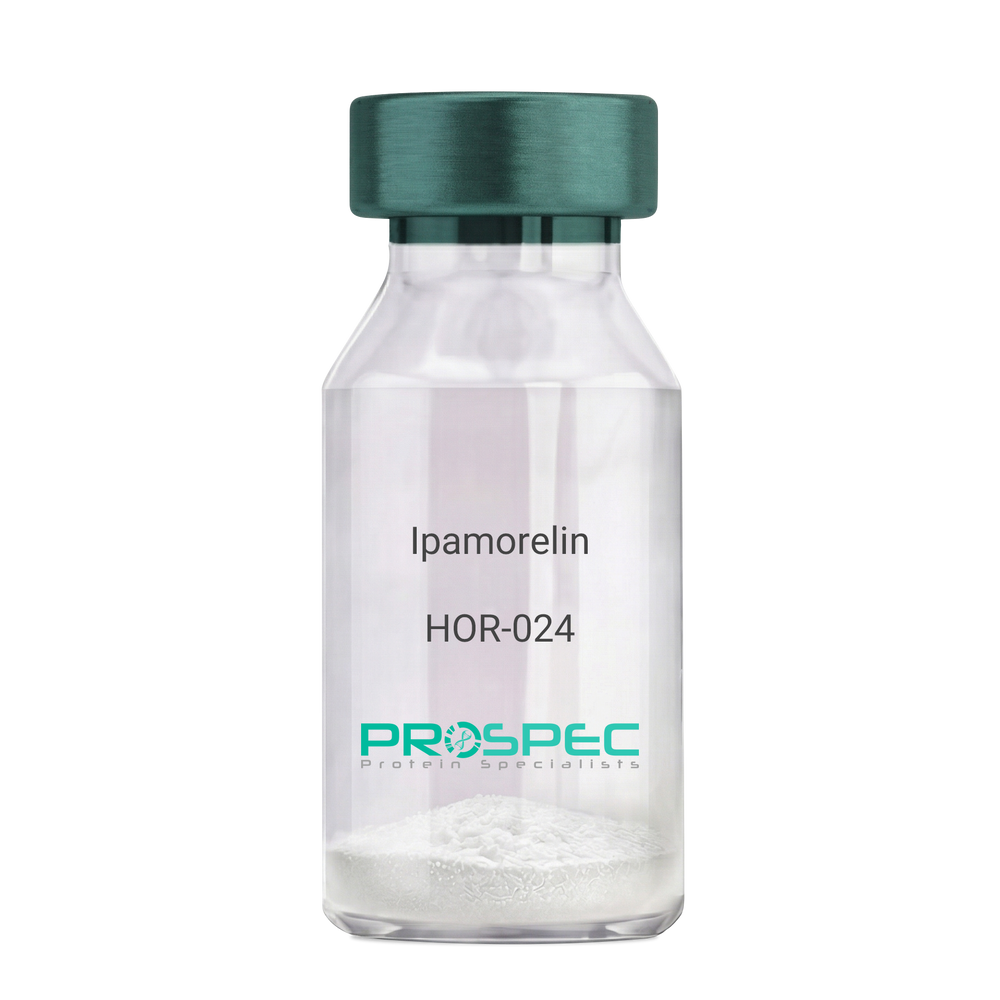
    Ipamorelin

    Description:

    Ipamorelin

    Ipamorelin

    Product # :

    HOR-024

    Price :

    Quantity :

    Shipping Method :

    Room Temp Icon

    Shipped at Room temp

    Add To Cart

    More Info

    • description
    • formulation
    • purity
    • More Info
    • Citations

    Description

    Ipamorelin Synthetic is a single, non-glycosylated polypeptide chain containing 4 amino acids, having a molecular mass of 711.85 Dalton and a Molecular formula of C38H49N9O5.

    Formulation

    The protein was lyophilized with no additives.

    Purity

    Greater than 97.0% as determined by analysis by RP-HPLC.

    Citations

    More Info

    • Introduction

      Ipamorelin is a peptide selective agonist of the ghrelin/growth hormone secretagogue receptor and a growth hormone secretagogue. Ipamorelin is a pentapeptide that was derived from GHRP1. Ipamorelin significantly increases plasma growth hormone levels in both animals and humans. Like pralmorelin and GHRP-6, ipamorelin does not affect prolactin, FSH, LH or TSH levels. However, unlike GHRP2 and GHRP6, but as growth hormone-releasing hormone (GHRH), ipamorelin does not stimulate the secretion of adrenocorticotropic hormone (ACTH) or cortisol, and is highly selective for inducing the secretion only of GH.

    • Synonyms

      Ipamorelin

    • Physical Appearance

      Sterile Filtered White lyophilized (freeze-dried) powder.

    • Stability

      Lyophilized Ipamorelin although stable at room temperature for 3 weeks, should be stored desiccated below -18°C. Upon reconstitution Ipamorelin should be stored at 4°C between 2-7 days and for future use below -18°C. For long term storage it is recommended to add a carrier protein (0.1% HSA or BSA). Please prevent freeze-thaw cycles.

    • Solubility

      It is recommended to reconstitute the lyophilized Ipamorelin in sterile 18MΩ-cm H2O not less than 100 µg/ml, which can then be further diluted to other aqueous solutions.

    • Amino Acid Sequence

      Aib-His-D-2-Nal-D-Phe-Lys-NH2.

    ProSpec's products are furnished for LABORATORY RESEARCH USE ONLY. The product may not be used as drugs, agricultural or pesticidal products, food additives or household chemicals.

    Ipamorelin
  • View Data Sheet

    Name :

    CCL19 Human

    Description:

    Macrophage Inflammatory Protein-3 Beta Human Recombinant (CCL19)

    Small inducible cytokine A19, CCL19, Macrophage inflammatory protein 3 beta, MIP-3- beta, EBI1-ligand chemokine, ELC, Beta chemokine exodus-3, CK beta-11, chemokine (C-C motif) ligand 19, CKb11, MIP3B, MIP-3b, SCYA19, MGC34433.

    Product # :

    CHM-287

    Price :

    Quantity :

    Shipping Method :

    Room Temp Icon

    Shipped at Room temp

    Add To Cart

    More Info

    • description
    • source
    • formulation
    • purity
    • biological activity
    • More Info
    • Citations

    Description

    Macrophage Inflammatory Protein-3 beta Human Recombinant produced in E.Coli is a single, non-glycosylated, polypeptide chain containing 77 amino acids and having a molecular mass of 8.8kDa. The MIP-3b is purified by proprietary chromatographic techniques.

    Source

    Escherichia Coli.

    Formulation

    Lyophilized without any additives.

    Purity

    Greater than 98.0% as determined by
    (a) Analysis by RP-HPLC.
    (b) Analysis by SDS-PAGE.

    Biological Activity

    The Activity of CCL19 is calculated by the ability to chemoattract Human T cells using a concentration of 7-40ng/ml corresponding to a Specific Activity of 25,000-143,000IU/mg.

    Citations

    More Info

    • Introduction

      Chemokine (C-C motif) ligand 19 (CCL19) is a small cytokine belonging to the CC chemokine family that is also known as EBI1 ligand chemokine (ELC) and macrophage inflammatory protein-3-beta (MIP-3-beta). CCL19 is expressed abundantly in thymus and lymph nodes, with moderate levels in trachea and colon and low levels in stomach, small intestine, lung, kidney and spleen. The gene for CCL19 is located on human chromosome 9. This chemokine elicits its effects on its target cells by binding to the chemokine receptor chemokine receptor CCR7. It attracts certain cells of the immune system, including dendritic cells and antigen-engaged B cells.

    • Synonyms

      Small inducible cytokine A19, CCL19, Macrophage inflammatory protein 3 beta, MIP-3- beta, EBI1-ligand chemokine, ELC, Beta chemokine exodus-3, CK beta-11, chemokine (C-C motif) ligand 19, CKb11, MIP3B, MIP-3b, SCYA19, MGC34433.

    • Physical Appearance

      Sterile Filtered White lyophilized (freeze-dried) powder.

    • Stability

      Lyophilized MIP-3b although stable at room temperature for 3 weeks, should be stored desiccated below -18°C. Upon reconstitution CCL19 should be stored at 4°C between 2-7 days and for future use below -18°C. For long term storage it is recommended to add a carrier protein (0.1% HSA or BSA).Please prevent freeze-thaw cycles.

    • Solubility

      It is recommended to reconstitute the lyophilized Macrophage Inflammatory Protein-3b in sterile 18MΩ-cm H2O not less than 100µg/ml, which can then be further diluted to other aqueous solutions.

    • Amino Acid Sequence

      GTNDAEDCCL SVTQKPIPGY IVRNFHYLLI KDGCRVPAVV FTTLRGRQLC APPDQPWVER IIQRLQRTSA KMKRRSS.

    • Background

      What is the molecular weight/Mw of CCL19 HUMAN Protein?
      CCL19 HUMAN Protein has a total Mw of 8.8kDa.

      What is the source or expression system of CCL19 HUMAN Protein?
      Escherichia Coli.

      What is the Purity of CCL19 HUMAN Protein?
      CCL19 HUMAN Protein is > 98% pure as determined by SDS-PAGE.

      What is the Biological Activity of CCL19 HUMAN Protein?
      The Activity of CCL19 is calculated by the ability to chemoattract Human T cells using a concentration of 7-40ng/ml corresponding to a Specific Activity of 25,000-143,000IU/mg.

      What is the amino acid sequence of CCL19 HUMAN Protein?
      GTNDAEDCCL SVTQKPIPGY IVRNFHYLLI KDGCRVPAVV FTTLRGRQLC APPDQPWVER IIQRLQRTSA KMKRRSS.

      What applications can CCL19 HUMAN Protein be used in?
      CCL19 HUMAN Protein can probably be used in western blot, ELISA and Lateral Flow.

      What is the endotoxin level for CCL19 HUMAN Protein?
      The endotoxin level is minimal, CCL19 HUMAN Protein was purified using conventional chromatography techniques.


    ProSpec's products are furnished for LABORATORY RESEARCH USE ONLY. The product may not be used as drugs, agricultural or pesticidal products, food additives or household chemicals.

    Mip 3B Human
  • View Data Sheet

    Name :

    TK1 Human

    Description:

    Thymidine Kinase 1 Human Recombinant

    Thymidine kinase 1 soluble, thymidine kinase cytosolic, TK2, EC 2.7.1.21.

    Product # :

    PKA-036

    Price :

    Quantity :

    Shipping Method :

    Ice Icon

    Shipped with Ice Packs

    Add To Cart

    More Info

    • description
    • source
    • formulation
    • purity
    • More Info
    • Citations

    Description

    TK1 Human Recombinant produced in E. coli is a single polypeptide chain containing 258 amino acids (1-234) and having a molecular mass of 28.0 kDa.TK1 is fused to a 24 amino acid His-tag at N-terminus & purified by proprietary chromatographic techniques.

    Source

    E.coli.

    Formulation

    The TK1 solution (1mg/1ml) contains 20mM Tris-HCl buffer (pH 8.0), 100mM NaCl, 1mM DTT and 20% glycerol.

    Purity

    Greater than 90% as determined by SDS-PAGE.

    Citations

    More Info

    • Introduction

      Thymidine Kinase 1 (TK1) is a phosphotransferase (a kinase): 2'-deoxythymidine kinase, ATP-thymidine 5'-phosphotransferase. TK1 is present in 2 forms in mammalian cells, TK1 and TK2. Thymidine kinases hold a main function in the synthesis of DNA and thus in cell division, as they are part of the distinctive reaction chain to introduce deoxythymidine (present in the body fluids as a result of degradation of DNA from food and from dead cells) into the DNA. Thymidine kinase is necessary for the action of many antiviral drugs. Thymidine kinase is used to select hybridoma cell lines in production of monoclonal antibodies.

    • Synonyms

      Thymidine kinase 1 soluble, thymidine kinase cytosolic, TK2, EC 2.7.1.21.

    • Physical Appearance

      Sterile Filtered colorless solution.

    • Stability

      Store at 4°C if entire vial will be used within 2-4 weeks. Store, frozen at -20°C for longer periods of time. For long term storage it is recommended to add a carrier protein (0.1% HSA or BSA).Avoid multiple freeze-thaw cycles.

    • Amino Acid Sequence

      MGSSHHHHHH SSGLVPRGSH MGSHMSCINL PTVLPGSPSK TRGQIQVILG PMFSGKSTEL MRRVRRFQIA QYKCLVIKYA KDTRYSSSFC THDRNTMEAL PACLLRDVAQ EALGVAVIGI DEGQFFPDIV EFCEAMANAG KTVIVAALDG TFQRKPFGAI LNLVPLAESV VKLTAVCMEC FREAAYTKRL GTEKEVEVIG GADKYHSVCR LCYFKKASGQ PAGPDNKENC PVPGKPGEAV AARKLFAPQQ ILQCSPAN.

    ProSpec's products are furnished for LABORATORY RESEARCH USE ONLY. The product may not be used as drugs, agricultural or pesticidal products, food additives or household chemicals.

    Tk1 Human
  • View Data Sheet

    Name :

    KLK3 Protein

    Description:

    Kallikrein-3 Recombinant Human

    Prostate-specific antigen, PSA, Gamma-seminoprotein, Seminin, Kallikrein-3, P-30 antigen, Semenogelase, KLK3, APS, hK3, KLK2A1.

    Product # :

    ENZ-1102

    Price :

    Quantity :

    Shipping Method :

    Room Temp Icon

    Shipped at Room temp

    Add To Cart

    More Info

    • description
    • source
    • formulation
    • purity
    • More Info
    • Citations

    Description

    Kallikrein-3 Human Recombinant produced in E.Coli is a single, non- glycosylated polypeptide chain containing 237 amino acids and having a molecular mass of 26.1kDa.KLK3 is purified by proprietary chromatographic techniques.

    Source

    Escherichia Coli.

    Formulation

    Lyophilized from a 0.2μm filtered concentrated solution in 20mM Tris-HCl, pH 8.0, 150mM NaCl and 3% trehalose.

    Purity

    Greater than 95.0% as determined by:
    (a) Analysis by RP-HPLC.
    (b) Analysis by SDS-PAGE.

    Citations

    More Info

    • Introduction

      Kallikrein-3 (KLK3) is a part of the kallikrein-related peptidase family. Kallikreins are a subgroup of serine proteases having various physiological functions. Numerous kallikreins take part in carcinogenesis and some may be prospective cancer and other disease biomarkers. Kallikrein-3 is 1 of the 15 kallikrein subfamily members located in a cluster on chromosome 19 and is a protease present in seminal plasma. KLK3 hydrolyzes semenogelin-1 consequently leading to the liquefaction of the seminal coagulum. KLK3 acts normally in the liquefaction of seminal coagulum, probably by hydrolysis of the high molecular mass seminal vesicle protein. Serum level of the KLK3 protein, called PSA in the clinical setting, is beneficial in the diagnosis and monitoring of prostatic carcinoma.

    • Synonyms

      Prostate-specific antigen, PSA, Gamma-seminoprotein, Seminin, Kallikrein-3, P-30 antigen, Semenogelase, KLK3, APS, hK3, KLK2A1.

    • Physical Appearance

      Sterile Filtered White lyophilized (freeze-dried) powder.

    • Stability

      Lyophilized KLK3 although stable at room temperature for 3 weeks, should be stored desiccated below -18°C. Upon reconstitution Kallikrein-3 should be stored at 4°C between 2-7 days and for future use below -18°C.Please prevent freeze-thaw cycles.

    • Solubility

      It is recommended to reconstitute the lyophilized Kallikrein-3 in sterile 18MΩ-cm H2O not less than 100µg/ml, which can then be further diluted to other aqueous solutions.

    • Amino Acid Sequence

      IVGGWECEKH SQPWQVLVAS RGRAVCGGVL VHPQWVLTAA HCIRNKSVIL LGRHSLFHPE DTGQVFQVSH SFPHPLYDMS LLKNRFLRPG DDSSHDLMLL RLSEPAELTDA VKVMDLPTQE PALGTTCYAS GWGSIEPEEF LTPKKLQCVD LHVISNDVCA QVHPQKVTKF MLCAGRWTGG KSTCSGDSGG PLVCNGVLQG ITSWGSEPCA LPERPSLYTK VVHYRKWIKD TIVANP.

    ProSpec's products are furnished for LABORATORY RESEARCH USE ONLY. The product may not be used as drugs, agricultural or pesticidal products, food additives or household chemicals.

    Klk3 Protein
  • View Data Sheet

    Name :

    EGF Human

    Description:

    Epidermal Growth Factor Human Recombinant

    Urogastrone, URG, EGF.

    Product # :

    CYT-217

    Price :

    Quantity :

    Shipping Method :

    Room Temp Icon

    Shipped at Room temp

    Add To Cart

    More Info

    • description
    • source
    • formulation
    • purity
    • biological activity
    • More Info
    • Citations

    Description

    Epidermal Growth Factor Human Recombinant produced in E.Coli is a single, non-glycosylated, polypeptide chain containing 53 amino acids and having a molecular mass of 6.2kDa. The EGF is purified by proprietary chromatographic techniques.

    Source

    Escherichia Coli.

    Formulation

    EGF was lyophilized from a concentrated (1mg/ml) solution containing PBS pH-7.4.

    Purity

    Greater than 98.0% as determined by SDS-PAGE.

    Biological Activity

    The ED50 as determined by a cell proliferation assay using murine Balb/c 3T3 cells is less than 0.1 ng/ml, corresponding to a specific activity of >1.0x10⁷ IU/mg.

    Citations

    More Info

    • Introduction

      Epidermal growth factor has a profound effect on the differentiation of specific cells in vivo and is a potent mitogenic factor for a variety of cultured cells of both ectodermal and mesodermal origin. The EGF precursor is believed to exist as a membrane-bound molecule which is proteolytically cleaved to generate the 53-amino acid peptide hormone that stimulates cells to divide. EGF stimulates the growth of various epidermal and epithelial tissues in vivo and in vitro and of some fibroblasts in cell culture.

    • Synonyms

      Urogastrone, URG, EGF.

    • Physical Appearance

      Sterile Filtered White lyophilized (freeze-dried) powder.

    • Stability

      Lyophilized Epidermal Growth Factor Recombinant although stable at room temperature for 3 weeks, should be stored desiccated below -18°C. Upon reconstitution EGF should be stored at 4°C between 2-7 days and for future use below -18°C.For long term storage it is recommended to add a carrier protein (0.1% HSA or BSA).Please prevent freeze-thaw cycles.

    • Solubility

      It is recommended to reconstitute the lyophilized Epidermal Growth Factor in sterile 18MΩ-cm H2O not less than 100µg/ml, which can then be further diluted to other aqueous solutions.

    • Amino Acid Sequence

      NSDSECPLSH DGYCLHDGVC MYIEALDKYA CNCVVGYIGE RCQYRDLKWW ELR.

    • Background

      About EGF:

      In the sphere of biomedical studies, epidermal boom factor (EGF) is a cornerstone that gives precious insights into the mechanisms underlying tissue healing, differentiation, and mobile proliferation. In this article we will explore the characteristics and uses of epidermal growth factor (EGF).

      Description:

      Epidermal growth factor (EGF) is a 6-kDa protein consisting of 53 amino acid residues and 3 intramolecular disulfide linkages. Human tissues, such as platelets, the parotid gland, and the submandibular gland, are rich in EGF. EGF, which was first discovered in human urine and the submaxillary glands of mice, functions as a major modulator of cell proliferation by attaching to its receptor, EGFR, which is found on the cell membrane. EGF triggers autophosphorylation of transmembrane protein tyrosine kinase EGFR upon binding, hence initiating downstream signaling cascades through pathways such as phosphatidylinositol and ras. Beyond the cell membrane, EGF has a variety of roles as it also initiates cytoplasmic processes such actin depolymerization and membrane ruffle formation. Studies indicate that EGF and its receptor might possibly be important components of the nucleus, highlighting the complexity of EGF-mediated cellular responses.

      Function:

      By attaching to the epidermal growth factor receptor (EGFR), EGF promotes the survival, differentiation, and multiplication of cells. This connection is essential for boosting many physiological processes and stimulating cell proliferation. The preservation of oro-esophageal and stomach tissue integrity is greatly supported by salivary EGF, which is regulated by dietary inorganic iodine. Its actions include the healing of gastric and oral ulcers, the inhibition of gastric acid secretion, the stimulation of DNA synthesis, and the protection of mucosal surfaces against harmful substances such as bile acids, gastric acid, and bacteria. Salivary EGF's role extends to repairing gastric tissue and addressing oro-esophagal issues, showcasing its healing ability in resolving oral and gastrointestinal ailments, including ulcers.

      Mechanism:

      EGF functions by forming a strong bond with the cell surface's epidermal growth factor receptor (EGFR), which triggers ligand- induced dimerization. This incident sets off the intrinsic protein-tyrosine kinase activity of EGFR, which in turn initiates a signal transduction cascade inside the cell. Numerous biochemical changes are brought about by this cascade, such as increased intracellular calcium levels, increased glycolysis and protein synthesis, and increased expression of particular genes, most notably the EGFR gene. These carefully planned alterations eventually promote DNA synthesis and cell division, illuminating the complex process by which EGF directs basic biological functions and modulates cellular responses.

      What is the molecular weight/Mw of EGF Protein?
      EGF Protein has a total Mw of 6.2kDa.

      What is the source or expression system of EGF Protein?
      Escherichia Coli.

      What is the Purity of EGF Protein?
      EGF Protein is >98% pure as determined by SDS-PAGE.

      What is the Biological Activity of EGF Protein?
      The ED50 as determined by a cell proliferation assay using murine Balb/c 3T3 cells is less than 0.1 ng/ml, corresponding to a specific activity of >1.0x10⁷ IU/mg.

      What is the amino acid sequence of EGF Protein?
      NSDSECPLSH DGYCLHDGVC MYIEALDKYA CNCVVGYIGE RCQYRDLKWW ELR.

      What applications can EGF Protein be used in?
      EGF Protein can probably be used in western blot, ELISA and Lateral Flow.

      What is the endotoxin level for EGF Protein?
      The endotoxin level is minimal, EGF Protein was purified using conventional chromatography techniques.

    ProSpec's products are furnished for LABORATORY RESEARCH USE ONLY. The product may not be used as drugs, agricultural or pesticidal products, food additives or household chemicals.

    Egf Human
  • View Data Sheet

    Name :

    Streptavidin

    Description:

    Streptavidin Recombinant

    Product # :

    PRO-791

    Price :

    Quantity :

    Shipping Method :

    Room Temp Icon

    Shipped at Room temp

    Add To Cart

    More Info

    • description
    • source
    • formulation
    • purity
    • More Info
    • Citations

    Description

    Streptavidin Streptomyces Avidinii Recombinant produced in E.Coli. The molecular weight per tetramer is approximately 52kDa.

    Source

    Escherichia Coli.

    Formulation

    Lyophilized in 10mM potassium phosphate buffer pH 6.5.

    Purity

    Greater than 98.0% as determined by SDS-PAGE and HPLC.

    Citations

    More Info

    • Introduction

      Streptavidin is a tetrameric protein secreted by Streptomyces avidinii which binds firmly to biotin. Streptavidin is widely used in molecular biology through its unique high affinity for the vitamin biotin. The dissociation constant (Kd) of the biotin-streptavidin complex is about ~10-15 mol/L. The strong affinity recognition of biotin and biotinylated molecules has made streptavidin one of the most important components in diagnostics and laboratory kits. The streptavidin/biotin system has one of the biggest free energies of association of yet observed for noncovalent binding of a protein and small ligand in aqueous solution (K_assoc = 10**14). The complexes are also extremely stable over a wide range of temperature and pH.

    • Physical Appearance

      Sterile Filtered White lyophilized (freeze-dried) powder.

    • Stability

      Streptavidin is shipped at ambient temperature, upon arrival store at -20°C.

    • Solubility

      It is recommended to reconstitute the lyophilized Streptavidin in sterile 18MΩ-cm H2O not less than 0.5mg/ml, which can then be further diluted to other aqueous solutions.

    • Amino Acid Sequence

      MAEAGITGTWYNQLGSTFIVTAGADGALTGTYESAVGNAESRYVLT
      GRYDSAPATDGSGTALGWTVAWKNNYRNAHSATTWSGQYVGGA
      EARINTQWLLTSGTTEANAWKSTLVGHDTFTKVKPSAAS.

    • Proteolytic Activity

      < 10-3 U/mg protein (Azocoll, 25 °C, 24 h, pH 8.0).

    • Specific Activity

      > 17U/mg (one unit binds 1 μg D-biotin).

    ProSpec's products are furnished for LABORATORY RESEARCH USE ONLY. The product may not be used as drugs, agricultural or pesticidal products, food additives or household chemicals.

    Streptavidin Recombinant
  • View Data Sheet

    Name :

    ADI1 Antibody

    Description:

    Acireductone Dioxygenase 1, Mouse Anti Human

    APL1, ARD, FLJ10913, HMFT1638, MTCBP-1, SIPL, 1,2-dihydroxy-3-keto-5-methylthiopentene dioxygenase, Acireductone dioxygenase, Fe-ARD, Membrane-type 1 matrix metalloproteinase cytoplasmic tail-binding protein 1, Submergence-induced protein-like factor.

    Product # :

    ANT-466

    Price :

    Quantity :

    Shipping Method :

    Ice Icon

    Shipped with Ice Packs

    Add To Cart

    More Info

    • formulation
    • More Info
    • Citations

    Formulation

    1mg/ml containing PBS, pH-7.4, 10% Glycerol and 0.02% Sodium Azide.

    Citations

    More Info

    • Introduction

      Acireductone dioxygenase 1 (ADI1) is a part of the acireductone dioxygenase family of metal-binding enzymes, which are involved in methionine salvage. ADI1 regulates mRNA processing in the nucleus, and carries out different functions depending on its localization. Related pseudogenes have been defined on chromosomes 8 and 20. ADI1 down-regulates cell migration arbitrated by MMP14.

    • Synonyms

      APL1, ARD, FLJ10913, HMFT1638, MTCBP-1, SIPL, 1,2-dihydroxy-3-keto-5-methylthiopentene dioxygenase, Acireductone dioxygenase, Fe-ARD, Membrane-type 1 matrix metalloproteinase cytoplasmic tail-binding protein 1, Submergence-induced protein-like factor.

    • Physical Appearance

      Sterile filtered colorless solution.

    • Immunogen

      Anti-human ADI1 mAb, clone PAT27E8A, is derived from hybridization of mouse F0 myeloma cells with spleen cells from BALB/c mice immunized with a recombinant human ADI1 protein 1-179 amino acids purified from E. coli.

    • Ig Subclass

      Mouse IgG1 heavy chain and Kappa light chain.

    • Clone

      PAT27E8A.

    • Applications

      The antibody has been tested by ELISA, Western blot analysis to assure specificity and reactivity. Since application varies, however, each investigation should be titrated by the reagent to obtain optimal results. Recommended dilution range for Western blot analysis is 1:500 ~ 1:5000. Recommended starting dilution is 1:1000.

    • Type

      Mouse Anti Human Monoclonal.

    • Storage Procedures

      For periods up to 1 month store at 4°C, for longer periods of time, store at -20°C. Prevent freeze thaw cycles.

    ProSpec's products are furnished for LABORATORY RESEARCH USE ONLY. The product may not be used as drugs, agricultural or pesticidal products, food additives or household chemicals.

    Adi1 Antibody
  • View Data Sheet

    Name :

    LDHA Antibody

    Description:

    Lactate Dehydrogenase A, Mouse Anti Human

    LDH-A, GSD11, LDH1, LDHM, PIG19, EC 1.1.1.27, lactate dehydrogenase M, LDH-M, LDH-1, L-lactate dehydrogenase A chain, LDH muscle subunit, Renal carcinoma antigen NY-REN-59, Cell proliferation-inducing gene 19 protein, LDHA.

    Product # :

    ANT-004

    Price :

    Quantity :

    Shipping Method :

    Ice Icon

    Shipped with Ice Packs

    Add To Cart

    More Info

    • formulation
    • More Info
    • Citations

    Formulation

    1mg/ml containing PBS, pH-7.4, & 0.1% Sodium Azide.

    Citations

    More Info

    • Introduction

      LDHA catalyzes the conversion of L-lactate and NAD to pyruvate and NADH in the final step of anaerobic glycolysis. LDHA is localized primarily in muscle tissue and is part of the lactate dehydrogenase family. Mutations in LDHA have been linked to exertional myoglobinuria. LDH1 is decreased in essential thrombocythemia. LDHA is induced through a non-genomic pathway of estrogen action. Reduction in LDH-A activity results in stimulation of mitochondrial respiration and decrease of mitochondrial membrane potential.

    • Synonyms

      LDH-A, GSD11, LDH1, LDHM, PIG19, EC 1.1.1.27, lactate dehydrogenase M, LDH-M, LDH-1, L-lactate dehydrogenase A chain, LDH muscle subunit, Renal carcinoma antigen NY-REN-59, Cell proliferation-inducing gene 19 protein, LDHA.

    • Immunogen

      Anti-human LDHA mAb, is derived from hybridization of mouse F0 myeloma cells with spleen cells from BALB/c mice immunized with recombinant human LDHA amino acids 1-332 purified from E. coli.

    • Ig Subclass

      Mouse IgG2b heavy chain and κ light chain.

    • Clone

      PAT1A4AT.

    • Applications

      LDHA antibody has been tested by ELISA, Western blot and Immunofluorescence analysis to assure specificity and reactivity. Since application varies, however, each investigation should be titrated by the reagent to obtain optimal results. Recommended dilution range for Western blot analysis and Immunofluorescence is 1:500 ~ 8,000.
      Recommended starting dilution is 1:1,000.

    • Type

      Mouse Anti Human Monoclonal.

    • Storage Procedures

      For periods up to 1 month store at 4°C, for longer periods of time, store at -20°C. Prevent freeze thaw cycles.

    • Purification Method

      LDHA antibody was purified from mouse ascitic fluids by protein-G affinity chromatography.

    ProSpec's products are furnished for LABORATORY RESEARCH USE ONLY. The product may not be used as drugs, agricultural or pesticidal products, food additives or household chemicals.

    Ldha Antibody
  • View Data Sheet

    Name :

    Leptin Mouse

    Description:

    Leptin Mouse Recombinant

    OB Protein, Obesity Protein, OBS, Obesity factor.

    Product # :

    CYT-351

    Price :

    Quantity :

    Shipping Method :

    Room Temp Icon

    Shipped at Room temp

    Add To Cart

    More Info

    • description
    • source
    • formulation
    • purity
    • biological activity
    • More Info
    • Citations

    Description

    Leptin Mouse Recombinant produced in E.Coli is a single, non-glycosylated, polypeptide chain containing 147 amino acids and having a molecular mass of 16 kDa. The Leptin is purified by proprietary chromatographic techniques.

    Source

    Escherichia Coli.

    Formulation

    The mouse Leptin was lyophilized from a concentrated (1mg/ml) solution containing 0.0045mM NaHCO3.

    Purity

    Greater than 95.0% as determined by:
    (a) Analysis by gel filtration analysis.
    (b) Analysis by SDS-PAGE.

    Biological Activity

    Biological activity of Mouse Leptin is performed by including proliferation of BAF/3 cells stably transfected with the long form of human leptin receptor.

    Citations

    More Info

    • Introduction

      A 16-kDa peptide hormone secreted from white adipocytes and implicated in the regulation of food intake and energy balance. Leptin provides the key afferent signal from fat cells in the feedback system that controls body fat stores.

    • Synonyms

      OB Protein, Obesity Protein, OBS, Obesity factor.

    • Physical Appearance

      Sterile Filtered White lyophilized (freeze-dried) powder.

    • Stability

      Lyophilized Leptin although stable at room temperature for 3 weeks, should be stored desiccated below -18°C. Upon reconstitution Leptin should be stored at 4°C between 2-7 days and for future use below -18°C.For long term storage it is recommended to add a carrier protein (0.1% HSA or BSA).Please prevent freeze-thaw cycles.

    • Solubility

      It is recommended to reconstitute the lyophilized Leptin in sterile 18MΩ-cm H2O not less than 100µg/ml, which can then be further diluted to other aqueous solutions.

    • Amino Acid Sequence

      AVPIQKVQDD TKTLIKTIVT RINDISHTQS VSAKQRVTGL DFIPGLHPIL SLSKMDQTLA VYQQVLTSLP SQNVLQIAND LENLRDLLHL LAFSKSCSLP QTSGLQKPES LDGVLEASLY STEVVALSRL QGSLQDILQQ LDVSPEC.

    • Protein content

      Protein quantitation was carried out by UV spectroscopy at 280 nm using the absorbency value of 0.20 as the extinction coefficient for a 0.1% (1mg/ml) solution at pH-8.0. This value is calculated by the PC GENE computer analysis program of protein sequences (IntelliGenetics).

    ProSpec's products are furnished for LABORATORY RESEARCH USE ONLY. The product may not be used as drugs, agricultural or pesticidal products, food additives or household chemicals.

    Leptin Mouse
  • View Data Sheet

    Name :

    GPI Antibody

    Description:

    Glucose-6-Phosphate Isomerase, Mouse Anti Human

    Glucose-6-phosphate isomerase, Phosphoglucose isomerase, Phosphohexose isomerase, Autocrine motility factor, Neuroleukin, Sperm antigen 36, GPI, PGI, PHI, AMF, NLK, SA-36, GNPI.

    Product # :

    ANT-613

    Price :

    Quantity :

    Shipping Method :

    Ice Icon

    Shipped with Ice Packs

    Add To Cart

    More Info

    • formulation
    • More Info
    • Citations

    Formulation

    1mg/ml containing PBS, pH-7.4, 10% Glycerol and 0.02% Sodium Azide.

    Citations

    More Info

    • Introduction

      Glucose-6-phosphate isomerase (GPI) is a part of the GPI family whose members encode multifunctional phosphoglucose isomerase proteins involved in energy pathways. GPI is a dimeric enzyme which catalyzes the reversible isomerization of glucose-6-phosphate and fructose-6-phosphate. Mammalian GPI also functions as a tumor-secreted cytokine and an angiogenic factor (AMF) which stimulates endothelial cell motility. In addition, GPI is a neurotrophic factor (Neuroleukin) for spinal and sensory neurons. GPI performs in different capacities inside and outside the cell. In the cytoplasm, GPI is involved in glycolysis and gluconeogenesis, while outside the cell it acts as a neurotrophic factor for spinal and sensory neurons.
      Defects in the GPI gene cause the nonspherocytic hemolytic anemia and a severe enzyme deficiency can be linked to hydrops fetalis, immediate neonatal death and neurological impairment.

    • Synonyms

      Glucose-6-phosphate isomerase, Phosphoglucose isomerase, Phosphohexose isomerase, Autocrine motility factor, Neuroleukin, Sperm antigen 36, GPI, PGI, PHI, AMF, NLK, SA-36, GNPI.

    • Physical Appearance

      Sterile filtered colorless solution.

    • Immunogen

      Anti-human GPI mAb, clone PAT22G2AT, is derived from hybridization of mouse F0 myeloma cells with spleen cells from BALB/c mice immunized with a recombinant human GPI protein 1-558 amino acids purified from E. coli.

    • Ig Subclass

      Mouse IgG2a heavy chain and k light chain.

    • Clone

      PAT22G2AT.

    • Applications

      The antibody has been tested by ELISA, Western blot analysis, Flow cytometry and ICC/IF to assure specificity and reactivity. Since application varies, however, each investigation should be titrated by the reagent to obtain optimal results.

    • Type

      Mouse Anti Human Monoclonal.

    • Storage Procedures

      For periods up to 1 month store at 4°C, for longer periods of time, store at -20°C. Prevent freeze thaw cycles.

    • Purification Method

      GPI antibody was purified from mouse ascitic fluids by protein-A affinity chromatography.

    ProSpec's products are furnished for LABORATORY RESEARCH USE ONLY. The product may not be used as drugs, agricultural or pesticidal products, food additives or household chemicals.

    Gpi Antibody
  • View Data Sheet

    Name :

    UGDH Antibody

    Description:

    UDP-Glucose Dehydrogenase, Mouse Anti Human

    GDH, UDP-GlcDH, UDPGDH, UGD, EC 1.1.1.22, UDP-Glc dehydrogenase, UDP-glucose 6-dehydrogenase, UGDH.

    Product # :

    ANT-648

    Price :

    Quantity :

    Shipping Method :

    Ice Icon

    Shipped with Ice Packs

    Add To Cart

    More Info

    • formulation
    • More Info
    • Citations

    Formulation

    1mg/ml containing PBS, pH-7.4, 10% Glycerol and 0.02% Sodium Azide.

    Citations

    More Info

    • Introduction

      UGDH is part of the UDP-glucose/GDP-mannose dehydrogenase family and is a widely expressed enzyme localized in the liver. UGDH transfers UDP-glucose to UDP-glucuronate and thus takes part in the biosynthesis of glycosaminoglycans such as hyaluronan, chondroitin sulfate, and heparan sulfate. These glycosylated products are ordinary molecules of the extracellular matrix and participate in signal transduction, cell migration, and cancer growth and metastasis.

    • Synonyms

      GDH, UDP-GlcDH, UDPGDH, UGD, EC 1.1.1.22, UDP-Glc dehydrogenase, UDP-glucose 6-dehydrogenase, UGDH.

    • Physical Appearance

      Sterile filtered colorless solution.

    • Immunogen

      Anti-human UGDH mAb, is derived from hybridization of mouse F0 myeloma cells with spleen cells from BALB/c mice immunized with recombinant human UGDH amino acids 1-494 purified from E. coli.

    • Ig Subclass

      Mouse IgG2b heavy chain and k light chain.

    • Clone

      PAT2G11AT.

    • Applications

      UGDH antibody has been tested by ELISA, Western blot analysis, Flow cytometry and ICC/IF to assure specificity and reactivity. Since application varies, however, each investigation should be titrated by the reagent to obtain optimal results.

    • Type

      Mouse Anti Human Monoclonal.

    • Storage Procedures

      For periods up to 1 month store at 4°C, for longer periods of time, store at -20°C. Prevent freeze thaw cycles.

    • Purification Method

      UGDH antibody was purified from mouse ascitic fluids by protein-A affinity chromatography.

    ProSpec's products are furnished for LABORATORY RESEARCH USE ONLY. The product may not be used as drugs, agricultural or pesticidal products, food additives or household chemicals.

    Ugdh Antibody
  • View Data Sheet

    Name :

    SAE1/SAE2 Human

    Description:

    SAE1/SAE2 Human Recombinant

    Product # :

    ENZ-1161

    Price :

    Quantity :

    Shipping Method :

    Ice Icon

    Shipped with Ice Packs

    Add To Cart

    More Info

    • description
    • source
    • formulation
    • purity
    • More Info
    • Citations

    Description

    SAE1/SAE2 Human Recombinant produced in SF9 is glycosylated, polypeptide chain containing 2 subunits (SAE1 subunit molecular mass is 41kDa & SAE2 subunit molecular mass is 91kDa). The subunits associate to form a complex. The SAE1/SAE2 is expressed with a -10xHis tag and purified by proprietary chromatographic techniques.

    Source

    Sf9 insect cells.

    Formulation

    SAE1/SAE2 is supplied in 20mM HEPES buffer pH-8.0, 200mM NaCl and 20% glycerol.

    Purity

    Greater than 95.0% as determined by SDS-PAGE.

    Citations

    More Info

    • Introduction

      SAE1 and SAE2 form heterodimers which take part in the posttranslational modification of proteins (sumoylation). This process regulates protein structure as well as intracellular localization of the target. SAE1/SAE2 may indicate on dermatomyositis (DM) as autoantibodies against those 2 proteins have been found in patients.

    • Physical Appearance

      Sterile Filtered colorless solution.

    • Stability

      Store at 4°C if entire vial will be used within 2-4 weeks. Store, frozen at -20°C for longer periods of time. Avoid multiple freeze-thaw cycles.

    • Immunological Functions

      1. Binds IgG-type human auto-antibodies.2. Standard ELISA test (checkerboard analysis of positive/negative samples) and immunodot test with positive/negative samples.

    • coating concentration

      0.3-0.8 µg/ml (depending on the type of ELISA plate and coating buffer). Suitable for labeling of functional groups.

    • Applications

      Western blot with anti SAE1/SAE2 autoantibody positive sample & Polyclonal anti-SAE1 and anti-SAE2 antibodies.

    ProSpec's products are furnished for LABORATORY RESEARCH USE ONLY. The product may not be used as drugs, agricultural or pesticidal products, food additives or household chemicals.

    Sae1 Sae2
  • View Data Sheet

    Name :

    Thyroglobulin Human

    Description:

    Thyroglobulin Human Recombinant

    Thyroglobulin, TGN, AITD3, TG.

    Product # :

    PRO-2803

    Price :

    Quantity :

    Shipping Method :

    Room Temp Icon

    Shipped at Room temp

    Add To Cart

    More Info

    • description
    • source
    • formulation
    • purity
    • More Info
    • sds-page
    • Citations

    Description

    Thyroglobulin Human produced in a mammalian cell line is a single, non-glycosylated polypeptide chain (1-2768 a.a.) and having a molecular mass of 304640 Dalton. Thyroglobulin Human is fused with GlyAlaProGly4SerHis10-tag at C-terminal and purified by proprietary chromatographic techniques.

    Source

    Mammalian cell line.

    Formulation

    Thyroglobulin was lyophilized from PBS, pH 7.4 and 5.4 % sucrose.

    Purity

    Greater than 95.0% as determined by SDS-PAGE.

    sds-page

    Thyroglobulin Recombinant Human SDS-PAGE - Product image 1

    Citations

    More Info

    • Synonyms

      Thyroglobulin, TGN, AITD3, TG.

    • Physical Appearance

      Sterile Filtered White lyophilized (freeze-dried) powder.

    • Stability

      Lyophilized Thyroglobulin although stable at room temperature for 3 weeks, should be stored desiccated below -18°C. Upon reconstitution Thyroglobulin should be stored at 4°C between 2-7 days and for future use below -18°C. For long term storage it is recommended to add a carrier protein (0.1% HSA or BSA). Please prevent freeze-thaw cycles.

    • Solubility

      It is recommended to reconstitute the lyophilized Thyroglobulin in sterile 18MΩ-cm H2O not less than 100µg/ml, which can then be further diluted to other aqueous solutions.

    • Background

      Thyroglobulin, a glycoprotein primarily produced in the thyroid gland, stands at the center of thyroid hormone synthesis. Comprising a series of tyrosine residues, thyroglobulin serves as the scaffold upon which thyroid hormones are assembled. Beyond its pivotal role in thyroid physiology, thyroglobulin has garnered significant attention in the realm of thyroid disease diagnostics, offering valuable insights into thyroid function and disorders. This research delves into the intricacies of thyroglobulin human recombinant protein, exploring its biochemical properties, physiological significance, and its crucial applications in both clinical and research settings.

      Structural Complexity of Thyroglobulin:

      Thyroglobulin is a large, dimeric protein boasting an intricate structure composed of multiple domains. Within its structure lie tyrosine residues crucial for iodine incorporation, a process fundamental for thyroid hormone synthesis. Its size and complexity reflect the sophistication of thyroid hormone production, as thyroglobulin acts as a reservoir for thyroid hormones within the thyroid follicles.

      Physiological Significance in Thyroid Function:

      Thyroglobulin plays a central role in the synthesis of triiodothyronine (T3) and thyroxine (T4), the thyroid hormones essential for regulating metabolism and overall body homeostasis. During thyroid hormone synthesis, thyroglobulin is secreted into the follicular lumen, where it undergoes iodination and subsequent proteolysis, releasing T3 and T4. This process highlights the indispensable nature of thyroglobulin in thyroid hormone production, making it a key biomolecule in thyroid physiology.

    ProSpec's products are furnished for LABORATORY RESEARCH USE ONLY. The product may not be used as drugs, agricultural or pesticidal products, food additives or household chemicals.

    Thyroglobulin Antigen
  • View Data Sheet

    Name :

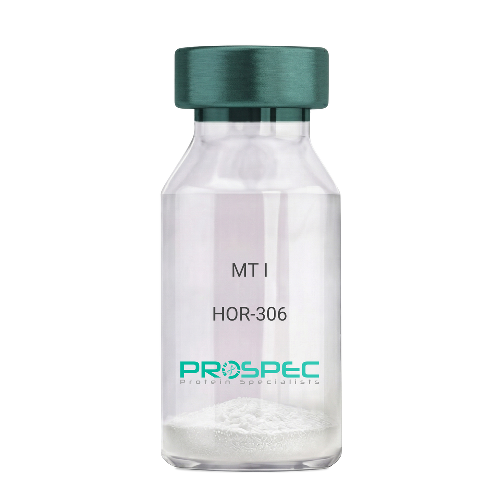
    MT I

    Description:

    Melanotan-I

    Melanotan-I, MT-I, Melanotan-1.

    Product # :

    HOR-306

    Price :

    Quantity :

    Shipping Method :

    Room Temp Icon

    Shipped at Room temp

    Add To Cart

    More Info

    • description
    • formulation
    • purity
    • More Info
    • Citations

    Description

    Melanotan-I has the amino acid sequence of Ser-Tyr-Ser-Nle-Glu-His-D-Phe-Arg-Trp-Gly-Lys-Pro-Val and a molecular weight of 1647.4 Dalton.

    Formulation

    The protein (1mg/ml) was lyophilized with no additives.

    Purity

    Greater than 98.0% as determined by RP-HPLC.

    Citations

    More Info

    • Synonyms

      Melanotan-I, MT-I, Melanotan-1.

    • Physical Appearance

      Sterile Filtered off-White lyophilized (freeze-dried) powder.

    • Stability

      Lyophilized Melanotan-I although stable at room temperature for 3 weeks, should be stored desiccated below -18°C. Upon reconstitution MT-I should be stored at 4°C between 2-7 days and for future use below -18°C.For long term storage it is recommended to add a carrier protein (0.1% HSA or BSA).Please prevent freeze-thaw cycles.

    • Solubility

      It is recommended to reconstitute the lyophilized Melanotan-I in sterile 1% acetic acid not less than 100 µg/ml, which can then be further diluted to other aqueous solutions.

    ProSpec's products are furnished for LABORATORY RESEARCH USE ONLY. The product may not be used as drugs, agricultural or pesticidal products, food additives or household chemicals.

    Melanotan I
  • View Data Sheet

    Name :

    Leptin Pufferfish

    Description:

    Leptin Pufferfish Recombinant

    OB Protein, Obesity Protein, OBS, Obesity factor.

    Product # :

    CYT-530

    Price :

    Quantity :

    Shipping Method :

    Room Temp Icon

    Shipped at Room temp

    Add To Cart

    More Info

    • description
    • source
    • formulation
    • purity
    • biological activity
    • More Info
    • Citations

    Description

    Leptin Pufferfish (Takifugu rubripes) Recombinant produced in E.Coli is a single, non-glycosylated, polypeptide chain having a molecular mass of 16 kDa. Bioactive Leptin Pufferfish (Takifugu rubripes) Recombinant was prepared according to the sequence published by Kurokawa et al. (2005)Peptides 26, 745-750 in two forms: monomer and covalent dimer. MS analysis revealed molecular masses of 15,291 and 30,585 Da, close to the theoretical values of 15,270 and 30,540 Da. CD spectra revealed high similarity to mammalian leptins. Other details of its preparation will be soon published by Yacobovitz et al (in press), General and Comparative Endocrinology.The Pufferfish Leptin is purified by proprietary chromatographic techniques.

    Source

    Escherichia Coli.

    Formulation

    The Pufferfish Leptin was lyophilized from a concentrated (0.85mg/ml) solution with 0.003mM NaHCO3.

    Purity

    Greater than 99.0% as determined by:
    (a) Analysis by SEC-HPLC.
    (b) Analysis by SDS-PAGE.

    Biological Activity

    Biological active as evidenced by inducing proliferation of BAF/3 cells stably transfected with the long form of human leptin receptor. The affinity of human leptin receptors is considerably lower campared to mammalian leptins.

    Citations

    More Info

    • Introduction

      A 16-kDa peptide hormone secreted from white adipocytes and implicated in the regulation of food intake and energy balance. Leptin provides the key afferent signal from fat cells in the feedback system that controls body fat stores.

    • Synonyms

      OB Protein, Obesity Protein, OBS, Obesity factor.

    • Physical Appearance

      Sterile Filtered White lyophilized (freeze-dried) powder.

    • Stability

      Lyophilized Pufferfish Leptin although stable at room temperature for 3 weeks, should be stored desiccated below -18°C. Upon reconstitution Leptin should be stored at 4°C between 2-7 days and for future use below -18°C.For long term storage it is recommended to add a carrier protein (0.1% HSA or BSA).Please prevent freeze-thaw cycles.

    • Solubility

      It is recommended to reconstitute the lyophilized Pufferfish Leptin in sterile 0.4% NaHCO3 pH-9 not less than 100µg/ml, which can then be further diluted to other aqueous solutions.

    • Amino Acid Sequence

      ALPGALDAMDVEKMKSKVTWKAQGLVARIDKHFPDRGLRFDTDKVE

      GSTSVVASLESYNNLISDRFGGVSQIKTEISSLAGYLNHWREGNCQE

      QQPKVWPRRNIFNHTVSLEALMRVREFLKLLQKNVDLLERC

    • Protein content

      Protein quantitation was carried out by UV spectroscopy at 280 nm using the absorbency value of 1.28 as the extinction coefficient for a 0.1% (1mg/ml) solution. This value is calculated by the PC GENE computer analysis program of protein sequences (IntelliGenetics).

    ProSpec's products are furnished for LABORATORY RESEARCH USE ONLY. The product may not be used as drugs, agricultural or pesticidal products, food additives or household chemicals.

    Leptin Pufferfish
  • View Data Sheet

    Name :

    Leptin Rat, PEG

    Description:

    Pegylated Rat Leptin Recombinant

    OB Protein, Obesity Protein, OBS, Obesity factor.

    Product # :

    CYT-592

    Price :

    Quantity :

    Shipping Method :

    Room Temp Icon

    Shipped at Room temp

    Add To Cart

    More Info

    • description
    • source
    • formulation
    • purity
    • biological activity
    • More Info
    • Citations

    Description

    Mono-Pegylated Leptin Rat Recombinant produced in E.Coli is a single, non-glycosylated, polypeptide chain containing 146 amino acids and an additional Ala at N-terminus having a molecular mass of 35.6 kDa (with 20 kDa PEG) as determined by mass spectometry. However due to enlarged hydrodymanic volume it runs on the SDS-PAGE as 48 kDa protein and in gel-filtration on Superdex 200 as over 100 kDa protein. Its half-life in circulation after SC injection was over 20 hours. Rat Leptin was purified by proprietary chromatographic techniques according to Salomon et al (2006) Protein Expression and Purification 47, 128–136 and then pegylated.

    Source

    Escherichia Coli.

    Formulation

    The protein was lyophilized from a concentrated (0.65mg/ml) solution with 0.003mM NaHCO3.

    Purity

    Greater than 99.0% as determined by:
    (a) Analysis by Gel-Filtration.
    (b) Analysis by SDS-PAGE.

    Biological Activity

    Pegylated Rat Leptin is capable of stimulatng proliferation of BAF/3 cells stably transfected with the long form of human leptin receptor. Its in vitro activity is only slightly lower than the non-pegylated antagonist but in vivo it has profound weight reducing effect (as compared to the non-pegylated leptin), resulting mainly from reduced food intake.

    Citations

    More Info

    • Introduction

      A 16-kDa peptide hormone secreted from white adipocytes and implicated in the regulation of food intake and energy balance. Leptin provides the key afferent signal from fat cells in the feedback system that controls body fat stores.

    • Synonyms

      OB Protein, Obesity Protein, OBS, Obesity factor.

    • Physical Appearance

      Sterile Filtered White lyophilized (freeze-dried) powder.

    • Stability

      Lyophilized pegylated Rat Leptin Antagonist although stable at room temperature for several weeks, should be stored desiccated below -18°C. Upon reconstitution of pegylated Rat Leptin at > 0.1 mg/ml and up to 2 mg/ml and filter sterilization Rat leptin can be stored at 4C or even room temperature for several weeks making it suitable for long term infusion studies using osmotic pumps. At lower concentration addition of a carrier protein (0.1% HSA or BSA) is suggested. Please prevent freeze-thaw cycles.

    • Solubility

      It is recommended to reconstitute the lyophilized pegylated Rat Leptin in sterile water or in sterile 0.4% NaHCO3 adjusted to pH-8.5, not less than 100µg/ml, which can then be further diluted to other aqueous solutions.

    ProSpec's products are furnished for LABORATORY RESEARCH USE ONLY. The product may not be used as drugs, agricultural or pesticidal products, food additives or household chemicals.

    Leptin Rat Pegylated
  • View Data Sheet

    Name :

    RGS16 Human

    Description:

    Regulator of G-Protein Signaling 16 Human Recombinant

    A28-RGS14, A28-RGS14P, RGS-R, Regulator of G-protein signaling 16, RGS16, Retinally abundant regulator of G-protein signaling, Retinal-specific RGS, hRGS-r, RGSR.

    Product # :

    PRO-798

    Price :

    Quantity :

    Shipping Method :

    Ice Icon

    Shipped with Ice Packs

    Add To Cart

    More Info

    • description
    • source
    • formulation
    • purity
    • More Info
    • Citations

    Description

    RGS16 Human Recombinant produced in E.Coli is a single, non-glycosylated, polypeptide chain containing 222 amino acids (1-202 a.a.) and having a molecular mass of 24.9 kDa. The RGS16 is fused to a 20 amino acid His Tag at N-terminal and purified by proprietary chromatographic techniques.

    Source

    Escherichia Coli.

    Formulation

    The RGS16 solution (0.5mg/ml) contains Phosphate Buffered Saline (pH7.4), 0.1M NaCl and 20% glycerol.

    Purity

    Greater than 90% as determined by SDS-PAGE.

    Citations

    More Info

    • Introduction

      RGS16 is part of the ''regulator of G protein signaling'' family. RGS16 inhibits signal transduction by increasing the GTPase activity of G protein alpha subunits thereby driving them into their inactive GDP-bound form. RGS16 Binds to G(i)-alpha and G(o)-alpha, but not to G(s)-alpha. RGS16 regulates the kinetics of signaling in the phototransduction cascade.

    • Synonyms

      A28-RGS14, A28-RGS14P, RGS-R, Regulator of G-protein signaling 16, RGS16, Retinally abundant regulator of G-protein signaling, Retinal-specific RGS, hRGS-r, RGSR.

    • Physical Appearance

      Sterile Filtered clear colorless solution.

    • Stability

      Store at 4°C if entire vial will be used within 2-4 weeks. Store, frozen at -20°C for longer periods of time. For long term storage it is recommended to add a carrier protein (0.1% HSA or BSA). Avoid multiple freeze-thaw cycles.

    • Amino Acid Sequence

      MGSSHHHHHH SSGLVPRGSH MCRTLAAFPT TCLERAKEFK TRLGIFLHKS ELGCDTGSTG KFEWGSKHSK ENRNFSEDVL GWRESFDLLL SSKNGVAAFH AFLKTEFSEE NLEFWLACEE FKKIRSATKL ASRAHQIFEE FICSEAPKEV NIDHETRELT RMNLQTATAT CFDAAQGKTR TLMEKDSYPR FLKSPAYRDL AAQASAASAT LSSCSLDEPS HT.

    ProSpec's products are furnished for LABORATORY RESEARCH USE ONLY. The product may not be used as drugs, agricultural or pesticidal products, food additives or household chemicals.

    Rgs16 Human
  • View Data Sheet

    Name :

    GHK-Cu

    Description:

    GHK-Cu

    Copper Tripeptide-1

    Product # :

    HOR-063

    Price :

    Quantity :

    Shipping Method :

    Room Temp Icon

    Shipped at Room temp

    Add To Cart

    More Info

    • description
    • formulation
    • purity
    • More Info
    • Citations

    Description

    GHK-Cu is a synthetic single, non-glycosylated polypeptide chain containing 3 amino acids, having a molecular mass of 401.91 Dalton and a Molecular formula of C14H22N6O4Cu.

    Formulation

    The protein was lyophilized with no additives.

    Purity

    Greater than 97.0% as determined by analysis by RP-HPLC.

    Citations

    More Info

    • Physical Appearance

      Sterile Filtered White lyophilized (freeze-dried) powder.

    • Stability

      Lyophilized GHK-Cu although stable at room temperature for 3 weeks, should be stored desiccated below -18°C. Upon reconstitution GHK-Cu should be stored at 4°C between 2-7 days and for future use below -18°C.
      For long term storage it is recommended to add a carrier protein (0.1% HSA or BSA).
      Please prevent freeze-thaw cycles.


    • Solubility

      It is recommended to reconstitute the lyophilized GHK-Cu in sterile 18MΩ-cm H2O not less than 100 µg/ml, which can then be further diluted to other aqueous solutions.

    • Amino Acid Sequence

      Gly-His-Lys.

    • Background

      GHK-Cu is located in human plasma and has an ability to modify gene expression to a healthier state, influencing more than 4,000 human genes. GHK-Cu can change pathological gene expression to a healthy mode, mainly in chronic conditions as metastatic cancer, COPD and Ulcerative Colitis. GHK-Cu was discovered to be effective in systemic repair and neuroprotection.

      What is the molecular weight/Mw of GHK-CU Protein?
      GHK-CU Protein has a total Mw of 0.40kDa.

      What is the Purity of GHK-CU Protein?
      GHK-CU Protein is >97% pure as determined by SDS-PAGE.

      What is the Biological Activity of GHK-CU Protein?
      The biological functionality of GHK-CU Protein will be determined in the future.

      What is the amino acid sequence of GHK-CU Protein?
      Gly-His-Lys.

      What applications can GHK-CU Protein be used in?
      GHK-CU Protein can probably be used in western blot, ELISA and Lateral Flow.

      What is the endotoxin level for GHK-CU Protein?
      The endotoxin level is minimal, GHK-CU Protein was purified using conventional chromatography techniques.

      Who supplies GHK-CU that is in stock and available to order?
      ProSpec has the protein in stock and available for immediate delivery.

      Where can I order bulk mg quantities of GHK-CU?
      ProSpec supplies bulk mg quantities at affordable prices that meets research and academic budgets.

    ProSpec's products are furnished for LABORATORY RESEARCH USE ONLY. The product may not be used as drugs, agricultural or pesticidal products, food additives or household chemicals.

    GHK-Cu
  • 1
  • …
  • 40
  • 41
  • 42
Your browser does not support the video tag.

Products

  • Cytokines
  • Growth Factors
  • Chemokines
  • CD Antigens
  • Neurotrophins
  • Hormones
  • Enzymes
  • Viral Antigens
  • Recombinant Proteins
  • Natural Proteins
  • Monoclonal Antibodies
  • Polyclonal Antibodies
  • Thermal Packaging

About

  • Company
  • Founder
  • Contribution
  • Contact Us
  • My Account

Ordering

  • Ordering Method
  • Ordering - Credit Card
  • Ordering PO
  • Shipping Fees
  • FAQ's

Legal

  • Terms & Conditions
  • Privacy
  • Site Map
  • info@prospecbio.com
  • Facebook
  • Instagram
  • Linkedin Profile

© 2026 ProSpec-Tany TechnoGene Ltd. All Rights Reserved.

  • Choosing a selection results in a full page refresh.
  • Opens in a new window.