Search results
1000 results found for “Visfatin”
Name
Description
Product #
Price
Quantity
Shipping Method
- View Data Sheet
Name :
CSTA Human, HisDescription:
Cystatin A Human Recombinant, His tag
Cystatin-A, Cystatin-AS, Stefin-A, CSTA, STF1, STFA.
Product # :
PRO-504Price :
Quantity :
Shipping Method :
Shipped with Ice Packs
More Info
- description
- source
- formulation
- More Info
- Citations
Description
CSTA produced in E.Coli is a single, non-glycosylated polypeptide chain containing 118 amino acids (1-98 a.a.) and having a molecular mass of 13.1kDa.CSTA is fused to a 20 amino acid His-tag at N-terminus & purified by proprietary chromatographic techniques.
Source
Escherichia Coli.
Formulation
Cystatin-A (1mg/ml) in 20mM Tris-HCl buffer (pH8.0), 1mM DTT and 10% glycerol.
Citations
More Info
-
Introduction
Cystatin-A is the intracellular inhibitor of cysteine proteinase cathepsin B (CatB) and belongs to family 1 of the cystatin superfamily of inhibitor proteins. CSTA is a stefin that functions as a cysteine protease inhibitor, forming tight complexes with papain and the cathepsins B, H, and L. It has been proposed that extracellular CatB together with other classes of proteinases are all subjected to a cascade-like manner of activation. CatB and its inhibitor Cystatin-A are involved in degradation of extracellular matrix proteins during tissue remodeling. CSTA is one of the precursor proteins of cornified cell envelope in keratinocytes and plays a role in epidermal development and maintenance. Cystatin-A has been reported to be found in high concentrations in epithelial cells, polymorphonuclear leukocytes and lymphoid tissue. Stefins may be used as prognostic and diagnostic tools for cancer.
-
Synonyms
Cystatin-A, Cystatin-AS, Stefin-A, CSTA, STF1, STFA.
-
Physical Appearance
Sterile Filtered clear solution.
-
Stability
Store at 4°C if entire vial will be used within 2-4 weeks. Store, frozen at -20°C for longer periods of time. For long term storage it is recommended to add a carrier protein (0.1% HSA or BSA).Avoid multiple freeze-thaw cycles.
-
Amino Acid Sequence
MGSSHHHHHH SSGLVPRGSH MIPGGLSEAK PATPEIQEIV DKVKPQLEEK TNETYGKLEA VQYKTQVVAG TNYYIKVRAG DNKYMHLKVF KSLPGQNEDL VLTGYQVDKN KDDELTGF.
ProSpec's products are furnished for LABORATORY RESEARCH USE ONLY. The product may not be used as drugs, agricultural or pesticidal products, food additives or household chemicals.
- View Data Sheet
Name :
IGFBP3 Human, HisDescription:
Insulin Like Growth Factor Binding Protein-3 Human Recombinant, His Tag
Insulin-like growth factor-binding protein 3, Insulin Like Growth Factor Binding Protein-3, His Tag, IGFBP3, IBP-3, IGF-binding protein 3, IGFBP-3, IBP3, BP-53, Insulin-like growth factor binding protein 3 isoform b precursor.
Product # :
CYT-879Price :
Quantity :
Shipping Method :
Shipped with Ice Packs
More Info
- description
- source
- formulation
- purity
- More Info
- SDS-PAGE
- Citations
Description
IGFBP3 Human Recombinant produced in E.Coli is a single, non-glycosylated polypeptide chain containing 285 amino acids (28-291a.a.) and having a molecular mass of 31kDa.IGFBP3 is fused to a 21 amino acid His-tag at N-terminus & purified by proprietary chromatographic techniques.
Source
Escherichia Coli.
Formulation
IGFBP3 protein solution (1mg/ml) containing 20mM Tris-HCl buffer (pH8.0) and 10% glycerol.
Purity
Greater than 90.0% as determined by analysis by SDS-PAGE.
SDS-PAGE
Citations
More Info
-
Introduction
IGFBP3 is a member of the insulin-like growth factor binding protein (IGFBP) family and encodes a protein with an IGFBP domain and a thyroglobulin type-I domain. The protein forms a ternary complex with insulin-like growth factor acid-labile subunit (IGFALS) and either insulin-like growth factor (IGF) I or II. In this form, it circulates in the plasma, prolonging the half-life of IGFs and altering their interaction with cell surface receptors. Alternate transcriptional splice variants, encoding different isoforms, have been characterized.
-
Synonyms
Insulin-like growth factor-binding protein 3, Insulin Like Growth Factor Binding Protein-3, His Tag, IGFBP3, IBP-3, IGF-binding protein 3, IGFBP-3, IBP3, BP-53, Insulin-like growth factor binding protein 3 isoform b precursor.
-
Physical Appearance
Sterile Filtered clear solution.
-
Stability
Store at 4°C if entire vial will be used within 2-4 weeks. Store, frozen at -20°C for longer periods of time. For long term storage it is recommended to add a carrier protein (0.1% HSA or BSA).Avoid multiple freeze-thaw cycles.
-
Amino Acid Sequence
MGSSHHHHHH SSGLVPRGSH MGASSAGLGP VVRCEPCDAR ALAQCAPPPA VCAELVREPG CGCCLTCALS EGQPCGIYTE RCGSGLRCQP SPDEARPLQA LLDGRGLCVN ASAVSRLRAY LLPAPPAPGN ASESEEDRSA GSVESPSVSS THRVSDPKFH PLHSKIIIIK KGHAKDSQRY KVDYESQSTD TQNFSSESKR ETEYGPCRRE MEDTLNHLKF LNVLSPRGVH IPNCDKKGFY KKKQCRPSKG RKRGFCWCVD KYGQPLPGYT TKGKEDVHCY SMQSK.
-
Background
What is the molecular weight/Mw of IGFBP3 HUMAN, HIS Protein?
IGFBP3 HUMAN, HIS Protein has a total Mw of 31kDa.
What is the source or expression system of IGFBP3 HUMAN, HIS Protein?
Escherichia Coli.
What is the Purity of IGFBP3 HUMAN, HIS Protein?
IGFBP3 HUMAN, HIS Protein is >90% pure as determined by SDS-PAGE.
What is the Biological Activity of IGFBP3 HUMAN, HIS Protein?
The biological functionality of IGFBP3 HUMAN, HIS Protein will be determined in the future.
What is the amino acid sequence of IGFBP3 HUMAN, HIS Protein?
MGSSHHHHHH SSGLVPRGSH MGASSAGLGP VVRCEPCDAR ALAQCAPPPA VCAELVREPG CGCCLTCALS EGQPCGIYTE RCGSGLRCQP SPDEARPLQA LLDGRGLCVN ASAVSRLRAY LLPAPPAPGN ASESEEDRSA GSVESPSVSS THRVSDPKFH PLHSKIIIIK KGHAKDSQRY KVDYESQSTD TQNFSSESKR ETEYGPCRRE MEDTLNHLKF LNVLSPRGVH IPNCDKKGFY KKKQCRPSKG RKRGFCWCVD KYGQPLPGYT TKGKEDVHCY SMQSK.
What applications can IGFBP3 HUMAN, HIS Protein be used in?
IGFBP3 HUMAN, HIS Protein can probably be used in western blot, ELISA and Lateral Flow.
What is the endotoxin level for IGFBP3 HUMAN, HIS Protein?
The endotoxin level is minimal, IGFBP3 HUMAN, HIS Protein was purified using conventional chromatography techniques.
ProSpec's products are furnished for LABORATORY RESEARCH USE ONLY. The product may not be used as drugs, agricultural or pesticidal products, food additives or household chemicals.
- View Data Sheet
Name :
CALB1 HumanDescription:
Calbindin-1 Human Recombinant
Calbindin, Vitamin D-dependent calcium-binding protein, avian-type, Calbindin D28, D-28K, CALB1, CAB27, CALB, calbindin 1 28kDa.
Product # :
PRO-721Price :
Quantity :
Shipping Method :
Shipped with Ice Packs
More Info
- description
- source
- formulation
- purity
- More Info
- Citations
Description
CALB1 Human Recombinant produced in E.Coli is a single, non-glycosylated, polypeptide chain containing 261 amino acids (1-261 a.a.) and having a molecular mass of 30kDa.The CALB1 is purified by proprietary chromatographic techniques.
Source
Escherichia Coli.
Formulation
The CALB1 protein solution contains 20mM Tris-HCl buffer (pH8.0), 1mM DTT, 10% glycerol and 2mM EDTA.
Purity
Greater than 95.0% as determined by SDS-PAGE.
Citations
More Info
-
Introduction
Calbindin 1 (CALB1) is a calcium binding protein that is a member of the troponin C superfamily. CALB1 plays a vital role in calcium regulation (including calcium transport and uptake, calcification of bone and teeth) and calcium associated signaling in neurons and transiently in embryological development. CALB1 also has a role in protecting neurons from apoptotic cell death. CALB1 buffers cytosolic calcium and may stimulate a membrane Ca2+-ATPase and a 3',5'-cyclic nucleotide phosphodiesterase. The biological function of CALB1 seems to be tied to the redox state of its five cysteine residues.
CALB1 has 4 active calcium-binding domains, and 2 modified domains that seemingly have lost their calcium-binding ability. CALB1 is expressed in neural tissues. In the brain, the CALB1 synthesis is independent of vitamin-D-derived hormones.
Disregulation of the CALB1 is associated with epilepsy, amyotrophic lateral sclerosis, Huntington's disease. The neurons in brains of Huntington disease patients are calbindin-depleted. -
Synonyms
Calbindin, Vitamin D-dependent calcium-binding protein, avian-type, Calbindin D28, D-28K, CALB1, CAB27, CALB, calbindin 1 28kDa.
-
Physical Appearance
Sterile Filtered colorless solution.
-
Stability
Store at 4°C if entire vial will be used within 2-4 weeks. Store, frozen at -20°C for longer periods of time. For long term storage it is recommended to add a carrier protein (0.1% HSA or BSA).Avoid multiple freeze-thaw cycles.
-
Amino Acid Sequence
MAESHLQSSL ITASQFFEIW LHFDADGSGY LEGKELQNLI QELQQARKKA GLELSPEMKT FVDQYGQRDD GKIGIVELAH VLPTEENFLL LFRCQQLKSC EEFMKTWRKY DTDHSGFIET EELKNFLKDL LEKANKTVDD TKLAEYTDLM LKLFDSNNDG KLELTEMARL LPVQENFLLK FQGIKMCGKE FNKAFELYDQ DGNGYIDENE LDALLKDLCE KNKQDLDINN ITTYKKNIMA LSDGGKLYRT DLALILCAGD N.
ProSpec's products are furnished for LABORATORY RESEARCH USE ONLY. The product may not be used as drugs, agricultural or pesticidal products, food additives or household chemicals.
- View Data Sheet
Name :
AFP HumanDescription:
Alpha-Fetoprotein Human
Alpha-fetoprotein, Alpha-fetoglobulin, Alpha-1-fetoprotein, AFP, FETA, HPAFP.
Product # :
PRO-406Price :
Quantity :
Shipping Method :
Shipped with Ice Packs
More Info
- description
- source
- formulation
- purity
- More Info
- Citations
Description
Human alpha-fetoprotein purified from pooled human cord serum.
Source
Human cord serum.
Formulation
AFP protein filtered (0.2µm) solution in Tris buffered saline pH 7.5 and less than 0.1% NaN3.
Purity
Greater than 95%.
Citations
More Info
-
Introduction
AFP is normally synthesized in the liver, intestinal tract, and yolk sac of the fetus. Antibody to AFP has been shown to be useful in detecting hepatocellular carcinomas (HCC) and germ cell neoplasms, especially yolk sac tumors.
-
Synonyms
Alpha-fetoprotein, Alpha-fetoglobulin, Alpha-1-fetoprotein, AFP, FETA, HPAFP.
-
Physical Appearance
Sterile Filtered clear solution.
-
Stability
Human Alpha-Fetoprotein should be stored at 2-8°C.
ProSpec's products are furnished for LABORATORY RESEARCH USE ONLY. The product may not be used as drugs, agricultural or pesticidal products, food additives or household chemicals.
- View Data Sheet
Name :
Lamin-A HumanDescription:
Lamin-A Human Recombinant
Prelamin-A/C, LMNA, LMN1, Lamin-A/C, 70 kDa lamin, Renal carcinoma antigen NY-REN-32, FPL, IDC, LFP, CDDC, EMD2, FPLD, HGPS, LDP1, LMNC, PRO1, CDCD1, CMD1A, FPLD2, LMNL1, CMT2B1, LGMD1B.
Product # :
PRO-690Price :
Quantity :
Shipping Method :
Shipped with Ice Packs
More Info
- description
- source
- formulation
- purity
- More Info
- Citations
Description
Recombinant Human Lamin A produced in E.Coli is a single, non-glycosylated polypeptide chain containing 645 amino acids and having a molecular mass of 70 kDa. Lamin-A protein is fused to a 6xHis tag at N-terminus and purified by conventional chromatography techniques.
Source
Escherichia Coli.
Formulation
The Lamin-A Protein solution (0.9mg/ml) contains 20mM phosphate buffer pH 7.0, 500mM NaCl, 1mM DTT, 1.5mM EDTA and 20% (v/v) Glycerol.
Purity
Greater than 90.0% as determined by SDS-PAGE.
Citations
More Info
-
Introduction
Lamin-A is a major component of the nuclear lamina, a dynamic meshwork located just under the nuclear envelope and it is encoded by lamin A/C gene (LMNA).
Lamin-A is synthesized as Prelamin A, a longer precursor that in vivo goes through a serial post-translational modifications that lead to mature Lamin A.
Diverse mutations in the Lamin A/C gene are associated with different diseases that are collectively called laminophaties, including Emery-Dreifuss muscular dystrophy, familiar partial lipodystrophy, limb girdle muscular dystrophy, dilated cardiomyopathy, Charcot-Marie-Tooth disease, and Hutchinson-Gilford progeria syndrome. -
Synonyms
Prelamin-A/C, LMNA, LMN1, Lamin-A/C, 70 kDa lamin, Renal carcinoma antigen NY-REN-32, FPL, IDC, LFP, CDDC, EMD2, FPLD, HGPS, LDP1, LMNC, PRO1, CDCD1, CMD1A, FPLD2, LMNL1, CMT2B1, LGMD1B.
-
Physical Appearance
Sterile filtered colorless solution.
-
Stability
Store at 4°C if entire vial will be used within 2-4 weeks. Store, frozen at -20°C for longer periods of time. Avoid multiple freeze-thaw cycles.
-
Amino Acid Sequence
HHHHHH-METPSQRRATRSGAQASSTPLSPTRITRLQEKEDLQELNDRLAVYIDRVHSLETENAGLRLRITES
EEVVSREVSGIKAAYEAELGDARKTLDSVAKERARLQLELSKVREEFKELKARNTKKEGDLIAAQA
RLKDLEALLNSKEAALSTALSEKRTLEGELHDLRGQVAKLEAALGEAKKQLQDEMLRRVDAENRL
QTMKEELDFQKNIYSEELRETKRRHETRLVEIDNGKQREFESRLADALQELRAQHEDQVEQYKKE
LEKTYSAKLDNARQSAERNSNLVGAAHEELQQSRIRIDSLSAQLSQLQKQLAAKEAKLRDLEDSLA
RERDTSRRLLAEKEREMAEMRARMQQQLDEYQELLDIKLALDMEIHAYRKLLEGEEERLRLSPSP
TSQRSRGRASSHSSQTQGGGSVTKKRKLESTESRSSFSQHARTSGRVAVEEVDEEGKFVRLRN
KSNEDQSMGNWQIKRQNGDDPLLTYRFPPKFTLKAGQVVTIWAAGAGATHSPPTDLVWKAQNT
WGCGNSLRTALINSTGEEVAMRKLVRSVTVVEDDEDEDGDDLLHHHHGSHCSSSGDPAEYNLRS
RTVLCGTCGQPADKASASGSGAQVGGPISSGSSASSVTVTRSYRSVGGSGGGSFGDNLVTRS
ProSpec's products are furnished for LABORATORY RESEARCH USE ONLY. The product may not be used as drugs, agricultural or pesticidal products, food additives or household chemicals.
- View Data Sheet
Name :
PCSK9 HumanDescription:
Proprotein Convertase Subtilisin/Kexin Type 9 Human Recombinant
Proprotein Convertase Subtilisin/Kexin Type 9, NARC1, Subtilisin/Kexin-Like Protease PC9, HCHOLA3, LDLCQ1, NARC-1, FH3, PC9, Convertase Subtilisin/Kexin Type 9 Preproprotein, Hypercholesterolemia, Autosomal Dominant 3, Neural Apoptosis Regulated Convertase 1, Neural Apoptosis-Regulated Convertase 1, Proprotein Convertase 9, EC 3.4.21.111, EC 3.4.21, EC 3.4.21, PCSK9.
Product # :
PRO-2043Price :
Quantity :
Shipping Method :
Shipped with Ice Packs
More Info
- description
- source
- formulation
- purity
- More Info
- Citations
Description
PCSK9 Human Recombinant produced in HEK cells is a single, glycosylated, polypeptide chain (Gln31-Gln692) containing a total of 672 amino acids, having a calculated molecular mass of 72.4kDa and fused to a 10 aa His tag at C-Terminus.
Source
HEK 293.
Formulation
PCSK9 filtered (0.4µm) solution at a concentration of 0.25-0.7mg/ml in phosphate buffered saline and 20 % (w/v) glycerol.
Purity
Greater than 95.0% as determined by SDS-PAGE.
Citations
More Info
-
Introduction
Proprotein Convertase Subtilisin/Kexin Type 9 (PCSK9) belongs to the subtilisin-like proprotein convertase family, which includes proteases that process protein and peptide precursors trafficking via regulated or constitutive branches of the secretory pathway. PCSK9 protein goes through an autocatalytic processing event with its prosegment in the ER and is constitutively secreted as an inactive protease into the extracellular matrix and trans-Golgi network. PCSK9 is expressed in the liver, intestine and kidney tissues and escorts specific receptors for lysosomal degradation. PCSK has a role in cholesterol and fatty acid metabolism. PCSK9 gene mutations are linked with autosomal dominant familial hypercholesterolemia.
-
Synonyms
Proprotein Convertase Subtilisin/Kexin Type 9, NARC1, Subtilisin/Kexin-Like Protease PC9, HCHOLA3, LDLCQ1, NARC-1, FH3, PC9, Convertase Subtilisin/Kexin Type 9 Preproprotein, Hypercholesterolemia, Autosomal Dominant 3, Neural Apoptosis Regulated Convertase 1, Neural Apoptosis-Regulated Convertase 1, Proprotein Convertase 9, EC 3.4.21.111, EC 3.4.21, EC 3.4.21, PCSK9.
-
Physical Appearance
Filtered colorless solution.
-
Stability
Store at 4°C if entire vial will be used within 2-4 weeks. Store, frozen at -20°C for longer periods of time. For long term storage it is recommended to add a carrier protein (0.1% HSA or BSA).Avoid multiple freeze-thaw cycles.
-
Amino Acid Sequence
QEDEDGDYEE LVLALRSEED GLAEAPEHGT TATFHRCAKD PWRLPGTYVV VLKEETHLSQ SERTARRLQA QAARRGYLTK ILHVFHGLLP GFLVKMSGDL LELALKLPHV DYIEEDSSVF AQSIPWNLER ITPPRYRADE YQPPDGGSLV EVYLLDTSIQ SDHREIEGRV MVTDFENVPE EDGTRFHRQA SKCDSHGTHL AGVVSGRDAG VAKGASMRSL RVLNCQGKGT VSGTLIGLEF IRKSQLVQPV GPLVVLLPLA GGYSRVLNAA CQRLARAGVV LVTAAGNFRD DACLYSPASA PEVITVGATN AQDQPVTLGT LGTNFGRCVD LFAPGEDIIG ASSDCSTCFV SQSGTSQAAA HVAGIAAMML SAEPELTLAE LRQRLIHFSA KDVINEAWFP EDQRVLTPNL VAALPPSTHG AGWQLFCRTV WSAHSGPTRM ATAVARCAPD EELLSCSSFS RSGKRRGERM EAQGGKLVCR AHNAFGGEGV YAIARCCLLP QANCSVHTAP PAEASMGTRV HCHQQGHVLT GCSSHWEVED LGTHKPPVLR PRGQPNQCVG HREASIHASC CHAPGLECKV KEHGIPAPQE QVTVACEEGW TLTGCSALPG TSHVLGAYAV DNTCVVRSRD VSTTGSTSEG AVTAVAICCR SRHLAQASQE LQHHHHHHHH HH.
ProSpec's products are furnished for LABORATORY RESEARCH USE ONLY. The product may not be used as drugs, agricultural or pesticidal products, food additives or household chemicals.
- View Data Sheet
Name :
Leptin AntibodyDescription:
Leptin, Mouse Anti-Human
OB Protein, Obesity Protein, OBS, Obesity factor.
Product # :
ANT-172Price :
Quantity :
Shipping Method :
Shipped at Room temp
More Info
- More Info
- Citations
Citations
More Info
-
Introduction
A 16-kDa peptide hormone secreted from white adipocytes and implicated in the regulation of food intake and energy balance. Leptin provides the key afferent signal from fat cells in the feedback system that controls body fat stores.
-
Synonyms
OB Protein, Obesity Protein, OBS, Obesity factor.
-
Solubility
Reconstitute with sterile H20. Mix gently, wash the sides of the vial and wait 30-60 seconds before use.
-
Immunogen
r.Human Leptin.
-
Ig Subclass
Mouse IgG1.
-
Clone
YD-HLEP4B6.
-
Applications
Direct ELISA, Western Blot, Immuneprecipitation.
-
Titer
By direct ELISA, 1:10,000 dilution will yield 0.8 O.D using alkaline phosphatase conjugated rabbit anti-mouse Ig (Jackson Laboratories).
-
Shipping Conditions
Antibody is shipped lyophilized at ambient temperature.
-
Type
Mouse Anti Human Monoclonal.
-
Storage Procedures
In lyophilized form, for long periods, store at 4C in a dry environment. After reconstitution, if not intended for use within a month, aliquot and store at -20C.
-
Purification Method
Ion exchange.
ProSpec's products are furnished for LABORATORY RESEARCH USE ONLY. The product may not be used as drugs, agricultural or pesticidal products, food additives or household chemicals.
- View Data Sheet
Name :
EGF Human, PichiaDescription:
Epidermal Growth Factor Human Recombinant, Pichia
Urogastrone, URG, EGF.
Product # :
CYT-332Price :
Quantity :
Shipping Method :
Shipped at Room temp
More Info
- description
- source
- formulation
- purity
- biological activity
- More Info
- Citations
Description
Epidermal Growth Factor Human Recombinant produced in Pichia Pastoris is a single, glycosylated, polypeptide chain containing 51 amino acids and having a molecular mass of 6KDa.The EGF is purified by proprietary chromatographic techniques.
Source
Pichia Pastoris.
Formulation
Lyophilized from a filtered concentrated solution in PBS, pH 7.4.
Purity
Greater than 98.0% as determined by(a) Analysis by RP-HPLC.
(b) Analysis by SDS-PAGE.Biological Activity
The ED₅₀, calculated by the dose-dependant proliferation of murine BALB/c 3T3 cells (measured by 3H-thymidine uptake) is < 0.1 ng/ml corresponding to a specific activity of 1 x 10⁷ Units/mg.
Citations
More Info
-
Introduction
Epidermal growth factor has a profound effect on the differentiation of specific cells in vivo and is a potent mitogenic factor for a variety of cultured cells of both ectodermal and mesodermal origin. The EGF precursor is believed to exist as a membrane-bound molecule which is proteolytically cleaved to generate the 53-amino acid peptide hormone that stimulates cells to divide. EGF stimulates the growth of various epidermal and epithelial tissues in vivo and in vitro and of some fibroblasts in cell culture.
-
Synonyms
Urogastrone, URG, EGF.
-
Physical Appearance
Sterile Filtered White lyophilized (freeze-dried) powder.
-
Stability
Lyophilized Epidermal Growth Factor Recombinant although stable at room temperature for 3 weeks, should be stored desiccated below -18°C. Upon reconstitution EGF should be stored at 4°C between 2-7 days and for future use below -18°C.For long term storage it is recommended to add a carrier protein (0.1% HSA or BSA).Please prevent freeze-thaw cycles.
-
Solubility
It is recommended to reconstitute the lyophilized Epidermal Growth Factor in sterile 18MΩ-cm H2O not less than 100µg/ml, which can then be further diluted to other aqueous solutions.
-
Amino Acid Sequence
NSDSECPLSH DGYCLHDGVC MYIEALDKYA CNCVVGYIGE RCQYRDLKWW E.
-
Background
Harnessing Pichia for Epidermal Growth Factor Human Recombinant Production: Novel Approaches and Therapeutic Implications
Abstract:
This research paper delves into a cutting-edge avenue of Epidermal Growth Factor (EGF) Human Recombinant production by leveraging Pichia as an expression host. Through a synthesis of advanced methodologies encompassing genetic engineering, fermentation, and bioinformatics, this study explores the potential of Pichia-based platforms for enhanced EGF yield and biological activity. The findings not only offer insights into efficient EGF production but also underscore the therapeutic prospects of this approach.
Introduction:
Epidermal Growth Factor (EGF) holds a crucial place in cellular processes. This paper explores a novel dimension of EGF Human Recombinant production utilizing Pichia expression systems, emphasizing both technical aspects and the potential impact on therapeutic applications.
Pichia as an Expression Host:
Pichia stands as a promising alternative to conventional expression platforms due to its robustness and eukaryotic machinery. This paper investigates the strategic integration of EGF gene into Pichia, utilizing tailored vectors and promoters for optimal protein production.
Genetic Engineering Strategies:
Precise genetic manipulation is pivotal for enhanced EGF yield. Gene codon optimization and signal peptide selection are meticulously undertaken to ensure proper protein folding and secretion in Pichia. Through these approaches, EGF expression and secretion are finely tuned, resulting in biologically active EGF.
Fermentation and Protein Purification:
Expression is followed by fermentation in controlled conditions, leading to EGF accumulation. This step is supplemented by purification processes like chromatography, ensuring high EGF purity. Biochemical assays validate the biological activity of the purified EGF, affirming its therapeutic potential.
Bioinformatics in EGF-Pichia Interaction:
Advanced bioinformatics analyses shed light on the intricate interactions between EGF and Pichia host. Structural modeling and molecular dynamics simulations provide insights into potential post-translational modifications and protein-protein interactions, enriching our understanding of EGF behavior in Pichia.
Therapeutic Implications:
Beyond production, the paper emphasizes the therapeutic significance of EGF produced in Pichia. Enhanced production efficiency directly impacts cost-effectiveness, broadening its accessibility for therapeutic use. The EGF-Pichia approach presents exciting avenues for wound healing therapies and targeted cancer interventions.
Challenges and Future Directions:
Despite the progress, challenges such as glycosylation patterns and scaling-up strategies remain. Future efforts should focus on refining glycosylation profiles to ensure consistent bioactivity and optimizing bioreactor designs to scale up production for clinical applications.
Conclusion:
In a synergy of advanced methodologies and therapeutic implications, the Pichia-based Epidermal Growth Factor Human Recombinant production presents an innovative paradigm. The intricate harmony between Pichia host and EGF production holds promise for novel therapies, underscoring the potential impact of this pioneering approach.
What is the molecular weight/Mw of EGF Protein?
EGF Protein has a total Mw of 6kDa.
What is the source or expression system of EGF Protein?
Pichia Pastoris.
What is the Purity of EGF Protein?
EGF Protein is >98% pure as determined by SDS-PAGE.
What is the Biological Activity of EGF Protein?
The ED₅₀, calculated by the dose-dependant proliferation of murine BALB/c 3T3 cells (measured by 3H-thymidine uptake) is < 0.1 ng/ml corresponding to a specific activity of 1 x 10⁷ Units/mg.
What is the amino acid sequence of EGF Protein?
NSDSECPLSH DGYCLHDGVC MYIEALDKYA CNCVVGYIGE RCQYRDLKWW E.
What applications can EGF Protein be used in?
EGF Protein can probably be used in western blot, ELISA and Lateral Flow.
What is the endotoxin level for EGF Protein?
The endotoxin level is minimal, EGF Protein was purified using conventional chromatography techniques.
ProSpec's products are furnished for LABORATORY RESEARCH USE ONLY. The product may not be used as drugs, agricultural or pesticidal products, food additives or household chemicals.
- View Data Sheet
Name :
ACTN1 AntibodyDescription:
Actinin Alpha 1, Mouse Anti Human
ACTN1, Actinin, Alpha 1, Alpha-Actinin Cytoskeletal Isoform, F-Actin Cross-Linking Protein, Non-Muscle Alpha-Actinin-1, BDPLT15, Actinin 1 Smooth Muscle, Alpha-Actinin-1.
Product # :
ANT-597Price :
Quantity :
Shipping Method :
Shipped with Ice Packs
More Info
- formulation
- More Info
- Citations
Formulation
1mg/ml containing PBS, pH-7.4, 10% Glycerol and 0.02% Sodium Azide.
Citations
More Info
-
Introduction
ACTN1 encodes a nonmuscle, cytoskeletal, alpha actinin isoform and maps to the same site as the structurally similar erythroid beta spectrin gene. Alpha actinins belong to the spectrin gene superfamily which represents a diverse group of cytoskeletal proteins, including the alpha and beta spectrins and dystrophins. Alpha actinin is an actin-binding protein with multiple roles in different cell types. In nonmuscle cells, the cytoskeletal isoform is found along microfilament bundles and adherens-type junctions, where it is involved in binding actin to the membrane. In contrast, skeletal, cardiac, and smooth muscle isoforms are localized to the Z-disc and analogous dense bodies, where they help anchor the myofibrillar actin filaments.
-
Synonyms
ACTN1, Actinin, Alpha 1, Alpha-Actinin Cytoskeletal Isoform, F-Actin Cross-Linking Protein, Non-Muscle Alpha-Actinin-1, BDPLT15, Actinin 1 Smooth Muscle, Alpha-Actinin-1.
-
Physical Appearance
Sterile filtered colorless solution.
-
Immunogen
Anti-human ACTN1 mAb, is derived from hybridization of mouse F0 myeloma cells with spleen cells from BALB/c mice immunized with a recombinant human ACTN1 protein 1-249 amino acids purified from E. coli.
-
Ig Subclass
Mouse IgG2a heavy chain and k light chain.
-
Clone
PAT1D10AT.
-
Applications
ACTN1 antibody has been tested by ELISA, Western blot analysis and Flow cytometry to assure specificity and reactivity. Since application varies, however, each investigation should be titrated by the reagent to obtain optimal results.
-
Type
Mouse Anti Human Monoclonal.
-
Storage Procedures
For periods up to 1 month store at 4°C, for longer periods of time, store at -20°C. Prevent freeze thaw cycles.
-
Purification Method
ACTN1 antibody was purified from mouse ascitic fluids by protein-A affinity chromatography.
ProSpec's products are furnished for LABORATORY RESEARCH USE ONLY. The product may not be used as drugs, agricultural or pesticidal products, food additives or household chemicals.
- View Data Sheet
Name :
HPRT PAT1D9AT AntibodyDescription:
Hypoxanthine-Guanine Phosphoribosyltransferase Clone PAT1D9AT, Mouse Anti Human
Hypoxanthine-Guanine Phosphoribosyltransferase , EC 2.4.2.8, HGPRT, HGPRTase, HPRT, HPRT1.
Product # :
ANT-727Price :
Quantity :
Shipping Method :
Shipped with Ice Packs
More Info
- formulation
- More Info
- Citations
Formulation
1mg/ml containing PBS, pH-7.4, 10% Glycerol and 0.02% Sodium Azide.
Citations
More Info
-
Introduction
HPRT1 has a main part in the generation of purine nucleotides through the purine salvage pathway. HPRT1 primarily functions to salvage purines from degraded DNA to renewed purine synthesis. Therefore, it performs as a catalyst in the reaction between guanine and phosphoribosyl pyrophosphate to form GMP.
-
Synonyms
Hypoxanthine-Guanine Phosphoribosyltransferase , EC 2.4.2.8, HGPRT, HGPRTase, HPRT, HPRT1.
-
Physical Appearance
Sterile filtered colorless solution.
-
Immunogen
Anti-human HPRT mAb, is derived from hybridization of mouse F0 myeloma cells with spleen cells from BALB/c mice immunized with recombinant human HPRT amino acids 1-218 purified from E. coli.
-
Ig Subclass
Mouse IgG2b heavy chain and κ light chain.
-
Clone
PAT1D9AT.
-
Applications
HPRT antibody has been tested by ELISA, Western blot analysis, ICC/IF and Flow cytometry to assure specificity and reactivity. Since application varies, however, each investigation should be titrated by the reagent to obtain optimal results.
-
Type
Mouse Anti Human Monoclonal.
-
Storage Procedures
For periods up to 1 month store at 4°C, for longer periods of time, store at -20°C. Prevent freeze thaw cycles.
-
Purification Method
HPRT antibody was purified from mouse ascitic fluids by protein-A affinity chromatography.
ProSpec's products are furnished for LABORATORY RESEARCH USE ONLY. The product may not be used as drugs, agricultural or pesticidal products, food additives or household chemicals.
- View Data Sheet
Name :
TNF alpha humanDescription:
Tumor Necrosis Factor-Alpha Human Recombinant
TNF-alpha, Tumor necrosis factor ligand superfamily member 2, TNF-a, Cachectin, DIF, TNFA, TNFSF2.
Product # :
CYT-223Price :
Quantity :
Shipping Method :
Shipped at Room temp
More Info
- description
- source
- formulation
- purity
- biological activity
- More Info
- Citations
Description
Tumor Necrosis Factor-a Human Recombinant produced in E.Coli is a single, non-glycosylated, polypeptide chain containing 158 amino acids (157 a.a. of the mature human TNF-alpha and an N-terminal methionine) and having a molecular mass of 17.5kDa. The TNF-alpha is purified by standard chromatographic techniques.
Source
Escherichia Coli.
Formulation
TNF-a Human was lyophilized from a concentrated 1mg/ml solution containing 20mM PB, pH-7.2, and 100mM NaCl.
Purity
Greater than 95.0% as determined by:
(a) Analysis by RP-HPLC.
(b) Analysis by SDS-PAGE.Biological Activity
The Specific Activity is >5.0×10⁷ IU/mg as determined by the cytolysis of murine L929 cells in the presence of Actinomycin D.Citations
More Info
-
Introduction
Tumor necrosis factor is a cytokine involved in systemic inflammation and is a member of a group of cytokines that all stimulate the acute phase reaction. TNF is mainly secreted by macrophages.
TNF causes apoptotic cell death, cellular proliferation, differentiation, inflammation, tumorigenesis and viral replication, TNF is also involved in lipid metabolism, and coagulation. TNF's primary role is in the regulation of immune cells.
Dysregulation and, in particular, overproduction of TNF have been implicated in a variety of human diseases- autoimmune diseases, INS resistance, and cancer. -
Synonyms
TNF-alpha, Tumor necrosis factor ligand superfamily member 2, TNF-a, Cachectin, DIF, TNFA, TNFSF2.
-
Physical Appearance
Sterile Filtered White lyophilized (freeze-dried) powder.
-
Stability
Lyophilized Tumor Necrosis Factor-a although stable at room temperature for 3 weeks, should be stored desiccated below -18°C. Upon reconstitution TNF-a should be stored at 4°C between 2-7 days and for future use below -18°C.For long term storage it is recommended to add a carrier protein (0.1% HSA or BSA).Please prevent freeze-thaw cycles.
-
Solubility
It is recommended to reconstitute the lyophilized Tumor Necrosis Factor-alpha in sterile 18MΩ-cm H2O not less than 100µg/ml, which can then be further diluted to other aqueous solutions.
-
Amino Acid Sequence
MVRSSSRTPS DKPVAHVVAN PQAEGQLQWL NRRANALLAN GVELRDNQLV VPSEGLYLIY SQVLFKGQGC PSTHVLLTHT ISRIAVSYQT KVNLLSAIKS PCQRETPEGA E AKPWYEPIY LGGVFQLEKG DRLSAEINRP DYLDFAESGQ VYFGIIAL.
-
Background
TNF Alpha Human: An Overview of Its Role and Importance in Immunology
TNF alpha human, also known as Tumor Necrosis Factor-alpha, is a critical cytokine in the immune system. Macrophages mainly produce this protein, and it plays a key role in inflammation and the acute phase reaction.
This protein is involved in various cellular functions, including cell death, differentiation, proliferation, and immune regulation.
Production and Properties
Tumor Necrosis Factor-alpha is produced recombinantly in E. coli and consists of a single, non-glycosylated polypeptide chain. It includes 157 amino acids of the mature human TNF-alpha and an N-terminal methionine, resulting in a molecular mass of approximately 17.5 kDa.
Furthermore, the protein is purified through standard chromatographic techniques to ensure high purity and biological activity.
Solubility and Usage
The lyophilized form of TNF appears as a sterile, white powder. It is recommended to reconstitute this powder in sterile water to achieve a solution of no less than 100µg/ml.
This solution can then be further diluted for various experimental applications. TNF alpha is used extensively in research, particularly for studying its effects on cell signaling and immune response.
Storage and Stability
For long-term storage, TNF should be kept desiccated below -18°C. Once reconstituted, it should be used within a week if stored at 4°C or kept below -18°C for future use. Avoiding freeze-thaw cycles is crucial to maintain the protein's functionality.
Biological Role and Implications
TNF alpha human is involved in the regulation of immune cells and is known for its role in inflammatory processes.
Dysregulation of TNF alpha production is linked to various diseases, such as autoimmune disorders, insulin resistance, and cancer. It is also a target for therapeutic interventions, particularly in conditions like rheumatoid arthritis and inflammatory bowel disease.
Mechanism of Action
TNF alpha can induce fever, apoptotic cell death, and can inhibit tumorigenesis and viral replication. Moreover, it is a potent mediator of the acute phase reaction, which influences the activity of various cells involved in systemic inflammation.
Research and Clinical Importance
Scientific research on TNF has provided insights into its complex role in disease mechanisms. Its interaction with receptors such as TNFRSF1A underscores its multifaceted effects across different organ systems, from liver function to brain activity.
Ongoing studies continue to explore its therapeutic potential, especially how it can be modulated to treat diseases without harmful side effects.
In essence, TNF alpha human is a versatile and powerful component of the immune system, important for both health and disease. Understanding its pathways and functions helps scientists develop better treatments for various inflammatory and autoimmune diseases.
ProSpec's products are furnished for LABORATORY RESEARCH USE ONLY. The product may not be used as drugs, agricultural or pesticidal products, food additives or household chemicals.
- View Data Sheet
Name :
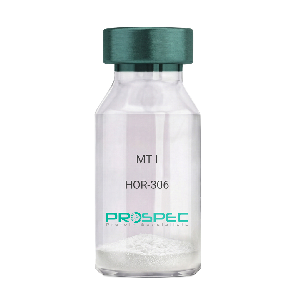
MT IDescription:
Melanotan-I
Melanotan-I, MT-I, Melanotan-1.
Product # :
HOR-306Price :
Quantity :
Shipping Method :
Shipped at Room temp
More Info
- description
- formulation
- purity
- More Info
- Citations
Description
Melanotan-I has the amino acid sequence of Ser-Tyr-Ser-Nle-Glu-His-D-Phe-Arg-Trp-Gly-Lys-Pro-Val and a molecular weight of 1647.4 Dalton.
Formulation
The protein (1mg/ml) was lyophilized with no additives.
Purity
Greater than 98.0% as determined by RP-HPLC.
Citations
More Info
-
Synonyms
Melanotan-I, MT-I, Melanotan-1.
-
Physical Appearance
Sterile Filtered off-White lyophilized (freeze-dried) powder.
-
Stability
Lyophilized Melanotan-I although stable at room temperature for 3 weeks, should be stored desiccated below -18°C. Upon reconstitution MT-I should be stored at 4°C between 2-7 days and for future use below -18°C.For long term storage it is recommended to add a carrier protein (0.1% HSA or BSA).Please prevent freeze-thaw cycles.
-
Solubility
It is recommended to reconstitute the lyophilized Melanotan-I in sterile 1% acetic acid not less than 100 µg/ml, which can then be further diluted to other aqueous solutions.
ProSpec's products are furnished for LABORATORY RESEARCH USE ONLY. The product may not be used as drugs, agricultural or pesticidal products, food additives or household chemicals.
- View Data Sheet
Name :
PON1 Human, HEKDescription:
Paraoxonase-1 Human Recombinant, HEK
Serum paraoxonase/arylesterase 1, Serum aryldialkylphosphatase 1, Aromatic esterase 1, A-esterase 1 , Serum aryldialkylphosphatase 1, paraoxonase 1, K-45, ESA, PON, MVCD5
Product # :
ENZ-1154Price :
Quantity :
Shipping Method :
Shipped with Ice Packs
More Info
- description
- source
- formulation
- purity
- biological activity
- More Info
- Citations
Description
PON1 Human Recombinant produced in HEK cells is a single, glycosylated, polypeptide chain (16-355 a.a) containing a total of 346 amino acids, having a molecular mass of 39.0kDa. PON1 is fused to a 6 amino acid His-tag at C-terminus,and is purified by proprietary chromatographic techniques.
Source
HEK293 Cells.
Formulation
The PON1 solution (0.25mg/ml) contains 20% Glycerol and Phosphate-Buffered Saline (pH 7.4).
Purity
Greater than 90.0% as determined by SDS-PAGE.
Biological Activity
Specific activity is > 2,500 pmol/min/ug. Defined by the amount of enzyme that hydrolyzes 1pmole of pnitrophenyl acetate to p-nitrophenol per minute at pH 7.5 at 37˚C.
Citations
More Info
-
Introduction
Paraoxonase-1 or PON1 is part of the paraoxonase group of proteins. PON1 is an enzyme, responsible to the toxic metabolites of a different of organophosphorus insecticides hydrolyzation. Furthermore, PON1 is a dominant anti-atherosclerotic part of HDL. The enzyme needs PPAR-gamma for activation, leading to synthesis and release of paraoxonase 1 from the liver tissue, resulting in atherosclerosis reduction. PON1 has many qualities for atheroprotective through inflammatory lipid peroxides metabolism. This enzyme can hydrolyze a large number of substrates, for example cyclic carbonates, lactones, nerve gases etc.
-
Synonyms
Serum paraoxonase/arylesterase 1, Serum aryldialkylphosphatase 1, Aromatic esterase 1, A-esterase 1 , Serum aryldialkylphosphatase 1, paraoxonase 1, K-45, ESA, PON, MVCD5
-
Physical Appearance
Sterile Filtered colorless solution.
-
Stability
Store at 4°C if entire vial will be used within 2-4 weeks. Store, frozen at -20°C for longer periods of time. For long term storage it is recommended to add a carrier protein (0.1% HSA or BSA).Avoid multiple freeze-thaw cycles.
-
Amino Acid Sequence
LFRNHQSSYQ TRLNALREVQ PVELPNCNLV KGIETGSEDL EILPNGLAFI SSGLKYPGIK SFNPNSPGKI LLMDLNEEDP TVLELGITGS KFDVSSFNPH GISTFTDEDN AMYLLVVNHP DAKSTVELFK FQEEEKSLLH LKTIRHKLLP NLNDIVAVGP EHFYGTNDHY FLDPYLQSWE MYLGLAWSYV VYYSPSEVRV VAEGFDFANG INISPDGKYV YIAELLAHKI HVYEKHANWT LTPLKSLDFN TLVDNISVDP ETGDLWVGCH PNGMKIFFYD SENPPASEVL RIQNILTEEP KVTQVYAENG TVLQGSTVAS VYKGKLLIGT VFHKALYCEL HHHHHH
ProSpec's products are furnished for LABORATORY RESEARCH USE ONLY. The product may not be used as drugs, agricultural or pesticidal products, food additives or household chemicals.
- View Data Sheet
Name :
LYG2 HumanDescription:
Lysozyme G-Like 2 Human Recombinant
Lysozyme G-Like 2, Lysozyme G-Like Protein 2, LYGH, EC 3.2.1.-.
Product # :
PRO-1245Price :
Quantity :
Shipping Method :
Shipped with Ice Packs
More Info
- description
- source
- formulation
- purity
- More Info
- Citations
Description
LYG2 Human Recombinant produced in E. coli is a single polypeptide chain containing 216 amino acids (20-212) and having a molecular mass of 23.9 kDa. LYG2 is fused to a 23 amino acid His-tag at N-terminus & purified by proprietary chromatographic techniques.
Source
E.coli.
Formulation
The LYG2 solution (0.5mg/1ml) contains 20mM Tris-HCl buffer (pH 8.0), 0.2M NaCl, 1mM DTT and 20% glycerol.
Purity
Greater than 90% as determined by SDS-PAGE.
Citations
More Info
-
Introduction
lysozyme G-like 2 and LYGH (LYG2), is a member of the glycosyl hydrolase 23 family. Lysozyme takes part in human innate immunity by causing bacterial cell lysis. LYG2 contains a SLT domain, a protein domain present in bacterial lytic transglycosylase (SLT) and in eukaryotic lysozymes (GEWL). SLT domain catalyzes the cleavage of the beta-1,4-glycosidic bond between N-acetylmuramic acid (MurNAc) and N-acetyglucosamine (GlcNAc).
-
Synonyms
Lysozyme G-Like 2, Lysozyme G-Like Protein 2, LYGH, EC 3.2.1.-.
-
Physical Appearance
Sterile Filtered colorless solution.
-
Stability
Store at 4°C if entire vial will be used within 2-4 weeks. Store, frozen at -20°C for longer periods of time. For long term storage it is recommended to add a carrier protein (0.1% HSA or BSA).Avoid multiple freeze-thaw cycles.
-
Amino Acid Sequence
MGSSHHHHHH SSGLVPRGSH MGSSYPFSHS MKPHLHPRLY HGCYGDIMTM KTSGATCDAN SVMNCGIRGS EMFAEMDLRA IKPYQTLIKE VGQRHCVDPA VIAAIISRES HGGSVLQDGW DHRGLKFGLM QLDKQTYHPV GAWDSKEHLS QATGILTERI KAIQKKFPTW SVAQHLKGGL SAFKSGIEAI ATPSDIDNDF VNDIIARAKF YKRQSF.
ProSpec's products are furnished for LABORATORY RESEARCH USE ONLY. The product may not be used as drugs, agricultural or pesticidal products, food additives or household chemicals.
- View Data Sheet
Name :
CST3 Human, PichiaDescription:
Cystatin C Human Recombinant, Pichia
Product # :
PRO-2743Price :
Quantity :
Shipping Method :
Shipped at Room temp
More Info
- description
- source
- formulation
- purity
- More Info
- Citations
Description
Recombinant Human Cystatin C is having a molecular mass of approximately 13kDa.
Source
Pichia pastoris.
Formulation
The protein was lyophilized from 20mM NH4HCO3.
Purity
Greater than 96.0%.
Citations
More Info
-
Introduction
Cystatins are a superfamily of cysteine proteinase inhibitors found in both plants and animals. They comprise a group of proteinase inhibitors, widely distributed in tissues and body fluids, and form tight complexes with cysteine proteases such as cathepsin B, H, L and S. Cystatin C, a secreted molecule of this family, is of interest from biochemical, medicine and evolutionary points of view. Cystatin C is increased in patients with malignant diseases, and is related to the insufficiency of renal function and appears to be a better marker than creatinine. On the other hand, low levels of cystatin C involve cause the breakdown of the elastic laminae and subsequently, the atherosclerosis and abdominal aortic aneurysm.
-
Physical Appearance
Sterile Filtered White lyophilized (freeze-dried) powder.
-
Stability
Human CST3 although stable at 4°C for 1 week, should be stored at -20°C.
-
Solubility
It is recommended to reconstitute the lyophilized CST3 Human in sterile 18MΩ-cm H2O at a concentration of 100µg/ml, which can then be further diluted to other aqueous solutions.
ProSpec's products are furnished for LABORATORY RESEARCH USE ONLY. The product may not be used as drugs, agricultural or pesticidal products, food additives or household chemicals.
- View Data Sheet
Name :
EGF (1-51), HumanDescription:
Epidermal Growth Factor (1-51 a.a.)Human Recombinant
Urogastrone, URG, EGF.
Product # :
CYT-1115Price :
Quantity :
Shipping Method :
Shipped at Room temp
More Info
- description
- source
- formulation
- purity
- biological activity
- More Info
- Citations
Description
Epidermal Growth Factor (1-51 a.a.) Human Recombinant produced in yeast is a single, glycosylated polypeptide chain containing 51 amino acids and having a molecular mass of 6.0kDa. The EGF is purified by proprietary chromatographic techniques.
Source
Saccharomyces cerevisiae
Formulation
Lyophilized from a 0.2μm filtered concentrated solution in PBS, pH 7.4.
Purity
Greater than 98.0% as determined by:
(a) Analysis by RP-HPLC.
(b) Analysis by SDS-PAGE.Biological Activity
The ED50 is determined by a cell proliferation assay using murine Balb/c 3T3 cells and is < than 0.1 ng/ml, corresponding to a specific activity of > 1.0 × 10⁷ IU/mg.
Citations
More Info
-
Introduction
Epidermal growth factor has a profound effect on the differentiation of specific cells in vivo and is a potent mitogenic factor for a variety of cultured cells of both ectodermal and mesodermal origin. The EGF precursor is believed to exist as a membrane-bound molecule which is proteolytically cleaved to generate the 53-amino acid peptide hormone that stimulates cells to divide. EGF stimulates the growth of several epidermal and epithelial tissues in vivo and in vitro and of some fibroblasts in cell culture.
-
Synonyms
Urogastrone, URG, EGF.
-
Physical Appearance
Sterile Filtered White lyophilized (freeze-dried) powder.
-
Stability
Lyophilized EGF although stable at room temperature for 3 weeks, should be stored desiccated below -18°C. Upon reconstitution Epidermal Growth Factor should be stored at 4°C between 2-7 days and for future use below -18°C.Please prevent freeze-thaw cycles.
-
Solubility
It is recommended to reconstitute the lyophilized Epidermal Growth Factor in sterile 18MΩ-cm H2O not less than 100µg/ml, which can then be further diluted to other aqueous solutions.
-
Amino Acid Sequence
NSDSECPLSH DGYCLHDGVC MYIEALDKYA CNCVVGYIGE RCQYRDLKWW E.
-
Background
Exploring the Potential of Epidermal Growth Factor (1-51 a.a.) Human Recombinant: Novel Insights and Therapeutic Prospects
Abstract:
Epidermal Growth Factor (EGF) stands as a pivotal cytokine orchestrating essential cellular processes. This concise research paper delves into the unique realm of Epidermal Growth Factor (1-51 a.a.) Human Recombinant, unveiling its intricate molecular dynamics, signaling cascades, and therapeutic promise. Employing cutting-edge methodologies encompassing in vitro assays and animal models, this study elucidates the multifaceted cellular responses sparked by this truncated EGF variant, paving the way for potential clinical applications.
Introduction:
The truncated form of EGF, spanning amino acids 1 to 51 (a.a.), carries distinct attributes that set it apart from the full-length counterpart. This paper centers on exploring the intriguing dimensions of Epidermal Growth Factor (1-51 a.a.) Human Recombinant, offering new insights into its interactions and potential utility.
Molecular Insights and Signaling Dynamics:
At the heart of its function lies the interplay between EGF (1-51 a.a.) and the epidermal growth factor receptor (EGFR). High-resolution structural analyses unveil the nuances of their binding interface, initiating a cascade of phosphorylation events that trigger canonical and non-canonical signaling pathways. The MAPK pathway and the PI3K/Akt pathway, intricately modulated by EGF (1-51 a.a.), propel cellular processes like proliferation, migration, and evasion of apoptosis.
In Vitro Profiling and Cellular Responses:
In dissecting the cellular responses, diverse in vitro assays have been employed. These encompass cell viability assays, wound healing assays, and intricate fluorescence resonance energy transfer (FRET) studies. These assays converge to illuminate the dynamic orchestration of EGF-induced cellular behaviors, showcasing its role in promoting cellular migration, division, and wound closure.
In Vivo Implications and Therapeutic Horizons:
Translating these insights into tangible therapeutic possibilities, in vivo studies present a compelling narrative. In animal models, EGF (1-51 a.a.) emerges as a potent player in cutaneous wound healing, fostering accelerated tissue regeneration. Moreover, its potential extends to oncology, as it not only influences tumor microenvironments but also demonstrates anti-apoptotic effects, hinting at its role in tailored cancer interventions.
Future Prospects and Challenges:
While these discoveries hold immense promise, challenges persist. The intricate network of signaling events demands further scrutiny, considering potential cross-talk and off-target effects. Refining delivery mechanisms and dosing regimens is essential for realizing the clinical potential of EGF (1-51 a.a.).
Conclusion:
In a synthesis of complex molecular insights and tangible therapeutic potential, Epidermal Growth Factor (1-51 a.a.) Human Recombinant emerges as a captivating subject. Its truncated structure and distinctive signaling cascades paint a canvas of cellular orchestration. As research advances, harnessing its therapeutic benefits could usher in novel interventions for wound healing and cancer therapy.
What is the molecular weight/Mw of EGF Protein?
EGF Protein has a total Mw of 6kDa.
What is the source or expression system of EGF Protein?
Saccharomyces cerevisiae
What is the Purity of EGF Protein?
EGF Protein is >98% pure as determined by SDS-PAGE.
What is the Biological Activity of EGF Protein?
The ED50 is determined by a cell proliferation assay using murine Balb/c 3T3 cells and is < than 0.1 ng/ml, corresponding to a specific activity of > 1.0 × 10⁷ IU/mg.
What is the amino acid sequence of EGF Protein?
NSDSECPLSH DGYCLHDGVC MYIEALDKYA CNCVVGYIGE RCQYRDLKWW E.
What applications can EGF Protein be used in?
EGF Protein can probably be used in western blot, ELISA and Lateral Flow.
What is the endotoxin level for EGF Protein?
The endotoxin level is minimal, EGF Protein was purified using conventional chromatography techniques.
ProSpec's products are furnished for LABORATORY RESEARCH USE ONLY. The product may not be used as drugs, agricultural or pesticidal products, food additives or household chemicals.
- View Data Sheet
Name :
SEPT3 HumanDescription:
Septin-3 Human Recombinant
Septin 3, SEP3, Neuronal-Specific Septin 3, Neuronal-Specific Septin-3, BK250D10.3, Neuronal-specific septin-3.
Product # :
PRO-2152Price :
Quantity :
Shipping Method :
Shipped with Ice Packs
More Info
- description
- source
- formulation
- purity
- More Info
- Citations
Description
SEPT3 Human Recombinant produced in E.Coli is a single, non-glycosylated polypeptide chain containing 381 amino acids (1-358 a.a) and having a molecular mass of 43.1kDa. SEPT3 is fused to a 23 amino acid His-tag at N-terminus & purified by proprietary chromatographic techniques.
Source
Escherichia Coli.
Formulation
SEPT3 protein solution (1mg/ml) containing Phosphate buffered saline (pH7.4), 10% glycerol and 1mM DTT.
Purity
Greater than 95% as determined by SDS-PAGE.
Citations
More Info
-
Introduction
Septin-3, also known as SEPT3 is a member of the septin family of GTPases. Members of this family are essential for cytokinesis. Furthermore, expression is up regulated by retinoic acid in a human teratocarcinoma cell line. The exact role of SEPT3 has not been determined yet. In addition, among its related pathways are Bacterial invasion of epithelial cells.
-
Synonyms
Septin 3, SEP3, Neuronal-Specific Septin 3, Neuronal-Specific Septin-3, BK250D10.3, Neuronal-specific septin-3.
-
Physical Appearance
Sterile filtered colorless solution.
-
Stability
Store at 4°C if entire vial will be used within 2-4 weeks. Store, frozen at -20°C for longer periods of time. For long term storage it is recommended to add a carrier protein (0.1% HSA or BSA).Avoid multiple freeze-thaw cycles.
-
Amino Acid Sequence
MGSSHHHHHH SSGLVPRGSH MGSMSKGLPE TRTDAAMSEL VPEPRPKPAV PMKPMSINSN LLGYIGIDTI IEQMRKKTMK TGFDFNIMVV GQSGLGKSTL VNTLFKSQVS RKASSWNREE KIPKTVEIKA IGHVIEEGGV KMKLTVIDTP GFGDQINNEN CWEPIEKYIN EQYEKFLKEE VNIARKKRIP DTRVHCCLYF ISPTGHSLRP LDLEFMKHLS KVVNIIPVIA KADTMTLEEK SEFKQRVRKE LEVNGIEFYP QKEFDEDLED KTENDKIRQE SMPFAVVGSD KEYQVNGKRV LGRKTPWGII EVENLNHCEF ALLRDFVIRT HLQDLKEVTH NIHYETYRAK RLNDNGGLPP GEGLLGTVLP PVPATPCPTA E.
ProSpec's products are furnished for LABORATORY RESEARCH USE ONLY. The product may not be used as drugs, agricultural or pesticidal products, food additives or household chemicals.
- View Data Sheet
Name :
Actin RabbitDescription:
Actin Rabbit
Product # :
PRO-517Price :
Quantity :
Shipping Method :
Shipped at Room temp
More Info
- description
- source
- formulation
- purity
- More Info
- Citations
Description
Ultra pure Actin consists in the alpha-skeletal muscle isoform and is purified from rabbit striated muscle.The purification method used (according to Spudich & Watts) results in a highly purified protein having a Molecular mass of 43,000 dalton.
Source
Rabbit Muscle.
Formulation
The protein was lyophilized from a 1mg/ml solution containing 10mM Tris/HCl buffer pH 8.0, 0.2mM CaCl2, 0.2mM ATP, 1mM DTT and 0.5% (w/v) SDS.
Purity
Greater than 98.0% as determined by SDS-PAGE.
Citations
More Info
-
Introduction
Actin is a muscle protein localized in the I band of the myofibrils; acting along with myosin, it is responsible for contraction and relaxation of muscle. Each actin protomer binds one molecule of ATP and has one high affinity site for either calcium or magnesium ions, as well as several low affinity sites. Actin exists as a monomer in low salt concentrations, but filaments form rapidly as salt concentration rises, with the consequent hydrolysis of ATP. It occurs in globular (G-actin) and fibrous (F-actin) forms. Actin is found in all eukaryotic cells (except for nematode sperm). Actin is one of the most highly-conserved proteins, differing by no more than 20% in species as diverse as algae and humans. Its other functions include cell motility, cell division and cytokinesis, vesicle and organelle movement, cell signaling, and the establishment and maintenance of cell junctions and cell shape.
-
Physical Appearance
Sterile Filtered White lyophilized (freeze-dried) powder.
-
Stability
Store the Lyophilized Actin between 2-8°C, do not freeze. Upon reconstitution Actin should be stored at 4°C between 2-7 days and for future use below -18°C. Please prevent freeze-thaw cycles.
-
Solubility
It is recommended to reconstitute the lyophilized Actin in sterile 18MΩ-cm H2O not less than 1mg/ml, which can then be further diluted to other aqueous solutions.
ProSpec's products are furnished for LABORATORY RESEARCH USE ONLY. The product may not be used as drugs, agricultural or pesticidal products, food additives or household chemicals.
- View Data Sheet
Name :
LA/SS-B Human, BiotinDescription:
LA / SS-B Human Recombinant, Biotinylated
Lupus La protein, Sjoegren syndrome type B antigen, SS-B, La ribonucleoprotein, La autoantigen, SSB, La, LARP3, LA/SS-B, La(SS-B).
Product # :
PRO-2562Price :
Quantity :
Shipping Method :
Shipped with Ice Packs
More Info
- description
- source
- formulation
- purity
- More Info
- Citations
Description
LA/SS-B Human Recombinant produced in SF9 is a single, glycosylated, polypeptide chain having a calculated molecular mass of 48 kDa. The LA/SS-B is expressed with a -6x His tag at N-terminus and purified by proprietary chromatographic techniques.
Source
Sf9 insect cells.
Formulation
The protein solution contains 20mM HEPES, pH 7.5, 400mM NaCl, 20% Glycerol.
Purity
Greater than 80.0% as determined by SDS-PAGE.
Citations
More Info
-
Introduction
The La protein is a 47 kDa polypeptide that frequently acts as an autoantigen in systemic lupus erythematosus and Sjogren's syndrome patients. La is involved in various aspects of RNA metabolism, including binding and protecting 3-prime UUU(OH) elements of newly RNA polymerase III - transcribed RNA, processing 5-prime and 3-prime ends of pre-tRNA precursors, acting as an RNA chaperone, and binding viral RNAs linked to hepatitis C virus. It occurs in both the nucleus and the cytoplasm, where it assumes different roles. In the nucleus, La protein facilitates the production of tRNAs, acting as an RNA polymerase III (RNAP III) transcription factor by attaching to the U-rich 3'UTR of nascent transcripts, aiding in their folding and maturation. In the cytoplasm, La protein facilitates the translation of specific mRNAs, acting as a translation factor. As an RNA binding protein (RBP), La protein associates with subsets of mRNAs which contain a 5'-terminal oligopyrimidine (5'TOP) motif known to direct protein synthesis. The binding of La protein to particular classes of RNA molecules regulates their downstream processing, guards them from endonuclease digestion, and organizes their export from the nucleus. La/SS-B appears to be readily disposed to proteolysis, which results in many smaller (42kD, 320, and 270) nevertheless still immunoreactive polypeptides. La/SS-B antigen is strongly conserved across species. Anti-La/SS-B autoantibodies were originally found as precipitating autoantibodies in sera of Sjogren's Syndrome patients and referred to as SjT. Anti-La/SS-B precipitins are most frequently found in Sjogren's Syndrome, Systemic Lupus Erythematosus (SLE) and Subacute Cutaneous Lupus. Also, there seems to be a correlation between anti-La/SS-B and the absence of nephritis in SLE patients.
-
Synonyms
Lupus La protein, Sjoegren syndrome type B antigen, SS-B, La ribonucleoprotein, La autoantigen, SSB, La, LARP3, LA/SS-B, La(SS-B).
-
Physical Appearance
Sterile Filtered clear solution.
-
Stability
Store at 4°C if entire vial will be used within 2-4 weeks. Store, frozen at -20°C for longer periods of time. Avoid multiple freeze-thaw cycles.
-
Immunological Functions
1. Binds IgG type human auto antibodies.2. Functional Streptavidin based ELISA test (analysis of positive/negative samples.)
ProSpec's products are furnished for LABORATORY RESEARCH USE ONLY. The product may not be used as drugs, agricultural or pesticidal products, food additives or household chemicals.
- View Data Sheet
Name :
TRAIL HumanDescription:
TRAIL / APO2 Ligand Human Recombinant
Tumor necrosis factor ligand superfamily member 10, TNF-related apoptosis-inducing ligand, Protein TRAIL, Apo-2 ligand, Apo-2L, CD253 antigen, TL2, APO2L, TNFSF10.
Product # :
CYT-443Price :
Quantity :
Shipping Method :
Shipped at Room temp
More Info
- description
- source
- formulation
- purity
- biological activity
- More Info
- Citations
Description
TRAIL/APO 2 Ligand Human Recombinant produced in E.Coli is a single, non-glycosylated polypeptide chain containing 168 amino acids (Met+Arg115-Gly281) and having a molecular mass of ~21kDa. The sTRAIL is purified by proprietary chromatographic techniques.
Source
Escherichia Coli.
Formulation
Lyophilized from a filtered (0.2µm) solution containing 20mM Tris-HCl pH 8.0 and 150mM NaCl.
Purity
Greater than 95.0% as determined by:
(a) Analysis by RP-HPLC.
(b) Analysis by SDS-PAGE.Biological Activity
The activity is determined by the cytolysis of Murine L929 cells in the presence of Actinomycin D, ED50 for this effect is less than 2ng/ml, corresponding to a specific activity of 5,000,000IU/mg.Citations
More Info
-
Introduction
TNF-related apoptosis-inducing ligand (TRAIL) is a ligand molecule which induces apoptosis. It is a type II transmembrane protein with homology to other members of the tumor necrosis factor family.
In humans, the gene that encodes for TRAIL is located at chromosome 3q26. TRAIL binds to the death receptors, DR4 and DR5. The process of apoptosis is caspase-8-dependent. This protein preferentially induces apoptosis in transformed and tumor cells, but does not appear to kill normal cells although it is expressed at a significant level in most normal tissues. -
Synonyms
Tumor necrosis factor ligand superfamily member 10, TNF-related apoptosis-inducing ligand, Protein TRAIL, Apo-2 ligand, Apo-2L, CD253 antigen, TL2, APO2L, TNFSF10.
-
Physical Appearance
Sterile Filtered White lyophilized (freeze-dried) powder.
-
Stability
Lyophilized APO 2 Ligand although stable at room temperature for 3 weeks, should be stored desiccated below -18°C. Upon reconstitution TRAIL should be stored at 4°C between 2-7 days and for future use below -18°C.For long term storage it is recommended to add a carrier protein (0.1% HSA or BSA).Please prevent freeze-thaw cycles.
-
Solubility
It is recommended to reconstitute the lyophilized TRAIL in sterile 18MΩ-cm H2O not less than 100µg/ml, which can then be further diluted to other aqueous solutions.
-
Amino Acid Sequence
MRERGPQRVA AHITGTRGRS NTLSSPNSKN EKALGRKINS WESSRSGHSF LSNLHLRNGE LVIHEKGFYY IYSQTYFRFQ EEIKENTKND KQMVQYIYKY TSYPDPILLM KSARNSCWSK DAEYGLYSIY QGGIFELKEN DRIFVSVTNE HLIDMDHEAS FFGAFLVG.
ProSpec's products are furnished for LABORATORY RESEARCH USE ONLY. The product may not be used as drugs, agricultural or pesticidal products, food additives or household chemicals.
- View Data Sheet
Name :
DsbA E.ColiDescription:
Disulfide Oxidoreductase E.Coli Recombinant
Disulfide oxidoreductase A, dsbA, rpbB, Disulfide oxidoreductase (DsbA) E Coli, DsDNA-binding protein A, Doublestranded DNA-binding protein, Disulfide oxidoreductase A periplasmic protein disulfide isomerase I, Thiol disulfide interchange protein dsbA.
Product # :
ENZ-1143Price :
Quantity :
Shipping Method :
Shipped with Ice Packs
More Info
- description
- source
- formulation
- purity
- More Info
- Citations
Description
DsbA E.Coli Recombinant produced in E.Coli is a single, non-glycosylated polypeptide chain containing 190 amino acids (20-208) and having a molecular mass of 21.2 kDa. DsbA E.Coli is purified by proprietary chromatographic techniques.
Source
Escherichia Coli.
Formulation
DsbA E.Coli protein (1mg/ml) is formulated in 20mM Tris-HCl buffer (pH 7.5) and 2mM EDTA.
Purity
Greater than 95.0% as determined by SDS-PAGE.
Citations
More Info
-
Introduction
Disulfide Oxidoreductase or DsbA is a protein, found in the plasma. It is part of the thioredoxin protein family. The protein creates disulfide bonds in the target proteins by contributing disulfide bond from its Cys30-Pro31-His32-Cys33 active site to a couple of cysteine residues.
-
Synonyms
Disulfide oxidoreductase A, dsbA, rpbB, Disulfide oxidoreductase (DsbA) E Coli, DsDNA-binding protein A, Doublestranded DNA-binding protein, Disulfide oxidoreductase A periplasmic protein disulfide isomerase I, Thiol disulfide interchange protein dsbA.
-
Physical Appearance
Sterile Filtered clear solution.
-
Stability
Store at 4°C if entire vial will be used within 2-4 weeks. Store, frozen at -20°C for longer periods of time. For long term storage it is recommended to add a carrier protein (0.1% HSA or BSA).Avoid multiple freeze-thaw cycles.
-
Amino Acid Sequence
MAQYEDGKQY TTLEKPVAGA PQVLEFFSFF CPHCYQFEEV LHISDNVKKK LPEGVKMTKY HVNFMGGDLG KDLTQAWAVA MALGVEDKVT VPLFEGVQKT QTIRSASDIR DVFINAGIKG EEYDAAWNSF VVKSLVAQQE KAAADVQLRG VPAMFVNGKY QLNPQGMDTS NMDVFVQQYA DTVKYLSEKK.
ProSpec's products are furnished for LABORATORY RESEARCH USE ONLY. The product may not be used as drugs, agricultural or pesticidal products, food additives or household chemicals.
- View Data Sheet
Name :
GFRA3 Human, Sf9Description:
GDNF Family Receptor Alpha 3 Human Recombinant, Sf9
GDNF Family Receptor Alpha3, GDNFR-alpha-3, GFR-alpha-3, GDNF Receptor Alpha-3, GDNFR3, GDNF Family Receptor Alpha-3, Glial Cell Line-Derived Neurotrophic Factor Receptor Alpha-3, GPI-Linked Receptor, GFRA3.
Product # :
CYT-1013Price :
Quantity :
Shipping Method :
Shipped with Ice Packs
More Info
- description
- source
- formulation
- purity
- More Info
- Citations
Description
GFRA3 Human Recombinant produced in Sf9 Baculovirus cells is a single, glycosylated polypeptide chain containing 585 amino acids (32-374) and having a molecular mass of 65.5kDa (Molecular size on SDS-PAGE will appear at approximately 70-100kDa). GFRA3 is fused to a 239 amino acid hIgG-His-Tag at C-terminus and purified by proprietary chromatographic techniques.
Source
Sf9, Baculovirus cells.
Formulation
GFRA3 protein solution (0.25mg/ml) containing Phosphate Buffered Saline (pH 7.4) and 10% glycerol.
Purity
Greater than 85.0% as determined by SDS-PAGE.
Citations
More Info
-
Introduction
GDNF Family Receptor Alpha 3 (GFRA3) belongs to the GDNF receptor family. GFRA3 creates a signaling receptor complex with RET tyrosine kinase receptor and binds the ligand, artemin (ARTN).
-
Synonyms
GDNF Family Receptor Alpha3, GDNFR-alpha-3, GFR-alpha-3, GDNF Receptor Alpha-3, GDNFR3, GDNF Family Receptor Alpha-3, Glial Cell Line-Derived Neurotrophic Factor Receptor Alpha-3, GPI-Linked Receptor, GFRA3.
-
Physical Appearance
Sterile Filtered clear solution.
-
Stability
Store at 4°C if entire vial will be used within 2-4 weeks. Store, frozen at -20°C for longer periods of time. For long term storage it is recommended to add a carrier protein (0.1% HSA or BSA).Avoid multiple freeze-thaw cycles.
-
Amino Acid Sequence
ADPDPLPTES RLMNSCLQAR RKCQADPTCS AAYHHLDSCT SSISTPLPSE EPSVPADCLE AAQQLRNSSL IGCMCHRRMK NQVACLDIYW TVHRARSLGN YELDVSPYED TVTSKPWKMN LSKLNMLKPD SDLCLKFAML CTLNDKCDRL RKAYGEACSG PHCQRHVCLR QLLTFFEKAA EPHAQGLLLC PCAPNDRGCG ERRRNTIAPN CALPPVAPNC LELRRLCFSD PLCRSRLVDF QTHCHPMDIL GTCATEQSRC LRAYLGLIGT AMTPNFVSNV NTSVALSCTC RGSGNLQEEC EMLEGFFSHN PCLTEAIAAK MRFHSQLFSQ DWPHPTFAVM AHQNENLEPK SCDKTHTCPP CPAPELLGGP SVFLFPPKPK DTLMISRTPE VTCVVVDVSH EDPEVKFNWY VDGVEVHNAK TKPREEQYNS TYRVVSVLTV LHQDWLNGKE YKCKVSNKAL PAPIEKTISK AKGQPREPQV YTLPPSRDEL TKNQVSLTCL VKGFYPSDIA VEWESNGQPE NNYKTTPPVL DSDGSFFLYS KLTVDKSRWQ QGNVFSCSVM HEALHNHYTQ KSLSLSPGKH HHHHH.
-
Background
What is the molecular weight/Mw of GFRA3 HUMAN, SF9 Protein?
GFRA3 HUMAN, SF9 Protein has a total Mw of 65.5kDa.
What is the source or expression system of GFRA3 HUMAN, SF9 Protein?
Sf9, Baculovirus cells.
What is the Purity of GFRA3 HUMAN, SF9 Protein?
GFRA3 HUMAN, SF9 Protein is >85% pure as determined by SDS-PAGE.
What is the Biological Activity of GFRA3 HUMAN, SF9 Protein?
The biological functionality of GFRA3 HUMAN, SF9 Protein will be determined in the future.
What is the amino acid sequence of GFRA3 HUMAN, SF9 Protein?
ADPDPLPTES RLMNSCLQAR RKCQADPTCS AAYHHLDSCT SSISTPLPSE EPSVPADCLE AAQQLRNSSL IGCMCHRRMK NQVACLDIYW TVHRARSLGN YELDVSPYED TVTSKPWKMN LSKLNMLKPD SDLCLKFAML CTLNDKCDRL RKAYGEACSG PHCQRHVCLR QLLTFFEKAA EPHAQGLLLC PCAPNDRGCG ERRRNTIAPN CALPPVAPNC LELRRLCFSD PLCRSRLVDF QTHCHPMDIL GTCATEQSRC LRAYLGLIGT AMTPNFVSNV NTSVALSCTC RGSGNLQEEC EMLEGFFSHN PCLTEAIAAK MRFHSQLFSQ DWPHPTFAVM AHQNENLEPK SCDKTHTCPP CPAPELLGGP SVFLFPPKPK DTLMISRTPE VTCVVVDVSH EDPEVKFNWY VDGVEVHNAK TKPREEQYNS TYRVVSVLTV LHQDWLNGKE YKCKVSNKAL PAPIEKTISK AKGQPREPQV YTLPPSRDEL TKNQVSLTCL VKGFYPSDIA VEWESNGQPE NNYKTTPPVL DSDGSFFLYS KLTVDKSRWQ QGNVFSCSVM HEALHNHYTQ KSLSLSPGKH HHHHH.
What applications can GFRA3 HUMAN, SF9 Protein be used in?
GFRA3 HUMAN, SF9 Protein can probably be used in western blot, ELISA and Lateral Flow.
What is the endotoxin level for GFRA3 HUMAN, SF9 Protein?
The endotoxin level is minimal, GFRA3 HUMAN, SF9 Protein was purified using conventional chromatography techniques.
ProSpec's products are furnished for LABORATORY RESEARCH USE ONLY. The product may not be used as drugs, agricultural or pesticidal products, food additives or household chemicals.
- View Data Sheet
Name :
KLK3 ProteinDescription:
Kallikrein-3 Recombinant Human
Prostate-specific antigen, PSA, Gamma-seminoprotein, Seminin, Kallikrein-3, P-30 antigen, Semenogelase, KLK3, APS, hK3, KLK2A1.
Product # :
ENZ-1102Price :
Quantity :
Shipping Method :
Shipped at Room temp
More Info
- description
- source
- formulation
- purity
- More Info
- Citations
Description
Kallikrein-3 Human Recombinant produced in E.Coli is a single, non- glycosylated polypeptide chain containing 237 amino acids and having a molecular mass of 26.1kDa.KLK3 is purified by proprietary chromatographic techniques.
Source
Escherichia Coli.
Formulation
Lyophilized from a 0.2μm filtered concentrated solution in 20mM Tris-HCl, pH 8.0, 150mM NaCl and 3% trehalose.
Purity
Greater than 95.0% as determined by:
(a) Analysis by RP-HPLC.
(b) Analysis by SDS-PAGE.Citations
More Info
-
Introduction
Kallikrein-3 (KLK3) is a part of the kallikrein-related peptidase family. Kallikreins are a subgroup of serine proteases having various physiological functions. Numerous kallikreins take part in carcinogenesis and some may be prospective cancer and other disease biomarkers. Kallikrein-3 is 1 of the 15 kallikrein subfamily members located in a cluster on chromosome 19 and is a protease present in seminal plasma. KLK3 hydrolyzes semenogelin-1 consequently leading to the liquefaction of the seminal coagulum. KLK3 acts normally in the liquefaction of seminal coagulum, probably by hydrolysis of the high molecular mass seminal vesicle protein. Serum level of the KLK3 protein, called PSA in the clinical setting, is beneficial in the diagnosis and monitoring of prostatic carcinoma.
-
Synonyms
Prostate-specific antigen, PSA, Gamma-seminoprotein, Seminin, Kallikrein-3, P-30 antigen, Semenogelase, KLK3, APS, hK3, KLK2A1.
-
Physical Appearance
Sterile Filtered White lyophilized (freeze-dried) powder.
-
Stability
Lyophilized KLK3 although stable at room temperature for 3 weeks, should be stored desiccated below -18°C. Upon reconstitution Kallikrein-3 should be stored at 4°C between 2-7 days and for future use below -18°C.Please prevent freeze-thaw cycles.
-
Solubility
It is recommended to reconstitute the lyophilized Kallikrein-3 in sterile 18MΩ-cm H2O not less than 100µg/ml, which can then be further diluted to other aqueous solutions.
-
Amino Acid Sequence
IVGGWECEKH SQPWQVLVAS RGRAVCGGVL VHPQWVLTAA HCIRNKSVIL LGRHSLFHPE DTGQVFQVSH SFPHPLYDMS LLKNRFLRPG DDSSHDLMLL RLSEPAELTDA VKVMDLPTQE PALGTTCYAS GWGSIEPEEF LTPKKLQCVD LHVISNDVCA QVHPQKVTKF MLCAGRWTGG KSTCSGDSGG PLVCNGVLQG ITSWGSEPCA LPERPSLYTK VVHYRKWIKD TIVANP.
ProSpec's products are furnished for LABORATORY RESEARCH USE ONLY. The product may not be used as drugs, agricultural or pesticidal products, food additives or household chemicals.
- View Data Sheet
Name :
APOM Human, HEKDescription:
Apolipoprotein-M Human Recombinant, HEK
G3a, HSPC336, NG20, Apolipoprotein M, APOM, Apo-M, MGC22400.
Product # :
CYT-026Price :
Quantity :
Shipping Method :
Shipped at Room temp
More Info
- description
- source
- formulation
- purity
- More Info
- Citations
Description
APOM HEK Human Recombinant Protein is 20 kDa protein containing 179 amino acid residues of the APOM Human and 13 additional amino acid residues of flag Tag.
Source
HEK293.
Formulation
APOM Human was filtered (0.4µm) and lyophilized from 0.5 mg/ml supplied in 20mM TRIS and 50mM NaCl, pH 7.5.
Purity
Greater than 90% as determined by SDS-PAGE.
Citations
More Info
-
Introduction
APOM is belongs to the lipocalin protein family and is associated with high density lipoproteins and to a lesser extent with low density lipoproteins and triglyceride-rich lipoproteins. APOM is secreted through the plasma membrane but remains membrane-bound, where it takes part in lipid transport.
-
Synonyms
G3a, HSPC336, NG20, Apolipoprotein M, APOM, Apo-M, MGC22400.
-
Physical Appearance
White lyophilized (freeze-dried) powder.
-
Stability
Store lyophilized APOM Human at -20°C. Aliquot the product after reconstitution to avoid repeated freezing/thawing cycles. Reconstituted APOM Human can be stored at 4°C for a limited period of time; it does not show any change after two weeks at 4°C.
-
Solubility
It is recommended to add deionized water to prepare a working stock solution of approximately 0.5mg/ml and let the lyophilized pellet dissolve completely. Product is not sterile! Please filter the product by an appropriate sterile filter before using it on cell culture.
-
Amino Acid Sequence
HVDYKDDDDK PAGCPEHSQL TTLGVDGKEF PEVHLGQWYF IAGAAPTKEE LATFDPVDNI VFNMAAGSAP MQLHLRATIR MKDGLCVPRK WIYHLTEGST DLRTEGRPDM KTELFSSSCP GGIMLNETGQ GYQRFLLYNR SPHPPEKCVE EFKSLTSCLD SKAFLLTPRN QEACELSNN
-
Applications
Cell culture and/or animal studies, ELISA and Western blotting.
-
Background
What is the molecular weight/Mw of APOM Protein?
APOM Protein has a total Mw of 20kDa.
What is the source or expression system of APOM Protein?
HEK293.
What is the Purity of APOM Protein?
APOM Protein is >90% pure as determined by SDS-PAGE.
What is the Biological Activity of APOM Protein?
The biological functionality of APOM Protein will be determined in the future.
What is the amino acid sequence of APOM Protein?
HVDYKDDDDK PAGCPEHSQL TTLGVDGKEF PEVHLGQWYF IAGAAPTKEE LATFDPVDNI VFNMAAGSAP MQLHLRATIR MKDGLCVPRK WIYHLTEGST DLRTEGRPDM KTELFSSSCP GGIMLNETGQ GYQRFLLYNR SPHPPEKCVE EFKSLTSCLD SKAFLLTPRN QEACELSNN
What applications can APOM Protein be used in?
APOM Protein can probably be used in western blot, ELISA and Lateral Flow.
What is the endotoxin level for APOM Protein?
The endotoxin level is minimal, APOM Protein was purified using conventional chromatography techniques.
ProSpec's products are furnished for LABORATORY RESEARCH USE ONLY. The product may not be used as drugs, agricultural or pesticidal products, food additives or household chemicals.