Search results
1000 results found for “Uteroglobin”
Name
Description
Product #
Price
Quantity
Shipping Method
- View Data Sheet
Name :
BTN3A1 HumanDescription:
Butyrophilin Subfamily 3 Member A1 Human Recombinant
Butyrophilin Subfamily 3 Member A1, BTF5, DJ45P21.3 (Butyrophilin, Subfamily 3, Member A1), Butyrophilin, Subfamily 3, Member A1, CD277 Antigen, BTN3.1, BT3.1, CD277, Butyrophilin subfamily 3 member A1.
Product # :
PRO-2454Price :
Quantity :
Shipping Method :
Shipped with Ice Packs
More Info
- description
- source
- formulation
- purity
- More Info
- Citations
Description
BTN3A1 Human Recombinant produced in Sf9 Baculovirus cells is a single, glycosylated polypeptide chain containing 464 amino acids (30-254a.a.) and having a molecular mass of 51.1kDa (Molecular size on SDS-PAGE will appear at approximately 50-70kDa).BTN3A1 is expressed with a 239 amino acid hIgG-His Tag at C-Terminus and purified by proprietary chromatographic techniques.
Source
Sf9, Baculovirus cells.
Formulation
BTN3A1 protein solution (0.5mg/ml) contains Phosphate Buffered Saline (pH 7.4) and 10% glycerol.
Purity
Greater than 90.0% as determined by SDS-PAGE.
Citations
More Info
-
Introduction
Butyrophilin sub family 3 member A1, also known as BTN3A1 is a member of the immunoglobulin superfamily. BTN3A1 is composed of an extracellular N-terminal IgV as well as a membrane proximal IgC domain followed by a transmembrane domain and also a cytoplasmic tail. BTN3A1 participates in T-cell activation and also in the adaptive immune response. Furthermore, BTN3A1 regulates the proliferation of activated T-cells & the release of cytokines and IFNG by activated T-cells. BTN3A1, Mediates the response of T-cells to infected as well as transformed cells which are categorized by high levels of phosphorylated metabolites, such as isopentenyl pyrophosphate.
-
Synonyms
Butyrophilin Subfamily 3 Member A1, BTF5, DJ45P21.3 (Butyrophilin, Subfamily 3, Member A1), Butyrophilin, Subfamily 3, Member A1, CD277 Antigen, BTN3.1, BT3.1, CD277, Butyrophilin subfamily 3 member A1.
-
Physical Appearance
Sterile Filtered colorless solution.
-
Stability
Store at 4°C if entire vial will be used within 2-4 weeks. Store, frozen at -20°C for longer periods of time. For long term storage it is recommended to add a carrier protein (0.1% HSA or BSA).Avoid multiple freeze-thaw cycles.
-
Amino Acid Sequence
QFSVLGPSGP ILAMVGEDAD LPCHLFPTMS AETMELKWVS SSLRQVVNVY ADGKEVEDRQ SAPYRGRTSI LRDGITAGKA ALRIHNVTAS DSGKYLCYFQ DGDFYEKALV ELKVAALGSD LHVDVKGYKD GGIHLECRST GWYPQPQIQW SNNKGENIPT VEAPVVADGV GLYAVAASVI MRGSSGEGVS CTIRSSLLGL EKTASISIAD PFFRSAQRWI AALAGLEPKS CDKTHTCPPC PAPELLGGPS VFLFPPKPKD TLMISRTPEV TCVVVDVSHE DPEVKFNWYV DGVEVHNAKT KPREEQYNST YRVVSVLTVL HQDWLNGKEY KCKVSNKALP APIEKTISKA KGQPREPQVY TLPPSRDELT KNQVSLTCLV KGFYPSDIAV EWESNGQPEN NYKTTPPVLD SDGSFFLYSK LTVDKSRWQQ GNVFSCSVMH EALHNHYTQK SLSLSPGKHH HHHH.
ProSpec's products are furnished for LABORATORY RESEARCH USE ONLY. The product may not be used as drugs, agricultural or pesticidal products, food additives or household chemicals.
- View Data Sheet
Name :
GP1BB HumanDescription:
Glycoprotein Ib Platelet Subunit Beta Human Recombinant
GPIBB, glycoprotein Ib platelet subunit beta, BDPLT1,GP-Ib beta, BS, CD42C ,GPIbbeta, Antigen CD42b-beta, GPIb-beta, GPIbB, Platelet glycoprotein Ib beta chain.
Product # :
PRO-2774Price :
Quantity :
Shipping Method :
Shipped with Ice Packs
More Info
- description
- source
- formulation
- purity
- More Info
- Citations
Description
GP1BB Human Recombinant is a single, glycosylated, polypeptide chain (26-147 a.a) containing a total of 131 amino acids, having a molecular mass of 14.0 kDa. GP1BB is fused to a 6 amino acid His-tag at C-terminus and is purified by proprietary chromatographic techniques.
Source
HEK293 Cells.
Formulation
The GP1BB solution (0.25mg/ml) contains 10% Glycerol and Phosphate-Buffered Saline (pH 7.4).
Purity
Greater than 95.0% as determined by SDS-PAGE.
Citations
More Info
-
Synonyms
GPIBB, glycoprotein Ib platelet subunit beta, BDPLT1,GP-Ib beta, BS, CD42C ,GPIbbeta, Antigen CD42b-beta, GPIb-beta, GPIbB, Platelet glycoprotein Ib beta chain.
-
Physical Appearance
Sterile Filtered colorless solution.
-
Stability
Store at 4°C if entire vial will be used within 2-4 weeks. Store, frozen at -20°C for longer periods of time. For long term storage it is recommended to add a carrier protein (0.1% HSA or BSA). Avoid multiple freeze-thaw cycles.
-
Amino Acid Sequence
DGSCPAPCSC AGTLVDCGRR GLTWASLPTA FPVDTTELVL TGNNLTALPP GLLDALPALR TAHLGANPWR CDCRLVPLRA WLAGRPERAP YRDLRCVAPP ALRGRLLPYL AEDELRAACA PGPLCHHHHH H.
-
Background
Glycoprotein 1b beta (GP1BB) is a crucial component of the platelet glycoprotein Ib-IX-V complex, playing a pivotal role in platelet function and hemostasis. This research aims to explore the structure, function, and implications of GP1BB protein in platelet adhesion, aggregation, and the development of thrombotic disorders. Understanding the molecular mechanisms and regulatory roles of GP1BB can provide valuable insights into its potential as a therapeutic target for thrombotic diseases.
Structure and Expression of GP1BB Protein:
GP1BB is a transmembrane protein consisting of an extracellular domain, a single transmembrane region, and a short cytoplasmic tail. It is predominantly expressed on the surface of platelets, where it interacts with von Willebrand factor (VWF) and other components of the glycoprotein Ib-IX-V complex. GP1BB is crucial for mediating platelet adhesion to damaged endothelium and the formation of stable platelet aggregates at the site of injury.
GP1BB and Platelet Adhesion:
GP1BB interacts with VWF, which is exposed upon vascular injury. This interaction facilitates the initial attachment of platelets to the damaged endothelium. GP1BB also participates in the formation of high-affinity bonds between platelets and VWF, contributing to the stabilization of platelet adhesion and the initiation of platelet aggregation. Dysregulation of GP1BB-mediated platelet adhesion can lead to abnormal clot formation and thrombotic disorders.
Implications of GP1BB in Thrombotic Disorders:
Defects or mutations in the GP1BB gene can result in Bernard-Soulier syndrome (BSS), a rare inherited bleeding disorder characterized by giant platelets, thrombocytopenia, and impaired platelet adhesion. BSS patients often experience excessive bleeding and bruising due to defective GP1BB function. On the other hand, abnormal activation or overexpression of GP1BB has been associated with increased risk of thrombotic events, such as myocardial infarction and stroke.
Therapeutic Strategies Targeting GP1BB:
Given its crucial role in platelet function and thrombotic disorders, GP1BB represents a potential target for therapeutic interventions. Strategies aimed at modulating GP1BB expression or function have been explored. This includes the development of monoclonal antibodies or small molecule inhibitors that selectively target GP1BB and interfere with its interaction with VWF or other binding partners. Such approaches aim to prevent excessive platelet adhesion and aggregation, reducing the risk of thrombotic events.
Challenges and Future Directions:
While targeting GP1BB shows promise, several challenges need to be addressed. The development of specific inhibitors or modulators of GP1BB that do not interfere with its physiological functions is crucial. Additionally, understanding the complex interplay between GP1BB and other platelet receptors and signaling pathways is essential for the development of effective therapeutic strategies.
Conclusion:
The study of GP1BB protein provides valuable insights into its central role in platelet adhesion, aggregation, and thrombotic disorders. Understanding the molecular mechanisms and functional implications of GP1BB opens avenues for the development of targeted therapies for thrombotic diseases. Further research on GP1BB protein, its interactions, and its modulation in pathological conditions will contribute to the development of novel therapeutic interventions to mitigate the risk of thrombotic events and improve patient outcomes.
ProSpec's products are furnished for LABORATORY RESEARCH USE ONLY. The product may not be used as drugs, agricultural or pesticidal products, food additives or household chemicals.
- View Data Sheet
Name :
PYGL HumanDescription:
Phosphorylase, Glycogen, Liver Human Recombinant
GSD6, Glycogen phosphorylase, liver form.
Product # :
ENZ-675Price :
Quantity :
Shipping Method :
Shipped with Ice Packs
More Info
- description
- source
- formulation
- purity
- More Info
- Citations
Description
PYGL Human Recombinant produced in E.Coli is a single, non-glycosylated polypeptide chain containing 879 amino acids (1-847 a.a) and having a molecular mass of 100.7kDa.PYGL is fused to a 32 amino acid His-tag at N-terminus & purified by proprietary chromatographic techniques.
Source
Escherichia Coli.
Formulation
PYGL protein solution (0.25mg/ml) in phosphate buffered saline (pH7.4), 30% glycerol and 1mM DTT.
Purity
Greater than 80.0% as determined by SDS-PAGE.
Citations
More Info
-
Introduction
Glycogen phosphorylase (PYGL) converts from inactive phosphorylase B to active phosphorylase A by phosphorylation of serine residue 15. Activity of the PYGL enzyme is further regulated by numerous allosteric effectors and hormonal controls. The liver isozyme supplies the glycemic demands of the body in general whereas the brain and muscle isozymes supply just those tissues.
-
Synonyms
GSD6, Glycogen phosphorylase, liver form.
-
Physical Appearance
Sterile Filtered colorless solution.
-
Stability
Store at 4°C if entire vial will be used within 2-4 weeks. Store, frozen at -20°C for longer periods of time. For long term storage it is recommended to add a carrier protein (0.1% HSA or BSA).Avoid multiple freeze-thaw cycles.
-
Amino Acid Sequence
MGSSHHHHHH SSGLVPRGSH MGSEFELRRQ ASMAKPLTDQ EKRRQISIRG IVGVENVAEL KKSFNRHLHF TLVKDRNVAT TRDYYFALAH TVRDHLVGRW IRTQQHYYDK CPKRVYYLSL EFYMGRTLQN TMINLGLQNA CDEAIYQLGL DIEELEEIEE DAGLGNGGLG RLAACFLDSM ATLGLAAYGY GIRYEYGIFN QKIRDGWQVE EADDWLRYGN PWEKSRPEFM LPVHFYGKVE HTNTGTKWID TQVVLALPYD TPVPGYMNNT VNTMRLWSAR APNDFNLRDF NVGDYIQAVL DRNLAENISR VLYPNDNFFE GKELRLKQEY FVVAATLQDI IRRFKASKFG STRGAGTVFD AFPDQVAIQL NDTHPALAIP ELMRIFVDIE KLPWSKAWEL TQKTFAYTNH TVLPEALERW PVDLVEKLLP RHLEIIYEIN QKHLDRIVAL FPKDVDRLRR MSLIEEEGSK RINMAHLCIV GSHAVNGVAK IHSDIVKTKV FKDFSELEPD KFQNKTNGIT PRRWLLLCNP GLAELIAEKI GEDYVKDLSQ LTKLHSFLGD DVFLRELAKV KQENKLKFSQ FLETEYKVKI NPSSMFDVQV KRIHEYKRQL LNCLHVITMY NRIKKDPKKL FVPRTVIIGG KAAPGYHMAK MIIKLITSVA DVVNNDPMVG SKLKVIFLEN YRVSLAEKVI PATDLSEQIS TAGTEASGTG NMKFMLNGAL TIGTMDGANV EMAEEAGEEN LFIFGMRIDD VAALDKKGYE AKEYYEALPE LKLVIDQIDN GFFSPKQPDL FKDIINMLFY HDRFKVFADY EAYVKCQDKV SQLYMNPKAW NTMVLKNIAA SGKFSSDRTI KEYAQNIWNV EPSDLKISLS NESNKVNGN.
ProSpec's products are furnished for LABORATORY RESEARCH USE ONLY. The product may not be used as drugs, agricultural or pesticidal products, food additives or household chemicals.
- View Data Sheet
Name :
ZG16 HumanDescription:
Zymogen Granule Protein 16 Homolog Human Recombinant
Zymogen granule membrane protein 16, Zymogen granule protein 16, hZG16, Secretory lectin ZG16, ZG16, JCLN, JCLN1, ZG16A, FLJ43571, FLJ92276, MGC34820, MGC183567.
Product # :
PRO-478Price :
Quantity :
Shipping Method :
Shipped with Ice Packs
More Info
- description
- source
- formulation
- purity
- More Info
- Citations
Description
ZG16 Human Recombinant produced in E. coli is a single polypeptide chain containing 174 amino acids (17-167) and having a molecular mass of 19kDa. ZG16 is fused to a 23 amino acid His-tag at N-terminus & purified by proprietary chromatographic techniques.
Source
Escherichia Coli.
Formulation
The ZG16 solution (0.25mg/ml) contains 20mM Tris-HCl buffer (pH 8.0), 0.15M NaCl, 20% glycerol and 1mM DTT.
Purity
Greater than 95% as determined by SDS-PAGE.
Citations
More Info
-
Introduction
Zymogen Granule Protein 16 Homolog (ZG16) is a member of the jacalin lectin family. ZG16 may have a role in protein trafficking and may act as a linker molecule between the submembranous matrix on the luminal side of zymogen granule membrane (ZGM) and aggregated secretory proteins during granule formation in the TGN. ZG16 is highly expressed in the liver, but can also be detected at lower levels in colon, ileum and jejunum.
-
Synonyms
Zymogen granule membrane protein 16, Zymogen granule protein 16, hZG16, Secretory lectin ZG16, ZG16, JCLN, JCLN1, ZG16A, FLJ43571, FLJ92276, MGC34820, MGC183567.
-
Physical Appearance
Sterile filtered colorless solution.
-
Stability
Store at 4°C if entire vial will be used within 2-4 weeks. Store, frozen at -20°C for longer periods of time. For long term storage it is recommended to add a carrier protein (0.1% HSA or BSA). Avoid multiple freeze-thaw cycles.
-
Amino Acid Sequence
MGSSHHHHHH SSGLVPRGSH MGSNAIQARS SSYSGEYGGG GGKRFSHSGN QLDGPITALR VRVNTYYIVG LQVRYGKVWS DYVGGRNGDL EEIFLHPGES VIQVSGKYKW YLKKLVFVTD KGRYLSFGKD SGTSFNAVPL HPNTVLRFIS GRSGSLIDAI GLHWDVYPTS CSRC.
ProSpec's products are furnished for LABORATORY RESEARCH USE ONLY. The product may not be used as drugs, agricultural or pesticidal products, food additives or household chemicals.
- View Data Sheet
Name :
Globular Adiponectin HumanDescription:
Globular Adiponectin Human Recombinant
Acrp30, AdipoQ, GBP-28, APM-1, ACDC.
Product # :
CYT-615Price :
Quantity :
Shipping Method :
Shipped at Room temp
More Info
- description
- source
- formulation
- purity
- biological activity
- More Info
- Citations
Description
gAcrp30 Human Recombinant produced in E.Coli is a non-glycosylated, Polypeptide chain containing 145 amino acids and having a molecular mass of 16.7kDa. The gAcrp30 is purified by proprietary chromatographic techniques.
Source
Escherichia Coli.
Formulation
Sterile filtered and lyophilized from 10mM sodium phosphate & 0.5mM DTT, pH 7.5.
Purity
Greater than 95% as determined by SDS-PAGE.
Biological Activity
gAcrp30 activity is determined by its ability to inhibit the proliferation of mouse M1 cells. The expected ED50 for this effect is 669ng/ml, corresponding to a specific activity of 1.5x10³units/mg.
Citations
More Info
-
Introduction
gAcrp30 globular protein, exists as a result of proteolytic processing of adiponectin. Adiponectin is manufactured and secreted solely by adipocytes, and is a highly obtained plasma protein, accounting for up to 0.05% of total serum protein. Similar to Adiponectin, gAcrp30 is able of lowering hyperglycemia and reversing INS resistance. In addition, gAcrp30 is an significant protein that is involved in promoting fat loss by signaling muscle to absorb and burn Free-Fatty Acids. AdipoR1 & AdipoR2 are the 2 signaling receptors for adiponectin and gAcrp30 that were recently been identified.
-
Synonyms
Acrp30, AdipoQ, GBP-28, APM-1, ACDC.
-
Stability
For long term, store lyophilized protein at -20°C. Aliquot the product after reconstitution to avoid repeated freezing/thawing cycles. Reconstituted protein can be stored at 4°C for a limited period of time two weeks.
-
Solubility
We recommended reconstituting gAcrp30 at a concentration of 0.1mg per ml with 10mM sodium phosphate & 0.5mM DTT, pH 7.5. which can then be further diluted to other aqueous solutions.
-
Amino Acid Sequence
MKGEPGEGAY VYRSAFSVGL ETYVTIPNMP IRFTKIFYNQ QNHYDGSTGK FHCNIPGLYY FAYHITVYMK DVKVSLFKKD KAMLFTYDQY QENNVDQASG SVLLHLEVGD QVWLQVYGEG ERNGLYADND NDSTFTGFLL YHDTN.
-
Background
What is the molecular weight/Mw of ADIPONECTIN Protein?
ADIPONECTIN Protein has a total Mw of 16.7kDa.
What is the source or expression system of ADIPONECTIN Protein?
Escherichia Coli.
What is the Purity of ADIPONECTIN Protein?
ADIPONECTIN Protein is >95% pure as determined by SDS-PAGE.
What is the Biological Activity of ADIPONECTIN Protein?
gAcrp30 activity is determined by its ability to inhibit the proliferation of mouse M1 cells. The expected ED50 for this effect is 669ng/ml, corresponding to a specific activity of 1.5x10³units/mg.
What is the amino acid sequence of ADIPONECTIN Protein?
MKGEPGEGAY VYRSAFSVGL ETYVTIPNMP IRFTKIFYNQ QNHYDGSTGK FHCNIPGLYY FAYHITVYMK DVKVSLFKKD KAMLFTYDQY QENNVDQASG SVLLHLEVGD QVWLQVYGEG ERNGLYADND NDSTFTGFLL YHDTN.
What applications can ADIPONECTIN Protein be used in?
ADIPONECTIN Protein can probably be used in western blot, ELISA and Lateral Flow.
What is the endotoxin level for ADIPONECTIN Protein?
The endotoxin level is minimal, ADIPONECTIN Protein was purified using conventional chromatography techniques.
ProSpec's products are furnished for LABORATORY RESEARCH USE ONLY. The product may not be used as drugs, agricultural or pesticidal products, food additives or household chemicals.
- View Data Sheet
Name :
SHH HumanDescription:
Sonic HedgeHog Human Recombinant
SHH, HHG-1, HHG1, Sonic hedgehog protein, TPT, HLP3, HPE3, SMMCI, TPTPS, MCOPCB5.
Product # :
CYT-676Price :
Quantity :
Shipping Method :
Shipped at Room temp
More Info
- description
- source
- formulation
- purity
- biological activity
- More Info
- Citations
Description
Sonic HedgeHog Recombinant Human produced in E.Coli is a single, non-glycosylated polypeptide chain containing 179 amino acids and having a molecular mass of 20.2kDa. The Cys at position 2 has been substituted with 2 Ile’s.
Source
Escherichia Coli.
Formulation
SHH is lyophilized from 10mM Na3PO4, pH 7.5.
Purity
Greater than 95.0% as determined by SDS-PAGE.
Biological Activity
The ED50 is measured by the dose-dependent induction of alkaline phosphatase production by CCL-226 fibroblasts and is 1.47μg/ml corresponding to a specific activity of 680U/mg.
Citations
More Info
-
Introduction
Recombinant Human Sonic Hedgehog is part of a small group of secreted proteins that are vital for development in both vertebrates and invertebrates. 3 mammalian hedgehog genes (sonic, desert, Indian) share about 60% homology. The Human Sonic Hedgehog is 99% homologous to the mouse gene. Sonic HedgeHog is a protein that is vital in guding the early embryo. It has been associated as the major inductive signal in patterning of the ventral neural tube, the anterior-posterior limb axis, and the ventral somites. Sonic HedgeHog binds to the patched receptor, which functions in association with smoothened, to activate the transcription of target genes. In the absence of sonic HedgeHog, patched receptor represses the constitutive signaling activity of smoothened. Sonic HedgeHog also regulates another factor, the gli oncogene. Sonic HedgeHog intercellular signal is essential for a various patterning events during development: signal produced by the notochord that induces ventral cell fate in the neural tube and somites, and the polarizing signal for patterning of the anterior-posterior axis of the developing limb bud. Sonic HedgeHog exhibits both floor plate- and motor neuron-inducing activity. Mutations in a long-range Sonic HedgeHog enhancer located in an intron of the limb region 1 gene result in preaxial polydactyly.
-
Synonyms
SHH, HHG-1, HHG1, Sonic hedgehog protein, TPT, HLP3, HPE3, SMMCI, TPTPS, MCOPCB5.
-
Physical Appearance
Sterile Filtered White lyophilized (freeze-dried) powder.
-
Stability
Lyophilized Human Sonic HedgeHog although stable at room temperature for 3 weeks, should be stored desiccated below -18°C. Upon reconstitution Sonic HedgeHog should be stored at 4°C between 2-7 days and for future use below -18°C.Please prevent freeze-thaw cycles.
-
Solubility
It is recommended to reconstitute the lyophilized SHH in sterile water not less than 100µg/ml, which can then be further diluted to other aqueous solutions.
-
Amino Acid Sequence
MIIGPGRGFG KRRHPKKLTP LAYKQFIPNV AEKTLGASGR YEGKISRNSE RFKELTPNYN PDIIFKDEEN TGADRLMTQR CKDKLNALAI SVMNQWPGVK LRVTEGWDED GHHSEESLHY EGRALDITTS DRDRSKYGML ARLAVEAGFD WVYYESKAHI HCSVKAENSV AAKSGGCFP
ProSpec's products are furnished for LABORATORY RESEARCH USE ONLY. The product may not be used as drugs, agricultural or pesticidal products, food additives or household chemicals.
- View Data Sheet
Name :
F7 HumanDescription:
Coagulation Factor VIIa Human Recombinant
Coagulation factor VII, EC 3.4.21.21, Serum prothrombin conversion accelerator, SPCA, Proconvertin, Eptacog alfa, F7.
Product # :
PRO-331Price :
Quantity :
Shipping Method :
Shipped at Room temp
More Info
- description
- source
- formulation
- purity
- biological activity
- More Info
- Citations
Description
Factor VIIa Human Recombinant produced in BHK is a glycosylated polypeptide two-chain dimer consisting of 406 amino acids with a molecular weight of 50kD.The Factor-VIIa is purified by proprietary chromatographic techniques.
Source
BHK cells (Baby Hamster Kidney Cells).
Formulation
The protein 1 mg/ml was lyophilized after from a sterile solution containing 10mg sucrose pH-6.
Purity
Greater than 98.0% as determined by analysis by SDS-PAGE.
Biological Activity
The potency per mg was tested and found to be 50,000Units/mg.Citations
More Info
-
Introduction
Coagulation factor VII is a vitamin K-dependent factor which is essential for hemostasis. It circulates in the blood as a zymogen which is later converted to an active form by factor IXa, factor Xa, factor XIIa, or thrombin by minor proteolysis. Upon activation of factor VII, a heavy chain with a catalytic domain and a light chain with 2 EGF-like domains are generated, and the two chains are held together by a disulfide bond. The presence of factor III and calcium ions further activates the coagulation cascade by converting factor IX to factor IXa and/or factor X to factor Xa. Alternative splicing of factor VII results in 2 transcripts. Defects in coagulation factor VII can cause coagulopathy. Coagulation factor VII initiates the extrinsic pathway of blood coagulation. Minor proteolysis converts factor VII to factor VIIa by factors Xa, XIIa, IXa, or thrombin. Factor VIIa also converts factor IX to factor IXa in the presence of tissue factor and calcium.
-
Synonyms
Coagulation factor VII, EC 3.4.21.21, Serum prothrombin conversion accelerator, SPCA, Proconvertin, Eptacog alfa, F7.
-
Physical Appearance
Sterile Filtered White lyophilized (freeze-dried) powder.
-
Stability
Lyophilized Factor-VIIa although stable at room temperature for 3 weeks, should be stored desiccated below -18°C. Upon reconstitution Factor-VIIa should be stored at 4°C between 2-7 days and for future use below -18°C.Please prevent freeze-thaw cycles.
-
Solubility
It is recommended to reconstitute the lyophilized Factor-VIIa in sterile 18MΩ-cm H2O not less than 100µg/ml, which can then be further diluted to other aqueous solutions.
ProSpec's products are furnished for LABORATORY RESEARCH USE ONLY. The product may not be used as drugs, agricultural or pesticidal products, food additives or household chemicals.
- View Data Sheet
Name :
SERPINA3Description:
Alpha-1 AntiChymotrypsin Human Recombinant
Alpha-1-antichymotrypsin, ACT, Cell growth-inhibiting gene 24/25 protein, SERPINA3, AACT, A1ACT, GIG24, GIG25, MGC88254.
Product # :
PRO-750Price :
Quantity :
Shipping Method :
Shipped with Ice Packs
More Info
- description
- source
- formulation
- purity
- More Info
- Citations
Description
SERPINA3 Human Recombinant fused with a 20 amino acid His tag at N-terminus produced in E.Coli is a single, non-glycosylated, polypeptide chain containing 421 amino acids (24-423 a.a.) and having a molecular mass of 47.6 kDa.The SERPINA3 is purified by proprietary chromatographic techniques.
Source
Escherichia Coli.
Formulation
The SERPINA3 solution contains 20mM Tris-HCl buffer pH-8, 1mM DTT, and 10% glycerol.
Purity
Greater than 95.0% as determined by SDS-PAGE.
Citations
More Info
-
Introduction
Alpha 1 ACT is an early-stage acute-phase plasma protein and a serpin that preferentially inactivates chymotrypsin, cathepsin G, and chymase. Alpha-1-ACT, a serine protease inhibitor, is tightly associated with amyloid plaques in Alzheimer's disease (AD) and in normal aged human and monkey brain.
Regulation of the serine proteases and serine protease inhibitors plays an important role in neuromuscular differentiation. Prostate specific antigen (PSA), a chymotrypsin-like serine protease, is predominantly complexed to Alpha-1-ACT. -
Synonyms
Alpha-1-antichymotrypsin, ACT, Cell growth-inhibiting gene 24/25 protein, SERPINA3, AACT, A1ACT, GIG24, GIG25, MGC88254.
-
Physical Appearance
Sterile Filtered colorless solution.
-
Stability
Store at 4°C if entire vial will be used within 2-4 weeks. Store, frozen at -20°C for longer periods of time. For long term storage it is recommended to add a carrier protein (0.1% HSA or BSA).Avoid multiple freeze-thaw cycles.
-
Amino Acid Sequence
MGSSHHHHHH SSGLVPRGSH MHPNSPLDEE NLTQENQDRG THVDLGLASA NVDFAFSLYK QLVLKAPDKN VIFSPLSIST ALAFLSLGAH NTTLTEILKG LKFNLTETSE AEIHQSFQHL LRTLNQSSDE LQLSMGNAMF VKEQLSLLDR FTEDAKRLYG SEAFATDFQD SAAAKKLIND YVKNGTRGKI TDLIKDLDSQ TMMVLVNYIF FKAKWEMPFD PQDTHQSRFY LSKKKWVMVP MMSLHHLTIP YFRDEELSCT VVELKYTGNA SALFILPDQD KMEEVEAMLL PETLKRWRDS LEFREIGELY LPKFSISRDY NLNDILLQLG IEEAFTSKAD LSGITGARNL AVSQVVHKAV LDVFEEGTEA SAATAVKITL LSALVETRTI VRFNRPFLMI IVPTDTQNIF FMSKVTNPKQ A.
ProSpec's products are furnished for LABORATORY RESEARCH USE ONLY. The product may not be used as drugs, agricultural or pesticidal products, food additives or household chemicals.
- View Data Sheet
Name :
REG3A HumanDescription:
Regenerating Islet-Derived 3 Alpha Human Recombinant
Regenerating islet-derived protein 3 alpha, Reg III-alpha, Pancreatitis-associated protein 1, REG3A, HIP, PAP, PAP1, REG3, INGAP, PAP-H, PBCGF, REG-III.
Product # :
PRO-421Price :
Quantity :
Shipping Method :
Shipped at Room temp
More Info
- description
- source
- formulation
- purity
- More Info
- Citations
Description
The Recombinant Human REG3A protein is produced with N-terminal fusion of His Tag. The REG3A His -Tagged Fusion Protein is an 18.4 kDa protein containing 149 amino acid residues of the Human REG3A and 16 additional amino acid residues- His Tag (underlined).
Source
Escherichia Coli.
Formulation
Filtered (0.4µm) and lyophilized from 0.5 mg/ml in 0.05M Acetate buffer pH-4.
Purity
Greater than 95.0% as determined by SDS-PAGE.
Citations
More Info
-
Introduction
Pancreatitis-associated protein (PAP) is a secretory protein not normally expressed in healthy pancreas but highly induced during acute pancreatitis. While PAP has been shown to be anti-bacterial and antiapoptotic in vitro, its definitive biological function in vivo is not clear. Using antisepse oligonucleotides, inhibition of PAP expression significantly worsened pancreatitis in a rat model. During pancreatitis, PAP released by the pancreas could mediate lung inflammation through induction of hepatic TNF- alpha expression and subsequent increase in circulating TNF-alpha.
PAP is activated in primary liver cancers. In normal liver, the protein is undetectable in normal mature hepatocytes and found only in some ductular cells, representing potential hepatic progenitor cells. PAP can be considered hepatic cytokine that combines mitogenic and anti-apoptotic functions regarding hepatocytes, and consequently acts as a growth factor in vivo to enhance liver regeneration. In pancreatic cancor, PAP was overexpressed in 79% (30 of 38) of pancreatic ductal adenocarcinoma, 19% (7 of 36) of chronic pancreatitis, and 29% (2 of 7) of mucinous cystadenoma. PAP was found in malignant ductular structures in pancreatic carcinomas as well as in benign proliferating ductules and acinar cells in chronic pancreatitis. Elevation of PAP in patients with pancreatic cancer is not merely explainable by concomitant pancreatitis, but seems to be due to increased PAP production by the cancer cells and is also correlated to tumour load as expressed by the UICC stages.
Epithelial expression of PAP was induced under intestinal mucosal inflammation initiated by exposure to commensal bacteria or DSS as well as inflamed IBD colon. Increased serum level of PAP diagnosed ileal location in active Crohn disease with a sensitivity of 60%, a specificity of 94%, a positive predictive value of 84% and a negative predictive value of 81%. Elevated serum PAP (> 50 ng/mL) is significantly associated with disease activity and ileal location of Crohn disease. -
Synonyms
Regenerating islet-derived protein 3 alpha, Reg III-alpha, Pancreatitis-associated protein 1, REG3A, HIP, PAP, PAP1, REG3, INGAP, PAP-H, PBCGF, REG-III.
-
Physical Appearance
Filtered White lyophilized (freeze-dried) powder.
-
Stability
Store lyophilized protein at -20°C. Aliquot the product after reconstitution to avoid repeated freezing/thawing cycles. Reconstituted protein can be stored at 4°C for a limited period of time; it does not show any change after two weeks at 4°C.
-
Solubility
It is recommended to add 0.1M Acetate buffer pH-4 to prepare a working stock solution of approximately 0.5mg/ml and let the lyophilized pellet dissolve completely. For conversion into higher pH value, we recommend intensive dilution by relevant buffer to a concentration of 10μg/ml. In higher concentrations the solubility of this antigen is limited. Product is not sterile! Please filter the product by an appropriate sterile filter before using it in the cell culture.
-
Amino Acid Sequence
MRGSHHHHHH GMASHMEEPQ RELPSARIRC PKGSKAYGSH CYALFLSPKS WTDADLACQK RPSGNLVSVL SGAEGSFVSS LVKSIGNSYS YVWIGLHDPT QGTEPNGEGW EWSSSDVMNY FAWERNPSTI SSPGHCASLS RSTAFLRWKD YNCNVRLPYV CKFTD.
-
Applications
Western blotting, ELISA.
ProSpec's products are furnished for LABORATORY RESEARCH USE ONLY. The product may not be used as drugs, agricultural or pesticidal products, food additives or household chemicals.
- View Data Sheet
Name :
COL4A3BP HumanDescription:
Collagen Type IV Alpha 3 Binding Protein Human Recombinant
COL4A3BP, FLJ20597, Collagen type IV alpha-3-binding protein, GPBP, STARD11, CERT, HCERT, CERTL, Ceramide Transfer Protein, Goodpasture antigen-binding protein, StAR-related lipid transfer protein 11, START domain-containing protein 11.
Product # :
PRO-837Price :
Quantity :
Shipping Method :
Shipped with Ice Packs
More Info
- description
- source
- formulation
- purity
- More Info
- Citations
Description
COL4A3BP Recombinant Human produced in E.Coli is a single, non-glycosylated polypeptide chain containing 290 amino acids (347-598 a.a.) and having a molecular mass of 33.1 kDa. The COL4A3BP is fused to 38 amino acid His-Tag at N-terminus and purified by proprietary chromatographic techniques.
Source
Escherichia Coli.
Formulation
COL4A3BP Human solution containing 20mM Tris HCL pH-8, 0.1M NaCl, & 10% glycerol.
Purity
Greater than 95.0% as determined by SDS-PAGE.
Citations
More Info
-
Introduction
COL4A3BP is a kinase that particularly phosphorylates the N-terminal region of the non-collagenous domain of the alpha 3 chain of type IV collagen, recognized as the Goodpasture antigen that is the outcome of an autoimmune reaction directed at COL4A3BP. One isoform of COL4A3BP participates in ceramide intracellular transport.
-
Synonyms
COL4A3BP, FLJ20597, Collagen type IV alpha-3-binding protein, GPBP, STARD11, CERT, HCERT, CERTL, Ceramide Transfer Protein, Goodpasture antigen-binding protein, StAR-related lipid transfer protein 11, START domain-containing protein 11.
-
Physical Appearance
Sterile filtered colorless solution.
-
Stability
Store at 4°C if entire vial will be used within 2-4 weeks. Store, frozen at -20°C for longer periods of time. For long term storage it is recommended to add a carrier protein (0.1% HSA or BSA).Avoid multiple freeze-thaw cycles.
-
Amino Acid Sequence
MRGSHHHHHH GMASMTGGQQ MGRDLYDDDD KDRWAGSMLH WPTSLPSGDA FSSVGTHRFV QKVEEMVQNH MTYSLQDVGG DANWQLVVEE GEMKVYRREV EENGIVLDPL KATHAVKGVT GHEVCNYFWN VDVRNDWETT IENFHVVETL ADNAIIIYQT HKRVWPASQR DVLYLSVIRK IPALTENDPE TWIVCNFSVD HDSAPLNNRC VRAKINVAMI CQTLVSPPEG NQEISRDNIL CKITYVANVN PGGWAPASVL RAVAKREYPK FLKRFTSYVQ EKTAGKPILF.
ProSpec's products are furnished for LABORATORY RESEARCH USE ONLY. The product may not be used as drugs, agricultural or pesticidal products, food additives or household chemicals.
- View Data Sheet
Name :
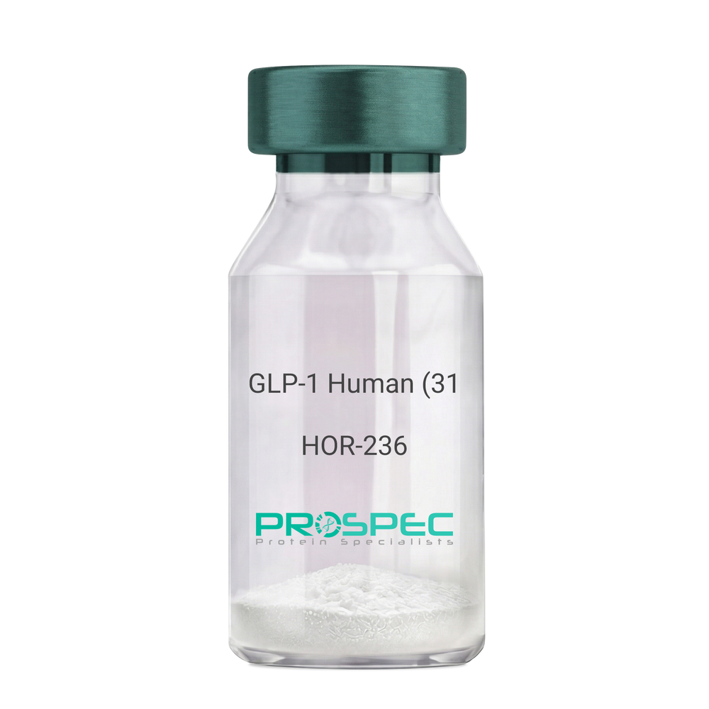
GLP-1 Human (31 a.a.)Description:
Glucagon Like Peptide-1 (31 a.a.) Human Recombinant
GLP1, GLP2, GRPP.
Product # :
HOR-236Price :
Quantity :
Shipping Method :
Shipped at Room temp
More Info
- description
- source
- formulation
- purity
- More Info
- Citations
Description
Glucagon Like Peptide-1 Human Recombinant produced in E.Coli is a single, non-glycosylated, polypeptide chain containing 31 amino acids and having a molecular mass of 3.3 kDa. The GLP-1 is purified by proprietary chromatographic techniques.
Source
Escherichia Coli.
Formulation
The Recombinant GLP-1 concentrated solution (1mg/ml) was lyophilized after extensive dialyses against 0.1mg sodium phosphate monobasic & 1.6mg sodium phosphate dibasic.
Purity
Greater than 98.0% as determined by:
(a) Analysis by RP-HPLC.
(b) Analysis by SDS-PAGE.Citations
More Info
-
Introduction
Glucagon-like peptide-1 (GLP-1) is derived from the transcription product of the proglucagon gene. The major source of GLP-1 in the body is the intestinal L cell that secretes GLP-1 as a guthormone. The biologically active forms of GLP-1 are: GLP-1-(7-37) and GLP-1-(7-36)NH2.
GLP-1 secretion by L cells is dependent on the presence of nutrients in the lumen of the small intestine. The secretagogues (agents that causes or stimulates secretion) of this hormone include major nutrients like carbohydrate, proteinand lipid. Once in the circulation, GLP-1 has a half life of less than 2 minutes, due to rapid degradation by the enzyme dipeptidyl peptidase-4.
GLP-1 possesses several physiological properties that make it a subject of intensive investigation as a potential treatment of diabetes mellitus. The known physiological functions of GLP-1 include: Increases insulin secretion from the pancreas in a glucose-dependent manner, decreases glucagon secretion from the pancreas, increases beta cells mass and insulin gene expression, inhibits acid secretion and gastric emptying in the stomach, decreases food intake by increasing satiety. -
Synonyms
GLP1, GLP2, GRPP.
-
Physical Appearance
Sterile Filtered White lyophilized (freeze-dried) powder.
-
Stability
Lyophilized Glucagon Like Peptide-1 although stable at room temperature for 3 weeks, should be stored desiccated below -18°C. Upon reconstitution GLP-1 should be stored at 4°C between 2-7 days and for future use below -18°C. For long term storage it is recommended to add a carrier protein (0.1% HSA or BSA).Please prevent freeze-thaw cycles.
-
Solubility
It is recommended to reconstitute the lyophilized Glucagon Like Peptide-1 in sterile 18MΩ-cm H2O not less than 100µg/ml, which can then be further diluted to other aqueous solutions.
-
Amino Acid Sequence
HAEGTFTSDV SSYLEGQAAK EFIAWLVKGR G.
ProSpec's products are furnished for LABORATORY RESEARCH USE ONLY. The product may not be used as drugs, agricultural or pesticidal products, food additives or household chemicals.
- View Data Sheet
Name :
AHSG HumanDescription:
Alpha-2-HS-Glycoprotein Human Recombinant
PRO2743, A2HS, AHS, FETUA, HSGA, Alpha-2-HS-glycoprotein, Alpha-2-Z-globulin, Ba-alpha-2-glycoprotein, Fetuin-A.
Product # :
PRO-1450Price :
Quantity :
Shipping Method :
Shipped with Ice Packs
More Info
- description
- source
- formulation
- purity
- More Info
- Citations
Description
AHSG Human Recombinant produced in E.Coli is a single, non-glycosylated polypeptide chain containing 372 amino acids (19-367 a.a) and having a molecular mass of 39.7kDa (Molecular size on SDS-PAGE will appear higher).AHSG is fused to a 23 amino acid His-tag at N-terminus & purified by proprietary chromatographic techniques.
Source
Escherichia Coli.
Formulation
AHSG protein solution (0.5mg/ml) containing 20mM Tris-HCl buffer (pH8.0), 10% glycerol 0.1M NaCl and 1mM DTT.
Purity
Greater than 90.0% as determined by SDS-PAGE.
Citations
More Info
-
Introduction
AHSG (fetuin-A) which is a glycoprotein present in the serum, is synthesized by hepatocytes. AHSG is one of the fetuin class of plasma binding proteins. It is involved in a number of functions, such as endocytosis, brain development and the formation of bone tissue. It is usually present in the cortical plate of the immature cerebral cortex and bone marrow hemopoietic matrix, hence it has been postulated that it participates in the development of the tissues. Yet, its exact significance is still vague. AHSG promotes endocytosis, hold opsonic properties and influences the mineral phase of the bone. Affinity for calcium and barium ions has been shown.
-
Synonyms
PRO2743, A2HS, AHS, FETUA, HSGA, Alpha-2-HS-glycoprotein, Alpha-2-Z-globulin, Ba-alpha-2-glycoprotein, Fetuin-A.
-
Physical Appearance
Sterile Filtered colorless solution.
-
Stability
Store at 4°C if entire vial will be used within 2-4 weeks. Store, frozen at -20°C for longer periods of time. For long term storage it is recommended to add a carrier protein (0.1% HSA or BSA).Avoid multiple freeze-thaw cycles.
-
Amino Acid Sequence
MGSSHHHHHH SSGLVPRGSH MGSAPHGPGL IYRQPNCDDP ETEEAALVAI DYINQNLPWG YKHTLNQIDE VKVWPQQPSG ELFEIEIDTL ETTCHVLDPT PVARCSVRQL KEHAVEGDCD FQLLKLDGKF SVVYAKCDSS PDSAEDVRKV CQDCPLLAPL NDTRVVHAAK AALAAFNAQN NGSNFQLEEI SRAQLVPLPP STYVEFTVSG TDCVAKEATE AAKCNLLAEK QYGFCKATLS EKLGGAEVAV TCTVFQTQPV TSQPQPEGAN EAVPTPVVDP DAPPSPPLGA PGLPPAGSPP DSHVLLAAPP GHQLHRAHYD LRHTFMGVVS LGSPSGEVSH PRKTRTVVQP SVGAAAGPVV PPCPGRIRHF KV.
ProSpec's products are furnished for LABORATORY RESEARCH USE ONLY. The product may not be used as drugs, agricultural or pesticidal products, food additives or household chemicals.
- View Data Sheet
Name :
ST6GAL1 Human, sf9Description:
ST6 Beta-Galactosamide Alpha-2,6-Sialyltranferase 1, sf9 Human Recombinant
ST6 Beta-Galactoside Alpha-2,6-Sialyltransferase 1, ST6 Beta-Galactosamide Alpha-2,6-Sialyltranferase 1, ST6Gal I, CMP-N-Acetylneuraminate-Beta-Galactosamide-Alpha-2,6-Sialyltransferase 1,B-Cell Antigen CD75, Alpha 2,6-ST 1, EC 2.4.99.1, ST6GalI, SIAT1, CMP-N-Acetylneuraminate Beta-Galactosamide Alpha-2,6-Sialyltransferase, Sialyltransferase 1 (Beta-Galactoside Alpha-2,6-Sialyltransferase) , ST6 N-Acetylgalactosaminide Alpha-2,6-Sialyltransferase 1, Sialyltransferase 1, ST6N.
Product # :
ENZ-950Price :
Quantity :
Shipping Method :
Shipped with Ice Packs
More Info
- description
- source
- formulation
- purity
- More Info
- Citations
Description
ST6GAL1 produced in Sf9 Baculovirus cells is a single, glycosylated polypeptide chain containing 389 amino acids (27-406 a.a.) and having a molecular mass of 44.6kDa. ST6GAL1 is expressed with a 6 amino acid His tag at C-Terminus and purified by proprietary chromatographic techniques.
Source
Sf9, Baculovirus cells.
Formulation
ST6GAL1 protein solution (0.5mg/ml) contains Phosphate Buffered Saline (pH 7.4) and 10% glycerol.
Purity
Greater than 90.0% as determined by SDS-PAGE.
Citations
More Info
-
Introduction
ST6GAL1 also known as ST6 Beta-Galactosamide Alpha-2,6-Sialyltranferase 1, is part of the glycosyltransferase family 29. ST6GAL1 is a type II membrane protein which catalyzes the transfer of sialic acid from CMP-sialic acid to galactose-containing substrates. Furthermore, ST6GAL1 is normally found in the Golgi however it can be proteolytically processed to a soluble form, ST6GAL1 is also involved in the generation of the cell-surface carbohydrate determinants as well differentiation antigens HB-6, CD75, and CD76.
-
Synonyms
ST6 Beta-Galactoside Alpha-2,6-Sialyltransferase 1, ST6 Beta-Galactosamide Alpha-2,6-Sialyltranferase 1, ST6Gal I, CMP-N-Acetylneuraminate-Beta-Galactosamide-Alpha-2,6-Sialyltransferase 1,B-Cell Antigen CD75, Alpha 2,6-ST 1, EC 2.4.99.1, ST6GalI, SIAT1, CMP-N-Acetylneuraminate Beta-Galactosamide Alpha-2,6-Sialyltransferase, Sialyltransferase 1 (Beta-Galactoside Alpha-2,6-Sialyltransferase) , ST6 N-Acetylgalactosaminide Alpha-2,6-Sialyltransferase 1, Sialyltransferase 1, ST6N.
-
Physical Appearance
Sterile Filtered colorless solution.
-
Stability
Store at 4°C if entire vial will be used within 2-4 weeks. Store, frozen at -20°C for longer periods of time. For long term storage it is recommended to add a carrier protein (0.1% HSA or BSA). Avoid multiple freeze-thaw cycles.
-
Amino Acid Sequence
ADPKEKKKGS YYDSFKLQTK EFQVLKSLGK LAMGSDSQSV SSSSTQDPHR GRQTLGSLRG LAKAKPEASF QVWNKDSSSK NLIPRLQKIW KNYLSMNKYK VSYKGPGPGI KFSAEALRCH LRDHVNVSMV EVTDFPFNTS EWEGYLPKES IRTKAGPWGR CAVVSSAGSL KSSQLGREID DHDAVLRFNG APTANFQQDV GTKTTIRLMN SQLVTTEKRF LKDSLYNEGI LIVWDPSVYH SDIPKWYQNP DYNFFNNYKT YRKLHPNQPF YILKPQMPWE LWDILQEISP EEIQPNPPSS GMLGIIIMMT LCDQVDIYEF LPSKRKTDVC YYYQKFFDSA CTMGAYHPLL YEKNLVKHLN QGTDEDIYLL GKATLPGFRT IHCHHHHHH.
ProSpec's products are furnished for LABORATORY RESEARCH USE ONLY. The product may not be used as drugs, agricultural or pesticidal products, food additives or household chemicals.
- View Data Sheet
Name :
AFP ProteinDescription:
Alpha Fetoprotein Human Recombinant
Alpha-Fetoprotein, Alpha-1-Fetoprotein, Alpha-Fetoglobulin, HPAFP, AFPD, FETA, HP, Alpha-fetoprotein.
Product # :
PRO-2229Price :
Quantity :
Shipping Method :
Shipped with Ice Packs
More Info
- description
- source
- formulation
- purity
- More Info
- Citations
Description
AFP produced in Sf9 Baculovirus cells is a single, glycosylated polypeptide chain containing 600 amino acids (19-609a.a.) and having a molecular mass of 67.5kDa (Molecular size on SDS-PAGE will appear at approximately 57-70kDa). AFP is expressed with a ADP at N-terminus and 6 amino acid His tag at C-Terminus and purified by proprietary chromatographic techniques.
Source
Sf9, Baculovirus cells.
Formulation
AFP protein solution (0.5mg/ml) contains Phosphate Buffered Saline (pH 7.4) and 10% glycerol.
Purity
Greater than 95.0% as determined by SDS-PAGE.
Citations
More Info
-
Introduction
AFP is normally synthesized in the liver, intestinal tract, and yolk sac of the fetus. Antibody to AFP has been shown to be useful in detecting hepatocellular carcinomas (HCC) and germ cell neoplasms, especially yolk sac tumors.
-
Synonyms
Alpha-Fetoprotein, Alpha-1-Fetoprotein, Alpha-Fetoglobulin, HPAFP, AFPD, FETA, HP, Alpha-fetoprotein.
-
Physical Appearance
Sterile Filtered colorless solution.
-
Stability
Store at 4°C if entire vial will be used within 2-4 weeks. Store, frozen at -20°C for longer periods of time. For long term storage it is recommended to add a carrier protein (0.1% HSA or BSA).Avoid multiple freeze-thaw cycles.
-
Amino Acid Sequence
ADPRTLHRNEYGI ASILDSYQCT AEISLADLAT IFFAQFVQEA TYKEVSKMVK DALTAIEKPT GDEQSSGCLE NQLPAFLEEL CHEKEILEKY GHSDCCSQSE EGRHNCFLAH KKPTPASIPL FQVPEPVTSC EAYEEDRETF MNKFIYEIAR RHPFLYAPTI LLWAARYDKI IPSCCKAENA VECFQTKAAT VTKELRESSL LNQHACAVMK NFGTRTFQAI TVTKLSQKFT KVNFTEIQKL VLDVAHVHEH CCRGDVLDCL QDGEKIMSYI CSQQDTLSNK ITECCKLTTL ERGQCIIHAE NDEKPEGLSP NLNRFLGDRD FNQFSSGEKN IFLASFVHEY SRRHPQLAVS VILRVAKGYQ ELLEKCFQTE NPLECQDKGE EELQKYIQES QALAKRSCGL FQKLGEYYLQ NAFLVAYTKK APQLTSSELM AITRKMAATA ATCCQLSEDK LLACGEGAAD IIIGHLCIRH EMTPVNPGVG QCCTSSYANR RPCFSSLVVD ETYVPPAFSD DKFIFHKDLC QAQGVALQTM KQEFLINLVK QKPQITEEQL EAVIADFSGL LEKCCQGQEQ EVCFAEEGQK LISKTRAALG VHHHHHH.
ProSpec's products are furnished for LABORATORY RESEARCH USE ONLY. The product may not be used as drugs, agricultural or pesticidal products, food additives or household chemicals.
- View Data Sheet
Name :
F8 ProteinDescription:
Coagulation Factor-VIII Human Recombinant
Coagulation factor VIII, Procoagulant component, Antihemophilic factor, AHF, F8, F8C, F8B, HEMA, FVIII, DXS1253E, F8 protein.
Product # :
PRO-318Price :
Quantity :
Shipping Method :
Shipped at Room temp
More Info
- description
- source
- formulation
- purity
- biological activity
- More Info
- Citations
Description
Antihemophilic Facor Human Recombinant produced in CHO is a glycosylated polypeptide chain having 2332 amino acids. The Factor-VIII is purified by proprietary chromatographic techniques.
Source
CHO cells (Chinese Hamster Ovarian Cells).
Formulation
Each 250IU vial was lyophilized from a solution containing 8mg Tween-80, 112mM NaCl, 40mg Mannitol, 10mg Trehalose, 1ng VWF and 4.2mM CaCl2.
Purity
Greater than 97.0% as determined by SDS-PAGE.
Biological Activity
The specific activity was found to be 7,058 IU/mg.Citations
More Info
-
Introduction
Coagulation factor VIII participates in the intrinsic pathway of blood coagulation; factor VIII is a cofactor for factor IXa which, in the presence of Ca+2 and phospholipids, converts factor X to the activated form Xa. This gene produces two alternatively spliced transcripts. Transcript variant 1 encodes a large glycoprotein, isoform a, which circulates in plasma and associates with von Willebrand factor in a noncovalent complex. This protein undergoes multiple cleavage events. Transcript variant 2 encodes a putative small protein, isoform b, which consists primarily of the phospholipid binding domain of factor VIIIc. This binding domain is essential for coagulant activity. Defects in this gene results in hemophilia A, a common recessive X-linked coagulation disorder.
-
Synonyms
Coagulation factor VIII, Procoagulant component, Antihemophilic factor, AHF, F8, F8C, F8B, HEMA, FVIII, DXS1253E, F8 protein.
-
Physical Appearance
Sterile Filtered White lyophilized (freeze-dried) powder.
-
Stability
Lyophilized Factor-VIII although stable at room temperature for 3 weeks, should be stored desiccated below -18°C. Upon reconstitution Factor-VIII should be stored at 4°C between 2-7 days and for future use below -18°C.Please prevent freeze-thaw cycles.
-
Solubility
It is recommended to reconstitute 250IU lyophilized Factor-VIII in 5ml sterile 18M-cm H2O, which can then be further diluted to other aqueous solutions.
ProSpec's products are furnished for LABORATORY RESEARCH USE ONLY. The product may not be used as drugs, agricultural or pesticidal products, food additives or household chemicals.
- View Data Sheet
Name :
SIRPA HumanDescription:
Signal-Regulatory Protein Alpha Human Recombinant
Signal-Regulatory Protein Alpha, SHPS1, CD172 Antigen-Like Family Member A, Inhibitory Receptor SHPS-1, Macrophage Fusion Receptor, PTPNS1, SIRP, P84, BIT, MFR, Brain-Immunoglobulin-Like Molecule With Tyrosine-Based Activation Motifs, Brain Ig-Like Molecule With Tyrosine-Based Activation Motifs, Protein Tyrosine Phosphatase, Non-Receptor Type Substrate 1, Tyrosine-Protein Phosphatase Non-Receptor Type Substrate 1, Tyrosine Phosphatase SHP Substrate 1, Signal-Regulatory Protein Alpha-1 , ignal-Regulatory Protein Alpha-2, Signal-Regulatory Protein Alpha-3, SHP Substrate 1, CD172a Antigen, Myd-1 Antigen, MyD-1 Antigen, Sirp-Alpha-1, Sirp-Alpha-2, Sirp-Alpha-3, CD172A, SHPS-1, MYD-1, MYD1, Tyrosine-protein phosphatase non-receptor type substrate 1.
Product # :
PRO-2080Price :
Quantity :
Shipping Method :
Shipped with Ice Packs
More Info
- description
- source
- formulation
- purity
- More Info
- Citations
Description
SIRPA Human Recombinant produced in E.Coli is a single, non-glycosylated polypeptide chain containing 370 amino acids (27-373 a.a) and having a molecular mass of 40.4kDa. SIRPA is fused to a 23 amino acid His-tag at N-terminus & purified by proprietary chromatographic techniques.
Source
Escherichia Coli.
Formulation
SIRPA protein solution (0.5mg/ml) containing PBS buffer (pH 7.4), 10% glycerol and 1mM DTT.
Purity
Greater than 90.0% as determined by SDS-PAGE.
Citations
More Info
-
Introduction
Signal-Regulatory Protein Alpha, SIRPA belongs to the signal-regulatory-protein (SIRP) family, as well as theimmunoglobulin super family. The members of the SIRP family are receptor-type transmembrane glycoproteins which areinvolved in the negative regulation of receptor tyrosine kinase-coupled signaling processes. SIRPA can bephosphorylated by tyrosine kinases. The phospho-tyrosine residues of this PTP have been shown to recruit SH2domain containing tyrosine phosphatases (PTP), and perform as substrates of PTPs. SIRPA take part insignal transduction mediated by a variety of growth factor receptors. CD47 has been shown to be a ligand for SIRPA.
-
Synonyms
Signal-Regulatory Protein Alpha, SHPS1, CD172 Antigen-Like Family Member A, Inhibitory Receptor SHPS-1, Macrophage Fusion Receptor, PTPNS1, SIRP, P84, BIT, MFR, Brain-Immunoglobulin-Like Molecule With Tyrosine-Based Activation Motifs, Brain Ig-Like Molecule With Tyrosine-Based Activation Motifs, Protein Tyrosine Phosphatase, Non-Receptor Type Substrate 1, Tyrosine-Protein Phosphatase Non-Receptor Type Substrate 1, Tyrosine Phosphatase SHP Substrate 1, Signal-Regulatory Protein Alpha-1 , ignal-Regulatory Protein Alpha-2, Signal-Regulatory Protein Alpha-3, SHP Substrate 1, CD172a Antigen, Myd-1 Antigen, MyD-1 Antigen, Sirp-Alpha-1, Sirp-Alpha-2, Sirp-Alpha-3, CD172A, SHPS-1, MYD-1, MYD1, Tyrosine-protein phosphatase non-receptor type substrate 1.
-
Physical Appearance
Sterile Filtered colorless solution.
-
Stability
Store at 4°C if entire vial will be used within 2-4 weeks. Store, frozen at -20°C for longer periods of time. For long term storage it is recommended to add a carrier protein (0.1% HSA or BSA).Avoid multiple freeze-thaw cycles.
-
Amino Acid Sequence
MGSSHHHHHH SSGLVPRGSH MGSGVAGEEE LQVIQPDKSV LVAAGETATL RCTATSLIPV GPIQWFRGAG PGRELIYNQK EGHFPRVTTV SDLTKRNNMD FSIRIGNITP ADAGTYYCVK FRKGSPDDVE FKSGAGTELS VRAKPSAPVV SGPAARATPQ HTVSFTCESH GFSPRDITLK WFKNGNELSD FQTNVDPVGE SVSYSIHSTA KVVLTREDVH SQVICEVAHV TLQGDPLRGT ANLSETIRVP PTLEVTQQPV RAENQVNVTC QVRKFYPQRL QLTWLENGNV SRTETASTVT ENKDGTYNWM SWLLVNVSAH RDDVKLTCQV EHDGQPAVSK SHDLKVSAHP KEQGSNTAAE NTGSNERNIY.
ProSpec's products are furnished for LABORATORY RESEARCH USE ONLY. The product may not be used as drugs, agricultural or pesticidal products, food additives or household chemicals.
- View Data Sheet
Name :
GROEL (27-573) HumanDescription:
GroEL (HSP60) (27-573 a.a.) Human Recombinant
CPN60, GROEL, HLD4, HSP-60, HSP60, HSP65, HuCHA60, SPG13, Chaperonin 60, 60 kDa chaperonin, P60 lymphocyte protein, 60 kDa heat shock protein, mitochondrial.
Product # :
HSP-055Price :
Quantity :
Shipping Method :
Shipped with Ice Packs
More Info
- description
- source
- formulation
- purity
- More Info
- Citations
Description
GROEL Human Recombinant produced in E.Coli is a single, non-glycosylated polypeptide chain containing 572 amino acids (27-573 a.a.) and having a molecular mass of 60kDa.GROEL is fused to a 25 amino acid His-tag at N-terminus & purified by proprietary chromatographic techniques.
Source
Escherichia Coli.
Formulation
GROEL protein solution (0.5mg/ml) contains 20mM Tris-HCl buffer, (pH 8.0) 0.1M NaCl and 10% glycerol.
Purity
Greater than 90.0% as determined by SDS-PAGE.
Citations
More Info
-
Introduction
GroEL, HSP60 is a chaperonin located in the mitochondria which is responsible for the transportation & refolding of proteins from the cytoplasm directly into the mitochondrial matrix. GroEL is regulated by the HSP10 cochaperonin, which is a single heptameric protein ring having a molecular mass of 10 kDa which form a unique complex with HSP60. HSP10, GroES coordinates the ATPase activity of the HSP60 subunits in order to allow the release of bound polypeptide in a manner that is productive for its correct folding.
-
Synonyms
CPN60, GROEL, HLD4, HSP-60, HSP60, HSP65, HuCHA60, SPG13, Chaperonin 60, 60 kDa chaperonin, P60 lymphocyte protein, 60 kDa heat shock protein, mitochondrial.
-
Physical Appearance
Sterile filtered colorless solution.
-
Stability
Store at 4°C if entire vial will be used within 2-4 weeks.Store, frozen at -20°C for longer periods of time. For long term storage it is recommended to add a carrier protein (0.1% HSA or BSA). Avoid multiple freeze-thaw cycles.
-
Amino Acid Sequence
MGSSHHHHHH SSGLVPRGSH MGSHMAKDVK FGADARALML QGVDLLADAV AVTMGPKGRT VIIEQSWGSP KVTKDGVTVA KSIDLKDKYK NIGAKLVQDV ANNTNEEAGD GTTTATVLAR SIAKEGFEKI SKGANPVEIR RGVMLAVDAV IAELKKQSKP VTTPEEIAQV ATISANGDKE IGNIISDAMK KVGRKGVITV KDGKTLNDEL EIIEGMKFDR GYISPYFINT SKGQKCEFQD AYVLLSEKKI SSIQSIVPAL EIANAHRKPL VIIAEDVDGE ALSTLVLNRL KVGLQVVAVK APGFGDNRKN QLKDMAIATG GAVFGEEGLT LNLEDVQPHD LGKVGEVIVT KDDAMLLKGK GDKAQIEKRI QEIIEQLDVT TSEYEKEKLN ERLAKLSDGV AVLKVGGTSD VEVNEKKDRV TDALNATRAA VEEGIVLGGG CALLRCIPAL DSLTPANEDQ KIGIEIIKRT LKIPAMTIAK NAGVEGSLIV EKIMQSSSEV GYDAMAGDFV NMVEKGIIDP TKVVRTALLD AAGVASLLTT AEVVVTEIPK EEKDPGMGAM GGMGGGMGGG MF.
ProSpec's products are furnished for LABORATORY RESEARCH USE ONLY. The product may not be used as drugs, agricultural or pesticidal products, food additives or household chemicals.
- View Data Sheet
Name :
AMBP HumanDescription:
Microglobulin Alpha-1 Protein Human
Alpha-1 Microglobulin, A1M.
Product # :
PRO-407Price :
Quantity :
Shipping Method :
Shipped at Room temp
More Info
- description
- source
- formulation
- purity
- More Info
- Citations
Description
Alpha 1-microglobulin (A1M) is an immunomodulatory protein with a broad spectrum of possible clinical applications and seems a promising marker for evaluation of tubular function.
Source
Purified from the urine of patients with chronic renal tubular proteinuria.
Formulation
Lyophilized from 0.02M NH4HCO3. May contain traces of buffer salts.
Purity
Greater than 96.0%.
Citations
More Info
-
Introduction
Alpha 1-microglobulin (A1M) is a lipocalin superfamily member (kernal lipocalins). A1M is a low molecular weight protein component of plasma. A1M is distributed in plasma and extravascular compartments of all organs. Alpha-1 Microglobulin is found in mammals, birds, amphibians and fish. The primary sites of A1M synthesis are the liver and the kidney. Around the opening of the lipocalin pocket three lysyl residues are situated; those residues carry yellow-brown modification derived from the binding and degradation of heme and kynurenin (a tryptophan metabolite). A1-Microglobulin’s reductase and dehydrogenase have broad biological substrate specificity properties due to its’ free cysteine side-chain which is located in a flexible loop. Alpha-1-microglobulin is glycosylated by three separate carbohydrate chains: two complex carbohydrates which are N-linked to asparagines at residues 17 and 96, and the other simple carbohydrate which is O-linked to threonine at position 5. The carbohydrates comprise 22% of the total molecular mass of the protein. The glycosylation varies between species. A1M exists in two forms- a free form and complexed to other macromolecules: in humans- complexed to immunoglobulin A (IgA), in rat- complexed to alpha-1-inhibitor-3. Free A1M is exceptionally heterogeneous in charge (therefore also known as protein HC), and is found tightly linked to a chromophore. The free Alpha-1-microglobulin is a monomeric protein composed of one 188 residue polypeptide and contains three cysteines, two of which (residues 75 and 173) form a conserved intra-molecular disulphide link. The chromophoric group is covalently bound to the free cysteine residue at position 34. A1M binds retinol as a major ligand, but this is probably distinct from its covalent chromophore. Half of all human plasma A1M (approximately 0.03mg/ml) forms a 1:1 complex with about 5% of plasma immunoglobulin A. The resulting macromolecular complexes’ molecular weight is 200000, and a plasma concentration of 0.3mg/ml. The complex can exhibit both antibody activity and affect many of the biological actions of free Alpha-1-microglobulin. Alpha-1-microglobulin was first discovered in pathological human urine. It was suggested that A1M might be involved in tissue defense against reactive oxygen species, oxidation by heme and kynurenin. Evidence also suggests that A1M functions in the regulation of the immune system. Other functions include: inhibition of stimulation of cultured lymphocytes by protein antigens; induction of cell division of lymphocytes, a mitogenic effect that can either be enhanced or inhibited by the action of other plasma components; inhibition of neutrophil granulocyte migration in vitro; and inhibition of chemotaxis.
Other functions include inhibition of stimulation of cultured lymphocytes by protein antigens; induction of cell division of lymphocytes, a mitogenic effect that can either be enhanced or inhibited by the action of other plasma components; inhibition of neutrophil granulocyte migration in vitro; and inhibition of chemotaxis. -
Synonyms
Alpha-1 Microglobulin, A1M.
-
Physical Appearance
Sterile Filtered Off-White lyophilized (freeze-dried) powder.
-
Stability
Human A1M although stable at room temperature for 3 weeks, should be stored between 2-8°C.
-
Solubility
Use phosphate buffer, pH>7.0 containing 0.15M NaCl, is recommended.
-
Human Virus Test
Starting material tested and certified negative for HIV I & II antibodies, Hepatitis B surface antigen, and Hepatitis C antibodies.
ProSpec's products are furnished for LABORATORY RESEARCH USE ONLY. The product may not be used as drugs, agricultural or pesticidal products, food additives or household chemicals.
- View Data Sheet
Name :
COL4A3 HumanDescription:
Collagen Type IV Alpha 3 Human Recombinant
Collagen alpha-3(IV) chain, Goodpasture antigen, COL4A3, Glomerular Basal Membrane, GBM.
Product # :
PRO-387Price :
Quantity :
Shipping Method :
Shipped with Ice Packs
More Info
- description
- source
- formulation
- purity
- More Info
- Citations
Description
Human α3 chain of collagen IV; identical with the antigen called "glomerular basal membrane antigen" (GBM). Recombinant antigen for solid (ELISA) and fluid phase diagnostic assays. Calculated Molecular weight: 53kDa. Calculated isoelectric point: pH 8.9. cDNA coding for a minicollagen version of the human collagen IV α3 chain fused to a hexa-histidine purification tag. The term minicollagen designates the removal of most of the epitope-less triplehelical collagenous region (situated between the N-terminal 7S domain and the C-terminal noncollagenous NC1 domain), which is a requirement for recombinant production of this antigen.
Source
Sf9 insect cells.
Formulation
The protein solution contains 20mM Hepes, pH 8.0 and 4M Urea.
Purity
Greater than 95.0% as determined by SDS-PAGE.
Citations
More Info
-
Introduction
Type IV collagen is a major structural component of basement membranes. It is a multimeric protein composed of 3 alpha subunits, which are encoded by 6 different genes, alpha 1 through alpha 6. Each of these alpha subunits can form a triple helix structure with 2 other subunits to form type IV collagen. The Goodpasture syndrome is a condition in which autoantibodies bind to the collagen molecules in the basement membranes of alveoli and glomeruli. The epitopes that elicit these autoantibodies are restricted basically to the non-collagenous C-terminal domain of the protein. There are numerous alternative transcripts that appear to be unique to the human COL4A3 and alternative splicing is limited to the six exons that encode this C-terminal domain. COL4A3 is also linked to an autosomal recessive form of Alport syndrome. The mutations contributing to the Alport syndrome are also situated within the exons that encode this C-terminal region. COL4A3 is organized in a head-to-head conformation with another type IV collagen gene so that each gene pair shares a common promoter. Several exons of COL4A3 are interspersed with exons of an uncharacterized gene which is on the opposite strand.
-
Synonyms
Collagen alpha-3(IV) chain, Goodpasture antigen, COL4A3, Glomerular Basal Membrane, GBM.
-
Physical Appearance
Sterile Filtered clear solution.
-
Stability
Recommendations for storage buffer: neutral to slightly alkaline pH and 4M urea as dissociating agent. Storage temperature: -70°C to -80°C. Please prevent freeze-thaw cycles.
-
Immunological Functions
1. Binds IgG-type human auto-antibodies. 2. Standard ELISA test (checkerboard analysis of positive/negative samples. 3. Immunodot test with positive/negative samples.
-
coating concentration
0.12-0.5 μg/ml (depending on the type of ELISA plate and coating buffer). Suitable for labeling of functional groups.
-
Note
It has been reported that the immunodominant epitope of GBM is a cryptic epitope that is not easily accessible to the corresponding autoantibodies. It is necessary to treat the protein under nonreducing conditions with a denaturant such as urea to unmask the epitopes (see Hellmark et al. in Autoantibodies, Peter, J.B. and Shoenfeld, Y., eds., Elsevier B.V., 1996, pp 291-298).
ProSpec's products are furnished for LABORATORY RESEARCH USE ONLY. The product may not be used as drugs, agricultural or pesticidal products, food additives or household chemicals.
- View Data Sheet
Name :
DsbG E.ColiDescription:
Thiol Disulfide Interchange Protein E.Coli Recombinant DsbG
Thiol:disulfide interchange protein dsbG, dsbG, ybdP, b0604, JW0597.
Product # :
HSP-024Price :
Quantity :
Shipping Method :
Shipped with Ice Packs
More Info
- description
- source
- formulation
- purity
- More Info
- Citations
Description
Recombinant DsbG produced in E.Coli is a single, non-glycosylated polypeptide chain containing 232 amino acids and having a molecular mass of 25.8 kDa. DsbG is purified by conventional chromatography techniques.
Source
Escherichia Coli.
Formulation
The DsbG protein solution contains 20mM Tris-HCl, pH-8, 2mM EDTA and 10% Glycerol.
Purity
Greater than 95.0% as determined by SDS-PAGE.
Citations
More Info
-
Introduction
Dsb proteins are in charge for the formation and rearrangement of disulfide bonds during the folding of secreted and membrane proteins in bacteria. DsbG has disulfide bond isomerase and chaperone activity. DsbG interacts with refolding intermediates of chemically denatured citrate synthase and prevents their aggregation in vitro. DsbG shares sequnce homology with DsbC. DsbG forms a stable periplasmic dimer and displays an equilibrium constant with glutathione comparable with DsbA and DsbC. DsbG is expressed at roughly 25% level of DsbC.
-
Synonyms
Thiol:disulfide interchange protein dsbG, dsbG, ybdP, b0604, JW0597.
-
Physical Appearance
Sterile filtered colorless solution.
-
Stability
Store at 4°C if entire vial will be used within 2-4 weeks. Store, frozen at -20°C for longer periods of time. For long term storage it is recommended to add a carrier protein (0.1% HSA or BSA).Avoid multiple freeze-thaw cycles.
-
Amino Acid Sequence
MEELPAPVKA IEKQGITIIK TFDAPGGMKG YLGKYQDMGV TIYLTPDGKH AISGYMYNEK GENLSNTLIE KEIYAPAGRE MWQRMEQSHW LLDGKKDAPV IVYVFADPFC PYCKQFWQQA RPWVDSGKVQ LRTLLVGVIK PESPATAAAI LASKDPAKTW QQYEASGGKL KLNVPANVST EQMKVLSDNE KLMDDLGANV TPAIYYMSKE NTLQQAVGLP DQKTLNIIMG NK.
ProSpec's products are furnished for LABORATORY RESEARCH USE ONLY. The product may not be used as drugs, agricultural or pesticidal products, food additives or household chemicals.
- View Data Sheet
Name :
GMNN AntibodyDescription:
Geminin, Mouse Anti Human
GMNN, Geminin, DNA Replication Inhibitor, Gem, RP3-369A17.3.
Product # :
ANT-319Price :
Quantity :
Shipping Method :
Shipped with Ice Packs
More Info
- formulation
- More Info
- Citations
Formulation
1mg/ml containing PBS, pH-7.4, & 0.1% Sodium Azide.
Citations
More Info
-
Introduction
Geminin is a 25 kDa nuclear protein, which inhibits DNA replication and is degraded during the mitotic phase of the cell cycle. Geminin controls replication by binding to the licensing factor Cdt1, and is involved in neural differentiation. In addition, Geminin directly interacts with Six3 and Hox homeodomain proteins during embryogenesis and inhibits their functions. Geminin can also promote DNA replication. Geminin has 2 roles in 2 different stages of the cell cycle: Geminin is a negative regulator of DNA replication during the “S phase” of the cell cycle. Inhibition of Geminin during the “S phase” (by RNAi) results in an additional round of replication of portions of the genome. During the “M phase” of the cell cycle (mitosis) Geminin stabilizes the replication factor Cdt1 promoting DNA replication during the next cell cycle. Moreover, inhibition of Geminin during mitosis (by RNAi) causes destabilization of Cdt1 protein and impairment of DNA replication during the next cell cycle. G
-
Synonyms
GMNN, Geminin, DNA Replication Inhibitor, Gem, RP3-369A17.3.
-
Immunogen
Anti-human Geminin mAb is derived from hybridization of mouse SP2/0 myeloma cells with spleen cells from BALB/c mice immunized with recombinant human Geminin amino acids 1-209 purified from E. coli.
-
Ig Subclass
Mouse IgG1 heavy chain and κ light chain.
-
Clone
P2H7AT.
-
Applications
Geminin antibody has been tested by ELISA and Western blot analysis to assure specificity and reactivity. Since application varies, however, each investigation should be titrated by the reagent to obtain optimal results. Recommended dilution range for Western blot analysis is 1:500 ~ 1,000. Recommended starting dilution is 1:500.
-
Type
Mouse Anti Human Monoclonal.
-
Storage Procedures
For periods up to 1 month store at 4°C, for longer periods of time, store at -20°C. Prevent freeze thaw cycles.
-
Purification Method
Geminin antibody was purified from mouse ascitic fluids by protein-G affinity chromatography.
ProSpec's products are furnished for LABORATORY RESEARCH USE ONLY. The product may not be used as drugs, agricultural or pesticidal products, food additives or household chemicals.
- View Data Sheet
Name :
SERPINA1Description:
Alpha 1 Antitrypsin Human
Alpha-1-antitrypsin, Alpha-1 protease inhibitor, Alpha-1-antiproteinase, SERPINA1, A1AT, PI, A1A, AAT, PI1, MGC9222, PRO2275, MGC23330.
Product # :
PRO-456Price :
Quantity :
Shipping Method :
Shipped at Room temp
More Info
- description
- formulation
- purity
- More Info
- Citations
Description
SERPINA1 extracted from human serum and having a 54kDa molecular weight is purified by proprietary chromatographic techniques.
Formulation
SERPINA1 is 0.02M NH4HCO3.
Purity
Greater than 96% as determined by SDS-PAGE.
Citations
More Info
-
Introduction
SERPINA1 is secreted and is a serine protease inhibitor which its targets include elastase, plasmin, collagenase, thrombin, leucocytic proteases, trypsin, chymotrypsin, and plasminogen activator. Defects in SERPINA1 gene can cause emphysema or liver disease. Antral SERPINA1 expression is particularly induced by H. pylori infection. lung and prostate cancers have shown a significant increase in SERPINA1 serum levels compared with healthy controls though breast cancers did not show a significant change. SERPINA1 is an endogenous inhibitor of serine proteases and inhibits the catalytic domain of human recombinant matriptase in vitro. Rise in SERPINA1 occurs as an acute phase response to tissue necrosis and inflammation. mutations in SERPINA1 and SLC11A1 genes change the balance between elastase produced by leukocytes during phagocytosis.
-
Synonyms
Alpha-1-antitrypsin, Alpha-1 protease inhibitor, Alpha-1-antiproteinase, SERPINA1, A1AT, PI, A1A, AAT, PI1, MGC9222, PRO2275, MGC23330.
-
Physical Appearance
Sterile Filtered White lyophilized (freeze-dried) powder.
-
Stability
Lyophilized SERPINA1 should be stored at 4°C.
-
Solubility
It is recommended to reconstitute the lyophilized SERPINA1 in 0.15M NaCl, which
can then be further diluted to other aqueous solutions.
ProSpec's products are furnished for LABORATORY RESEARCH USE ONLY. The product may not be used as drugs, agricultural or pesticidal products, food additives or household chemicals.
- View Data Sheet
Name :
BP-1 HumanDescription:
BP-1 Human Recombinant
Homeobox protein DLX-4, DLX-7, DLX-8, Beta protein 1, BP1, DLX7, DLX8, DLX9, DLX4.
Product # :
PRO-463Price :
Quantity :
Shipping Method :
Shipped with Ice Packs
More Info
- description
- source
- formulation
- More Info
- Citations
Description
BP-1 Human Recombinant (aa 103-168) expressed in E.coli, shows a 35 kDa band on SDS-PAGE.The BP-1 is purified by proprietary chromatographic techniques.
Source
Escherichia Coli.
Formulation
BP-1 protein in 50mM Tris-Acetate, pH7.5, 1mM EDTA and 20% Glycerol.
Citations
More Info
-
Introduction
BP-1 gene, a member of the DLX homeobox gene family, is overexpressed in 80% of breast cancer patients, and thus represents a potential new target for diagnosis and treatment.
Members of the Dlx gene family contain a homeobox that is related to that of Distal-less (Dll), a gene expressed in the head and limbs of the developing fruit fly (Drosophila). The Distal-less (Dlx) family of genes comprises at least 6 different members, DLX1-DLX6. The DLX proteins are postulated to play a role in forebrain and craniofacial development. Three transcript variants have been described for this gene, however, the full length nature of one variant has not been described. Studies of the two splice variants revealed that one encoded isoform functions as a repressor of the beta-globin gene while the other isoform lacks that function. -
Synonyms
Homeobox protein DLX-4, DLX-7, DLX-8, Beta protein 1, BP1, DLX7, DLX8, DLX9, DLX4.
-
Physical Appearance
Sterile Filtered clear solution.
-
Stability
Store vial at -20°C to -80°C. When stored at the recommended temperature, this protein is stable for 12 months.Please prevent freeze-thaw cycles.
ProSpec's products are furnished for LABORATORY RESEARCH USE ONLY. The product may not be used as drugs, agricultural or pesticidal products, food additives or household chemicals.
- View Data Sheet
Name :
GDF15 HumanDescription:
Growth and Differentiation Factor 15 Human Recombinant
GDF-15, MIC1, MIC-1, NAG-1, PDF, PLAB, PTGFB, Growth/differentiation factor 15, Placental bone morphogenetic protein, Placental TGF-beta, Macrophage inhibitory cytokine 1, Prostate differentiation factor, NSAID-activated gene 1 protein, NSAID-regulated gene 1 protein, NRG-1, GDF15.
Product # :
CYT-335Price :
Quantity :
Shipping Method :
Shipped at Room temp
More Info
- description
- source
- formulation
- purity
- More Info
- Citations
Description
GDF15 Human Recombinant produced in E.Coli is a homodimeric, non-glycosylated, Polypeptide chain containing 2x113 amino acids and having a molecular mass of 24.8 kDa. The GDF15 is purified by proprietary chromatographic techniques.
Source
Escherichia Coli.
Formulation
GDF15 is lyophilized from a sterile filtered solution containing 0.1% Trifluoroacetic Acid (TFA).
Purity
Greater than 95.0% as determined by SDS-PAGE.
Citations
More Info
-
Introduction
GDF15 is part of the TGF-Beta superfamily that is involved in regulating inflammatory and apoptotic pathways in injured tissues and throughout disease processes. GDF15 is most abundant in the liver. Its expression in liver can be considerably up-regulated in during injury of organs such as liver, kidney, heart and lung. GDF-15 promotes proliferation or growth arrest and differentiation due to differences in cellular differentiation. GDF15 prevents apoptosis in cerebellar granule neurons by activating Akt and inhibiting endogenously active ERK. GDF15 is a novel autocrine/endocrine factor that antagonizes the hypertrophic response and loss of ventricular performance.
-
Synonyms
GDF-15, MIC1, MIC-1, NAG-1, PDF, PLAB, PTGFB, Growth/differentiation factor 15, Placental bone morphogenetic protein, Placental TGF-beta, Macrophage inhibitory cytokine 1, Prostate differentiation factor, NSAID-activated gene 1 protein, NSAID-regulated gene 1 protein, NRG-1, GDF15.
-
Physical Appearance
Sterile Filtered White lyophilized (freeze-dried) powder.
-
Stability
Lyophilized GDF15 although stable at room temperature for 3 weeks, should be stored desiccated below -18°C. Upon reconstitution GDF15 should be stored at 4°C between 2-7 days and for future use below -18°C.For long term storage it is recommended to add a carrier protein (0.1% HSA or BSA).Please prevent freeze-thaw cycles.
-
Solubility
It is recommended to reconstitute the lyophilized GDF15 in sterile 18M-cm H2O at a concentration of 100µg/ml, which can then be further diluted to other aqueous solutions.
-
Amino Acid Sequence
MARNGDHCPL GPGRCCRLHT VRASLEDLGW ADWVLSPREV QVTMCIGACP SQFRAANMHA QIKTSLHRLK PDTVPAPCCV PASYNPMVLI QKTDTGVSLQ TYDDLLAKDC HCI.
-
Background
What is the molecular weight/Mw of GDF15 HUMAN Protein?
GDF15 HUMAN Protein has a total Mw of 24.8kDa.
What is the source or expression system of GDF15 HUMAN Protein?
Escherichia Coli.
What is the Purity of GDF15 HUMAN Protein?
GDF15 HUMAN Protein is >95% pure as determined by SDS-PAGE.
What is the Biological Activity of GDF15 HUMAN Protein?
The biological functionality of GDF15 HUMAN Protein will be determined in the future.
What is the amino acid sequence of GDF15 HUMAN Protein?
MARNGDHCPL GPGRCCRLHT VRASLEDLGW ADWVLSPREV QVTMCIGACP SQFRAANMHA QIKTSLHRLK PDTVPAPCCV PASYNPMVLI QKTDTGVSLQ TYDDLLAKDC HCI.
What applications can GDF15 HUMAN Protein be used in?
GDF15 HUMAN Protein can probably be used in western blot, ELISA and Lateral Flow.
What is the endotoxin level for GDF15 HUMAN Protein?
The endotoxin level is minimal, GDF15 HUMAN Protein was purified using conventional chromatography techniques.
ProSpec's products are furnished for LABORATORY RESEARCH USE ONLY. The product may not be used as drugs, agricultural or pesticidal products, food additives or household chemicals.