Search results
1000 results found for “PPARG”
Name
Description
Product #
Price
Quantity
Shipping Method
- View Data Sheet
Name :
MAPRE2 HumanDescription:
Microtubule-Associated Protein, RP/EB Family, Member 2 Human Recombinant
Microtubule-associated protein RP/EB family member 2, APC-binding protein EB2, End-binding protein 2, EB2, MAPRE2, RP1, EB1.
Product # :
PRO-932Price :
Quantity :
Shipping Method :
Shipped with Ice Packs
More Info
- description
- source
- formulation
- purity
- More Info
Description
MAPRE2 Human Recombinant produced in E.Coli is a single, non-glycosylated polypeptide chain containing 347 amino acids (1-327 a.a.) and having a molecular mass of 39.2kDa.MAPRE2 is fused to a 20 amino acid His-tag at N-terminus & purified by proprietary chromatographic techniques.
Source
Escherichia Coli.
Formulation
MAPRE2 protein solution (1mg/ml) containing 20mM Tris-HCl buffer (pH8.0), 20% glycerol, 0.1M NaCl and 1mM DTT.
Purity
Greater than 85% as determined by SDS-PAGE.
More Info
-
Introduction
MAPRE2 (RP1 or EB2) is an evolutionarily conserved protein, which associates with the tips of growing microtubules, and regulates microtubule dynamics and their interactions with intracellular structures. The EB proteins regulate microtubule function through CLIP proteins. MAPRE2 shares noteworthy homology to the adenomatous polyposis coli (APC) protein-binding EB1 gene family. MAPRE2 is necessary for spindle symmetry during mitosis. MAPRE2 is believed to have a role in the tumorigenesis of colorectal cancers and the proliferative control of normal cells.
-
Synonyms
Microtubule-associated protein RP/EB family member 2, APC-binding protein EB2, End-binding protein 2, EB2, MAPRE2, RP1, EB1.
-
Physical Appearance
Sterile Filtered clear solution.
-
Stability
Store at 4°C if entire vial will be used within 2-4 weeks. Store, frozen at -20°C for longer periods of time. For long term storage it is recommended to add a carrier protein (0.1% HSA or BSA).Avoid multiple freeze-thaw cycles.
-
Amino Acid Sequence
MGSSHHHHHH SSGLVPRGSH MPGPTQTLSP NGENNNDIIQ DNNGTIIPFR KHTVRGERSY SWGMAVNVYS TSITQETMSR HDIIAWVNDI VSLNYTKVEQ LCSGAAYCQF MDMLFPGCIS LKKVKFQAKL EHEYIHNFKL LQASFKRMNV DKVIPVEKLV KGRFQDNLDF IQWFKKFYDA NYDGKEYDPV EARQGQDAIP PPDPGEQIFN LPKKSHHANS PTAGAAKSSP AAKPGSTPSR PSSAKRASSS GSASKSDKDL ETQVIQLNEQ VHSLKLALEG VEKERDFYFG KLREIELLCQ EHGQENDDLV QRLMDILYAS EEHEGHTEEP EAEEQAHEQQ PPQQEEY.
ProSpec's products are furnished for LABORATORY RESEARCH USE ONLY. The product may not be used as drugs, agricultural or pesticidal products, food additives or household chemicals.
- View Data Sheet
Name :
GDNF RatDescription:
Glial-Derived Neurotrophic Factor Rat Recombinant
ATF1, ATF2, HFB1-GDNF, GDNF.
Product # :
CYT-403Price :
Quantity :
Shipping Method :
Shipped at Room temp
More Info
- description
- source
- formulation
- purity
- biological activity
- More Info
Description
Glial derived Neurotrophic Factor Rat Recombinant produced in E.Coli is a homodimer, non-glycosylated, polypeptide chain containing 2 x 134 amino acids and having a total molecular mass of 29.8 kDa.
Source
Escherichia Coli.
Formulation
GDNF was lyophilized from a sterile solution containing 1xPBS, pH 7.4.
Purity
Greater than 98.0% as determined by HPLC analysis and by SDS-PAGE.
Biological Activity
Recombinant rat GDNF has full biological activity when compared to standards. The ED50, determined by a cell proliferation assay using rat C6 cells, is less than 0.2ng/ml corresponding to a specific activity of more than 5,000,000IU/mg.More Info
-
Introduction
GDNF promotes the survival and differentiation of neurons in culture, and is able to prevent apoptosis of motor neurons induced by axotomy. The encoded protein is processed to a mature secreted form that exists as a homodimer. The mature form of the protein is a ligand for the product of the RET (rearranged during transfection) protooncogene. In addition to the transcript encoding GDNF, two additional alternative transcripts encoding distinct proteins, referred to as astrocyte-derived trophic factors, have also been described. Mutations in this gene may be associated with Hirschsprung disease.
GDNF enhances survival and morphological differentiation of neurons and increases their high-affinity uptake. -
Synonyms
ATF1, ATF2, HFB1-GDNF, GDNF.
-
Physical Appearance
Sterile Filtered White lyophilized (freeze-dried) powder.
-
Stability
Lyophilized Glial-derived Neurotrophic Factor although stable at room temperature for 3 weeks, should be stored desiccated below -18°C. Upon reconstitution GDNF should be stored at 4°C between 2-7 days and for future use below -18°C.For long term storage it is recommended to add a carrier protein (0.1% HSA or BSA).Please prevent freeze-thaw cycles.
-
Solubility
It is recommended to reconstitute the lyophilized Glial Derived Neurotrophic Factor in sterile 18MΩ-cm H2O not less than 100µg/ml, which can then be further diluted to other aqueous solutions.
-
Amino Acid Sequence
SPDKQAALP RRENRNRQAAA ASPENSRGKG RRGQRGKNRG CVLTAIHLNV TDLGLGYETK EELIFRYCSG SCESAETMYD KILKNLSRSR RLTSDKVGQA CCRPVAFDDD LSFLDDNLVY HILRKHSAKR CGCI
-
Background
What is the molecular weight/Mw of GDNF RAT Protein?
GDNF RAT Protein has a total Mw of 29.8kDa.
What is the source or expression system of GDNF RAT Protein?
Escherichia Coli.
What is the Purity of GDNF RAT Protein?
GDNF RAT Protein is >98% pure as determined by SDS-PAGE.
What is the Biological Activity of GDNF RAT Protein?
Recombinant rat GDNF has full biological activity when compared to standards. The ED50, determined by a cell proliferation assay using rat C6 cells, is less than 0.2ng/ml corresponding to a specific activity of more than 5,000,000IU/mg.
What is the amino acid sequence of GDNF RAT Protein?
SPDKQAALP RRENRNRQAAA ASPENSRGKG RRGQRGKNRG CVLTAIHLNV TDLGLGYETK EELIFRYCSG SCESAETMYD KILKNLSRSR RLTSDKVGQA CCRPVAFDDD LSFLDDNLVY HILRKHSAKR CGCI
What applications can GDNF RAT Protein be used in?
GDNF RAT Protein can probably be used in western blot, ELISA and Lateral Flow.
What is the endotoxin level for GDNF RAT Protein?
The endotoxin level is minimal, GDNF RAT Protein was purified using conventional chromatography techniques.
ProSpec's products are furnished for LABORATORY RESEARCH USE ONLY. The product may not be used as drugs, agricultural or pesticidal products, food additives or household chemicals.
- View Data Sheet
Name :
SGTA AntibodyDescription:
Small Glutamine-Rich Tetratricopeptide Repeat-Containing Protein Alpha, Mouse Anti Human
alphaSGT, hSGT, SGT, Vpu-binding protein, UBP, Small glutamine-rich tetratricopeptide repeat-containing protein alpha, Alpha-SGT, SGT1.
Product # :
ANT-631Price :
Quantity :
Shipping Method :
Shipped with Ice Packs
More Info
- formulation
- More Info
Formulation
1mg/ml containing PBS, pH-7.4, 10% Glycerol and 0.02% Sodium Azide.
More Info
-
Introduction
SGTA is a ubiquitously expressed protein that contains three TPR protein-protein interaction duplicates. SGTA takes part as a component of the androgen receptor (AR)-chaperone-cochaperone complex, functions as a cochaperone and participates in androgen signaling. SGTA binds directly to HSC70 and HSP70 and mediates their ATPase activity. SGTA gene encodes a protein that is able to interact with the chief nonstructural protein of parvovirus H-1 and 70-kDa heat shock cognate. In addition, SGTA interacts with Vpu and Gag from HIV-1, SARS-CoV accessory protein 7a, DNAJC5 and DNAJC5B. Since this transcript is expressed universally in several tissues, SGTA serves a housekeeping function. While being involved in apoptosis and androgen signaling, SGTA is a possible molecule for polycystic ovary syndrome, a disorder characterized by androgen excess, obesity and menstrual disturbances.
-
Synonyms
alphaSGT, hSGT, SGT, Vpu-binding protein, UBP, Small glutamine-rich tetratricopeptide repeat-containing protein alpha, Alpha-SGT, SGT1.
-
Physical Appearance
Sterile filtered colorless solution.
-
Immunogen
Anti-human SGTA mAb, is derived from hybridization of mouse F0 myeloma cells with spleen cells from BALB/c mice immunized with a recombinant human SGTA protein 1-313 amino acids purified from E. coli.
-
Ig Subclass
Mouse IgG1 heavy chain and k light chain.
-
Clone
PAT19E8AT.
-
Applications
The antibody has been tested by ELISA, Western blot analysis, Flow cytometry and ICC/IF to assure specificity and reactivity. Since application varies, however, each investigation should be titrated by the reagent to obtain optimal results.
-
Type
Mouse Anti Human Monoclonal.
-
Storage Procedures
For periods up to 1 month store at 4°C, for longer periods of time, store at -20°C. Prevent freeze thaw cycles.
-
Purification Method
SGTA antibody was purified from mouse ascitic fluids by protein-A affinity chromatography.
ProSpec's products are furnished for LABORATORY RESEARCH USE ONLY. The product may not be used as drugs, agricultural or pesticidal products, food additives or household chemicals.
- View Data Sheet
Name :
IL15 PAT7G9AT AntibodyDescription:
Interleukin-15 PAT7G9AT Mouse Anti-Human
IL-15, MGC9721, Interleukin-15.
Product # :
ANT-776Price :
Quantity :
Shipping Method :
Shipped with Ice Packs
More Info
- formulation
- More Info
Formulation
1mg/ml containing PBS, pH-7.4, 10% Glycerol and 0.02% Sodium Azide.
More Info
-
Synonyms
IL-15, MGC9721, Interleukin-15.
-
Physical Appearance
Sterile filtered colorless solution.
-
Immunogen
Anti-human IL15 mAb, is derived from hybridization of mouse F0 myeloma cells with spleen cells from BALB/c mice immunized with a recombinant human IL15 protein 49-162 amino acids purified from E. coli.
-
Ig Subclass
Mouse IgG2a heavy chain and Kappa light chain.
-
Clone
PAT7G9T.
-
Applications
The antibody has been tested by ELISA, Western blot analysis, FACS and ICC/IF to assure specificity and reactivity. Since application varies, however, each investigation should be titrated by the reagent to obtain optimal results.
-
Background
Interleukin-15 which is produced by a wide variety of cells and tissues is a pleiotropic cytokine which takes part in innate and adaptive immunity. IL- 15 is well known due to its involvement in a trans-signaling mechanism where IL-15 receptor alpha from 1 subset of cells presents IL-15 to close cells that express IL-2Rβ/γc. IL-15 like IL-2, stimulates the proliferation of Tlymphocytes and induces the generation of cytotoxic T-lymphocytes (CTLs) and lymphokine-activated killer (LAK) cells. IL-15 and IL-2 may also compete for the same receptor, and thus negatively regulate each other's activity.
-
Type
Mouse Anti Human Monoclonal.
-
Storage Procedures
For periods up to 1 month store at 4°C, for longer periods of time, store at -20°C. Prevent freeze thaw cycles.
-
Purification Method
IL15 antibody was purified by protein-A affinity chromatography.
ProSpec's products are furnished for LABORATORY RESEARCH USE ONLY. The product may not be used as drugs, agricultural or pesticidal products, food additives or household chemicals.
- View Data Sheet
Name :
ITGB1 AntibodyDescription:
Mouse Anti Human Integrin Beta 1
Integrin beta-1, Fibronectin receptor subunit beta, Glycoprotein IIa, GPIIA, VLA-4 subunit beta, CD29, ITGB1, FNRB, MDF2, MSK12, Integrin beta 1, CD29, VLAB.
Product # :
ANT-715Price :
Quantity :
Shipping Method :
Shipped with Ice Packs
More Info
- formulation
- More Info
Formulation
1mg/ml containing PBS, pH-7.4, 10% Glycerol and 0.02% Sodium Azide.
More Info
-
Introduction
Integrins are heterodimeric proteins consist of alpha and beta subunits. There are more than 18 alpha and 8 beta subunits discovered in mammals. Integrin family members are membrane receptors that participates in cell adhesion and recognition in a variety of processes including embryogenesis, hemostasis, tissue repair, immune response and metastatic diffusion of tumor cells. ITGB1 encodes a beta subunit.
-
Synonyms
Integrin beta-1, Fibronectin receptor subunit beta, Glycoprotein IIa, GPIIA, VLA-4 subunit beta, CD29, ITGB1, FNRB, MDF2, MSK12, Integrin beta 1, CD29, VLAB.
-
Physical Appearance
Sterile filtered colorless solution.
-
Immunogen
Anti-human ITGB1 mAb, is derived from hybridization of mouse F0 myeloma cells with spleen cells from BALB/c mice immunized with a recombinant human ITGB1 protein 21-461 amino acids purified from E. coli.
-
Ig Subclass
Mouse IgG1 heavy chain and k light chain.
-
Clone
PAT47E2AT.
-
Applications
ITGB1 antibody has been tested by ELISA and Western blot analysis to assure specificity and reactivity. Since application varies, however, each investigation should be titrated by the reagent to obtain optimal results.
-
Type
Mouse Anti Human Monoclonal.
-
Storage Procedures
For periods up to 1 month store at 4°C, for longer periods of time, store at -20°C. Prevent freeze thaw cycles.
-
Purification Method
ITGB1 antibody was purified from mouse ascitic fluids by protein-A affinity chromatography.
ProSpec's products are furnished for LABORATORY RESEARCH USE ONLY. The product may not be used as drugs, agricultural or pesticidal products, food additives or household chemicals.
- View Data Sheet
Name :
TLR7 AntibodyDescription:
Toll-like receptor 7, Mouse Anti Human
Toll-like receptor 7, TLR7.
Product # :
ANT-403Price :
Quantity :
Shipping Method :
Shipped with Ice Packs
More Info
- formulation
- More Info
Formulation
1mg/ml containing PBS, pH-7.4, & 0.1% Sodium Azide.
More Info
-
Introduction
TLR7 belongs to the Toll-like receptor (TLR) family that plays a fundamental role in pathogen recognition and activation of innate immunity. TLRs are highly conserved from Drosophila to humans and share structural and functional similarities. TLRs recognize pathogen-associated molecular patterns (PAMPs) which are expressed on infectious agents, and mediate the production of cytokines necessary for the development of effective immunity. The various TLRs exhibit different patterns of expression. TLR7 is principally expressed in lung, placenta, and spleen, and lies in close proximity to TLR8, on chromosome X.
-
Synonyms
Toll-like receptor 7, TLR7.
-
Immunogen
Anti-human TLR7 mAb, is derived from hybridization of mouse SP2/O myeloma cells with spleen cells from BALB/c mice immunized with recombinant human TLR7 amino acids 451-500 purified from E. coli.
-
Ig Subclass
Mouse IgG1 heavy chain and κ light chain.
-
Clone
P4F4AT.
-
Applications
TLR7 antibody has been tested by ELISA and Western blot analysis to assure specificity and reactivity. Since application varies, however, each investigation should be titrated by the reagent to obtain optimal results. Recommended dilution range for Western blot analysis is 1:500 ~ 2,000. Recommended starting dilution is 1:1,000.
-
Type
Mouse Anti Human Monoclonal.
-
Storage Procedures
For periods up to 1 month store at 4°C, for longer periods of time, store at -20°C. Prevent freeze thaw cycles.
-
Purification Method
TLR7 antibody was purified from mouse ascitic fluids by protein-G affinity chromatography.
ProSpec's products are furnished for LABORATORY RESEARCH USE ONLY. The product may not be used as drugs, agricultural or pesticidal products, food additives or household chemicals.
- View Data Sheet
Name :
KLRD1 AntibodyDescription:
Mouse Anti Human Killer Cell Lectin Like Receptor D1
Killer Cell Lectin Like Receptor D1, Killer Cell Lectin-Like Receptor Subfamily D, Member 1, NK Cell Receptor, CD94 Antigen, CD94, KP43, Killer Cell Lectin-Like Receptor Subfamily D Member 1, Natural Killer Cells Antigen CD94, KLRD1.
Product # :
ANT-717Price :
Quantity :
Shipping Method :
Shipped with Ice Packs
More Info
- formulation
- More Info
Formulation
1mg/ml containing PBS, pH-7.4, 10% Glycerol and 0.02% Sodium Azide.
More Info
-
Introduction
KLRD1, also known as Killer Cell Lectin Like Receptor D1, is expressed on the surface of natural killer cells in the innate immune system. KLRD1 functions as a receptor for the recognition of MHC class I HLA-E molecules by NK cells and some cytotoxic T-cells. KLRD1 can create disulfide-bonded heterodimer with NKG2 family members. CD94 & NKG2 complex interacts with (HLA)-E, Human Leukocyte Antigen on target cells on the surface of natural killer cells.
-
Synonyms
Killer Cell Lectin Like Receptor D1, Killer Cell Lectin-Like Receptor Subfamily D, Member 1, NK Cell Receptor, CD94 Antigen, CD94, KP43, Killer Cell Lectin-Like Receptor Subfamily D Member 1, Natural Killer Cells Antigen CD94, KLRD1.
-
Physical Appearance
Sterile filtered colorless solution.
-
Immunogen
Anti-human KLRD1 mAb, is derived from hybridization of mouse F0 myeloma cells with spleen cells from BALB/c mice immunized with a recombinant human KLRD1 protein 32-279 amino acids purified from E. coli.
-
Ig Subclass
Mouse IgG1 heavy chain and k light chain.
-
Clone
PAT13E3AT.
-
Applications
KLRD1 antibody has been tested by ELISA and Western blot analysis to assure specificity and reactivity. Since application varies, however, each investigation should be titrated by the reagent to obtain optimal results.
-
Type
Mouse Anti Human Monoclonal.
-
Storage Procedures
For periods up to 1 month store at 4°C, for longer periods of time, store at -20°C. Prevent freeze thaw cycles.
-
Purification Method
KLRD1 antibody was purified from mouse ascitic fluids by protein-A affinity chromatography.
ProSpec's products are furnished for LABORATORY RESEARCH USE ONLY. The product may not be used as drugs, agricultural or pesticidal products, food additives or household chemicals.
- View Data Sheet
Name :
PPIF AntibodyDescription:
Cyclophilin-F, Mouse Anti Human
Oeptidylprolyl Isomerase F, PPIF, CYP-D, CYP3, SnuCyp-20, Peptidyl-prolyl cis-trans isomerase mitochondrial, Cyclophilin F, FLJ90798, MGC117207, peptidylprolyl isomerase F.
Product # :
ANT-389Price :
Quantity :
Shipping Method :
Shipped with Ice Packs
More Info
- formulation
- More Info
Formulation
1mg/ml containing PBS, pH-7.4, & 0.1% Sodium Azide.
More Info
-
Introduction
PPIF is a part of the peptidyl-prolyl cis-trans isomerase (PPIase) family. PPIF accelerates the folding of proteins. It catalyzes the cis-trans isomerization of proline imidic peptide bonds in oligopeptides. PPIF is key component of the mitochondrial permeability transition pore in the inner mitochondrial membrane. Activation of this pore is thought to be involved in the induction of apoptotic and necrotic cell death.
-
Synonyms
Oeptidylprolyl Isomerase F, PPIF, CYP-D, CYP3, SnuCyp-20, Peptidyl-prolyl cis-trans isomerase mitochondrial, Cyclophilin F, FLJ90798, MGC117207, peptidylprolyl isomerase F.
-
Immunogen
Anti-human Cyclophilin-F mAb is derived from hybridization of mouse F0 myeloma cells with spleen cells from BALB/c mice immunized with recombinant human Cyclophilin-F amino acids 30-207 purified from E. coli.
-
Ig Subclass
Mouse IgG2b heavy chain and κ light chain.
-
Clone
PAT1F5AT.
-
Applications
Cyclophilin-F antibody has been tested by ELISA, Western blot and immunohistochemistry analysis to assure specificity and reactivity. Since application varies, however, each investigation should be titrated by the reagent to obtain optimal results. Recommended dilution range for Western blot is 1:500~1:1,000 and immunohistochemistry analysis is 1:50~100. Recommended starting dilution for Western blot is 1:500 and Immunohistochemistry is 1:50.
-
Type
Mouse Anti Human Monoclonal.
-
Storage Procedures
For periods up to 1 month store at 4°C, for longer periods of time, store at -20°C. Prevent freeze thaw cycles.
-
Purification Method
Cyclophilin-F antibody was purified from mouse ascitic fluids by protein-G affinity chromatography.
ProSpec's products are furnished for LABORATORY RESEARCH USE ONLY. The product may not be used as drugs, agricultural or pesticidal products, food additives or household chemicals.
- View Data Sheet
Name :
NRIP3 HumanDescription:
Nuclear Receptor-Interacting Protein 3 Human Recombinant
C11orf14, NY-SAR-105, Nuclear receptor-interacting protein 3, Sarcoma antigen NY-SAR-105, NRIP3.
Product # :
PRO-1384Price :
Quantity :
Shipping Method :
Shipped with Ice Packs
More Info
- description
- source
- formulation
- purity
- More Info
Description
NRIP3 Human Recombinant produced in E.Coli is a single, non-glycosylated polypeptide chain containing 264 amino acids (1-241 a.a.) and having a molecular mass of 29.4kDa. NRIP3 is fused to a 23 amino acid His-tag at N-terminus & purified by proprietary chromatographic techniques.
Source
E.coli.
Formulation
NRIP3 protein solution (0.5mg/ml) contains 20mM Tris-HCl buffer (pH 8.0), 0.15M NaCl, 20% glycerol and 1mM DTT.
Purity
Greater than 85% as determined by SDS-PAGE.
More Info
-
Introduction
Nuclear Receptor-Interacting Protein (NRIP3)is a 241 amino acid protein which is encoded by a gene that maps to human chromosome 11.Diseases associated with NRIP3 include sarcoma, and acute myeloid leukemia.
-
Synonyms
C11orf14, NY-SAR-105, Nuclear receptor-interacting protein 3, Sarcoma antigen NY-SAR-105, NRIP3.
-
Physical Appearance
Sterile Filtered colorless solution.
-
Stability
Store at 4°C if entire vial will be used within 2-4 weeks. Store, frozen at -20°C for longer periods of time. For long term storage it is recommended to add a carrier protein (0.1% HSA or BSA).Avoid multiple freeze-thaw cycles.
-
Amino Acid Sequence
MGSSHHHHHH SSGLVPRGSH MGSMFYSGLL TEGGRKETDM REAASLRQQR RMKQAVQFIH KDSADLLPLD GLKKLGSSKD MQPHNILQRR LMETNLSKLR SGPRVPWASK TNKLNQAKSE GLKKSEEDDM ILVSCQCAGK DVKALVDTGC LYNLISLACV DRLGLKEHVK SHKHEGEKLS LPRHLKVVGQ IEHLVITLGS LRLDCPAAVV DDNEKNLSLG LQTLRSLKCI INLDKHRLIM GKTDKEEIPF VETVSLNEDN TSEA.
ProSpec's products are furnished for LABORATORY RESEARCH USE ONLY. The product may not be used as drugs, agricultural or pesticidal products, food additives or household chemicals.
- View Data Sheet
Name :
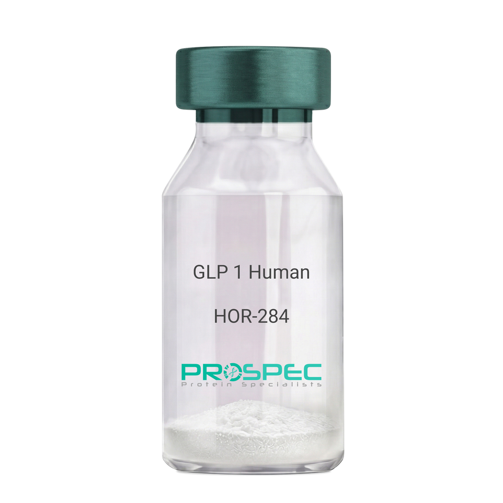
GLP 1 HumanDescription:
Human Glucagon Like Peptide-1
GLP1, Glucagon Like Peptide-1, Incretin hormone.
Product # :
HOR-284Price :
Quantity :
Shipping Method :
Shipped at Room temp
More Info
- description
- formulation
- purity
- More Info
Description
Glucagon Like Peptide-1 is a single, non-glycosylated, polypeptide chain containing 30 amino acids and having a molecular mass of 3297.7 Dalton.The GLP-1 is purified by proprietary chromatographic techniques.
Formulation
The GLP-1 peptide was lyophilized with no additives.
Purity
Greater than 96.0% as determined by RP-HPLC.
More Info
-
Introduction
Glucagon-like peptide-1 (GLP-1) is derived from the transcription product of the proglucagon gene. The major source of GLP-1 in the body is the intestinal L cell that secretes GLP-1 as a guthormone. The biologically active forms of GLP-1 are: GLP-1-(7-37) and GLP-1-(7-36)NH2.
GLP-1 secretion by L cells is dependent on the presence of nutrients in the lumen of the small intestine. The secretagogues (agents that causes or stimulates secretion) of this hormone include major nutrients like carbohydrate, proteinand lipid. Once in the circulation, GLP-1 has a half life of less than 2 minutes, due to rapid degradation by the enzyme dipeptidyl peptidase-4.
GLP-1 possesses several physiological properties that make it a subject of intensive investigation as a potential treatment of diabetes mellitus. The known physiological functions of GLP-1 include: Increases insulin secretion from the pancreas in a glucose-dependent manner, decreases glucagon secretion from the pancreas, increases beta cells mass and insulin gene expression, inhibits acid secretion and gastric emptying in the stomach, decreases food intake by increasing satiety. -
Synonyms
GLP1, Glucagon Like Peptide-1, Incretin hormone.
-
Physical Appearance
Sterile Filtered White lyophilized (freeze-dried) powder.
-
Stability
Lyophilized Glucagon Like Peptide-1 although stable at room temperature for 3 weeks, should be stored desiccated below -18°C. Upon reconstitution GLP-1 should be stored at 4°C between 2-7 days and for future use below -18°C. For long term storage it is recommended to add a carrier protein (0.1% HSA or BSA).Please prevent freeze-thaw cycles.
-
Solubility
It is recommended to reconstitute the lyophilized Glucagon Like Peptide-1 in sterile H2O at 0.5mg/ml, which can then be further diluted to other aqueous solutions.
-
Amino Acid Sequence
H-His-Ala-Glu-Gly-Thr-Phe-Thr-Ser-Asp-Val-Ser-Ser-Tyr-Leu-Glu-Gly-Gln-Ala-Ala-Lys-Glu-Phe-Ile-Ala-Trp-Leu-Val-Lys-Gly-Arg-NH2.
ProSpec's products are furnished for LABORATORY RESEARCH USE ONLY. The product may not be used as drugs, agricultural or pesticidal products, food additives or household chemicals.
- View Data Sheet
Name :
T.Vaginalis P65Description:
Trichomonas Vaginalis P65 Recombinant
Product # :
PRO-1920Price :
Quantity :
Shipping Method :
Shipped with Ice Packs
More Info
- description
- source
- formulation
- purity
- More Info
Description
The Recombinant Trichomonas vaginalis p65, E.Coli derived migrates around 67kda on SDS-PAGE. The full length protein is purified by standard chromatography techniques.
Source
Escherichia Coli.
Formulation
T. Vaginalis p65protein solution in PBS and 25Mm K2CO3.
Purity
Protein is >90% pure as determined by 10% PAGE (coomassie staining).
More Info
-
Introduction
Trichomonas vaginalis, an anaerobic, flagellated protozoan parasite, is most dominant protozoan causing sexual transmitted disease. T. vaginalis induced vaginal tract infection occurs as multiple steps involving interactions between host macromolecules and protozoan virulent factor. Adhension protein P65 of T. vaginalis has a vital part in assisting the penetration into vaginal tract surface.
-
Physical Appearance
Sterile Filtered clear solution.
-
Stability
T. Vaginalis p65 Recombinant although stable at 4°C for 1 week, should be stored below -18°C. Please prevent freeze thaw cycles.
ProSpec's products are furnished for LABORATORY RESEARCH USE ONLY. The product may not be used as drugs, agricultural or pesticidal products, food additives or household chemicals.
- View Data Sheet
Name :
NTRK2 HumanDescription:
Neurotrophic Receptor Tyrosine Kinase 2 Human Recombinant
GP145-TrkB, trk-B, TRKB, NTRK2
Product # :
CYT-1159Price :
Quantity :
Shipping Method :
Shipped with Ice Packs
More Info
- description
- source
- formulation
- purity
- More Info
Description
NTRK2 Human produced in Sf9 Baculovirus cells is a single, glycosylated polypeptide chain containing 407 amino acids (32-430a.a) and having a molecular mass of 45.2kDa.NTRK2 is fused to an 8 amino acid His tag at C-terminus and purified by proprietary chromatographic techniques.
Source
Sf9, Baculovirus cells.
Formulation
The NTRK2 solution (0. 5mg/ml) contains 10% Glycerol and Phosphate-Buffered Saline (pH 7.4).
Purity
Greater than 95% as determined by SDS-PAGE.
More Info
-
Introduction
Neurotrophic Receptor Tyrosine Kinase 2, also referred to NTRK2, is a tyrosine-protein kinase receptor which takes part in the development & the maturation of the central and the peripheral nervous systems through regulation of neuron survival, migration, proliferation, differentiation and synapse formation & plasticity. NTRK2 acts in learning and memory by regulating both short term synaptic function and long-term potentiation. The substrates that are known for the TRK family receptors are: SHC1, PI-3 kinase and PLC-gamma-1.
-
Synonyms
GP145-TrkB, trk-B, TRKB, NTRK2
-
Physical Appearance
Sterile Filtered colorless solution.
-
Stability
Store at 4°C if entire vial will be used within 2-4 weeks. Store, frozen at -20°C for longer periods of time. For long term storage it is recommended to add a carrier protein (0.1% HSA or BSA).Avoid multiple freeze-thaw cycles.
-
Amino Acid Sequence
CPTSCKCSAS RIWCSDPSPG IVAFPRLEPN SVDPENITEI FIANQKRLEI INEDDVEAYV GLRNLTIVDS GLKFVAHKAF LKNSNLQHIN FTRNKLTSLS RKHFRHLDLS ELILVGNPFT CSCDIMWIKT LQEAKSSPDT QDLYCLNESS KNIPLANLQI PNCGLPSANL AAPNLTVEEG KSITLSCSVA GDPVPNMYWD VGNLVSKHMN ETSHTQGSLR ITNISSDDSG KQISCVAENL VGEDQDSVNL TVHFAPTITF LESPTSDHHW CIPFTVKGNP KPALQWFYNG AILNESKYIC TKIHVTNHTE YHGCLQLDNP THMNNGDYTL IAKNEYGKDE KQISAHFMGW PGIDDGANPN YPDVIYEDYG TAANDIGDTT NRSNEIPSTD VTDKTGREHL EHHHHHH
ProSpec's products are furnished for LABORATORY RESEARCH USE ONLY. The product may not be used as drugs, agricultural or pesticidal products, food additives or household chemicals.
- View Data Sheet
Name :
PAK4 HumanDescription:
p21 Activated Kinase 4 Human Recombinant
PAK-4, p21 (CDKN1A)-Activated Kinase 4, p21-Activated Kinase 4, Serine/threonine-protein kinase PAK 4, KIAA1142, PAK4.
Product # :
PKA-259Price :
Quantity :
Shipping Method :
Shipped with Ice Packs
More Info
- description
- source
- formulation
- purity
- More Info
Description
PAK4 Human Recombinant produced in E.Coli is a single, non-glycosylated polypeptide chain containing 628 amino acids (1-591 a.a.) and having a molecular mass of 68.3 kDa.PAK4 is fused to 37 amino acid His-Tag at N-terminus and is purified by standard chromatography techniques.
Source
Escherichia Coli.
Formulation
The PAK4 protein (0.5mg/ml) contains 25mM HEPES pH-7.5, 150mM NaCl, 5mM MgCl2 and 30% glycerol.
Purity
Greater than 90% as determined by SDS-PAGE.
More Info
-
Introduction
PAK4 is part of the group B family of p21-activated kinases (PAK). It's known as an effector protein for cell division cycle 42(Cdc42) and protein that plays an important role in regulating cytoskeletal organization and cell morphology. PAK4 expression is eminent in many cancer cell lines, and is involved in tumorigenesis. PAK proteins are significant effectors that associate Rho GTPases to cytoskeleton reorganization and nuclear signaling. PAK proteins are targets for the small GTP binding proteins Cdc42 and Rac and have been implicated in a wide range of biological activities. PAK4 cooperates specifically with the GTP-bound form of Cdc42Hs and weakly activates the JNK family of MAP kinases. PAK4 is a mediator of filopodia formation and is involved in the reorganization of the actin cytoskeleton. PAK4 activates the JNK pathway. PAK4 phosphorylates and inactivates the protein phosphatase SSH1, leading to increased inhibitory phosphorylation of the actin binding/depolymerizing factor cofilin. Decreased cofilin activity may lead to stabilization of actin filaments.
-
Synonyms
PAK-4, p21 (CDKN1A)-Activated Kinase 4, p21-Activated Kinase 4, Serine/threonine-protein kinase PAK 4, KIAA1142, PAK4.
-
Physical Appearance
Sterile filtered colorless solution.
-
Stability
Store at 4°C if entire vial will be used within 2-4 weeks. Store, frozen at -20°C for longer periods of time. For long term storage it is recommended to add a carrier protein (0.1% HSA or BSA). Avoid multiple freeze-thaw cycles.
-
Amino Acid Sequence
MRGSHHHHHH GMASMTGGQQ MGRDLYDDDD KDRWGSHMFG KRKKRVEISA PSNFEHRVHT GFDQHEQKFT GLPRQWQSLI EESARRPKPL VDPACITSIQ PGAPKTIVRG SKGAKDGALT LLLDEFENMS VTRSNSLRRD SPPPPARARQ ENGMPEEPAT TARGGPGKAG SRGRFAGHSE AGGGSGDRRR AGPEKRPKSS REGSGGPQES SRDKRPLSGP DVGTPQPAGL ASGAKLAAGR PFNTYPRADT DHPSRGAQGE PHDVAPNGPS AGGLAIPQSS SSSSRPPTRA RGAPSPGVLG PHASEPQLAP PACTPAAPAV PGPPGPRSPQ REPQRVSHEQ FRAALQLVVD PGDPRSYLDN FIKIGEGSTG IVCIATVRSS GKLVAVKKMD LRKQQRRELL FNEVVIMRDY QHENVVEMYN
SYLVGDELWV VMEFLEGGAL TDIVTHTRMN EEQIAAVCLA VLQALSVLHA QGVIHRDIKS DSILLTHDGR VKLSDFGFCA QVSKEVPRRK SLVGTPYWMA PELISRLPYG PEVDIWSLGI MVIEMVDGEP PYFNEPPLKA MKMIRDNLPP RLKNLHKVSP SLKGFLDRLL VRDPAQRATA AELLKHPFLA KAGPPASIVP LMRQNRTR.
ProSpec's products are furnished for LABORATORY RESEARCH USE ONLY. The product may not be used as drugs, agricultural or pesticidal products, food additives or household chemicals.
- View Data Sheet
Name :
PCID2 HumanDescription:
PCI Domain Containing 2 Human Recombinant
PCI Domain Containing 2, CSN12-Like Protein, PCI Domain-Containing Protein 2, F10, PCI domain-containing protein 2.
Product # :
PRO-2074Price :
Quantity :
Shipping Method :
Shipped with Ice Packs
More Info
- description
- source
- formulation
- purity
- More Info
Description
PCID2 Human Recombinant produced in E.Coli is a single, non-glycosylated polypeptide chain containing 341 amino acids (80-399 a.a) and having a molecular mass of 39.3kDa. PCID2 is fused to a 21 amino acid His-tag at N-terminus & purified by proprietary chromatographic techniques.
Source
E.coli.
Formulation
PCID2 protein solution (1.0 mg/ml) containing 20mM Tris-HCl buffer (pH 8.0), 10% glycerol and 0.4M Urea.
Purity
Greater than 85% as determined by SDS-PAGE.
More Info
-
Introduction
PCI Domain Containing 2 (PCID2) is expressed in immature and early-stage B lymphocytes and regulates the expression of mitotic checkpoint protein MAD2. PCID2 is a component of the TREX-2 complex, transcription and export complex 2, this complex functions in docking export-competent ribonucleoprotein particles (mRNPs) to the nuclear entrance of the nuclear pore complex, nuclear basket.
-
Synonyms
PCI Domain Containing 2, CSN12-Like Protein, PCI Domain-Containing Protein 2, F10, PCI domain-containing protein 2.
-
Physical Appearance
Sterile Filtered colorless solution.
-
Stability
Store at 4°C if entire vial will be used within 2-4 weeks. Store, frozen at -20°C for longer periods of time. For long term storage it is recommended to add a carrier protein (0.1% HSA or BSA).Avoid multiple freeze-thaw cycles.
-
Amino Acid Sequence
MGSSHHHHHH SSGLVPRGSH MAYKCQTVIV QSFLRAFQAH KEENWALPVM YAVALDLRVF ANNADQQLVK KGKSKVGDML EKAAELLMSC FRVCASDTRA GIEDSKKWGM LFLVNQLFKI YFKINKLHLC KPLIRAIDSS NLKDDYSTAQ RVTYKYYVGR KAMFDSDFKQ AEEYLSFAFE HCHRSSQKNK RMILIYLLPV KMLLGHMPTV ELLKKYHLMQ FAEVTRAVSE GNLLLLHEAL AKHEAFFIRC GIFLILEKLK IITYRNLFKK VYLLLKTHQL SLDAFLVALK FMQVEDVDID EVQCILANLI YMGHVKGYIS HQHQKLVVSK QNPFPPLSTV C.
ProSpec's products are furnished for LABORATORY RESEARCH USE ONLY. The product may not be used as drugs, agricultural or pesticidal products, food additives or household chemicals.
- View Data Sheet
Name :
KIR3DL1 HumanDescription:
Killer Cell Immunoglobulin-Like Receptor, 3 Domains Long Cytoplasmic Tail 1 Human Recombinant
Killer cell immunoglobulin-like receptor 3DL1, MHC class I NK cell receptor, Natural killer-associated transcript 3, NKAT-3, p70 natural killer cell receptor clones CL-2/CL-11, HLA-BW4-specific inhibitory NK cell receptor, CD158 antigen-like family member E, CD158e antigen, KIR3DL1, CD158E, NKAT3, NKB1, KIR, NKB1B, CD158E1, MGC119726, MGC119728, MGC126589, MGC126591.
Product # :
PRO-427Price :
Quantity :
Shipping Method :
Shipped with Ice Packs
More Info
- description
- source
- formulation
- purity
- More Info
Description
Recombinant KIR3DL1 produced in E.Coli is a single, non-glycosylated polypeptide chain containing 132 amino acids and having a molecular mass of 15 kDa.The KIR3DL1 is purified by proprietary chromatographic techniques.
Source
Escherichia Coli.
Formulation
The protein (1mg/ml) contains 25mM Tris-HCl (pH7.5) and 100mM NaCl.
Purity
Greater than 95.0% as determined by(a) Analysis by RP-HPLC.
(b) Analysis by SDS-PAGE.More Info
-
Introduction
Killer-cell immunoglobulin-like receptors (KIRs), are a family of cell surface glycoproteins found on Natural Killer (NK) Cells, which are important cells of the immune system. They control the killing function of these cells by interacting with MHC class I molecules, which are expressed on all cell types. This interaction allows them to identify virally infected cells or tumor cells that have a distinctive low level of Class I MHC on their surface. The majority of KIRs are inhibitory, which means that their recognition of MHC suppresses the cytotoxic activity of their NK cell. Only a limited number of KIRs have the capacity to activate cells.
The KIR genes are found in a cluster on chromosome 19q13.4 within the 1 Mb leukocyte receptor complex (LRC). KIR molecules are extremely polymorphic, meaning their gene sequences differ significantly between individuals, so that different individuals have different arrays/repertoires of KIR genes.
The KIR proteins are categorized by the number of extracellular immunoglobulin domains (2D or 3D) and by whether they have a long (L) or short (S) cytoplasmic domain. KIR proteins with the long cytoplasmic domain transduce inhibitory signals upon ligand binding via an immune tyrosine-based inhibitory motif (ITIM). Whereas KIR proteins with the short cytoplasmic domain lack the ITIM motif and instead associate with the TYRO protein tyrosine kinase binding protein to transduce activating signals.
The three Ig-domain from of inhibitory killer cell Ig-like receptor 1(KIR3DL1, NKB1, nkat3, p70KIR) is a NK cell receptor for polymorphic HLA-B determinant. KIR3DLl recognizes the Bw4 determinant defined by sequence motifs at positions 77-83 of the HLA-B heavy chain. The cytoplasmic tail of KIR, which contains two immunoreceptor tyrosine-based inhibition motifs (ITIMs), mediates inhibitory signal transduction that prevents killer cell-mediated cytotoxicity. A His-tag fusion protein of KIR3DL1 cytoplasmic tail (361-444aa) was overexpressed as insoluble protein aggregates (inclusion bodies). -
Synonyms
Killer cell immunoglobulin-like receptor 3DL1, MHC class I NK cell receptor, Natural killer-associated transcript 3, NKAT-3, p70 natural killer cell receptor clones CL-2/CL-11, HLA-BW4-specific inhibitory NK cell receptor, CD158 antigen-like family member E, CD158e antigen, KIR3DL1, CD158E, NKAT3, NKB1, KIR, NKB1B, CD158E1, MGC119726, MGC119728, MGC126589, MGC126591.
-
Physical Appearance
Sterile filtered colorless solution.
-
Stability
Store at 4°C if entire vial will be used within 2-4 weeks. Store, frozen at -20°C for longer periods of time. For long term storage it is recommended to add a carrier protein (0.1% HSA or BSA).Avoid multiple freeze-thaw cycles.
-
Amino Acid Sequence
MRGSHHHHHH GMASMTGGQQ MGRDLYDDDD KDRWGSTSGT IDKLDIEFHLWCSNKKNAAV MDQEPAGNRT ANSEDSDEQD PEEVTYAQLD HCVFTQRKIT RPSQRPKTPP TDTILYTELP NAKPRSKVVS CP.
ProSpec's products are furnished for LABORATORY RESEARCH USE ONLY. The product may not be used as drugs, agricultural or pesticidal products, food additives or household chemicals.
- View Data Sheet
Name :
CD116 Human, sf9Description:
GM-CSF Receptor Alpha Sf9 Human Recombinant
Colony Stimulating Factor 2 Receptor Alpha Subunit, Colony Stimulating Factor 2 Receptor, Alpha, Low-Affinity (Granulocyte-Macrophage), Alpha-GM-CSF Receptor, GM-CSF-R-Alpha, CD116 Antigen, GMCSFR-Alpha, GMR-Alpha, CDw116, CSF2RY, CSF2R, Granulocyte-Macrophage Colony-Stimulating Factor Receptor Subunit Alpha, Granulocyte-Macrophage Colony-Stimulating Factor Receptor Alpha Chain, GM-CSF Receptor Alpha Subunit, AlphaGMR, CSF2RAX, CSF2RAY, CSF2RX, GMCSFR, CD116, SMDP4, GMR.
Product # :
CYT-1044Price :
Quantity :
Shipping Method :
Shipped with Ice Packs
More Info
- description
- source
- formulation
- purity
- biological activity
- More Info
Description
CSF2RA produced in Sf9 Baculovirus cells is a single, glycosylated polypeptide chain containing 310 amino acids (20-320a.a.) and having a molecular mass of 35.9kDa. (Molecular size on SDS-PAGE will appear at approximately 40-57kDa). CSF2RA is expressed with a 9 amino acid His tag at C-Terminus and purified by proprietary chromatographic techniques.
Source
Sf9, Baculovirus cells.
Formulation
CSF2RA protein solution (0.5mg/ml) contains Phosphate Buffered Saline (pH 7.4) and 10% glycerol.
Purity
Greater than 90.0% as determined by SDS-PAGE.
Biological Activity
Measured by its ability to inhibit GM-CSF dependent proliferation of TF1 human erythroleukemic cells. The ED50 for this effect is less or equal to 10ug/ml in the presence of 0.5ng/ml GM-CSF.
More Info
-
Introduction
GM-CSF Receptor Alpha (CSF2RA) is the alpha subunit of the heterodimeric receptor for colony stimulating factor 2, a cytokine which controls the production, differentiation, and function of granulocytes and macrophages. CSFR2 is also a member of the cytokine family of receptors. In addition, this gene is found in the pseudoautosomal region (PAR) of the X and Y chromosomes. Multiple transcript variants encoding various isoforms have been found for this gene, while some of the isoforms being membrane-bound and others being soluble. Diseases associated with CSF2RA include surfactant metabolism dysfunction, pulmonary 4, and csf2ra-related pulmonary surfactant metabolism dysfunction.
-
Synonyms
Colony Stimulating Factor 2 Receptor Alpha Subunit, Colony Stimulating Factor 2 Receptor, Alpha, Low-Affinity (Granulocyte-Macrophage), Alpha-GM-CSF Receptor, GM-CSF-R-Alpha, CD116 Antigen, GMCSFR-Alpha, GMR-Alpha, CDw116, CSF2RY, CSF2R, Granulocyte-Macrophage Colony-Stimulating Factor Receptor Subunit Alpha, Granulocyte-Macrophage Colony-Stimulating Factor Receptor Alpha Chain, GM-CSF Receptor Alpha Subunit, AlphaGMR, CSF2RAX, CSF2RAY, CSF2RX, GMCSFR, CD116, SMDP4, GMR.
-
Physical Appearance
Sterile Filtered colorless solution.
-
Stability
Store at 4°C if entire vial will be used within 2-4 weeks. Store, frozen at -20°C for longer periods of time. For long term storage it is recommended to add a carrier protein (0.1% HSA or BSA). Avoid multiple freeze-thaw cycles.
-
Amino Acid Sequence
ADPLIPEKSD LRTVAPASSL NVRFDSRTMN LSWDCQENTT FSKCFLTDKK NRVVEPRLSN NECSCTFREI CLHEGVTFEV HVNTSQRGFQ QKLLYPNSGR EGTAAQNFSC FIYNADLMNC TWARGPTAPR DVQYFLYIRN SKRRREIRCP YYIQDSGTHV GCHLDNLSGL TSRNYFLVNG TSREIGIQFF DSLLDTKKIE RFNPPSNVTV RCNTTHCLVR WKQPRTYQKL SYLDFQYQLD VHRKNTQPGT ENLLINVSGD LENRYNFPSS EPRAKHSVKI RAADVRILNW SSWSEAIEFG SDDGHHHHHH
-
Background
GM-CSF Receptor Alpha Human Recombinant: A Glimpse into Its Potential and Implications
Abstract:
Granulocyte-Macrophage Colony Stimulating Factor (GM-CSF) receptor alpha, a pivotal component in the GM-CSF signaling pathway, has been the focal point of numerous studies pertaining to hematopoiesis and immune responses. This paper provides an overview of the GM-CSF receptor alpha human recombinant, exploring its characteristics, production techniques, and potential therapeutic applications.
Introduction
GM-CSF, a cytokine responsible for the differentiation and proliferation of white blood cells, functions by binding to its receptor, GM-CSF receptor. The alpha subunit (GM-CSFRα) of this receptor plays a crucial role in ligand binding and is essential for initiating cellular responses. Modern biotechnological advancements have led to the successful production of its human recombinant form, offering new avenues in medical research.
Recombinant GM-CSFRα:
Production and Features Recombinant GM-CSFRα is synthesized using cutting-edge recombinant DNA technologies, predominantly in bacterial or mammalian expression systems. This human recombinant form retains its ability to bind to GM-CSF, maintaining its biological functionality and providing myriad research opportunities.
Therapeutic and Clinical Prospects
- Autoimmune Diseases: GM-CSF is often overexpressed in various autoimmune disorders. By utilizing recombinant GM-CSFRα as a potential decoy receptor, it's feasible to mitigate the effects of excessive GM-CSF, offering a new therapeutic strategy.
- Hematopoietic Disorders: Given its integral role in white blood cell development, recombinant GM-CSFRα might hold promise in treatments or as a diagnostic tool for certain hematological conditions.
- Research Paradigm: Beyond therapeutic applications, the recombinant GM-CSFRα can serve as an invaluable research tool to elucidate the nuances of GM-CSF signaling, aiding in the understanding of immune response mechanisms.
Conclusion:
GM-CSF receptor alpha human recombinant stands at the forefront of exciting research and therapeutic potential. While its full capabilities are yet to be realized, current insights underscore its significance in the realms of immunology and medicine.
What is the molecular weight/Mw of CD116 Protein?
CD116 Protein has a total Mw of 35.9kDa.
What is the source or expression system of CD116 Protein?
Sf9, Baculovirus cells.
What is the Purity of CD116 Protein?
CD116 Protein is >90% pure as determined by SDS-PAGE.
What is the Biological Activity of CD116 Protein?
Measured by its ability to inhibit GM-CSF dependent proliferation of TF1 human erythroleukemic cells. The ED50 for this effect is less or equal to 10ug/ml in the presence of 0.5ng/ml GM-CSF.
What is the amino acid sequence of CD116 Protein?
CD116 Protein is composed from 310 amino acids.
What applications can CD116 Protein be used in?
CD116 Protein can probably be used in western blot, ELISA and Lateral Flow.
What is the endotoxin level for CD116 Protein?
The endotoxin level is minimal, CD116 Protein was purified using conventional chromatography techniques
ProSpec's products are furnished for LABORATORY RESEARCH USE ONLY. The product may not be used as drugs, agricultural or pesticidal products, food additives or household chemicals.
- View Data Sheet
Name :
SFRP4 Human, sf9Description:
Secreted Frizzled-Related Protein 4 Human Recombinant, sf9
Secreted Frizzled Related Protein 4, Secreted Frizzled-Related Protein 4, Frizzled Protein, Human Endometrium, SFRP-4, FRPHE Secreted Frizzled-Related Protein 4; Secreted Frizzled-Related Protein 4, FRP-4, PYL, SFRP4, FRPHE.
Product # :
PRO-2368Price :
Quantity :
Shipping Method :
Shipped with Ice Packs
More Info
- description
- source
- formulation
- purity
- More Info
Description
SFRP4 produced in Sf9 Baculovirus cells is a single, glycosylated polypeptide chain containing 337 amino acids (19-346 a.a.) and having a molecular mass of 38.9kDa (Molecular size on SDS-PAGE will appear at approximately 40-57kDa). SFRP4 is expressed with a 9 amino acid His tag at C-Terminus and purified by proprietary chromatographic techniques.
Source
Sf9, Baculovirus cells.
Formulation
SFRP4 protein solution (0.5mg/ml) contains Phosphate Buffered Saline (pH 7.4) and 10% glycerol.
Purity
Greater than 90.0% as determined by SDS-PAGE.
More Info
-
Introduction
Secreted frizzled-related protein 4 (SFRP4) belongs to the SFRP family which contains a cysteine-rich domain homologous to the putative Wnt-binding site of Frizzled proteins. SFRPs serve as soluble modulators of Wnt signaling. SFRP4 may serve as a regulator of adult uterine morphology and function. SFRP4 increases apoptosis during ovulation possibly via modulation of FZ1/FZ4/WNT4 signaling. SFRP4 also has phosphaturic effects by specifically inhibiting sodium-dependent phosphate uptake. SFRP4 is expressed in proliferative endometrium and several types of ovarian, endometrial and Brest tumors. SFRP4 is expressed in mesenchymal cells and in cardiomyocytes. SFRP4 expression in ventricular myocardium correlates with apoptosis related gene expression. SFRP4 is up-regulated in failing myocardium.
-
Synonyms
Secreted Frizzled Related Protein 4, Secreted Frizzled-Related Protein 4, Frizzled Protein, Human Endometrium, SFRP-4, FRPHE Secreted Frizzled-Related Protein 4; Secreted Frizzled-Related Protein 4, FRP-4, PYL, SFRP4, FRPHE.
-
Physical Appearance
Sterile Filtered colorless solution.
-
Stability
Store at 4°C if entire vial will be used within 2-4 weeks. Store, frozen at -20°C for longer periods of time. For long term storage it is recommended to add a carrier protein (0.1% HSA or BSA). Avoid multiple freeze-thaw cycles.
-
Amino Acid Sequence
ADPVRGAPCE AVRIPMCRHM PWNITRMPNH LHHSTQENAI LAIEQYEELV DVNCSAVLRF FLCAMYAPIC TLEFLHDPIK PCKSVCQRAR DDCEPLMKMY NHSWPESLAC DELPVYDRGV CISPEAIVTD LPEDVKWIDI TPDMMVQERP LDVDCKRLSP DRCKCKKVKP TLATYLSKNY SYVIHAKIKA VQRSGCNEVT TVVDVKEIFK SSSPIPRTQV PLITNSSCQC PHILPHQDVL IMCYEWRSRM MLLENCLVEK WRDQLSKRSI QWEERLQEQR RTVQDKKKTA GRTSRSNPPK PKGKPPAPKP ASPKKNIKTR SAQKRTNPKR VHHHHHH
ProSpec's products are furnished for LABORATORY RESEARCH USE ONLY. The product may not be used as drugs, agricultural or pesticidal products, food additives or household chemicals.
- View Data Sheet
Name :
SNRPE HumanDescription:
Small Nuclear Ribonucleoprotein Polypeptide E Human Recombinant
Small nuclear ribonucleoprotein E, snRNP-E, Sm protein E, Sm-E, SmE, SNRPE, B-raf.
Product # :
PRO-104Price :
Quantity :
Shipping Method :
Shipped with Ice Packs
More Info
- description
- source
- formulation
- purity
- More Info
Description
SNRPE Human Recombinant fused with a 20 amino acid His tag at N-terminus produced in E.Coli is a single, non-glycosylated, polypeptide chain containing 112 amino acids (1-92 a.a.) and having a molecular mass of 12.9kDa. The SNRPE is purified by proprietary chromatographic techniques.
Source
Escherichia Coli.
Formulation
The SNRPE solution (0.25 mg/ml) contains 20mM Tris-HCl buffer (pH8.0), 0.2M NaCl, 5mM DTT, 1mM EDTA and 30% glycerol.
Purity
Greater than 95.0% as determined by SDS-PAGE.
More Info
-
Introduction
SNRPE facilitates the cytoplasmic construction of the UsnRNPs by binding to a conserved Sm site on UsnRNA and forming a stable snRNP core complex. While a core protein to UsnRNP, the SNRPE connects with the entire U family of snRNAs including U1–U6. In addition, SNRPE interacts with DDX20 and Small nuclear ribonucleoprotein polypeptide F.
-
Synonyms
Small nuclear ribonucleoprotein E, snRNP-E, Sm protein E, Sm-E, SmE, SNRPE, B-raf.
-
Physical Appearance
Sterile Filtered colorless solution.
-
Stability
Store at 4°C if entire vial will be used within 2-4 weeks. Store, frozen at -20°C for longer periods of time. For long term storage it is recommended to add a carrier protein (0.1% HSA or BSA).Avoid multiple freeze-thaw cycles.
-
Amino Acid Sequence
MGSSHHHHHH SSGLVPRGSH MAYRGQGQKV QKVMVQPINL IFRYLQNRSR IQVWLYEQVN MRIEGCIIGF DEYMNLVLDD AEEIHSKTKS RKQLGRIMLK GDNITLLQSV SN.
ProSpec's products are furnished for LABORATORY RESEARCH USE ONLY. The product may not be used as drugs, agricultural or pesticidal products, food additives or household chemicals.
- View Data Sheet
Name :
NRBF2 HumanDescription:
Nuclear Receptor Binding Factor 2 Human Recombinant
Nuclear receptor binding factor 2, COPR1, COPR2, NRBF-2, Nuclear receptor-binding factor 2, Comodulator of PPAR and RXR, NRBF2, COPR.
Product # :
PRO-1692Price :
Quantity :
Shipping Method :
Shipped with Ice Packs
More Info
- description
- source
- formulation
- purity
- More Info
Description
NRBF2 Human Recombinant produced in E.coli is a single, non-glycosylated polypeptide chain containing 266 amino acids (45-287) and having a molecular mass of 29.9 kDa.NRBF2 is fused to a 23 amino acid His-tag at N-terminus & purified by proprietary chromatographic techniques.
Source
Escherichia Coli.
Formulation
The NRBF2 solution (0.5mg/ml) contains 20mM Tris-HCl buffer (pH 8.0), 0.15M NaCl, 20% glycerol and 1mM DTT.
Purity
Greater than 85.0% as determined by SDS-PAGE.
More Info
-
Introduction
Nuclear receptor binding factor 2 (NRBF2) which works as transcriptional activator, modulates transcriptional activation by target nuclear receptors.
-
Synonyms
Nuclear receptor binding factor 2, COPR1, COPR2, NRBF-2, Nuclear receptor-binding factor 2, Comodulator of PPAR and RXR, NRBF2, COPR.
-
Physical Appearance
Sterile Filtered colorless solution.
-
Stability
Store at 4°C if entire vial will be used within 2-4 weeks. Store, frozen at -20°C for longer periods of time. For long term storage it is recommended to add a carrier protein (0.1% HSA or BSA).Avoid multiple freeze-thaw cycles.
-
Amino Acid Sequence
MGSSHHHHHH SSGLVPRGSH MGSMKLTQSE QAHLSLELQR DSHMKQLLLI QERWKRAQRE ERLKAQQNTD KDAAAHLQTS HKPSAEDAEG QSPLSQKYSP STEKCLPEIQ GIFDRDPDTL LYLLQQKSEP AEPCIGSKAP KDDKTIIEEQ ATKIADLKRH VEFLVAENER LRKENKQLKA EKARLLKGPI EKELDVDADF VETSELWSLP PHAETATASS TWQKFAANTG KAKDIPIPNL PPLDFPSPEL PLMELSEDIL KGFMNN.
ProSpec's products are furnished for LABORATORY RESEARCH USE ONLY. The product may not be used as drugs, agricultural or pesticidal products, food additives or household chemicals.
- View Data Sheet
Name :
VPS26A HumanDescription:
Vacuolar Protein Sorting 26A Human Recombinant
Vacuolar protein sorting-associated protein 26A, Vesicle protein sorting 26A, hVPS26, VPS26A, VPS26, HB58, PEP8A, Hbeta58.
Product # :
PRO-1161Price :
Quantity :
Shipping Method :
Shipped with Ice Packs
More Info
- description
- source
- formulation
- purity
- More Info
Description
VPS26A Human Recombinant produced in E.Coli is a single, non-glycosylated polypeptide chain containing 350 amino acids (1-327 a.a) and having a molecular mass of 40.6kDa.VPS26A is fused to a 23 amino acid His-tag at N-terminus & purified by proprietary chromatographic techniques.
Source
E.coli.
Formulation
VPS26A protein solution (0.5mg/ml) containing 20mM Tris-HCl buffer (pH8.0), 10% glycerol and 0.15M NaCl.
Purity
Greater than 85% as determined by SDS-PAGE.
More Info
-
Introduction
Vacuolar protein sorting-associated protein 26A (VPS26A) is a vital element of the retromer complex, which is required to recover lysosomal enzyme receptors (IGF2R and M6PR) from endosomes to the trans-Golgi network. Furthermore, VPS26A is essential in the regulation of transcytosis of the polymeric immunoglobulin receptor (pIgR-pIgA). Expression analysis in yeast and mammalian cells reveal that VPS26A interacts directly with VPS35 that functions as the core of the retromer complex.
-
Synonyms
Vacuolar protein sorting-associated protein 26A, Vesicle protein sorting 26A, hVPS26, VPS26A, VPS26, HB58, PEP8A, Hbeta58.
-
Physical Appearance
Sterile Filtered colorless solution.
-
Stability
Store at 4°C if entire vial will be used within 2-4 weeks. Store, frozen at -20°C for longer periods of time. For long term storage it is recommended to add a carrier protein (0.1% HSA or BSA).Avoid multiple freeze-thaw cycles.
-
Amino Acid Sequence
MGSSHHHHHH SSGLVPRGSH MGSMSFLGGF FGPICEIDIV LNDGETRKMA EMKTEDGKVE KHYLFYDGES VSGKVNLAFK QPGKRLEHQG IRIEFVGQIE LFNDKSNTHE FVNLVKELAL PGELTQSRSY DFEFMQVEKP YESYIGANVR LRYFLKVTIV RRLTDLVKEY DLIVHQLATY PDVNNSIKME VGIEDCLHIE FEYNKSKYHL KDVIVGKIYF LLVRIKIQHM ELQLIKKEIT GIGPSTTTET ETIAKYEIMD GAPVKGESIP IRLFLAGYDP TPTMRDVNKK FSVRYFLNLV LVDEEDRRYF KQQEIILWRK APEKLRKQRT NFHQRFESPE SQASAEQPEM.
ProSpec's products are furnished for LABORATORY RESEARCH USE ONLY. The product may not be used as drugs, agricultural or pesticidal products, food additives or household chemicals.
- View Data Sheet
Name :
RAET1E Human, Sf9Description:
Retinoic Acid Early Transcript 1E Human Recombinant, Sf9
Retinoic Acid Early Transcript 1E, Lymphocyte Effector Toxicity Activation Ligand, RAE-1-Like Transcript 4, NKG2DL4, N2DL-4, LETAL, ULBP4, RL-4, NKG2D Ligand 4, BA350J20.7, RAET1E2, N2DL4.
Product # :
PRO-2467Price :
Quantity :
Shipping Method :
Shipped with Ice Packs
More Info
- description
- source
- formulation
- purity
- More Info
Description
RAET1E Human Recombinant produced in Sf9 Insect cells is a single, glycosylated polypeptide chain containing 204 amino acids (31-225 a.a.) and having a molecular mass of 23.4kDa (Molecular size on SDS-PAGE will appear at approximately 28-40kDa).RAET1E is expressed with a 6 amino acids His tag at C-Terminus and purified by proprietary chromatographic techniques.
Source
Sf9, Baculovirus cells.
Formulation
RAET1E protein solution (0.5mg/ml) contains Phosphate Buffered Saline (pH 7.4) and 10% glycerol.
Purity
Greater than 90.0% as determined by SDS-PAGE.
More Info
-
Introduction
RAET1E is a member of the MHC class I family. MHC class I family contains main histocompatibility complex (MHC) class I-related genes positioned in a cluster on chromosome 6q24.2-q25.3. RAET1E and RAET1G protein are different from other RAET1 proteins since they have type I membrane-spanning sequences at their C termini instead glycosylphosphatidylinositol anchor sequences.RAET1E acts as a ligand for NKG2D receptor, expressed on the surface of numerous types of immune cells, involves in innate adaptive immune reactions.RAET1E delivers signals to NK cells and advances tumor immune surveillance by inducing the growth of anti-tumor cytotoxic lymphocyte.
-
Synonyms
Retinoic Acid Early Transcript 1E, Lymphocyte Effector Toxicity Activation Ligand, RAE-1-Like Transcript 4, NKG2DL4, N2DL-4, LETAL, ULBP4, RL-4, NKG2D Ligand 4, BA350J20.7, RAET1E2, N2DL4.
-
Physical Appearance
Sterile Filtered colorless solution.
-
Stability
Store at 4°C if entire vial will be used within 2-4 weeks. Store, frozen at -20°C for longer periods of time. For long term storage it is recommended to add a carrier protein (0.1% HSA or BSA).Avoid multiple freeze-thaw cycles.
-
Amino Acid Sequence
ADPHSLCFNF TIKSLSRPGQ PWCEAQVFLN KNLFLQYNSD NNMVKPLGLL GKKVYATSTW GELTQTLGEV GRDLRMLLCD IKPQIKTSDP STLQVEMFCQ REAERCTGAS WQFATNGEKS LLFDAMNMTW TVINHEASKI KETWKKDRGL EKYFRKLSKG DCDHWLREFL GHWEAMPEPT VSPVNASDIH WSSSSLPDHH HHHH.
ProSpec's products are furnished for LABORATORY RESEARCH USE ONLY. The product may not be used as drugs, agricultural or pesticidal products, food additives or household chemicals.
- View Data Sheet
Name :
GHRP4Description:
Growth Hormone Releasing Peptide-4
GHRP-4, GHRP4.
Product # :
HOR-022Price :
Quantity :
Shipping Method :
Shipped at Room temp
More Info
- description
- formulation
- purity
- More Info
Description
Growth Hormone Releasing Peptide-4 Synthetic is a single, non-glycosylated polypeptide chain containing 4 amino acids, having a molecular mass of 607.73 Dalton and a Molecular formula of C34H37N7O4.
Formulation
The protein was lyophilized with no additives.
Purity
Greater than 97.0% as determined by analysis by RP-HPLC.
More Info
-
Introduction
GH-releasing peptides (GHRPs) are synthetic peptides that like GHRH effect on pituitary somatotrophs to stimulate GH release. The GHRP4 is one of several synthetic met-enkephalin analogs that owns unnatural D-amino acids. They were developed for their growth hormone (GH) releasing activity and called GH secretatogues. They lack opioid activity but are potent stimulators of GH release. These secretatogues are distinct from the growth hormone releasing hormone (GHRH or GHRF) as they share no sequence relation and derive their function through action at a completely different receptor, the ghrelin receptor.
-
Synonyms
GHRP-4, GHRP4.
-
Physical Appearance
Sterile Filtered White lyophilized (freeze-dried) powder.
-
Stability
Lyophilized Growth Hormone Releasing Peptide-4 although stable at room temperature for 3 weeks, should be stored desiccated below -18°C. Upon reconstitution GHRP4 should be stored at 4°C between 2-7 days and for future use below -18°C. For long term storage it is recommended to add a carrier protein (0.1% HSA or BSA). Please prevent freeze-thaw cycles.
-
Solubility
It is recommended to reconstitute the lyophilized GHRP4 in sterile 18MΩ-cm H2O not less than 100 µg/ml, which can then be further diluted to other aqueous solutions.
-
Amino Acid Sequence
(D-Trp)-Ala-Trp-(D-Phe)-NH2.
ProSpec's products are furnished for LABORATORY RESEARCH USE ONLY. The product may not be used as drugs, agricultural or pesticidal products, food additives or household chemicals.
- View Data Sheet
Name :
EDAR AntibodyDescription:
Ectodysplasin A Receptor, Mouse Anti Human
Ectodysplasin A Receptor, DL, Ectodysplasin 1, Anhidrotic Receptor, Anhidrotic Ectodysplasin Receptor 1, Ectodermal Dysplasia Receptor, Ectodysplasin-A Receptor, Downless Homolog, EDA-A1 Receptor, ECTD10A, ECTD10B, EDA3, HRM1, ED3, Tumor Necrosis Factor Receptor Superfamily Member EDAR, Downless, Mouse, Homolog Of, EDA-A1R, EDA1R, ED1R, ED5, Tumor necrosis factor receptor superfamily member EDAR.
Product # :
ANT-560Price :
Quantity :
Shipping Method :
Shipped with Ice Packs
More Info
- formulation
- More Info
Formulation
1mg/ml containing PBS, pH-7.4, 10% Glycerol and 0.02% Sodium Azide.
More Info
-
Introduction
Ectodysplasin A Receptor, also known as EDAR belongs to the tumor necrosis factor receptor family. EDAR is a receptor for the soluble ligand ectodysplasin A, and is capable of activating the nuclear factor-kappaB, JNK, as well as caspase-independent cell death pathways. EDAR is necessary for the development of hair, teeth, and other ectodermal derivatives. Furthermore, mutations in EDAR resulted in autosomal dominant and recessive forms of hypohidrotic ectodermal dysplasia.
-
Synonyms
Ectodysplasin A Receptor, DL, Ectodysplasin 1, Anhidrotic Receptor, Anhidrotic Ectodysplasin Receptor 1, Ectodermal Dysplasia Receptor, Ectodysplasin-A Receptor, Downless Homolog, EDA-A1 Receptor, ECTD10A, ECTD10B, EDA3, HRM1, ED3, Tumor Necrosis Factor Receptor Superfamily Member EDAR, Downless, Mouse, Homolog Of, EDA-A1R, EDA1R, ED1R, ED5, Tumor necrosis factor receptor superfamily member EDAR.
-
Physical Appearance
Sterile filtered colorless solution.
-
Immunogen
Anti-human EDAR mAb, is derived from hybridization of mouse F0 myeloma cells with spleen cells from BALB/c mice immunized with a recombinant human EDAR protein 27-448 amino acids purified from E. coli.
-
Ig Subclass
Mouse IgG2a heavy chain and k light chain.
-
Clone
PAT19E8AT.
-
Applications
EDAR antibody has been tested by ELISA, Western blot analysis to assure specificity and reactivity. Since application varies, however, each investigation should be titrated by the reagent to obtain optimal results. Recommended starting dilution is 1:1000.
-
Type
Mouse Anti Human Monoclonal.
-
Storage Procedures
For periods up to 1 month store at 4°C, for longer periods of time, store at -20°C. Prevent freeze thaw cycles.
-
Purification Method
EDAR antibody was purified from mouse ascitic fluids by protein-A affinity chromatography.
ProSpec's products are furnished for LABORATORY RESEARCH USE ONLY. The product may not be used as drugs, agricultural or pesticidal products, food additives or household chemicals.
- View Data Sheet
Name :
PMVK AntibodyDescription:
Phosphomevalonate Kinase, Mouse Anti Human
Phosphomevalonate kinase, PMKase, hPMK, PMVK, PMKI, PMK, PMKA, PMKASE, HUMPMKI.
Product # :
ANT-574Price :
Quantity :
Shipping Method :
Shipped with Ice Packs
More Info
- formulation
- More Info
Formulation
1mg/ml containing PBS, pH-7.4, 10% Glycerol and 0.02% Sodium Azide.
More Info
-
Introduction
Phosphomevalonate kinase (PMVK) is a cytosolic enzyme. PMVK is expressed highly in the heart, liver, skeletal muscle, kidney and pancreas and slightly lower in the brain, placenta, and lung. PMVK catalyzes the conversion of mevalonate 5-phosphate into mevalonate 5-diphosphate in the fifth reaction of the cholesterol biosynthetic pathway. Induced by sterol, PMVK participates in isopentenyl diphosphate biosynthesis via the mevalonate pathway.
-
Synonyms
Phosphomevalonate kinase, PMKase, hPMK, PMVK, PMKI, PMK, PMKA, PMKASE, HUMPMKI.
-
Physical Appearance
Sterile filtered colorless solution.
-
Immunogen
Anti-human PMVK mAb, is derived from hybridization of mouse F0 myeloma cells with spleen cells from BALB/c mice immunized with recombinant human PMVK amino acids 1-192 purified from E. coli.
-
Ig Subclass
Mouse IgG2a heavy chain and k light chain.
-
Clone
PAT21C10AT.
-
Applications
PMVK antibody has been tested by ELISA, Western blot analysis, ICC/IF and Flow cytometry to assure specificity and reactivity. Since application varies, however, each investigation should be titrated by the reagent to obtain optimal results.
-
Type
Mouse Anti Human Monoclonal.
-
Storage Procedures
For periods up to 1 month store at 4°C, for longer periods of time, store at -20°C. Prevent freeze thaw cycles.
-
Purification Method
PMVK antibody was purified from mouse ascitic fluids by protein-A affinity chromatography.
ProSpec's products are furnished for LABORATORY RESEARCH USE ONLY. The product may not be used as drugs, agricultural or pesticidal products, food additives or household chemicals.