Search results
1000 results found for “Methylmalonic Aciduria”
Name
Description
Product #
Price
Quantity
Shipping Method
- View Data Sheet
Name :
HaptoglobinDescription:
Haptoglobin Human Recombinant
Haptoglobin, HP, BP, HPA1S, MGC111141, HP2-ALPHA-2.
Product # :
PRO-567Price :
Quantity :
Shipping Method :
Shipped at Room temp
More Info
- description
- source
- formulation
- purity
- More Info
- Citations
Description
Haptoglobin Human Recombinant produced in E.Coli is a single, non-glycosylated, polypeptide chain containing fusion protein with His tag and having a total Mw of 33 kDa (4 kDa His-tag).
Source
Escherichia Coli.
Formulation
Each mg was lyophilized with 1xPBS, 0.4% SDS and 4mM DTT.
Purity
Greater than 90.0% as determined by SDS-PAGE.
Citations
More Info
-
Introduction
Haptoglobin is a glycoprotein which is synthesized in the liver and circulates in the blood. Haptoglobin is produced typically by hepatocytes but also by other tissues: e.g. skin, lung, and kidney. It is a positive acute phase protein that binds free hemoglobin and removes it from the circulation to prevent kidney injury, and iron loss following hemolysis. The haptoglobin-hemoglobin complex is subsequently removed by the reticuloendothelial system (generally the spleen). As the reticuloendothelial system removes the haptoglobin-hemoglobin complex from the body, haptoglobin levels are reduced in hemolytic anaemias. In the course of binding hemoglobin, haptoglobin sequesters the iron inside hemoglobin, preventing iron-utilizing bacteria from benefitting from hemolysis.
Haptoglobin consists of two A- and two B-chains, connected by disulfide bonds. Three major haptoglobin phenotypes are known to exist (Hp 1-1, Hp 2-1, and Hp 2-2). Hp 1-1 is biologically the most effective in binding free hemoglobin and suppressing inflammatory responses associated with free hemoglobin. Hp 2-2 is biologically the least active, and Hp 2-1 is moderately active. Haptoglobin’s molecular mass ranges from 8-200 kDa.
Reduced levels can be seen in haemolysis and impaired liver function. High levels are a marker for acute or chronic inflammation. Ahaptoglobinemia or hypohaptoglobinemia are caused by mutations in the haptoglobin gene and/or its regulatory regions. Haptoglobin is also linked to diabetic nephropathy, the incidence of coronary artery disease in type 1 diabetes, Crohn's disease, inflammatory disease behavior, primary sclerosing cholangitis, susceptibility to idiopathic Parkinson's disease, and a reduced incidence of Plasmodium falciparum malaria. -
Synonyms
Haptoglobin, HP, BP, HPA1S, MGC111141, HP2-ALPHA-2.
-
Physical Appearance
Sterile Filtered White lyophilized (freeze-dried) powder.
-
Stability
Lyophilized Haptoglobin although stable at room temperature for 3 weeks, should be stored desiccated below -18°C. Upon reconstitution Haptoglobin should be stored at 4°C between 2-7 days and for future use below -18°C.For long term storage it is recommended to add a carrier protein (0.1% HSA or BSA).Please prevent freeze-thaw cycles.
-
Solubility
It is recommended to reconstitute the lyophilized Haptoglobin in sterile 18MΩ-cm H2O not less than 100µg/ml, which can then be further diluted to other aqueous solutions.
-
Amino Acid Sequence
L ILGGHLDAKG SFPWQAKMVS HHNLTTGATL INEQWLLTTA KNLFLNHSEN ATAKDIAPTL TLYVGKKQLV EIEKVVLHPN YSQVDIGLIK LKQKVSVNER VMPICLPSKD YAEVGRVGYV SGWGRNANFK FTDHLKYVML PVADQDQCIR HYEGSTVPEK KTPKSPVGVQ PILNEHTFCA GMSKYQEDTC YGDAGSAFAV HDLEEDTWYA TGILSFDKSC AVAEYGVYVK VTSIQDWVQK TIAEN
ProSpec's products are furnished for LABORATORY RESEARCH USE ONLY. The product may not be used as drugs, agricultural or pesticidal products, food additives or household chemicals.
- View Data Sheet
Name :
Visfatin HumanDescription:
Visfatin Human Recombinant
PBEF, Pre-B cell colony-enhancing factor, Nicotinamide phosphoribosyltransferase NAmPRTase, Nampt, MGC117256, DKFZP666B131, 1110035O14Rik.
Product # :
CYT-318Price :
Quantity :
Shipping Method :
Shipped at Room temp
More Info
- description
- source
- formulation
- purity
- biological activity
- More Info
- Citations
Description
Visfatin Human Recombinant produced in E.Coli is a single, non-glycosylated, polypeptide chain containing 466 amino acids. The total molecular mass is 52.6kDa (calculated). The Visfatin is purified by Flag-affinity chromatography.
Source
Escherichia Coli.
Formulation
Visfatin was lyophilized with no additives.
Purity
Greater than 90.0% as determined by SDS-PAGE.
Biological Activity
The activity is determined by its ability to induce IL-6, IL-1 beta and TNF alpha production from human PBMCs at 100ng/ml.Citations
More Info
-
Introduction
Excess adiposity is the most important risk in the development of type 2 diabetes mellitus (T2DM). Adipose tissue produces several proteins (adipocytokines) such as leptin, adiponectin, resistin, tumor necrosis factor-a, and IL-6, that modulate sensitivity and appear to play an important role in the pathogenesis, diabetes, dyslipidemia, inflammation, and atherosclerosis. Visfatin, also known as pre-B cell colony-enhancing factor (PBEF), is a cytokine that is highly expressed in visceral fat and was originally isolated as a secreted factor that synergizes with IL-7 and stem cell factors to promote the growth of B cell precursors. Visfatin homologs have been identified in carp, invertebrate mollusks, and bacteria, as well as in vertebrates, including humans and the mouse. It has been postulated to play a role in innate immunity.
Visfatin exerts mimetic effects that are dose-dependent and quantitatively similar to stimulating muscle and adipocyte glucose transport, and in inhibiting hepatocyte glucose production. Intravenous injection of recombinant visfatin in mice decreased plasma glucose in a dose-dependent fashion. In keeping with its mimetic effects, visfatin was as effective in reducing hyperglycemia in deficient diabetic mice. Visfatin was also found to be bound to and activate receptor, causing receptor phosphorylation and the activation of downstream signaling molecules. However, visfatin did not compete for binding to the receptor, indicating that the two proteins were recognized by different regions of the receptor. Thus, visfatin might play a role in glucose homeostasis and dysregulation in biosynthesis or signal transduction, and might contribute to the pathogenesis of diabetes. -
Synonyms
PBEF, Pre-B cell colony-enhancing factor, Nicotinamide phosphoribosyltransferase NAmPRTase, Nampt, MGC117256, DKFZP666B131, 1110035O14Rik.
-
Physical Appearance
Filtered White lyophilized (freeze-dried) powder.
-
Stability
Lyophilized Visfatin although stable at room temperature for 3 weeks, should be stored desiccated below -18°C. Upon reconstitution Visfatin should be stored at 4°C between 2-7 days and for future use below -18°C.For long term storage it is recommended to add a carrier protein (0.1% HSA or BSA).Please prevent freeze-thaw cycles.
-
Solubility
Centrifuge vial before opening. When reconstituting the product, gently pipet and wash down the sides of the vial to ensure full recovery of the protein into solution. It is recommended to reconstitute the lyophilized product with 20 mM HCl at a concentration of 0.1 mg/mL, which can be further diluted into other aqueous solutions. Wait several minutes for full reconstitution and solubility.
-
Amino Acid Sequence
MPPNTSKVYS YFECREKKTE NSKLRKVKYE ETVFYGLQYI LNKYLKGKVV TKEKIQEAKD VYKEHFQDDV FNEKGWNYIL EKYDGHLPIE IKAVPEGFVI PRGNVLFTVE NTDPECYWLT NWIETILVQS WYPITVATNS REQKKILAKY LLETSGNLDG LEYKLHDFGY RGVSSQETAG IGASAHLVNF KGTDTVAGLA LIKKYYGTKD PVPGYSVPAA EHSTITAWGK DHEKDAFEHI VTQFSSVPVS VVSDSYDIYN ACEKIWGEDL RHLIVSRSTQ APLIIRPDSG NPLDTVLKVL EILGKKFPVT ENSKGYKLLP PYLRVIQGDG VDINTLQEIV EGMKQKMWSI ENIAFGSGGG LLQKLTRDLL NCSFKCSYVV TNGLGINVFK DPVADPNKRS KKGRLSLHRT PAGNFVTLEE GKGDLEEYGQ DLLHTVFKNG KVTKSYSFDE IRKNAQLNIE LEAAHH.
-
Background
About Visfatin Human
Visfatin is a cytokine expressed in visceral fat that was originally isolated as a secreted
element that synergized with stem cell factors and IL-7. One of its main functions is to
enhance the development of B cell precursors.The cytokine is also known as the “Pre-B Cell Colony-Enhancing Factor (PBEF).” It has been
identified in vertebrates, including mice and humans, and it’s being studied due to its link
to inflammatory conditions, beta cell function, and cardiovascular disease.
What’s the Function of Visfatin Human Recombinant?Visfatin human recombinant is produced in E. Coli. It’s a single, non-glycosylated,
polypeptide chain that contains 466 amino acids, it’s purified by FLAG-affinity
chromatography, and it contains a total molecular mass of 52.6 kDa.
What Are the Main Applications of Visfatin Human Recombinant?The cytokine is being researched because of its involvement in glucose homeostasis,
dysregulation in biosynthesis and signal transduction, and the pathogenesis of diabetes.
Visfatin human recombinant is tailored exclusively for laboratory research, ensuring
experts can get further answers regarding the cytokine’s involvement in different
processes, including pathogenesis, diabetes, inflammation, dyslipidemia, and
atherosclerosis.Findings can also help during the identification of high-risk people for cardiovascular
disease and diabetes.
ProSpec's products are furnished for LABORATORY RESEARCH USE ONLY. The product may not be used as drugs, agricultural or pesticidal products, food additives or household chemicals.
- View Data Sheet
Name :
RPRD1A HumanDescription:
Regulation Of Nuclear Pre-MRNA Domain Containing 1A Human Recombinant
Regulation of Nuclear Pre-MRNA Domain Containing 1A, RPRD1A, P15RS, Cyclin-Dependent Kinase 2B-Inhibitor-Related Protein, Cyclin-Dependent Kinase Inhibitor 2B-Related Protein (P15INK4B-Related Protein), P15INK4B-Related Protein, HsT3101, Regulation of Nuclear Pre-MRNA Domain-Containing Protein 1A, Cyclin-Dependent Kinase Inhibitor 2B-Related Protein, FLJ10656.
Product # :
PRO-1945Price :
Quantity :
Shipping Method :
Shipped with Ice Packs
More Info
- description
- source
- formulation
- purity
- More Info
- Citations
Description
RPRD1A Human Recombinant produced in E.coli is a single, non-glycosylated polypeptide chain containing 337 amino acids (1-312) and having a molecular mass of 38.4 kDa.RPRD1A is fused to a 25 amino acid His-tag at N-terminus & purified by proprietary chromatographic techniques.
Source
Escherichia Coli.
Formulation
The RPRD1A solution (0.5mg/ml) contains 20mM Tris-HCl buffer (pH 8.0), 0.15M NaCl, 1mM DTT and 10% glycerol.
Purity
Greater than 95.0% as determined by SDS-PAGE.
Citations
More Info
-
Introduction
Regulation Of Nuclear Pre-MRNA Domain Containing 1A (RPRD1A) is upregulated in cells overexpressing cyclin-dependent kinase inhibitor p15(INK4b) and may have a part in cell cycle regulation. RPRD1A interacts with phosphorylated C-terminal heptapeptide repeat domain (CTD) of the largest RNA polymerase II subunit POLR2A, and partakes in dephosphorylation of the CTD. RPRD1A May function as a negative regulator of cyclin-D1 (CCND1) and cyclin-E (CCNE1) in the cell cycle.
-
Synonyms
Regulation of Nuclear Pre-MRNA Domain Containing 1A, RPRD1A, P15RS, Cyclin-Dependent Kinase 2B-Inhibitor-Related Protein, Cyclin-Dependent Kinase Inhibitor 2B-Related Protein (P15INK4B-Related Protein), P15INK4B-Related Protein, HsT3101, Regulation of Nuclear Pre-MRNA Domain-Containing Protein 1A, Cyclin-Dependent Kinase Inhibitor 2B-Related Protein, FLJ10656.
-
Physical Appearance
Sterile Filtered clear solution.
-
Stability
Store at 4°C if entire vial will be used within 2-4 weeks. Store, frozen at -20°C for longer periods of time. For long term storage it is recommended to add a carrier protein (0.1% HSA or BSA).Avoid multiple freeze-thaw cycles.
-
Amino Acid Sequence
MGSSHHHHHH SSGLVPRGSH MGSEFMSAFS EAALEKKLSE LSNSQQSVQT LSLWLIHHRK HSRPIVTVWE RELRKAKPNR KLTFLYLAND VIQNSKRKGP EFTKDFAPVI VEAFKHVSSE TDESCKKHLG RVLSIWEERS VYENDVLEQL KQALYGDKKP RKRTYEQIKV DENENCSSLG SPSEPPQTLD LVRALQDLEN AASGDAAVHQ RIASLPVEVQ EVSLLDKITD KESGERLSKM VEDACMLLAD YNGRLAAEID DRKQLTRMLA DFLRCQKEAL AEKEHKLEEY KRKLARVSLV RKELRSRIQS LPDLSRLPNV TGSHMHLPFA GDIYSED.
ProSpec's products are furnished for LABORATORY RESEARCH USE ONLY. The product may not be used as drugs, agricultural or pesticidal products, food additives or household chemicals.
- View Data Sheet
Name :
SERF2 HumanDescription:
Small EDRK-Rich Factor 2 Human Recombinant
Small EDRK-rich factor 2, Gastric cancer-related protein VRG107, Protein 4F5-related, 4F5re, h4F5rel, FAM2C, HsT17089.
Product # :
PRO-1730Price :
Quantity :
Shipping Method :
Shipped with Ice Packs
More Info
- description
- source
- formulation
- purity
- More Info
- Citations
Description
SERF2 Human Recombinant produced in E.Coli is a single, non-glycosylated polypeptide chain containing 82 amino acids (1-59 a.a) and having a molecular mass of 9.3kDa.SERF2 is fused to a 23 amino acid His-tag at N-terminus & purified by proprietary chromatographic techniques.
Source
Escherichia Coli.
Formulation
SERF2 protein solution (0.25mg/ml) containing 20mM Tris-HCl buffer (pH 8.0), 0.15M NaCl and 30% glycerol.
Purity
Greater than 85.0% as determined by SDS-PAGE.
Citations
More Info
-
Introduction
SERF2 (small EDRK-rich factor 2) is a member of the SERF family. SERF2 is a protein-coding gene. Among the diseases associated with SERF2 are spinal muscular atrophy, and muscular atrophy.
-
Synonyms
Small EDRK-rich factor 2, Gastric cancer-related protein VRG107, Protein 4F5-related, 4F5re, h4F5rel, FAM2C, HsT17089.
-
Physical Appearance
Sterile Filtered clear solution.
-
Stability
Store at 4°C if entire vial will be used within 2-4 weeks. Store, frozen at -20°C for longer periods of time. For long term storage it is recommended to add a carrier protein (0.1% HSA or BSA).Avoid multiple freeze-thaw cycles.
-
Amino Acid Sequence
MGSSHHHHHH SSGLVPRGSH MGSMTRGNQR ELARQKNMKK QSDSVKGKRR DDGLSAAARK QRDSEIMQQK QKKANEKKEE PK
ProSpec's products are furnished for LABORATORY RESEARCH USE ONLY. The product may not be used as drugs, agricultural or pesticidal products, food additives or household chemicals.
- View Data Sheet
Name :
GPD2 HumanDescription:
Glycerol-3-Phosphate Dehydrogenase 2 Human Recombinant
Glycerol-3-phosphate dehydrogenase mitochondrial, glycerol-3-phosphate dehydrogenase 2 (mitochondrial), GPDH-M, GPD-M, mtGPD, GPD2, GDH2, GPDM, mGPDH.
Product # :
ENZ-437Price :
Quantity :
Shipping Method :
Shipped with Ice Packs
More Info
- description
- source
- formulation
- purity
- More Info
- Citations
Description
GPD2 Human Recombinant produced in E.Coli is single, a non-glycosylated, Polypeptide chain containing 558 amino acids fragment (43-600) corresponding to the GlpA domain fragment of the mature protein, having a total molecular mass of 66.26kDa and fused with a 4.5kDa amino-terminal hexahistidine tag. The GPD2 is purified by proprietary chromatographic techniques.
Source
Escherichia Coli.
Formulation
GPD2 protein solution is supplied in 20mM Tris-HCl pH 8, 1mM EDTA and 50% glycerol.
Purity
Greater than 95.0% as determined by SDS-PAGE.
Citations
More Info
-
Introduction
GPD2 (Mitochondrial glycerol-3-phosphate dehydrogenase) is a Ca2+-sensitive, FAD-binding protein, which is located on the outer surface of the inner mitochondrial membrane. Two isoforms have been identified for mGPD: Isoform 1 is comprised of 727 a.a. residues, while isoform 2 lacks 126 a.a. residues of the N-terminus. GPD2 catalyses the oxidation of glycerol-3-phosphate to DHAP (dihydroxyacetone phosphate) with associated reduction of the enzyme-bound FAD. GPD2 is a testis-specific promoter of mitochondrial GPDH. GPD2 along with a cytosolic NAD-linked GPD forms the glycerol phosphate shuttle that uses the interconversion of G-3-P and DHAP to transfer reducing equivalents into mitochondria, which results in the reoxidation of NADH produced during glycolysis.
GPD2 deficiency contributes to the impairment of glucose-stimulated INS discharge in a number of animal models of non-INS dependent diabetes mellitus. GPD2 up-regulation as a result of a highly glycolytic environment contributes to the general increase in ROS generation and may lead to the progression of prostate cancer. -
Synonyms
Glycerol-3-phosphate dehydrogenase mitochondrial, glycerol-3-phosphate dehydrogenase 2 (mitochondrial), GPDH-M, GPD-M, mtGPD, GPD2, GDH2, GPDM, mGPDH.
-
Physical Appearance
Sterile Filtered clear solution.
-
Stability
Store at 4°C if entire vial will be used within 2-4 weeks. Store, frozen at -20°C for longer periods of time. Please avoid freeze thaw cycles.
ProSpec's products are furnished for LABORATORY RESEARCH USE ONLY. The product may not be used as drugs, agricultural or pesticidal products, food additives or household chemicals.
- View Data Sheet
Name :
ATF HumanDescription:
Apo Transferrin Human
Serotransferrin, Transferrin, Siderophilin, Beta-1-metal-binding globulin, TF, PRO1557, PRO2086, DKFZp781D0156, Apo Transferrin, ATF.
Product # :
PRO-325Price :
Quantity :
Shipping Method :
Shipped at Room temp
More Info
- description
- source
- formulation
- purity
- More Info
- Citations
Description
Human Apo Transferrin is a glycoprotein of approximately 77 kDa.
Source
Human serum.
Formulation
The protein was lyophilized from 20mM NH4HC03 solution. May contain traces of buffer salts.
Purity
Greater than 98.0% as determined by coomassie blue stained SDS-PAGE and Cellulose Acetate electrophoresis.
Citations
More Info
-
Introduction
Transferrin is the iron-transport protein of vertebrate serum and donates iron to cells through interaction with a specific membrane receptor, CD71. Transferrin appears to be indispensable for most cells growing in tissue culture.
It is referred to frequently as a growth factor because, in analogy to other growth factor-receptor interactions, proliferating cells express high numbers of transferrin receptors, and the binding of transferrin to their receptors is needed for cells to initiate and maintain their DNA synthesis. Apart from its role as an iron transport protein transferrin acts as a cytokine and has functions that may not be related to its iron-carrying capacity.
Human Transferrin is a crucial component for the cultivation of mammalian cells in-vitro. Human Transferrin is Critical for long-term cells growth in-vitro. Human Transferrin is used as detoxificant in media by binding contaminating metal ions. Human Transferrin is often used as a nutrient in fermentation media for recombinant protein and biopharmaceutical production. Additional common uses of Human Transferrin areMolecular weight, Affinity purification of anti-human transferrin antibodies and also as receptor mediated transfection of molecules such as DNA, into cells. -
Synonyms
Serotransferrin, Transferrin, Siderophilin, Beta-1-metal-binding globulin, TF, PRO1557, PRO2086, DKFZp781D0156, Apo Transferrin, ATF.
-
Physical Appearance
Sterile Filtered White lyophilized (freeze-dried) powder.
-
Stability
Store the lyophilized Apo Transferrin between 2-8°C, do not freeze. Upon reconstitution Apo Transferrin should be stored at 4°C between 2-7 days and for future use below -18°C.Please prevent freeze-thaw cycles.
-
Solubility
It is recommended to reconstitute the lyophilized Apo Transferrin in sterile 18MΩ-cm H2O not less than 100µg/ml, which can then be further diluted to other aqueous solutions.
-
Human Virus Test
Plasma from each donor has been tested and found negative for antibody to HIV-1, HIV-2, HCV, HBSAG and Syphilis.
-
Iron Content
The Iron content was estimated by ICP and was found to be <6 ppm.
ProSpec's products are furnished for LABORATORY RESEARCH USE ONLY. The product may not be used as drugs, agricultural or pesticidal products, food additives or household chemicals.
- View Data Sheet
Name :
CCBL1 HumanDescription:
Cysteine Conjugate-Beta Lyase Cytoplasmic Human Recombinant
Cysteine Conjugate-Beta Lyase, Cytoplasmic, Glutamine Transaminase K, Cysteine Conjugate-Beta Lyase; Cytoplasmic (Glutamine Transaminase K, Kyneurenine Aminotransferase), Kynurenine--Oxoglutarate Transaminase I, Glutamine--Phenylpyruvate Transaminase, Cysteine-S-Conjugate Beta-Lyase, Kynurenine Aminotransferase I, Kyneurenine Aminotransferase, KATI, GTK, Glutamine-Phenylpyruvate Aminotransferase, Kynurenine--Oxoglutarate Transaminase 1, Beta-Lysase, Kidney, EC 4.4.1.13, EC 2.6.1.64, EC 2.6.1.7, KAT1, Kynurenine--oxoglutarate transaminase 1.
Product # :
ENZ-878Price :
Quantity :
Shipping Method :
Shipped with Ice Packs
More Info
- description
- source
- formulation
- purity
- More Info
- Citations
Description
CCBL1 Human Recombinant produced in E.Coli is a single, non-glycosylated polypeptide chain containing 445 amino acids (1-422 a.a) and having a molecular mass of 50.3kDa. CCBL1 is fused to a 23 amino acid His-tag at N-terminus & purified by proprietary chromatographic techniques.
Source
Escherichia Coli.
Formulation
CCBL1 protein solution (1mg/ml) containing Phosphate buffered saline (pH7.4), 10% glycerol and 1mM DTT.
Purity
Greater than 90.0% as determined by SDS-PAGE.
Citations
More Info
-
Introduction
Cysteine Conjugate-Beta Lyase Cytoplasmic also known as CCBL1 is a member of the class-I pyridoxal-phosphate-dependent aminotransferase family. CCBL1 catalyzes the irreversible transamination of the L-tryptophan metabolite L-kynurenine to form kynurenic acid (KA) it also metabolizes the cysteine conjugates of certain halogenated alkenes and alkanes to form reactive metabolites. Furthermore, CCBL1 catalyzes the beta-elimination of S-conjugates and Se-conjugates of L-(seleno) cysteine, resulting in the cleavage of the C-S or C-Se bond.
-
Synonyms
Cysteine Conjugate-Beta Lyase, Cytoplasmic, Glutamine Transaminase K, Cysteine Conjugate-Beta Lyase; Cytoplasmic (Glutamine Transaminase K, Kyneurenine Aminotransferase), Kynurenine--Oxoglutarate Transaminase I, Glutamine--Phenylpyruvate Transaminase, Cysteine-S-Conjugate Beta-Lyase, Kynurenine Aminotransferase I, Kyneurenine Aminotransferase, KATI, GTK, Glutamine-Phenylpyruvate Aminotransferase, Kynurenine--Oxoglutarate Transaminase 1, Beta-Lysase, Kidney, EC 4.4.1.13, EC 2.6.1.64, EC 2.6.1.7, KAT1, Kynurenine--oxoglutarate transaminase 1.
-
Physical Appearance
Sterile Filtered colorless solution.
-
Stability
Store at 4°C if entire vial will be used within 2-4 weeks. Store, frozen at -20°C for longer periods of time. For long term storage it is recommended to add a carrier protein (0.1% HSA or BSA).Avoid multiple freeze-thaw cycles.
-
Amino Acid Sequence
MGSSHHHHHH SSGLVPRGSH MGSMAKQLQA RRLDGIDYNP WVEFVKLASE HDVVNLGQGF PDFPPPDFAV EAFQHAVSGD FMLNQYTKTF GYPPLTKILA SFFGELLGQE IDPLRNVLVT VGGYGALFTA FQALVDEGDE VIIIEPFFDC YEPMTMMAGG RPVFVSLKPG PIQNGELGSS SNWQLDPMEL AGKFTSRTKA LVLNTPNNPL GKVFSREELE LVASLCQQHD VVCITDEVYQ WMVYDGHQHI SIASLPGMWE RTLTIGSAGK TFSATGWKVG WVLGPDHIMK HLRTVHQNSV FHCPTQSQAA VAESFEREQL LFRQPSSYFV QFPQAMQRCR DHMIRSLQSV GLKPIIPQGS YFLITDISDF KRKMPDLPGA VDEPYDRRFV KWMIKNKGLV AIPVSIFYSV PHQKHFDHYI RFCFVKDEAT LQAMDEKLRK WKVEL.
ProSpec's products are furnished for LABORATORY RESEARCH USE ONLY. The product may not be used as drugs, agricultural or pesticidal products, food additives or household chemicals.
- View Data Sheet
Name :
CTSW HumanDescription:
Cathepsin-W Human Recombinant
Cathepsin W (Lymphopain), LYPN, lymphopain, EC 3.4.22.-.
Product # :
ENZ-762Price :
Quantity :
Shipping Method :
Shipped with Ice Packs
More Info
- description
- source
- formulation
- purity
- More Info
- Citations
Description
CTSW Human Recombinant produced in E.coli is a single, non-glycosylated polypeptide chain containing 378 amino acids (22-376) and having a molecular mass of 42.0 kDa. CTSW is fused to a 23 amino acid His-tag at N-terminus.
Source
Escherichia Coli.
Formulation
The CTSW solution (0.5mg/ml) contains 20mM Tris-HCl buffer (pH 8.0), 0.4M Urea and 10% glycerol.
Purity
Greater than 85% as determined by SDS-PAGE.
Citations
More Info
-
Introduction
CTSW protein belongs to the peptidase C1 family. CTSW is a cysteine proteinase with a detailed role in the regulation and mechanism of T-cell cytolytic activity. The encoded CTSW is linked to the membrane inside the endoplasmic reticulum of natural killer and cytotoxic T-cells. CTSW expression is up-regulated by interleukin-2.
-
Synonyms
Cathepsin W (Lymphopain), LYPN, lymphopain, EC 3.4.22.-.
-
Physical Appearance
Sterile Filtered clear solution.
-
Stability
Store at 4°C if entire vial will be used within 2-4 weeks. Store, frozen at -20°C for longer periods of time. For long term storage it is recommended to add a carrier protein (0.1% HSA or BSA).Avoid multiple freeze-thaw cycles.
-
Amino Acid Sequence
MGSSHHHHHH SSGLVPRGSH MGSIRGPLRA QDLGPQPLEL KEAFKLFQIQ FNRSYLSPEE HAHRLDIFAH NLAQAQRLQE EDLGTAEFGV TPFSDLTEEE FGQLYGYRRA AGGVPSMGRE IRSEEPEESV PFSCDWRKVA GAISPIKDQK NCNCCWAMAA AGNIETLWRI SFWDFVDVSV QELLDCGRCG DGCHGGFVWD AFITVLNNSG LASEKDYPFQ GKVRAHRCHP KKYQKVAWIQ DFIMLQNNEH RIAQYLATYG PITVTINMKP LQLYRKGVIK ATPTTCDPQL VDHSVLLVGF GSVKSEEGIW AETVSSQSQP QPPHPTPYWI LKNSWGAQWG EKGYFRLHRG SNTCGITKFP LTARVQKPDM KPRVSCPP.
ProSpec's products are furnished for LABORATORY RESEARCH USE ONLY. The product may not be used as drugs, agricultural or pesticidal products, food additives or household chemicals.
- View Data Sheet
Name :
SCGB1A1 Human, HisDescription:
Uteroglobin Human Recombinant, His Tag
Uteroglobin, Clara cell phospholipid-binding protein, CCPBP, Clara cells 10 kDa secretory protein, CC10, Secretoglobin family 1A member 1, Urinary protein 1, UP-1, UP1, Urine protein 1, SCGB1A1, CCSP, UGB, CC16.
Product # :
CYT-752Price :
Quantity :
Shipping Method :
Shipped at Room temp
More Info
- description
- source
- formulation
- purity
- More Info
- Citations
Description
Uteroglobin Human Recombinant produced in E.Coli is a single, non-glycosylated, polypeptide chain containing 80 amino acids and including a 10 a.a N-terminal His tag. The total molecular mass is 9.2kDa (calculated).
Source
Escherichia Coli.
Formulation
Filtered (0.4 µm) and lyophilized from 0.5 mg/ml in 0.05M phosphate buffer and 0.075M NaCl, pH 7.4.
Purity
Greater than 90.0% as determined by SDS-PAGE.
Citations
More Info
-
Introduction
Uteroglobin (SCGB1A1) which belongs to the Secretoglobin (SCGBs) superfamily, is a multifunctional protein that exerts anti-inflammatory and anti-tumorigenic effects by binding small hydrophobic molecules such as phospholipids and prostaglandins. Uteroglobin is involved in numerous functions including anti-inflammation, inhibition of phospholipase A2 and the sequestering of hydrophobic ligands. SCGB1A1 is expressed by Clara cells, the non-ciliated, non-mucous secretory cells predominant in lung bronchioles, and by other epithelia which communicate with the external environment. On top of sequestering pro-inflammatory mediators and carcinogens, Uteroglobin is implicated in the inhibition of cell migration and invasion, platelet aggregation, and T cell differentiation. SCGB1A1 gene defects are associated with a susceptibility to asthma.
-
Synonyms
Uteroglobin, Clara cell phospholipid-binding protein, CCPBP, Clara cells 10 kDa secretory protein, CC10, Secretoglobin family 1A member 1, Urinary protein 1, UP-1, UP1, Urine protein 1, SCGB1A1, CCSP, UGB, CC16.
-
Physical Appearance
Filtered White lyophilized (freeze-dried) powder.
-
Stability
Store lyophilized protein at -20°C. Aliquot the product after reconstitution to avoid repeated freezing/thawing cycles. Reconstituted protein can be stored at 4°C for a limited period of time; it does not show any change after two weeks at 4°C.
-
Solubility
It is recommended to add deionized water to prepare a working stock solution of approximately 0.5mg/ml and let the lyophilized pellet dissolve completely. SCGB1A1 is not sterile! Please filter the product by an appropriate sterile filter before using it in the cell culture.
-
Amino Acid Sequence
MKHHHHHHASEICPSFQRVI ETLLMDTPSS YEAAMELFSP DQDMREAGAQ LKKLVDTLPQ KPRESIIKLM EKIAQSSLCN.
ProSpec's products are furnished for LABORATORY RESEARCH USE ONLY. The product may not be used as drugs, agricultural or pesticidal products, food additives or household chemicals.
- View Data Sheet
Name :
LCAT Human, HEKDescription:
Lecithin-Cholesterol Acyltransferase Human Recombinant, HEK
Phosphatidylcholine-sterol acyltransferase, Lecithin-cholesterol acyltransferase, Phospholipid-cholesterol acyltransferase, LCAT.
Product # :
ENZ-254Price :
Quantity :
Shipping Method :
Shipped at Room temp
More Info
- description
- source
- formulation
- purity
- More Info
- Citations
Description
LCAT Human Recombinant produced in HEK is a single, glycosylated, polypeptide chain containing 429 amino acids (25-440) which includes a 13 amino acid Flag Tag fused at N-terminus and having a total molecular mass of 48.5 kDa. LCAT Human Recombinant is purified by proprietary chromatographic techniques.
Source
Human Embryonic Kidney 293 cells
Formulation
The LCAT protein was lyophilized from 0.4um filtered solution at a concentration of 0.5mg/ml containing 20mM Tris buffer, and 50mM NaCl, pH 7.5.
Purity
Greater than 95.0% as determined by SDS-PAGE.
Citations
More Info
-
Introduction
LCAT is an extracellular cholesterol esterifying enzyme, lecithin-cholesterol acyltransferase. The esterification of cholesterol is required for cholesterol transport. LCAT is a essential enzyme in the extracellular metabolism of plasma lipoproteins.
-
Synonyms
Phosphatidylcholine-sterol acyltransferase, Lecithin-cholesterol acyltransferase, Phospholipid-cholesterol acyltransferase, LCAT.
-
Physical Appearance
Filtered White lyophilized (freeze-dried) powder.
-
Stability
Lyophilized LCAT although stable at room temperature for 3 weeks, should be stored desiccated below -18°C. Upon reconstitution LCAT should be stored at 4°C between 2-7 days and for future use below -18°C.For long term storage it is recommended to add a carrier protein (0.1% HSA or BSA).Please prevent freeze-thaw cycles.
-
Solubility
Add deionized water to prepare a working stock solution of approximately 0.5 mg/mL and let the lyophilized pellet dissolve completely. LCAT HEK is not sterile! Please filter the product by an appropriate sterile filter before using it in the cell culture.
-
Amino Acid Sequence
HVDYKDDDDK PAGFWLLNVL FPPHTTPKAE LSNHTRPVIL VPGCLGNQLE AKLDKPDVVN WMCYRKTEDF FTIWLDLNMF LPLGVDCWID NTRVVYNRSS GLVSNAPGVQ IRVPGFGKTY SVEYLDSSKL AGYLHTLVQN LVNNGYVRDE TVRAAPYDWR LEPGQQEEYY RKLAGLVEEM HAAYGKPVFL IGHSLGCLHL LYFLLRQPQA WKDRFIDGFI SLGAPWGGSI KPMLVLASGD NQGIPIMSSI KLKEEQRITT TSPWMFPSRM AWPEDHVFIS TPSFNYTGRD FQRFFADLHF EEGWYMWLQS RDLLAGLPAP GVEVYCLYGV GLPTPRTYIY DHGFPYTDPV GVLYEDGDDT VATRSTELCG LWQGRQPQPV HLLPLHGIQH LNMVFSNLTL EHINAILLGA YRQGPPASPT ASPEPPPPE
ProSpec's products are furnished for LABORATORY RESEARCH USE ONLY. The product may not be used as drugs, agricultural or pesticidal products, food additives or household chemicals.
- View Data Sheet
Name :
F7 HumanDescription:
Coagulation Factor VIIa Human Recombinant
Coagulation factor VII, EC 3.4.21.21, Serum prothrombin conversion accelerator, SPCA, Proconvertin, Eptacog alfa, F7.
Product # :
PRO-331Price :
Quantity :
Shipping Method :
Shipped at Room temp
More Info
- description
- source
- formulation
- purity
- biological activity
- More Info
- Citations
Description
Factor VIIa Human Recombinant produced in BHK is a glycosylated polypeptide two-chain dimer consisting of 406 amino acids with a molecular weight of 50kD.The Factor-VIIa is purified by proprietary chromatographic techniques.
Source
BHK cells (Baby Hamster Kidney Cells).
Formulation
The protein 1 mg/ml was lyophilized after from a sterile solution containing 10mg sucrose pH-6.
Purity
Greater than 98.0% as determined by analysis by SDS-PAGE.
Biological Activity
The potency per mg was tested and found to be 50,000Units/mg.Citations
More Info
-
Introduction
Coagulation factor VII is a vitamin K-dependent factor which is essential for hemostasis. It circulates in the blood as a zymogen which is later converted to an active form by factor IXa, factor Xa, factor XIIa, or thrombin by minor proteolysis. Upon activation of factor VII, a heavy chain with a catalytic domain and a light chain with 2 EGF-like domains are generated, and the two chains are held together by a disulfide bond. The presence of factor III and calcium ions further activates the coagulation cascade by converting factor IX to factor IXa and/or factor X to factor Xa. Alternative splicing of factor VII results in 2 transcripts. Defects in coagulation factor VII can cause coagulopathy. Coagulation factor VII initiates the extrinsic pathway of blood coagulation. Minor proteolysis converts factor VII to factor VIIa by factors Xa, XIIa, IXa, or thrombin. Factor VIIa also converts factor IX to factor IXa in the presence of tissue factor and calcium.
-
Synonyms
Coagulation factor VII, EC 3.4.21.21, Serum prothrombin conversion accelerator, SPCA, Proconvertin, Eptacog alfa, F7.
-
Physical Appearance
Sterile Filtered White lyophilized (freeze-dried) powder.
-
Stability
Lyophilized Factor-VIIa although stable at room temperature for 3 weeks, should be stored desiccated below -18°C. Upon reconstitution Factor-VIIa should be stored at 4°C between 2-7 days and for future use below -18°C.Please prevent freeze-thaw cycles.
-
Solubility
It is recommended to reconstitute the lyophilized Factor-VIIa in sterile 18MΩ-cm H2O not less than 100µg/ml, which can then be further diluted to other aqueous solutions.
ProSpec's products are furnished for LABORATORY RESEARCH USE ONLY. The product may not be used as drugs, agricultural or pesticidal products, food additives or household chemicals.
- View Data Sheet
Name :
MUC1 ( 47 a.a.) HumanDescription:
Mucin-1 (47 a.a.) Human Recombinant
KL-6, ADMCKD1, ADMCKD, Ca15-3, CD227.
Product # :
PRO-2829Price :
Quantity :
Shipping Method :
Shipped at Room temp
More Info
- description
- source
- formulation
- purity
- More Info
- Citations
Description
The MUC1Human is created as a recombinant protein with a 4kda N-terminal fusion of His Tag. The MUC1His-Tagged Fusion Protein, produced in E. coli, is a 10kDa protein containing 47 amino acid residues of the MUC1Human, 1094-1140 amino acids.
Source
Escherichia Coli.
Formulation
Each mg was lyophilized with 1xPBS, 0.4% SDS and 4mM DTT.
Purity
Greater than 90% as determined by SDS-PAGE.
Citations
More Info
-
Synonyms
KL-6, ADMCKD1, ADMCKD, Ca15-3, CD227.
-
Physical Appearance
Filtered White lyophilized (freeze-dried) powder.
-
Stability
Store lyophilized MUC1at -20°C. Aliquot the product after reconstitution to avoid repeated freezing/thawing cycles. Reconstituted protein can be stored at 4°C for a limited period of time; it does not show any change after two weeks at 4°C.
-
Solubility
It is recommended to add deionized water to prepare a working stock solution of approximately 0.5mg/ml and let the lyophilized pellet dissolve completely. Product is not sterile! Please filter the product by an appropriate sterile filter before using it on cell culture.
-
Amino Acid Sequence
L R P G S V V V Q L T L A F R E G T I N V H D V E T Q F N Q Y K T E A A S R Y N L T I S D V
S G
-
Background
Human Mucin-1 also known as MUC1, is a glycoprotein with extensive O-linked glycosylation of its extracellular domain.
MUC1 has alpha and beta subunits that form a heterodimeric complex.
The N-terminal alpha subunit roles in cell-adhesion and the C-terminal beta subunit is involved in cell signaling.
Mucins line the apical surface of epithelial cells in the stomach, lungs, intestines, eyes and other tissues.
Mucins protect the body from infection by pathogen binding to oligosaccharides in the extracellular domain, preventing the pathogen from reaching the cell surface.
Overexpression of MUC1 can be related to various cancers.
ProSpec's products are furnished for LABORATORY RESEARCH USE ONLY. The product may not be used as drugs, agricultural or pesticidal products, food additives or household chemicals.
- View Data Sheet
Name :
GLB1 HumanDescription:
Galactosidase-Beta 1 Human Recombinant
lacZ, beta-gal, b-gal, Acid beta-galactosidase, Lactase, Elastin receptor 1
Product # :
ENZ-1136Price :
Quantity :
Shipping Method :
Shipped with Ice Packs
More Info
- description
- source
- formulation
- purity
- More Info
- Citations
Description
GLB1 Human produced in Sf9 Baculovirus cells is a single, glycosylated polypeptide chain containing 662 amino acids (24-677 a.a.) and having a molecular mass of 74.6 kDa.GLB1 is fused to an 8 amino acid His tag at C-Terminus and purified by proprietary chromatographic techniques.
Source
Sf9, Baculovirus cells.
Formulation
GLB1 Human protein (0.25mg/ml) is formulated in 10% glycerol and Phosphate-Buffered Saline (pH 7.4).
Purity
Greater than 90.0% as determined by SDS-PAGE.
Citations
More Info
-
Introduction
Beta-galactosidase or GLB1 is a B-galactosidase found in the lysosome. This enzyme hydrolyzes the finale B-galactose from the precursors ganglioside & keratin sulfate. GLB1 has a crucial part in the creation of elastogenesis (extracellular elastic fibers) and connective tissue development. This enzyme is similar to elastin-binding protein which is a key part in non-integrin cell surface receptor. In cells that extort elastin GLB1 is linked to tropoelastin intracellularly and act as recycling molecular chaperone.
-
Synonyms
lacZ, beta-gal, b-gal, Acid beta-galactosidase, Lactase, Elastin receptor 1
-
Physical Appearance
Sterile Filtered colorless solution.
-
Stability
Store at 4°C if entire vial will be used within 2-4 weeks. Store, frozen at -20°C for longer periods of time. For long term storage it is recommended to add a carrier protein (0.1% HSA or BSA).Avoid multiple freeze-thaw cycles.
-
Amino Acid Sequence
LRNATQRMFE IDYSRDSFLK DGQPFRYISG SIHYSRVPRF YWKDRLLKMK MAGLNAIQTY VPWNFHEPWP GQYQFSEDHD VEYFLRLAHE LGLLVILRPG PYICAEWEMG GLPAWLLEKE SILLRSSDPD YLAAVDKWLG VLLPKMKPLL YQNGGPVITV QVENEYGSYF ACDFDYLRFL QKRFRHHLGD DVVLFTTDGA HKTFLKCGAL QGLYTTVDFG TGSNITDAFL SQRKCEPKGP LINSEFYTGW LDHWGQPHST IKTEAVASSL YDILARGASV NLYMFIGGTN FAYWNGANSP YAAQPTSYDY DAPLSEAGDL TEKYFALRNI IQKFEKVPEG PIPPSTPKFA YGKVTLEKLK TVGAALDILC PSGPIKSLYP LTFIQVKQHY GFVLYRTTLP QDCSNPAPLS SPLNGVHDRA YVAVDGIPQG VLERNNVITL NITGKAGATL DLLVENMGRV NYGAYINDFK GLVSNLTLSS NILTDWTIFP LDTEDAVRSH LGGWGHRDSG HHDEAWAHNS SNYTLPAFYM GNFSIPSGIP DLPQDTFIQF PGWTKGQVWI NGFNLGRYWP ARGPQLTLFV PQHILMTSAP NTITVLELEW APCSSDDPEL CAVTFVDRPV IGSSVTYDHP SKPVEKRLMP PPPQKNKDSW LDHVLEHHHH HH
ProSpec's products are furnished for LABORATORY RESEARCH USE ONLY. The product may not be used as drugs, agricultural or pesticidal products, food additives or household chemicals.
- View Data Sheet
Name :
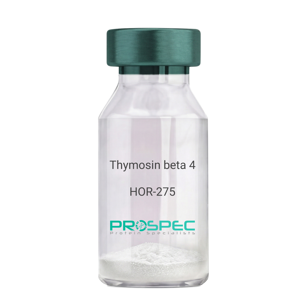
Thymosin beta 4Description:
Thymosin β4
Thymosin beta-4. TB500, TB-500
Product # :
HOR-275Price :
Quantity :
Shipping Method :
Shipped at Room temp
More Info
- description
- formulation
- purity
- More Info
- Citations
Description
Thymosin b4 is a 43 amino acid peptide which is regarded as the main intracellular G-actin sequestering peptide. It has a molecular weight of 4963.55 Da, and its molecular formula is: C212H350N56O78S1. Extracellular Thymosin b4 may contribute to physiological processes such as angiogenesis, wound healing, and regulation of inflammation.
Formulation
The protein (1mg/ml) was lyophilized with no additives.
Purity
Greater than 98.0% as determined by RP-HPLC.
Citations
More Info
-
Introduction
Thymosin is a hormone secreted from the thymus. Its primary function is to stimulate the production of T cells, which are an important part of the immune system. Thymosin also assists in the development of B cells to plasma cells to produce antibodies. The predominant form of thymosin, thymosin b4, is a member of a highly conserved family of actin monomer-sequestering proteins. b-thymosins are the primary regulators of unpolymerized actin, and are essential for maintaining the small cytoplasmic pool of free G-actin monomers required for rapid filament elongation and allowing for the flux of monomers between the thymosin-bound pool and F-actin.
-
Synonyms
Thymosin beta-4. TB500, TB-500
-
Physical Appearance
Sterile Filtered White lyophilized (freeze-dried) powder.
-
Stability
Lyophilized Thymosin b4 although stable at room temperature for 3 weeks, should be stored desiccated below -18°C. Upon reconstitution T beta 4 should be stored at 4°C between 2-7 days and for future use below -18°C.For long term storage it is recommended to add a carrier protein (0.1% HSA or BSA).Please prevent freeze-thaw cycles.
-
Solubility
It is recommended to reconstitute the lyophilized Thymosin beta-4 in sterile 18MΩ-cm H2O not less than 100 µg/ml, which can then be further diluted to other aqueous solutions.
-
Amino Acid Sequence
Thymosin b4 has an a.a. sequence of Ac-Ser-Asp-Lys-Pro-Asp-Met-Ala-Glu-Ile-Glu-Lys-Phe-Asp-Lys-Ser-Lys-Leu-Lys-Lys-Thr-Glu-Thr-Gln-Glu-Lys-Asn-Pro-Leu-Pro-Ser-Lys-Glu-Thr-Ile-Glu-Gln-Glu-Lys-Gln-Ala-Gly-Glu-Ser-OH.
-
Background
What is the molecular weight/Mw of THYMOSIN BETA 4 Protein?
THYMOSIN BETA 4 Protein has a total Mw of 4.96kDa.
What is the Purity of THYMOSIN BETA 4 Protein?
THYMOSIN BETA 4 Protein is >98% pure as determined by SDS-PAGE.
What is the Biological Activity of THYMOSIN BETA 4 Protein?
The biological functionality of THYMOSIN BETA 4 Protein will be determined in the future.
What is the amino acid sequence of THYMOSIN BETA 4 Protein?
Thymosin b4 has an a.a. sequence of Ac-Ser-Asp-Lys-Pro-Asp-Met-Ala-Glu-Ile-Glu-Lys-Phe-Asp-Lys-Ser-Lys-Leu-Lys-Lys-Thr-Glu-Thr-Gln-Glu-Lys-Asn-Pro-Leu-Pro-Ser-Lys-Glu-Thr-Ile-Glu-Gln-Glu-Lys-Gln-Ala-Gly-Glu-Ser-OH.
What applications can THYMOSIN BETA 4 Protein be used in?
THYMOSIN BETA 4 Protein can probably be used in western blot, ELISA and Lateral Flow.
What is the endotoxin level for THYMOSIN BETA 4 Protein?
The endotoxin level is minimal, THYMOSIN BETA 4 Protein was purified using conventional chromatography techniques.
Who supplies THYMOSIN BETA 4 that is in stock and available to order?
ProSpec has the protein in stock and available for immediate delivery.
Where can I order bulk mg quantities of THYMOSIN BETA 4?
ProSpec supplies bulk mg quantities at affordable prices that meets research and academic budgets.
ProSpec's products are furnished for LABORATORY RESEARCH USE ONLY. The product may not be used as drugs, agricultural or pesticidal products, food additives or household chemicals.
- View Data Sheet
Name :
CXCL9 Human, HisDescription:
MIG Human Recombinant (CXCL9), His Tag
C-X-C motif chemokine 9, CMK, crg-10, Humig, MIG, SCYB9, Gamma-interferon-induced monokine, Monokine induced by interferon-gamma, HuMIG, Small-inducible cytokine B9, CXCL9.
Product # :
CHM-016Price :
Quantity :
Shipping Method :
Shipped with Ice Packs
More Info
- description
- source
- formulation
- purity
- More Info
- SDS-PAGE
- Citations
Description
MIG Human Recombinant produced in E.Coli is a single, non-glycosylated polypeptide chain containing 126 amino acids (23-125 a.a.) and having a molecular mass of 14kDa.MIG is fused to a 23 amino acid His-tag at N-terminus & purified by proprietary chromatographic techniques.
Source
Escherichia Coli.
Formulation
MIG protein solution (0.25mg/ml) contains 20mM Tris-HCl buffer (pH 8.0), 0.15M NaCl and 30% glycerol.
Purity
Greater than 85.0% as determined by SDS-PAGE.
SDS-PAGE
Citations
More Info
-
Introduction
MIG (CXCL9) is a small cytokine which is part of the CXC chemokine family. MIG, which is also recognized as monokine is induced by gamma interferon. MIG is related to 2 other CXC chemokines named CXCL10 and CXCL11, whose genes are located next to the gene for CXCL9 on human chromosome 4. CXCL9, CXCL10 and CXCL11 obtain their chemotactic functions by interacting with the chemokine receptor CXCR3.
-
Synonyms
C-X-C motif chemokine 9, CMK, crg-10, Humig, MIG, SCYB9, Gamma-interferon-induced monokine, Monokine induced by interferon-gamma, HuMIG, Small-inducible cytokine B9, CXCL9.
-
Physical Appearance
Sterile Filtered colorless solution.
-
Stability
Store at 4°C if entire vial will be used within 2-4 weeks. Store, frozen at -20°C for longer periods of time. For long term storage it is recommended to add a carrier protein (0.1% HSA or BSA).Avoid multiple freeze-thaw cycles.
-
Amino Acid Sequence
MGSSHHHHHH SSGLVPRGSH MGSTPVVRKG RCSCISTNQG TIHLQSLKDL KQFAPSPSCE KIEIIATLKN GVQTCLNPDS ADVKELIKKW EKQVSQKKKQ KNGKKHQKKK VLKVRKSQRS RQKKTT.
-
Background
What is the molecular weight/Mw of CXCL9 HUMAN, HIS Protein?
CXCL9 HUMAN, HIS Protein has a total Mw of 14kDa.
What is the source or expression system of CXCL9 HUMAN, HIS Protein?
Escherichia Coli.
What is the Purity of CXCL9 HUMAN, HIS Protein?
CXCL9 HUMAN, HIS Protein is > 85% pure as determined by SDS-PAGE.
What is the Biological Activity of CXCL9 HUMAN, HIS Protein?
The biological functionality of CXCL9 HUMAN, HIS Protein will be determined in the future.
What is the amino acid sequence of CXCL9 HUMAN, HIS Protein?
MGSSHHHHHH SSGLVPRGSH MGSTPVVRKG RCSCISTNQG TIHLQSLKDL KQFAPSPSCE KIEIIATLKN GVQTCLNPDS ADVKELIKKW EKQVSQKKKQ KNGKKHQKKK VLKVRKSQRS RQKKTT.
What applications can CXCL9 HUMAN, HIS Protein be used in?
CXCL9 HUMAN, HIS Protein can probably be used in western blot, ELISA and Lateral Flow.
What is the endotoxin level for CXCL9 HUMAN, HIS Protein?
The endotoxin level is minimal, CXCL9 HUMAN, HIS Protein was purified using conventional chromatography techniques.Who supplies recombinant CXCL9 HUMAN, HIS that is in stock and available to order?
ProSpec has the protein in stock and available for immediate delivery.
Where can I order bulk mg quantities of CXCL9 HUMAN, HIS?
ProSpec supplies bulk mg quantities at affordable prices that meets research and academic budgets.
ProSpec's products are furnished for LABORATORY RESEARCH USE ONLY. The product may not be used as drugs, agricultural or pesticidal products, food additives or household chemicals.
- View Data Sheet
Name :
ALDH2 MouseDescription:
Aldehyde Dehydrogenase 2 Mouse Recombinant
Aldehyde dehydrogenase, mitochondrial, AHD-M1, ALDH class 2, ALDH-E2, ALDHI.
Product # :
ENZ-879Price :
Quantity :
Shipping Method :
Shipped with Ice Packs
More Info
- description
- source
- formulation
- purity
- More Info
- Citations
Description
ALDH2 Mouse Recombinant produced in E.Coli is a single, non-glycosylated polypeptide chain containing 523 amino acids (20-519 a.a) and having a molecular mass of 56.8kDa. ALDH2 is fused to a 23 amino acid His-tag at N-terminus & purified by proprietary chromatographic techniques.
Source
Escherichia Coli.
Formulation
ALDH2 protein e solution (0.5mg/ml) containing Phosphate buffered salin(pH7.4), 20% glycerol and 1mM DTT.
Purity
Greater than 85% as determined by SDS-PAGE.
Citations
More Info
-
Introduction
ALDH2 is part of the aldehyde dehydrogenase family of proteins which catalyze the chemical transformation from acetaldehyde to acetic acid. ALDH2 is the second enzyme of the major oxidative pathway of alcohol metabolism. ALDH2 has 2 major liver isoforms: cytosolic and mitochondrial, which differ by their electrophoretic mobilities, kinetic properties, and subcellular localizations. Nearly all Caucasians have 2 major isozymes, whereas roughly 50% of Orientals have only the cytosolic isozyme, omitting the mitochondrial isozyme. The extremely higher rate of acute alcohol intoxication with Orientals compared to Caucasians is due to the fact of the absence of mitochondrial isozyme. ALDH2 has a low Km for acetaldehydes, and is localized in mitochondrial matrix.
-
Synonyms
Aldehyde dehydrogenase, mitochondrial, AHD-M1, ALDH class 2, ALDH-E2, ALDHI.
-
Physical Appearance
Sterile Filtered clear solution.
-
Stability
Store at 4°C if entire vial will be used within 2-4 weeks. Store, frozen at -20°C for longer periods of time. For long term storage it is recommended to add a carrier protein (0.1% HSA or BSA).Avoid multiple freeze-thaw cycles.
-
Amino Acid Sequence
MGSSHHHHHH SSGLVPRGSH MGSSAAATSA VPAPNHQPEV FCNQIFINNE WHDAVSRKTF PTVNPSTGEV ICQVAEGNKE DVDKAVKAAR AAFQLGSPWR RMDASDRGRL LYRLADLIER DRTYLAALET LDNGKPYVIS YLVDLDMVLK CLRYYAGWAD KYHGKTIPID GDFFSYTRHE PVGVCGQIIP WNFPLLMQAW KLGPALATGN VVVMKVAEQT PLTALYVANL IKEAGFPPGV VNIVPGFGPT AGAAIASHEG VDKVAFTGST EVGHLIQVAA GSSNLKRVTL ELGGKSPNII MSDADMDWAV EQAHFALFFN QGQCCCAGSR TFVQENVYDE FVERSVARAK SRVVGNPFDS RTEQGPQVDE TQFKKILGYI KSGQQEGAKL LCGGGAAADR GYFIQPTVFG DVKDGMTIAK EEIFGPVMQI LKFKTIEEVV GRANDSKYGL AAAVFTKDLD KANYLSQALQ AGTVWINCYD VFGAQSPFGG YKMSGSGREL GEYGLQAYTE VKTVTVKVPQ KNS.
ProSpec's products are furnished for LABORATORY RESEARCH USE ONLY. The product may not be used as drugs, agricultural or pesticidal products, food additives or household chemicals.
- View Data Sheet
Name :
GAPDH Mouse, ActiveDescription:
GAPDH Mouse Recombinant, Active
Glyceraldehyde-3-phosphate dehydrogenase, GAPDH, Peptidyl-cysteine S-nitrosylase GAPDH, Gapdh, Gapd, Glyceraldehyde-3-phosphate dehydrogenase isoform 2, G3PD, GAPD, HEL-S-162eP.
Product # :
ENZ-986Price :
Quantity :
Shipping Method :
Shipped with Ice Packs
More Info
- description
- source
- formulation
- purity
- biological activity
- More Info
- Citations
Description
GAPDH Mouse Recombinant produced in E.Coli is a single, non-glycosylated polypeptide chain containing 356 amino acids (1-333a.a.) and having a molecular mass of 38.2kDa.GAPDH is fused to a 23 amino acid His-tag at N-terminus & purified by proprietary chromatographic techniques.
Source
E.coli.
Formulation
GAPDH protein solution (0.25mg/ml) containing Phosphate buffered saline (pH7.4), 20% glycerol and 1mM DTT.
Purity
Greater than 95% as determined by SDS-PAGE.
Biological Activity
Specific activity is > 40 units/mg, and is defined as the amount of enzyme that convert 1.0 umole of glyceraldehyde-3-phosphate to 1,3-Bisphosphoglycerate per minute at pH 8.5 at 37C.Citations
More Info
-
Introduction
GAPDH is a catalytic enzyme normally known to play a role in glycolysis. GAPDH exists as a tetramer composed of 36-kDa subunits and has a range of intracellular functions. GAPDH catalyzes the reversible reduction of 1,3-bisphosphoglycerate to glyceraldehyde 3-phosphophate in the presence of NADPH. Besides functioning as a glycolytic enzyme in cytoplasm, GAPDH has function in intracellular processes such as membrane fusion, microtubule bundling, phosphotransferase activity, nuclear RNA export, DNA replication and DNA repair. GAPDH catalyzes a vital energy-yielding step in carbohydrate metabolism, the reversible oxidative phosphorylation of glyceraldehyde-3-phosphate in the presence of inorganic phosphate and nicotinamide adenine dinucleotide (NAD). The enzyme exists as a tetramer of identical chains.
-
Synonyms
Glyceraldehyde-3-phosphate dehydrogenase, GAPDH, Peptidyl-cysteine S-nitrosylase GAPDH, Gapdh, Gapd, Glyceraldehyde-3-phosphate dehydrogenase isoform 2, G3PD, GAPD, HEL-S-162eP.
-
Physical Appearance
Sterile Filtered colorless solution.
-
Stability
Store at 4°C if entire vial will be used within 2-4 weeks. Store, frozen at -20°C for longer periods of time. For long term storage it is recommended to add a carrier protein (0.1% HSA or BSA).Avoid multiple freeze-thaw cycles.
-
Amino Acid Sequence
MGSSHHHHHH SSGLVPRGSH MGSMVKVGVN GFGRIGRLVT RAAICSGKVE IVAINDPFID LNYMVYMFQY DSTHGKFNGT VKAENGKLVI NGKPITIFQE RDPTNIKWGE AGAEYVVEST GVFTTMEKAG AHLKGGAKRV IISAPSADAP MFVMGVNHEK YDNSLKIVSN ASCTTNCLAP LAKVIHDNFG IVEGLMTTVH AITATQKTVD GPSGKLWRDG RGAAQNIIPA STGAAKAVGK VIPELNGKLT GMAFRVPTPN VSVVDLTCRL EKPAKYDDIK KVVKQASEGP LKGILGYTED QVVSCDFNSN SHSSTFDAGA GIALNDNFVK LISWYDNEYG YSNRVVDLMA YMASKE.
ProSpec's products are furnished for LABORATORY RESEARCH USE ONLY. The product may not be used as drugs, agricultural or pesticidal products, food additives or household chemicals.
- View Data Sheet
Name :
CRYBB1 HumanDescription:
Crystallin Beta B1 Human Recombinant
EC 1.17.4.1, RR2M, Beta-B1 crystallin, CATCN3.
Product # :
HSP-033Price :
Quantity :
Shipping Method :
Shipped with Ice Packs
More Info
- description
- source
- formulation
- purity
- More Info
- Citations
Description
CRYBB1 Recombinant Human produced in E.Coli is a single, non-glycosylated polypeptide chain containing 260 amino acids (1-252 a.a.) and having a molecular mass of 29.1 kDa. The CRYBB1 is fused to an 8 amino acid His-Tag at C-terminus and purified by proprietary chromatographic techniques.
Source
Escherichia Coli.
Formulation
The CRYBB1 solution (1mg/ml) contains 20mM Tris-HCl pH-8, 1mM DTT & 10% glycerol.
Purity
Greater than 90.0% as determined by SDS-PAGE.
Citations
More Info
-
Introduction
Crystallins are the main structural proteins of the vertebrate eye lens, where they maintain the transparency and refractive index of the lens. Crystallins are divided into 3 fprotein families, α, β, & γ families. Because lens central fiber cells lose their nuclei during development, these crystallins are prepared and then retained throughout life, making them extremely stable proteins. CRYBB1 is a beta basic group member and undergoes extensive cleavage at its N-terminal extension during lens maturation.
-
Synonyms
EC 1.17.4.1, RR2M, Beta-B1 crystallin, CATCN3.
-
Physical Appearance
Sterile filtered colorless solution.
-
Stability
Store at 4°C if entire vial will be used within 2-4 weeks. Store, frozen at -20°C for longer periods of time.For long term storage it is recommended to add a carrier protein (0.1% HSA or BSA).Avoid multiple freeze-thaw cycles.
-
Amino Acid Sequence
MSQAAKASAS ATVAVNPGPD TKGKGAPPAG TSPSPGTTLA PTTVPITSAK AAELPPGNYR LVVFELENFQ GRRAEFSGEC SNLADRGFDR VRSIIVSAGP WVAFEQSNFR GEMFILEKGE YPRWNTWSSS YRSDRLMSFR PIKMDAQEHK ISLFEGANFK GNTIEIQGDD APSLWVYGFS DRVGSVKVSS GTWVGYQYPG YRGYQYLLEP GDFRHWNEWG AFQPQMQSLR RLRDKQWHLE GSFPVLATEP PKRSHHHHHH.
ProSpec's products are furnished for LABORATORY RESEARCH USE ONLY. The product may not be used as drugs, agricultural or pesticidal products, food additives or household chemicals.
- View Data Sheet
Name :
MGLL Human, ActiveDescription:
Monoglyceride Lipase Human Recombinant, Active
Monoglyceride lipase, MGL, HU-K5, Lysophospholipase homolog, Lysophospholipase-like, Monoacylglycerol lipase, MAGL, MGLL, HUK5.
Product # :
ENZ-983Price :
Quantity :
Shipping Method :
Shipped with Ice Packs
More Info
- description
- source
- formulation
- purity
- biological activity
- More Info
- Citations
Description
MGLL Human Recombinant fused with a 20 amino acid His tag at N-terminus produced in E.Coli is a single, non-glycosylated, polypeptide chain containing 333 amino acids (1-313 a.a.) and having a molecular mass of 36.4kDa. The MGLL is purified by proprietary chromatographic techniques.
Source
Escherichia Coli.
Formulation
The MGLL solution (0.5mg/ml) contains 20mM Tris-HCl Buffer (pH 8.0) and 10% Glycerol.
Purity
Greater than 85.0% as determined by SDS-PAGE.
Biological Activity
Specific activity is > 170 units/mg, and is defined as the amount of enzyme that hydrolyze 1.0 umole of p-nitrophenyl butyrate to pnitrophenol per minute at pH 7.5 at 25C.Citations
More Info
-
Introduction
MGLL is a membrane-associated member of the serine hydrolase superfamily. MGLL is expressed in abundance in skeletal muscle and adipose tissue. MGLL functions jointly with hormone-sensitive lipase (LIPE) to hydrolyze intracellular triglyceride stores in adipocytes and other cells to fatty acids and glycerol. MGLL may also complement lipoprotein lipase (LPL) in completing hydrolysis of monoglycerides resulting from degradation of lipoprotein triglycerides.
-
Synonyms
Monoglyceride lipase, MGL, HU-K5, Lysophospholipase homolog, Lysophospholipase-like, Monoacylglycerol lipase, MAGL, MGLL, HUK5.
-
Physical Appearance
Sterile Filtered colorless solution.
-
Stability
Store at 4°C if entire vial will be used within 2-4 weeks. Store, frozen at -20°C for longer periods of time. For long term storage it is recommended to add a carrier protein (0.1% HSA or BSA).Avoid multiple freeze-thaw cycles.
-
Amino Acid Sequence
MGSSHHHHHH SSGLVPRGSH METGPEDPSS MPEESSPRRT PQSIPYQDLP HLVNADGQYL FCRYWKPTGT PKALIFVSHG AGEHSGRYEE LARMLMGLDL LVFAHDHVGH GQSEGERMVV SDFHVFVRDV LQHVDSMQKD YPGLPVFLLG HSMGGAIAIL TAAERPGHFA GMVLISPLVL ANPESATTFK VLAAKVLNLV LPNLSLGPID SSVLSRNKTE VDIYNSDPLI CRAGLKVCFG IQLLNAVSRV ERALPKLTVP FLLLQGSADR LCDSKGAYLL MELAKSQDKT LKIYEGAYHV LHKELPEVTN SVFHEINMWV SQRTATAGTA SPP.
ProSpec's products are furnished for LABORATORY RESEARCH USE ONLY. The product may not be used as drugs, agricultural or pesticidal products, food additives or household chemicals.
- View Data Sheet
Name :
IMPAD1 Human, ActiveDescription:
Inositol Monophosphatase Domain Containing 1 Human Recombinant, BioActive
Inositol monophosphatase 3, IMP 3, IMPase 3, EC 3.1.3.25, EC 3.1.3.7, Golgi 3-prime phosphoadenosine 5-prime phosphate 3-prime phosphatase, Golgi-resident PAP phosphatase, gPAPP, Inositol monophosphatase domain-containing protein 1, Inositol-1(or 4)-monophosphatase 3, Myo-inositol monophosphatase A3, IMPAD1, IMPA3, GPAPP, IMP-3
Product # :
PRO-2638Price :
Quantity :
Shipping Method :
Shipped with Ice Packs
More Info
- description
- source
- formulation
- purity
- biological activity
- More Info
- Citations
Description
IMPAD1 Human Recombinant produced in E.Coli is a single, non-glycosylated polypeptide chain containing 349 amino acids (34-359 a.a.) and having a molecular mass of 37.6kDa.IMPAD1 is fused to a 23 amino acid His-tag at N-terminus & purified by proprietary chromatographic techniques.
Source
Escherichia Coli.
Formulation
The IMPAD1 solution (0.25mg/ml) contains Phosphate-Buffered Saline (pH 7.4).
Purity
Greater than 90.0% as determined by SDS-PAGE.
Biological Activity
Specific activity is > 3300pmol/min/ug, and is defined as its ability to dephosphorylate adenosine 3'5'-diphosphate sodium slat at pH7.5, 25˚C.
Citations
More Info
-
Introduction
Inositol Monophosphatase Domain Containing 1 or IMPAD1 is a protein, part of the inositol monophosphatase group of proteins. The protein is found in Golgi apparatus and enhances phosphoadenosine phosphate hydrolysis to adenosine monophosphate. When Mutation in the IMPAD1 gene occurs leads to GRAPP type chondrodysplasia and therefore joint dislocations. On long arm chromosome 1 a pseudogene can be found.
-
Synonyms
Inositol monophosphatase 3, IMP 3, IMPase 3, EC 3.1.3.25, EC 3.1.3.7, Golgi 3-prime phosphoadenosine 5-prime phosphate 3-prime phosphatase, Golgi-resident PAP phosphatase, gPAPP, Inositol monophosphatase domain-containing protein 1, Inositol-1(or 4)-monophosphatase 3, Myo-inositol monophosphatase A3, IMPAD1, IMPA3, GPAPP, IMP-3
-
Physical Appearance
Sterile Filtered colorless solution.
-
Stability
Store at 4°C if entire vial will be used within 2-4 weeks. Store, frozen at -20°C for longer periods of time. For long term storage it is recommended to add a carrier protein (0.1% HSA or BSA).Avoid multiple freeze-thaw cycles.
-
Amino Acid Sequence
MGSSHHHHHH SSGLVPRGSH MGSGRFSLFG LGGEPGGGAA GPAAAADGGT VDLREMLAVS VLAAVRGGDE VRRVRESNVL HEKSKGKTRE GAEDKMTSGD VLSNRKMFYL LKTAFPSVQI NTEEHVDAAD QEVILWDHKI PEDILKEVTT PKEVPAESVT VWIDPLDATQ EYTEDLRKYV TTMVCVAVNG KPMLGVIHKP FSEYTAWAMV DGGSNVKARS SYNEKTPRIV VSRSHSGMVK QVALQTFGNQ TTIIPAGGAG YKVLALLDVP DKSQEKADLY IHVTYIKKWD ICAGNAILKA LGGHMTTLSG EEISYTGSDG IEGGLLASIR MNHQALVRKL PDLEKTGHK
ProSpec's products are furnished for LABORATORY RESEARCH USE ONLY. The product may not be used as drugs, agricultural or pesticidal products, food additives or household chemicals.
- View Data Sheet
Description:
TRAIL/APO 2 Ligand (114-281 a.a.) Human Recombinant, Active
Tumor necrosis factor ligand superfamily member 10, TNF-related apoptosis-inducing ligand, Protein TRAIL, Apo-2 ligand, Apo-2L, CD253 antigen, TL2, APO2L, TNFSF10.
Product # :
CYT-1014Price :
Quantity :
Shipping Method :
Shipped with Ice Packs
More Info
- description
- source
- formulation
- purity
- biological activity
- More Info
- Citations
Description
Soluble TNF-related apoptosis-inducing ligand Human Recombinant produced in E.Coli is a single, non-glycosylated polypeptide chain containing 169 amino acids (114-281) and having a molecular mass of 19.6 kDa. The sTRAIL is purified by proprietary chromatographic techniques.
Source
Escherichia Coli.
Formulation
TRAIL protein solution (1mg/ml) containing 20mM Tris-HCl pH-7.5, 300mM NaCl, 0.1mM DTT & 10% glycerol.
Purity
Greater than 95.0% as determined by SDS-PAGE.
Biological Activity
Measured in a cell cytotoxicity assay using Jurkat human T lymphocyte. The ED50 for this effect is less or equal to 1 ng/ml.
Citations
More Info
-
Introduction
TNF-related apoptosis-inducing ligand (TRAIL) is a ligand molecule which induces apoptosis. It is a type II transmembrane protein with homology to other members of the tumor necrosis factor family.
In humans, the gene that encodes for TRAIL is located at chromosome 3q26.
TRAIL binds to the death receptors, DR4 and DR5. The process of apoptosis is caspase-8-dependent. This protein preferentially induces apoptosis in transformed and tumor cells, but does not appear to kill normal cells although it is expressed at a significant level in most normal tissues. -
Synonyms
Tumor necrosis factor ligand superfamily member 10, TNF-related apoptosis-inducing ligand, Protein TRAIL, Apo-2 ligand, Apo-2L, CD253 antigen, TL2, APO2L, TNFSF10.
-
Physical Appearance
Sterile Filtered colorless liquid.
-
Stability
Store at 4°C if entire vial will be used within 2-4 weeks. Store, frozen at -20°C for longer periods of time. For long term storage it is recommended to add a carrier protein (0.1% HSA or BSA).Avoid multiple freeze-thaw cycles.
-
Amino Acid Sequence
MVRERGPQRV AAHITGTRGR SNTLSSPNSK NEKALGRKIN SWESSRSGHS FLSNLHLRNGELVIHEKGFY YIYSQTYFRF QEEIKENTKN DKQMVQYIYK YTSYPDPILL MKSARNSCWSKDAEYGLYSI YQGGIFELKE NDRIFVSVTN EHLIDMDHEA SFFGAFLVG.
ProSpec's products are furnished for LABORATORY RESEARCH USE ONLY. The product may not be used as drugs, agricultural or pesticidal products, food additives or household chemicals.
- View Data Sheet
Name :
BMP 2 Human, HEKDescription:
Bone Morphogenetic protein-2 Human Recombinant, HEK
BMP-2, BMP2A.
Product # :
CYT-080Price :
Quantity :
Shipping Method :
Shipped at Room temp
More Info
- description
- source
- formulation
- purity
- biological activity
- More Info
- Citations
Description
BMP-2 Human Recombinant produced in HEK cells is a glycosylated disulfide-linked homodimer, having a molecular weight range of 28kDa due to glycosylation. The BMP2 is purified by proprietary chromatographic techniques.
Source
HEK.
Formulation
The BMP2 was lyophilized from 0.67mg/ml in 2xPBS + 6% ethanol.
Purity
Greater than 95.0% as determined by analysis by SDS-PAGE.
Biological Activity
The specific activity as determined by the dose dependent induction of alkaline phosphatase production in the ATDC-5 cell line (Mouse chondrogenic cell line) was found to be 6.53ng/ml.
Citations
More Info
-
Introduction
BMP2 belongs to the transforming growth factor-beta (TGFB) superfamily. Bone morphogenic protein induces bone formation. BMP2 is a candidate gene for the autosomal dominant disease of fibrodysplasia (myositis) ossificans progressiva.
-
Synonyms
BMP-2, BMP2A.
-
Physical Appearance
Sterile Filtered White lyophilized (freeze-dried) powder.
-
Stability
Lyophilized BMP2 although stable at room temperature for 3 weeks, should be stored desiccated below -18°C. Upon reconstitution BMP-2 should be stored at 4°C between 2-7 days and for future use below -18°C.For long term storage it is recommended to add a carrier protein (0.1% HSA or BSA).Please prevent freeze-thaw cycles.
-
Solubility
It is recommended to reconstitute the lyophilized BMP-2 in sterile 4mM HCl containing 0.1% endotoxin-free recombinant HSA.
-
Amino Acid Sequence
QAKHKQRKRLKSSCKRHPLYVDFSDVGWNDWIVAPPGYHAFYCHGECPFPLADHLNST NHAIVQTLVNSVNSKIPKACCVPTELSAISMLYLDENEKVVLKNYQDMVVEGCGCR.
-
Background
What is the molecular weight/Mw of BMP2 Protein?
BMP2 Protein has a total Mw of 28kDa.
What is the source or expression system of BMP2 Protein?
Hek.
What is the Purity of BMP2 Protein?
BMP2 Protein is >95% pure as determined by SDS-PAGE.
What is the Biological Activity of BMP2 Protein?
The specific activity as determined by the dose dependent induction of alkaline phosphatase production in the ATDC-5 cell line (Mouse chondrogenic cell line) was found to be 6.53ng/ml.
What is the amino acid sequence of BMP2 Protein?
QAKHKQRKRLKSSCKRHPLYVDFSDVGWNDWIVAPPGYHAFYCHGECPFPLADHLNST NHAIVQTLVNSVNSKIPKACCVPTELSAISMLYLDENEKVVLKNYQDMVVEGCGCR.
What applications can BMP2 Protein be used in?
BMP2 Protein can probably be used in western blot, ELISA and Lateral Flow.
What is the endotoxin level for BMP2 Protein?
The endotoxin level is minimal, BMP2 Protein was purified using conventional chromatography techniques.
ProSpec's products are furnished for LABORATORY RESEARCH USE ONLY. The product may not be used as drugs, agricultural or pesticidal products, food additives or household chemicals.
- View Data Sheet
Name :
CDNF MouseDescription:
Cerebral Dopamine Neurotrophic Factor Mouse Recombinant
Cerebral dopamine neurotrophic factor, ARMET-like protein 1, Conserved dopamine neurotrophic factor, Cdnf, Armetl1, 9330140G23.
Product # :
CYT-729Price :
Quantity :
Shipping Method :
Shipped at Room temp
More Info
- description
- source
- formulation
- purity
- biological activity
- More Info
- Citations
Description
CDNF Mouse Recombinant produced in E.coli is a single, non-glycosylated polypeptide chain containing 163 amino acids and having a molecular mass of 18.5kDa.The CDNF is purified by proprietary chromatographic techniques.
Source
Escherichia Coli.
Formulation
CDNF protein was lyophilized from a 0.2µm filtered concentrated solution in 1xPBS, pH 7.4.
Purity
Greater than 97.0% as determined by:
(a) Analysis by RP-HPLC.
(b) Analysis by SDS-PAGE.Biological Activity
CDNF Mouse is able to enhance neurite outgrowth of E16-E18 rat embryonic cortical neurons when immobilized at 5-30 µg/mL on a nitrocellulose-coated microplate.Citations
More Info
-
Introduction
CDNF is a member of the ARMET family and acts as a trophic factor for dopamine neurons. CDNF inhibits the 6-hydroxydopamine (6-OHDA)-induced degeneration of dopaminergic neurons. When CDNF controlled after 6-OHDA-lesioning, it reestablishes the dopaminergic function and inhibits the degeneration of dopaminergic neurons in substantia nigra. CDNF is universally expressed in neuronal and non-neuronal tissues. The highest levels in the brain are found in the optic nerve and corpus callosum.
-
Synonyms
Cerebral dopamine neurotrophic factor, ARMET-like protein 1, Conserved dopamine neurotrophic factor, Cdnf, Armetl1, 9330140G23.
-
Physical Appearance
Sterile Filtered White lyophilized (freeze-dried) powder.
-
Stability
Lyophilized CDNF although stable at room temperature for 3 weeks, should be stored desiccated below -18°C. Upon reconstitution CDNF should be stored at 4°C between 2-7 days and for future use below -18°C. For long term storage it is recommended to add a carrier protein (0.1% HSA or BSA). Please prevent freeze-thaw cycles.
-
Solubility
It is recommended to reconstitute the lyophilized CDNF in sterile 18M-cm H2O not less than 100µg/ml, which can then be further diluted to other aqueous solutions.
-
Amino Acid Sequence
QGLEAGVGPR ADCEVCKEFL DRFYNSLLSR GIDFSADTIE KELLNFCSDA KGKENRLCYY LGATTDAATK ILGEVTRPMS VHIPAVKICE KLKKMDSQIC ELKYGKKLDL ASVDLWKMRV AELKQILQRW GEECRACAEK SDYVNLIREL APKYVEIYPQ TEL.
-
Background
What is the molecular weight/Mw of CDNF Protein?
CDNF Protein has a total Mw of 18.5kDa.
What is the source or expression system of CDNF Protein?
Escherichia Coli.
What is the Purity of CDNF Protein?
CDNF Protein is >97% pure as determined by SDS-PAGE.
What is the Biological Activity of CDNF Protein?
CDNF Mouse is able to enhance neurite outgrowth of E16-E18 rat embryonic cortical neurons when immobilized at 5-30 µg/mL on a nitrocellulose-coated microplate.
What is the amino acid sequence of CDNF Protein?
QGLEAGVGPR ADCEVCKEFL DRFYNSLLSR GIDFSADTIE KELLNFCSDA KGKENRLCYY LGATTDAATK ILGEVTRPMS VHIPAVKICE KLKKMDSQIC ELKYGKKLDL ASVDLWKMRV AELKQILQRW GEECRACAEK SDYVNLIREL APKYVEIYPQ TEL.
What applications can CDNF Protein be used in?
CDNF Protein can probably be used in western blot, ELISA and Lateral Flow.
What is the endotoxin level for CDNF Protein?
The endotoxin level is minimal, CDNF Protein was purified using conventional chromatography techniques.
ProSpec's products are furnished for LABORATORY RESEARCH USE ONLY. The product may not be used as drugs, agricultural or pesticidal products, food additives or household chemicals.
- View Data Sheet
Name :
CST7 HumanDescription:
Cystatin 7 Human Recombinant
Cystatin-F, Cystatin-7, Cystatin-like metastasis-associated protein, CMAP, Leukocystatin, Cystatin 7, CST7, CMAP.
Product # :
PRO-2222Price :
Quantity :
Shipping Method :
Shipped with Ice Packs
More Info
- description
- source
- formulation
- purity
- More Info
- Citations
Description
CST7 Human Recombinant produced in E. coli is a single polypeptide chain containing 132 amino acids (20-145) and having a molecular mass of 15.3kDa.CST7 is fused to a 7 amino acid His-tag at C-terminus & purified by proprietary chromatographic techniques.
Source
Escherichia Coli.
Formulation
The CST7 solution (0.5mg/1ml) contains phosphate buffered saline (pH7.4).
Purity
Greater than 95.0% as determined by SDS-PAGE.
Citations
More Info
-
Introduction
Cystatin 7 (CST7) is a glycosylated cysteine protease inhibitor with a putative role in immune regulation through inhibition of a unique target in the hematopoietic system. Cystatin-7 is comprised of multiple cystatin-like sequences. The superfamily is comprised of 3 inhibitory families: the type 1 cystatins (stefins), type 2 cystatins and the kininogens. Some members are active cysteine protease inhibitors, while others have lost or possibly never had this inhibitory activity. Type 2 cystatin proteins are a class of cysteine proteinase inhibitors found in various human fluids and secretions. CST7 protein expression has been observed in numerous human cancer cell lines established from malignant tumors.
-
Synonyms
Cystatin-F, Cystatin-7, Cystatin-like metastasis-associated protein, CMAP, Leukocystatin, Cystatin 7, CST7, CMAP.
-
Physical Appearance
Sterile Filtered clear solution.
-
Stability
Store at 4°C if entire vial will be used within 2-4 weeks. Store, frozen at -20°C for longer periods of time. For long term storage it is recommended to add a carrier protein (0.1% HSA or BSA).Avoid multiple freeze-thaw cycles.
-
Amino Acid Sequence
GPSPDTCSQD LNSRVKPGFP KTIKTNDPGV LQAARYSVEK FNNCTNDMFL FKESRITRAL VQIVKGLKYM LEVEIGRTTC KKNQHLRLDD CDFQTNHTLK QTLSCYSEVW VVPWLQHFEV PVLRCHHHHH HH.
ProSpec's products are furnished for LABORATORY RESEARCH USE ONLY. The product may not be used as drugs, agricultural or pesticidal products, food additives or household chemicals.