Skip to content

High-quality research proteins.

  • 1-800-805-6531

  • Mon - Sat 8.00-18.00, Sun - Closed

  • info@prospecbio.com

  • PO Box 6591 East Brunswick NJ 08816

  • Products
    • Cytokines
    • Growth factors
    • Chemokines
    • Cd antigens
    • Neurotrophins
    • Hormones
    • Enzymes
    • Viral antigens
    • Recombinant proteins
    • Natural proteins
    • Monoclonal antibodies
    • Polyclonal antibodies
    • Thermal Packaging
  • About
    • Company
    • Founder
    • Contribution
  • Customers
    • Eastern Asia
    • Eastern Europe
    • North America
    • South America
    • South East Asia
    • Southern Asia
    • Western Europe
  • Ordering
    • Ordering Method
    • Ordering - Credit Card
    • Ordering - PO
    • Shipping Fees
    • FAQs
  • Services
    • DNA Cloning
    • Protein Expression
    • Monoclonal Antibody
    • Fermentation
    • Technology License
  • Contact us
Log in

Country/region

  • USD $ | Albania
  • USD $ | Andorra
  • USD $ | Argentina
  • USD $ | Armenia
  • USD $ | Australia
  • USD $ | Austria
  • USD $ | Azerbaijan
  • USD $ | Belarus
  • USD $ | Belgium
  • USD $ | Bolivia
  • USD $ | Brazil
  • USD $ | Bulgaria
  • USD $ | Cambodia
  • USD $ | Canada
  • USD $ | Chile
  • USD $ | China
  • USD $ | Colombia
  • USD $ | Costa Rica
  • USD $ | Croatia
  • USD $ | Cyprus
  • USD $ | Czechia
  • USD $ | Denmark
  • USD $ | Estonia
  • USD $ | Finland
  • USD $ | France
  • USD $ | Georgia
  • USD $ | Germany
  • USD $ | Greece
  • USD $ | Hong Kong SAR
  • USD $ | Hungary
  • USD $ | Iceland
  • USD $ | India
  • USD $ | Ireland
  • USD $ | Israel
  • USD $ | Italy
  • USD $ | Japan
  • USD $ | Kazakhstan
  • USD $ | Latvia
  • USD $ | Liechtenstein
  • USD $ | Lithuania
  • USD $ | Madagascar
  • USD $ | Malta
  • USD $ | Mexico
  • USD $ | Moldova
  • USD $ | Monaco
  • USD $ | Netherlands
  • USD $ | New Zealand
  • USD $ | North Macedonia
  • USD $ | Norway
  • USD $ | Panama
  • USD $ | Paraguay
  • USD $ | Peru
  • USD $ | Philippines
  • USD $ | Poland
  • USD $ | Portugal
  • USD $ | Romania
  • USD $ | Singapore
  • USD $ | Slovakia
  • USD $ | Slovenia
  • USD $ | South Africa
  • USD $ | South Korea
  • USD $ | Spain
  • USD $ | Sri Lanka
  • USD $ | Sweden
  • USD $ | Switzerland
  • USD $ | Taiwan
  • USD $ | Thailand
  • USD $ | Türkiye
  • USD $ | Ukraine
  • USD $ | United Kingdom
  • USD $ | United States
  • USD $ | Uruguay
  • USD $ | Vatican City
  • USD $ | Venezuela
  • USD $ | Vietnam
  • Facebook
  • Instagram
logoProSpec - Recombinant Protein Manufacturer for academic and R&D industry
Become a distributor
Account Log in
Wishlist
Cart Cart(0)
  • Products
    +
    • Cytokines
    • Growth factors
    • Chemokines
    • Cd antigens
    • Neurotrophins
    • Hormones
    • Enzymes
    • Viral antigens
    • Recombinant proteins
    • Natural proteins
    • Monoclonal antibodies
    • Polyclonal antibodies
    • Thermal Packaging
  • About
    +
    • Company
    • Founder
    • Contribution
  • Customers
    +
    • Eastern Asia
    • Eastern Europe
    • North America
    • South America
    • South East Asia
    • Southern Asia
    • Western Europe
  • Ordering
    +
    • Ordering Method
    • Ordering - Credit Card
    • Ordering - PO
    • Shipping Fees
    • FAQs
  • Services
    +
    • DNA Cloning
    • Protein Expression
    • Monoclonal Antibody
    • Fermentation
    • Technology License
  • Contact us

Item added to your cart

View cart
View All
  • Cytokines
  • Growth factors
  • Chemokines
  • Cd antigens
  • Neurotrophins
  • Hormones
  • Enzymes
  • Viral antigens
  • Recombinant proteins
  • Natural proteins
  • Monoclonal antibodies
  • Polyclonal antibodies
  • Thermal Packaging
  • Tumor Necrosis Factor

    Tumor Necrosis Factor

    View All
  • 4-1BB

    4-1BB

    View All
  • Adiponectin

    Adiponectin

    View All
  • AIF1

    AIF1

    View All
  • Other Cytokines

    Other Cytokines

    View All
  • AITRL

    AITRL

    View All
  • Angiopoietin

    Angiopoietin

    View All
  • Apolipoprotein

    Apolipoprotein

    View All
  • B-Cell Activating Factor

    B-Cell Activating Factor

    View All
  • Beta Defensin

    Beta Defensin

    View All
  • Bone Morphogenetic Protein

    Bone Morphogenetic Protein

    View All
  • B type Natriuretic Peptide

    B type Natriuretic Peptide

    View All
  • BST

    BST

    View All
  • Betacellulin

    Betacellulin

    View All
  • Cardiotrophin

    Cardiotrophin

    View All
  • Activin

    Activin

    View All
  • Fibroblast Growth Factor

    Fibroblast Growth Factor

    View All
  • Other Growth Factors

    Other Growth Factors

    View All
  • CSF

    CSF

    View All
  • CTGF

    CTGF

    View All
  • IGFBP

    IGFBP

    View All
  • VEGF Protein

    VEGF Protein

    View All
  • EGF

    EGF

    View All
  • Epigen

    Epigen

    View All
  • Erythropoietin

    Erythropoietin

    View All
  • Insulin-Like Growth Factor

    Insulin-Like Growth Factor

    View All
  • Growth Hormone

    Growth Hormone

    View All
  • HDGF

    HDGF

    View All
  • Hepatocyte Growth Factor

    Hepatocyte Growth Factor

    View All
  • Insulin

    Insulin

    View All
  • BCA-1/ BLC (CXCL13)

    BCA-1/ BLC (CXCL13)

    View All
  • BRAK (CXCL14)

    BRAK (CXCL14)

    View All
  • C-10 (CCL6)

    C-10 (CCL6)

    View All
  • Other Chemokines

    Other Chemokines

    View All
  • MEC (CCL28)

    MEC (CCL28)

    View All
  • LD78-beta (CCL3L1)

    LD78-beta (CCL3L1)

    View All
  • CTACK (CCL27)

    CTACK (CCL27)

    View All
  • CXCL16

    CXCL16

    View All
  • CXCL17

    CXCL17

    View All
  • Platelet Factor-4 (CXCL4)

    Platelet Factor-4 (CXCL4)

    View All
  • ENA-78 (CXCL5)

    ENA-78 (CXCL5)

    View All
  • Eotaxin (CCL11,24,26)

    Eotaxin (CCL11,24,26)

    View All
  • Exodus-2 (CCL21)

    Exodus-2 (CCL21)

    View All
  • Fractalkine (CX3CL1)

    Fractalkine (CX3CL1)

    View All
  • CXCL6

    CXCL6

    View All
  • Other CD Antigens

    Other CD Antigens

    View All
  • CD14

    CD14

    View All
  • CD164

    CD164

    View All
  • CD1

    CD1

    View All
  • CD2

    CD2

    View All
  • CD200

    CD200

    View All
  • CD207

    CD207

    View All
  • CD226

    CD226

    View All
  • CD23

    CD23

    View All
  • CD244

    CD244

    View All
  • CD247

    CD247

    View All
  • CD27

    CD27

    View All
  • CD274

    CD274

    View All
  • CD300

    CD300

    View All
  • CD33

    CD33

    View All
  • Other Neurotrophins

    Other Neurotrophins

    View All
  • Beta-NGF

    Beta-NGF

    View All
  • BDNF

    BDNF

    View All
  • CDNF

    CDNF

    View All
  • CNTF

    CNTF

    View All
  • GDNF

    GDNF

    View All
  • Glia Maturation Factor

    Glia Maturation Factor

    View All
  • MANF

    MANF

    View All
  • Midkine

    Midkine

    View All
  • Neuroglobin

    Neuroglobin

    View All
  • Neurotrophic factors

    Neurotrophic factors

    View All
  • Neuregulin

    Neuregulin

    View All
  • Neuropilin

    Neuropilin

    View All
  • Pigment Epithelium-Derived Factor

    Pigment Epithelium-Derived Factor

    View All
  • Pleiotrophin

    Pleiotrophin

    View All
  • Peptide Hormones

    Peptide Hormones

    View All
  • Other Hormones

    Other Hormones

    View All
  • HCG

    HCG

    View All
  • Endothelin

    Endothelin

    View All
  • Exendin

    Exendin

    View All
  • FSH

    FSH

    View All
  • Glucagon

    Glucagon

    View All
  • GHRP

    GHRP

    View All
  • GLP

    GLP

    View All
  • Thyrostimulin

    Thyrostimulin

    View All
  • Inhibin A

    Inhibin A

    View All
  • LHRH

    LHRH

    View All
  • Melanotan

    Melanotan

    View All
  • Procalcitonin

    Procalcitonin

    View All
  • PTH

    PTH

    View All
  • Peroxiredoxin

    Peroxiredoxin

    View All
  • Peptidase

    Peptidase

    View All
  • Synthetase

    Synthetase

    View All
  • Transferase

    Transferase

    View All
  • Hydrolase

    Hydrolase

    View All
  • Dehydrogenase

    Dehydrogenase

    View All
  • ACE2

    ACE2

    View All
  • Esterase

    Esterase

    View All
  • Protein Kinases

    Protein Kinases

    View All
  • Other Enzymes

    Other Enzymes

    View All
  • Phosphatase

    Phosphatase

    View All
  • Deaminase

    Deaminase

    View All
  • Oxygenase

    Oxygenase

    View All
  • Adenylate Kinase

    Adenylate Kinase

    View All
  • Lyase

    Lyase

    View All
  • Chagas

    Chagas

    View All
  • SARS

    SARS

    View All
  • Influenza

    Influenza

    View All
  • ASFV

    ASFV

    View All
  • Astrovirus

    Astrovirus

    View All
  • Borrelia

    Borrelia

    View All
  • Helicobacter Pylori

    Helicobacter Pylori

    View All
  • Chikungunya

    Chikungunya

    View All
  • Chlamydia

    Chlamydia

    View All
  • Cytomegalo

    Cytomegalo

    View All
  • Dengue

    Dengue

    View All
  • EBV

    EBV

    View All
  • Ebola

    Ebola

    View All
  • Feline Leukemia Virus

    Feline Leukemia Virus

    View All
  • Hepatitis A

    Hepatitis A

    View All
  • Other

    Other

    View All
  • Actin

    Actin

    View All
  • ADAM

    ADAM

    View All
  • Complement Component

    Complement Component

    View All
  • Ag85

    Ag85

    View All
  • Eukaryotic Translation Initiation Factor

    Eukaryotic Translation Initiation Factor

    View All
  • Anterior Gradient Protein

    Anterior Gradient Protein

    View All
  • Heat Shock Protein

    Heat Shock Protein

    View All
  • Alpha-2-HS-Glycoprotein

    Alpha-2-HS-Glycoprotein

    View All
  • Hemoglobin

    Hemoglobin

    View All
  • Allergy

    Allergy

    View All
  • Anaplasma

    Anaplasma

    View All
  • Angiogenin

    Angiogenin

    View All
  • Ankyrin Repeat Domain

    Ankyrin Repeat Domain

    View All
  • Annexin

    Annexin

    View All
  • Other Natural Proteins

    Other Natural Proteins

    View All
  • Aprotinin

    Aprotinin

    View All
  • Transferrin

    Transferrin

    View All
  • Avidin

    Avidin

    View All
  • Anti Coagulation Factors

    Anti Coagulation Factors

    View All
  • Natural Albumin

    Natural Albumin

    View All
  • Natural Coagulation Factors

    Natural Coagulation Factors

    View All
  • Fibronectin

    Fibronectin

    View All
  • Thrombin

    Thrombin

    View All
  • Anti Human Enzyme

    Anti Human Enzyme

    View All
  • Other Monoclonal Antibodies

    Other Monoclonal Antibodies

    View All
  • Anti Human Cytokine

    Anti Human Cytokine

    View All
  • Anti Human Heat Shock Protein

    Anti Human Heat Shock Protein

    View All
  • Anti Mouse Lymphocyte

    Anti Mouse Lymphocyte

    View All
  • Anti Human Chemokine

    Anti Human Chemokine

    View All
  • Anti Human Lymphocyte

    Anti Human Lymphocyte

    View All
  • Anti Viral Monoclonal

    Anti Viral Monoclonal

    View All
  • Anti Mouse Cytokine

    Anti Mouse Cytokine

    View All
  • Anti-GST Monoclonal

    Anti-GST Monoclonal

    View All
  • Other Polyclonal Antibodies

    Other Polyclonal Antibodies

    View All
  • Anti Viral

    Anti Viral

    View All
  • Anti-GST Monoclonal

    Anti-GST Monoclonal

    View All
Back

Search results

1000 results found for “Inhibin A”

Name

Description

Product #

Price

Quantity

Shipping Method

  • View Data Sheet

    Name :

    STAT1 Antibody

    Description:

    STAT1, Mouse Anti Human

    Signal transducer and activator of transcription 1-alpha/beta, Transcription factor ISGF-3 components p91/p84, STAT1, ISGF-3, STAT91, DKFZp686B04100.

    Product # :

    ANT-468

    Price :

    Quantity :

    Shipping Method :

    Ice Icon

    Shipped with Ice Packs

    Add To Cart

    More Info

    • formulation
    • More Info
    • Citations

    Formulation

    1mg/ml containing PBS, pH-7.4, 10% Glycerol and 0.02% Sodium Azide.

    Citations

    More Info

    • Introduction

      STAT1 is a member of the Signal Transducers and Activators of Transcriptionfamily of transcription factors. STAT1 is involved in upregulating genes due to a signal by either type Ior type IIinterferons. In response to IFN-?stimulation, STAT1 forms homodimers or heterodimers with STAT3that bind to the GAS (Interferon-Gamma Activated Sequence) promoter element; in response to either IFN-? or IFN-?stimulation, STAT1 forms a heterodimer with STAT2that can bind the ISRE (Interferon Stimulated Response Element) promoter element. In either case, binding of the promoter element leads to an increased expression of ISG (Interferon Stimulated Genes).

    • Synonyms

      Signal transducer and activator of transcription 1-alpha/beta, Transcription factor ISGF-3 components p91/p84, STAT1, ISGF-3, STAT91, DKFZp686B04100.

    • Physical Appearance

      Sterile filtered colorless solution.

    • Immunogen

      Anti-human STAT1 mAb, clone PAT1F4A, is derived from hybridization of mouse F0 myeloma cells with spleen cells from BALB/c mice immunized with a recombinant human STAT1 protein 1-712 amino acids purified from E. coli.

    • Ig Subclass

      Mouse IgG1 heavy chain and Kappa light chain.

    • Clone

      PAT1F4A.

    • Applications

      The antibody has been tested by ELISA, Western blot analysis to assure specificity and reactivity. Since application varies, however, each investigation should be titrated by the reagent to obtain optimal results. Recommended dilution range for Western blot analysis is 1:500 ~ 1:1000. Recommended starting dilution is 1:500.

    • Type

      Mouse Anti Human Monoclonal.

    • Storage Procedures

      For periods up to 1 month store at 4°C, for longer periods of time, store at -20°C. Prevent freeze thaw cycles.

    ProSpec's products are furnished for LABORATORY RESEARCH USE ONLY. The product may not be used as drugs, agricultural or pesticidal products, food additives or household chemicals.

    Stat1 Antibody
  • View Data Sheet

    Name :

    PAIP2 Antibody

    Description:

    Polyadenylate-Binding Protein-Interacting Protein 2, Mouse Anti Human

    Poly(A) Binding Protein Interacting Protein 2, PABP- interacting protein 2, HSPC218, PAIP-2, PAIP2A, Polyadenylate-binding protein-interacting protein 2, PAIP2, MGC72018.

    Product # :

    ANT-094

    Price :

    Quantity :

    Shipping Method :

    Ice Icon

    Shipped with Ice Packs

    Add To Cart

    More Info

    • formulation
    • More Info
    • Citations

    Formulation

    1mg/ml containing PBS, pH-7.4, & 0.1% Sodium Azide.

    Citations

    More Info

    • Introduction

      PAIP2 has a role as a repressor in the regulation of translation initiation of poly(A)-containing mRNAs. PAIP2 inhibitory activity on translation is mediated through its action on PABPC1. PAIP2 displaces the interaction of PABPC1 with poly(A) RNA and competes with PAIP1 for binding to PABPC1. PAIP2 association with PABPC1 results in disruption of the cytoplasmic poly(A) RNP structure organization.

    • Synonyms

      Poly(A) Binding Protein Interacting Protein 2, PABP- interacting protein 2, HSPC218, PAIP-2, PAIP2A, Polyadenylate-binding protein-interacting protein 2, PAIP2, MGC72018.

    • Physical Appearance

      Sterile Filtered colorless solution.

    • Immunogen

      Anti-human PAIP2 mAb is derived from hybridization of mouse F0 myeloma cells with spleen cells from BALB/c mice immunized with recombinant human CMBL 1-127 amino acids purified from E. coli.

    • Ig Subclass

      Mouse IgG2b heavy chain and ? light chain.

    • Clone

      PAT1C12AT.

    • Applications

      PAIP2 antibody has been tested by ELISA, Western blot analysis to assure specificity and reactivity. Since application varies, however, each investigation should be titrated by the reagent to obtain optimal results. Recommended dilution range for Western blot analysis is 1:500 ~ 1:5000. Recommended starting dilution is 1:1000.

    • Type

      Mouse Anti Human Monoclonal.

    • Storage Procedures

      For periods up to 1 month store at 4°C, for longer periods of time, store at -20°C. Prevent freeze thaw cycles.

    • Purification Method

      PAIP2 antibody was purified from mouse ascitic fluids by protein-G affinity chromatography.

    ProSpec's products are furnished for LABORATORY RESEARCH USE ONLY. The product may not be used as drugs, agricultural or pesticidal products, food additives or household chemicals.

    Paip2 Antibody
  • View Data Sheet

    Name :

    Cathepsin H Antibody

    Description:

    Cathepsin H, Mouse Anti Human

    Cathepsin H, CTSH, CPSB, ACC-4, ACC-5, MGC1519, minichain, DKFZp686B24257.

    Product # :

    ANT-405

    Price :

    Quantity :

    Shipping Method :

    Ice Icon

    Shipped with Ice Packs

    Add To Cart

    More Info

    • formulation
    • More Info
    • Citations

    Formulation

    1mg/ml containing PBS, pH-7.4, & 0.1% Sodium Azide.

    Citations

    More Info

    • Introduction

      Cathepsin H is a lysosomal cysteine proteinase important in the overall degradation of lysosomal proteins. Cathepsin H is composed of a dimer of disulfide-linked heavy and light chains, both produced from a single protein precursor. Cathepsin H, which belongs to the peptidase C1 protein family, can act both as an aminopeptidase and as an endopeptidase. Increased expression of the Cathepsin H gene has been associated with malignant progression of prostate tumors.

    • Synonyms

      Cathepsin H, CTSH, CPSB, ACC-4, ACC-5, MGC1519, minichain, DKFZp686B24257.

    • Immunogen

      Anti-human Cathepsin H mAb, is derived from hybridization of mouse SP2/O myeloma cells with spleen cells from BALB/c mice immunized with recombinant human Cathepsin H amino acids 23-335 purified from E. coli.

    • Ig Subclass

      Mouse IgG2a heavy chain and κ light chain.

    • Clone

      P3G8AT.

    • Applications

      Cathepsin H antibody has been tested by ELISA and Western blot analysis to assure specificity and reactivity. Since application varies, however, each investigation should be titrated by the reagent to obtain optimal results. Recommended dilution range for Western blot analysis is 1:500 ~ 2,000. Recommended starting dilution is 1:1,000.

    • Type

      Mouse Anti Human Monoclonal.

    • Storage Procedures

      For periods up to 1 month store at 4°C, for longer periods of time, store at -20°C. Prevent freeze thaw cycles.

    • Purification Method

      Cathepsin H antibody was purified from mouse ascitic fluids by protein-G affinity chromatography.

    ProSpec's products are furnished for LABORATORY RESEARCH USE ONLY. The product may not be used as drugs, agricultural or pesticidal products, food additives or household chemicals.

    Cathepsin H Antibody
  • View Data Sheet

    Name :

    PPIC Antibody

    Description:

    Peptidylprolyl Isomerase C, Mouse Anti Human

    Peptidyl-prolyl cis-trans isomerase C, PPIase C, Cyclophilin C, Rotamase C, PPIC, CYPC.

    Product # :

    ANT-069

    Price :

    Quantity :

    Shipping Method :

    Ice Icon

    Shipped with Ice Packs

    Add To Cart

    More Info

    • formulation
    • More Info
    • Citations

    Formulation

    1mg/ml containing PBS, pH-7.4, & 0.1% Sodium Azide.

    Citations

    More Info

    • Introduction

      Peptidylprolyl Isomerase C (PPIC) belongs to the peptidyl-prolyl cis-trans isomerase (PPIase) family. PPIases catalyze the cis-trans isomerization of proline imidic peptide bonds in oligopeptides and hasten the folding of proteins. Like other PPIases, PPIC binds immunosuppressant cyclosporin A.

    • Synonyms

      Peptidyl-prolyl cis-trans isomerase C, PPIase C, Cyclophilin C, Rotamase C, PPIC, CYPC.

    • Immunogen

      Anti-human PPIC mAb, is derived from hybridization of mouse FO myeloma cells with spleen cells from BALB/c mice immunized with recombinant human PPIC amino acids 39-197 purified from E. coli.

    • Ig Subclass

      Mouse IgG2b heavy chain and k light chain.

    • Clone

      PAT3C6AT.

    • Applications

      PPIC antibody has been tested by ELISA, Western blot analysis to assure specificity and reactivity. Since application varies, however, each investigation should be titrated by the reagent to obtain optimal results. Recommended dilution range for Western blot analysis is 1:1000.

    • Type

      Mouse Anti Human Monoclonal.

    • Storage Procedures

      For periods up to 1 month store at 4°C, for longer periods of time, store at -20°C. Prevent freeze thaw cycles.

    • Purification Method

      PPIC antibody was purified from mouse ascitic fluids by protein-G affinity chromatography.

    ProSpec's products are furnished for LABORATORY RESEARCH USE ONLY. The product may not be used as drugs, agricultural or pesticidal products, food additives or household chemicals.

    Ppic Antibody
  • View Data Sheet

    Name :

    PRDX1 Antibody

    Description:

    Peroxiredoxin-1, Mouse Anti Human

    Peroxiredoxin-1, EC 1.11.1.15, Thioredoxin peroxidase 2, Thioredoxin-dependent peroxide reductase 2, Proliferation-associated gene protein, Natural killer cell-enhancing factor A, NKEF-A, PRDX1, TDPX2, PRDX-1, PAG, PAGA, PRX1, PAGB, PRXI, MSP23, NKEFA.

    Product # :

    ANT-394

    Price :

    Quantity :

    Shipping Method :

    Ice Icon

    Shipped with Ice Packs

    Add To Cart

    More Info

    • formulation
    • More Info
    • Citations

    Formulation

    1mg/ml containing PBS, pH-7.4, & 0.1% Sodium Azide.

    Citations

    More Info

    • Introduction

      PRDX1 is part of the peroxiredoxin family of antioxidant enzymes, which reduce hydrogen peroxide and alkyl hydroperoxides. PRDX1 is an important protector of red blood cells against reactive oxygen species and in tumor prevention. PRDX1 is antioxidant protective in cells, and contributes to the antiviral activity of CD8(+) T-cells. PRDX1 has a proliferative effect and is involved in cancer development or progression. Peroxiredoxin-1 is plays a role in redox regulation of the cell. Peroxiredoxin decreases peroxides with reducing equivalents provided through the thioredoxin system but not from glutaredoxin. Peroxiredoxin is involved in eliminating peroxides generated during metabolism. Peroxiredoxin participates in the signaling cascades of growth factors and TNF-alpha by regulating the intracellular concentrations of h(2)o(2).

    • Synonyms

      Peroxiredoxin-1, EC 1.11.1.15, Thioredoxin peroxidase 2, Thioredoxin-dependent peroxide reductase 2, Proliferation-associated gene protein, Natural killer cell-enhancing factor A, NKEF-A, PRDX1, TDPX2, PRDX-1, PAG, PAGA, PRX1, PAGB, PRXI, MSP23, NKEFA.

    • Immunogen

      Anti-human PRDX1 mAb is derived from hybridization of mouse F0 myeloma cells with spleen cells from BALB/c mice immunized with recombinant human PRDX1 amino acids 1-199 purified from E. coli.

    • Ig Subclass

      Mouse IgG1 heavy chain and κ light chain.

    • Clone

      PAT1D8AT.

    • Applications

      PRDX1 antibody has been tested by ELISA and Western blot analysis to assure specificity and reactivity. Since application varies, however, each investigation should be titrated by the reagent to obtain optimal results. Recommended dilution range for Western blot analysis is 1:1000 ~ 2000. Recommended starting dilution is 1:1000.

    • Type

      Mouse Anti Human Monoclonal.

    • Storage Procedures

      For periods up to 1 month store at 4°C, for longer periods of time, store at -20°C. Prevent freeze thaw cycles.

    • Purification Method

      PRDX1 antibody was purified from mouse ascitic fluids by protein-G affinity chromatography.

    ProSpec's products are furnished for LABORATORY RESEARCH USE ONLY. The product may not be used as drugs, agricultural or pesticidal products, food additives or household chemicals.

    Prdx1 Antibody
  • View Data Sheet

    Name :

    GSTP1 Antibody

    Description:

    Glutathione S-Transferase pi 1, Mouse Anti Human

    Glutathione S-transferase P 1, Gst P1, GST YF-YF, GST class-pi, GST-piB, Preadipocyte growth factor.

    Product # :

    ANT-729

    Price :

    Quantity :

    Shipping Method :

    Ice Icon

    Shipped with Ice Packs

    Add To Cart

    More Info

    • formulation
    • More Info
    • Citations

    Formulation

    1mg/ml containing PBS, pH-7.4, 10% Glycerol and 0.02% Sodium Azide.

    Citations

    More Info

    • Introduction

      GSTP1 is a polymorphic gene encoding active, functionally different GSTP1 variant proteins that are believed to function in xenobiotic metabolism and have a part in susceptibility to cancer, and other diseases. GSTP1 is a glutathione S-transferase belonging to the pi class. GST family enzymes play a significant role in detoxification by catalyzing the conjugation of many hydrophobic and electrophilic compounds with reduced glutathione. Based upon the biochemical, immunologic and structural properties of the soluble GSTs they are grouped into 4 main classes: alpha, mu, pi, and theta. The GSTP1 enzyme acts by catalyzing the reaction of glutathione with an acceptor molecule to form a Sulfur-substituted glutathione. The reactions employing glutathione contribute the transformation of a broad range of electrophiles, including reactive products of lipid, protein, carcinogens, therapeutic drugs, environmental toxins, and products of oxidative stress.
      The GSTP1 inactivation through CpG hypermethylation is frequent in pituitary adenomas and may be a factor in aggressive pituitary tumor behavior. GSTP1 is may be a transcriptional target of the p53 tumor suppressor gene. Single-nucleotide polymorphism in GSTP1 is linked to modified protein binding, which influence GSTP1's contribution to carcinogen and drug metabolism, and possibly disease pathogenesis and/or drug response. GST-pi might have central roles in proliferation of androgen-independent human prostate cancer cells.

    • Synonyms

      Glutathione S-transferase P 1, Gst P1, GST YF-YF, GST class-pi, GST-piB, Preadipocyte growth factor.

    • Physical Appearance

      Sterile filtered colorless solution.

    • Immunogen

      Anti-human GSTP1 mAb, is derived from hybridization of mouse F0 myeloma cells with spleen cells from BALB/c mice immunized with recombinant human GSTP1 amino acids 1-210 purified from E. coli.

    • Ig Subclass

      Mouse IgG1 heavy chain and κ light chain.

    • Clone

      PAT12C10AT

    • Applications

      GSTP1 antibody has been tested by ELISA, Western blot analysis, ICC/IF and Flow cytometry to assure specificity and reactivity. Since application varies, however, each investigation should be titrated by the reagent to obtain optimal results.

    • Type

      Mouse Anti Human Monoclonal.

    • Storage Procedures

      For periods up to 1 month store at 4°C, for longer periods of time, store at -20°C. Prevent freeze thaw cycles.

    • Purification Method

      GSTP1 antibody was purified from mouse ascitic fluids by protein-A affinity chromatography.

    ProSpec's products are furnished for LABORATORY RESEARCH USE ONLY. The product may not be used as drugs, agricultural or pesticidal products, food additives or household chemicals.

    Gstp1 Antibody
  • View Data Sheet

    Name :

    TNF a Rabbit

    Description:

    Tumor Necrosis Factor-Alpha Rabbit Recombinant

    Tumor necrosis factor, Cachectin, TNF-alpha, Tumor necrosis factor ligand superfamily member 2, TNF-a, TNF, TNFA, TNFSF2. 

    Product # :

    CYT-008

    Price :

    Quantity :

    Shipping Method :

    Room Temp Icon

    Shipped at Room temp

    Add To Cart

    More Info

    • description
    • source
    • formulation
    • purity
    • biological activity
    • More Info
    • Citations

    Description

    Tumor Necrosis Factor-a Rabbit Recombinant consists of three identical polypeptide chains of 158 amino acids combined to form a compact, bell-shaped homotrimer. TNF-alpha was produced in E.Coli is a non-glycosylated, polypeptide chain having a molecular mass of 17.4 kDa for the individual subunit. The TNF-alpha is purified by standard chromatographic techniques.

    Source

    Escherichia Coli.

    Formulation

    TNF-alpha Rabbit was lyophilized after extensive dialysis against 20mM PB, pH7.4, 300mM NaCl.

    Purity

    Greater than 95% as determined by:
    (a) Analysis by RP-HPLC.
    (b) Analysis by SDS-PAGE.

    Biological Activity

    The ED50 as determined by the cytolysis of murine L929 cells in the presence of Actinomycin D is less than 0.03ng/ml, corresponding to a Specific Activity of 30,000,000 IU/mg.

    Citations

    More Info

    • Introduction

      Tumor necrosis factor is a cytokine involved in systemic inflammation and is a member of a group of cytokines that all stimulate the acute phase reaction. TNF is mainly secreted by macrophages.
      TNF causes apoptotic cell death, cellular proliferation, differentiation, inflammation, tumorigenesis and viral replication, TNF is also involved in lipid metabolism, and coagulation. TNF's primary role is in the regulation of immune cells.
      Dysregulation and, in particular, overproduction of TNF have been implicated in a variety of human diseases- autoimmune diseases, insulin resistance, and cancer.

    • Synonyms

      Tumor necrosis factor, Cachectin, TNF-alpha, Tumor necrosis factor ligand superfamily member 2, TNF-a, TNF, TNFA, TNFSF2.

    • Physical Appearance

      Sterile Filtered White lyophilized (freeze-dried) powder.

    • Stability

      Lyophilized Tumor Necrosis Factor-a although stable at room temperature for 3 weeks, should be stored desiccated below -18°C. Upon reconstitution TNF-a should be stored at 4°C between 2-7 days and for future use below -18°C.For long term storage it is recommended to add a carrier protein (0.1% HSA or BSA).Please prevent freeze-thaw cycles.

    • Solubility

      It is recommended to reconstitute the lyophilized Tumor Necrosis Factor-alpha in sterile 18MΩ-cm H2O not less than 100µg/ml, which can then be further diluted to other aqueous solutions.

    • Amino Acid Sequence

      The sequence of the first five N-terminal amino acids was determined and was found to be Met-Ser-Ala-Ser-Arg.

    ProSpec's products are furnished for LABORATORY RESEARCH USE ONLY. The product may not be used as drugs, agricultural or pesticidal products, food additives or household chemicals.

    Tnf A Rabbit
  • View Data Sheet

    Name :

    HMOX1 Antibody

    Description:

    Heme Oxygenase 1, Mouse Anti Human

    HO-1, HSP32, bK286B10, HMOX-1, Heme oxygenase 1, HMOX1, HO, HO1.

    Product # :

    ANT-683

    Price :

    Quantity :

    Shipping Method :

    Ice Icon

    Shipped with Ice Packs

    Add To Cart

    More Info

    • formulation
    • More Info
    • Citations

    Formulation

    1mg/ml containing PBS, pH-7.4, 10% Glycerol and 0.02% Sodium Azide.

    Citations

    More Info

    • Introduction

      HMOX1 cleaves the heme ring at the alpha methene bridge to form Biliverdin. Biliverdin is then converted to Bilirubin by Biliverdin reductase. In physiological state, the highest activity of HMOX1 is found in the spleen, where senescent erythrocytes are sequestrated and destroyed. Heme Oxygenase-1 is involved in the regulation of cardiovascular function and its adaptive response to a variety of stressors. HMOX1 is induced in the colon of ulcerative colitis. HMOX1 is found to overexpress with a higher extent of intraplaque angiogenesis implies a multi-faceted role for HMOX1 in modulating the progression of atherosclerosis. HMOX1 expression reduced LPS-stimulated secretion of MCP-1, IL-6, IL-10, and TNF-alpha in murine and human macrophages.

    • Synonyms

      HO-1, HSP32, bK286B10, HMOX-1, Heme oxygenase 1, HMOX1, HO, HO1.

    • Physical Appearance

      Sterile filtered colorless solution.

    • Immunogen

      Anti-human HMOX1 mAb, is derived from hybridization of mouse F0 myeloma cells with spleen cells from BALB/c mice immunized with recombinant human HMOX1 amino acids 1-266 purified from E. coli.

    • Ig Subclass

      Mouse IgG2a heavy chain and K light chain.

    • Clone

      PAT1D6AT.

    • Applications

      HMOX1 antibody has been tested by ELISA, Western blot analysis, Flow cytometry and ICC/IF to assure specificity and reactivity. Since application varies, however, each investigation should be titrated by the reagent to obtain optimal results.

    • Type

      Mouse Anti Human Monoclonal.

    • Storage Procedures

      For periods up to 1 month store at 4°C, for longer periods of time, store at -20°C. Prevent freeze thaw cycles.

    • Purification Method

      HMOX1 antibody was purified from mouse ascitic fluids by protein-A affinity chromatography.

    ProSpec's products are furnished for LABORATORY RESEARCH USE ONLY. The product may not be used as drugs, agricultural or pesticidal products, food additives or household chemicals.

    Hmox1 Antibody
  • View Data Sheet

    Name :

    YWHAQ Antibody

    Description:

    Tyr-3/Trp- 5 Monooxygenase Activation Protein Theta, Mouse Anti Human

    14-3-3 theta, 14-3-3 tau, 14-3-3 T-cell, HS1, YWHAQ, 1C5, 14-3-3 Tau, Tyr-3/Trp- 5 Monooxygenase Activation Protein Theta.

    Product # :

    ANT-387

    Price :

    Quantity :

    Shipping Method :

    Ice Icon

    Shipped with Ice Packs

    Add To Cart

    More Info

    • formulation
    • More Info
    • Citations

    Formulation

    1mg/ml containing PBS, pH-7.4, 10% glycerol & 0.02% Sodium Azide.

    Citations

    More Info

    • Introduction

      The 14-3-3 family of proteins plays a key regulatory role in signal transduction, checkpoint control, apoptotic and nutrient-sensing pathways. 14-3-3 proteins are highly conserved and ubiquitously expressed. There are at least seven isoforms, β, γ, ε, σ, ζ, τ and η that have been identified in mammals. The 14-3-3 tau, a subtype of the 14-3-3 family of proteins, was found in T Cells, brain and testes. This 14-3-3 tau is upregulated in patients with amyotrophic lateral sclerosis.

    • Synonyms

      14-3-3 theta, 14-3-3 tau, 14-3-3 T-cell, HS1, YWHAQ, 1C5, 14-3-3 Tau, Tyr-3/Trp- 5 Monooxygenase Activation Protein Theta.

    • Immunogen

      Anti-human YWHAQ mAb is derived from hybridization of mouse F0 myeloma cells with spleen cells from BALB/c mice immunized with recombinant human YWHAQ amino acids 1-245 purified from E. coli.

    • Ig Subclass

      Mouse IgG1 heavy chain and κ light chain.

    • Clone

      PAT1A1AT.

    • Applications

      YWHAQ antibody has been tested by ELISA and Western blot analysis to assure specificity and reactivity. Since application varies, however, each investigation should be titrated by the reagent to obtain optimal results. Recommended dilution range for Western blot analysis is 1:1000 ~ 2000. Recommended starting dilution is 1:1000.

    • Type

      Mouse Anti Human Monoclonal.

    • Storage Procedures

      For periods up to 1 month store at 4°C, for longer periods of time, store at -20°C. Prevent freeze thaw cycles.

    • Purification Method

      YWHAQ antibody was purified from mouse ascitic fluids by protein-G affinity chromatography.

    ProSpec's products are furnished for LABORATORY RESEARCH USE ONLY. The product may not be used as drugs, agricultural or pesticidal products, food additives or household chemicals.

    Ywhaq Antibody
  • View Data Sheet

    Name :

    NAA30 Human

    Description:

    N Alpha-Acetyltransferase 30, NatC Catalytic Subunit Human Recombinant

    N(Alpha)-Acetyltransferase 30, NatC Catalytic Subunit, C14orf35, NAT12, N-Acetyltransferase 12 (GCN5-Related, Putative), N-Acetyltransferase 12, NatC Catalytic Subunit, MAK3, N-Acetyltransferase MAK3 Homolog, Chromosome 14 Open Reading Frame 35, Mak3p, NAT12P, N-Alpha-Acetyltransferase 30, N-Alpha-Acetyltransferase 30, NatC Catalytic Subunit, Putative N-Acetyltransferase, EC 2.3.1.88.

    Product # :

    ENZ-720

    Price :

    Quantity :

    Shipping Method :

    Ice Icon

    Shipped with Ice Packs

    Add To Cart

    More Info

    • description
    • source
    • formulation
    • purity
    • More Info
    • Citations

    Description

    NAA30 Human Recombinant produced in E.Coli is a single, non-glycosylated polypeptide chain containing 385 amino acids (1-362 a.a) and having a molecular mass of 41.7kDa.NAA30 is fused to a 23 amino acid His-tag at N-terminus & purified by proprietary chromatographic techniques.

    Source

    Escherichia Coli.

    Formulation

    NAA30 protein solution (0.25mg/ml) containing 20mM Tris-HCl buffer (pH 8.0), 0.15M NaCl, 10% glycerol and 1mM DTT.

    Purity

    Greater than 90.0% as determined by SDS-PAGE.

    Citations

    More Info

    • Introduction

      N-alpha-acetyltransferase 30 (NAA30) is catalytic subunit of the N-terminal acetyltransferase C (NatC) complex. NAA30 catalyzes acetylation of the N-terminal methionine residues of peptides beginning with Met-Leu-Ala and Met-Leu-Gly. In addition, NAA30 is essential for the lysosomal localization and function of ARL8B. The disease Eastern equine encephalitis has been associated with NAA30.

    • Synonyms

      N(Alpha)-Acetyltransferase 30, NatC Catalytic Subunit, C14orf35, NAT12, N-Acetyltransferase 12 (GCN5-Related, Putative), N-Acetyltransferase 12, NatC Catalytic Subunit, MAK3, N-Acetyltransferase MAK3 Homolog, Chromosome 14 Open Reading Frame 35, Mak3p, NAT12P, N-Alpha-Acetyltransferase 30, N-Alpha-Acetyltransferase 30, NatC Catalytic Subunit, Putative N-Acetyltransferase, EC 2.3.1.88.

    • Physical Appearance

      Sterile Filtered clear solution.

    • Stability

      Store at 4°C if entire vial will be used within 2-4 weeks. Store, frozen at -20°C for longer periods of time. For long term storage it is recommended to add a carrier protein (0.1% HSA or BSA).Avoid multiple freeze-thaw cycles.

    • Amino Acid Sequence

      MGSSHHHHHH SSGLVPRGSH MGSMAEVPPG PSSLLPPPAP PAPAAVEPRC PFPAGAALAC CSEDEEDDEE HEGGGSRSPA GGESATVAAK GHPCLRCPQP PQEQQQLNGL ISPELRHLRA AASLKSKVLS VAEVAATTAT PDGGPRATAT KGAGVHSGER PPHSLSSNAR TAVPSPVEAA AASDPAAARN GLAEGTEQEE EEEDEQVRLL SSSLTADCSL RSPSGREVEP GEDRTIRYVR YESELQMPDI MRLITKDLSE PYSIYTYRYF IHNWPQLCFL AMVGEECVGA IVCKLDMHKK MFRRGYIAML AVDSKYRRNG IGTNLVKKAI YAMVEGDCDE VVLETEITNK SALKLYENLG FVRDKRLFRY YLNGVDALRL KLWLR

    ProSpec's products are furnished for LABORATORY RESEARCH USE ONLY. The product may not be used as drugs, agricultural or pesticidal products, food additives or household chemicals.

    Naa30 Human
  • View Data Sheet

    Name :

    SST

    Description:

    Somatostatin

    Growth hormone release-inhibiting factor, SST, SMS, SMST, GHIH.

    Product # :

    HOR-299

    Price :

    Quantity :

    Shipping Method :

    Room Temp Icon

    Shipped at Room temp

    Add To Cart

    More Info

    • description
    • formulation
    • purity
    • More Info
    • Citations

    Description

    Somatostatin Synthetic is a single, non-glycosylated polypeptide chain containing 14 amino acids, having a molecular mass of 1637.9 Dalton and a Molecular formula of C76H104N18O19S2. The CAS# is 38916-34-6.

    Formulation

    The protein was lyophilized with no additives.

    Purity

    Greater than 98.0% as determined by:
    (a) Analysis by RP-HPLC.
    (b) Analysis by SDS-PAGE.

    Citations

    More Info

    • Introduction

      Somatostatin (also known as growth hormone inhibiting hormone (GHIH) or somatotropin release-inhibiting hormone (SRIF) is a peptide hormone that regulates the endocrine systemand affects neurotransmission and cell proliferation via interaction with G-protein-coupled somatostatin receptors and inhibition of the release of numerous secondary hormones. Somatostatin has two active forms produced by alternative cleavage of a single preproprotein: one of 14 amino acids, the other of 28 amino acids.

    • Synonyms

      Growth hormone release-inhibiting factor, SST, SMS, SMST, GHIH.

    • Physical Appearance

      Sterile Filtered White lyophilized (freeze-dried) powder.

    • Stability

      Lyophilized SST although stable at room temperature for 3 weeks, should be stored desiccated below -18°C. Upon reconstitution Somatostatin should be stored at 4°C between 2-7 days and for future use below -18°C.For long term storage it is recommended to add a carrier protein (0.1% HSA or BSA).Please prevent freeze-thaw cycles.

    • Solubility

      It is recommended to reconstitute the lyophilized Somatostatin in sterile 18MΩ-cm H2O not less than 100 µg/ml, which can then be further diluted to other aqueous solutions.

    • Amino Acid Sequence

      Ala-Gly-Cys-Lys-Asn-Phe- Phe-Trp-Lys-Thr-Phe-Thr-Ser-Cys-OH.

    ProSpec's products are furnished for LABORATORY RESEARCH USE ONLY. The product may not be used as drugs, agricultural or pesticidal products, food additives or household chemicals.

    Somatostatin
  • View Data Sheet

    Name :

    AZGP1 Human

    Description:

    Alpha-2-Glycoprotein 1 Zinc-Binding Human

    Zn-alpha-2-glycoprotein, Zn-alpha-2-GP, AZGP1, ZAG, Zinc-alpha-2-glycoprotein, ZNGP1, ZA2G.

    Product # :

    PRO-1605

    Price :

    Quantity :

    Shipping Method :

    Room Temp Icon

    Shipped at Room temp

    Add To Cart

    More Info

    • description
    • source
    • formulation
    • purity
    • More Info
    • Citations

    Description

    The Human Zinc-Alpha 2 Glycoprotein produced from Human Serum has a molecular mass of 32.14kDa (calculated without glycosylation) containing 278 amino acid residues.

    Source

    Human Serum.

    Formulation

    ZA2G protein filtered (0.4µm) and lyophilized in 0.5mg/ml in 20mM TRIS and 50mM NaCl, pH 8.0.

    Purity

    Greater than 80% as determined by SDS-PAGE.

    Citations

    More Info

    • Introduction

      Zinc-alpha-2-glycoprotein (ZAG) is found in body fluids such as serum, sweat, and seminal and breast cyst fluids. It is identical in amino acid sequence to tumor-derived lipid mobilizing factor (LMF), a protein associated with the dramatic loss of adipose body stores in cancer cachexia, and has been shown to stimulate lipolysis by adipocytes in vivo and in vitro. A role for ZAG has been proposed in the regulation of body weight, and age-dependent changes in genetically influenced obesity, and also it regulates melanin production by normal and malignant melanocytes. It has also recently been classified as a novel adipokine in that it is produced by both white and brown fat adipocytes and may act in a local autocrine fashion in the reduction of adiposity in cachexia. Controlling ZAG/LMF's activity could be life-saving in the management of certain cancers and other cachexiainducing conditions, and its possible normal role in body fat store homeostasis is deserving of understanding in its own right. ZAG exhibits a class I major histocompatibility complex (MHC) fold but is a soluble protein rather than being anchored to plasma membranes and does not associate with alpha-2-microglobulin in humans. Like antigen-presenting MHC class I proteins, ZAG has an open apical groove, and X-ray crystallography of human derived ZAG revealed an unidentifiable electron density in a similar position to that occupied by antigenic peptides in classical MHC proteins and glycolipids in isoforms of CD1. This presumptive ligand is not a peptide, and the groove is too small to hold a glycolipid such as is presented by CD1 isoforms. By analogy with all other MHC class I-related proteins that have an open apical groove [some do not ], occupancy by a ligand is probably crucial to ZAG's biological function. Despite all of the structural and biochemical evidence that ZAG binds a ligand, none has so far been found by extraction from protein isolated from biological fluids. This difficulty could be because the ligand is labile, heterogeneous, or readily lost during purification procedures. Knowing more about how ZAG interacts with the compounds it has been found to bind, both natural and artificial, will inform searches for the elusive ligand(s) and its/their role in ZAG's signaling function.

    • Synonyms

      Zn-alpha-2-glycoprotein, Zn-alpha-2-GP, AZGP1, ZAG, Zinc-alpha-2-glycoprotein, ZNGP1, ZA2G.

    • Physical Appearance

      Filtered White lyophilized (freeze-dried) powder.

    • Stability

      Store lyophilized protein at -20°C. Aliquot the product after reconstitution to avoid repeated freezing/thawing cycles. Reconstituted protein can be stored at 4°C for a limited period of time; it does not show any change after two weeks at 4°C.

    • Solubility

      It is recommended to add deionized water to prepare a working stock solution of approximately 0.5 mg/ml and let the lyophilized pellet dissolve completely. Product is not sterile! Please filter the product by an appropriate sterile filter before using it in the cell culture.

    • Amino Acid Sequence

      QENQDGRYSL TYIYTGLSKH VEDVPAFQAL GSLNDLQFFR YNSKDRKSQP MGLWRQVEGM EDWKQDSQLQ KAREDIFMET LKDIVEYYND SNGSHVLQGR FGCEIENNRS SGAFWKYYYD GKDYIEFNKE IPAWVPFDPA AQITKQKWEA EPVYVQRAKA YLEEECPATL RKYLKYSKNI LDRQDPPSVV VTSHQAPGEK KKLKCLAYDF YPGKIDVHWT RAGEVQEPEL RGDVLHNGNG TYQSWVVVAV PPQDTAPYSC HVQHSSLAQP LVVPWEAS.

    • Human Virus Test

      Blood samples from each donor have been tested and found negative for HBsAg, anti-HCV, HIV Ag/Ab and syphilis.

    ProSpec's products are furnished for LABORATORY RESEARCH USE ONLY. The product may not be used as drugs, agricultural or pesticidal products, food additives or household chemicals.

    Human Azgp1
  • View Data Sheet

    Name :

    Adipotide

    Description:

    Adipotide

    Product # :

    HOR-060

    Price :

    Quantity :

    Shipping Method :

    Room Temp Icon

    Shipped at Room temp

    Add To Cart

    More Info

    • description
    • formulation
    • purity
    • More Info
    • Citations

    Description

    Adipotide is a synthetic single, non-glycosylated polypeptide chain containing 25 amino acids, having a molecular mass of 2555 Dalton and a Molecular formula of C111H204N36O28S2.

    Formulation

    The protein was lyophilized with no additives.

    Purity

    Greater than 97.0% as determined by analysis by RP-HPLC.

    Citations

    More Info

    • Physical Appearance

      Sterile Filtered White lyophilized (freeze-dried) powder.

    • Stability

      Lyophilized Adipotide although stable at room temperature for 3 weeks, should be stored desiccated below -18°C. Upon reconstitution Adipotide should be stored at 4°C between 2-7 days and for future use below -18°C.
      For long term storage it is recommended to add a carrier protein (0.1% HSA or BSA).
      Please prevent freeze-thaw cycles.

    • Solubility

      It is recommended to reconstitute the lyophilized Adipotide in sterile 18MΩ-cm H2O not less than 100 µg/ml, which can then be further diluted to other aqueous solutions.

    • Amino Acid Sequence

      H-Cys-Lys-Gly-Gly-Arg-Ala-Lys-AspCys-Gly-Gly-d-Lys-d-Leu-d-Ala-d-Lys-d-Leu-d-Ala-d-Lys-d-Lys-d-Leud-Ala-d-Lys-d-Leu-d-Ala-d-Lys-OH (disulfide bond).

    • Background

      Adipotide is a proapoptotic peptidomimetic which selectively destroy the blood vessels supplying white adipose tissue. Adipotide plays a main role in translational metabolic research. Adipotide is the most potent proof-of-concept for non-hormonal, vascular-targeted fat reduction.

    ProSpec's products are furnished for LABORATORY RESEARCH USE ONLY. The product may not be used as drugs, agricultural or pesticidal products, food additives or household chemicals.

    Adipotide
  • View Data Sheet

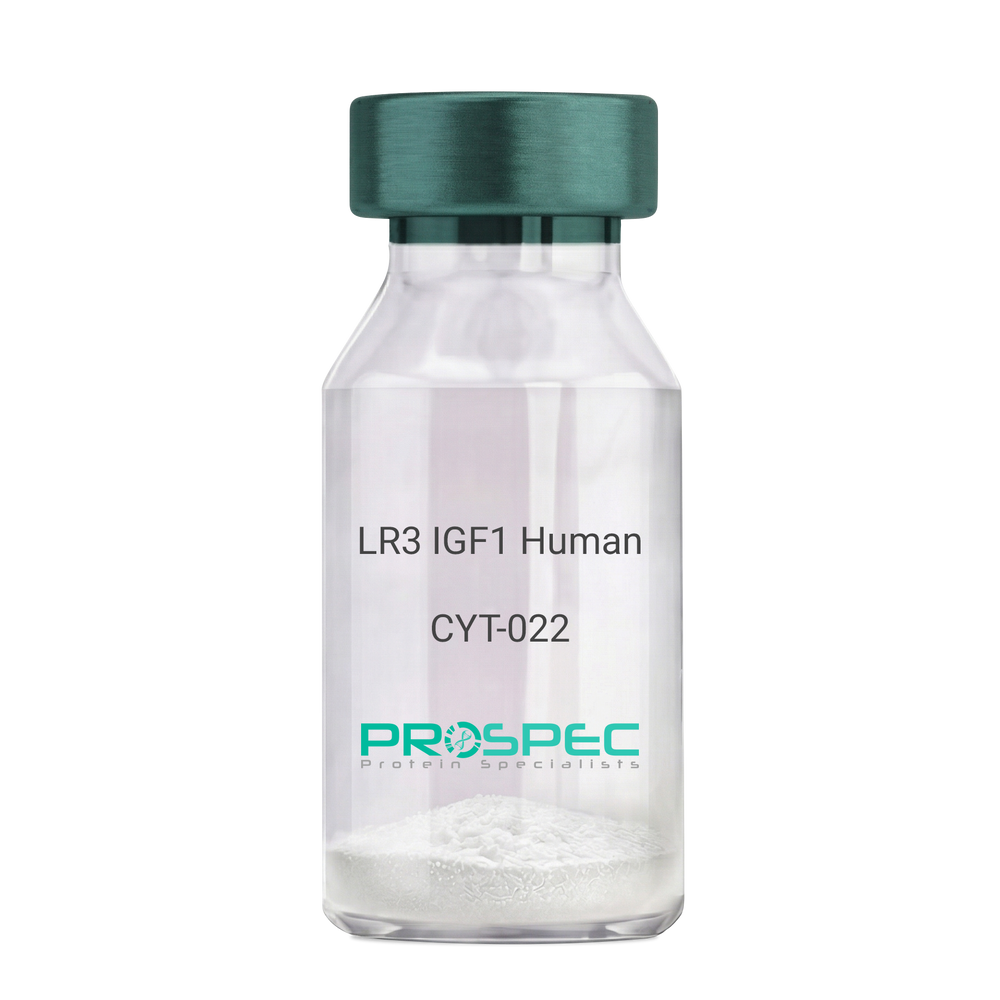
    Name :

    IGF1 LR3 Human

    Description:

    LR3 Insulin Like Growth Factor-1 Human Recombinant

    R3 IGF1, R3 IGF-1, R3IGF1, R3IGF-1, LONG IGF1, LONG IGF-1, LONG R3 IGF1, LONG R3IGF1, LONG R3 IGF-1, LONG R3IGF-1.

    Product # :

    CYT-022

    Price :

    Quantity :

    Shipping Method :

    Room Temp Icon

    Shipped at Room temp

    Add To Cart

    More Info

    • description
    • source
    • formulation
    • purity
    • biological activity
    • More Info
    • sds-page
    • Citations

    Description

    The LR3 is a long-term analog of human IGF-1, specifically designed and manufactured for mammalian cell culture to support large-scale manufacturing of recombinant biopharmaceuticals. Recombinant Human LR3 Insulin Like Growth Factor-1 produced in E.Coli is a single, non-glycosylated, polypeptide chain containing 83 amino acids and having a molecular mass of 9.1kDa.

    Source

    Escherichia Coli.

    Formulation

    Lyophilized from a 0.2µm filtered concentrated solution in 20mM PB, pH 7.2.

    Purity

    Greater than 97.0% as determined by SDS-PAGE.

    Biological Activity

    The ED50 as determined by the stimulation of protein synthesis in L6 myoblasts is less than 10ng/ml, corresponding to a specific activity of 100,000units/mg.

    sds-page

    LR3 IGF1 sds-page - Product image 1

    Citations

    More Info

    • Introduction

      IGF-1 (Insulin-like growth factor-1) is a major hormonal mediator of statural growth. Under regular circumstances, GH (growth hormone) binds to its receptor in the liver, and other tissues, and stimulates the synthesis/secretion of IGF-1. In target tissues, the Type 1 IGF receptor, that is homologous to the insulin receptor, is activated by IGF-1, leading to intracellular signaling which stimulates multiple processes leading to statural growth. IGF-1 metabolic actions are partly directed at stimulating the uptake of glucose, fatty acids, and amino acids so that metabolism supports growing tissues.

    • Synonyms

      R3 IGF1, R3 IGF-1, R3IGF1, R3IGF-1, LONG IGF1, LONG IGF-1, LONG R3 IGF1, LONG R3IGF1, LONG R3 IGF-1, LONG R3IGF-1.

    • Physical Appearance

      Sterile Filtered White lyophilized (freeze-dried) powder.

    • Stability

      Lyophilized LR3 IGF1 although stable at room temperature for 3 weeks, should be stored desiccated below -18°C. Upon reconstitution the LR3 IGF1 should be stored at 4°C between 2-7 days and for future use below -18°C.For long term storage it is recommended to add a carrier protein (0.1% HSA or BSA).Please prevent freeze-thaw cycles.

    • Solubility

      It is recommended to reconstitute the lyophilized LR3 IGF1 in sterile 18M-cm H2O at a concentration of 100µg/ml, which can then be further diluted to other aqueous solutions.

    • Amino Acid Sequence

      MFPAMPLSSLFVNGPRTLCGAELVDALQFVCGDRGFYFNKPTGYGSSSRRAPQTGIV DECCFRSCDLRRLEMYCAPLKPAKSA.

    • Background

      What is the molecular weight/Mw of IGF1 LR3 HUMAN Protein?
      IGF1 LR3 HUMAN Protein has a total Mw of 9.1kDa.

      What is the source or expression system of IGF1 LR3 HUMAN Protein?
      Escherichia Coli.

      What is the Purity of IGF1 LR3 HUMAN Protein?
      IGF1 LR3 HUMAN Protein is >98% pure as determined by SDS-PAGE.

      What is the Biological Activity of IGF1 LR3 HUMAN Protein?
      The ED50 as determined by the stimulation of protein synthesis in L6 myoblasts is less than 10ng/ml, corresponding to a specific activity of 100,000units/mg.

      What is the amino acid sequence of IGF1 LR3 HUMAN Protein?
      MFPAMPLSSLFVNGPRTLCGAELVDALQFVCGDRGFYFNKPTGYGSSSRRAPQTGIV DECCFRSCDLRRLEMYCAPLKPAKSA.

      What applications can IGF1 LR3 HUMAN Protein be used in?
      IGF1 LR3 HUMAN Protein can probably be used in western blot, ELISA and Lateral Flow.

      What is the endotoxin level for IGF1 LR3 HUMAN Protein?
      The endotoxin level is minimal, IGF1 LR3 HUMAN Protein was purified using conventional chromatography techniques.


    ProSpec's products are furnished for LABORATORY RESEARCH USE ONLY. The product may not be used as drugs, agricultural or pesticidal products, food additives or household chemicals.

    Long R3 Igf1 Human
  • View Data Sheet

    Name :

    ALDH5A1 Antibody

    Description:

    Aldehyde Dehydrogenase 5 A1, Mouse Anti Human

    Succinate-semialdehyde dehydrogenase mitochondrial, Aldehyde dehydrogenase family 5 member A1, NAD(+)-dependent succinic semialdehyde dehydrogenase, ALDH5A1, SSADH, SSDH.

    Product # :

    ANT-621

    Price :

    Quantity :

    Shipping Method :

    Ice Icon

    Shipped with Ice Packs

    Add To Cart

    More Info

    • formulation
    • More Info
    • Citations

    Formulation

    1mg/ml containing PBS, pH-7.4, 10% Glycerol and 0.02% Sodium Azide.

    Citations

    More Info

    • Introduction

      ALDH5A1 is a mitochondrial NAD(+)-dependent succinic semialdehyde dehydrogenase, which is a member of the aldehyde dehydrogenase family of proteins. The ALDH5A1 protein functions as a mediator to the NADP+-dependent oxidation of aldehydes into acids and has an imperative role in the detoxification of alcohol-derived acetaldehyde, as well as in lipid peroxidation and in the metabolism of corticosteroids, biogenic amines and neurotransmitters. ALDH5A1 is expressed in various tissues, including the liver, heart, lung, brain, kidney and placenta. Deficiency in the ALDH5A1 enzyme, known as 4-hydroxybutyricaciduria, is a rare inborn error in the metabolism of the neurotransmitter 4-aminobutyric acid (GABA). In response to this defect, physiologic fluids from patients accumulate GHB, which is a compound with numerous neuromodulatory properties.

    • Synonyms

      Succinate-semialdehyde dehydrogenase mitochondrial, Aldehyde dehydrogenase family 5 member A1, NAD(+)-dependent succinic semialdehyde dehydrogenase, ALDH5A1, SSADH, SSDH.

    • Physical Appearance

      Sterile filtered colorless solution.

    • Immunogen

      Anti-human ALDH5A1 mAb, is derived from hybridization of mouse F0 myeloma cells with spleen cells from BALB/c mice immunized with a recombinant human ALDH5A1 protein 48-535 amino acids purified from E. coli.

    • Ig Subclass

      Mouse IgG2a heavy chain and k light chain.

    • Clone

      PAT17F5AT.

    • Applications

      The antibody has been tested by ELISA, Western blot analysis, Flow cytometry and ICC/IF to assure specificity and reactivity. Since application varies, however, each investigation should be titrated by the reagent to obtain optimal results.

    • Type

      Mouse Anti Human Monoclonal.

    • Storage Procedures

      For periods up to 1 month store at 4°C, for longer periods of time, store at -20°C. Prevent freeze thaw cycles.

    • Purification Method

      ALDH5A1 antibody was purified from mouse ascitic fluids by protein-A affinity chromatography.

    ProSpec's products are furnished for LABORATORY RESEARCH USE ONLY. The product may not be used as drugs, agricultural or pesticidal products, food additives or household chemicals.

    Aldh5A1 Antibody
  • View Data Sheet

    Name :

    IgA 1+2 antibody

    Description:

    IgA 1&2, Mouse Anti-Human

    Product # :

    ANT-201

    Price :

    Quantity :

    Shipping Method :

    Room Temp Icon

    Shipped at Room temp

    Add To Cart

    More Info

    • formulation
    • More Info
    • Citations

    Formulation

    1mg/ml in PBS (after reconstitution).

    Citations

    More Info

    • Solubility

      Reconstitute with H20 to get a 1 mg/ml concentration. Mix gently, wash the sides of the vial and wait 30-60 seconds before use.

    • Immunogen

      Purified hIgA.

    • Ig Subclass

      Mouse IgG1.

    • Note

      This antibody recognizes both human IgA1 and human IgA2 subtypes.

    • Titer

      By direct ELISA, 1:20,000 dilution will yield >1.0 O.D using alkaline phosphatase conjugated rabbit anti-mouse Ig (Jackson Laboratories).

    • Shipping Conditions

      Antibody is shipped lyophilized at ambient temperature.

    • Type

      Mouse Anti Human Monoclonal.

    • Storage Procedures

      In lyophilized form, for long periods, store at 4°C in a dry environment. After reconstitution, if not intended for use within a month, aliquot and store at -20°C.Please prevent freeze-thaw cycles.

    • Purification Method

      Ion exchange column.

    ProSpec's products are furnished for LABORATORY RESEARCH USE ONLY. The product may not be used as drugs, agricultural or pesticidal products, food additives or household chemicals.

    Iga 12 Antibody
  • View Data Sheet

    Name :

    PPP3CA antibody

    Description:

    Mouse Anti Human Protein Phosphatase 3, Catalytic subunit, Alpha Isozyme

    Serine/threonine-protein phosphatase 2B catalytic subunit alpha isoform, CAM-PRP catalytic subunit, Calmodulin-dependent calcineurin A subunit alpha isoform, PPP3CA, CALNA, CAN, CALN, CCN1, CNA1, PPP2B, CALNA1.

    Product # :

    ANT-737

    Price :

    Quantity :

    Shipping Method :

    Ice Icon

    Shipped with Ice Packs

    Add To Cart

    More Info

    • formulation
    • More Info
    • Citations

    Formulation

    1mg/ml containing PBS, pH-7.4, 10% Glycerol and 0.02% Sodium Azide.

    Citations

    More Info

    • Introduction

      PPP3CA (aka Calcineurin A) is a major soluble calmodulin binding protein in the brain and a Ca2+/calmodulin dependent serine/threonine protein phosphatase, with a relatively limited substrate specificity. PPP3CA activates the T cells of the immune system and can be blocked by drugs. PPP3CA activates NFATc (a transcription factor) by dephosphorylating it. The activated NFATc is subsequently translocated into the nucleus, where it upregulates the expression of interleukin 2.

    • Synonyms

      Serine/threonine-protein phosphatase 2B catalytic subunit alpha isoform, CAM-PRP catalytic subunit, Calmodulin-dependent calcineurin A subunit alpha isoform, PPP3CA, CALNA, CAN, CALN, CCN1, CNA1, PPP2B, CALNA1.

    • Physical Appearance

      Sterile filtered colorless solution.

    • Immunogen

      Anti-human PPP3CA mAb, is derived from hybridization of mouse F0 myeloma cells with spleen cells from BALB/c mice immunized with a recombinant human PPP3CA protein 1-511 amino acids purified from E. coli.

    • Ig Subclass

      Mouse IgG2b heavy chain and k light chain.

    • Clone

      PAT1E11AT.

    • Applications

      PPP3CA antibody has been tested by ELISA and Western blot analysis to assure specificity and reactivity. Since application varies, however, each investigation should be titrated by the reagent to obtain optimal results.

    • Type

      Mouse Anti Human Monoclonal.

    • Storage Procedures

      For periods up to 1 month store at 4°C, for longer periods of time, store at -20°C. Prevent freeze thaw cycles.

    • Purification Method

      PPP3CA antibody was purified from mouse ascitic fluids by protein-A affinity chromatography.

    ProSpec's products are furnished for LABORATORY RESEARCH USE ONLY. The product may not be used as drugs, agricultural or pesticidal products, food additives or household chemicals.

    Ppp3Ca Antibody
  • View Data Sheet

    Name :

    LITAF Antibody

    Description:

    Lipopolysaccharide-induced TNF factor, Mouse Anti Human

    Lipopolysaccharide-induced tumor necrosis factor-alpha factor, LPS-induced TNF-alpha factor, p53-induced gene 7 protein, Small integral membrane protein of lysosome/late endosome, LITAF, PIG7, SIMPLE, TP53I7, FLJ38636, MGC116698, MGC116700, MGC116701, MGC125274, MGC125275, MGC125276.

    Product # :

    ANT-443

    Price :

    Quantity :

    Shipping Method :

    Ice Icon

    Shipped with Ice Packs

    Add To Cart

    More Info

    • formulation
    • More Info
    • Citations

    Formulation

    1mg/ml containing PBS, pH-7.4, & 0.02% Sodium Azide and 10% Glycerol.

    Citations

    More Info

    • Introduction

      Lipopolysaccharide is a potent stimulator of monocytes and macrophages, causing secretion of tumor necrosis factor-alpha (TNF-alpha) and other inflammatory mediators. LITAF is a lipopolysaccharide-induced TNF-alpha factor, which is a DNA-binding protein and can mediate the TNF-alpha expression by direct binding to the promoter region of the TNF-alpha gene. The transcription of the LITAF gene is induced by tumor suppressor p53 and has been implicated in the p53-induced apoptotic pathway. Mutations in the LITAF gene cause Charcot-Marie-Tooth disease type 1C (CMT1C) and may be involved in the carcinogenesis of extramammary Paget''s disease (EMPD).

    • Synonyms

      Lipopolysaccharide-induced tumor necrosis factor-alpha factor, LPS-induced TNF-alpha factor, p53-induced gene 7 protein, Small integral membrane protein of lysosome/late endosome, LITAF, PIG7, SIMPLE, TP53I7, FLJ38636, MGC116698, MGC116700, MGC116701, MGC125274, MGC125275, MGC125276.

    • Physical Appearance

      Sterile Filtered clear solution.

    • Immunogen

      Anti-human LITAF mAb, is derived from hybridization of mouse F0 myeloma cells with spleen cells from BALB/c mice immunized with recombinant human LITAF amino acids 1-161 purified from E. coli.

    • Ig Subclass

      Mouse IgG2b heavy chain and k light chain.

    • Clone

      PAT5C10AT.

    • Applications

      LITAF antibody has been tested by ELISA and Western blot analysis to assure specificity and reactivity. Since application varies, however, each investigation should be titrated by the reagent to obtain optimal results. Recommended dilution range for Western blot analysis is 1:1000. Recommended starting dilution is 1:1000.

    • Type

      Mouse Anti Human Monoclonal.

    • Storage Procedures

      For periods up to 1 month store at 4°C, for longer periods of time, store at -20°C. Prevent freeze thaw cycles.

    • Purification Method

      LITAF antibody was purified from mouse ascitic fluids by protein-G affinity chromatography.

    ProSpec's products are furnished for LABORATORY RESEARCH USE ONLY. The product may not be used as drugs, agricultural or pesticidal products, food additives or household chemicals.

    Litaf Antibody
  • View Data Sheet

    Name :

    GPI Antibody

    Description:

    Glucose-6-Phosphate Isomerase, Mouse Anti Human

    Glucose-6-phosphate isomerase, Phosphoglucose isomerase, Phosphohexose isomerase, Autocrine motility factor, Neuroleukin, Sperm antigen 36, GPI, PGI, PHI, AMF, NLK, SA-36, GNPI.

    Product # :

    ANT-613

    Price :

    Quantity :

    Shipping Method :

    Ice Icon

    Shipped with Ice Packs

    Add To Cart

    More Info

    • formulation
    • More Info
    • Citations

    Formulation

    1mg/ml containing PBS, pH-7.4, 10% Glycerol and 0.02% Sodium Azide.

    Citations

    More Info

    • Introduction

      Glucose-6-phosphate isomerase (GPI) is a part of the GPI family whose members encode multifunctional phosphoglucose isomerase proteins involved in energy pathways. GPI is a dimeric enzyme which catalyzes the reversible isomerization of glucose-6-phosphate and fructose-6-phosphate. Mammalian GPI also functions as a tumor-secreted cytokine and an angiogenic factor (AMF) which stimulates endothelial cell motility. In addition, GPI is a neurotrophic factor (Neuroleukin) for spinal and sensory neurons. GPI performs in different capacities inside and outside the cell. In the cytoplasm, GPI is involved in glycolysis and gluconeogenesis, while outside the cell it acts as a neurotrophic factor for spinal and sensory neurons.
      Defects in the GPI gene cause the nonspherocytic hemolytic anemia and a severe enzyme deficiency can be linked to hydrops fetalis, immediate neonatal death and neurological impairment.

    • Synonyms

      Glucose-6-phosphate isomerase, Phosphoglucose isomerase, Phosphohexose isomerase, Autocrine motility factor, Neuroleukin, Sperm antigen 36, GPI, PGI, PHI, AMF, NLK, SA-36, GNPI.

    • Physical Appearance

      Sterile filtered colorless solution.

    • Immunogen

      Anti-human GPI mAb, clone PAT22G2AT, is derived from hybridization of mouse F0 myeloma cells with spleen cells from BALB/c mice immunized with a recombinant human GPI protein 1-558 amino acids purified from E. coli.

    • Ig Subclass

      Mouse IgG2a heavy chain and k light chain.

    • Clone

      PAT22G2AT.

    • Applications

      The antibody has been tested by ELISA, Western blot analysis, Flow cytometry and ICC/IF to assure specificity and reactivity. Since application varies, however, each investigation should be titrated by the reagent to obtain optimal results.

    • Type

      Mouse Anti Human Monoclonal.

    • Storage Procedures

      For periods up to 1 month store at 4°C, for longer periods of time, store at -20°C. Prevent freeze thaw cycles.

    • Purification Method

      GPI antibody was purified from mouse ascitic fluids by protein-A affinity chromatography.

    ProSpec's products are furnished for LABORATORY RESEARCH USE ONLY. The product may not be used as drugs, agricultural or pesticidal products, food additives or household chemicals.

    Gpi Antibody
  • View Data Sheet

    Name :

    GSTT1 Antibody

    Description:

    Glutathione S-Transferase Theta-1, Mouse Anti Human

    Glutathione S-transferase theta-1, GST class-theta-1, Glutathione transferase T1-1, GSTT1.

    Product # :

    ANT-591

    Price :

    Quantity :

    Shipping Method :

    Ice Icon

    Shipped with Ice Packs

    Add To Cart

    More Info

    • formulation
    • More Info
    • Citations

    Formulation

    1mg/ml containing PBS, pH-7.4, 10% Glycerol and 0.02% Sodium Azide.

    Citations

    More Info

    • Introduction

      GSTT1 belongs to a superfamily of proteins which catalyze the conjugation of reduced glutathione to a variety of electrophilic and hydrophobic compounds. GSTT1 is one of the GSTs’ four main classes: alpha, mu, pi and theta (which includes GSTT1 and GSTT2). GSTT1 is involved in activation and detoxification reactions and catalyzes the conjugation of industrial chemicals, such as epoxybutane, ethylene oxides, halomethane with glutathione. GSTT1 is found in erythrocytes, at low levels in the liver as well as in Clara and ciliated cells at the alveolar/bronchiolar junction in the lung. The GSTT1 gene is deficient in 38% of the population. The GSTTI enzyme deficiency might influence the individual risk for development of acquired aplastic anemia and acute myeloid leukemia. The presence or absence of the GSTT1 gene is concurrent with GSST1+ (the conjugator) and GSTT1- (the non-conjugator) phenotypes correspondingly. The GSTT1+ phenotype is able to catalyze the glutathione conjugation of dichloromethane. GSTT1-null genotypes are seen as having a higher risk of developing leukoplakia. Germline genetic polymorphism in GSTT1 is linked to breast cancer.

    • Synonyms

      Glutathione S-transferase theta-1, GST class-theta-1, Glutathione transferase T1-1, GSTT1.

    • Physical Appearance

      Sterile filtered colorless solution.

    • Immunogen

      Anti-human GSTT1 mAb, is derived from hybridization of mouse F0 myeloma cells with spleen cells from BALB/c mice immunized with recombinant human GSTT1 amino acids 1-240 purified from E. coli.

    • Ig Subclass

      Mouse IgG2b heavy chain and k light chain.

    • Clone

      PAT38D11AT.

    • Applications

      GSTT1 antibody has been tested by ELISA, Western blot analysis, ICC/IF and Flow cytometry to assure specificity and reactivity. Since application varies, however, each investigation should be titrated by the reagent to obtain optimal results.

    • Type

      Mouse Anti Human Monoclonal.

    • Storage Procedures

      For periods up to 1 month store at 4°C, for longer periods of time, store at -20°C. Prevent freeze thaw cycles.

    • Purification Method

      GSTT1 antibody was purified from mouse ascitic fluids by protein-A affinity chromatography.

    ProSpec's products are furnished for LABORATORY RESEARCH USE ONLY. The product may not be used as drugs, agricultural or pesticidal products, food additives or household chemicals.

    Gstt1 Antibody
  • View Data Sheet

    Name :

    CRABP2 Antibody

    Description:

    Cellular Retinoic Acid binding Protein 2, Mouse Anti Human

    RBP6, CRABP-II, CRABP2, RETINOIC ACID-BINDING PROTEIN CELLULAR TYPE II, Cellular retinoic acid-binding protein 2, Cellular retinoic acid-binding protein II.

    Product # :

    ANT-356

    Price :

    Quantity :

    Shipping Method :

    Ice Icon

    Shipped with Ice Packs

    Add To Cart

    More Info

    • formulation
    • More Info
    • Citations

    Formulation

    1mg/ml containing PBS, pH-7.4, & 0.1% Sodium Azide.

    Citations

    More Info

    • Introduction

      CRABP2 NCBI Accession No: NP_001869 regulates the access of retinoic acid to the nuclear retinoic acid receptors. CRABP2 is involved in a regulatory feedback mechanism that controls the action of retinoic acid on cell differentiation. CRABP2 is involved in the conversion of vitamin A into its intracellular active form retinoic acid, which regulate the genes responsible for lipid metabolism and adipocyte differentiation. CRABP2 gene is located on chromosome 1q21-23 and this region has been linked with related disorders such as familial combined hyperlipidemia (FCHL) and type 2 diabetes mellitus. CRABP proteins are of low molecular weight having an important function in retinoic acid-mediated regulation of human skin growth and differentiation.

    • Synonyms

      RBP6, CRABP-II, CRABP2, RETINOIC ACID-BINDING PROTEIN CELLULAR TYPE II, Cellular retinoic acid-binding protein 2, Cellular retinoic acid-binding protein II.

    • Immunogen

      Anti-human CRABP2 mAb is derived from hybridization of mouse F0 myeloma cells with spleen cells from BALB/c mice immunized with recombinant human CRABP2 amino acids 1-138 purified from E. coli.

    • Ig Subclass

      Mouse IgG2a heavy chain and κ light chain.

    • Clone

      PAT2E11AT.

    • Applications

      CRABP2 antibody has been tested by ELISA and Western blot analysis to assure specificity and reactivity. Since application varies, however, each investigation should be titrated by the reagent to obtain optimal results. Recommended dilution range for Western blot analysis is 1:250 ~ 1000. Recommended starting dilution is 1:250.

    • Type

      Mouse Anti Human Monoclonal.

    • Storage Procedures

      For periods up to 1 month store at 4°C, for longer periods of time, store at -20°C. Prevent freeze thaw cycles.

    • Purification Method

      CRABP2 antibody was purified from mouse ascitic fluids by protein-G affinity chromatography.

    ProSpec's products are furnished for LABORATORY RESEARCH USE ONLY. The product may not be used as drugs, agricultural or pesticidal products, food additives or household chemicals.

    Crabp2 Antibody
  • View Data Sheet

    Name :

    Lypressin

    Description:

    Lypressin

    Product # :

    HOR-278

    Price :

    Quantity :

    Shipping Method :

    Room Temp Icon

    Shipped at Room temp

    Add To Cart

    More Info

    • description
    • formulation
    • purity
    • More Info
    • Citations

    Description

    Lypressin is a hormone used to prevent or control the frequent urination, increased thirst, and loss of water associated with diabetes insipidus (water diabetes). Its molecular weight is 1056.2 Dalton having an amino acid sequence of Cys-Tyr-Phe-Gln-Asn-Cys-Pro-Lys-Gly-NH2. The Molecular Formula is: C46H65N13O12S2. The CAS Number is: 50-57-7.

    Formulation

    The protein (1mg/ml) was lyophilized with no additives.

    Purity

    Greater than 99.0% as determined by RP-HPLC.

    Citations

    More Info

    • Physical Appearance

      Sterile Filtered White lyophilized (freeze-dried) powder.

    • Stability

      Store the lyophilized Lypressin between 2-8°C, do not freeze. Upon reconstitution Lypressin should be stored at 4°C between 2-7 days and for future use below -18°C. Please prevent freeze-thaw cycles.

    • Solubility

      It is recommended to reconstitute the lyophilized Lypressin in sterile 18MΩ-cm H2O not less than 100 µg/ml, which can then be further diluted to other aqueous solutions.
      Lypressin is also soluble in 1% Acetic Acid.

    ProSpec's products are furnished for LABORATORY RESEARCH USE ONLY. The product may not be used as drugs, agricultural or pesticidal products, food additives or household chemicals.

    Lypressin
  • View Data Sheet

    Name :

    Leuprorelin Human

    Description:

    Leuprolide Human

    Leuprolide, Leuprorelin.

    Product # :

    HOR-260

    Price :

    Quantity :

    Shipping Method :

    Room Temp Icon

    Shipped at Room temp

    Add To Cart

    More Info

    • description
    • formulation
    • purity
    • More Info
    • Citations

    Description

    Leuprolide Human Synthetic is a single, non-glycosylated, polypeptide chain containing 9 amino acids and having a molecular mass of 1209 Dalton. The molecular formula is C59H84N16O12.

    Formulation

    The Leuprorelin was lyophilized from a concentrated (1mg/ml) solution with no additives.

    Purity

    Greater than 98.0% as determined by RP-HPLC.

    Citations

    More Info

    • Introduction

      Leuprorelin (INN) or leuprolide acetate (USAN) is a gonadotropin-releasing hormone agonist (GnRH agonist).
      By causing constant stimulation of the pituitaryGnRH receptors, it initially causes stimulation (flare), but thereafter decreases pituitary secretion (downregulation) of gonadotropins luteinizing hormone(LH) and follicle-stimulating hormone(FSH).
      Like other GnRH agonists, leuprolide may be used in the treatment of hormone-responsive cancers such as prostate canceror breast cancer, estrogen-dependent conditions (such as endometriosisor uterine fibroids), to treat precocious puberty, and to control ovarian stimulation in IVF.

    • Synonyms

      Leuprolide, Leuprorelin.

    • Physical Appearance

      Sterile Filtered White lyophilized (freeze-dried) powder.

    • Stability

      Lyophilized Leoprolide although stable at room temperature for 3 weeks, should be stored desiccated below -18°C. Upon reconstitution Leuprorelin should be stored at 4°C between 2-7 days and for future use below -18°C.For long term storage it is recommended to add a carrier protein (0.1% HSA or BSA).Please prevent freeze-thaw cycles.

    • Solubility

      It is recommended to reconstitute the lyophilized Leoprolide in sterile 18MΩ-cm H2O not less than 100µg/ml, which can then be further diluted to other aqueous solutions.

    • Amino Acid Sequence

      pGlu-His-Trp-Ser-Tyr-D-Leu-Leu-Arg-Pro-NHEt.

    ProSpec's products are furnished for LABORATORY RESEARCH USE ONLY. The product may not be used as drugs, agricultural or pesticidal products, food additives or household chemicals.

    Leuprolide Human
  • View Data Sheet

    Name :

    Tirzepatide

    Description:

    Tirzepatide

    Product # :

    HOR-057

    Price :

    Quantity :

    Shipping Method :

    Room Temp Icon

    Shipped at Room temp

    Add To Cart

    More Info

    • description
    • formulation
    • purity
    • More Info
    • Citations

    Description

    Tirzepatide is a synthetic single, non-glycosylated polypeptide chain containing 39 amino acids, having a molecular mass of 4813 Dalton and a Molecular formula of C187H291N45O59.

    Formulation

    The protein was lyophilized with no additives.

    Purity

    Greater than 97.0% as determined by analysis by RP-HPLC.

    Citations

    More Info

    • Physical Appearance

      Sterile Filtered White lyophilized (freeze-dried) powder.

    • Stability

      Lyophilized Tirzepatide although stable at room temperature for 3 weeks, should be stored desiccated below -18°C. Upon reconstitution Tirzepatide should be stored at 4°C between 2-7 days and for future use below -18°C.
      For long term storage it is recommended to add a carrier protein (0.1% HSA or BSA).
      Please prevent freeze-thaw cycles.

    • Solubility

      It is recommended to reconstitute the lyophilized Tirzepatide in sterile 18MΩ-cm H2O not less than 100 µg/ml, which can then be further diluted to other aqueous solutions.

    • Amino Acid Sequence

      His-Ala-Glu-Gly-Thr-Phe-Thr-Ser-Asp-Tyr-Ser-Ile-Aib-Leu-Asp-Lys-Ile-Ala-Gln-Lys (Eicosanedioyl-isoGlu-PEG2-PEG2)-Ala-Phe-Val-Gln-Trp-Leu-Ile-Ala-Gly-Gly-Pro-Ser-Ser-Gly-Ala-Pro-Pro-Pro-Ser-NH2.

    • Background

      Tirzepatide is a first-in-class dual glucose-dependent insulinotropic polypeptide and glucagon-like peptide-1 receptor agonist. Recent development suggests Tirzepatide as organ protection and systemic metabolic health while at first, it was only approved for Type 2 Diabetes and Chronic Weight Management. Preclinical data also indicates a promising future in neuroprotection and most likely to expand its clinical utility into neurodegenerative pathologies.

      What is the molecular weight/Mw of TIRZEPATIDE Protein?
      TIRZEPATIDE Protein has a total Mw of 4.8kDa.

      What is the Purity of TIRZEPATIDE Protein?
      TIRZEPATIDE Protein is >97% pure as determined by SDS-PAGE.

      What is the Biological Activity of TIRZEPATIDE Protein?
      The biological functionality of TIRZEPATIDE Protein will be determined in the future.

      What is the amino acid sequence of TIRZEPATIDE Protein?
      His-Ala-Glu-Gly-Thr-Phe-Thr-Ser-Asp-Tyr-Ser-Ile-Aib-Leu-Asp-Lys-Ile-Ala-Gln-Lys (Eicosanedioyl-isoGlu-PEG2-PEG2)-Ala-Phe-Val-Gln-Trp-Leu-Ile-Ala-Gly-Gly-Pro-Ser-Ser-Gly-Ala-Pro-Pro-Pro-Ser-NH2.
      What applications can TIRZEPATIDE Protein be used in?
      TIRZEPATIDE Protein can probably be used in western blot, ELISA and Lateral Flow.

      What is the endotoxin level for TIRZEPATIDE Protein?
      The endotoxin level is minimal, TIRZEPATIDE Protein was purified using conventional chromatography techniques.

      Who supplies TIRZEPATIDE that is in stock and available to order?
      ProSpec has the protein in stock and available for immediate delivery.

      Where can I order bulk mg quantities of TIRZEPATIDE?
      ProSpec supplies bulk mg quantities at affordable prices that meets research and academic budgets.

    ProSpec's products are furnished for LABORATORY RESEARCH USE ONLY. The product may not be used as drugs, agricultural or pesticidal products, food additives or household chemicals.

    Tirzepatide
  • 1
  • …
  • 40
  • 41
  • 42
Your browser does not support the video tag.

Products

  • Cytokines
  • Growth Factors
  • Chemokines
  • CD Antigens
  • Neurotrophins
  • Hormones
  • Enzymes
  • Viral Antigens
  • Recombinant Proteins
  • Natural Proteins
  • Monoclonal Antibodies
  • Polyclonal Antibodies
  • Thermal Packaging

About

  • Company
  • Founder
  • Contribution
  • Contact Us
  • My Account

Ordering

  • Ordering Method
  • Ordering - Credit Card
  • Ordering PO
  • Shipping Fees
  • FAQ's

Legal

  • Terms & Conditions
  • Privacy
  • Site Map
  • info@prospecbio.com
  • Facebook
  • Instagram
  • Linkedin Profile

© 2026 ProSpec-Tany TechnoGene Ltd. All Rights Reserved.

  • Choosing a selection results in a full page refresh.
  • Opens in a new window.