Search results
1000 results found for “Anti Viral Monoclonal”
Name
Description
Product #
Price
Quantity
Shipping Method
- View Data Sheet
Name :
HCV Core 24Description:
Hepatitis C Virus Core 24 Recombinant
Product # :
HCV-240Price :
Quantity :
Shipping Method :
Shipped with Ice Packs
More Info
- description
- formulation
- purity
- More Info
Description
The HCV Core 24 genotype-1b, E.coli derived recombinant protein, contains the HCV core nucleocapsid immunodominant regions. The protein is fused with b-galactosidase (114 kDa) at N-terminus.
Formulation
20mM Tris-Hcl pH 8, 1.5M urea and 10mM b-mercaptoethanol.
Purity
HCV Core 24 protein is >95% pure as determined by 10% PAGE (coomassie staining).
More Info
-
Introduction
HCV is a small 50nm, enveloped, single-stranded, positive sense RNAvirus in the family Flaviviridae.
HCV has a high rate of replication with approximately one trillion particles produced each day in an infected individual. Due to lack of proofreading by the HCV RNA polymerase, the HCV has an exceptionally high mutation rate, a factor that may help it elude the host's immune response. Hepatitis C virus is classified into six genotypes(1-6) with several subtypes within each genotype. The preponderance and distribution of HCV genotypes varies globally. Genotype is clinically important in determining potential response to interferon-based therapy and the required duration of such therapy. Genotypes 1 and 4 are less responsive to interferon-based treatment than are the other genotypes (2, 3, 5 and 6). -
Stability
HCV-Core 24 although stable at 4°C for 1 week, should be stored below -18°C. Please prevent freeze thaw cycles.
-
Applications
HCV-core 24 antigen is suitable for ELISA and Western blots, excellent antigen for detection of HCV with minimal specificity problems.
-
Specificity
Immunoreactive with sera of HCV-infected individuals.
-
Purification Method
HCV-Core 24 protein was purified by proprietary chromatographic technique.
ProSpec's products are furnished for LABORATORY RESEARCH USE ONLY. The product may not be used as drugs, agricultural or pesticidal products, food additives or household chemicals.
- View Data Sheet
Name :
Borrelia VisE1Description:
Borrelia Burgdorferi VlsE1 Recombinant
Product # :
BOR-024Price :
Quantity :
Shipping Method :
Shipped with Ice Packs
More Info
- description
- source
- formulation
- purity
- More Info
Description
Recombinant Borrelia VisE1 (variable major protein like sequence E1) produced in E.coli is a non-glycosylated, polypeptide chain having a calculated molecular mass of 43kDa. Borrelia VisE1 is expressed with a -10x His tag at N-terminus and purified by proprietary chromatographic techniques.
Source
Escherichia Coli.
Formulation
Borrelia VisE1 is supplied in 20mM HEPES buffer pH-8.0, 200mM NaCl and 20% glycerol.
Purity
Greater than 95.0% as determined by SDS-PAGE.
More Info
-
Introduction
Borrelia is a part of the genus of bacteria of the spirochete phylum. Borrelia causes borreliosis, which is a zoonotic, vector-borne disease transmitted mainly by ticks and some by lice, depending on the species. Of the 36 known species of Borrelia, 12 are distinguished to cause Lyme disease or borreliosis and are transmitted by ticks. The main Borrelia species causing Lyme disease are Borrelia burgdorferi, Borrelia afzelii, and Borrelia garinii. Variable major protein like sequence E1 protein (VlsE1) is a borrelial surface protein which is the most sensitive protein for IgG antibody detection in all stages of Lyme disease.
-
Physical Appearance
Sterile Filtered clear solution.
-
Stability
Store at 4°C if entire vial will be used within 2-4 weeks. Store, frozen at -20°C for longer periods of time. Avoid multiple freeze-thaw cycles.
-
Immunological Functions
1. Binds IgG- and IgM-type human antibodies.2.Immunodot test with Lyme disease positive/negative samples.
-
Applications
Western blot with Patient sample.
ProSpec's products are furnished for LABORATORY RESEARCH USE ONLY. The product may not be used as drugs, agricultural or pesticidal products, food additives or household chemicals.
- View Data Sheet
Name :
HIV-1 TAT Clade-B, BiotinDescription:
HIV-1 TAT Clade-B Recombinant, Biotin Labeled
Product # :
HIV-128Price :
Quantity :
Shipping Method :
Shipped with Ice Packs
More Info
- description
- source
- formulation
- purity
- More Info
Description
HIV-1 TAT Clade-B Biotin Recombinant- produced in E.coli is a single, non-glycosylated, polypeptide chain containing 86 amino acids encoded by two exons and having chain having a molecular mass of 14kDa and was biotinylated with NHS biotin.
Source
Escherichia Coli.
Formulation
1xPBS & 0.05% glycerol.
Purity
Greater than 90.0% as determined by HPLC analysis & SDS-PAGE.
More Info
-
Introduction
Human immunodeficiency virus type-1 (HIV-1) regulatory Tat protein plays an essential role in viral replication and infectivity. The HIV-1 TAT is a molecule of 86 aminoacids encoded by two exons. The product of the first exon is sufficient for the transactivation of the HIV-1 promoter. Biotin molecules can be coupled easily to either antigens (such as avidin, streptavidin, Fc binding proteins like Protein A or G) or antibodies and they can be used in a variety of detection systems.
Biotin can be conjugated to many proteins without significantly changing their biological activity. Biotin binds with high affinity to avidin, streptavidin and NeutrAvidin protein.
Every single protein can react with several molecules of biotin, each of which, in turn, can bind a molecule of avidin. This greatly increases the sensitivity of many assay procedures. The avidin-biotin interaction is the strongest known noncovalent biological interaction (Ka = 1015 M-1) between protein and ligand. The bound formation between biotin and avidin is very rapid and, once formed, withstands extremes of pH, organic solvents and other denaturing agents. The avidinbiotin complex can withstand up to 3 M guanidine•HCl. Biotin can be released by 8 M guanidine•HCl at pH 1.5, or by autoclaving. -
Physical Appearance
Sterile Filtered colorless solution.
-
Stability
HIV-1 TAT biotin recombinant although stable at 4°C for 1 week, should be stored below -18°C. Please prevent freeze-thaw cycles.
-
Specificity
Immunoreactive with all sera of HIV-1 infected individuals.
ProSpec's products are furnished for LABORATORY RESEARCH USE ONLY. The product may not be used as drugs, agricultural or pesticidal products, food additives or household chemicals.
- View Data Sheet
Name :
H3N2 WyomingDescription:
H3N2 Influenza-A Virus Wyoming/3/2003 Recombinant
Product # :
IHA-012Price :
Quantity :
Shipping Method :
Shipped with Ice Packs
More Info
- description
- source
- formulation
- purity
- More Info
Description
Recombinant Full-Length H3N2 A/Wyoming/2003/3 is glycosylated with N-linked sugars, produced using baculovirus vectors in insect cells and its Mw is 72,000 Dalton. Accession number: AY531033.
Source
Baculovirus Insect Cells
Formulation
The Recombinant H3N2 A/Wyoming/2003/3 solution contains 10mM Sodium phosphate, pH 7.4, 150mM NaCl and 0.005% Tween-20.
Purity
Greater than 90.0% as determined by SDS-PAGE.
More Info
-
Introduction
H3N2 is a subtype of the influenza A virus. Its name derives from the forms of the two kinds of proteinson the surface of its coat, hemagglutinin(H) and neuraminidase(N). H3N2 exchanges genes for internal proteins with other influenza subtypes. H3N2 has tended to dominate in prevalence over H1N1, H1N2, and influenza B. H3N2 strain descended from H2N2by antigenic shift, in which genes from multiple subtypes re-assorted to form a new virus. Both the H2N2and H3N2 strains contained genesfrom avian influenzaviruses.
-
Physical Appearance
Sterile Filtered colorless solution.
-
Stability
H3N2 A/Wyoming/2003/3 Recombinant should be stored at 4°C.
ProSpec's products are furnished for LABORATORY RESEARCH USE ONLY. The product may not be used as drugs, agricultural or pesticidal products, food additives or household chemicals.
- View Data Sheet
Name :
SARS Spike (306-515), Sf9Description:
Coronavirus Spike Receptor Binding Domain (306-515 a.a.)Recombinant, Sf9
Spike glycoprotein, S glycoprotein, Peplomer protein, E2 glycoprotein precursor, Severe acute repiratory Syndrome-related Coronavirus, SARS, SRAS-CoV, SARS-CoV1, E2.
Product # :
SARS-047Price :
Quantity :
Shipping Method :
Shipped with Ice Packs
More Info
- description
- source
- formulation
- purity
- biological activity
- More Info
Description
SARS Spike produced in Sf9 Baculovirus cells is a single, glycosylated polypeptide chain containing 219 amino acids (306-515 aa) and having a molecular mass of 24.7kDa.SARS Spike is fused to a 6 amino acid His tag at C-terminus and purified by proprietary chromatographic techniques.
Source
Sf9, Baculovirus cells.
Formulation
The SARS Spike (306-515) solution (0.5mg/ml) contains Phosphate-Buffered Saline (pH 7.4) and 10% Glycerol.
Purity
Greater than 95.0% as determined by SDS-PAGE.
Biological Activity
Measured by its binding ability in a functional ELISA with Human ACE-2 (CAT# enz-1159).
More Info
-
Introduction
SARS Coronavirus is an enveloped virus containing 3 outer structural proteins, namely the membrane (M), envelope (E), and spike (S) proteins. Spike (S)-glycoprotein of the virus interacts with a cellular receptor and mediates membrane fusion to allow viral entry into susceptible target cells. Accordingly, S-protein takes part in virus infection cycle and is the primary target of neutralizing antibodies.
-
Synonyms
Spike glycoprotein, S glycoprotein, Peplomer protein, E2 glycoprotein precursor, Severe acute repiratory Syndrome-related Coronavirus, SARS, SRAS-CoV, SARS-CoV1, E2.
-
Physical Appearance
Sterile Filtered colorless solution.
-
Stability
Store at 4°C if entire vial will be used within 2-4 weeks. Store, frozen at -20°C for longer periods of time. For long term storage it is recommended to add a carrier protein (0.1% HSA or BSA). Avoid multiple freeze-thaw cycles.
-
Amino Acid Sequence
ADPRVVPSGD VVRFPNITNL CPFGEVFNAT KFPSVYAWER KKISNCVADY SVLYNSTFFS TFKCYGVSAT KLNDLCFSNV YADSFVVKGD DVRQIAPGQT GVIADYNYKL PDDFMGCVLA WNTRNIDATS TGNYNYKYRY LRHGKLRPFE RDISNVPFSP DGKPCTPPAL NCYWPLNDYG FYTTTGIGYQ PYRVVVLSFE LLNAPATVCG PKLHHHHHH
ProSpec's products are furnished for LABORATORY RESEARCH USE ONLY. The product may not be used as drugs, agricultural or pesticidal products, food additives or household chemicals.
- View Data Sheet
Name :
Frataxin AntibodyDescription:
Frataxin, Mouse Anti Human
FXN, Friedreich ataxia protein, Frataxin mitochondrial, FRDA, X25, FA, CyaY, FARR, MGC57199, Frataxin.
Product # :
ANT-671Price :
Quantity :
Shipping Method :
Shipped with Ice Packs
More Info
- formulation
- More Info
Formulation
1mg/ml containing PBS, pH-7.4, 10% Glycerol and 0.02% Sodium Azide.
More Info
-
Introduction
Frataxin is a mitochondrial iron binding protein that is part of the FRATAXIN family. Frataxin functions in regulating mitochondrial iron transport and respiration. The expansion of intronic trinucleotide repeat GAA results in Friedreich ataxia. Frataxin plays a role in iron homeostasis. Frataxin is an anti-apoptotic protein which prevents mitochondrial damage and reactive oxygen species (ROS) production.
-
Synonyms
FXN, Friedreich ataxia protein, Frataxin mitochondrial, FRDA, X25, FA, CyaY, FARR, MGC57199, Frataxin.
-
Physical Appearance
Sterile filtered colorless solution.
-
Immunogen
Anti-human Frataxin mAb, is derived from hybridization of mouse F0 myeloma cells with spleen cells from BALB/c mice immunized with a recombinant human Frataxin protein 42-210 amino acids purified from E. coli.
-
Ig Subclass
Mouse IgG1 heavy chain and k light chain.
-
Clone
PAT10E11AT.
-
Applications
The antibody has been tested by ELISA, Western blot analysis and ICC/IF to assure specificity and reactivity. Since application varies, however, each investigation should be titrated by the reagent to obtain optimal results.
-
Type
Mouse Anti Human Monoclonal.
-
Storage Procedures
For periods up to 1 month store at 4°C, for longer periods of time, store at -20°C. Prevent freeze thaw cycles.
-
Purification Method
Frataxin antibody was purified from mouse ascitic fluids by protein-A affinity chromatography.
ProSpec's products are furnished for LABORATORY RESEARCH USE ONLY. The product may not be used as drugs, agricultural or pesticidal products, food additives or household chemicals.
- View Data Sheet
Name :
H3N2 PanamaDescription:
H3N2 Influenza-A Virus Panama/2007/99
Product # :
IHA-006Price :
Quantity :
Shipping Method :
Shipped with Ice Packs
More Info
- description
- formulation
- purity
- More Info
Description
Allantoic fluid of 10 days old embryonated eggs, inoculated with influenza A virus, strain A/Panama/2007/99. The Influenza Virus was purified by Ultracentrifugation with 10-40 % sucrose gradient.
Formulation
The H3N2 A/Panama/2007/99 solution contains STE, 0.09% sodium azide (NaN3) and 0.005% thimerosal.
Stir thoroughly prior to its use.Purity
Greater than 90.0% as determined byAnalysis by SDS-PAGE.
More Info
-
Introduction
H3N2 is a subtype of the influenza A virus. Its name derives from the forms of the two kinds of proteinson the surface of its coat, hemagglutinin(H) and neuraminidase(N). H3N2 exchanges genes for internal proteins with other influenza subtypes. H3N2 has tended to dominate in prevalence over H1N1, H1N2, and influenza B. H3N2 strain descended from H2N2 by antigenic shift, in which genes from multiple subtypes re-assorted to form a new virus. Both the H2N2 and H3N2 strains contained genes from avian influenza viruses.
-
Physical Appearance
Opaque suspension.
-
Stability
A/Panama/2007/99 although stable 4°C for 4 weeks, should be stored below -18°C. Please prevent freeze-thaw cycles.
-
Inactivation
Thimerosal and beta propiolactone treatmentThis product has been treated in a manner consistent with methods of inactivation. Generally accepted good laboratory practices appropriate to microbiological/viral safe handling practices and techniques are required at work.
-
Immunological Activity
Tested with anti-influenza A monoclonal antibodies in ELISA.Serological studies of influenza A virus, immunogen for antibody production.
ProSpec's products are furnished for LABORATORY RESEARCH USE ONLY. The product may not be used as drugs, agricultural or pesticidal products, food additives or household chemicals.
- View Data Sheet
Name :
UBE2L6 AntibodyDescription:
Ubiquitin Conjugating Enzyme E2L 6, Mouse Anti Human
RIG-B, UBCH8, MGC40331, UBE2L6, Ubiquitin/ISG15-conjugating enzyme E2 L6, Ubiquitin-protein ligase L6, Ubiquitin carrier protein L6, Retinoic acid-induced gene B protein.
Product # :
ANT-386Price :
Quantity :
Shipping Method :
Shipped with Ice Packs
More Info
- formulation
- More Info
Formulation
1mg/ml containing PBS, pH-7.4, & 0.1% Sodium Azide.
More Info
-
Introduction
UBE2L6 belongs to the E2 ubiquitin-conjugating enzyme family. UBE2L6 enzyme is very similar in its primary structure to the enzyme encoded by UBE2L3 gene. UBE2L6 catalyzes the covalent attachment of ubiquitin to other proteins. UBE2L6 functions in the e6/e6-ap-induced ubiquitination of p53/tp53.
-
Synonyms
RIG-B, UBCH8, MGC40331, UBE2L6, Ubiquitin/ISG15-conjugating enzyme E2 L6, Ubiquitin-protein ligase L6, Ubiquitin carrier protein L6, Retinoic acid-induced gene B protein.
-
Immunogen
Anti-human UBE2L6 mAb is derived from hybridization of mouse F0 myeloma cells with spleen cells from BALB/c mice immunized with recombinant human UBE2L6 amino acids 1-152 purified from E. coli.
-
Ig Subclass
Mouse IgG2b heavy chain and κ light chain.
-
Clone
Pk1H3AT.
-
Applications
UBE2L6 antibody has been tested by ELISA, Western blot and immunohistochemistry analysis to assure specificity and reactivity. Since application varies, however, each investigation should be titrated by the reagent to obtain optimal results. Recommended dilution range for Western blot is 1:1,000~1:3,000 and immunohistochemistry analysis is 1:50~100. Recommended starting dilution for Western blot is 1:2,000 and Immunohistochemistry is 1:50.
-
Type
Mouse Anti Human Monoclonal.
-
Storage Procedures
For periods up to 1 month store at 4°C, for longer periods of time, store at -20°C. Prevent freeze thaw cycles.
-
Purification Method
UBE2L6 antibody was purified from mouse ascitic fluids by protein-G affinity chromatography.
ProSpec's products are furnished for LABORATORY RESEARCH USE ONLY. The product may not be used as drugs, agricultural or pesticidal products, food additives or household chemicals.
- View Data Sheet
Name :
Leptin AntibodyDescription:
Leptin, Mouse Anti-Human
OB Protein, Obesity Protein, OBS, Obesity factor.
Product # :
ANT-172Price :
Quantity :
Shipping Method :
Shipped at Room temp
More Info
- More Info
More Info
-
Introduction
A 16-kDa peptide hormone secreted from white adipocytes and implicated in the regulation of food intake and energy balance. Leptin provides the key afferent signal from fat cells in the feedback system that controls body fat stores.
-
Synonyms
OB Protein, Obesity Protein, OBS, Obesity factor.
-
Solubility
Reconstitute with sterile H20. Mix gently, wash the sides of the vial and wait 30-60 seconds before use.
-
Immunogen
r.Human Leptin.
-
Ig Subclass
Mouse IgG1.
-
Clone
YD-HLEP4B6.
-
Applications
Direct ELISA, Western Blot, Immuneprecipitation.
-
Titer
By direct ELISA, 1:10,000 dilution will yield 0.8 O.D using alkaline phosphatase conjugated rabbit anti-mouse Ig (Jackson Laboratories).
-
Shipping Conditions
Antibody is shipped lyophilized at ambient temperature.
-
Type
Mouse Anti Human Monoclonal.
-
Storage Procedures
In lyophilized form, for long periods, store at 4C in a dry environment. After reconstitution, if not intended for use within a month, aliquot and store at -20C.
-
Purification Method
Ion exchange.
ProSpec's products are furnished for LABORATORY RESEARCH USE ONLY. The product may not be used as drugs, agricultural or pesticidal products, food additives or household chemicals.
- View Data Sheet
Name :
GNMT AntibodyDescription:
Glycine N-Methyltransferase, Mouse Anti Human
Glycine N-methyltransferase, GNMT.
Product # :
ANT-590Price :
Quantity :
Shipping Method :
Shipped with Ice Packs
More Info
- formulation
- More Info
Formulation
1mg/ml containing PBS, pH-7.4, 10% Glycerol and 0.02% Sodium Azide.
More Info
-
Introduction
GNMT is an enzyme that catalyzes the conversion of S-adenosyl-L-methionine with glycine to S-adenosyl-L-homocysteine and sarcosine. GNMT is located in the cytoplasm and acts as a homotetramer. Defects in the GNMT gene causes of GNMT deficiency (hypermethioninemia). GNMT affects DNA methylation by regulating the ratio of S-adenosylmethionine to S-adenosylhomocystine and is involved in the detoxification pathway in liver cells. GNMT expression is diminished in human hepatocellular carcinoma (HCC). GNMT catalyzes the methylation of glycine by using s- adenosylmethionine (adomet) to form n-methylglycine (sarcosine) with the concomitant production of s-adenosylhomocysteine (adohcy). GNMT plays an essential role in the regulation of tissue concentration of adomet and of metabolism of methionine.
-
Synonyms
Glycine N-methyltransferase, GNMT.
-
Physical Appearance
Sterile filtered colorless solution.
-
Immunogen
Anti-human GNMT mAb, is derived from hybridization of mouse F0 myeloma cells with spleen cells from BALB/c mice immunized with recombinant human GNMT amino acids 1-295 purified from E. coli.
-
Ig Subclass
Mouse IgG2a heavy chain and k light chain.
-
Clone
PAT5D1AT.
-
Applications
GNMT antibody has been tested by ELISA, Western blot analysis and ICC/IF analysis to assure specificity and reactivity. Since application varies, however, each investigation should be titrated by the reagent to obtain optimal results.
-
Type
Mouse Anti Human Monoclonal.
-
Storage Procedures
For periods up to 1 month store at 4°C, for longer periods of time, store at -20°C. Prevent freeze thaw cycles.
-
Purification Method
GNMT antibody was purified from mouse ascitic fluids by protein-A affinity chromatography.
ProSpec's products are furnished for LABORATORY RESEARCH USE ONLY. The product may not be used as drugs, agricultural or pesticidal products, food additives or household chemicals.
- View Data Sheet
Name :
CHIKV E1Description:
Chikungunya E1 Recombinant
Product # :
CHI-004Price :
Quantity :
Shipping Method :
Shipped with Ice Packs
More Info
- description
- source
- formulation
- purity
- More Info
Description
Recombinant Chikungunya E1 produced in E.coli having a molecular weight of 42kDa.
Source
Escherichia Coli.
Formulation
Sterile Filtered solution containing PBS and 25mM K2CO3.
Purity
Protein is >90% pure as determined by SDS-PAGE.
More Info
-
Introduction
Chikungunya is an infection caused by the chikungunya virus which is passed to humans by two species of mosquito of the genus Aedes: A. albopictus and A. aegypti. Animal reservoirs of the virus include monkeys, birds, cattle, and rodents. The features of the disease are a sudden onset of fever 2-4 days after exposure. The fever typically lasts 2-7 days, while the associated joint pains usually last weeks or months but sometimes years. The mortality rate is a little less than 1 in 1,000. The disease has occurred in outbreaks in Asia, Europe and the Americas since 2004. CHIKV is a single-stranded positive-sense RNA genome, 11,800 nts long which encodes 2 open reading frames. The nucleocapsid is tightly enveloped by a host-derived lipid bilayer (envelope) supporting the virus-encoded envelope proteins. 80 glycoprotein spikes are C- terminally anchored within the viral envelope. The structural polyprotein is translated from a viral sub genomic mRNA, while as the 5 structural proteins (capsid, E3, E2, 6K, E1) are translated as a single polyprotein, from which capsid (C) is cleaved off to encapsidate. The envelope polyprotein precursor E3-E2-6K-E1 is translocated to the endoplasmatic reticulum. Polyprotein is processed by host signalases, resulting in E3, E2 & E1 forming viral hetero-trimeric spikes. The viral spikes majorly contains E2 and E1 facilitate cell receptor recognition, cell entry thru pH-dependent endocytosis and support viral budding.
-
Physical Appearance
Sterile filtered colorless solution.
-
Stability
CHIKV E1 although stable at 4°C for 1 week, should be stored below -18°C. Please prevent freeze thaw cycles.
-
Amino Acid Sequence
PNTVGVPYKTLVNRPGYSPMVLEMELLSVTLEPTLSLDYITCEYKTVIPSPYVKCCGTAECKDKSLPDYSC
KVFTGVYPFMWGGAYCFCDTENTQLSEAHVEKSESCKTEFASAYRAHTASASAKLRVLYQGNNVTVSAY
ANGDHAVTVKDAKFIVGPMSSAWTPFDNKIVVYKGDVYNMDYPPFGAGRPGQFGDIQSRTPESEDVYAN
TQLVLQRPSAGTVHVPYSQAPSGFKYWLKERGASLQHTAPFGCQIATNPVRAMNCAVGNMPISIDIPDAAF
TRVVDAPSLTDMSCEVPACTHSSDFGGVAIIKYAASKKGKCAVHSMTNAVTIREAEIEVEGNSQLQISFSTAL
ASAEFRVQVCSTQVHCAAECHPPKDHIVNYPASHTTLGVQDISVTAMSWVQKITG
-
Purification Method
Purified by proprietary chromatographic technique.
ProSpec's products are furnished for LABORATORY RESEARCH USE ONLY. The product may not be used as drugs, agricultural or pesticidal products, food additives or household chemicals.
- View Data Sheet
Name :
GALE AntibodyDescription:
UDP-Galactose-4-Epimerase, Mouse Anti Human
UDP-glucose 4-epimerase, EC=5.1.3.2, Galactowaldenase, UDP-galactose 4 epimerase, GALE, SDR1E1, FLJ95174, FLJ97302.
Product # :
ANT-627Price :
Quantity :
Shipping Method :
Shipped with Ice Packs
More Info
- formulation
- More Info
Formulation
1mg/ml containing PBS, pH-7.4, 10% Glycerol and 0.02% Sodium Azide.
More Info
-
Introduction
GALE is an enzyme that participates as the third enzyme in the Leloir pathway of galactose metabolism. GALE is a homodimeric epimerase localized in bacterial, plant, and mammalian cells. GALE inhances the reverse chemical reaction, the conversion of UDP-glucose to UDP-galactose. UDP-galactose builds galactose-containing proteins and fats, which have a crucial part in chemical signaling, building cellular structures, transporting molecules, and producing energy.
-
Synonyms
UDP-glucose 4-epimerase, EC=5.1.3.2, Galactowaldenase, UDP-galactose 4 epimerase, GALE, SDR1E1, FLJ95174, FLJ97302.
-
Physical Appearance
Sterile filtered colorless solution.
-
Immunogen
Anti-human GALE mAb, is derived from hybridization of mouse F0 myeloma cells with spleen cells from BALB/c mice immunized with a recombinant human GALE protein 1-348 amino acids purified from E. coli.
-
Ig Subclass
Mouse IgG1 heavy chain and k light chain.
-
Clone
PAT6G10AT.
-
Applications
The antibody has been tested by ELISA, Western blot analysis, Flow cytometry and ICC/IF to assure specificity and reactivity. Since application varies, however, each investigation should be titrated by the reagent to obtain optimal results.
-
Type
Mouse Anti Human Monoclonal.
-
Storage Procedures
For periods up to 1 month store at 4°C, for longer periods of time, store at -20°C. Prevent freeze thaw cycles.
-
Purification Method
GALE antibody was purified from mouse ascitic fluids by protein-A affinity chromatography.
ProSpec's products are furnished for LABORATORY RESEARCH USE ONLY. The product may not be used as drugs, agricultural or pesticidal products, food additives or household chemicals.
- View Data Sheet
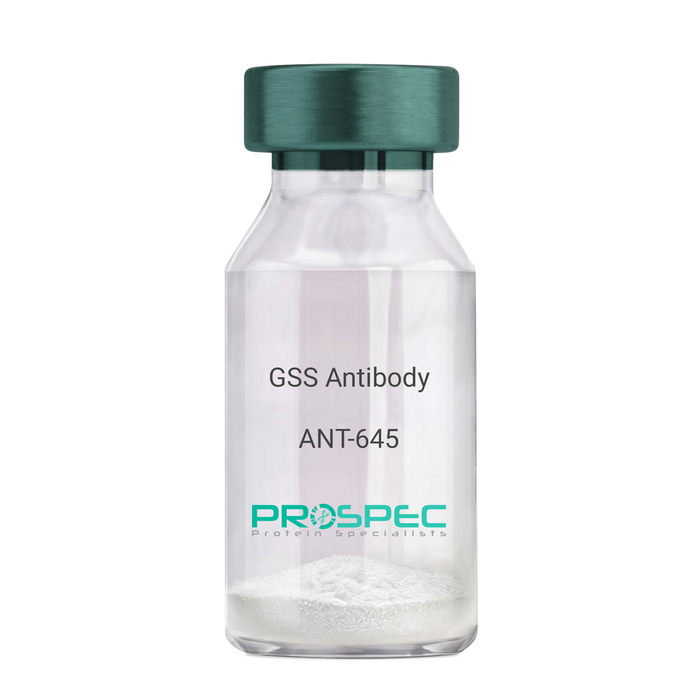
Name :
GSS AntibodyDescription:
Glutathione Synthetase, Mouse Anti Human
Glutathione synthetase, GSH synthetase, GSH-S, Glutathione synthase, GSHS, MGC14098, GSS.
Product # :
ANT-645Price :
Quantity :
Shipping Method :
Shipped with Ice Packs
More Info
- formulation
- More Info
Formulation
1mg/ml containing PBS, pH-7.4, 10% Glycerol and 0.02% Sodium Azide.
More Info
-
Introduction
Glutathione synthetase (GSS) is the second enzyme in the glutathione biosynthesis pathway. GSS catalyses the condensation of gamma-glutamylcysteine and glycine to form glutathione. Defects in the GSS are the cause of glutathione synthetase deficiency aka GSS deficiency or 5-oxoprolinuria or pyroglutamic aciduria, which is a severe form characterized by an increased rate of hemolysis and defective function of the central nervous system.
-
Synonyms
Glutathione synthetase, GSH synthetase, GSH-S, Glutathione synthase, GSHS, MGC14098, GSS.
-
Physical Appearance
Sterile filtered colorless solution.
-
Immunogen
Anti-human GSS mAb, is derived from hybridization of mouse F0 myeloma cells with spleen cells from BALB/c mice immunized with recombinant human GSS amino acids 1-474 purified from E. coli.
-
Ig Subclass
Mouse IgG1 heavy chain and k light chain.
-
Clone
PAT12F2AT.
-
Applications
GSS antibody has been tested by ELISA, Western blot analysis and ICC/IF to assure specificity and reactivity. Since application varies, however, each investigation should be titrated by the reagent to obtain optimal results.
-
Type
Mouse Anti Human Monoclonal.
-
Storage Procedures
For periods up to 1 month store at 4°C, for longer periods of time, store at -20°C. Prevent freeze thaw cycles.
-
Purification Method
GSS antibody was purified from mouse ascitic fluids by protein-A affinity chromatography.
ProSpec's products are furnished for LABORATORY RESEARCH USE ONLY. The product may not be used as drugs, agricultural or pesticidal products, food additives or household chemicals.
- View Data Sheet
Name :
HIV-1 TAT Clade-CDescription:
HIV-1 TAT Clade-C Recombinant
Product # :
HIV-116Price :
Quantity :
Shipping Method :
Shipped with Ice Packs
More Info
- description
- source
- formulation
- purity
- More Info
Description
HIV-1 TAT Recombinant- produced in E.coli is a single, non-glycosylated, polypeptide chain containing 101 amino acids encoded by two exons and having chain having a molecular mass of 21 kDa.
Source
Escherichia Coli.
Formulation
0.1% glycerol.
Purity
Greater than 90.0% as determined by SDS-PAGE.
More Info
-
Introduction
Human immunodeficiency virus type-1 (HIV-1) regulatory Tat protein plays an essential role in viral replication and infectivity. In addition, during acute infection, Tat is released extracellularly by infected cells and is taken up by neighboring cells where it transactivates viral replication and increases virus infectivity.
HIV-1 Tat activates transcription of HIV-1 viral genes by inducing phosphorylation of the C-terminal domain (CTD) of RNA polymerase II (RNAPII). Tat can also disturb cellular metabolism by inhibiting proliferation of antigen-specific T lymphocytes and by inducing cellular apoptosis. Tat-induced apoptosis of T-cells is attributed, in part, to the distortion of microtubules polymerization. LIS1 is a microtubule-associated protein that facilitates microtubule polymerization. -
Physical Appearance
Sterile filtered colorless clear solution.
-
Stability
Store at 4°C if entire vial will be used within 2-4 weeks. Store, frozen at -20°C for longer periods of time. Avoid multiple freeze-thaw cycles.
ProSpec's products are furnished for LABORATORY RESEARCH USE ONLY. The product may not be used as drugs, agricultural or pesticidal products, food additives or household chemicals.
- View Data Sheet
Name :
HCV NS5 Genotype-2bDescription:
Hepatitis C Virus NS5 Genotype-2b Recombinant
Product # :
HCV-231Price :
Quantity :
Shipping Method :
Shipped with Ice Packs
More Info
- description
- source
- formulation
- purity
- More Info
Description
The E.coli derived recombinant protein contains the HCV NS5 Genotype 2b immunodominant regions, amino acids 2212-2313. The protein is fused to a GST tag at N-Terminus.
Source
Escherichia Coli.
Formulation
1.5M urea, 25 mM Tris-HCl, pH-8, 0.2% Triton-X & 50% Glycerol.
Purity
HCV NS5 Genotype-2b protein is >95% pure as determined by 10% PAGE (coomassie staining).
More Info
-
Introduction
HCV is a small 50nm, enveloped, single-stranded, positive sense RNAvirus in the family Flaviviridae.
HCV has a high rate of replication with approximately one trillion particles produced each day in an infected individual. Due to lack of proofreading by the HCV RNA polymerase, the HCV has an exceptionally high mutation rate, a factor that may help it elude the host's immune response. Hepatitis C virus is classified into six genotypes(1-6) with several subtypes within each genotype. The preponderance and distribution of HCV genotypes varies globally. Genotype is clinically important in determining potential response to interferon-based therapy and the required duration of such therapy. Genotypes 1 and 4 are less responsive to interferon-based treatment than are the other genotypes (2, 3, 5 and 6). -
Stability
HCV NS5 Genotype-2b although stable at 4°C for 1 week, should be stored below -18°C. Please prevent freeze thaw cycles.
-
Applications
HCV NS5 Genotype-2b antigen in ELISA and Western blots, excellent antigen for detection of HCV with minimal specificity problems.
-
Specificity
Immunoreactive with sera of HCV-infected individuals.
-
Purification Method
HCV NS5 Genotype-2b protein was purified by proprietary chromatographic technique.
ProSpec's products are furnished for LABORATORY RESEARCH USE ONLY. The product may not be used as drugs, agricultural or pesticidal products, food additives or household chemicals.
- View Data Sheet
Name :
REXO2 AntibodyDescription:
RNA Exonuclease 2, Mouse Anti Human
REX2, RNA exonuclease 2 homolog (S. cerevisiae), CGI-114, RFN, SFN, Oligoribonuclease, mitochondrial, RNA exonuclease 2 homolog, Small fragment nuclease,REXO2, SMFN.
Product # :
ANT-490Price :
Quantity :
Shipping Method :
Shipped with Ice Packs
More Info
- formulation
- More Info
Formulation
1mg/ml containing PBS, pH-7.4, 10% Glycerol and 0.02% Sodium Azide.
More Info
-
Introduction
EXO2 is a 3'-to-5' exonuclease specific for small (primarily 5 nucleotides or less in length) single-stranded RNA and DNA oligomers. RNA Exonuclease 2 (REXO2) takes part in DNA repair, replication, and recombination, and in RNA processing and degradation. EXO2 is involved in resistance of human cells to UV-C-induced cell death via its part in the DNA repair process.
-
Synonyms
REX2, RNA exonuclease 2 homolog (S. cerevisiae), CGI-114, RFN, SFN, Oligoribonuclease, mitochondrial, RNA exonuclease 2 homolog, Small fragment nuclease,REXO2, SMFN.
-
Physical Appearance
Sterile filtered colorless solution.
-
Immunogen
Anti-human REXO2 mAb, clone PAT59D9A, is derived from hybridization of mouse F0 myeloma cells with spleen cells from BALB/c mice immunized with a recombinant human REXO2 protein 26-237 amino acids purified from E. coli.
-
Ig Subclass
Mouse IgG2b heavy chain and Kappa light chain.
-
Clone
PAT59D9AT.
-
Applications
The antibody has been tested by ELISA, Western blot analysis to assure specificity and reactivity. Since application varies, however, each investigation should be titrated by the reagent to obtain optimal results. Recommended starting dilution is 1:500.
-
Type
Mouse Anti Human Monoclonal.
-
Storage Procedures
For periods up to 1 month store at 4°C, for longer periods of time, store at -20°C. Prevent freeze thaw cycles.
-
Purification Method
REXO2 antibody was purified from mouse ascitic fluids by protein-A affinity chromatography.
ProSpec's products are furnished for LABORATORY RESEARCH USE ONLY. The product may not be used as drugs, agricultural or pesticidal products, food additives or household chemicals.
- View Data Sheet
Name :
GLUL AntibodyDescription:
Glutamine Synthetase, Mouse Anti Human
GLNS, EC 6.3.1.2, EC 4.1.1.15, GLUL, Glutamine Synthetase, GS, Glutamate decarboxylase, Glutamate--ammonia ligase, PIG43, PIG59.
Product # :
ANT-705Price :
Quantity :
Shipping Method :
Shipped with Ice Packs
More Info
- formulation
- More Info
Formulation
1mg/ml containing PBS, pH-7.4, 10% Glycerol and 0.02% Sodium Azide.
More Info
-
Introduction
GLUL catalyzes the synthesis of glutamine from glutamate and ammonia. Glutamine is a major source of energy and that takes part in cell proliferation, inhibition of apoptosis, and cell signaling. GLUL is expressed during early fetal stages, and has a role in maintaining body pH by removing ammonia from circulation. Mutations in GLUL gene are related with congenital glutamine deficiency.
-
Synonyms
GLNS, EC 6.3.1.2, EC 4.1.1.15, GLUL, Glutamine Synthetase, GS, Glutamate decarboxylase, Glutamate--ammonia ligase, PIG43, PIG59.
-
Physical Appearance
Sterile filtered colorless solution.
-
Immunogen
Anti-human GLUL mAb, is derived from hybridization of mouse F0 myeloma cells with spleen cells from BALB/c mice immunized with recombinant human GLUL amino acids 1-373 purified from E. coli.
-
Ig Subclass
Mouse IgG1 heavy chain and κ light chain.
-
Clone
PAT8D7AT.
-
Applications
GLUL antibody has been tested by ELISA and Western blot analysis to assure specificity and reactivity. Since application varies, however, each investigation should be titrated by the reagent to obtain optimal results.
-
Type
Mouse Anti Human Monoclonal.
-
Storage Procedures
For periods up to 1 month store at 4°C, for longer periods of time, store at -20°C. Prevent freeze thaw cycles.
-
Purification Method
GLUL antibody was purified from mouse ascitic fluids by protein-A affinity chromatography.
ProSpec's products are furnished for LABORATORY RESEARCH USE ONLY. The product may not be used as drugs, agricultural or pesticidal products, food additives or household chemicals.
- View Data Sheet
Name :
PGK1 AntibodyDescription:
Phosphoglycerate Kinase 1, Mouse Anti Human
Phosphoglycerate kinase 1, Primer recognition protein 2, Cell migration-inducing gene 10 protein, PRP 2, PGKA, MIG10, MGC8947, MGC117307, MGC142128, PGK1, Pgk-1.
Product # :
ANT-604Price :
Quantity :
Shipping Method :
Shipped with Ice Packs
More Info
- formulation
- More Info
Formulation
1mg/ml containing PBS, pH-7.4, 10% Glycerol and 0.02% Sodium Azide.
More Info
-
Introduction
PGK1 is an X-linked enzyme that has a major role in the glycolytic pathway. PGK1 is a glycolytic enzyme which catalyzes the conversion of 1,3-diphosphoglycerate to 3-phosphoglycerate, generating an ATP molecule. PGK1 may also act as a cofactor for polymerase alpha. Defects in the PGK1 gene are usually associated with chronic hemolytic anemia, though it can be accompanied by either mental retardation or muscular disease (rhabdomyolysis). Overexpression of PGK1 and its signalling targets are possibly an expression-pathway in diffuse primary gastric carcinomas promoting peritoneal dissemination. It was shown that PGK1 is differentially expressed in the dorsolateral prefrontal cortex from patients with schizophrenia.
-
Synonyms
Phosphoglycerate kinase 1, Primer recognition protein 2, Cell migration-inducing gene 10 protein, PRP 2, PGKA, MIG10, MGC8947, MGC117307, MGC142128, PGK1, Pgk-1.
-
Physical Appearance
Sterile filtered colorless solution.
-
Immunogen
Anti-human PGK1 mAb, is derived from hybridization of mouse F0 myeloma cells with spleen cells from BALB/c mice immunized with a recombinant human PGK1 protein 1-417 amino acids purified from E. coli.
-
Ig Subclass
Mouse IgG1 heavy chain and k light chain.
-
Clone
PAT2F4AT.
-
Applications
PGK1 antibody has been tested by ELISA, Western blot analysis, ICC/IF and Flow cytometry to assure specificity and reactivity. Since application varies, however, each investigation should be titrated by the reagent to obtain optimal results.
-
Type
Mouse Anti Human Monoclonal.
-
Storage Procedures
For periods up to 1 month store at 4°C, for longer periods of time, store at -20°C. Prevent freeze thaw cycles.
-
Purification Method
PGK1 antibody was purified from mouse ascitic fluids by protein-A affinity chromatography.
ProSpec's products are furnished for LABORATORY RESEARCH USE ONLY. The product may not be used as drugs, agricultural or pesticidal products, food additives or household chemicals.
- View Data Sheet
Name :
ACAT2 AntibodyDescription:
Acetyl-Coenzyme A acetyltransferase 2, Mouse Anti Human
Acetyl-CoA acetyltransferase cytosolic, Cytosolic acetoacetyl-CoA thiolase, ACAT2, Acetyl CoA transferase-like protein, ACAT-2.
Product # :
ANT-561Price :
Quantity :
Shipping Method :
Shipped with Ice Packs
More Info
- formulation
- More Info
Formulation
1mg/ml containing PBS, pH-7.4, 10% Glycerol and 0.02% Sodium Azide.
More Info
-
Introduction
ACAT2 enzyme participates in lipid metabolism. ACAT2 takes part in lipoprotein assembly, catalyzing cholesterol esterification in mammalian cells. ACAT2 is an integral membrane protein that localizes to the endoplasmic reticulum of human intestinal cells. ACAT2 deficiency contributes to severe mental retardation and hypotonus.
-
Synonyms
Acetyl-CoA acetyltransferase cytosolic, Cytosolic acetoacetyl-CoA thiolase, ACAT2, Acetyl CoA transferase-like protein, ACAT-2.
-
Physical Appearance
Sterile filtered colorless solution.
-
Immunogen
Anti-human ACAT2 mAb, clone PAT55D5AT, is derived from hybridization of mouse F0 myeloma cells with spleen cells from BALB/c mice immunized with a recombinant human ACAT2 protein 1-397 amino acids purified from E. coli.
-
Ig Subclass
Mouse IgG2b heavy chain and k light chain.
-
Clone
PAT55D5AT.
-
Applications
The antibody has been tested by ELISA, Western blot analysis to assure specificity and reactivity. Since application varies, however, each investigation should be titrated by the reagent to obtain optimal results. Recommended starting dilution is 1:1000.
-
Type
Mouse Anti Human Monoclonal.
-
Storage Procedures
For periods up to 1 month store at 4°C, for longer periods of time, store at -20°C. Prevent freeze thaw cycles.
-
Purification Method
ACAT2 antibody was purified from mouse ascitic fluids by protein-A affinity chromatography.
ProSpec's products are furnished for LABORATORY RESEARCH USE ONLY. The product may not be used as drugs, agricultural or pesticidal products, food additives or household chemicals.
- View Data Sheet
Name :
TOMM20 AntibodyDescription:
Mouse Anti Human Translocase Of Outer Mitochondrial Membrane 20
Mmitochondrial import receptor subunit TOM20 homolog, TOMM20, MAS20, MOM19, KIAA0016, Outer mitochondrial membrane receptor Tom20, Mitochondrial 20 kDa outer membrane protein.
Product # :
ANT-712Price :
Quantity :
Shipping Method :
Shipped with Ice Packs
More Info
- formulation
- More Info
Formulation
1mg/ml containing PBS, pH-7.4, 10% Glycerol and 0.02% Sodium Azide.
More Info
-
Introduction
Mmitochondrial import receptor subunit TOMM20 homolog (TOMM20) is a member of the Tom20 family. The Tom machinery consists of import receptors for the initial binding of cytosolically synthesized preproteins and a general import pore (GIP) for the membrane translocation of various preproteins into the mitochondria. TOMM20 acts as the transit peptide receptor at the surface of the mitochondrion outer membrane and facilitates the movement of preproteins into the TOM40 translocation pore.
-
Synonyms
Mmitochondrial import receptor subunit TOM20 homolog, TOMM20, MAS20, MOM19, KIAA0016, Outer mitochondrial membrane receptor Tom20, Mitochondrial 20 kDa outer membrane protein.
-
Physical Appearance
Sterile filtered colorless solution.
-
Immunogen
Anti-human TOMM20 mAb, is derived from hybridization of mouse F0 myeloma cells with spleen cells from BALB/c mice immunized with a recombinant human TOMM20 protein 25-145 amino acids purified from E. coli.
-
Ig Subclass
Mouse IgG1 heavy chain and k light chain.
-
Clone
PAT1B2AT.
-
Applications
TOMM20 antibody has been tested by ELISA and Western blot analysis to assure specificity and reactivity. Since application varies, however, each investigation should be titrated by the reagent to obtain optimal results.
-
Type
Mouse Anti Human Monoclonal.
-
Storage Procedures
For periods up to 1 month store at 4°C, for longer periods of time, store at -20°C. Prevent freeze thaw cycles.
-
Purification Method
TOMM20 antibody was purified from mouse ascitic fluids by protein-A affinity chromatography.
ProSpec's products are furnished for LABORATORY RESEARCH USE ONLY. The product may not be used as drugs, agricultural or pesticidal products, food additives or household chemicals.
- View Data Sheet
Name :
GSR AntibodyDescription:
Glutathione Reductase, Mouse Anti Human
Glutathione reductase mitochondrial, GR, GRase, GSR, GLUR, GRD1.
Product # :
ANT-549Price :
Quantity :
Shipping Method :
Shipped with Ice Packs
More Info
- formulation
- More Info
Formulation
1mg/ml containing PBS, pH-7.4, 10% Glycerol and 0.02% Sodium Azide.
More Info
-
Introduction
Glutathione reductase (GSR) belongs to the class-I pyridine nucleotide-disulfide oxidoreductase family. The GSR enzyme is a homodimeric flavoprotein and has a role in maintaining glutathione (GSH) in its reduced form by catalyzing the reduction of glutathione disulfide (GSSG): GSSG + NADPH + H+ ->2GSH + NADP+. In the majority of eukaryotic cells, GSR upholds the ratio of [GSH] / [GSSG], and partakes in quite a few critical functions such as the detoxification of reactive oxygen species as well as protein and DNA biosynthesis.
-
Synonyms
Glutathione reductase mitochondrial, GR, GRase, GSR, GLUR, GRD1.
-
Physical Appearance
Sterile filtered colorless solution.
-
Immunogen
Anti-human GSR mAb, is derived from hybridization of mouse F0 myeloma cells with spleen cells from BALB/c mice immunized with recombinant human GSR amino acids 43-522 purified from E. coli.
-
Ig Subclass
Mouse IgG1 heavy chain and k light chain.
-
Clone
PAT11D10AT.
-
Applications
GSR antibody has been tested by ELISA, Western blot analysis to assure specificity and reactivity. Since application varies, however, each investigation should be titrated by the reagent to obtain optimal results. Recommended starting dilution is 1:500.
-
Type
Mouse Anti Human Monoclonal.
-
Storage Procedures
For periods up to 1 month store at 4°C, for longer periods of time, store at -20°C. Prevent freeze thaw cycles.
-
Purification Method
GSR antibody was purified from mouse ascitic fluids by protein-A affinity chromatography.
ProSpec's products are furnished for LABORATORY RESEARCH USE ONLY. The product may not be used as drugs, agricultural or pesticidal products, food additives or household chemicals.
- View Data Sheet
Name :
HIV-1 CRFDescription:
HIV-1 Circulating Recombinant Form
Product # :
HIV-008Price :
Quantity :
Shipping Method :
Shipped at Room temp
More Info
- description
- source
- formulation
- purity
- More Info
Description
HIV-1 CRF Recombinant produced in E.Coli is a single, non-glycosylated polypeptide chain containing 101 amino acids and having a molecular mass of 20.1kDa.
Source
Escherichia Coli.
Formulation
Lyophilized with 10% glycerol.
Purity
Greater than 90.0% as determined by SDS-PAGE.
More Info
-
Introduction
HIV-1 circulating recombinant forms (CRFs) are part of viral recombinant lineages that play a major role in the global epidemic. HIV-1 CRF is 1 of 2 genes which dominate the epidemic in Burkina Faso. Multiple HIV-1 subtypes and circulating recombinant forms are known to cocirculate in Africa. In West Africa, the high prevalence of HIV-1 CRFand cocirculation of subtype Aand other complex intersubtype recombinants has been highly documented. Cocirculation of HIV-1 CRFstrain with other genetic subtypes may lead to novel and complex HIV-1 recombinants.
-
Physical Appearance
Sterile Filtered White lyophilized (freeze-dried) powder.
-
Stability
Lyophilized HIV-1 CRF although stable at room temperature for 1 week, should be stored desiccated below -18°C. Upon reconstitution HIV-1 CRF should be stored at 4°C between 2-7 days and for future use below -18°C. For long-term storage it is recommended to add a carrier protein (0.1% HSA or BSA). Please prevent freeze-thaw cycles.
-
Solubility
It is recommended to reconstitute the lyophilized HIV-1 CRF in sterile 18MΩ-cm H2O not less than 100µg/ml, which can then be further diluted to other aqueous solutions.
-
Applications
Western Blotting, SDS Page
ProSpec's products are furnished for LABORATORY RESEARCH USE ONLY. The product may not be used as drugs, agricultural or pesticidal products, food additives or household chemicals.
- View Data Sheet
Name :
HCV Core NS3Description:
Hepatitis C Virus Nucleocapsid (core) NS3 Recombinant
Product # :
HCV-008Price :
Quantity :
Shipping Method :
Shipped with Ice Packs
More Info
- description
- source
- formulation
- purity
- More Info
Description
Recombinant HCV protein produced in E.Coli containing the Core (130 a.a.) and NS3 (236 a.a.) excluding NS4 and NS5 domains having a total Mw of 45kDa and purified by proprietary chromatographic techniques.
Source
Escherichia Coli.
Formulation
Sterile filtered solution containing PBS, 1M urea and 25mM Arginine.
Purity
Greater than 95.0% as determined by SDS-PAGE.
More Info
-
Introduction
HCV is a small 50nm, enveloped, single-stranded, positive sense RNAvirus in the family Flaviviridae.
HCV has a high rate of replication with approximately one trillion particles produced each day in an infected individual. Due to lack of proofreading by the HCV RNA polymerase, the HCV has an exceptionally high mutation rate, a factor that may help it elude the host's immune response. Hepatitis C virus is classified into six genotypes(1-6) with several subtypes within each genotype. The preponderance and distribution of HCV genotypes varies globally. Genotype is clinically important in determining potential response to interferon-based therapy and the required duration of such therapy. Genotypes 1 and 4 are less responsive to interferon-based treatment than are the other genotypes (2, 3, 5 and 6). -
Physical Appearance
Sterile filtered colorless solution.
-
Stability
Store at 4°C if entire vial will be used within 2-4 weeks. Store, frozen at -20°C for longer periods of time.For long term storage it is recommended to add a carrier protein (0.1% HSA or BSA). Avoid multiple freeze-thaw cycles.
ProSpec's products are furnished for LABORATORY RESEARCH USE ONLY. The product may not be used as drugs, agricultural or pesticidal products, food additives or household chemicals.
- View Data Sheet
Name :
HCV E2Description:
Hepatitis C Virus E2 Recombinant
Product # :
HCV-005Price :
Quantity :
Shipping Method :
Shipped with Ice Packs
More Info
- description
- source
- formulation
- purity
- More Info
Description
Recombinant Hepatitis C Virus E2 Genotype-1 produced in E. coli is a single polypeptide chain containing 226 amino acids (aa 482-671) and having a molecular mass of 25.4kDa (NCBI Accession#NP_671491).HCV E2 is fused to a 36 amino acid His-tag at N-terminus.
Source
Escherichia Coli.
Formulation
The HCV E2 solution (0.25mg/ml) contains 20mM Tris-HCl buffer (pH 8.0), 0.4M Urea and 10% glycerol.
Purity
Greater than 80.0% as determined by SDS-PAGE.
More Info
-
Introduction
E1 and E2 glycoproteins form a heterodimer which is involved in virus attachment to the host cell, virion internalization via clathrin-dependent endocytosis and fusion with host membrane. E1/E2 heterodimer binds to human LDLR, CD81 and SCARB1/SR-BI receptors, however this binding insufficient for infection, some additional liver specific cofactors may be required. The fusion function may perhaps be conducted by E1. E2 hinders human EIF2AK2/PKR activation, preventing the establishment of an antiviral state. E2 is a viral ligand for CD209/DC-SIGN and CLEC4M/DC-SIGNR, which are respectively located on dendritic cells (DCs), and on liver sinusoidal endothelial cells and macrophage-like cells of lymph node sinuses. These interactions allow seizure of circulating HCV particles by these cells and subsequent diffusion to permissive cells.
-
Physical Appearance
Sterile Filtered colorless solution.
-
Stability
Store at 4°C if entire vial will be used within 2-4 weeks. Store, frozen at -20°C for longer periods of time. For long term storage it is recommended to add a carrier protein (0.1% HSA or BSA).Avoid multiple freeze-thaw cycles.
-
Amino Acid Sequence
MRGSHHHHHH GMASMTGGQQ MGRDLYDDDD KDRWGSERPY CWHYPPRPCG IVPAKSVCGP VYCFTPSPVV VGTTDRSGAP TYSWGANDTD VFVLNNTRPP LGNWFGCTWM NSTGFTKVCG APPCVIGGVG NNTLLCPTDC FRKHPEATYS RCGSGPWITP RCMVDYPYRL WHYPCTINYT IFKVRMYVGG VEHRLEAACN WTRGERCDLE DRDRSELSPL LLSTTQ.
ProSpec's products are furnished for LABORATORY RESEARCH USE ONLY. The product may not be used as drugs, agricultural or pesticidal products, food additives or household chemicals.