Search results
1000 results found for “serglycin”
Name
Description
Product #
Price
Quantity
Shipping Method
- View Data Sheet
Name :
ITGB4 HumanDescription:
Integrin Beta 4 Human Recombinant
Integrin beta-4, GP150, CD104, ITGB4, Integrin Subunit Beta 4, CD104 Antigen, Integrin, Beta 4, Integrin Beta-4.
Product # :
PRO-2528Price :
Quantity :
Shipping Method :
Shipped with Ice Packs
More Info
- description
- source
- formulation
- purity
- More Info
Description
ITGB4 produced in Sf9 Baculovirus cells is a single, glycosylated polypeptide chain containing 691 amino acids (28-710a.a.) and having a molecular mass of 77.5kDa. (Molecular size on SDS-PAGE will appear at approximately 70-100kDa). ITGB4 is expressed with an 8 amino acid His tag at C-Terminus and purified by proprietary chromatographic techniques.
Source
Sf9, Baculovirus cells.
Formulation
ITGB4 protein solution (0.5mg/ml) contains Phosphate Buffered Saline (pH 7.4) & 10% glycerol.
Purity
Greater than 90.0% as determined by SDS-PAGE.
More Info
-
Introduction
ITGB4 (integrin beta-4 isoform 1) belongs to the Integrin beta family. ITGB4 forms noncovalent heterodimers with Integrin alpha 6 and takes part in the formation of epithelial hemidesmosomes. ITGB4 has a vital structural role in the hemidesmosome of epithelial cells and is needed for the regulation of keratinocyte motility & polarity. ITGB4 leans towards the association with alpha 6 subunit and is expected to take a key role in the biology of invasive carcinoma.
-
Synonyms
Integrin beta-4, GP150, CD104, ITGB4, Integrin Subunit Beta 4, CD104 Antigen, Integrin, Beta 4, Integrin Beta-4.
-
Physical Appearance
Sterile Filtered colorless solution.
-
Stability
Store at 4°C if entire vial will be used within 2-4 weeks. Store, frozen at -20°C for longer periods of time. For long term storage it is recommended to add a carrier protein (0.1% HSA or BSA). Avoid multiple freeze-thaw cycles.
-
Amino Acid Sequence
NRCKKAPVKS CTECVRVDKD CAYCTDEMFR DRRCNTQAEL LAAGCQRESI VVMESSFQIT EETQIDTTLR RSQMSPQGLR VRLRPGEERH FELEVFEPLE SPVDLYILMD FSNSMSDDLD NLKKMGQNLA RVLSQLTSDY TIGFGKFVDK VSVPQTDMRP EKLKEPWPNS DPPFSFKNVI SLTEDVDEFR NKLQGERISG NLDAPEGGFD AILQTAVCTR DIGWRPDSTH LLVFSTESAF HYEADGANVL AGIMSRNDER CHLDTTGTYT QYRTQDYPSV PTLVRLLAKH NIIPIFAVTN YSYSYYEKLH TYFPVSSLGV LQEDSSNIVE LLEEAFNRIR SNLDIRALDS PRGLRTEVTS KMFQKTRTGS FHIRRGEVGI YQVQLRALEH VDGTHVCQLP EDQKGNIHLK PSFSDGLKMD AGIICDVCTC ELQKEVRSAR CSFNGDFVCG QCVCSEGWSG QTCNCSTGSL SDIQPCLREG EDKPCSGRGE CQCGHCVCYG EGRYEGQFCE YDNFQCPRTS GFLCNDRGRC SMGQCVCEPG WTGPSCDCPL SNATCIDSNG GICNGRGHCE CGRCHCHQQS LYTDTICEIN YSAIHPGLCE DLRSCVQCQA WGTGEKKGRT CEECNFKVKM VDELKRAEEV VVRCSFRDED DDCTYSYTME GDGAPGPNST VLVHKKKDCP PGSLEHHHHH H
ProSpec's products are furnished for LABORATORY RESEARCH USE ONLY. The product may not be used as drugs, agricultural or pesticidal products, food additives or household chemicals.
- View Data Sheet
Name :
GAGE12F HumanDescription:
G Antigen 12F Human Recombinant
G Antigen 12F, GAGE-12F, G Antigen 12F/G/I.
Product # :
PRO-1736Price :
Quantity :
Shipping Method :
Shipped with Ice Packs
More Info
- description
- source
- formulation
- purity
- More Info
Description
GAGE12F Human Recombinant produced in E.Coli is a single, non-glycosylated polypeptide chain containing 140 amino acids (1-117aa) and having a molecular mass of 15.4kDa.GAGE12F is fused to a 23 amino acid His-tag at N-terminus & purified by proprietary chromatographic techniques.
Source
Escherichia Coli.
Formulation
GAGE12F protein solution (0.25mg/ml) containing 20mM Tris-HCl buffer (pH 8.0), 0.15M NaCl and 10% glycerol.
Purity
Greater than 85.0% as determined by SDS-PAGE.
More Info
-
Introduction
GAGE12F is a member of the GAGE multigene family expressed in a large range of tumors whereas in normal tissues, his expression is restricted to germ cells. These genes are organized in clustered repeats, and have a high degree of forecasted sequence identity, however differ by scattered single nucleotide substitution. Their sequences contain either the antigenic peptide YYWPRPRRY or YRPRPRRY which is recognized by cytotoxic T-cells.
-
Synonyms
G Antigen 12F, GAGE-12F, G Antigen 12F/G/I.
-
Physical Appearance
Sterile Filtered clear solution.
-
Stability
Store at 4°C if entire vial will be used within 2-4 weeks. Store, frozen at -20°C for longer periods of time. For long term storage it is recommended to add a carrier protein (0.1% HSA or BSA).Avoid multiple freeze-thaw cycles.
-
Amino Acid Sequence
MGSSHHHHHH SSGLVPRGSH MGSMSWRGRS TYYWPRPRRY VQPPEMIGPM RPEQFSDEVE PATPEEGEPA TQRQDPAAAQ EGEDEGASAG QGPKPEAHSQ EQGHPQTGCE CEDGPDGQEM DPPNPEEVKT PEEGEKQSQC
ProSpec's products are furnished for LABORATORY RESEARCH USE ONLY. The product may not be used as drugs, agricultural or pesticidal products, food additives or household chemicals.
- View Data Sheet
Name :
TPGS2 HumanDescription:
Tubulin Polyglutamylase Complex Subunit 2 Human Recombinant
Tubulin polyglutamylase complex subunit 2, C18orf10; HMFN0601, HsT3006, L17, PGs2, TPGS2.
Product # :
PRO-1668Price :
Quantity :
Shipping Method :
Shipped with Ice Packs
More Info
- description
- source
- formulation
- purity
- More Info
Description
TPGS2 Human Recombinant produced in E.coli is a single, non-glycosylated polypeptide chain containing 323 amino acids (1-300) and having a molecular mass of 35.7kDa.TPGS2 is fused to a 23 amino acid His-tag at N-terminus & purified by proprietary chromatographic techniques.
Source
Escherichia Coli.
Formulation
The TPGS2 solution (0.25mg/ml) contains 20mM Tris-HCl buffer (pH 8.0), 0.2M NaCl, 50% glycerol, 2mM DTT and 2mM EDTA.
Purity
Greater than 85.0% as determined by SDS-PAGE.
More Info
-
Introduction
Tubulin Polyglutamylase Complex Subunit 2 (TPGS2) is a component of the neuronal polyglutamylase complex, which takes part in post-translational addition of glutamate residues to C-terminal tubulin tails. Alternatively spliced transcript variants encoding multiple isoforms have been monitored for TPGS2.
-
Synonyms
Tubulin polyglutamylase complex subunit 2, C18orf10; HMFN0601, HsT3006, L17, PGs2, TPGS2.
-
Physical Appearance
Sterile Filtered colorless solution.
-
Stability
Store at 4°C if entire vial will be used within 2-4 weeks. Store, frozen at -20°C for longer periods of time. For long term storage it is recommended to add a carrier protein (0.1% HSA or BSA).Avoid multiple freeze-thaw cycles.
-
Amino Acid Sequence
MGSSHHHHHH SSGLVPRGSH MGSMEEEASS PGLGCSKPHL EKLTLGITRI LESSPGVTEV TIIEKPPAER HMISSWEQKN NCVMPEDVKN FYLMTNGFHM TWSVKLDEHI IPLGSMAINS ISKLTQLTQS SMYSLPNAPT LADLEDDTHE ASDDQPEKPH FDSRSVIFEL DSCNGSGKVC LVYKSGKPAL AEDTEIWFLD RALYWHFLTD TFTAYYRLLI THLGLPQWQY AFTSYGISPQ AKQWFSMYKP ITYNTNLLTE ETDSFVNKLD PSKVFKSKNK IVIPKKKGPV QPAGGQKGPS GPSGPSTSST SKSSSGSGNP TRK.
ProSpec's products are furnished for LABORATORY RESEARCH USE ONLY. The product may not be used as drugs, agricultural or pesticidal products, food additives or household chemicals.
- View Data Sheet
Name :
STC 1 HumanDescription:
Stanniocalcin-1 Human Recombinant
Stanniocalcin-1, STC, STC-1.
Product # :
HOR-259Price :
Quantity :
Shipping Method :
Shipped at Room temp
More Info
- description
- source
- formulation
- purity
- More Info
Description
Stanniocalcin-1 Human Recombinant produced in 293 cell line is a single, glycosylated, polypeptide chain containing 240 amino acids and having a total molecular mass of 25.9 kDa. The Stanniocalcin contains 10 residues form the C-Terminal Flag- tag. Stanniocalcin is purified by proprietary chromatographic techniques.
Source
293 cell line (Human embryonic kidney).
Formulation
Filtered (0.4µm) and lyophilized in 0.5mg/ml in 20mM Tris buffer, 50mM NaCl, pH 7.5.
Purity
Greater than 95.0% as determined by SDS-PAGE.
More Info
-
Introduction
Stanniocalcin 1 (STC1) is the mammalian homologue of STC, which was originally identified as a calcium/phosphate-regulating hormone in bony fishes. In contrast, STC1 may play an autocrine and paracrine role with pleiotropic effects in mammals. It is expressed in a wide variety of tissues, but unexpectedly is not detected in the circulation under normal circumstances, which is possibly caused by its attaching to soluble and tethered forms of a high-affinity binding protein. STC-1 can affect calcium homeostasis, bone and muscle mass and structure, and angiogenesis through effects on osteoblasts, osteoclasts, myoblasts/myocytes, and endothelial cells in mouse model. Differential regulation of myocardial STC1 protein expression was reported in heart failure. In addition, STC1 may regulate calcium currents in cardiomyocytes and may contribute to the alterations in calcium homeostasis of the failing heart. STC1 was found to be a selective modulator of hepatocyte growth factor (HGF)-induced endothelial migration and morphogenesis, an inhibitor of macrophage chemotaxis and chemokinesis, suppressor of progesterone and luteinization inhibitor. Together with STC-2, it may play important roles in the processes of implantation and decidualization in the rat. In terminally differentiated adipocytes, it may function as a "survival factor", which contributes to the maintenance of the integrity of mature adipose tissue. In context with its possible role in gestation, a Big STC, a three highermolecular- mass variant has been described. STC1 was identified as one of hypoxia-responsive genes coupled to hypoxia-driven angiogenesis.
Current research indicates that STC-1 might be a useful molecular marker to detect tumor cells in blood and bone marrow from patients with various types of malignancies. -
Synonyms
Stanniocalcin-1, STC, STC-1.
-
Physical Appearance
White lyophilized (freeze-dried) powder.
-
Stability
Lyophilized STC-1 although stable at room temperature for 3 weeks, should be stored desiccated below -18°C. Upon reconstitution Stanniocalcin should be stored at 4°C between 2-7 days and for future use below -18°C.For long term storage it is recommended to add a carrier protein (0.1% HSA or BSA).Please prevent freeze-thaw cycles.
-
Solubility
Add deionized water to a working concentration approximately 0.5mg/ml and let the lyophilized pellet dissolve completely. Product is not sterile! Please filter the product by appropriate sterile filter before using it in the cell culture.
-
Amino Acid Sequence
THEAEQNDSV SPRKSRVAAQ NSAEVVRCLN SALQVGCGAF ACLENSTCDT DGMYDICKSF LYSAAKFDTQ GKAFVKESLK CIANGVTSKV FLAIRRCSTF QRMIAEVQEE CYSKLNVCSI AKRNPEAITE VVQLPNHFSN RYYNRLVRSL LECDEDTVST IRDSLMEKIG PNMASLFHIL QTDHCAQTHP RADFNRRRTN EPQKLKVLLR NLRGEEDSPS HIKRTSHESA ASDYKDDDDK.
ProSpec's products are furnished for LABORATORY RESEARCH USE ONLY. The product may not be used as drugs, agricultural or pesticidal products, food additives or household chemicals.
- View Data Sheet
Name :
Streptavidin-NCDescription:
Streptavidin-NC Recombinant
Product # :
PRO-338Price :
Quantity :
Shipping Method :
Shipped with Ice Packs
More Info
- description
- source
- formulation
- purity
- More Info
Description
Recombinant Streptavidin-NC produced in E.Coli is a single, non-glycosylated, polypeptide chain having a molecular mass of 24kDa. Streptavidin-NC is engineered to bind to nitrocellulose.
Source
Escherichia Coli.
Formulation
The sterile solution contains 10mM K2HPO4-KH2PO4, pH 7.3.
Purity
Greater than 93.0% as determined by SDS-PAGE.
More Info
-
Introduction
Streptavidin is a tetrameric protein secreted by Streptomyces avidinii which binds firmly to biotin. Streptavidin is wilyde used in molecular biology through its unique high affinity for the vitamin biotin. The dissociation constant (Kd) of the biotin-streptavidin complex is about ~10-15 mol/L. The strong affinity recognition of biotin and biotinylated molecules has made streptavidin one of the most important components in diagnostics and laboratory kits. The streptavidin/biotin system has one of the biggest free energies of association of yet observed for noncovalent binding of a protein and small ligand in aqueous solution (K_assoc = 10**14). The complexes are also extremely stable over a wide range of temperature and pH.
-
Physical Appearance
Sterile Liquid formulation.
-
Stability
Streptavidin-NC although stable at 4°C for 3 weeks, should be stored below -18°C. Please prevent freeze thaw cycles.
ProSpec's products are furnished for LABORATORY RESEARCH USE ONLY. The product may not be used as drugs, agricultural or pesticidal products, food additives or household chemicals.
- View Data Sheet
Name :
EGFR AntibodyDescription:
Epidermal Growth Factor Receptor, Mouse Anti Human
Epidermal growth factor receptor, EC 2.7.10.1, Receptor tyrosine-protein kinase ErbB-1, ERBB, mENA, ERBB1, EGFR.
Product # :
ANT-042Price :
Quantity :
Shipping Method :
Shipped with Ice Packs
More Info
- formulation
- More Info
Formulation
1mg/ml containing PBS, pH-7.4, & 0.1% Sodium Azide.
More Info
-
Introduction
The epidermal growth factor receptor (EGF R) subfamily of receptor tyrosine kinases comprises four members: EGF R (also known as HER1, ErbB1 or ErbB), ErbB2 (Neu, HER-2), ErbB3 (HER-3), and ErbB4 (HER-4). All family members are type I transmembrane glycoprotein that has an extracellular domain which contains two cysteine-rich domains separated by a spacer region that is involved in ligand-binding, and a cytoplasmic domain which has a membrane-proximal tyrosine kinase domain and a C-terminal tail with multiple tyrosine autophosphorylation sites. The human EGF R gene encodes a 1210 amino acid (aa) residue precursor with a 24 aa putative signal peptide, a 621 aa extracellular domain, a 23 aa transmembrane domain, and a 542 aa cytoplasmic domain. EGF R has been shown to bind a subset of the EGF family ligands, including EGF, amphiregulin, TGF-a , betacellulin, epiregulin, heparin-binding EGF and neuregulin-2 in the absence of a co-receptor. Ligand binding induces EGF R homodimerization as well as heterdimerization with ErbB2, resulting in kinase activation, tyrosine phosphorylation and cell signaling. EGF R can also be recruited to form heterodimers with the ligand-activated ErbB3 or ErbB4. EGF R signaling has been shown to regulate multiple biological functions including cell proliferation, differentiation, motility and apoptosis. In addition, EGF R signaling has also been shown to play a role in carcinogenesis.
-
Synonyms
Epidermal growth factor receptor, EC 2.7.10.1, Receptor tyrosine-protein kinase ErbB-1, ERBB, mENA, ERBB1, EGFR.
-
Physical Appearance
Sterile filtered colorless solution.
-
Immunogen
Anti-human EGFR mAb, is derived from hybridization of mouse FO myeloma cells with spleen cells from BALB/c mice immunized with recombinant human EGFR amino acids 424-605 purified from E. coli.
-
Ig Subclass
Mouse IgG2b heavy chain and k light chain.
-
Clone
PAT6E3AT.
-
Applications
EGFR antibody has been tested by ELISA, Western blot analysis to assure specificity and reactivity. Since application varies, however, each investigation should be titrated by the reagent to obtain optimal results. Recommended dilution range for Western blot analysis is 1:500 ~ 1000.
Recommended starting dilution is 1:500. -
Type
Mouse Anti Human Monoclonal.
-
Storage Procedures
For periods up to 1 month store at 4°C, for longer periods of time, store at -20°C. Prevent freeze thaw cycles.
-
Purification Method
EGFR antibody was purified from mouse ascitic fluids by protein-G affinity chromatography.
ProSpec's products are furnished for LABORATORY RESEARCH USE ONLY. The product may not be used as drugs, agricultural or pesticidal products, food additives or household chemicals.
- View Data Sheet
Name :
Trypsin-2 HumanDescription:
Trypsin-2 Human Recombinant
Product # :
PRO-770Price :
Quantity :
Shipping Method :
Shipped at Room temp
More Info
- description
- source
- formulation
- purity
- biological activity
- More Info
Description
Recombinant Human Trypsin-2 expressed in E.Coli having an Mw of 24kDa is purified by standard chromatography techniques.
Source
Escherichia Coli.
Formulation
The protein was lyophilized without any additives.
Purity
Greater than 90% as determined by SDS-PAGE.
Biological Activity
2,800 units/mg.
More Info
-
Introduction
Trypsin (EC3.4.21.4) is part of the serine protease family. Trypsin cleaves lysine and arginine at the C-terminal side of the peptide. The hydrolysis rate is slower if an acidic residue is on either sides of the cleavage site and no cleavage occurs if a proline residue is on the carboxyl side of the cleavage site. Trypsin optimum pH is pH-7 to 9. Trypsin will also hydrolyze ester and amide linkages of synthetic derivatives of amino acids such as: benzoyl L-arginine ethyl ester (BAEE), p-toluenesulfonyl- L-arginine methyl ester (TAME), tosyl-L-arginine methyl ester, N-α-benzoyl-L-arginine p-nitroanilide (BAPNA), L-lysyl-p-nitroanilide, and benzoyl-L-tyrosine ethyl ester (BTEE). Serine protease inhibitors that inhibit recombinant trypsin include TLCK (N-p-tosyl-L-lysine chloromethyl ketone), PMSF (phenylmethanesulfonyl fluoride), benzamidine, soybean trypsin inhibitor, and ovomucoid.
-
Physical Appearance
Sterile Filtered lyophilized powder.
-
Stability
Recombinant Human Trypsin although stable at room temp for 1 week, should be stored desiccated below -18°C. For long term storage it is recommended to add a carrier protein (0.1% HSA or BSA). Please prevent freeze-thaw cycles.
-
Solubility
It is recommended to reconstitute the lyophilized Human Trypsin in sterile 1mM HCl or 50mM HAC not less than 100µg/ml, which can then be further diluted to other aqueous solutions.
-
Applications
Trypsin digestion: the suggested ratio is 1:50 to 1:1000 (w/w).
-
Unit Definition
One USP unit of trypsin activity will produce a Delta A253 of 0.003 per minute in a reaction volume of 3.0ml at pH7.6 and 25°C, with BAEE as a substrate (1cm light path).
ProSpec's products are furnished for LABORATORY RESEARCH USE ONLY. The product may not be used as drugs, agricultural or pesticidal products, food additives or household chemicals.
- View Data Sheet
Name :
SNCA HumanDescription:
Alpha Synuclein Human Recombinant
Alpha-synuclein, Non-A beta component of AD amyloid, Non-A4 component of amyloid precursor, NACP, PD1, PARK1, PARK4, MGC110988, a-Synuclein, SNCA.
Product # :
PRO-393Price :
Quantity :
Shipping Method :
Shipped with Ice Packs
More Info
- description
- source
- formulation
- purity
- More Info
Description
a-Synuclein Human Recombinant produced in E.Coli is a single, non-glycosylated polypeptide chain of 140 amino acids having a molecular mass of 14.4kDa (Real molecular weight on SDS-PAGE will be shift up). The Recombinant Human a-Synuclein is purified by proprietary chromatographic techniques.
Source
Escherichia Coli.
Formulation
The protein (1mg/ml) contains 20mM Tris-HCl buffer (pH 7.5), 0.1M NaCl and 1mM MgCl2.
Purity
Greater than 95.0% as determined by SDS-PAGE.
More Info
-
Introduction
a-Synuclein (amino acids 1-140), an acidic neuronal protein of 140 amino acids, is extremely heat-resistant and is natively unfolded with an extended structure primarily composed of random coils. a-synuclein has been suggested to be implicated in the pathogenesis of Parkinson’s disease and related neurodegenerative disorders, and more recently, to be an important regulatory component of vesicular transport in neuronal cells. Moreover, recent studies have shown that a-synuclein has chaperone activity and that this activity is lost upon removing its C-terminal acidic tail (amino acids 96-140).
-
Synonyms
Alpha-synuclein, Non-A beta component of AD amyloid, Non-A4 component of amyloid precursor, NACP, PD1, PARK1, PARK4, MGC110988, a-Synuclein, SNCA.
-
Physical Appearance
Sterile filtered colorless solution.
-
Stability
Store at 4°C if entire vial will be used within 2-4 weeks. Store, frozen at -20°C for longer periods of time. For long term storage it is recommended to add a carrier protein (0.1% HSA or BSA). Avoid multiple freeze-thaw cycles.
-
Amino Acid Sequence
MDVFMKGLSK AKEGVVAAAE KTKQGVAEAA GKTKEGVLYV GSKTKEGVVH GVATVAEKTK EQVTNVGGAV VTGVTAVAQK TVEGAGSIAA ATGFVKKDQL GKNEEGAPQE GILEDMPVDP DNEAYEMPSE EGYQDYEPEA.
ProSpec's products are furnished for LABORATORY RESEARCH USE ONLY. The product may not be used as drugs, agricultural or pesticidal products, food additives or household chemicals.
- View Data Sheet
Name :
ProNGF HumanDescription:
Pro-Nerve Growth Factor Human Recombinant
Human Pro-NGF, ProNGF, NGFB.
Product # :
CYT-426Price :
Quantity :
Shipping Method :
Shipped at Room temp
More Info
- description
- source
- formulation
- purity
- More Info
Description
Pro-NGF Human Recombinant produced in E.Coli is a single, non-glycosylated, polypeptide chain containing 224 amino acids and having a molecular mass of 25 kDa.ProNGF Human Recombinant is purified by proprietary chromatographic techniques.
Source
Escherichia Coli.
Formulation
ProNGF was lyophilized from a 0.2 μM filtered solution of 20m Tris-HCL, 0.5M NaCl, 5% Trehalose, 5% Mannitol. 0.01% Tween-80 and 1mM EDTA pH-8.
Purity
Greater than 95.0% as determined by SDS-PAGE.
More Info
-
Synonyms
Human Pro-NGF, ProNGF, NGFB.
-
Physical Appearance
Sterile Filtered White lyophilized (freeze-dried) powder.
-
Stability
Lyophilized ProNGF although stable at room temperature for 3 weeks, should be stored desiccated below -18°C. Upon reconstitution ProNGF should be stored at 4°C between 2-7 days and for future use below -18°C. Please prevent freeze-thaw cycles.
-
Solubility
It is recommended to reconstitute the lyophilized ProNGF in distilled water to a concentration no less than 100 µg/ml, which can then be further diluted to other aqueous solutions.
-
Amino Acid Sequence
MEPHSESNVPAGHTIPQAHWTKLQHSLDTALRRARSAPAAAIAARVAGQTRNI
TVDPRLFKKRRLRSPRVLFSTQPPREAADTQDLDFEVGGAAPFNRTHRSKRS
SSHPIFHRGEFSVCDSVSVWVGDKTTATDIKGKEVMVLGEVNINNSVFKQYFFET
KCRDPNPVDSGCRGIDSKHWNSYCTTTHTFVKALTMDGKQAAWRFIRIDTAC
VCVLSRKAVRRA. -
Background
Pro-Nerve Growth Factor Human Recombinant: Unveiling its Potential in Neuroregulation and Disease Pathogenesis
Abstract:
Pro-Nerve Growth Factor (Pro-NGF) human recombinant is a crucial precursor protein involved in neuronal development, survival, and degenerative processes. This research paper aims to provide a comprehensive analysis of Pro-NGF, including its characteristics, processing mechanisms, and implications in neuroregulation and disease pathogenesis. Additionally, innovative methodologies for the production and manipulation of Pro-NGF human recombinant are proposed, highlighting its potential as a therapeutic target for neurological disorders and neurodegenerative diseases.
Introduction:
Neuroregulation and maintenance of neuronal health are intricate processes governed by a network of signaling molecules. Pro-NGF, the precursor form of Nerve Growth Factor (NGF), acts as a key player in neuronal development, synaptic plasticity, and cell survival. This paper delves into the distinctive features of Pro-NGF and presents novel approaches for the production and manipulation of Pro-NGF human recombinant, aiming to unravel its role in neuroregulation and disease pathogenesis.
Characteristics and Processing Mechanisms:
Pro-NGF is initially synthesized as an inactive precursor, requiring proteolytic cleavage for conversion into mature NGF. The processing of Pro-NGF involves the action of proteases, such as furin, and the formation of distinct protein complexes. The balance between Pro-NGF and mature NGF levels plays a critical role in modulating neuronal function and fate, influencing processes such as neuronal survival, axonal growth, and synaptic plasticity.
Production and Manipulation of Pro-NGF Human Recombinant:
Efficient production methodologies and manipulation strategies are crucial for studying the role of Pro-NGF in neuroregulation and disease pathogenesis. Recombinant protein expression systems, including mammalian cell culture and bacterial expression systems, have been employed to produce functional Pro-NGF human recombinant. Techniques such as mutagenesis, protein purification, and specific inhibitors targeting Pro-NGF processing pathways enable the manipulation of Pro-NGF levels and investigation of its downstream effects.
Implications in Neuroregulation and Disease Pathogenesis:
Pro-NGF human recombinant holds significant potential in understanding the intricate mechanisms underlying neuroregulation and disease pathogenesis. Dysregulation of Pro-NGF processing and altered Pro-NGF/mature NGF ratios have been implicated in various neurological disorders, including Alzheimer's disease, Parkinson's disease, and ischemic stroke. Manipulating Pro-NGF levels and the balance between its mature form may offer therapeutic strategies for modulating neurotrophic signaling and promoting neuronal health in these conditions.
Conclusion:
Pro-NGF human recombinant emerges as a key regulator in neuroregulation and disease pathogenesis, offering promising avenues for therapeutic intervention. Enhancing our understanding of Pro-NGF processing mechanisms and its downstream signaling cascades will provide valuable insights into neurodevelopment, neurodegeneration, and potential therapeutic strategies. Targeting Pro-NGF as a therapeutic intervention may hold immense promise in treating neurological disorders and promoting neuronal health.
What is the molecular weight / Mw of ProNGF Protein?
ProNGF Protein has a total Mw of 25kDa.
What is the source or expression system of ProNGF Protein?
Escherichia Coli.
What is the Purity of ProNGF Protein?
ProNGF Protein is >95% pure as determined by SDS-PAGE.
What is the Biological Activity of ProNGF Protein?
The biological functionality of ProNGF Protein will be determined in the future.
What is the amino acid sequence of ProNGF Protein?
MEPHSESNVPAGHTIPQAHWTKLQHSLDTALRRARSAPAAAIAARVAGQTRNI
TVDPRLFKKRRLRSPRVLFSTQPPREAADTQDLDFEVGGAAPFNRTHRSKRS
SSHPIFHRGEFSVCDSVSVWVGDKTTATDIKGKEVMVLGEVNINNSVFKQYFFET
KCRDPNPVDSGCRGIDSKHWNSYCTTTHTFVKALTMDGKQAAWRFIRIDTAC
VCVLSRKAVRRA
What applications can ProNGF Protein be used in?
Tissue Factor Protein can probably be used in western blot, ELISA and Lateral Flow.
What is the endotoxin level for ProNGF Protein?
The endotoxin level is minimal, ProNGF Protein was purified using conventional chromatography techniques
ProSpec's products are furnished for LABORATORY RESEARCH USE ONLY. The product may not be used as drugs, agricultural or pesticidal products, food additives or household chemicals.
- View Data Sheet
Name :
CD68 Human, 38kDaDescription:
CD68 Human Recombinant, 38kDa
Macrosialin, Gp110, CD68 antigen, CD68, SCARD1, DKFZp686M18236.
Product # :
PRO-293Price :
Quantity :
Shipping Method :
Shipped with Ice Packs
More Info
- description
- source
- formulation
- More Info
Description
CD68 Human Recombinant full length expressed in E.coli, shows a 38 kDa band on SDS-PAGE.CD68 is purified by proprietary chromatographic techniques.
Source
Escherichia Coli.
Formulation
CD68 at 100µg/1ml in 50mM Tris-Acetate, pH7.5, 1mM EDTA, 20% Glycerol.
More Info
-
Introduction
CD68 encodes a 110-kD transmembrane glycoprotein that is highly expressed by human monocytes and tissue macrophages. It is a member of the lysosomal/endosomal-associated membrane glycoprotein (LAMP) family. The protein primarily localizes to lysosomes and endosomes with a smaller fraction circulating to the cell surface. It is a type I integral membrane protein with a heavily glycosylated extracellular domain and binds to tissue- and organ-specific lectins or selectins. The protein is also a member of the scavenger receptor family. Scavenger receptors typically function to clear cellular debris, promote phagocytosis, and mediate the recruitment and activation of macrophages. Alternative splicing results in multiple transcripts encoding different isoforms.
-
Synonyms
Macrosialin, Gp110, CD68 antigen, CD68, SCARD1, DKFZp686M18236.
-
Physical Appearance
Sterile Filtered clear solution.
-
Stability
Store vial at -20°C to -80°C. When stored at the recommended temperature, this protein is stable for 12 months.Please prevent freeze-thaw cycles.
ProSpec's products are furnished for LABORATORY RESEARCH USE ONLY. The product may not be used as drugs, agricultural or pesticidal products, food additives or household chemicals.
- View Data Sheet
Name :
Omentin AntibodyDescription:
Omentin, Mouse Anti Human
Intelectin-1, HL1, LFR, HL-1, INTL, ITLN, hIntL.
Product # :
ANT-505Price :
Quantity :
Shipping Method :
Shipped with Ice Packs
More Info
- formulation
- More Info
Formulation
1mg/ml containing PBS, pH-7.4, 10% Glycerol and 0.02% Sodium Azide.
More Info
-
Introduction
Omentin is a recently recognized gene highly localized to the mental tissue (visceral adipose tissue). Omentin is present in the stromal vascular cells in the adipose tissue rather than in the adipocytes. Omentin is predominantly expressed in the visceral adipose tissue than the subcutaneous tissue, with the omentin mRNA being 150 times higher in the visceral adipose tissue. Omentin has also been detected in human blood using western blot analysis, and seems to increase insulin-stimulated glucose uptake in 3T3-L1 adipocytes in mice. Omentin seems to increase Akt phosphorylation irrespective of insulin presence. Its role in glucose metabolism and obesity remains to be described; an insulin-sensitizing action is possible.Differences in Omentin expression has been noted in adipose tissue from normals and patients with inflammatory bowel disease although its significance is unknown
-
Synonyms
Intelectin-1, HL1, LFR, HL-1, INTL, ITLN, hIntL.
-
Physical Appearance
Sterile filtered colorless solution.
-
Immunogen
Anti-human Omentin mAb, clone PAT6C11AT, is derived from hybridization of mouse F0 myeloma cells with spleen cells from BALB/c mice immunized with a recombinant human Omentin protein 17-313 amino acids purified from E. coli.
-
Ig Subclass
Mouse IgG1 heavy chain and Kappa light chain.
-
Clone
PAT6C11AT.
-
Applications
The antibody has been tested by ELISA and Western blot analysis to assure specificity and reactivity. Since application varies, however, each investigation should be titrated by the reagent to obtain optimal results. Recommended starting dilution is 1:250.
-
Type
Mouse Anti Human Monoclonal.
-
Storage Procedures
For periods up to 1 month store at 4°C, for longer periods of time, store at -20°C. Prevent freeze thaw cycles.
-
Purification Method
Omentin antibody was purified by protein-A affinity chromatography.
ProSpec's products are furnished for LABORATORY RESEARCH USE ONLY. The product may not be used as drugs, agricultural or pesticidal products, food additives or household chemicals.
- View Data Sheet
Name :
XPA AntibodyDescription:
Xeroderma Pigmentosum Complementation Group A, Mouse Anti Human
XP1, XPAC, DNA repair protein complementing XP-A cells.
Product # :
ANT-504Price :
Quantity :
Shipping Method :
Shipped with Ice Packs
More Info
- formulation
- More Info
Formulation
1mg/ml containing PBS, pH-7.4, 10% Glycerol and 0.02% Sodium Azide.
More Info
-
Introduction
DNA repair protein complementing XP-A cells, (XPA), is a member of the XPA family. XPA protein takes a part in DNA excision repair. It Inductees repair by binding to damaged sites with different affinities depending on the photoproduct and the transcriptional state of the region. Defects in XPA is the reason of xeroderma pigmentosum complementation group A (XP-A), which is infrequent human autosomal recessive disease which characterized by solar sensitivity, high predisposition for developing cancers on areas exposed to sunlight, also may cause to neurological abnormalities.
-
Synonyms
XP1, XPAC, DNA repair protein complementing XP-A cells.
-
Physical Appearance
Sterile filtered colorless solution.
-
Immunogen
Anti-human XPA mAb, clone PAT71H3, is derived from hybridization of mouse F0 myeloma cells with spleen cells from BALB/c mice immunized with a recombinant human XPA protein 1-273 amino acids purified from E. coli.
-
Ig Subclass
Mouse IgG1 heavy chain and k light chain.
-
Clone
PAT71H3AT.
-
Applications
The antibody has been tested by ELISA, Western blot analysis, ICC/IF to assure specificity and reactivity. Since application varies, however, each investigation should be titrated by the reagent to obtain optimal results.
-
Type
Mouse Anti Human Monoclonal.
-
Storage Procedures
For periods up to 1 month store at 4°C, for longer periods of time, store at -20°C. Prevent freeze thaw cycles.
-
Purification Method
XPA antibody was purified from mouse ascitic fluids by protein-A affinity chromatography.
ProSpec's products are furnished for LABORATORY RESEARCH USE ONLY. The product may not be used as drugs, agricultural or pesticidal products, food additives or household chemicals.
- View Data Sheet
Name :
Vimentin Antibody, BiotinDescription:
Vimentin, Mouse Anti Human Antibody, Biotin
Product # :
ANT-246Price :
Quantity :
Shipping Method :
Shipped at Room temp
More Info
- formulation
- More Info
Formulation
1mg/ml in PBS (after reconstitution).
More Info
-
Introduction
Vimentin expression in human malignant glioma cells depends on cellular density, algorithms of drug delivery and chemo/radio treatment. Vimentin and detyrosinated microtubules provide structural support for the extensive microtentacles observed in detached tumor cells and a mechanism to promote successful metastatic spread. Primary colorectal carcinomas display aberrant expression of vimentin, and have activated Notch and TGFbeta signaling pathways. Vimentin is a strong arterial substrate for transglutaminases. Transglutaminase-mediated vimentin dimerization results in a novel unifying pathway by which vasodilatory and remodeling responses may be regulated. Ablation of vimentin expression inhibits migration and invasion of colon and breast cancer cell lines.
-
Solubility
Reconstitute with H20. Mix gently, wash the sides of the vial and wait 30-60 seconds before use.
-
Immunogen
Purified Human Vimentin.
-
Ig Subclass
mouse IgG1.
-
Clone
hVimentin.
-
Applications
Immunohistochemistry.
-
Cross reactivity
Vimentin antibody will recognize most mammalian Vimentin except mouse and rat.
-
Titer
For intra-cellular staining, use 5-10µl/1,000,000 cells. Wash twice with PBS/Azide before adding the appropriate amount of secondary fluorescent reagent.
-
Shipping Conditions
Antibody is shipped lyophilized at ambient temperature.
-
Available Conjugates
This antibody is also available conjugated to FITC and non conjugate form.
-
Type
Mouse Anti Human Monoclonal Antibody.
-
Storage Procedures
In lyophilized form, for long periods, store at 4°C in a dry environment. After reconstitution, if not intended for use within a month, aliquot and store at -20°C.
-
Purification Method
Ion exchange column.
ProSpec's products are furnished for LABORATORY RESEARCH USE ONLY. The product may not be used as drugs, agricultural or pesticidal products, food additives or household chemicals.
- View Data Sheet
Name :
MMP 7 HumanDescription:
Matrix Metalloproteinase-7 Human Recombinant
Matrilysin, EC 3.4.24.23, Pump-1 protease, Uterine metalloproteinase, Matrix metalloproteinase-7, MMP-7, Matrin, MPSL1, PUMP-1, MMP7.
Product # :
ENZ-867Price :
Quantity :
Shipping Method :
Shipped with Ice Packs
More Info
- description
- source
- formulation
- purity
- More Info
Description
MMP-7 Human Recombinant produced in E.Coli is a single, non-glycosylated polypeptide chain containing 174 amino acids (95-267 a.a) and having a molecular mass of 19.2kDa.MMP7 is purified by proprietary chromatographic techniques.
Source
Escherichia Coli.
Formulation
MMP7 protein solution (1mg/ml) containing 20mM Tris 8.0 and 10% glycerol.
Purity
Greater than 90% as determined by SDS-PAGE.
More Info
-
Introduction
Matrix metalloproteinase-7 (MMP-7) also known as matrilysin and PUMP (EC 3.4.24.23) cleaves a number of substrates including collagen types IV and X, elastin, fibronectin, gelatin, laminin and proteoglycans. MMP-7 is closely related to the stromelysin family members but is encoded by a different gene. MMP-7 is the smallest of all the MMPs consisting of a pro-peptide domain and a catalytic domain. It lacks the hemopexin-like domain common to other members of the MMPs. MMP-7 is secreted as a 28 kDa proenzyme and can be activated in vitro by organomercurials and trypsin and in vivo by MMP-3 to a 18 kDa active MMP-7 enzyme. Once activated, MMP-7 can activate pro-MMP-1 and pro-MMP-9 but not pro-MMP-2. MMP-7 is widely expressed having been reported in elevated levels in cycling endometrium as well as in colorectal cancers and adenomas, hepatocellular carcinomas, rectal carcinomas, and approximately 50% of gliomas.
-
Synonyms
Matrilysin, EC 3.4.24.23, Pump-1 protease, Uterine metalloproteinase, Matrix metalloproteinase-7, MMP-7, Matrin, MPSL1, PUMP-1, MMP7.
-
Physical Appearance
Sterile Filtered colorless solution.
-
Stability
Store at 4°C if entire vial will be used within 2-4 weeks. Store, frozen at -20°C for longer periods of time. For long term storage it is recommended to add a carrier protein (0.1% HSA or BSA). Avoid multiple freeze-thaw cycles.
-
Amino Acid Sequence
MYSLFPNSPK WTSKVVTYRI VSYTRDLPHI TVDRLVSKAL NMWGKEIPLH FRKVVWGTAD IMIGFARGAH GDSYPFDGPG NTLAHAFAPG TGLGGDAHFD EDERWTDGSS LGINFLYAAT HELGHSLGMG HSSDPNAVMY PTYGNGDPQN FKLSQDDIKG IQKLYGKRSN SRKK.
ProSpec's products are furnished for LABORATORY RESEARCH USE ONLY. The product may not be used as drugs, agricultural or pesticidal products, food additives or household chemicals.
- View Data Sheet
Name :
ODC1 antibodyDescription:
Ornithine Decarboxylase 1, Mouse Anti Human
ODC, Ornithine decarboxylase 1, EC 4.1.1.17.
Product # :
ANT-739Price :
Quantity :
Shipping Method :
Shipped with Ice Packs
More Info
- formulation
- More Info
Formulation
1mg/ml containing PBS, pH-7.4, 10% Glycerol and 0.02% Sodium Azide.
More Info
-
Introduction
Ornithine decarboxylase is the preliminary and rate-limiting enzyme in the biosynthetic pathway of polyamines and it takes part in the alteration of ornithine to putrescine. ODC1 is a key member of various biological processes, such as cell growth, transformation, differentiation and apoptosis. Overexpression of the ODC1 gene has a vital part in cell proliferation and the progress of cancer.
-
Synonyms
ODC, Ornithine decarboxylase 1, EC 4.1.1.17.
-
Physical Appearance
Sterile filtered colorless solution.
-
Immunogen
Anti-human ODC1 mAb, is derived from hybridization of mouse F0 myeloma cells with spleen cells from BALB/c mice immunized with recombinant human ODC1 amino acids 1-461 purified from E. coli.
-
Ig Subclass
Mouse IgG2b heavy chain and k light chain.
-
Clone
PAT9B3AT.
-
Applications
ODC1 antibody has been tested by ELISA, Western blot analysis, Flow cytometry and ICC/IF to assure specificity and reactivity. Since application varies, however, each investigation should be titrated by the reagent to obtain optimal results.
-
Type
Mouse Anti Human Monoclonal.
-
Storage Procedures
For periods up to 1 month store at 4°C, for longer periods of time, store at -20°C. Prevent freeze thaw cycles.
-
Purification Method
ODC1 antibody was purified from mouse ascitic fluids by protein-A affinity chromatography.
ProSpec's products are furnished for LABORATORY RESEARCH USE ONLY. The product may not be used as drugs, agricultural or pesticidal products, food additives or household chemicals.
- View Data Sheet
Name :
EPO, Clone PAT1C12ATDescription:
Erythropoietin clone PAT1C12AT, Mouse Anti Human
EPO-a, EPO-alpha, Epoetin, EP, MGC138142.
Product # :
ANT-744Price :
Quantity :
Shipping Method :
Shipped with Ice Packs
More Info
- formulation
- More Info
Formulation
1mg/ml containing PBS, pH-7.4, 10% Glycerol and 0.02% Sodium Azide.
More Info
-
Introduction
EPO is a member of the EPO/TPO family and encodes a secreted, glycosylated cytokine composed of 4 alpha helical bundles. EPO is found in the plasma and regulates red cell production by promoting erythroid differentiation and initiating hemoglobin synthesis. EPO also has neuroprotective activity against a variety of potential brain injuries and antiapoptotic functions in several tissue types.
-
Synonyms
EPO-a, EPO-alpha, Epoetin, EP, MGC138142.
-
Physical Appearance
Sterile filtered colorless solution.
-
Stability
For periods up to 1 month store at 4°C, for longer periods of time, store at -20°C. Prevent freeze thaw cycles
-
Ig Subclass
Mouse IgG1 heavy chain and k light chain.
-
Clone
PAT1C12AT.
-
Type
Mouse Anti Human Monoclonal.
-
Purification Method
EPO antibody was purified from mouse ascitic fluids by protein-A affinity chromatography.
ProSpec's products are furnished for LABORATORY RESEARCH USE ONLY. The product may not be used as drugs, agricultural or pesticidal products, food additives or household chemicals.
- View Data Sheet
Name :
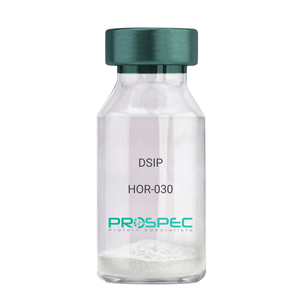
DSIPDescription:
Delta Sleep Inducing Peptide
Product # :
HOR-030Price :
Quantity :
Shipping Method :
Shipped at Room temp
More Info
- description
- formulation
- purity
- More Info
Description
DSIP Synthetic is a single, non-glycosylated polypeptide chain containing 15 amino acids, having a molecular mass of 1419.55 Dalton and a Molecular formula of C62H98N16O22.
Formulation
The protein was lyophilized with no additives.
Purity
Greater than 97.0% as determined by analysis by RP-HPLC.
More Info
-
Physical Appearance
Sterile Filtered White lyophilized (freeze-dried) powder.
-
Stability
Lyophilized DSIP although stable at room temperature for 3 weeks, should be stored desiccated below -18°C. Upon reconstitution DSIP should be stored at 4°C between 2-7 days and for future use below -18°C. For long term storage it is recommended to add a carrier protein (0.1% HSA or BSA). Please prevent freeze-thaw cycles.
-
Solubility
It is recommended to reconstitute the lyophilized DSIP in sterile 18MΩ-cm H2O not less than 100 µg/ml, which can then be further diluted to other aqueous solutions.
-
Amino Acid Sequence
H-Gly-Glu-Pro-Pro-Pro-Gly-Lys-Pro-Ala-Asp-Asp-Ala-Gly-Leu-Val-OH
-
Background
Delta Sleep-Inducing Peptide (DSIP), also known as Sleep-Promoting Peptide, is a neuropeptide that has been the subject of extensive research due to its potential role in sleep regulation, stress response, and neuroprotection. This nonapeptide, first isolated from the cerebral venous blood of rabbits during sleep, has been shown to induce slow-wave sleep, modulate pain perception, and exhibit potential antioxidant and immunomodulatory properties.
DSIP's primary function is its interaction with the sleep regulatory system. By modulating the release of certain neurotransmitters, DSIP can influence sleep patterns, particularly promoting slow-wave sleep, the most restorative stage of sleep. Studies by Kovalzon et al. (2011) have demonstrated that DSIP can enhance sleep quality in rats, suggesting potential applications in sleep disorders and the promotion of healthy sleep patterns.
In addition to its sleep-inducing effects, DSIP has been shown to possess neuroprotective properties. Research by Zolotarev et al. (2014) found that DSIP could protect neurons from oxidative stress, suggesting potential applications in the treatment of neurodegenerative diseases such as Alzheimer's and Parkinson's disease.
Given its sleep-inducing and neuroprotective effects, DSIP has been proposed as a potential therapeutic agent for a variety of conditions, including sleep disorders, chronic pain, and neurodegenerative diseases. For instance, a study by Spong et al. (2016) found that DSIP could improve sleep quality in patients with chronic insomnia, indicating its potential as a therapeutic agent in the treatment of sleep disorders.
While research on DSIP is promising, it is important to note that most studies have been conducted in animals or in vitro. More research is needed to fully understand the potential effects and applications of DSIP in humans. However, the existing body of research suggests that DSIP could be a promising tool in the treatment of sleep disorders, chronic pain, and neurodegenerative diseases.
ProSpec's products are furnished for LABORATORY RESEARCH USE ONLY. The product may not be used as drugs, agricultural or pesticidal products, food additives or household chemicals.
- View Data Sheet
Name :
HB-EGF RatDescription:
Proheparin-Binding EGF-like Growth Factor Rat Recombinant
Proheparin-binding EGF-like growth factor, Heparin-binding EGF-like growth factor, HB-EGF, HBEGF, Dtr, Hegfl, GFHB.
Product # :
CYT-170Price :
Quantity :
Shipping Method :
Shipped at Room temp
More Info
- description
- source
- formulation
- purity
- biological activity
- More Info
Description
HB-EGF Rat Recombinant produced in E.Coli is a single, non-glycosylated, polypeptide chain containing 86 amino acids and having a molecular mass of 9.7kDa.The HB-EGF is purified by proprietary chromatographic techniques.
Source
Escherichia Coli.
Formulation
The protein was filtered (0.2µm) and lyophilized from a concentrated solution containing PBS, 300mM NaCl, pH 7.4 and 5% trehalose.
Purity
Greater than 95.0% as determined by:
(a) Analysis by RP-HPLC.
(b) Analysis by SDS-PAGE.Biological Activity
The ED50 as determined by a cell proliferation assay using balb/c 3T3 cells is < 1.0 ng/ml, corresponding to a specific activity of > 1.0×10⁶ units/mg.
More Info
-
Introduction
HB-EGF is an EGF related growth factor which signals via the EGF receptor, and stimulates the proliferation of SMC (smooth muscle cells), fibroblasts, epithelial cells and keratinocytes. HB-EGF is expressed in various cell types and tissues, including vascular endothelial cells and SMC, macrophages, skeletal muscle, keratinocytes and particular tumor cells. HB-EGF’s ability to explicitly bind heparin and heparin sulfate proteoglycans is dissimilar from other EGF-like molecules, and might be related to the enhanced mitogenic activity, relative to EGF, that HB-EGF exerts on smooth muscle cells.
-
Synonyms
Proheparin-binding EGF-like growth factor, Heparin-binding EGF-like growth factor, HB-EGF, HBEGF, Dtr, Hegfl, GFHB.
-
Physical Appearance
Sterile Filtered White lyophilized (freeze-dried) powder.
-
Stability
Lyophilized Rat HB-EGF Recombinant although stable at room temperature for 3 weeks, should be stored desiccated below -18°C. Upon reconstitution HB-EGF should be stored at 4°C between 2-7 days and for future use below -18°C.For long term storage it is recommended to add a carrier protein (0.1% HSA or BSA).Please prevent freeze-thaw cycles.
-
Solubility
It is recommended to reconstitute the lyophilized Rat HB-EGF in sterile 18M-cm H2O not less than 100 µg/ml, which can then be further diluted to other aqueous solutions.
-
Amino Acid Sequence
DLEGTDLDLF KVAFSSKPQA LATPGKEKNG KKKRKGKGLG KKRDPCLKKY KDYCIHGECR YLKELRIPSC HCLPGYHGQR CHGLTL.
-
Background
What is the molecular weight/Mw of HB-EGF Protein?
HB-EGF Protein has a total Mw of 9.7kDa.
What is the source or expression system of HB-EGF Protein?
Escherichia Coli.
What is the Purity of HB-EGF Protein?
HB-EGF Protein is >95% pure as determined by SDS-PAGE.
What is the Biological Activity of HB-EGF Protein?
The ED50 as determined by a cell proliferation assay using balb/c 3T3 cells is < 1.0 ng/ml, corresponding to a specific activity of > 1.0×10⁶ units/mg.
What is the amino acid sequence of HB-EGF Protein?
DLEGTDLDLF KVAFSSKPQA LATPGKEKNG KKKRKGKGLG KKRDPCLKKY KDYCIHGECR YLKELRIPSC HCLPGYHGQR CHGLTL.
What applications can HB-EGF Protein be used in?
HB-EGF Protein can probably be used in western blot, ELISA and Lateral Flow.
What is the endotoxin level for HB-EGF Protein?
The endotoxin level is minimal, HB-EGF Protein was purified using conventional chromatography techniques.
ProSpec's products are furnished for LABORATORY RESEARCH USE ONLY. The product may not be used as drugs, agricultural or pesticidal products, food additives or household chemicals.
- View Data Sheet
Name :
DeslorelinDescription:
Deslorelin
Product # :
HOR-240Price :
Quantity :
Shipping Method :
Shipped at Room temp
More Info
- description
- formulation
- purity
- More Info
Description
Deslorelin is a potent LHRH/GnRH agonist has a molecular formula of C64H83N17O12, pGlu-His-Trp-Ser-Tyr-D-Trp-Leu-Arg-Pro-NHC2H5 having an Mw of 1284.4 Dalton.
Formulation
The Deslorelin peptide was lyophilized with no additives.
Purity
Greater than 99.0% as determined by RP-HPLC.
More Info
-
Introduction
Deslorelin is being studied in the treatment of cancer as a way to block sex hormones made by the ovaries or testicles. It belongs to the family of drugs called gonadotropin-releasing hormone analogs. It is used for the induction of ovulation in mares. Deslorelin binds to and activates pituitary gonadotropin releasing hormone (GnRH) receptors. Continuous, prolonged administration of goserelin in males results in pituitary GnRH receptor desensitization and inhibition of pituitary secretion of follicle stimulating hormone (FSH) and luteinizing hormone (LH), leading to a significant decline in testosterone production; in females, prolonged administration results in a decrease in estradiol production.
-
Physical Appearance
Sterile Filtered White lyophilized (freeze-dried) powder.
-
Stability
Lyophilized Deslorelin although stable at room temperature for 3 weeks, should be stored desiccated below -18°C. Upon reconstitution Deslorelin should be stored at 4°C between 2-7 days and for future use below -18°C. For long term storage it is recommended to add a carrier protein (0.1% HSA or BSA).Please prevent freeze-thaw cycles.
-
Solubility
It is recommended to reconstitute the lyophilized Deslorelin in sterile 18MΩ-cm H2O not less than 100µg/ml, which can then be further diluted to other aqueous solutions.
ProSpec's products are furnished for LABORATORY RESEARCH USE ONLY. The product may not be used as drugs, agricultural or pesticidal products, food additives or household chemicals.
- View Data Sheet
Name :
EGF Mouse ProteinDescription:
Epidermal Growth Factor Mouse Recombinant
Urogastrone, URG, EGF.
Product # :
CYT-326Price :
Quantity :
Shipping Method :
Shipped at Room temp
More Info
- description
- source
- formulation
- purity
- biological activity
- More Info
Description
Epidermal Growth Factor Mouse Recombinant produced in E.Coli is a single, non-glycosylated, polypeptide chain containing 53 amino acids including 3 intramolecular disulfide-bonds and having a molecular mass of 6 kDa.The EGF is purified by proprietary chromatographic techniques.
Source
Escherichia Coli.
Formulation
The protein was lyophilized with no additives.
Purity
Greater than 98.0% as determined by:
(a) Analysis by RP-HPLC.
(b) Analysis by SDS-PAGE.Biological Activity
The activity is determined by the dose-dependent proliferation of mouse BALB/c 3T3 cells and is typically less than 0.1ng/ml.More Info
-
Introduction
Epidermal growth factor has a profound effect on the differentiation of specific cells in vivo and is a potent mitogenic factor for a variety of cultured cells of both ectodermal and mesodermal origin. The EGF precursor is believed to exist as a membrane-bound molecule which is proteolytically cleaved to generate the 53-amino acid peptide hormone that stimulates cells to divide. EGF stimulates the growth of various epidermal and epithelial tissues in vivo and in vitro and of some fibroblasts in cell culture.
-
Synonyms
Urogastrone, URG, EGF.
-
Physical Appearance
Sterile Filtered White lyophilized (freeze-dried) powder.
-
Stability
Lyophilized Epidermal Growth Factor Recombinant although stable at room temperature for 3 weeks, should be stored desiccated below -18°C. Upon reconstitution EGF should be stored at 4°C between 2-7 days and for future use below -18°C.For long term storage it is recommended to add a carrier protein (0.1% HSA or BSA).Please prevent freeze-thaw cycles.
-
Solubility
It is recommended to reconstitute the lyophilized Epidermal Growth Factor in sterile 18MΩ-cm H2O not less than 100µg/ml, which can then be further diluted to other aqueous solutions.
-
Amino Acid Sequence
NSYPGCPSSY DGYCLNGGVC MHIESLDSYT CNCVIGYSGD RCQTRDLRWW ELR.
-
Background
Exploring Novel Frontiers: Epidermal Growth Factor Mouse Recombinant and its Potential Therapeutic Implications
Abstract:
This research paper delves into the uncharted realm of Epidermal Growth Factor Mouse Recombinant (EGF-MR), unraveling its intricate molecular attributes, cellular signaling, and therapeutic prospects. Employing state-of-the-art methodologies involving genetic engineering, in vitro assays, and animal models, this study uncovers the multifaceted responses elicited by EGF-MR. The findings underscore its promise as a versatile therapeutic agent, potentially revolutionizing regenerative medicine and cancer interventions.
Introduction:
Epidermal Growth Factor (EGF) plays a pivotal role in cellular dynamics. This paper ventures into the nuanced landscape of Epidermal Growth Factor Mouse Recombinant (EGF-MR), delving into its unique molecular characteristics and exploring the therapeutic horizons it presents.
Molecular Insights and Receptor Binding:
EGF-MR's interaction with the epidermal growth factor receptor (EGFR) sets the stage for intricate intracellular events. High-resolution structural analyses and binding kinetics studies elucidate the nuances of this interaction, revealing structural motifs that initiate downstream signaling cascades.
Cellular Signaling and Functional Responses:
EGF-MR initiates canonical and non-canonical signaling pathways, including the mitogen-activated protein kinase (MAPK) and phosphoinositide 3-kinase (PI3K)/Akt pathways. Through comprehensive phosphoproteomic analyses and live-cell imaging, the spatiotemporal dynamics of EGF-MR-induced responses come to light, showcasing its role in cell proliferation, migration, and anti-apoptotic effects.
Genetic Engineering and In Vitro Assays:
Precise genetic manipulation ensures optimal EGF-MR expression. Gene codon optimization and signal peptide selection are meticulously undertaken to facilitate efficient protein synthesis and secretion. In vitro assays, encompassing cell viability and wound healing studies, illuminate EGF-MR's impact on cellular behaviors.
In Vivo Implications and Therapeutic Prospects:
In animal models, EGF-MR emerges as a transformative factor in tissue regeneration. Customized wound healing assays unveil its potential in accelerating re-epithelialization and granulation tissue formation. Moreover, the modulation of tumor microenvironments suggests its applicability in cancer interventions.
Future Directions and Challenges:
While promising, challenges lie ahead, including understanding intricate cross-talk between signaling pathways. Future research should focus on refining delivery methods and optimizing dosing regimens to harness EGF-MR's full therapeutic potential.
Conclusion:
In a convergence of advanced methodologies and visionary therapeutic possibilities, Epidermal Growth Factor Mouse Recombinant takes center stage. Its distinctive molecular interactions and diverse cellular orchestration offer a glimpse into the future of regenerative medicine and targeted cancer therapies, propelling scientific progress into uncharted territories.
What is the molecular weight/Mw of EGF Protein?
EGF Protein has a total Mw of 6 kDa.
What is the source or expression system of EGF Protein?
Escherichia Coli.
What is the Purity of EGF Protein?
EGF Protein is >98% pure as determined by SDS-PAGE.
What is the Biological Activity of EGF Protein?
The activity is determined by the dose-dependent proliferation of mouse BALB/c 3T3 cells and is typically less than 0.1ng/ml.
What is the amino acid sequence of EGF Protein?
NSYPGCPSSY DGYCLNGGVC MHIESLDSYT CNCVIGYSGD RCQTRDLRWW ELR.
What applications can EGF Protein be used in?
EGF Protein can probably be used in western blot, ELISA and Lateral Flow.
What is the endotoxin level for EGF Protein?
The endotoxin level is minimal, EGF Protein was purified using conventional chromatography techniques.
ProSpec's products are furnished for LABORATORY RESEARCH USE ONLY. The product may not be used as drugs, agricultural or pesticidal products, food additives or household chemicals.
- View Data Sheet
Name :
FGF 21 MouseDescription:
Fibroblast Growth Factor-21 Mouse Recombinant
Fibroblast growth factor 21, FGF-21.
Product # :
CYT-339Price :
Quantity :
Shipping Method :
Shipped at Room temp
More Info
- description
- source
- formulation
- purity
- More Info
Description
Fibroblast Growth Factor -21 Mouse Recombinant produced in E.Coli is a single, non-glycosylated, polypeptide chain containing 183 amino acids including N-terminal Methionin and having a molecular mass of 20.1 kDa. The FGF-21 is purified by proprietary chromatographic techniques.
Source
Escherichia Coli.
Formulation
Filtered (0.4µm) and lyophilized from 0.5mg/ml in 20mM TRIS, 20mM NaCl, pH 7.5.
Purity
Greater than 95.0% as determined by SDS-PAGE.
More Info
-
Introduction
The FGFs are a family of more than 20 small (~17–26 kDa) secreted peptides. The initial characterization of these proteins focused on their ability to stimulate fibroblast proliferation. This mitogenic activity was mediated through FGF receptors (FGFRs) 1, 2, or 3. A fourth closely related tyrosine kinase receptor (FGFR4) was able to bind the FGFs but did not lead to a mitogenic response.
FGFs modulate cellular activity via at least 5 distinct subfamilies of high-affinity FGF receptors (FGFRs): FGFR-1, -2, -3, and -4, all with intrinsic tyrosine kinase activity and, except for FGFR-4, multiple splice isoforms, and FGFR-5, which lacks an intracellular kinase domain. There is growing evidence that FGFRs can be important for regulation of glucose and lipid homeostasis. The overexpression of a dominant negative form of FGFR-1 in ? cells leads to diabetes in mice, which thus implies that proper FGF signaling is required for normal ? cell function and glycemia maintenance. FGFR-2 appears to be a key molecule during pancreatic development. Moreover, FGFR-4 has been implicated in cholesterol metabolism and bile acid synthesis.
FGF-19, has been shown to cause resistance to diet-induced obesity desensitization and to improve glucose, and lipid profiles in diabetic rodents. Since these effects, at least in part, are mediated through the observed changes in metabolic rates, FGF-19 can be considered as a regulator of energy expenditure.
FGF-21 is preferentially expressed in liver, but an exact knowledge of FGF-21 bioactivity and its mode of action have been lacking to date. FGF-21 is a potent activator of glucose uptake on adipocytes, protects animals from diet-induced obesity when overexpressed in transgenic mice, and lowers blood glucose and triglyceride levels when therapeutically administered to diabetic rodents. -
Synonyms
Fibroblast growth factor 21, FGF-21.
-
Physical Appearance
Filtered white lyophilized powder.
-
Stability
Lyophilized FGF-21 Mouse Recombinant although stable at room temperature for 3 weeks, should be stored desiccated below -18°C. Upon reconstitution Fibroblast Growth Factor 21 should be stored at 4°C between 2-7 days and for future use below -18°C.For long term storage it is recommended to add a carrier protein (0.1% HSA or BSA).Please prevent freeze-thaw cycles.
-
Solubility
It is recommended to add deionized water to prepare a working stock solution of approximately 0.5mg/ml and let the lyophilized pellet dissolve completely. Product is not sterile! Please filter the product by an appropriate sterile filter before using it in the cell culture. Add DTT (0.2mM) and NaCl (0.1-0.15M) before freezing to prevent potential aggregation.
-
Amino Acid Sequence
MAY PIPDSSPLLQ FGGQVRQRYL YTDDDQDTEA HLEIREDGTV VGAAHRSPES LLELKALKPG VIQILGVKAS RFLCQQPDGA LYGSPHFDPE ACSFRELLLE DGYNVYQSEA HGLPLRLPQK DSPNQDATSW GPVRFLPMPG LLHEPQDQAG FLPPEPPDVG SSDPLSMVEP LQGRSPSYAS.
-
Background
What is the molecular weight/Mw of FGF21 MOUSE Protein?
FGF21 MOUSE Protein has a total Mw of 20.1kDa.
What is the source or expression system of FGF21 MOUSE Protein?
Escherichia Coli.
What is the Purity of FGF21 MOUSE Protein?
FGF21 MOUSE Protein is >95% pure as determined by SDS-PAGE.
What is the Biological Activity of FGF21 MOUSE Protein?
The biological functionality of FGF21 MOUSE Protein will be determined in the future.
What is the amino acid sequence of FGF21 MOUSE Protein?
MAY PIPDSSPLLQ FGGQVRQRYL YTDDDQDTEA HLEIREDGTV VGAAHRSPES LLELKALKPG VIQILGVKAS RFLCQQPDGA LYGSPHFDPE ACSFRELLLE DGYNVYQSEA HGLPLRLPQK DSPNQDATSW GPVRFLPMPG LLHEPQDQAG FLPPEPPDVG SSDPLSMVEP LQGRSPSYAS.
What applications can FGF21 MOUSE Protein be used in?
FGF21 MOUSE Protein can probably be used in western blot, ELISA and Lateral Flow.
What is the endotoxin level for FGF21 MOUSE Protein?
The endotoxin level is minimal, FGF21 MOUSE Protein was purified using conventional chromatography techniques.
ProSpec's products are furnished for LABORATORY RESEARCH USE ONLY. The product may not be used as drugs, agricultural or pesticidal products, food additives or household chemicals.
- View Data Sheet
Name :
SAA EquineDescription:
Serum Amyloid A (APO-SAA) Equine Recombinant
Product # :
CYT-1222Price :
Quantity :
Shipping Method :
Shipped at Room temp
More Info
- description
- source
- formulation
- purity
- More Info
Description
SAA Equine produced in E.coli is a single, non-glycosylated polypeptide chain (1-110 a.a) containing 120 a.a and having a molecular mass of 13,580 Dalton. SAA s fused with a 10 amino acids affinity tag at N-Terminus and purified by proprietary chromatographic techniques.
Source
Escherichia Coli.
Formulation
SAA was lyophilized from 0.01M HCl (pH 2.0).
Purity
Greater than 95.0% as determined by SDS-PAGE.
More Info
-
Physical Appearance
Sterile Filtered White lyophilized (freeze-dried) powder.
-
Stability
Lyophilized SAA although stable at room temperature for 3 weeks, should be stored desiccated below -18°C. Upon reconstitution Serum Amyloid A (APO-SAA) should be stored at 4°C between 2-7 days and for future use below -18°C. For long term storage it is recommended to add a carrier protein (0.1% HSA or BSA). Please prevent freeze-thaw cycles.
-
Solubility
It is recommended to reconstitute the lyophilized SAA in 0.01M HCl (pH 2.0) not less than 100µg/ml, which can then be further diluted to other aqueous solutions.
-
Background
Elevated SAA levels serve as sensitive markers for various inflammatory disorders, including infections, autoimmune diseases, and cancer. SAA assays, often utilizing recombinant SAA proteins, are invaluable in clinical diagnostics, aiding in disease monitoring and treatment evaluation. Moreover, the targeted modulation of SAA activities presents novel therapeutic strategies, offering potential interventions in conditions characterized by excessive inflammation.
SAA Recombinant Protein, as a representative of the intricate SAA family, embodies the complexities of inflammation, immune regulation, and lipid metabolism. Its versatile roles in health and disease underscore its significance in biology and medicine. By unraveling the molecular intricacies of SAA, scientists pave the way for innovative diagnostic methods, therapeutic interventions, and a deeper understanding of the intricate interplay between inflammation and various physiological processes. This research not only illuminates the multifaceted nature of SAA but also holds the promise of transformative advancements in both clinical practice and scientific discovery.
ProSpec's products are furnished for LABORATORY RESEARCH USE ONLY. The product may not be used as drugs, agricultural or pesticidal products, food additives or household chemicals.
- View Data Sheet
Name :
SARS Nucleocapsid, 1-49 a.a.Description:
SARS-Associated Coronavirus Nuclecapsid (1-49 a.a.), Recombinant
Product # :
SARS-005Price :
Quantity :
Shipping Method :
Shipped with Ice Packs
More Info
- description
- source
- formulation
- purity
- More Info
Description
The E.coli derived recombinant protein contains the Nucleocapsid protein 1-49 amino acids immunodominant regions.
Source
Escherichia Coli.
Formulation
SARS Nucleocaspid protein solution is supplied in PBS.
Purity
Protein is >90% pure as determined SDS-PAGE.
More Info
-
Introduction
The SARS Nucleocaspid is a protein that has a crucial structural importance, by making the virions associates with the RNA genome and therefore create a nucleocapsid that has the specific helical form. There has been found 3 different domain structures to the nucleocapsid antigen. This protein contains 3 outer structural proteins, the membrane (M), envelope (E), and spike (S) proteins.
-
Physical Appearance
Sterile Filtered clear solution.
-
Stability
Protein is shipped on ice packs. Upon arrival, Store at -20°C.
-
Specificity
Immunoreactive with sera of SARS-infected individuals.
-
Purification Method
Purified by proprietary chromatographic technique.
ProSpec's products are furnished for LABORATORY RESEARCH USE ONLY. The product may not be used as drugs, agricultural or pesticidal products, food additives or household chemicals.
- View Data Sheet
Name :
SARS Mosaic S(M)Description:
SARS-Associated Coronavirus Spike Mosaic S (M) Recombinant
Product # :
SARS-256Price :
Quantity :
Shipping Method :
Shipped with Ice Packs
More Info
- description
- formulation
- purity
- More Info
Description
The E.Coli derived 38 kDa recombinant mosaic protein contains the middle section of the Spike protein 408-470, 540-573 amino acids immunodominant regions.
Formulation
25mM Tris-HCl, 0.4% sarcosyl, 0.25% Triton –100 and 50% glycerol.
Purity
SARS Mosaic protein is >95% pure as determined by 10% PAGE (Coomassie staining).
More Info
-
Introduction
SARS Coronavirus is an enveloped virus containing three outer structural proteins, namely the membrane (M), envelope (E), and spike (S) proteins. Spike (S)-glycoprotein of the virus interacts with a cellular receptor and mediates membrane fusion to allow viral entry into susceptible target cells. Accordingly, S-protein plays an important role in virus infection cycle and is the primary target of neutralizing antibodies.
-
Stability
SARS Mosaic protein is shipped at ambient temperature. Upon arrival, store at -20C.
-
Applications
SARS Mosaic Antigen in ELISA and Western blots, excellent antigen for detection of SARS with minimal specificity problems.
-
Specificity
Immunoreactive with sera of SARS-infected individuals.
-
Purification Method
SARS Mosaic protein was purified by proprietary chromatographic technique.
ProSpec's products are furnished for LABORATORY RESEARCH USE ONLY. The product may not be used as drugs, agricultural or pesticidal products, food additives or household chemicals.