Search results
1000 results found for “Transferase”
Name
Description
Product #
Price
Quantity
Shipping Method
- View Data Sheet
Name :
FGF19 Human, HEKDescription:
Fibroblast Growth Factor-19 Human Recombinant, HEK
fibroblast growth factor 19, FGF19.
Product # :
CYT-1180Price :
Quantity :
Shipping Method :
Shipped with Ice Packs
More Info
- description
- source
- formulation
- purity
- More Info
- Citations
Description
FGF19 Mouse Recombinant produced in HEK293 cells is a single, glycosylated polypeptide chain (a.a 23-216) containing 205 amino acids and having a molecular mass of 23.0 kDa.FGF19 is fused to a 6 amino acid His-tag at C-terminus & purified by proprietary chromatographic techniques.
Source
HEK293 cells.
Formulation
FGF19 protein (0.5mg/ml) contains 20mM Tris-HCl(pH8.0), 20% glycerol and 0.1M NaCl.
Purity
Greater than 95.0% as determined by SDS-PAGE.
Citations
More Info
-
Introduction
Fibroblast Growth Factor-19 (FGF-19) is a member of the FGF family. FGF-19 interacts with FGFR1, FGFR2, FGFR3 and FGFR4. T FGF-19 takes part in the suppression of bile acid biosynthesis through downregulation of CYP7A1 expression, following positive regulation of the JNK and EPK1/2 cascades. FGF-19 stimulates glucose uptake in adiposytes and is a high affinity, heparin dependent ligand for FGFR4.
-
Synonyms
fibroblast growth factor 19, FGF19.
-
Physical Appearance
Sterile Filtered colorless solution.
-
Stability
Store at 4°C if entire vial will be used within 2-4 weeks. Store, frozen at -20°C for longer periods of time. For long term storage it is recommended to add a carrier protein (0.1% HSA or BSA).Avoid multiple freeze-thaw cycles.
-
Amino Acid Sequence
DGSHMRPLAF SDAGPHVHYG WGDPIRLRHL YTSGPHGLSS CFLRIRADGV VDCARGQSAH SLLEIKAVAL RTVAIKGVHS VRYLCMGADG KMQGLLQYSE EDCAFEEEIR PDGYNVYRSE KHRLPVSLSS AKQRQLYKNR GFLPLSHFLP MLPMVPEEPE DLRGHLESDM FSSPLETDSM DPFGLVTGLE AVRSPSFEKH HHHHH
-
Background
What is the molecular weight/Mw of FGF19 HUMAN,HEK Protein?
FGF19 HUMAN,HEK Protein has a total Mw of 23kDa.
What is the source or expression system of FGF19 HUMAN,HEK Protein?
HEK293 cells.
What is the Purity of FGF19 HUMAN,HEK Protein?
FGF19 HUMAN,HEK Protein is >95% pure as determined by SDS-PAGE.
What is the Biological Activity of FGF19 HUMAN,HEK Protein?
The biological functionality of FGF19 HUMAN,HEK Protein will be determined in the future.
What is the amino acid sequence of FGF19 HUMAN,HEK Protein?
DGSHMRPLAF SDAGPHVHYG WGDPIRLRHL YTSGPHGLSS CFLRIRADGV VDCARGQSAH SLLEIKAVAL RTVAIKGVHS VRYLCMGADG KMQGLLQYSE EDCAFEEEIR PDGYNVYRSE KHRLPVSLSS AKQRQLYKNR GFLPLSHFLP MLPMVPEEPE DLRGHLESDM FSSPLETDSM DPFGLVTGLE AVRSPSFEKH HHHHH
What applications can FGF19 HUMAN,HEK Protein be used in?
FGF19 HUMAN,HEK Protein can probably be used in western blot, ELISA and Lateral Flow.
What is the endotoxin level for FGF19 HUMAN,HEK Protein?
The endotoxin level is minimal, FGF19 HUMAN,HEK Protein was purified using conventional chromatography techniques.
ProSpec's products are furnished for LABORATORY RESEARCH USE ONLY. The product may not be used as drugs, agricultural or pesticidal products, food additives or household chemicals.
- View Data Sheet
Name :
ACE2 (18-615) HumanDescription:
Angiotensin Converting Enzyme 2 (18-615 a.a.), Human Recombinant
Product # :
ENZ-1121Price :
Quantity :
Shipping Method :
Shipped with Ice Packs
More Info
- description
- source
- formulation
- purity
- biological activity
- More Info
- Citations
Description
The CHO derived ACE2 Human recombinant protein contains the extracellular domain amino acids 18- 615 fused to Fc tag at C-terminal and has a molecular weight of ~130 kDa. ACE2 Protein binds to SARS Coronavirus-2 [ CoV-2019 ] Spike receptor binding domain.
Source
CHO Cells
Formulation
ACE2 Human protein solution is supplied in 50mM Tris-HCl, pH7.5, and 90mM glycine.
Purity
Protein is >95% pure as determined SDS-PAGE.
Biological Activity
ACE2 activity was measured by its binding ability in a functional ELISA.
The immobilized Recombinant Human ACE2 protein binds to SARS CoV2 Spike protein Receptor Binding Domain at 2ug per ml.
Citations
More Info
-
Introduction
ACE-2 (Angiotensin converting enzyme 2) an enzyme bound to cell membranes in various organs such as intestines arteries , lungs, heart & kidney. ACE2 an entry receptor of SARS coronaviruses as well as SARS-CoV-2,.The coronavirus spike (S) glycoprotein is a class I viral fusion antigen located on the external envelope of the virion that takes part in a critical part in viral infection by identifying host cell receptors and facilitating fusion of the viral and cellular membranes. 2 main domains in coronavirus S1 have been recognized, the N-terminal domain and C-terminal domain. One or the other and/or both S1 domains function as a receptor-binding domain. SARS-CoV + MERS-CoV equally use C-domain to attach their receptors.ACE2 is a type I transmembrane antigen with an extracellular N-terminal domain having the catalytic site and an intracellular C-terminal tail. ACE2 obtains a signal peptide, a transmembrane domain, and a single metalloproteinase active site containing an HEXXH zinc-binding domain. ACE-2 plays a role as a mono-carboxypeptidase which degrades Ang I to produce the nonapeptide Ang 1–9 and Ang II to create the heptapeptide Ang 1–7.
-
Physical Appearance
Sterile Filtered clear solution.
-
Stability
ACE-2 Human Recombinant Protein is shipped on ice packs. Upon arrival, Store at -20°C.
-
Background
Research on Angiotensin Converting Enzyme 2 Human Recombinant: Structure, Function, and Therapeutic Implications.
Introduction:
Angiotensin Converting Enzyme 2 (ACE2) is a key enzyme involved in the renin-angiotensin system (RAS) and plays a crucial role in cardiovascular homeostasis. In recent years, ACE2 has garnered significant attention due to its pivotal role as the cellular receptor for the severe acute respiratory syndrome coronavirus 2 (SARS-CoV-2), the virus responsible for the global COVID-19 pandemic. This research paper aims to provide an overview of ACE2, including its structure, function, and potential therapeutic implications.ACE2 is a transmembrane protein that belongs to the zinc metalloprotease family. It is expressed in various tissues, including the cardiovascular system, lungs, kidneys, and gastrointestinal tract. ACE2 acts as a counter-regulatory enzyme within the RAS by catalyzing the conversion of angiotensin II (Ang II) to angiotensin-(1-7) (Ang-(1-7)). This enzymatic conversion results in the downregulation of the vasoconstrictor effects of Ang II and the promotion of vasodilation, anti-inflammatory responses, and tissue protection.
The recent emergence of SARS-CoV-2 has highlighted ACE2's role as the primary receptor for viral entry into host cells. The binding of the viral spike protein to ACE2 enables the virus to enter and infect target cells, primarily in the respiratory system. This interaction has raised interest in ACE2 as a potential target for therapeutic interventions against COVID-19.
In addition to its involvement in viral infections, ACE2 has been implicated in various cardiovascular and respiratory disorders. Dysregulation of ACE2 expression and activity has been associated with hypertension, heart failure, acute respiratory distress syndrome (ARDS), and pulmonary fibrosis. Understanding the intricate balance of ACE2 activity and its interaction with the RAS is crucial for elucidating the pathophysiology of these diseases and developing targeted therapeutic approaches.
Several therapeutic strategies targeting ACE2 have been explored, including the development of ACE2-based recombinant proteins. Recombinant ACE2 has shown potential as a therapeutic agent by acting as a decoy receptor for SARS-CoV-2, thereby preventing viral entry into host cells. Moreover, modulation of ACE2 expression and activity holds promise for managing cardiovascular and respiratory diseases.
In conclusion, Angiotensin Converting Enzyme 2 (ACE2) plays a critical role in cardiovascular homeostasis and has emerged as a key player in the context of viral infections, particularly the COVID-19 pandemic. This research aims to provide insights into the structure, function, and potential therapeutic implications of ACE2. By unraveling the complexities of ACE2 biology, we strive to pave the way for the development of innovative therapeutic strategies against COVID-19 and other related diseases.
ProSpec's products are furnished for LABORATORY RESEARCH USE ONLY. The product may not be used as drugs, agricultural or pesticidal products, food additives or household chemicals.
- View Data Sheet
Name :
HSD17B11 HumanDescription:
Hydroxysteroid (17-beta) Dehydrogenase 11 Human Recombinant
17-beta-hydroxysteroid dehydrogenase 11, 17-beta-HSD 11, 17bHSD11, 17betaHSD11, 17-beta-hydroxysteroid dehydrogenase XI, 17-beta-HSD XI, 17betaHSDXI, Cutaneous T-cell lymphoma-associated antigen HD-CL-03, CTCL-associated antigen HD-CL-03, Dehydrogenase/reductase SDR family member 8, Retinal short-chain dehydrogenase/reductase 2, retSDR2, HSD17B11, DHRS8, PAN1B, SDR16C2, 17BHSD11.
Product # :
ENZ-049Price :
Quantity :
Shipping Method :
Shipped with Ice Packs
More Info
- description
- source
- formulation
- purity
- More Info
- Citations
Description
HSD17B11 Human Recombinant fused with a 21 amino acid His tag at N-terminus produced in E.Coli is a single, non-glycosylated, polypeptide chain containing 287 amino acids (20-285 a.a.) and having a molecular mass of 31.4kDa. The HSD17B11 is purified by proprietary chromatographic techniques.
Source
Escherichia Coli.
Formulation
The HSD17B11 solution (0.5 mg/ml) contains 20mM Tris-HCl buffer (pH8.0), 0.2M NaCl, 5mM DTT and 20% glycerol.
Purity
Greater than 95.0% as determined by SDS-PAGE.
Citations
More Info
-
Introduction
Dehydrogenase/reductase SDR family member 8 (HSD17B11) is a member of the HSD17B family of proteins, which regulate the availability of steroids within various tissues throughout the body. HSD17B11 is widely expressed with the highest levels found in the retina, pancreas, kidney, liver, lung, adrenal, small intestine, ovary and heart as well as in steroidogenic cells. HSD17B11 converts androstan-3-?,17-?-diol (3-?-diol) to androsterone, suggesting it may participate in androgen metabolism during steroidogenesis.
-
Synonyms
17-beta-hydroxysteroid dehydrogenase 11, 17-beta-HSD 11, 17bHSD11, 17betaHSD11, 17-beta-hydroxysteroid dehydrogenase XI, 17-beta-HSD XI, 17betaHSDXI, Cutaneous T-cell lymphoma-associated antigen HD-CL-03, CTCL-associated antigen HD-CL-03, Dehydrogenase/reductase SDR family member 8, Retinal short-chain dehydrogenase/reductase 2, retSDR2, HSD17B11, DHRS8, PAN1B, SDR16C2, 17BHSD11.
-
Physical Appearance
Sterile Filtered colorless solution.
-
Stability
Store at 4°C if entire vial will be used within 2-4 weeks. Store, frozen at -20°C for longer periods of time. For long term storage it is recommended to add a carrier protein (0.1% HSA or BSA).Avoid multiple freeze-thaw cycles.
-
Amino Acid Sequence
MGSSHHHHHH SSGLVPRGSH MESFVKLFIP KRRKSVTGEI VLITGAGHGI GRLTAYEFAK LKSKLVLWDI NKHGLEETAA KCKGLGAKVH TFVVDCSNRE DIYSSAKKVK AEIGDVSILV NNAGVVYTSD LFATQDPQIE KTFEVNVLAH FWTTKAFLPA MTKNNHGHIV TVASAAGHVS VPFLLAYCSS KFAAVGFHKT LTDELAALQI TGVKTTCLCP NFVNTGFIKN PSTSLGPTLE PEEVVNRLMH GILTEQKMIF IPSSIAFLTT LERILPERFL AVLKQKI.
ProSpec's products are furnished for LABORATORY RESEARCH USE ONLY. The product may not be used as drugs, agricultural or pesticidal products, food additives or household chemicals.
- View Data Sheet
Name :
PLA2G2E HumanDescription:
Secreted Phospholipase A2-IIE Human Recombinant
Group IIE secretory phospholipase A2, EC 3.1.1.4, Phosphatidylcholine 2-acylhydrolase GIIE, GIIE sPLA2, sPLA(2)-IIE, sPLA2-IIE, PLA2G2E.
Product # :
ENZ-327Price :
Quantity :
Shipping Method :
Shipped at Room temp
More Info
- description
- source
- formulation
- purity
- More Info
- Citations
Description
Secreted Phospholipase A2-IIE Human Recombinant manufactured with N-terminal His-Tag. PLA2G2E His-Tagged Fusion Protein is 15.8 kDa protein containing 123 amino acid residues of the human secreted phospholipase A2-IIE and 16 additional amino acid residues – His-Tag (underlined).
Source
Escherichia Coli.
Formulation
Sterile filtered and lyophilized from 0.5 mg/ml in 0.05M Acetate buffer pH-4.
Purity
Greater than 95% as determined by SDS PAGE.
Citations
More Info
-
Introduction
Phospholipase A2 (PLA2) catalyzes the hydrolysis of the sn-2 position of membrane glycerophospholipids to liberate arachidonic acid (AA), a precursor of eicosanoids including prostaglandins and leukotrienes. The same reaction also produces lysophosholipids, which represent another class of lipid mediators.
The secretory PLA2 (sPLA2) family, in which 10 isozymes have been identified, consists of low molecular weight, Ca2+-requiring secretory enzymes that have been implicated in a number of biological processes, such as modification of eicosanoid generation, inflammation, and host defense.This enzyme has been proposed to hydrolyze phosphatidylcholine (PC) in lipoproteins to liberate lyso-PC and free fatty acids in the arterial wall, thereby facilitating the accumulation of bioactive lipids and modified lipoproteins in atherosclerotic foci.
In mice, sPLA2 expression significantly influences HDL particle size and composition and demonstrate that an induction of sPLA2 is required for the decrease in plasma HDL cholesterol in response to inflammatory stimuli. Instillation of bacteria into the bronchi was associated with surfactant degradation and a decrease in large:small ratio of surfactant aggregates in rats. -
Synonyms
Group IIE secretory phospholipase A2, EC 3.1.1.4, Phosphatidylcholine 2-acylhydrolase GIIE, GIIE sPLA2, sPLA(2)-IIE, sPLA2-IIE, PLA2G2E.
-
Physical Appearance
Sterile Filtered lyophilized (freeze-dried) powder.
-
Stability
Store lyophilized protein at -20°C. Aliquot the product after reconstitution to avoid repeated freezing/thawing cycles. Reconstituted protein can be stored at 4°C for a limited period of time; it does not show any change after two weeks at 4°C.
-
Solubility
Add 0.2 ml of 0.1M Acetate buffer pH-4 and let the lyophilized pellet dissolve completely. For conversion into higher pH value, we recommend intensive dilution by relevant buffer to a concentration of 10 μg/ml. In higher concentrations the solubility of this antigen is limited.
-
Amino Acid Sequence
MRGSHHHHHH GMASHMNLVQ FGVMIEKMTG KSALQYNDYG CYCGIGGSHW PVDQTDWCCH AHDCCYGRLE KLGCEPKLEK YLFSVSERGI FCAGRTTCQR LTCECDKRAA LCFRRNLGTY NRKYAHYPNK LCTGPTPPC
ProSpec's products are furnished for LABORATORY RESEARCH USE ONLY. The product may not be used as drugs, agricultural or pesticidal products, food additives or household chemicals.
- View Data Sheet
Name :
GlpK E. coliDescription:
Glycerol kinase E. Coli Recombinant
Glycerol kinase, glycerol 3-phosphotransferase, Glycerokinase, GK.
Product # :
PKA-038Price :
Quantity :
Shipping Method :
Shipped with Ice Packs
More Info
- description
- source
- formulation
- purity
- More Info
- Citations
Description
GlpK E. Coli Recombinant produced in E. coli is a single, non-glycosylated polypeptide chain containing 525 amino acids (1-502 a.a) and having a molecular mass of 58.6 kDa.GlpK is fused to a 23 amino acid His-tag at N-terminus& purified by proprietary chromatographic techniques.
Source
Escherichia Coli.
Formulation
GlpK protein solution (1mg/ml) containing Phosphate buffered saline (pH7.4),10% glycerol and 1mM DTT.
Purity
Greater than 95.0% as determined by SDS-PAGE.
Citations
More Info
-
Introduction
GlpK also known as glycerol kinase, is a member of the FGGY kinase family. GlpK catalyzes the transfer of a phosphate group from ATP to glycerol, thereby forming glycerol phosphate. Furthermore, this intermediate can then be converted to dihydroxyacetone phosphate (DHAP), which is utilized in either glycolysis or gluconeogenesis. The activity of GlpK is affected by numerous metabolites. The non-competitive allosteric inhibition by fructose 1,6-bisphosphate (FBP) triggers modifications in the quaternary structure of Glpk.
-
Synonyms
Glycerol kinase, glycerol 3-phosphotransferase, Glycerokinase, GK.
-
Physical Appearance
Sterile Filtered clear solution.
-
Stability
Store at 4°C if entire vial will be used within 2-4 weeks.Store, frozen at -20°C for longer periods of time. For long term storage it is recommended to add a carrier protein (0.1% HSA or BSA).Avoid multiple freeze-thaw cycles.
-
Amino Acid Sequence
MGSSHHHHHH SSGLVPRGSH MGSMTEKKYI VALDQGTTSS RAVVMDHDAN IISVSQREFE QIYPKPGWVE HDPMEIWATQ SSTLVEVLAK ADISSDQIAA IGITNQRETT IVWEKETGKP IYNAIVWQCR RTAEICEHLK RDGLEDYIRS NTGLVIDPYF SGTKVKWILD HVEGSRERAR RGELLFGTVD TWLIWKMTQG RVHVTDYTNA SRTMLFNIHT LDWDDKMLEV LDIPREMLPE VRRSSEVYGQ TNIGGKGGTR IPISGIAGDQ QAALFGQLCV KEGMAKNTYG TGCFMLMNTG EKAVKSENGL LTTIACGPTG EVNYALEGAV FMAGASIQWL RDEMKLINDA YDSEYFATKV QNTNGVYVVP AFTGLGAPYW DPYARGAIFG LTRGVNANHI IRATLESIAY QTRDVLEAMQ ADSGIRLHAL RVDGGAVANN FLMQFQSDIL GTRVERPEVR EVTALGAAYL AGLAVGFWQN LDELQEKAVI EREFRPGIET TERNYRYAGW KKAVKRAMAW EEHDE.
ProSpec's products are furnished for LABORATORY RESEARCH USE ONLY. The product may not be used as drugs, agricultural or pesticidal products, food additives or household chemicals.
- View Data Sheet
Name :
UBE2C HumanDescription:
Ubiquitin Conjugating enzyme E2C Human Recombinant
Ubiquitin-conjugating enzyme E2 C, EC 6.3.2.19, Ubiquitin-protein ligase C, Ubiquitin carrier protein C, Ubc10, UBCH10, dJ447F3.2.
Product # :
ENZ-346Price :
Quantity :
Shipping Method :
Shipped with Ice Packs
More Info
- description
- source
- formulation
- purity
- More Info
- Citations
Description
UBE2C Human Recombinant produced in E.coli is a single, non-glycosylated polypeptide chain containing 202 amino acids (1-179) and having a molecular mass of 22.1 kDa.The UBE2C is fused to a 23 amino acid His-Tag at N-terminus and purified by proprietary chromatographic techniques.
Source
Escherichia Coli.
Formulation
UBE2C protein 1mg/ml is supplied in 20mM Tris-HCL, pH-8, 0.15M NaCl, 1mM DTT and 10% Glycerol.
Purity
Greater than 90.0% as determined by Analysis by SDS-PAGE.
Citations
More Info
-
Introduction
UbcH10 is an essential mediator of mitotic destruction events and cell cycle progression. It catalyzes the destruction of cyclins A and B in conjunction with the anaphase-promoting complex, and therefore, plays an important role in the control of the cell exit from mitosis This activity is essential at then end of mitosis for the inactivation of their partner kinase Cdc2 and exit from mitosis into G1 of the next cell cycle. In addition, UbcH10 bears homology to yeast PAS2, a gene that is essential for biogenesis of peroxisomes. UbcH10 is useful for in vitro ubiquitinylation reactions.
-
Synonyms
Ubiquitin-conjugating enzyme E2 C, EC 6.3.2.19, Ubiquitin-protein ligase C, Ubiquitin carrier protein C, Ubc10, UBCH10, dJ447F3.2.
-
Physical Appearance
Sterile Filtered clear solution.
-
Stability
Store at 4°C if entire vial will be used within 2-4 weeks. Store, frozen at -20°C for longer periods of time. For long term storage it is recommended to add a carrier protein (0.1% HSA or BSA).Avoid multiple freeze-thaw cycles.
-
Amino Acid Sequence
MGSSHHHHHH SSGLVPRGSH MGSMASQNRD PAATSVAAAR KGAEPSGGAA RGPVGKRLQQ ELMTLMMSGD KGISAFPESD NLFKWVGTIH GAAGTVYEDL RYKLSLEFPS GYPYNAPTVK FLTPCYHPNV DTQGNICLDI LKEKWSALYD VRTILLSIQS LLGEPNIDSP LNTHAAELWK NPTAFKKYLQ ETYSKQVTSQ EP
ProSpec's products are furnished for LABORATORY RESEARCH USE ONLY. The product may not be used as drugs, agricultural or pesticidal products, food additives or household chemicals.
- View Data Sheet
Name :
UBE2D1 HumanDescription:
Ubiquitin Conjugating Enzyme E2D1 Human Recombinant
Ubiquitin-conjugating enzyme E2 D1, Stimulator of Fe transport, SFT, UBC4/5 homolog, UbcH5, Ubiquitin carrier protein D1, Ubiquitin-conjugating enzyme E2(17)KB 1, Ubiquitin-conjugating enzyme E2-17 kDa 1, Ubiquitin-protein ligase D1, UBE2D1, SFT, UBC5A, UBCH5A, E2(17)KB1.
Product # :
ENZ-116Price :
Quantity :
Shipping Method :
Shipped with Ice Packs
More Info
- description
- source
- formulation
- purity
- More Info
- Citations
Description
UBE2D1 produced in E.Coli is a single, non-glycosylated polypeptide chain containing 170 amino acids (1-147 a.a.) and having a molecular mass of 19kDa.UBE2D1 is fused to a 23 amino acid His-tag at N-terminus & purified by proprietary chromatographic techniques.
Source
Escherichia Coli.
Formulation
UBE2D1 solution (1mg/ml) containing 20mM Tris-HCl buffer (pH8.0), 40% glycerol and 0.1M NaCl.
Purity
Greater than 95.0% as determined by SDS-PAGE.
Citations
More Info
-
Introduction
UBE2D1 is a member of the ubiquitin-conjugating enzyme family. Ubiquitination involves at least 3 classes of enzymes: ubiquitin-activating enzymes (E1s), ubiquitin-conjugating enzymes (E2s) and ubiquitin-protein ligases (E3s). UBE2D1 is strongly related to a stimulator of iron transport (SFT), and is up-regulated in hereditary hemochromatosis. In addition, UBE2D1 functions in the ubiquitination of the tumor-suppressor protein p53 and the hypoxia-inducible transcription factor HIF1alpha by interacting with the E1 ubiquitin-activating enzyme and the E3 ubiquitin-protein ligases.
-
Synonyms
Ubiquitin-conjugating enzyme E2 D1, Stimulator of Fe transport, SFT, UBC4/5 homolog, UbcH5, Ubiquitin carrier protein D1, Ubiquitin-conjugating enzyme E2(17)KB 1, Ubiquitin-conjugating enzyme E2-17 kDa 1, Ubiquitin-protein ligase D1, UBE2D1, SFT, UBC5A, UBCH5A, E2(17)KB1.
-
Physical Appearance
Sterile filtered colorless solution.
-
Stability
UBE2D1 Human Recombinant although stable at 4°C for 1 week, should be stored below -18°C. Please prevent freeze thaw cycles.
-
Amino Acid Sequence
MGSSHHHHHH SSGLVPRGSH MGSMALKRIQ KELSDLQRDP PAHCSAGPVG DDLFHWQATI MGPPDSAYQG GVFFLTVHFP TDYPFKPPKI AFTTKIYHPN INSNGSICLD ILRSQWSPAL TVSKVLLSIC SLLCDPNPDD PLVPDIAQIY KSDKEKYNRH AREWTQKYAM.
ProSpec's products are furnished for LABORATORY RESEARCH USE ONLY. The product may not be used as drugs, agricultural or pesticidal products, food additives or household chemicals.
- View Data Sheet
Name :
UBE2I Human HisDescription:
Ubiquitin-Conjugating Enzyme E2I Human Recombinant, His Tag
SUMO-conjugating enzyme UBC9, EC 6.3.2.-, SUMO-protein ligase, Ubiquitin-conjugating enzyme E2 I, Ubiquitin-protein ligase I, Ubiquitin carrier protein I, Ubiquitin carrier protein 9, p18, UBC9, C358B7.1.
Product # :
ENZ-274Price :
Quantity :
Shipping Method :
Shipped at Room temp
More Info
- description
- source
- formulation
- purity
- More Info
- Citations
Description
Ubiquitin-Conjugating Enzyme E2I Human Recombinant produced in E.coli is a 19.5 kDa protein containing 171 amino acids.The UBE2I protein contains 6xHis tag and is purified by proprietary chromatographic techniques.
Source
Escherichia Coli.
Formulation
Lyophilized from a 0.2μm filtered concentrated (1mg/ml) solution in 1X PBS and 1mM DTT, pH 7.5.
Purity
Greater than 95.0% as determined by(a) Analysis by RP-HPLC.
(b) Analysis by SDS-PAGE.Citations
More Info
-
Introduction
Human Ubquitin Conjugating Enzyme 9 (Ubc9) is a member of the E2 family and is specific for the conjugation of SUMO to a variety of target proteins. SUMO conjugation to target proteins is mediated by a different, but analogous, pathway to ubiquitinylation. This E2 is unusual in that it interacts directly with protein substrates that are modified by sumolyation, and may play a role in substrate recognition. Ubc9 can mediate the conjugation of SUMO-1 to a variety of proteins including RanGAP1, I?B?, and PML without the requirement of an E3 ligase.
-
Synonyms
SUMO-conjugating enzyme UBC9, EC 6.3.2.-, SUMO-protein ligase, Ubiquitin-conjugating enzyme E2 I, Ubiquitin-protein ligase I, Ubiquitin carrier protein I, Ubiquitin carrier protein 9, p18, UBC9, C358B7.1.
-
Physical Appearance
Sterile Filtered white lyophilized powder.
-
Stability
Lyophilized UBE2I although stable at room temperature for 3 weeks, should be stored desiccated below -18°C. Upon reconstitution UBE2I should be stored at 4°C between 2-7 days and for future use below -18°C.For long term storage it is recommended to add a carrier protein (0.1% HSA or BSA).Please prevent freeze-thaw cycles.
-
Solubility
It is recommended to reconstitute the lyophilized UBE2I in sterile water not less than 100µg/ml, which can then be further diluted to other aqueous solutions.
-
Amino Acid Sequence
MHHHHHHAMGTLNMSGIALSRLAQERKAWRKDHPFGFVAVPTKNPDGT
MNLMNWECAIPGKKGTPWEGGLFKLRMLFKDDYPSSPPKCKFEPPLFH
PNVYPSGTVCLSILEEDKDWRPAITIKQILLGIQELLNEPNIQDPAQAEAYTI
YCQNRVEYEKRVRAQAKKFAPS.
ProSpec's products are furnished for LABORATORY RESEARCH USE ONLY. The product may not be used as drugs, agricultural or pesticidal products, food additives or household chemicals.
- View Data Sheet
Name :
PSPH HumanDescription:
Phosphoserine Phosphatase Human Recombinant
Phosphoserine phosphatase, EC 3.1.3.3, PSP, O-phosphoserine phosphohydrolase, PSPase, L-3-phosphoserine phosphatase, PSPH.
Product # :
PKA-224Price :
Quantity :
Shipping Method :
Shipped with Ice Packs
More Info
- description
- source
- formulation
- purity
- More Info
- Citations
Description
Phosphoserine Phosphatase Human Recombinant produced in E.Coli is a single, non-glycosylated polypeptide chain containing 225 amino acids and having a molecular mass of 25 kDa. PSP was overexpressed in E. coli and purified by conventional chromatography.
Source
Escherichia Coli.
Formulation
The protein contains 20mM Hepes pH 7.5, 1mM DTT &100mM KCl2.
Purity
Greater than 95.0% as determined by:
(a) Analysis by RP-HPLC.
(b) Analysis by SDS-PAGE.Citations
More Info
-
Introduction
Human Phosphoserine phosphatase (hPSP) is an important enzyme in the phosphorylated pathway of serine biosynthesis, which contributes a major portion of the endogenous L-serine. Similar to known L-3-phosphoserine phosphatases, it catalyzed the Mg2+-dependent hydrolysis of L-phosphoserine and an exchange reaction between L-serine and L-phosphoserine. Recently, its complex structures reveal that the open-closed environmental change of the active site, generated -helical bundle domain, is important to substrate by local rearrangement of the recognition and hydrolysis.
-
Synonyms
Phosphoserine phosphatase, EC 3.1.3.3, PSP, O-phosphoserine phosphohydrolase, PSPase, L-3-phosphoserine phosphatase, PSPH.
-
Physical Appearance
Sterile filtered colorless solution.
-
Stability
Store at 4°C if entire vial will be used within 2-4 weeks. Store, frozen at -20°C for longer periods of time. For long term storage it is recommended to add a carrier protein (0.1% HSA or BSA). Avoid multiple freeze-thaw cycles.
-
Amino Acid Sequence
MVSHSELRKL FYSADAVCFD VDSTVIREEG IDELAKICGV EDAVSEMTRR AMGGAVPFKA ALTERLALIQ PSREQVQRLI AEQPPHLTPG IRELVSRLQE RNVQVFLISG GFRSIVEHVA SKLNIPATNV FANRLKFYFN GEYAGFDETQ PTAESGGKGK VIKLLKEKFH FKKIIMIGDG ATDMEACPPA DAFIGFGGNV IRQQVKDNAK WYITDFVELL GELEE.
ProSpec's products are furnished for LABORATORY RESEARCH USE ONLY. The product may not be used as drugs, agricultural or pesticidal products, food additives or household chemicals.
- View Data Sheet
Name :
COQ9 HumanDescription:
Coenzyme Q9 Human Recombinant
Ubiquinone biosynthesis protein COQ9, mitochondrial, C16orf49, COQ10D5, C16orf49, HSPC326, PSEC0129.
Product # :
ENZ-898Price :
Quantity :
Shipping Method :
Shipped with Ice Packs
More Info
- description
- source
- formulation
- purity
- More Info
- Citations
Description
COQ9 Human Recombinant produced in E.Coli is a single, non-glycosylated polypeptide chain containing 297 amino acids (45-318 a.a) and having a molecular mass of 33.3kDa.COQ9 is fused to a 23 amino acid His-tag at N-terminus & purified by proprietary chromatographic techniques.
Source
Escherichia Coli.
Formulation
COQ9 protein solution (1mg/ml) in Phosphate buffered saline (pH7.4), 10% glycerol and 1mM DTT.
Purity
Greater than 90.0% as determined by SDS-PAGE.
Citations
More Info
-
Introduction
Ubiquinone biosynthesis protein COQ9 (COQ9) represents a mitochondrial ubiquinone biosynthesis gene. The COQ9 protein is likely required for biosynthesis of coenzyme Q10, since mutations at this locus have been linked with autosomal-recessive neonatal-onset primary coenzyme Q10 deficiency.
-
Synonyms
Ubiquinone biosynthesis protein COQ9, mitochondrial, C16orf49, COQ10D5, C16orf49, HSPC326, PSEC0129.
-
Physical Appearance
Sterile Filtered colorless solution.
-
Stability
Store at 4°C if entire vial will be used within 2-4 weeks. Store, frozen at -20°C for longer periods of time. For long term storage it is recommended to add a carrier protein (0.1% HSA or BSA).Avoid multiple freeze-thaw cycles.
-
Amino Acid Sequence
MGSSHHHHHH SSGLVPRGSH MGSRSSDEQK QQPPNSFSQQ HSETQGAEKP DPESSHSPPR YTDQGGEEEE DYESEEQLQH RILTAALEFV PAHGWTAEAI AEGAQSLGLS SAAASMFGKD GSELILHFVT QCNTRLTRVL EEEQKLVQLG QAEKRKTDQF LRDAVETRLR MLIPYIEHWP RALSILMLPH NIPSSLSLLT SMVDDMWHYA GDQSTDFNWY TRRAMLAAIY NTTELVMMQD SSPDFEDTWR FLENRVNDAM NMGHTAKQVK STGEALVQGL MGAAVTLKNL TGLNQRR.
ProSpec's products are furnished for LABORATORY RESEARCH USE ONLY. The product may not be used as drugs, agricultural or pesticidal products, food additives or household chemicals.
- View Data Sheet
Name :
RPP30 HumanDescription:
Ribonuclease P/MRP 30kDa Subunit Human Recombinant
Ribonuclease P protein subunit p30, RNaseP protein p30, RNase P subunit 2, RPP30, RNASEP2, TSG15, FLJ38491, RP11-320F15.1.
Product # :
ENZ-040Price :
Quantity :
Shipping Method :
Shipped with Ice Packs
More Info
- description
- source
- formulation
- purity
- More Info
- Citations
Description
RPP30 Human Recombinant fused with a 23 amino acid His tag at N-terminus produced in E.Coli is a single, non-glycosylated, polypeptide chain containing 291 amino acids (1-268 a.a.) and having a molecular mass of 31.8kDa. The RPP30 is purified by proprietary chromatographic techniques.
Source
Escherichia Coli.
Formulation
The RPP30 solution (0.5 mg/ml) contains 20mM Tris-HCl buffer (pH8.0), 20% glycerol, 5mM DTT, 200mM NaCl and 1mM EDTA.
Purity
Greater than 90.0% as determined by SDS-PAGE.
Citations
More Info
-
Introduction
Ribonuclease P protein subunit p30 (RPP30) is a member of the eukaryotic/archaeal RNase P protein component 3 family. RPP30 is component of ribonuclease P, which is a protein complex that generates mature tRNA molecules by cleaving their 5'-ends. Ribonuclease P (RNase P) is small nuclear ribonucleoprotein (snRNPs) which acts on RNA substrates in vitro. In addition, RNase P which accumulate in the nucleolus, have a similar RNA component and several protein subunits in common.
-
Synonyms
Ribonuclease P protein subunit p30, RNaseP protein p30, RNase P subunit 2, RPP30, RNASEP2, TSG15, FLJ38491, RP11-320F15.1.
-
Physical Appearance
Sterile Filtered colorless solution.
-
Stability
Store at 4°C if entire vial will be used within 2-4 weeks. Store, frozen at -20°C for longer periods of time. For long term storage it is recommended to add a carrier protein (0.1% HSA or BSA).Avoid multiple freeze-thaw cycles.
-
Amino Acid Sequence
MGSSHHHHHH SSGLVPRGSH MGSMAVFADL DLRAGSDLKA LRGLVETAAH LGYSVVAINH IVDFKEKKQE IEKPVAVSEL FTTLPIVQGK SRPIKILTRL TIIVSDPSHC NVLRATSSRA RLYDVVAVFP KTEKLFHIAC THLDVDLVCI TVTEKLPFYF KRPPINVAID RGLAFELVYS PAIKDSTMRR YTISSALNLM QICKGKNVII SSAAERPLEI RGPYDVANLG LLFGLSESDA KAAVSTNCRA ALLHGETRKT AFGIISTVKK PRPSEGDEDC LPASKKAKCE G.
ProSpec's products are furnished for LABORATORY RESEARCH USE ONLY. The product may not be used as drugs, agricultural or pesticidal products, food additives or household chemicals.
- View Data Sheet
Name :
BDH1 HumanDescription:
3-Hydroxybutyrate Dehydrogenase, Type 1 Human Recombinant
D-beta-hydroxybutyrate dehydrogenase, mitochondrial, BDH, 3-hydroxybutyrate dehydrogenase, BDH1, SDR9C1.
Product # :
ENZ-215Price :
Quantity :
Shipping Method :
Shipped with Ice Packs
More Info
- description
- source
- formulation
- purity
- More Info
- Citations
Description
BDH1 Human Recombinant produced in E.coli is a single, non-glycosylated polypeptide chain containing 318 amino acids (47-343) and having a molecular mass of 35.3kDa.BDH1 is fused to a 21 amino acid His-tag at N-terminus & purified by proprietary chromatographic techniques.
Source
E.coli.
Formulation
The BDH1 solution (0.25mg/ml) contains 20mM Tris-HCl buffer (pH8.0), 30% glycerol, 0.2M NaCl, 2mM DTT and 0.1mM PMSF.
Purity
Greater than 85% as determined by SDS-PAGE.
Citations
More Info
-
Introduction
BDH1 belongs to the short-chain dehydrogenase/reductase gene family. BDH1 forms a homotetrameric lipid-requiring enzyme of the mitochondrial membrane and has a specific necessity for phosphatidylcholine for optimal enzymatic activity. BDH1 catalyzes the interconversion of acetoacetate and (R)-3-hydroxybutyrate, the 2 main ketone bodies formed during fatty acid catabolism.
-
Synonyms
D-beta-hydroxybutyrate dehydrogenase, mitochondrial, BDH, 3-hydroxybutyrate dehydrogenase, BDH1, SDR9C1.
-
Physical Appearance
Sterile Filtered colorless solution.
-
Stability
Store at 4°C if entire vial will be used within 2-4 weeks. Store, frozen at -20°C for longer periods of time. For long term storage it is recommended to add a carrier protein (0.1% HSA or BSA).Avoid multiple freeze-thaw cycles.
-
Amino Acid Sequence
MGSSHHHHHH SSGLVPRGSH MASAAEPVGS KAVLVTGCDS GFGFSLAKHL HSKGFLVFAG CLMKDKGHDG VKELDSLNSD RLRTVQLNVC SSEEVEKVVE IVRSSLKDPE KGMWGLVNNA GISTFGEVEF TSLETYKQVA EVNLWGTVRM TKSFLPLIRR AKGRVVNISS MLGRMANPAR SPYCITKFGV EAFSDCLRYE MYPLGVKVSV VEPGNFIAAT SLYSPESIQA IAKKMWEELP EVVRKDYGKK YFDEKIAKME TYCSSGSTDT SPVIDAVTHA LTATTPYTRY HPMDYYWWLR MQIMTHLPGA ISDMIYIR.
ProSpec's products are furnished for LABORATORY RESEARCH USE ONLY. The product may not be used as drugs, agricultural or pesticidal products, food additives or household chemicals.
- View Data Sheet
Name :
CASP3 Human, Sf9Description:
Caspase 3 Apoptosis-Related Cysteine Peptidase Human Recombinant, Sf9
CASP3, CPP32, CPP32B, SCA-1, CASP-3, Apopain, Cysteine protease CPP32, CPP-32, Protein Yama, SREBP cleavage activity 1.
Product # :
ENZ-1106Price :
Quantity :
Shipping Method :
Shipped with Ice Packs
More Info
- description
- source
- formulation
- purity
- biological activity
- More Info
- Citations
Description
CASP3 Human produced in Sf9 Baculovirus cells is a single, glycosylated polypeptide chain containing 256 amino acids (29-277 a.a.) and having a molecular mass of 29.4kDa (Migrates at 13.5-18kDa on SDS-PAGE under reducing conditions). CASP3 is expressed with a 6 amino acid His tag at C-Terminus and purified by proprietary chromatographic techniques.
Source
Sf9, Baculovirus cells.
Formulation
CASP3 protein solution (0.5mg/ml) containing 20mM HEPES buffer (pH 7.5), 0.1M NaCl, 1mM EDTA, 20% Glycerol and 1mM DTT.
Purity
Greater than 90.0% as determined by SDS-PAGE.
Biological Activity
Specific activity is greater than 5,000 pmol/min/ug. One unit will liberate 1 pmoles of Ac-DEVD-AFC to Ac-DEVD and AFC per minute at pH7.5 at 25C.
Citations
More Info
-
Introduction
Caspase 3 Apoptosis-Related Cysteine Peptidase (CASP3) belongs to the cysteine-aspartic acid protease (caspase) family. Sequential activation of caspases plays a key role in the execution-phase of cell apoptosis. Caspases exist as inactive proenzymes which undergo proteolytic processing at conserved aspartic residues to generate 2 subunits, large and small, that dimerize to create the active enzyme. CASP3 protein cleaves and activates caspases 6, 7 and 9, and the protein itself is processed by caspases 8, 9 and 10. CASP3 is the leading caspase involved in the cleavage of amyloid-beta 4A precursor protein, which is linked with neuronal death in Alzheimer's disease. In addition, CASP3 is involved in the cleavage of huntingtin. CASP3 also cleaves and activates sterol regulatory element binding proteins (SREBPs) between the basic helix-loop-helix leucine zipper domain and the membrane attachment domain. CASP3 initiates cell adhesion in sympathetic neurons through RET cleavage.
-
Synonyms
CASP3, CPP32, CPP32B, SCA-1, CASP-3, Apopain, Cysteine protease CPP32, CPP-32, Protein Yama, SREBP cleavage activity 1.
-
Physical Appearance
Sterile Filtered colorless solution.
-
Stability
Store at 4°C if entire vial will be used within 2-4 weeks. Store, frozen at -20°C for longer periods of time. For long term storage it is recommended to add a carrier protein (0.1% HSA or BSA).Avoid multiple freeze-thaw cycles.
-
Amino Acid Sequence
MSGISLDNSY KMDYPEMGLC IIINNKNFHK STGMTSRSGT DVDAANLRET FRNLKYEVRN
KNDLTREEIV ELMRDVSKED HSKRSSFVCV LLSHGEEGII FGTNGPVDLK KITNFFRGDR
CRSLTGKPKL FIIQACRGTE LDCGIETDSG VDDDMACHKI PVEADFLYAY STAPGYYSWR
NSKDGSWFIQ SLCAMLKQYA DKLEFMHILT RVNRKVATEF ESFSFDATFH AKKQIPCIVS MLTKELYFYH HHHHHH
ProSpec's products are furnished for LABORATORY RESEARCH USE ONLY. The product may not be used as drugs, agricultural or pesticidal products, food additives or household chemicals.
- View Data Sheet
Name :
SEPSECS MouseDescription:
Selenocysteinyl-tRNA(Sec) synthase Mouse Recombinant
AA986712, D5Ertd135e, SecS, SLA, SLA/LP autoantigen, SLA-p35, Selenocysteinyl-tRNA(Sec) synthase, Selenocysteine synthase, Liver-pancreas antigen, Soluble liver antigen, Sec synthase, UGA suppressor tRNA-associated protein, SepSecS.
Product # :
ENZ-1081Price :
Quantity :
Shipping Method :
Shipped with Ice Packs
More Info
- description
- source
- formulation
- purity
- More Info
- Citations
Description
SEPSECS produced in E.Coli is a single, non-glycosylated polypeptide chain containing 527 amino acids (1-504 a.a.) and having a molecular mass of 57.7kDa.SEPSECS is fused to a 23 amino acid His-tag at N-terminus & purified by proprietary chromatographic techniques.
Source
Escherichia Coli.
Formulation
The SEPSECS protein solution (0.25 mg/ml) is formulated in 20mM Tris-HCl buffer (pH7.5) 1mM DTT, 0.2M NaCl and 50% glycerol.
Purity
Greater than 90% as determined by SDS-PAGE.
Citations
More Info
-
Introduction
SEPSECS catalyzes the last step of sec synthesis by converting O-phosphoseryl-tRNA(sec) to selenocysteinyl-tRNA(sec) using selenophosphate as the selenium donor. Furthermore, SEPSECS protein is considered a specific marker of autoimmune hepatitis.
-
Synonyms
AA986712, D5Ertd135e, SecS, SLA, SLA/LP autoantigen, SLA-p35, Selenocysteinyl-tRNA(Sec) synthase, Selenocysteine synthase, Liver-pancreas antigen, Soluble liver antigen, Sec synthase, UGA suppressor tRNA-associated protein, SepSecS.
-
Physical Appearance
Sterile Filtered clear solution.
-
Stability
Store at 4°C if entire vial will be used within 2-4 weeks. Store, frozen at -20°C for longer periods of time. For long term storage it is recommended to add a carrier protein (0.1% HSA or BSA).Avoid multiple freeze-thaw cycles
-
Amino Acid Sequence
MGSSHHHHHH SSGLVPRGSH MGSMNPESFA AGERRVSPAY VRQGCEARRA HEHLIRLLLE QGKCPEDGWD ESTLELFLHE LAVMDSNNFL GNCGVGEREG RVASALVARR HYRFIHGIGR SGDISAVQPK AAGSSLLNKI TNSLVLNVIK LAGVHSVASC FVVPMATGMS LTLCFLTLRH
KRPKAKYIIW PRIDQKSCFK SMVTAGFEPV VIENVLEGDE LRTDLKAVEA KIQELGPEHI LCLHSTTACF APRVPDRLEE LAVICANYDI PHVVNNAYGL QSSKCMHLIQ QGARVGRIDA FVQSLDKNFM VPVGGAIIAG FNEPFIQDIS KMYPGRASAS PSLDVLITLL SLGCSGYRKL
LKERKEMFVY LSTQLKKLAE AHNERLLQTP HNPISLAMTL KTIDGHHDKA VTQLGSMLFT RQVSGARAVP LGNVQTVSGH TFRGFMSHAD NYPCAYLNAA AAIGMKMQDV DLFIKRLDKC LNIVRKEQTR ASVVSGADRN KAEDADIEEM ALKLDDVLGD VGQGPAL.
ProSpec's products are furnished for LABORATORY RESEARCH USE ONLY. The product may not be used as drugs, agricultural or pesticidal products, food additives or household chemicals.
- View Data Sheet
Name :
POR (43-677) HumanDescription:
P450 Oxidoreductase Human Recombinant
P450 (Cytochrome) Oxidoreductase, EC 1.6.2.4, CYPOR, P450R, CPR, NADPH-Dependent Cytochrome P450 Reductase, NADPH--Cytochrome P450 Reductase, NADPH--cytochrome P450 reductase.
Product # :
ENZ-1186Price :
Quantity :
Shipping Method :
Shipped with Ice Packs
More Info
- description
- source
- formulation
- purity
- biological activity
- More Info
- Citations
Description
POR Human Recombinant produced in Sf9 Insect cells is a single, glycosylated, polypeptide chain (43-677 a.a) containing a total of 642 amino acids, having a molecular mass of 73.0 kDa. POR is fused to a 6 amino acid His-tag at C-terminus,and is purified by proprietary chromatographic techniques.
Source
Sf9, Baculovirus cells.
Formulation
POR protein solution (0.5mg/ml) contains 10% Glycerol and Phosphate-Buffered Saline (pH 7.4).
Purity
Greater than 95.0% as determined by SDS-PAGE.
Biological Activity
Specific activity is > 1,500 pmol/min/mg. Defined by the amount of enzyme that reduction of 1 pmole cytochrome-C by NADPH/min. at pH-8 25C.
Citations
More Info
-
Synonyms
P450 (Cytochrome) Oxidoreductase, EC 1.6.2.4, CYPOR, P450R, CPR, NADPH-Dependent Cytochrome P450 Reductase, NADPH--Cytochrome P450 Reductase, NADPH--cytochrome P450 reductase.
-
Physical Appearance
Sterile filtered colorless solution.
-
Stability
Store at 4°C if entire vial will be used within 2-4 weeks. Store, frozen at -20°C for longer periods of time. For long term storage it is recommended to add a carrier protein (0.1% HSA or BSA).Avoid multiple freeze-thaw cycles.
-
Amino Acid Sequence
MLFRKKKEEV PEFTKIQTLT SSVRESSFVE KMKKTGRNII VFYGSQTGTA EEFANRLSKD AHRYGMRGMS ADPEEYDLAD LSSLPEIDNA LVVFCMATYG EGDPTDNAQD FYDWLQETDV DLSGVKFAVF GLGNKTYEHF NAMGKYVDKR LEQLGAQRIF ELGLGDDDGN LEEDFITWRE QFWPAVCEHF GVEATGEESS IRQYELVVHT DIDAAKVYMG EMGRLKSYEN QKPPFDAKNP FLAAVTTNRK LNQGTERHLM HLELDISDSK IRYESGDHVA VYPANDSALV NQLGKILGAD LDVVMSLNNL DEESNKKHPF PCPTSYRTAL TYYLDITNPP RTNVLYELAQ YASEPSEQEL LRKMASSSGE GKELYLSWVV EARRHILAIL QDCPSLRPPI DHLCELLPRL QARYYSIASS SKVHPNSVHI CAVVVEYETK AGRINKGVAT NWLRAKEPAG ENGGRALVPM FVRKSQFRLP FKATTPVIMV GPGTGVAPFI GFIQERAWLR QQGKEVGETL LYYGCRRSDE DYLYREELAQ FHRDGALTQL NVAFSREQSH KVYVQHLLKQ DREHLWKLIE GGAHIYVCGD ARNMARDVQN TFYDIVAELG AMEHAQAVDY IKKLMTKGRY SLDVWSHHHH HH.
-
Background
P450 Oxidoreductase (POR) is a vital enzyme that plays a crucial role in the electron transfer system, specifically in the cytochrome P450 (CYP) enzyme family. POR acts as an electron donor for various CYP enzymes involved in drug metabolism, steroid biosynthesis, and detoxification processes. This research aims to explore the function, regulation, and significance of POR protein in human cells, shedding light on its role in maintaining cellular homeostasis and drug metabolism.
Function of POR Protein:
POR protein serves as an essential component in the redox reactions of the CYP enzymes. It transfers electrons from NADPH to the CYP enzymes, allowing them to catalyze a wide range of reactions involved in the metabolism of endogenous compounds, drugs, and toxins. Through its electron transfer function, POR enables the activation or inactivation of substrates, contributing to the regulation of cellular processes such as hormone synthesis, drug clearance, and xenobiotic detoxification.
Regulation of POR Protein:
The expression and activity of POR protein are tightly regulated to ensure proper functioning of the CYP enzymes. Several factors influence POR expression, including genetic variations, environmental stimuli, and hormonal signals. Transcriptional regulation of POR involves binding of specific transcription factors to its promoter region. Additionally, post-translational modifications, such as phosphorylation and protein-protein interactions, modulate POR activity, influencing its electron transfer efficiency and interaction with CYP enzymes.
Role of POR Protein in Drug Metabolism:
One of the prominent functions of POR protein is its involvement in drug metabolism. POR collaborates with CYP enzymes in the biotransformation of a wide array of drugs, converting them into more soluble and easily excretable forms. The interplay between POR and CYP enzymes determines the pharmacokinetics and therapeutic efficacy of numerous drugs. Understanding the role of POR in drug metabolism is crucial for predicting drug-drug interactions, optimizing drug dosing, and minimizing the risk of adverse reactions.
Significance of POR Protein in Disease States:
Emerging evidence suggests that POR protein dysregulation can contribute to various disease states. Mutations in the POR gene have been linked to disorders such as Antley-Bixler syndrome and disordered steroidogenesis, highlighting the critical role of POR in development and endocrine function. Moreover, altered POR expression and activity have been implicated in drug resistance and toxicity, as well as in the pathogenesis of certain cancers.
Conclusion:
The investigation of P450 Oxidoreductase (POR) protein in human cells provides valuable insights into its function, regulation, and significance in various physiological and pathological processes. Understanding the interplay between POR and CYP enzymes is essential for deciphering drug metabolism pathways, predicting drug interactions, and developing personalized therapeutic strategies. Further research is warranted to unravel the intricate mechanisms governing POR activity and its potential as a therapeutic target.
ProSpec's products are furnished for LABORATORY RESEARCH USE ONLY. The product may not be used as drugs, agricultural or pesticidal products, food additives or household chemicals.
- View Data Sheet
Name :
GLB1 HumanDescription:
Galactosidase-Beta 1 Human Recombinant
lacZ, beta-gal, b-gal, Acid beta-galactosidase, Lactase, Elastin receptor 1
Product # :
ENZ-1136Price :
Quantity :
Shipping Method :
Shipped with Ice Packs
More Info
- description
- source
- formulation
- purity
- More Info
- Citations
Description
GLB1 Human produced in Sf9 Baculovirus cells is a single, glycosylated polypeptide chain containing 662 amino acids (24-677 a.a.) and having a molecular mass of 74.6 kDa.GLB1 is fused to an 8 amino acid His tag at C-Terminus and purified by proprietary chromatographic techniques.
Source
Sf9, Baculovirus cells.
Formulation
GLB1 Human protein (0.25mg/ml) is formulated in 10% glycerol and Phosphate-Buffered Saline (pH 7.4).
Purity
Greater than 90.0% as determined by SDS-PAGE.
Citations
More Info
-
Introduction
Beta-galactosidase or GLB1 is a B-galactosidase found in the lysosome. This enzyme hydrolyzes the finale B-galactose from the precursors ganglioside & keratin sulfate. GLB1 has a crucial part in the creation of elastogenesis (extracellular elastic fibers) and connective tissue development. This enzyme is similar to elastin-binding protein which is a key part in non-integrin cell surface receptor. In cells that extort elastin GLB1 is linked to tropoelastin intracellularly and act as recycling molecular chaperone.
-
Synonyms
lacZ, beta-gal, b-gal, Acid beta-galactosidase, Lactase, Elastin receptor 1
-
Physical Appearance
Sterile Filtered colorless solution.
-
Stability
Store at 4°C if entire vial will be used within 2-4 weeks. Store, frozen at -20°C for longer periods of time. For long term storage it is recommended to add a carrier protein (0.1% HSA or BSA).Avoid multiple freeze-thaw cycles.
-
Amino Acid Sequence
LRNATQRMFE IDYSRDSFLK DGQPFRYISG SIHYSRVPRF YWKDRLLKMK MAGLNAIQTY VPWNFHEPWP GQYQFSEDHD VEYFLRLAHE LGLLVILRPG PYICAEWEMG GLPAWLLEKE SILLRSSDPD YLAAVDKWLG VLLPKMKPLL YQNGGPVITV QVENEYGSYF ACDFDYLRFL QKRFRHHLGD DVVLFTTDGA HKTFLKCGAL QGLYTTVDFG TGSNITDAFL SQRKCEPKGP LINSEFYTGW LDHWGQPHST IKTEAVASSL YDILARGASV NLYMFIGGTN FAYWNGANSP YAAQPTSYDY DAPLSEAGDL TEKYFALRNI IQKFEKVPEG PIPPSTPKFA YGKVTLEKLK TVGAALDILC PSGPIKSLYP LTFIQVKQHY GFVLYRTTLP QDCSNPAPLS SPLNGVHDRA YVAVDGIPQG VLERNNVITL NITGKAGATL DLLVENMGRV NYGAYINDFK GLVSNLTLSS NILTDWTIFP LDTEDAVRSH LGGWGHRDSG HHDEAWAHNS SNYTLPAFYM GNFSIPSGIP DLPQDTFIQF PGWTKGQVWI NGFNLGRYWP ARGPQLTLFV PQHILMTSAP NTITVLELEW APCSSDDPEL CAVTFVDRPV IGSSVTYDHP SKPVEKRLMP PPPQKNKDSW LDHVLEHHHH HH
ProSpec's products are furnished for LABORATORY RESEARCH USE ONLY. The product may not be used as drugs, agricultural or pesticidal products, food additives or household chemicals.
- View Data Sheet
Name :
GLO1 MouseDescription:
Glyoxalase-I Mouse Recombinant
Lactoylglutathione lyase, Aldoketomutase, Glyoxalase I, Glx I, Ketone-aldehyde mutase, Methylglyoxalase, S-D-lactoylglutathione methylglyoxal lyase.
Product # :
ENZ-953Price :
Quantity :
Shipping Method :
Shipped with Ice Packs
More Info
- description
- source
- formulation
- purity
- biological activity
- More Info
- Citations
Description
GLO1 Mouse Recombinant produced in Sf9 Baculovirus cells is a single, glycosylated polypeptide chain containing 192 amino acids (1-184a.a.) and having a molecular mass of 21.8kDa (Molecular size on SDS-PAGE will appear at approximately 28-40kDa). GLO1 is expressed with an 8 amino acid His tag at C-Terminus and purified by proprietary chromatographic techniques.
Source
Sf9, Baculovirus cells.
Formulation
GLO1 protein solution (0.5mg/ml) containing Phosphate Buffered Saline (pH 7.4) and 10% glycerol.
Purity
Greater than 95.0% as determined by SDS-PAGE.
Biological Activity
Specific activity is > 210 units/mg, and is defined as the amount of enzyme that will form 1.0 µmol of S-lactoylglutathione from methylglyoxal and reduced glutathione per minute at pH 6.5 at 25C.Citations
More Info
-
Introduction
GLO1 is involved in the catalysis and formation of S-lactoyl-glutathione from methylglyoxal condensation and reduced glutatione. GLO1 is linked to HLA and is localized to 6p21.3-p21.1, between HLA and the centromere. GLO1 enzyme is ubundantly expressed and present in numerous tumor cell lines, in which its concentration is often upregulated ubiquitisly. GLO1 is a major susceptible gene for autism in an ethnic Chinese population from Taiwan. GLO1 might be involved in the pathophysiology of mood disorders. GLO1 plays a role in the pathophysiology of mood disorders. Overexpression of GLO1 is associated with kidney tumor.
-
Synonyms
Lactoylglutathione lyase, Aldoketomutase, Glyoxalase I, Glx I, Ketone-aldehyde mutase, Methylglyoxalase, S-D-lactoylglutathione methylglyoxal lyase.
-
Physical Appearance
Sterile Filtered colorless solution.
-
Stability
Store at 4°C if entire vial will be used within 2-4 weeks. Store, frozen at -20°C for longer periods of time. For long term storage it is recommended to add a carrier protein (0.1% HSA or BSA).Avoid multiple freeze-thaw cycles.
-
Amino Acid Sequence
MAEPQPASSG LTDETAFSCC SDPDPSTKDF LLQQTMLRIK DPKKSLDFYT RVLGLTLLQK LDFPAMKFSL YFLAYEDKND IPKDKSEKTA WTFSRKATLE LTHNWGTEDD ETQSYHNGNS DPRGFGHIGI AVPDVYSACK RFEELGVKFV KKPDDGKMKG LAFIQDPDGY WIEILNPNKI ATIILEHHHH HH.
ProSpec's products are furnished for LABORATORY RESEARCH USE ONLY. The product may not be used as drugs, agricultural or pesticidal products, food additives or household chemicals.
- View Data Sheet
Name :
UCHL3 MouseDescription:
Ubiquitin Carboxyl-Terminal Esterase L3 Mouse Recombinant
Ubiquitin carboxyl-terminal hydrolase isozyme L3, UCH-L3, UCHL3, Ubiquitin Carboxyl-Terminal Esterase L3, Ubiquitin thioesterase L3, Uchl3, Ubiquitin carboxyl-terminal esterase L3.
Product # :
ENZ-978Price :
Quantity :
Shipping Method :
Shipped with Ice Packs
More Info
- description
- source
- formulation
- purity
- biological activity
- More Info
- Citations
Description
UCHL3 Mouse Recombinant produced in Sf9 Baculovirus cells is a single, glycosylated polypeptide chain containing 238 amino acids (1-230) and having a molecular mass of 27.2kDa (Molecular size on SDS-PAGE will appear at approximately 28-40kDa).UCHL3 is fused to 8 amino acid His-Tag at C-terminus and purified by proprietary chromatographic techniques.
Source
Sf9, Baculovirus cells.
Formulation
UCHL3 protein solution (0.5mg/ml) containing Phosphate buffered saline (pH7.4) and 10% glycerol.
Purity
Greater than 90.0% as determined by SDS-PAGE.
Biological Activity
Specific activity is > 9,000 pmol/min/mg, and is defined as the amount of enzyme that hydrolysis 1.0 pmole of ubiquitin-AMC per minute at pH 7.5, at 37°C.Citations
More Info
-
Introduction
Ubiquitin carboxyl-terminal hydrolase isozyme L3 belongs to a gene family whose products hydrolyze small C-terminal adducts of ubiquitin to produce the ubiquitin monomer. UCHL3 takes part in the regulation of neuronal development and spermatogenesis and is associated to neurodegenerative diseases. UCHL3 has a 54% homology to UCHL1.
-
Synonyms
Ubiquitin carboxyl-terminal hydrolase isozyme L3, UCH-L3, UCHL3, Ubiquitin Carboxyl-Terminal Esterase L3, Ubiquitin thioesterase L3, Uchl3, Ubiquitin carboxyl-terminal esterase L3.
-
Physical Appearance
Sterile Filtered clear solution.
-
Stability
Store at 4°C if entire vial will be used within 2-4 weeks. Store, frozen at -20°C for longer periods of time. For long term storage it is recommended to add a carrier protein (0.1% HSA or BSA).Avoid multiple freeze-thaw cycles.
-
Amino Acid Sequence
MEGQRWLPLE ANPEVTNQFL KQLGLHPNWQ FVDVYGMEPE LLSMVPRPVC AVLLLFPITE KYEVFRTEEE EKIKSQGQDV TSSVYFMKQT ISNACGTIGL IHAIANNKDK MHFESGSTLK KFLEESVSMS PEERAKFLEN YDAIRVTHET SAHEGQTEAP SIDEKVDLHF IALVHVDGHL YELDGRKPFP INHGKTSDET LLEDAIEVCK KFMERDPDEL RFNAIALSAA LEHHHHHH.
ProSpec's products are furnished for LABORATORY RESEARCH USE ONLY. The product may not be used as drugs, agricultural or pesticidal products, food additives or household chemicals.
- View Data Sheet
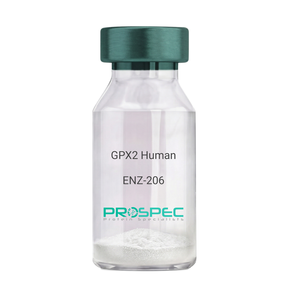
Name :
GPX2 HumanDescription:
Glutathione Peroxidase 2 Human Recombinant
Glutathione peroxidase 2, GPx-2, GSHPx-2, Gastrointestinal glutathione peroxidase, Glutathione peroxidase-gastrointestinal, GPx-GI, GSHPx-GI, Glutathione peroxidase-related protein 2, GPRP-2, GPX2, GPRP, GI-GPx.
Product # :
ENZ-206Price :
Quantity :
Shipping Method :
Shipped with Ice Packs
More Info
- description
- source
- formulation
- purity
- More Info
- Citations
Description
GPX2 Human Recombinant produced in E.coli is a single, non-glycosylated polypeptide chain containing 210 amino acids (1-190) and having a molecular mass of 24.1kDa.GPX2 is fused to a 20 amino acid His-tag at N-terminus & purified by proprietary chromatographic techniques.
Source
Escherichia Coli.
Formulation
The GPX2 solution (0.25mg/ml) contains 20mM Tris-HCl buffer (pH7.5), 40% glycerol, 0.15M NaCl and 1mM DTT.
Purity
Greater than 95% as determined by SDS-PAGE.
Citations
More Info
-
Introduction
Glutathione peroxidase 2 (GPX2) is a member of the glutathione peroxidase family, consisting of 8 known glutathione peroxidases (Gpx1-8) in humans. Glutathione peroxidase functions in the detoxification of hydrogen peroxide, and is one of the most important antioxidant enzymes in humans. GPX2 may have a major role in protecting mammals from the toxicity of ingested organic hydroperoxides. GPX2 is one of only a few proteins known in higher vertebrates to contain selenocysteine, which appears at the active site of glutathione peroxidase and is coded by the nonsense (stop) codon TGA.
-
Synonyms
Glutathione peroxidase 2, GPx-2, GSHPx-2, Gastrointestinal glutathione peroxidase, Glutathione peroxidase-gastrointestinal, GPx-GI, GSHPx-GI, Glutathione peroxidase-related protein 2, GPRP-2, GPX2, GPRP, GI-GPx.
-
Physical Appearance
Sterile filtered colorless solution.
-
Stability
Store at 4°C if entire vial will be used within 2-4 weeks. Store, frozen at -20°C for longer periods of time. For long term storage it is recommended to add a carrier protein (0.1% HSA or BSA).Avoid multiple freeze-thaw cycles.
-
Amino Acid Sequence
MGSSHHHHHH SSGLVPRGSH MAFIAKSFYD LSAISLDGEK VDFNTFRGRA VLIENVASLC GTTTRDFTQL NELQCRFPRR LVVLGFPCNQ FGHQENCQNE EILNSLKYVR PGGGYQPTFT LVQKCEVNGQ NEHPVFAYLK DKLPYPYDDP FSLMTDPKLI IWSPVRRSDV AWNFEKFLIG PEGEPFRRYS RTFPTINIEP DIKRLLKVAI.
ProSpec's products are furnished for LABORATORY RESEARCH USE ONLY. The product may not be used as drugs, agricultural or pesticidal products, food additives or household chemicals.
- View Data Sheet
Name :
DHODH HumanDescription:
Dihydroorotate Dehydrogenase Human Recombinant
Dihydroorotate dehydrogenase (quinone), Dihydroorotate oxidase, human complement of yeast URA1, DHOdehase, POADS, EC 1.3.5.2, EC 1.3.3.1.
Product # :
ENZ-642Price :
Quantity :
Shipping Method :
Shipped with Ice Packs
More Info
- description
- source
- formulation
- purity
- More Info
- Citations
Description
DHODH Human Recombinant produced in E. coli is a single polypeptide chain containing 390 amino acids (31-395) and having a molecular mass of 42.3 kDa.DHODH is fused to a 25 amino acid His-tag at N-terminus & purified by proprietary chromatographic techniques.
Source
E.coli.
Formulation
The DHODH solution (0.5mg/1ml) contains 20mM Tris-HCl buffer (pH 8.0), 100mM NaCl, 1mM DTT and 20% glycerol.
Purity
Greater than 90% as determined by SDS-PAGE.
Citations
More Info
-
Introduction
Dihydroorotate dehydrogenase quinone (DHODH) is a member of the dihydroorotate dehydrogenase family. DHODH is a mitochondrial protein found on the outer surface of the inner mitochondrial membrane. DHODH catalyzes the 4th enzymatic step, the ubiquinone-mediated oxidation of dihydroorotate to orotate (with quinone as electron acceptor), in de novo pyrimidine biosynthesis. DHODH gene defects cause the postaxial acrofacial dysostosis (POADS), also known as Miller syndrome.
-
Synonyms
Dihydroorotate dehydrogenase (quinone), Dihydroorotate oxidase, human complement of yeast URA1, DHOdehase, POADS, EC 1.3.5.2, EC 1.3.3.1.
-
Physical Appearance
Sterile Filtered colorless solution.
-
Stability
Store at 4°C if entire vial will be used within 2-4 weeks. Store, frozen at -20°C for longer periods of time. For long term storage it is recommended to add a carrier protein (0.1% HSA or BSA).Avoid multiple freeze-thaw cycles.
-
Amino Acid Sequence
GSSHHHHHH SSGLVPRGSH MGSHMTGDER FYAEHLMPTL QGLLDPESAH RLAVRFTSLG LLPRARFQDS DMLEVRVLGH KFRNPVGIAA GFDKHGEAVD GLYKMGFGFV EIGSVTPKPQ EGNPRPRVFR LPEDQAVINR YGFNSHGLSV VEHRLRARQQ KQAKLTEDGL PLGVNLGKNK TSVDAAEDYA EGVRVLGPLA DYLVVNVSSP NTAGLRSLQG KAELRRLLTK VLQERDGLRR VHRPAVLVKI APDLTSQDKE DIASVVKELG IDGLIVTNTT VSRPAGLQGA LRSETGGLSG KPLRDLSTQT IREMYALTQG RVPIIGVGGV SSGQDALEKI RAGASLVQLY TALTFWGPPV VGKVKRELEA LLKEQGFGGV TDAIGADHRR.
ProSpec's products are furnished for LABORATORY RESEARCH USE ONLY. The product may not be used as drugs, agricultural or pesticidal products, food additives or household chemicals.
- View Data Sheet
Name :
TGFBR1 Human, ActiveDescription:
Transforming Growth Factor Beta Receptor 1 Human Recombinant, Active
TGFBR1, AAT5, ACVRLK4, ALK-5, ALK5, ESS1, LDS1, LDS1A, LDS2A, MSSE, SKR4, tbetaR-I, TGFR-1.
Product # :
PKA-135Price :
Quantity :
Shipping Method :
Shipped with Ice Packs
More Info
- description
- source
- formulation
- purity
- biological activity
- More Info
- Citations
Description
TGFBR1 produced in Sf9 insect cells is a single, glycosylated polypeptide chain containing 342 amino acids (27-126a.a.) and having a molecular mass of 38kDa. TGFBR1 is expressed with 242 amino acid hIgG-His-Tag at C-Terminus and purified by proprietary chromatographic techniques.
Source
Sf9, Baculovirus cells.
Formulation
TGFBR1 protein solution (0.5mg/ml) Phosphate Buffered Saline (pH 7.4) containing 10% glycerol.
Purity
Greater than 90.0% as determined by SDS-PAGE.
Biological Activity
≤ 2 ug/ml, measured by its binding ability in a functional ELISA with Mouse CD105.
Citations
More Info
-
Synonyms
TGFBR1, AAT5, ACVRLK4, ALK-5, ALK5, ESS1, LDS1, LDS1A, LDS2A, MSSE, SKR4, tbetaR-I, TGFR-1.
-
Physical Appearance
Sterile filtered colorless solution.
-
Stability
Store at 4°C if entire vial will be used within 2-4 weeks. Store, frozen at -20°C for longer periods of time. For long term storage it is recommended to add a carrier protein (0.1% HSA or BSA).Avoid multiple freeze-thaw cycles.
-
Amino Acid Sequence
ADLLLPGATA LQCFCHLCTK DNFTCVTDGL CFVSVTETTD KVIHNSMCIA EIDLIPRDRP FVCAPSSKTG SVTTTYCCNQ DHCNKIELPT TVKSSPGLGP VELVEPKSCD KTHTCPPCPA PELLGGPSVF LFPPKPKDTL MISRTPEVTC VVVDVSHEDP EVKFNWYVDG VEVHNAKTKP REEQYNSTYR VVSVLTVLHQ DWLNGKEYKC KVSNKALPAP IEKTISKAKG QPREPQVYTL
PPSRDELTKN QVSLTCLVKG FYPSDIAVEW ESNGQPENNY KTTPPVLDSD GSFFLYSKLT VDKSRWQQGN VFSCSVMHEA LHNHYTQKSL SLSPGKHHHH HH. -
Background
Transforming Growth Factor Beta Receptor 1 (TGFBR1), a transmembrane protein crucial in the TGF-β signaling pathway, holds a paramount position in regulating diverse cellular processes. Its intricate involvement in development, immune responses, tissue homeostasis, and disease has elevated TGFBR1 to a central role in biology and medicine. This research embarks on a comprehensive exploration of the TGFBR1 protein, unraveling its structural complexities, signaling mechanisms, and its far-reaching implications in various physiological and pathological contexts. By dissecting the intricacies of TGFBR1, scientists aim to decode the fundamental cellular processes it governs and explore potential therapeutic avenues in the domains of cancer, fibrosis, and immunology.
Structural Complexity of TGFBR1:
TGFBR1 is a serine/threonine kinase receptor with an extracellular ligand-binding domain, a transmembrane domain, and an intracellular kinase domain. Its structure allows it to interact with TGF-β ligands and initiate downstream signaling cascades. Understanding the three-dimensional architecture of TGFBR1 is pivotal for deciphering its interactions with ligands, co-receptors, and intracellular signaling partners, shedding light on the molecular intricacies of its function.
Signaling Pathways and Physiological Functions:
Upon ligand binding, TGFBR1 phosphorylates downstream effectors, regulating processes like cell proliferation, differentiation, apoptosis, and immune responses. TGF-β signaling mediated by TGFBR1 is vital in embryogenesis, tissue repair, and immune tolerance. Dysregulation of this pathway is implicated in numerous diseases, including cancer, fibrosis, and autoimmune disorders, underscoring the significance of TGFBR1 in maintaining cellular and tissue homeostasis.
TGFBR1 in Cancer Biology:
TGFBR1's dual role as a tumor suppressor and a promoter of cancer progression reflects its complexity in cancer biology. In early stages, TGFBR1 signaling suppresses cell growth and promotes apoptosis, acting as a defense against tumorigenesis. However, in advanced stages, cancer cells exploit TGFBR1 signaling to facilitate invasion, metastasis, and immune evasion. Understanding the context-dependent nature of TGFBR1's functions in cancer is pivotal for developing targeted therapies.
Targeting TGFBR1 in Therapeutics:
Given its critical roles in various diseases, TGFBR1 has emerged as an attractive target for therapeutic interventions. In cancer, efforts are underway to develop small molecule inhibitors and monoclonal antibodies that modulate TGFBR1 signaling, aiming to curb tumor progression. Additionally, in fibrotic disorders, targeting TGFBR1 offers hope for halting the pathological tissue remodeling characteristic of these diseases, providing potential treatments for conditions such as pulmonary fibrosis and liver cirrhosis.
TGFBR1 Protein, with its intricate signaling mechanisms and diverse physiological roles, stands at the crossroads of fundamental cellular processes and disease pathogenesis. Its involvement in development, immune regulation, cancer, and tissue homeostasis underscores its significance in biology and medicine. As researchers delve deeper into the complexities of TGFBR1, they pave the way for innovative therapies and a deeper understanding of diseases, ultimately shaping the future of healthcare and scientific exploration. This research not only illuminates the pivotal role of TGFBR1 but also holds the promise of transformative advancements in medicine and our understanding of cellular signaling pathways.
ProSpec's products are furnished for LABORATORY RESEARCH USE ONLY. The product may not be used as drugs, agricultural or pesticidal products, food additives or household chemicals.
- View Data Sheet
Name :
MutY E.ColiDescription:
Adenine DNA Glycosylase E.Coli Recombinant
ECK2956, JW2928, mica, mutB, mutY.
Product # :
ENZ-702Price :
Quantity :
Shipping Method :
Shipped with Ice Packs
More Info
- description
- source
- formulation
- purity
- More Info
- Citations
Description
MutY Recombinant produced in E.coli is a single, non-glycosylated polypeptide chain containing 373 amino acids (1-350) and having a molecular mass of 41.5kDa. MutY is fused to a 23 amino acid His-tag at N-terminus & purified by proprietary chromatographic techniques.
Source
E.coli.
Formulation
The MutY solution (0.25mg/ml) contains 20mM Tris-HCl buffer (pH 8.0), 0.1M NaCl and 20% glycerol.
Purity
Greater than 90% as determined by SDS-PAGE.
Citations
More Info
-
Introduction
Adenine DNA glycosylase (mutY) is an adenine DNA glycosylase active on DNA substrates including A/G, A/8-oxoG, or A/C mismatches and also has a weak guanine glycosylase activity on G/8- oxoG-containing DNA. mutY is essential for the prevention of mutations resulting from oxidative DNA impairment. Rising levels of mutY in A549 cells exposed to oxygen and infrared radiation leads to improvements in cell survival. mutY is common in neurons where mitochondrial genomes exposed to reactive oxygen species that damage DNA must uphold integrity over the whole mammalian life span.
-
Synonyms
ECK2956, JW2928, mica, mutB, mutY.
-
Physical Appearance
Sterile Filtered colorless solution.
-
Stability
Store at 4°C if entire vial will be used within 2-4 weeks. Store, frozen at -20°C for longer periods of time. For long term storage it is recommended to add a carrier protein (0.1% HSA or BSA).Avoid multiple freeze-thaw cycles.
-
Amino Acid Sequence
MGSSHHHHHH SSGLVPRGSH MGSMQASQFS AQVLDWYDKY GRKTLPWQID KTPYKVWLSE VMLQQTQVAT VIPYFERFMA RFPTVTDLAN APLDEVLHLW TGLGYYARAR NLHKAAQQVA TLHGGKFPET FEEVAALPGV GRSTAGAILS LSLGKHFPIL DGNVKRVLAR CYAVSGWPGK KEVENKLWSL SEQVTPAVGV ERFNQAMMDL GAMICTRSKP KCSLCPLQNG CIAAANNSWA LYPGKKPKQT LPERTGYFLL LQHEDEVLLA QRPPSGLWGG LYCFPQFADE ESLRQWLAQR QIAADNLTQL TAFRHTFSHF HLDIVPMWLP VSSFTGCMDE GNALWYNLAQ PPSVGLAAPV ERLLQQLRTG APV.
ProSpec's products are furnished for LABORATORY RESEARCH USE ONLY. The product may not be used as drugs, agricultural or pesticidal products, food additives or household chemicals.
- View Data Sheet
Name :
SUMF1 Human, Sf9Description:
Sulfatase Modifying Factor 1 Human Recombinant, Sf9
SUMF1, AAPA3037, FGE, UNQ3037, Formylglycine-generating enzyme, C-alpha-formylglycine-generating enzyme 1, Sulfatase-modifying factor 1.
Product # :
PRO-2619Price :
Quantity :
Shipping Method :
Shipped with Ice Packs
More Info
- description
- source
- formulation
- purity
- More Info
- Citations
Description
SUMF1 produced in Sf9 Insect cells is a single, glycosylated polypeptide chain containing 347 amino acids (34-374.a.) and having a molecular mass of 38.1kDa (Molecular size on SDS-PAGE will appear at approximately 40-57kDa). SUMF1 is expressed with a 6 amino acid His tag at C-Terminus and purified by proprietary chromatographic techniques.
Source
Sf9, Insect cells.
Formulation
SUMF1 protein solution (0.25mg/ml) contains Phosphate Buffered Saline (pH 7.4) and 10% glycerol.
Purity
Greater than 85.0% as determined by SDS-PAGE.
Citations
More Info
-
Introduction
SUMF1 is a part of the SUMF protein family. SUMF1 catalyzes the hydrolysis of sulfate esters by oxidizing a cysteine residue in the substrate sulfatase to an active site 3-oxoalanine residue called C-alpha-formylglycine. Mutations in this gene will cause multiple sulfatase deficiency meaning a lysosomal storage disorder.
-
Synonyms
SUMF1, AAPA3037, FGE, UNQ3037, Formylglycine-generating enzyme, C-alpha-formylglycine-generating enzyme 1, Sulfatase-modifying factor 1.
-
Physical Appearance
Sterile Filtered colorless solution.
-
Stability
Store at 4°C if entire vial will be used within 2-4 weeks. Store, frozen at -20°C for longer periods of time. For long term storage it is recommended to add a carrier protein (0.1% HSA or BSA). Avoid multiple freeze-thaw cycles.
-
Amino Acid Sequence
SQEAGTGAGA GSLAGSCGCG TPQRPGAHGS SAAAHRYSRE ANAPGPVPGE RQLAHSKMVP IPAGVFTMGT DDPQIKQDGE APARRVTIDA FYMDAYEVSN TEFEKFVNST GYLTEAEKFG DSFVFEGMLS EQVKTNIQQA VAAAPWWLPV KGANWRHPEG PDSTILHRPD HPVLHVSWND AVAYCTWAGK RLPTEAEWEY SCRGGLHNRL FPWGNKLQPK GQHYANIWQG EFPVTNTGED GFQGTAPVDA FPPNGYGLYN IVGNAWEWTS DWWTVHHSVE ETLNPKGPPS GKDRVKKGGS YMCHRSYCYR YRCAARSQNT PDSSASNLGF RCAADRLPTM DHHHHHH.
ProSpec's products are furnished for LABORATORY RESEARCH USE ONLY. The product may not be used as drugs, agricultural or pesticidal products, food additives or household chemicals.
- View Data Sheet
Name :
sRANKL HumanDescription:
RANK Ligand Soluble Human Recombinant
Soluble Receptor Activator of NFkB Ligand, TNFSF11, TRANCE, TNF-related activation-induced cytokine, OPGL, ODF, Osteoclast differentiation factor, Tumor necrosis factor ligand superfamily member 11, Receptor activator of nuclear factor kappa B ligand, RANKL, Osteoprotegerin ligand, CD254 antigen, sRANKL, sOdf, hRANKL2.
Product # :
CYT-334Price :
Quantity :
Shipping Method :
Shipped at Room temp
More Info
- description
- source
- formulation
- purity
- biological activity
- More Info
- HPLC, SDS-PAGE
- Citations
Description
sRANKL Human Recombinant produced in E.Coli is a single, non-glycosylated polypeptide chain containing 175 amino acids and having a molecular mass of 19.7kDa.
Source
Escherichia Coli.
Formulation
The protein was lyophilized from a concentrated (1mg/ml) solution containing 10mM Sodium phosphate, pH-7.5.
Purity
Greater than 95.0% as determined by analysis by SDS-PAGE.
Biological Activity
The activity of RAW-Blue was measured to be 46.96 ng/ml, corresponding to a specific activity of 2.1x10⁴ units/mg.
HPLC, SDS-PAGE
Citations
More Info
-
Introduction
RANKL binds to tnfrsf11b/opg and to tnfrsf11a/rank. Osteoclast differentiation and activation factor. Augments the ability of dendritic cells to stimulate naive t-cell proliferation. May be an important regulator of interactions between t-cells and dendritic cells and may play a role in the regulation of the t-cell-dependent immune response. sRANKL may also play an important role in enhanced bone-resorption in humoral hypercalcemia of malignancy.
-
Synonyms
Soluble Receptor Activator of NFkB Ligand, TNFSF11, TRANCE, TNF-related activation-induced cytokine, OPGL, ODF, Osteoclast differentiation factor, Tumor necrosis factor ligand superfamily member 11, Receptor activator of nuclear factor kappa B ligand, RANKL, Osteoprotegerin ligand, CD254 antigen, sRANKL, sOdf, hRANKL2.
-
Physical Appearance
Sterile Filtered White lyophilized (freeze-dried) powder.
-
Stability
Lyophilized TNFSF11 although stable at room temperature for 3 weeks, should be stored desiccated below -18°C. Upon reconstitution sRANKL should be stored at 4°C between 2-7 days and for future use below -18°C.For long term storage it is recommended to add a carrier protein (0.1% HSA or BSA).Please prevent freeze-thaw cycles.
-
Solubility
It is recommended to reconstitute the lyophilized sRANKL in sterile 18MΩ-cm H2O at a concentration of 100µg/ml, which can then be further diluted to other aqueous solutions.
-
Amino Acid Sequence
EKAMVDGSW LDLAKRSKLE AQPFAHLTIN ATDIPSGSHK VSLSSWYHDR GWAKISNMTF SNGKLIVNQD GFYYLYANIC FRHHETSGDL ATEYLQLMVY VTKTSIKIPS SHTLMKGGST KYWSGNSEFH FYSINVGGFF KLRSGEEISI EVSNPSLLDP DQDATYFGAF KVRDID.
ProSpec's products are furnished for LABORATORY RESEARCH USE ONLY. The product may not be used as drugs, agricultural or pesticidal products, food additives or household chemicals.