Search results
1000 results found for “Thrombin”
Name
Description
Product #
Price
Quantity
Shipping Method
- View Data Sheet
Name :
CTF1 RatDescription:
Cardiotrophin-1 Rat Recombinant
Cardiotrophin-1, CT-1, Ctf1.
Product # :
CYT-199Price :
Quantity :
Shipping Method :
Shipped at Room temp
More Info
- description
- source
- formulation
- purity
- biological activity
- More Info
Description
Cardiotrophin-1 Rat Recombinant produced in E.Coli is a single, non-glycosylated, polypeptide chain containing 203 amino acids and having a molecular mass of 21.4kDa.The CTF1 Rat is purified by proprietary chromatographic techniques.
Source
Escherichia Coli.
Formulation
Lyophilized from a 0.2µm filtered concentrated solution in PBS, pH 7.4.
Purity
Greater than 95.0% as determined by SDS-PAGE and HPLC analyses.
Biological Activity
The ED50 as determined by the dose-dependent proliferation of TF-1 cells was found to be < 0.5ng/ml, corresponding to a specific activity of > 2.0×10⁶ units/mg.More Info
-
Introduction
Cardiotrophin 1 (CT-1) is a 201 amino acid member of the interleukin-6 superfamily. It was identified by its ability to induce hypertrophic response in cardiac myocytes. CT-1 mRNA levels were found both in cardiac myocytes and in cardiac nonmyocytes. CT 1 was also detected in abundance in normal adult human lung and was expressed in both fetal and adult airway smooth muscle cells. CT 1 activates gp130 dependent signaling and stimulates the Janus kinase/signal transducers and activators of transcription (JAK/STAT) pathway to transduce hypertrophic and cytoprotective signals in cardiac myocytes.
CT 1 has also a neurotrophic function. CTF1 deficiency causes increased motoneuron cell death in spinal cord and brainstem nuclei of mice during a period between embryonic day 14 and the first postnatal week. Moreover, CT-1 is a hepatocyte survival factor that efficiently reduces hepatocellular damage in animal models of acute liver injury. Cardiotrophin 1 expression is augmented after hypoxic stimulation and it can protect cardiac cells when added either prior to simulated ischaemia or at the time of reoxygenation following simulated ischaemia. Cardiotrophin 1 can induce expression of the protective heat shock proteins (hsps) in cardiac cells.
Cardiotrophin-1 increased ventricular expression of ANP, brain natriuretic peptide (BNP) and angiotensinogen mRNA.
Cardiophin 1 levels were significantly elevated in patients with heart failure, patients with dilatative cardiomyopathy, moderate/severe mitral regurgitation, stable and unstable angina and after acute myocardial infarction. -
Synonyms
Cardiotrophin-1, CT-1, Ctf1.
-
Physical Appearance
Sterile Filtered White lyophilized (freeze-dried) powder.
-
Stability
Lyophilized Cardiotrophin-1 Rat although stable at room temperature for 3 weeks, should be stored desiccated below -18°C. Upon reconstitution CTF1 Rat should be stored at 4°C between 2-7 days and for future use below -18°C.Please prevent freeze-thaw cycles.
-
Solubility
It is recommended to reconstitute the lyophilized CTF1 in sterile 4mM HCl not less than 100µg/ml, which can then be further diluted to other aqueous solutions.
-
Amino Acid Sequence
MSQREGSLED HQTDSSFSFL PHLEAKIRQT HNLARLLTKY ADQLLEEYVQ QQGEPFGLPG FSPPRLPLAG LSGPAPSHAG LPVSERLRQD AAALSALPAL LDAVRRRQAE LNPRAPRLLR SLEDAARQVR ALGAAVETVL AALGAAARGP VPEPVATSAL FTSNSAAGVF SAKVLGLHVC GLYGEWVSRT EGDLGQLVPG GVA.
-
Background
What is the molecular weight/Mw of CTF1 Protein?
CTF1 Protein has a total Mw of 21.4kDa.
What is the source or expression system of CTF1 Protein?
Escherichia Coli.
What is the Purity of CTF1 Protein?
CTF1 Protein is >95% pure as determined by SDS-PAGE.
What is the Biological Activity of CTF1 Protein?
The ED50 as determined by the dose-dependent proliferation of TF-1 cells was found to be < 0.5ng/ml, corresponding to a specific activity of > 2.0×10⁶ units/mg.
What is the amino acid sequence of CTF1 Protein?
MSQREGSLED HQTDSSFSFL PHLEAKIRQT HNLARLLTKY ADQLLEEYVQ QQGEPFGLPG FSPPRLPLAG LSGPAPSHAG LPVSERLRQD AAALSALPAL LDAVRRRQAE LNPRAPRLLR SLEDAARQVR ALGAAVETVL AALGAAARGP VPEPVATSAL FTSNSAAGVF SAKVLGLHVC GLYGEWVSRT EGDLGQLVPG GVA.
What applications can CTF1 Protein be used in?
CTF1 Protein can probably be used in western blot, ELISA and Lateral Flow.
What is the endotoxin level for CTF1 Protein?
The endotoxin level is minimal, CTF1 Protein was purified using conventional chromatography techniques.
ProSpec's products are furnished for LABORATORY RESEARCH USE ONLY. The product may not be used as drugs, agricultural or pesticidal products, food additives or household chemicals.
- View Data Sheet
Name :
EPCAM Human, sf9Description:
Epithelial Cell Adhesion Molecule Human Recombinant, Sf9
Epithelial Cell Adhesion Molecule , Tumor-Associated Calcium Signal Transducer 1, Major Gastrointestinal Tumor-Associated Protein GA733-2, Adenocarcinoma-Associated Antigen, Cell Surface Glycoprotein Trop-1, Epithelial Glycoprotein 314, TACSTD1, EGP314, MIC18, TROP1, M4S1, KSA, Membrane Component, Chromosome 4, Surface Marker (35kD Glycoprotein), Antigen Identified By Monoclonal Antibody AUA1, Human Epithelial Glycoprotein-2, Epithelial Cell Surface Antigen, Epithelial Glycoprotein, KS 1/4 Antigen, CD326 Antigen, GA733-2, HEGP314, HNPCC8, Ep-CAM, DIAR5, EGP-2, EGP40, KS1/4, MK-1, M1S2, ESA, EGP, EPCAM.
Product # :
PRO-2238Price :
Quantity :
Shipping Method :
Shipped with Ice Packs
More Info
- description
- source
- formulation
- purity
- More Info
Description
EPCAM produced in Sf9 Baculovirus cells is a single, glycosylated polypeptide chain (24-265 a.a.) and fused to a 6 aa His Tag at C-terminus containing a total of 248 amino acids and having a molecular mass of 28.2kDa.EPCAM shows multiple bands between 28-40kDa on SDS-PAGE, reducing conditions and purified by proprietary chromatographic techniques.
Source
Sf9, Baculovirus cells.
Formulation
EPCAM protein solution (1mg/ml) contains Phosphate buffered saline (pH7.4) and 10% glycerol.
Purity
Greater than 95.0% as determined by SDS-PAGE.
More Info
-
Introduction
EPCAM is a carcinoma-associated antigen and belongs to a family which includes at least 2 type I membrane proteins. The EPCAM protein has a role in embryonic stem cells proliferation and differentiation. EPCAM is used as a target for immunotherapy treatment of human carcinomas. EPCAM is expressed on most normal epithelial cells and gastrointestinal carcinomas and acts as a homotypic calcium-independent cell adhesion molecule. Epithelial cell adhesion molecules (EPCAM) can act as a physical homophilic interaction molecule between intestinal epithelial cells (IECs) and intraepithelial lymphocytes (IELs) at the mucosal epithelium for supplying immunological barrier as a first line of defense against mucosal infection. EPCAM gene mutations result in congenital tufting enteropathy.
-
Synonyms
Epithelial Cell Adhesion Molecule , Tumor-Associated Calcium Signal Transducer 1, Major Gastrointestinal Tumor-Associated Protein GA733-2, Adenocarcinoma-Associated Antigen, Cell Surface Glycoprotein Trop-1, Epithelial Glycoprotein 314, TACSTD1, EGP314, MIC18, TROP1, M4S1, KSA, Membrane Component, Chromosome 4, Surface Marker (35kD Glycoprotein), Antigen Identified By Monoclonal Antibody AUA1, Human Epithelial Glycoprotein-2, Epithelial Cell Surface Antigen, Epithelial Glycoprotein, KS 1/4 Antigen, CD326 Antigen, GA733-2, HEGP314, HNPCC8, Ep-CAM, DIAR5, EGP-2, EGP40, KS1/4, MK-1, M1S2, ESA, EGP, EPCAM.
-
Physical Appearance
Sterile filtered colorless solution.
-
Stability
Store at 4°C if entire vial will be used within 2-4 weeks. Store, frozen at -20°C for longer periods of time. For long term storage it is recommended to add a carrier protein (0.1% HSA or BSA).Avoid multiple freeze-thaw cycles.
-
Amino Acid Sequence
QEECVCENYK LAVNCFVNNN RQCQCTSVGA QNTVICSKLA AKCLVMKAEM NGSKLGRRAK PEGALQNNDG LYDPDCDESG LFKAKQCNGT STCWCVNTAG VRRTDKDTEI TCSERVRTYW IIIELKHKAR EKPYDSKSLR TALQKEITTR YQLDPKFITS ILYENNVITI DLVQNSSQKT QNDVDIADVA YYFEKDVKGE SLFHSKKMDL TVNGEQLDLD PGQTLIYYVD EKAPEFSMQG LKHHHHHH
ProSpec's products are furnished for LABORATORY RESEARCH USE ONLY. The product may not be used as drugs, agricultural or pesticidal products, food additives or household chemicals.
- View Data Sheet
Name :
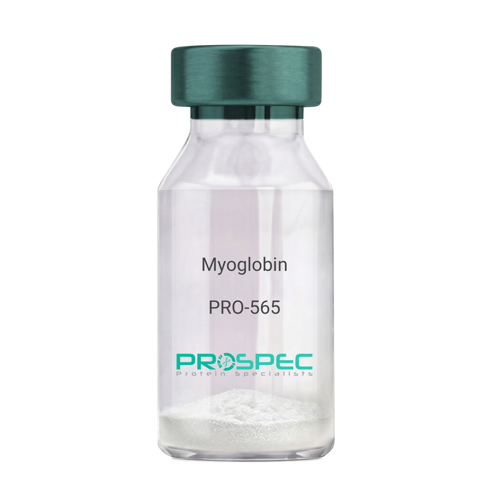
MyoglobinDescription:
Myoglobin Human
Myoglobin, MB, PVALB, MGC13548.
Product # :
PRO-565Price :
Quantity :
Shipping Method :
Shipped with Ice Packs
More Info
- description
- source
- formulation
- purity
- More Info
Description
Human Myoglobin produced in Human Cardiac Tissues having a molecular mass of 17.5kDa. Myoglobin is released from recently injured myocardial cells within a few hours of Infarction. Peak levels are reached more quickly than CK-MB or Troponin complex.
Source
Human Cardiac Tissues.
Formulation
The protein solution is in 0.05M phosphate buffer pH 7.5 containing 0.15M NaCl and 0.09% NaN3. Filtered through a 0.2µM membrane.
Purity
Greater than 96.0%.
More Info
-
Introduction
Myoglobin is a member of the globin superfamily and can be found in skeletal and cardiac muscles. It is a haemoprotein that contributs to intracellular oxygen storage and transcellular facilitated diffusion of oxygen. Myoglobin has a single-chain globular structure of 153 amino acids, containing a heme prosthetic group (iron-containing porphyrin) in the core around which the remaining apoprotein folds. Myoglobin has 8 alpha helices and a hydrophobic core. Myoglobin’s molecular weight is 16.7 kDa, and it is the primary oxygen-carrying pigment of muscle tissues. The binding of oxygen in myoglobin is different from the cooperative oxygen binding in hemoglobin, since positive collaboration is a property of multimeric/oligomeric proteins only. Instead, the binding of oxygen by myoglobin is uninfluenced by the oxygen pressure in the surrounding tissue. Myoglobin is frequently referred to as having an "instant binding tenacity" to oxygen given its hyperbolic oxygen dissociation curve. Different organisms are able to hold their breaths longer due to high concentrations of myoglobin in their muscle cells. Myoglobin is responsible for the pigments that make meat red. The color of the meat is partly determined by the charge of the iron atom in myoglobin and the oxygen attached to it. Myoglobin is found in Type I muscle, Type II A and Type II B, but it is mostly deemed that myoglobin is not found in smooth muscle. Myoglobin is discharged from damaged muscle tissue (rhabdomyolysis), which contains very high concentrations of myoglobin. Even though the released myoglobin is filtered by the kidneys, it is toxic to the renal tubular epithelium and thus may cause acute renal failure.
-
Synonyms
Myoglobin, MB, PVALB, MGC13548.
-
Physical Appearance
Sterile Filtered red solution.
-
Stability
Human Myoglobin should be stored at 2-8°C.
-
Human Virus Test
Starting material donor tested and certified negative for HIV I & II antibodies, Hepatitis B surface antigen, and Hepatitis C antibodies.
ProSpec's products are furnished for LABORATORY RESEARCH USE ONLY. The product may not be used as drugs, agricultural or pesticidal products, food additives or household chemicals.
- View Data Sheet
Name :
MIF Human His NDescription:
Macrophage Migration Inhibitory Factor Human, Recombinant His Tag N-Terminus
Phenylpyruvate tautomerase, Glycosylation-inhibiting factor, GIF, MMIF, MIF.
Product # :
CYT-431Price :
Quantity :
Shipping Method :
Shipped with Ice Packs
More Info
- description
- source
- formulation
- purity
- More Info
Description
MIF human Recombinant, fused to 40 a.a. His-tag at N-terminus, was cloned into an E. coli expression vector and was purified to apparent homogeneity by using conventional column chromatography techniques. Macrophage Inducing Factor Human Recombinant ( 1-115 a.a. ) is a single, non-glycosylated, polypeptide chain having a total amino acids of 155 and molecular mass of 17kDa.
Source
Escherichia Coli.
Formulation
1mg/ml solution containing PBS pH-7.4.
Purity
Greater than 95.0% as determined by:
(a) Analysis by RP-HPLC.
(b) Analysis by SDS-PAGE.More Info
-
Introduction
The cytokine Macrophage migration inhibitory factor (MIF) has been identified to be secreted by the pituitary gland and the monocyte/macrophage and to play an important role in endotoxic shock. MIF has the unique property of being released from macrophages and T cells in response to physiological concentrations of glucocorticoids. The secretion of MIF is tightly regulated and decreases at high, anti-inflammatory steroid concentration.
-
Synonyms
Phenylpyruvate tautomerase, Glycosylation-inhibiting factor, GIF, MMIF, MIF.
-
Physical Appearance
Sterile filtered colorless solution.
-
Stability
Liquid MIF although stable 4°C for 1 week, should be stored below -18°C. Please prevent freeze-thaw cycles.
-
Amino Acid Sequence
MRGSHHHHHHGMASMTGGQQMGRDLYDDDDKDRWGSMPMFIVNTNV PRASVPDGFL SELTQQLAQA TGKPPQYIAV HVVPDQLMAF GGSSEPCALC LHSIGKIGGA QNRSYSKLLC GLLAERLRIS PDRVYINYYD MNAANVGWNN STFA.
ProSpec's products are furnished for LABORATORY RESEARCH USE ONLY. The product may not be used as drugs, agricultural or pesticidal products, food additives or household chemicals.
- View Data Sheet
Name :
PDGF AA HumanDescription:
Platelet-Derived Growth Factor AA Human Recombinant
Glioma-derived growth factor, GDGF, Osteosarcoma-derived Growth Factor, ODGF, PDGF-AA, PDGF-1.
Product # :
CYT-341Price :
Quantity :
Shipping Method :
Shipped at Room temp
More Info
- description
- source
- formulation
- purity
- biological activity
- More Info
Description
Platelet-Derived Growth Factor AA Human Recombinant is a homodimeric, non-glycosylated, polypeptide chain containing 2 x 125 amino acids and having a total molecular mass of 28511 Dalton. PDGF-AA is purified by proprietary chromatographic techniques.
Source
Escherichia Coli.
Formulation
Lyophilized without any additives.
Purity
Greater than 95.0% as determined by SDS-PAGE.
Biological Activity
The ED50, calculated by the dose-dependent proliferation of murine 3T3 indicator cells is < 0.32ng/ml, corresponding to a Specific Activity of 3,125x103units/mg.More Info
-
Introduction
PDGF-AA, PDGF-BB and PDGF-AB, are potent mitogens for a variety of cell types including smooth muscle cells, connective tissue cells, bone and cartilage cells, and some blood cells. The PDGF is stored in platelet alpha-granules and released upon platelet activation. The PDGF is involved in a number of biological processes, including hyperplasia, chemotaxis, embryonic neuron development, and respiratory tubule epithelial cell development. Two distinct signaling receptors used by PDGF
have been identified and named PDGFR-alpha and PDGFR-beta. PDGFR-alpha is high-affinity receptor for each of the three PDGF forms. On the other hand, PDGFR-beta interacts with only PDGF-BB and PDGF-AB. -
Synonyms
Glioma-derived growth factor, GDGF, Osteosarcoma-derived Growth Factor, ODGF, PDGF-AA, PDGF-1.
-
Physical Appearance
Sterile Filtered White lyophilized (freeze-dried) powder.
-
Stability
Lyophilized Platelet-Derived Growth Factor AA although stable at room temperature for 3 weeks, should be stored desiccated below -18°C. Upon reconstitution PDGF-AA should be stored at 4°C between 2-7 days and for future use below -18°C.For long term storage it is recommended to add a carrier protein (0.1% HSA or BSA).Please prevent freeze-thaw cycles.
-
Solubility
It is recommended to reconstitute the lyophilized Platelet-derived Growth Factor-AA in sterile 18MΩ-cm H2O not less than 100µg/ml, which can then be further diluted to other aqueous solutions.
-
Amino Acid Sequence
SIEEAVPAVC KTRTVIYEIP RSQVDPTSAN FLIWPPCVEV KRCTGCCNTS SVKCQPSRVH HRSVKVAKVE YVRKKPKLKE VQVRLEEHLE CACATTSLNP DYREEDTGRP RESGKKRKRK RLKPT.
ProSpec's products are furnished for LABORATORY RESEARCH USE ONLY. The product may not be used as drugs, agricultural or pesticidal products, food additives or household chemicals.
- View Data Sheet
Name :
TNF a RabbitDescription:
Tumor Necrosis Factor-Alpha Rabbit Recombinant
Tumor necrosis factor, Cachectin, TNF-alpha, Tumor necrosis factor ligand superfamily member 2, TNF-a, TNF, TNFA, TNFSF2.
Product # :
CYT-008Price :
Quantity :
Shipping Method :
Shipped at Room temp
More Info
- description
- source
- formulation
- purity
- biological activity
- More Info
Description
Tumor Necrosis Factor-a Rabbit Recombinant consists of three identical polypeptide chains of 158 amino acids combined to form a compact, bell-shaped homotrimer. TNF-alpha was produced in E.Coli is a non-glycosylated, polypeptide chain having a molecular mass of 17.4 kDa for the individual subunit. The TNF-alpha is purified by standard chromatographic techniques.
Source
Escherichia Coli.
Formulation
TNF-alpha Rabbit was lyophilized after extensive dialysis against 20mM PB, pH7.4, 300mM NaCl.
Purity
Greater than 95% as determined by:
(a) Analysis by RP-HPLC.
(b) Analysis by SDS-PAGE.Biological Activity
The ED50 as determined by the cytolysis of murine L929 cells in the presence of Actinomycin D is less than 0.03ng/ml, corresponding to a Specific Activity of 30,000,000 IU/mg.
More Info
-
Introduction
Tumor necrosis factor is a cytokine involved in systemic inflammation and is a member of a group of cytokines that all stimulate the acute phase reaction. TNF is mainly secreted by macrophages.
TNF causes apoptotic cell death, cellular proliferation, differentiation, inflammation, tumorigenesis and viral replication, TNF is also involved in lipid metabolism, and coagulation. TNF's primary role is in the regulation of immune cells.
Dysregulation and, in particular, overproduction of TNF have been implicated in a variety of human diseases- autoimmune diseases, insulin resistance, and cancer. -
Synonyms
Tumor necrosis factor, Cachectin, TNF-alpha, Tumor necrosis factor ligand superfamily member 2, TNF-a, TNF, TNFA, TNFSF2.
-
Physical Appearance
Sterile Filtered White lyophilized (freeze-dried) powder.
-
Stability
Lyophilized Tumor Necrosis Factor-a although stable at room temperature for 3 weeks, should be stored desiccated below -18°C. Upon reconstitution TNF-a should be stored at 4°C between 2-7 days and for future use below -18°C.For long term storage it is recommended to add a carrier protein (0.1% HSA or BSA).Please prevent freeze-thaw cycles.
-
Solubility
It is recommended to reconstitute the lyophilized Tumor Necrosis Factor-alpha in sterile 18MΩ-cm H2O not less than 100µg/ml, which can then be further diluted to other aqueous solutions.
-
Amino Acid Sequence
The sequence of the first five N-terminal amino acids was determined and was found to be Met-Ser-Ala-Ser-Arg.
ProSpec's products are furnished for LABORATORY RESEARCH USE ONLY. The product may not be used as drugs, agricultural or pesticidal products, food additives or household chemicals.
- View Data Sheet
Name :
TNFSF8 HumanDescription:
CD30 Ligand Human Recombinant
Tumor necrosis factor ligand superfamily member 8, CD30 ligand, CD30-L, CD153, TNFSF8, CD30L, CD30LG, CD30 Antigen Ligand, CD153 Antigen.
Product # :
CYT-824Price :
Quantity :
Shipping Method :
Shipped with Ice Packs
More Info
- description
- source
- formulation
- purity
- More Info
Description
TNFSF8 Human Recombinant produced in E.Coli is a single, non-glycosylated polypeptide chain containing 195 amino acids (63-234 a.a.) and having a molecular mass of 22kDa.TNFSF8 is fused to a 23 amino acid His-tag at N-terminus & purified by proprietary chromatographic techniques.
Source
Escherichia Coli.
Formulation
TNFSF8 protein solution (0.5mg/ml) contains 20mM Tris-HCl buffer (pH 8.0), 10% glycerol and 0.4M Urea.
Purity
Greater than 80% as determined by SDS-PAGE.
More Info
-
Introduction
CD30 Ligand (TNFSF8) is a cytokine which is a member of the tumor necrosis factor (TNF) ligand family. The TNFSF8 cytokine is a ligand for TNFRSF8/CD30, which is a cell surface antigen and a marker for Hodgkin lymphoma and related hematologic malignancies. The employment of the TNFSF8 cytokine expressed on B cell surface has an inhibitory role in modulating Ig class switch. TNFSF8 enhances cell proliferation of some lymphoma cell lines, while inducing cell death and reducing cell proliferation of other lymphoma cell lines. The pleiotropic biological activities of the TNFSF8 cytokine on different CD30+ lymphoma cell lines has a pathophysiologic role in Hodgkin's and some non-Hodgkin's lymphomas.
-
Synonyms
Tumor necrosis factor ligand superfamily member 8, CD30 ligand, CD30-L, CD153, TNFSF8, CD30L, CD30LG, CD30 Antigen Ligand, CD153 Antigen.
-
Physical Appearance
Sterile Filtered clear solution.
-
Stability
Store at 4°C if entire vial will be used within 2-4 weeks. Store, frozen at -20°C for longer periods of time.For long term storage it is recommended to add a carrier protein (0.1% HSA or BSA).Please avoid freeze thaw cycles.
-
Amino Acid Sequence
MGSSHHHHHH SSGLVPRGSH MGSQRTDSIP NSPDNVPLKG GNCSEDLLCI LKRAPFKKSW AYLQVAKHLN KTKLSWNKDG ILHGVRYQDG NLVIQFPGLY FIICQLQFLV QCPNNSVDLK LELLINKHIK KQALVTVCES GMQTKHVYQN LSQFLLDYLQ VNTTISVNVD TFQYIDTSTF PLENVLSIFL YSNSD.
ProSpec's products are furnished for LABORATORY RESEARCH USE ONLY. The product may not be used as drugs, agricultural or pesticidal products, food additives or household chemicals.
- View Data Sheet
Name :
TREM2 PAT1ETAT AntibodyDescription:
Triggering receptor expressed on myeloid cells 2 Clone PAT1ETAT, Mouse Anti Human
Triggering receptor expressed on myeloid cells 2, Triggering receptor expressed on monocytes 2, TREM-2, TREM2, Trem2a, Trem2b, Trem2c.
Product # :
ANT-777Price :
Quantity :
Shipping Method :
Shipped with Ice Packs
More Info
- formulation
- More Info
Formulation
1mg/ml containing PBS, pH-7.4, 10% Glycerol and 0.02% Sodium Azide.
More Info
-
Synonyms
Triggering receptor expressed on myeloid cells 2, Triggering receptor expressed on monocytes 2, TREM-2, TREM2, Trem2a, Trem2b, Trem2c.
-
Immunogen
Anti-human TREM2 mAb, is derived from hybridization of mouse F0 myeloma cells with spleen cells from BALB/c mice immunized with recombinant human TREM2 amino acids 19-161 purified from E. coli.
-
Ig Subclass
Mouse IgG1 heavy chain and κ light chain.
-
Clone
PAT1ETAT.
-
Applications
TREM2 antibody has been tested by ELISA, ICC/IF, FACS and Western blot analysis to assure specificity and reactivity. Since application varies, however, each investigation should be titrated by the reagent to obtain optimal results.
-
Background
TREM2 is a membrane protein which forms a receptor signaling complex with TYROBP. TREM2 takes part in chronic inflammation by triggering the production of constitutive inflammatory cytokines. TREM2 deficiency is a source of polycystic lipomembranous osteodysplasia with sclerosing leukoencephalopathy.
-
Type
Mouse Anti Human Monoclonal.
-
Storage Procedures
For periods up to 1 month store at 4°C, for longer periods of time, store at -20°C. Prevent freeze thaw cycles.
-
Purification Method
TREM2 antibody was purified from mouse ascitic fluids by protein-A affinity chromatography.
ProSpec's products are furnished for LABORATORY RESEARCH USE ONLY. The product may not be used as drugs, agricultural or pesticidal products, food additives or household chemicals.
- View Data Sheet
Name :
Welqut ProteaseDescription:
Welqut Protease Staphylococcus aureus Recombinant
Product # :
ENZ-1113Price :
Quantity :
Shipping Method :
Shipped with Ice Packs
More Info
- description
- source
- formulation
- purity
- More Info
Description
Welqut Protease Recombinant is a single, non-glycosylated polypeptide chain containing 204 amino acids and having a molecular mass of 22kDa. The Welqut Protease is purified by proprietary chromatographic techniques.
Source
Escherichia Coli.
Formulation
Welqut Protease contains 10 mM Na2HPO4, 50% glycerol, 1.8 mM KH2PO4, pH 7.3, 140 mM NaCl and 2.7 mM KCl.
Purity
Greater than 97.0% as determined by SDS-PAGE.
More Info
-
Introduction
WELQut Protease is an extremely specific and recombinant serine protease from Staphylococcus aureus. The WELQut Protease identifies and accurately cleaves recombinant proteins that has a recognition sequence added to them, with the amino acid sequence Trp, Glu, Leu, Gln, X (any amino acid). WELQut Protease cut externally from the recognition sequence, therefor doesn’t leave extra amino acids bound to the target protein. The protease isn’t temperature sensitive (works in 4-30°C) or pH sensitive (pH 6.5-9.0), also, there is no need in any particular buffers.
-
Physical Appearance
Sterile Filtered colorless solution.
-
Stability
Store at 4°C if entire vial will be used within 2-4 weeks. Store, frozen at -20°C for longer periods of time. For long term storage it is recommended to add a carrier protein (0.1% HSA or BSA).Avoid multiple freeze-thaw cycles.
-
Unit Definition
Each unit is defined as the amount of enzyme required to cleave ≥99% of 100μg of a control protein in 16 h at 20°C. Enzyme activity is assayed in 100μl 100 mM Tris-HCl (pH 8.0).
ProSpec's products are furnished for LABORATORY RESEARCH USE ONLY. The product may not be used as drugs, agricultural or pesticidal products, food additives or household chemicals.
- View Data Sheet
Name :
HDGF2 HumanDescription:
Hepatoma-Derived Growth Factor-2 Human Recombinant
Hepatoma-derived growth factor-related protein 3, HRP-3, Hepatoma-derived growth factor 2, HDGF-2, HDGFRP3, HDGF2, CGI-142.
Product # :
CYT-132Price :
Quantity :
Shipping Method :
Shipped with Ice Packs
More Info
- description
- source
- formulation
- purity
- More Info
- SDS-PAGE
Description
HDGF2 Human Recombinant produced in E.Coli is a single, non-glycosylated polypeptide chain containing 227 amino acids (1-203 a.a.) and having a molecular mass of 25.1kDa (Molecular weight on SDS-PAGE will appear higher). HDGF2 is fused to a 24 amino acid His-tag at N-terminus & purified by proprietary chromatographic techniques.
Source
Escherichia Coli.
Formulation
HDGF2 protein solution (0.5mg/ml) containing 20mM Tris-HCl buffer (pH 8.0), 1mM DTT, 10% glycerol and 0.1M NaCl.
Purity
Greater than 90% as determined by SDS-PAGE.
SDS-PAGE
More Info
-
Introduction
Hepatoma-derived growth factor, related protein 3 (HDGFRP3/HDGF2) is the original member of a family of polypeptides labeled HDGF related proteins (HRPs). The HDGF2 protein augments DNA synthesis and may play a part in cell proliferation. HDGF2 is expressed primarily in the testis and the brain, to an intermediate extent in the heart, and to a slight extent in the ovaries, kidneys, spleen, and liver in humans.
-
Synonyms
Hepatoma-derived growth factor-related protein 3, HRP-3, Hepatoma-derived growth factor 2, HDGF-2, HDGFRP3, HDGF2, CGI-142.
-
Physical Appearance
Sterile Filtered clear solution.
-
Stability
Store at 4°C if entire vial will be used within 2-4 weeks. Store, frozen at -20°C for longer periods of time. For long term storage it is recommended to add a carrier protein (0.1% HSA or BSA).Avoid multiple freeze-thaw cycles.
-
Amino Acid Sequence
MGSSHHHHHH SSGLVPRGSH MGSHMARPRP REYKAGDLVF AKMKGYPHWP ARIDELPEGA VKPPANKYPI FFFGTHETAF LGPKDLFPYK EYKDKFGKSN KRKGFNEGLW EIENNPGVKF TGYQAIQQQS SSETEGEGGN TADASSEEEG DRVEEDGKGK RKNEKAGSKR KKSYTSKKSS
KQSRKSPGDE DDKDCKEEEN KSSSEGGDAG NDTRNTTSDL QKTSEGT. -
Background
What is the molecular weight/Mw of HDGF2 HUMAN Protein?
HDGF2 HUMAN Protein has a total Mw of 25.1kDa.
What is the source or expression system of HDGF2 HUMAN Protein?
Escherichia Coli.
What is the Purity of HDGF2 HUMAN Protein?
HDGF2 HUMAN Protein is >90% pure as determined by SDS-PAGE.
What is the Biological Activity of HDGF2 HUMAN Protein?
The biological functionality of HDGF2 HUMAN Protein will be determined in the future.
What is the amino acid sequence of HDGF2 HUMAN Protein?
MGSSHHHHHH SSGLVPRGSH MGSHMARPRP REYKAGDLVF AKMKGYPHWP ARIDELPEGA VKPPANKYPI FFFGTHETAF LGPKDLFPYK EYKDKFGKSN KRKGFNEGLW EIENNPGVKF TGYQAIQQQS SSETEGEGGN TADASSEEEG DRVEEDGKGK RKNEKAGSKR KKSYTSKKSS
KQSRKSPGDE DDKDCKEEEN KSSSEGGDAG NDTRNTTSDL QKTSEGT.
What applications can HDGF2 HUMAN Protein be used in?
HDGF2 HUMAN Protein can probably be used in western blot, ELISA and Lateral Flow.
What is the endotoxin level for HDGF2 HUMAN Protein?
The endotoxin level is minimal, HDGF2 HUMAN Protein was purified using conventional chromatography techniques.
ProSpec's products are furnished for LABORATORY RESEARCH USE ONLY. The product may not be used as drugs, agricultural or pesticidal products, food additives or household chemicals.
- View Data Sheet
Name :
LysostaphinDescription:
Lysostaphin Recombinant
Lysostaphin, EC 3.4.24.75, Glycyl-glycine endopeptidase.
Product # :
ENZ-269Price :
Quantity :
Shipping Method :
Shipped at Room temp
More Info
- description
- source
- formulation
- purity
- biological activity
- More Info
Description
Lysostaphin Recombinant produced in E.Coli is a non-glycosylated polypeptide chain having a molecular mass of 26.92 kDa.
Source
Escherichia Coli.
Formulation
The protein was lyophilized without any additives.
Purity
98% as determined by RP-HPLC.
Biological Activity
Assessed by the decrease in turbidity of a suspension of heat-killed Staphylococcus aureus at pH-8, 30°C. Lysostaphin is a zinc enzyme therefore EDTA is an inhibitory factor.
More Info
-
Introduction
Lysostaphin, an endopeptidase specific for the cell wall peptidoglycan of staphylococci, is an extremely potent anti-staphylococcal agent. Lysostaphin is used as a research and diagnostic tool. Because it lyses staphylococci efficiently, it is widely used when preparing staphylococcal DNA or other cellular components for genetic and biochemical studies and for the preparation of protoplasts for transformation. Preparation and analysis of bacterial DNA has become a powerful tool used by clinical and other microbiologists in epidemiological studies aimed at tracing sources of infection or bacterial contamination.
-
Synonyms
Lysostaphin, EC 3.4.24.75, Glycyl-glycine endopeptidase.
-
Physical Appearance
Sterile Filtered lyophilized powder.
-
Stability
Lyophilized Lysostaphin although stable at room temperature for 2 weeks, should be stored desiccated below -18°C. Upon reconstitution Lysostaphin can be stored at 4°C up to 3 weeks and for future use below -18°C.Please prevent freeze-thaw cycles.
-
Solubility
It is recommended to reconstitute the lyophilized Lysostaphin in 20mM sodium acetate, pH 4.5 for optimal stability, which can then be further diluted.
-
Protein content
Protein quantitation was carried out by two independent methods 1. UV spectroscopy at 280 nm using the absorbency value of 2.02 as the extinction coefficient for a 0.1% (1mg/ml) solution. This value is calculated by the PC GENE computer analysis program of protein sequences (IntelliGenetics). 2. Analysis RP-HPLC, using a calibrated solution of Lysostaphin as a Reference standard.
-
Specific Activity
Determined to be 3,540 units/mg.
ProSpec's products are furnished for LABORATORY RESEARCH USE ONLY. The product may not be used as drugs, agricultural or pesticidal products, food additives or household chemicals.
- View Data Sheet
Name :
CDC34 HumanDescription:
Cell Division Cycle 34 Human Recombinant
UB2R1, CDC-34, Ubiquitin-conjugating enzyme E2 R1, Ubiquitin-protein ligase R1, Ubiquitin-conjugating enzyme E2-32 kDa complementing, E2-CDC34, CDC34, EC 6.3.2.19, UBC3, UBE2R1.
Product # :
ENZ-1103Price :
Quantity :
Shipping Method :
Shipped with Ice Packs
More Info
- description
- source
- formulation
- purity
- More Info
Description
Cell Division Cycle 34 Human Recombinant produced in E.Coli is a single, non- glycosylated polypeptide chain containing 236 amino acids and having a molecular mass of 26.7kDa. CDC34 is purified by proprietary chromatographic techniques.
Source
Escherichia Coli.
Formulation
CDC34 is supplied as a 0.2 μm filtered solution conteining 50mM HEPES, pH 7.0, 10 % Glycerol, 125mM NaCl, , 5 % Trehalose and 1 mM DTT.
Purity
Greater than 95.0% as determined by:
(a) Analysis by RP-HPLC.
(b) Analysis by SDS-PAGE.More Info
-
Introduction
CDC34 takes part in the control of cell cycle and DNA replication. Cdc34 in association with different E3 complexes, including SCF, targets many different substrates for ubiquitination and degradation during cell division, signal transduction, and development. Cdc34 substrates that have been characterized include B-Myb, Wee1, MyoD, ATF5, p27Xic1, and p27Kip1. Additionally, substrates such as p21Cip1, E2F, cyclin E, and cyclin D are putative substrates of Cdc34 by virtue of their SCF requirement for proteolysis. Cdc34 is self-associate through a domain in the C-terminus, and is phosphorylated and ubiquitinylated in vivo. CDC34is useful for in vitro ubiquitinylation reactions.
-
Synonyms
UB2R1, CDC-34, Ubiquitin-conjugating enzyme E2 R1, Ubiquitin-protein ligase R1, Ubiquitin-conjugating enzyme E2-32 kDa complementing, E2-CDC34, CDC34, EC 6.3.2.19, UBC3, UBE2R1.
-
Physical Appearance
Sterile Filtered colorless solution.
-
Stability
Store at 4°C if entire vial will be used within 2-4 weeks. Store, frozen at -20°C for longer periods of time. For long term storage it is recommended to add a carrier protein (0.1% HSA or BSA).Avoid multiple freeze-thaw cycles.
-
Amino Acid Sequence
MARPLVPSSQ KALLLELKGL QEEPVEGFRV TLVDEGDLYN WEVAIFGPPN TYYEGGYFKA RLKFPIDYPY SPPAFRFLTK MWHPNIYETG DVCISILHPP VDDPQSGELP SERWNPTQNV RTILLSVISL LNEPNTFSPA NVDASVMYRK WKESKGKDRE YTDIIRKQVL GTKVDAERDG VKVPTTLAEY CVKTKAPAPD EGSDLFYDDY YEDGEVEEEA DSCFGDDEDD SGTEES.
ProSpec's products are furnished for LABORATORY RESEARCH USE ONLY. The product may not be used as drugs, agricultural or pesticidal products, food additives or household chemicals.
- View Data Sheet
Name :
CXCL14 Human, HisDescription:
BRAK Human Recombinant (CXCL14), His-Tag
C-X-C motif chemokine 14, Small-inducible cytokine B14, Chemokine BRAK, Bolekine, NJAC, KS1, Kec, BMAC, MIP-2g, SCYB14, CXCL14, BRAK, MGC10687.
Product # :
CHM-239Price :
Quantity :
Shipping Method :
Shipped at Room temp
More Info
- description
- source
- formulation
- purity
- More Info
Description
CXCL14 Human Recombinant produced in E.Coli is a single, non-glycosylated, polypeptide chain containing 88 amino acids and having a molecular mass of 10.66 kDa. The Human BRAK contains a 10 a.a. fusion His tag at N-Terminus. The BRAK is purified by proprietary chromatographic techniques.
Source
Escherichia Coli.
Formulation
CXCL14 filtered (0.4µm) and lyophilized from a concentrated (0.5mg/ml) solution containing 20mM Tris buffer & 20mM NaCl pH-7.5.
Purity
Greater than 95.0% as determined by:
(a) Analysis by RP-HPLC.
(b) Analysis by SDS-PAGE.More Info
-
Introduction
CXCL14 is involved in immunoregulatory and inflammatory processes. BRAK protein is structurally related to the CXC (Cys-X-Cys) subfamily of cytokines. CXCL14 displays chemotactic activity for monocytes but not for lymphocytes, dendritic cells, neutrophils or macrophages. CXCL14 is involved in the homeostasis of monocyte-derived macrophages.
-
Synonyms
C-X-C motif chemokine 14, Small-inducible cytokine B14, Chemokine BRAK, Bolekine, NJAC, KS1, Kec, BMAC, MIP-2g, SCYB14, CXCL14, BRAK, MGC10687.
-
Physical Appearance
Filtered White lyophilized (freeze-dried) powder.
-
Stability
Lyophilized BRAK although stable at room temperature for 3 weeks, should be stored desiccated below -18°C. Upon reconstitution BRAK should be stored at 4°C between 2-7 days and for future use below -18°C.For long term storage it is recommended to add a carrier protein (0.1% HSA or BSA).Please prevent freeze-thaw cycles.
-
Solubility
It is recommended to reconstitute the lyophilized CXCL14 in sterile 18MΩ-cm H2O not less than 100µg/ml, which can then be further diluted to other aqueous solutions. Product is not sterile! Please filter the product by an appropriate sterile filter before using it in the cell culture.
-
Amino Acid Sequence
MKHHHHHHAS SKCKCSRKGP KIRYSDVKKL EMKPKYPHCE EKMVIITTKS VSRYRGQEHC LHPKLQSTKR FIKWYNAWNE KRRVYEE.
-
Background
What is the molecular weight/Mw of CXCL14 HUMAN, HIS Protein?
CXCL14 HUMAN, HIS Protein has a total Mw of 10.66kDa.
What is the source or expression system of CXCL14 HUMAN, HIS Protein?
Escherichia Coli.
What is the Purity of CXCL14 HUMAN, HIS Protein?
CXCL14 HUMAN, HIS Protein is >95% pure as determined by SDS-PAGE.
What is the Biological Activity of CXCL14 HUMAN, HIS Protein?
The biological functionality of CXCL14 HUMAN, HIS Protein will be determined in the future.
What is the amino acid sequence of CXCL14 HUMAN, HIS Protein?
MKHHHHHHAS SKCKCSRKGP KIRYSDVKKL EMKPKYPHCE EKMVIITTKS VSRYRGQEHC LHPKLQSTKR FIKWYNAWNE KRRVYEE.
What applications can CXCL14 HUMAN, HIS Protein be used in?
CXCL14 HUMAN, HIS Protein can probably be used in western blot, ELISA and Lateral Flow.
What is the endotoxin level for CXCL14 HUMAN, HIS Protein?
The endotoxin level is minimal, CXCL14 HUMAN, HIS Protein was purified using conventional chromatography techniques.
ProSpec's products are furnished for LABORATORY RESEARCH USE ONLY. The product may not be used as drugs, agricultural or pesticidal products, food additives or household chemicals.
- View Data Sheet
Name :
TNF a Mutant HumanDescription:
Tumor Necrosis Factor-Alpha Mutant Human Recombinant
TNF-alpha, Tumor necrosis factor ligand superfamily member 2, TNF-a, Cachectin, DIF, TNFA, TNFSF2.
Product # :
CYT-384Price :
Quantity :
Shipping Method :
Shipped at Room temp
More Info
- description
- source
- formulation
- purity
- biological activity
- More Info
Description
Tumor Necrosis Factor-a Variant Human Recombinant produced in E.Coli is a single, non-glycosylated, polypeptide chain containing 151 amino acids and having a molecular mass of 16598 Dalton. The TNF-alpha Variant is purified by standard chromatographic techniques.
Source
Escherichia Coli.
Formulation
The protein was lyophilized after extensive dialysis against 0.5x PBS pH -7.
Purity
Greater than 95.0% as determined by:
(a) Analysis by RP-HPLC.
(b) Analysis by SDS-PAGE.Biological Activity
The ED50 as determined by the cytolysis of murine L929 cells in the presence of Actinomycin D is < 0.05ng/ml, corresponding to a Specific Activity of 20,000,000 units/mg.More Info
-
Introduction
The clinical use of the potent anti-tumor activity of TNF-a has been limited by the proinflammatory side effects including fever, dose-limiting hypotension, hepatotoxicity, intravascular thrombosis, and hemorrhage. Designing clinically applicable TNF-a mutants with low systemic toxicity has been an intense pharmacological interest. Human TNF-a, which binds to the murine TNF-R55 but not to the mouse TNF-R75, exhibits retained anti-tumor activity and reduced systemic toxicity in mice compared with murine TNF-a, which binds to both murine TNF receptors. Based on these results, many TNF-? mutants that selectively bind to TNF-R55 have been designed. These mutants displayed cytotoxic activities on tumor cell lines in vitro, and exhibited lower systemic toxicity in vivo.
Recombinant Human TNF-a Variant/Mutant compared with the wild-type, has an amino acid sequence deletion from a.a. 1-7, and the following a.a. substitutes Arg8, Lys9, Arg10 and Phe157 which is proven tohave more activity and with less inflammatory side effect in vivo. -
Synonyms
TNF-alpha, Tumor necrosis factor ligand superfamily member 2, TNF-a, Cachectin, DIF, TNFA, TNFSF2.
-
Physical Appearance
Sterile Filtered White lyophilized (freeze-dried) powder.
-
Stability
Lyophilized Tumor Necrosis Factor-a Variant although stable at room temperature for 3 weeks, should be stored desiccated below -18°C. Upon reconstitution TNF-a Variant should be stored at 4°C between 2-7 days and for future use below -18°C.For long term storage it is recommended to add a carrier protein (0.1% HSA or BSA).Please prevent freeze-thaw cycles.
-
Solubility
It is recommended to reconstitute the lyophilized Tumor Necrosis Factor-alpha Variant in sterile 18MΩ-cm H2O not less than 100µg/ml, which can then be further diluted to other aqueous solutions.
-
Amino Acid Sequence
MRKRKPVAHV VANPQAEGQL QWLNRRANAL LANGVELRDN
QLVVPSEGLY LIYSQVLFKG QGCPSTHVLL THTISRIAVS YQTKVNLLSA IKSPCQRETP EGAEAKPWYE PIYLGGVFQL EKGDRLSAEI NRPDYLDFAE SGQVYFGIIAF.
ProSpec's products are furnished for LABORATORY RESEARCH USE ONLY. The product may not be used as drugs, agricultural or pesticidal products, food additives or household chemicals.
- View Data Sheet
Name :
CTGF (182-250 a.a.) HumanDescription:
Connective Tissue Growth Factor Human Recombinant (182-250 a.a.)
CCN2, NOV2, HCS24, IGFBP8, MGC102839, CTGF, Connective Tissue Growth Factor.
Product # :
CYT-526Price :
Quantity :
Shipping Method :
Shipped at Room temp
More Info
- description
- source
- formulation
- purity
- More Info
Description
The Connective Tissue Growth Factor amino acids 182-250, produced in E.Coli, is a fusion protein with His Tag (4 kDa), having a total molecular mass of 15 kDa.
Source
Escherichia Coli.
Formulation
Lyophilized without any additives.
Purity
Greater than 90.0% as determined by SDS-PAGE.
More Info
-
Introduction
Connective Tissue Growth Factor belongs to the CCN family of proteins. The CCN family presently consists of six members in human also known as: Cyr61 (Cystein rich 61), CTGF (Connective Tissue Growth Factor), Nov (Nephroblastoma Overexpressed gene), WISP-1, 2 and 3 (Wnt-1 Induced Secreted Proteins). The CCN genes encode secreted proteins associated with the Extracellular Matrix (ECM) and cell membrane. CCN proteins are matricellular proteins which are involved in the regulation of various cellular functions including: proliferation, differentiation, survival, adhesion and migration. They are expressed in derivatives of the three embryonic sheets and are implicated in the development of kidney, nervous system, muscle, bone marrow, cartilage and bone. During adulthood, they are implicated in wound healing, bone fracture repair, and pathologies such as: fibrosis, vascular ailments and tumorigenesis.
Full length secreted CCN proteins can show an antiproliferative activity, whereas truncated isoforms are likely to stimulate proliferation and behave as oncogenes. The full length protein consists of four modulesModule I shares partial identity with the N-terminal part of the Insulin-like Growth Factor Binding Proteins (IGFBPs).
Module II includes a stretch of 70amino acid residues – which shares sequence identity with the Von Willebrand Factor Type C repeat (VWC).
Module III contains sequences sharing identity with the Thrombospondin type 1 repeat (TSP1) (WSXCSXXCG), which is thought to be implicated in the binding of sulfated glycoconjugates and to be important for cell adhesion. Module IV, also designated CT, is encoded by exon5. It is the leasts conserved one of the four domains at the level of nucleotide sequence, but it appears to be critical for several of the biological functions attributed to the CCN proteins. Module IV resembles the CT domain of several extracellular protein including, Von Willebrand's factor and mucins. Sequence similarities to heparin-binding motifs are also found within this domain. Proteolysis of the secreted full-length CCN proteins that has been reported in the case of CCN2 and CCN3 might result in the production of CCN-derived peptides with high affinity for ligands that full-length CNN proteins bind only poorly. Amino-truncated CCN2 isoforms were biologically active whereas no specific biological activity has been attributed to the truncated CCN3. Although the molecular processes underlying the production of these secreted isoforms is presently unknown, it is important to note that proteolysis occur at the same amino acid residues in both CCN2 and CCN3. An elevated expression of CCN2 has also been detected by Northern blotting in human invasive mammary ductal carcinomas, dermatofibromas, pyogenic granuloma, endothelial cells of angiolipomas and angioleiomyomas, and in pancreatic tumors. A study performed with chondrosarcomas representative of various histological grades established that CCN2 expression was closely correlated with increasing levels of malignancy. In agreement with CCN2 playing a role in brain tumor angiogenesis, immunocytochemistry studies indicated that both glioblastoma tumor cells and proliferating endothelial cells stained positive for CCN2. In astrocytomas, CCN2 expression was particularly elevated in high grade tumors, with a marked effect of CCN2 on cell proliferation. Downregulation of CCN2 expression in these cells was associated with a growth arrest at the G1/S transition while over-expression of CCN2 induced a two-fold increase of the number of cells in the G1 phase. Gene profiling analysis allowed to identify a set of about 50 genes whose expression might account for the proliferative activity of CCN2 in these cells.
CCN2 was seen in a higher proportion of mononuclear cells of patients with acute lymphoblastic leukemia. -
Synonyms
CCN2, NOV2, HCS24, IGFBP8, MGC102839, CTGF, Connective Tissue Growth Factor.
-
Physical Appearance
Sterile Filtered white lyophilized powder.
-
Stability
Lyophilized CTGF although stable at room temperature for 3 weeks, should be stored desiccated below -18°C. Upon reconstitution CTGF should be stored at 4°C between 2-7 days and for future use below -18°C.For long-term storage it is recommended to add a carrier protein (0.1% HSA or BSA).Please prevent freeze-thaw cycles.
-
Solubility
It is recommended to reconstitute the lyophilized CTGF in sterile 18MΩ-cm H2O not less than 100 µg/ml, which can then be further diluted to other aqueous solutions.
-
Background
What is the molecular weight/Mw of CTGF Protein?
CTGF Protein has a total Mw of 15kDa.
What is the source or expression system of CTGF Protein?
Escherichia Coli.
What is the Purity of CTGF Protein?
CTGF Protein is >90% pure as determined by SDS-PAGE.
What is the Biological Activity of CTGF Protein?
The biological functionality of CTGF Protein will be determined in the future.
What is the amino acid sequence of CTGF Protein?
CTGF Protein is composed from 180-250 amino acids.
What applications can CTGF Protein be used in?
CTGF Protein can probably be used in western blot, ELISA and Lateral Flow.
What is the endotoxin level for CTGF Protein?
The endotoxin level is minimal, CTGF Protein was purified using conventional chromatography techniques.
ProSpec's products are furnished for LABORATORY RESEARCH USE ONLY. The product may not be used as drugs, agricultural or pesticidal products, food additives or household chemicals.
- View Data Sheet
Name :
GH HumanDescription:
Growth Hormone Human Recombinant
GH1, GH, GHN, GH-N, hGH-N,Pituitary growth hormone, Growth hormone 1, Somatotropin.
Product # :
CYT-202Price :
Quantity :
Shipping Method :
Shipped at Room temp
More Info
- description
- source
- formulation
- purity
- biological activity
- More Info
Description
GH Human Recombinant produced in E.Coli is a single, non-glycosylated, polypeptide chain containing 191 amino acids and having a molecular mass of 22kDa. Growth Hormone is purified by proprietary chromatographic techniques.
Source
Escherichia Coli.
Formulation
GH protein lyophilized from a 0.2µm filtered concentrated solution containing mannitol, and glycine.
Purity
Greater than 96.0% as determined by SDS-PAGE.
Biological Activity
The ED50 was determined by the proliferation assay of rat lymphoma NB2-11 cells and was found to be less than 0.1ng/ml.
More Info
-
Introduction
GH is a member of the somatotropin/prolactin family of hormones which play an important role in growth control. The gene, along with four other related genes, is located at the growth hormone locus on chromosome 17 where they are interspersed in the same transcriptional orientation; an arrangement which is thought to have evolved by a series of gene duplications. The five genes share a remarkably high degree of sequence identity. Alternative splicing generates additional isoforms of each of the five growth hormones, leading to further diversity and potential for specialization. This particular family member is expressed in the pituitary but not in placental tissue as is the case for the other four genes in the growth hormone locus. Mutations in or deletions of the gene lead to growth hormone deficiency and short stature.
-
Synonyms
GH1, GH, GHN, GH-N, hGH-N,Pituitary growth hormone, Growth hormone 1, Somatotropin.
-
Physical Appearance
Sterile Filtered White lyophilized (freeze-dried) powder.
-
Stability
Lyophilized GH although stable at room temperature for 3 weeks, should be stored desiccated below -18°C. Upon reconstitution HGH should be stored at 4°C between 2-7 days and for future use below -18°C.For long term storage it is recommended to add a carrier protein (0.1% HSA or BSA).Please prevent freeze-thaw cycles.
-
Solubility
It is recommended to reconstitute the lyophilized HGH in sterile 18MΩ-cm H2O not less than 100µg/ml, which can then be further diluted to other aqueous solutions.
-
Amino Acid Sequence
FPTIPLSRLF DNAMLRAHRL HQLAFDTYQE FEEAYIPKEQ KYSFLQNPQT SLCFSESIPT PSNREETQQK SNLELLRISL LLIQSWLEPV QFLRSVFANS LVYGASDSNV YDLLKDLEEG IQTLMGRLED GSPRTGQIFK QTYSKFDTNS HNDDALLKNY GLLYCFRKDM DKVETFLRIV QCRSVEGSCG F.
ProSpec's products are furnished for LABORATORY RESEARCH USE ONLY. The product may not be used as drugs, agricultural or pesticidal products, food additives or household chemicals.
- View Data Sheet
Name :
HBsAg adwDescription:
Hepatitis B Surface Antigen, adw Recombinant
Product # :
HBS-872Price :
Quantity :
Shipping Method :
Shipped with Ice Packs
More Info
- description
- source
- formulation
- purity
- More Info
Description
HbsAg adw produced Pichia Pastoris, having a molecular weight of approximately 24.0 kDa as shown on SDS-PAGE.
Source
Pichia Pastoris.
Formulation
Sterile Filtered solution containing 20mM Phosphate Buffer, 154mM sodium chloride, pH 7.1.
Purity
Greater than 95.0% as determined by SDS-PAGE.
More Info
-
Introduction
HBsAg is the surface antigenof the Hepatitis-B-Virus (HBV). The capsidof a virus has different surface proteins from the rest of the virus. The antigen is a protein that binds specifically on one of these surface proteins. It is commonly referred to as the Australian Antigen.
-
Physical Appearance
Sterile Filtered pale solution.
-
Stability
HBsAg Should be stored at 4°C.DO NOT FREEZE.
ProSpec's products are furnished for LABORATORY RESEARCH USE ONLY. The product may not be used as drugs, agricultural or pesticidal products, food additives or household chemicals.
- View Data Sheet
Name :
CST6 HumanDescription:
Cystatin E/M Human Recombinant
Cystatin E/M, cystatin 6, Cystatin M, Cystatin-E, Cysteine proteinase inhibitor.
Product # :
PRO-892Price :
Quantity :
Shipping Method :
Shipped with Ice Packs
More Info
- description
- source
- formulation
- purity
- More Info
Description
CST6 Human Recombinant produced in E.coli is a single, non-glycosylated polypeptide chain containing 142 amino acids (29-149) and having a molecular mass of 15.9 kDa.The CST6 is fused to a 21 amino acid His-Tag at N-terminus and purified by proprietary chromatographic techniques.
Source
E.coli.
Formulation
The CST6 solution (0.5mg/ml) contains 20mM Tris-HCl buffer (pH 8.0), 1mM DTT, 0.1M NaCl and 10% glycerol.
Purity
Greater than 95% as determined by SDS-PAGE.
More Info
-
Introduction
CST6 belongs to the type 2 cystatin family. Unlike other family members that act as active cysteine protease inhibitors, CST6 restrain the inhibition of cathepsin B but is not active against cathepsin C. CST6 are secretable proteins that influence osteogenesis and bone resorption, regulation of the hepatocyte growth factor receptors, and the response to systemic inflammation.
-
Synonyms
Cystatin E/M, cystatin 6, Cystatin M, Cystatin-E, Cysteine proteinase inhibitor.
-
Physical Appearance
Sterile Filtered colorless solution.
-
Stability
Store at 4°C if entire vial will be used within 2-4 weeks. Store, frozen at -20°C for longer periods of time. For long term storage it is recommended to add a carrier protein (0.1% HSA or BSA).Avoid multiple freeze-thaw cycles.
-
Amino Acid Sequence
MGSSHHHHHH SSGLVPRGSH MRPQERMVGE LRDLSPDDPQ VQKAAQAAVA SYNMGSNSIY YFRDTHIIKA QSQLVAGIKY FLTMEMGSTD CRKTRVTGDH VDLTTCPLAA GAQQEKLRCD FEVLVVPWQN SSQLLKHNCV QM.
ProSpec's products are furnished for LABORATORY RESEARCH USE ONLY. The product may not be used as drugs, agricultural or pesticidal products, food additives or household chemicals.
- View Data Sheet
Name :
CST6 Human, ActiveDescription:
Cystatin E/M, BioActive Human Recombinant
Cystatin E/M, cystatin 6, Cystatin M, Cystatin-E, Cysteine proteinase inhibitor.
Product # :
PRO-2633Price :
Quantity :
Shipping Method :
Shipped with Ice Packs
More Info
- description
- source
- formulation
- purity
- biological activity
- More Info
Description
CST6 Human Recombinant produced in E.Coli is a single, non-glycosylated polypeptide chain containing 142 amino acids (29-149 a.a.) and having a molecular mass of 15.9kDa.CST6 is fused to a 21 amino acid His tag at N-Terminus and purified by proprietary chromatographic techniques.
Source
E.coli.
Formulation
The CST6 solution (0.5mg/1ml) contains 10% glycerol, 20mM Tris-HCl buffer (pH 8.0), 0.1M NaCl and 1mM DTT.
Purity
Greater than 90.0% as determined by SDS-PAGE.
Biological Activity
The IC50 value is < 10nM. The inhibitory function of Cystatin 6 on protease activity of papain was measured by a fluorometric assay using Z-FR-AMC at pH 7.5 at 25˚C.
More Info
-
Introduction
Cystatin E/M or CST6 is part of the cystatin type 2 family. Part of the cystatin type 2 family members can act as cysteine protease inhibitors, whereas cystatin E/M regulates cathepsin B inhibitors and not cathepsin C. cystatin E/M is a protein the when secreted, has an effect on osteogenesis and bone resorption, insulin regulation, response to systemic inflammation & hepatocyte growth factor receptors.
-
Synonyms
Cystatin E/M, cystatin 6, Cystatin M, Cystatin-E, Cysteine proteinase inhibitor.
-
Physical Appearance
Sterile Filtered colorless solution.
-
Stability
Store at 4°C if entire vial will be used within 2-4 weeks. Store, frozen at -20°C for longer periods of time. For long term storage it is recommended to add a carrier protein (0.1% HSA or BSA).Avoid multiple freeze-thaw cycles.
-
Amino Acid Sequence
MGSSHHHHHH SSGLVPRGSH MRPQERMVGE LRDLSPDDPQ VQKAAQAAVA SYNMGSNSIY YFRDTHIIKA QSQLVAGIKY FLTMEMGSTD CRKTRVTGDH VDLTTCPLAA GAQQEKLRCD FEVLVVPWQN SSQLLKHNCV QM
ProSpec's products are furnished for LABORATORY RESEARCH USE ONLY. The product may not be used as drugs, agricultural or pesticidal products, food additives or household chemicals.
- View Data Sheet
Name :
Adiponectin Human, HEKDescription:
Adiponectin Human Recombinant, HEK
Acrp30, AdipoQ, GBP-28, APM-1, ACDC.
Product # :
CYT-434Price :
Quantity :
Shipping Method :
Shipped at Room temp
More Info
- description
- source
- formulation
- purity
- biological activity
- More Info
- sds-page
Description
Acrp30 Human Recombinant produced in HEK cells is a glycosylated polypeptide chain containing 234 amino acids (19-244a.a.) and having a molecular mass of 25.5kDa. Acrp30 is expressed with a FLAG tag at C-Terminus and purified by proprietary chromatographic techniques.
Source
HEK293 (Human Embryonic Kidney cell line).
Formulation
Filtered (0.4µm) and lyophilized from 0.5mg/ml in 50mM phosphate Buffer, 75mM NaCl, pH 7.4.
Purity
Greater than 98% as determined by HPLC and SDS PAGE.
Biological Activity
In vitro gluconeogenesis assay in primary hepatocytes was performed, showing the Adiponectin human derived from mammalian cells can inhibit glucose production. The ED50 was ~6 µg/ml.
sds-page
More Info
-
Introduction
Adiponectin is a recently discovered 244 amino acid protein, the product of the apM1 gene, which is physiologically active and specifically and highly expressed in adipose cells (Adipokine). The protein belongs to the soluble defense collagen super family; it has a collagen-like domain structurally homologous with collagen VIII and X and complement factor C1q-like globular domain. APM-1 forms homotrimers, which are the building blocks for higher order complexes found circulating in serum.
-
Synonyms
Acrp30, AdipoQ, GBP-28, APM-1, ACDC.
-
Physical Appearance
White lyophilized (freeze-dried) powder.
-
Stability
For long term, store lyophilized AdipoQ at -20°C. Aliquot the product after reconstitution to avoid repeated freezing/thawing cycles. Reconstituted protein can be stored at 4°C for a limited period of time; it does not show any change after two weeks at 4°C. The lyophilized protein remains stable for 24 months when stored at -20°C.
-
Solubility
Add deionized water to prepare a working stock solution of approximately 0.5mg/ml and let the lyophilized pellet dissolve completely. Product not sterile! Please filter the product by an appropriate sterile filter before using it in cell culture.
-
Amino Acid Sequence
ETTTQGPGVL LPLPKGACTG WMAGIPGHPG HNGAPGRDGR DGTPGEKGEK GDPGLIGPKG DIGETGVPGA EGPRGFPGIQ GRKGEPGEGA YVYRSAFSVG LETYVTIPNM PIRFTKIFYN QQNHYDGSTG KFHCNIPGLY YFAYHIVYMK DVKVSLFKKD KAMLFTYDQY QENNVDQASG SVLLHLEVGD QVWLQVYGEG ERNGLYADND NDSTFTGFLL YHDTNDYKDDDDK.
-
Background
What is the molecular weight/Mw of adiponectin Protein?
Adiponectin Protein has a total Mw of 25.5kDa.
What is the source or expression system of adiponectin Protein?
HEK293.
What is the Purity of adiponectin Protein?
adiponectin Protein is >98% pure as determined by SDS-PAGE.
What is the Biological Activity of adiponectin Protein?
In vitro gluconeogenesis assay in primary hepatocytes was performed, showing the Adiponectin human derived from mammalian cells can inhibit glucose production. The ED50 was ~6 µg/ml.
What is the amino acid sequence of adiponectin Protein?
ETTTQGPGVL LPLPKGACTG WMAGIPGHPG HNGAPGRDGR DGTPGEKGEK GDPGLIGPKG DIGETGVPGA EGPRGFPGIQ GRKGEPGEGA YVYRSAFSVG LETYVTIPNM PIRFTKIFYN QQNHYDGSTG KFHCNIPGLY YFAYHIVYMK DVKVSLFKKD KAMLFTYDQY QENNVDQASG SVLLHLEVGD QVWLQVYGEG ERNGLYADND NDSTFTGFLL YHDTNDYKDDDDK.
What applications can adiponectin Protein be used in?
adiponectin Protein can probably be used in western blot, ELISA and Lateral Flow.
What is the endotoxin level for adiponectin Protein?
The endotoxin level is minimal, adiponectin Protein was purified using conventional chromatography techniques.
ProSpec's products are furnished for LABORATORY RESEARCH USE ONLY. The product may not be used as drugs, agricultural or pesticidal products, food additives or household chemicals.
- View Data Sheet
Name :
HERPUD1 HumanDescription:
HERPUD1 Human Recombinant
Homocysteine-responsive endoplasmic reticulum-resident ubiquitin-like domain member 1 protein, HERPUD1, Methyl methanesulfonate (MMF)-inducible fragment protein 1, HERP, KIAA0025, MIF1, Mif1, SUP.
Product # :
PRO-2105Price :
Quantity :
Shipping Method :
Shipped with Ice Packs
More Info
- description
- source
- formulation
- purity
- More Info
Description
HERPUD1 Human Recombinant produced in E. coli is a single polypeptide chain containing 286 amino acids (1-263) and having a molecular mass of 31.6kDa.HERPUD1 is fused to a 23 amino acid His-tag at N-terminus & purified by proprietary chromatographic techniques.
Source
Escherichia Coli.
Formulation
The HERPUD1 solution (0.5mg/1ml) contains Phosphate buffer saline (pH 7.4) and 10% glycerol.
Purity
Greater than 85.0% as determined by SDS-PAGE.
More Info
-
Introduction
HERPUD1 is a multi-pass membrane protein which is a part of the endoplasmic reticulum quality control system. HERPUD1 owns 1 N-terminal ubiquitin-like domain and is expressed highly in the brain. HERPUD1 is also known as ER-associated degradation (ERAD) which takes part in ubiquitin-dependent degradation of misfolded endoplasmic reticulum proteins.
-
Synonyms
Homocysteine-responsive endoplasmic reticulum-resident ubiquitin-like domain member 1 protein, HERPUD1, Methyl methanesulfonate (MMF)-inducible fragment protein 1, HERP, KIAA0025, MIF1, Mif1, SUP.
-
Physical Appearance
Sterile Filtered clear solution.
-
Stability
Store at 4°C if entire vial will be used within 2-4 weeks. Store, frozen at -20°C for longer periods of time. For long term storage it is recommended to add a carrier protein (0.1% HSA or BSA).Avoid multiple freeze-thaw cycles.
-
Amino Acid Sequence
MGSSHHHHHH SSGLVPRGSH MGSMESETEP EPVTLLVKSP NQRHRDLELS GDRGWSVGHL KAHLSRVYPE RPRPEDQRLI YSGKLLLDHQ CLRDLLPKQE KRHVLHLVCN VKSPSKMPEI NAKVAESTEE PAGSNRGQYP EDSSSDGLRQ REVLRNLSSP GWENISRPEA AQQAFQGLGP GFSGYTPYGW LQLSWFQQIY ARQYYMQYLA ATAASGAFVP PPSAQEIPVV SAPAPAPIHN QFPAENQPAN QNAAPQVVVN PGANQNLRMN AQGGPIVEED DEINRD.
ProSpec's products are furnished for LABORATORY RESEARCH USE ONLY. The product may not be used as drugs, agricultural or pesticidal products, food additives or household chemicals.
- View Data Sheet
Name :
SCGB2A2 Human, HEKDescription:
Mammaglobin-A Human Recombinant, HEK
Mammaglobin-A, Mammaglobin-1, Secretoglobin family 2A member 2, SCGB2A2, MGB1, UGB2, MGC71974.
Product # :
PRO-2038Price :
Quantity :
Shipping Method :
Shipped at Room temp
More Info
- description
- source
- formulation
- purity
- More Info
Description
Mammaglobin-A Human Recombinant (Fc Chimera) produced in HEK cells is a single, glycosylated, polypeptide chain (Gly19-Phe93) containing a total of 321 amino acids, having a calculated molecular mass of 36.3kDa. The SCGB2A2 protein is fused to a 2 aa C-terminal linker, a 6 aa C-terminal His tag, a 7 aa TEV site and a 231 aa Human IgG1 fragment (Pro100-Lys330).
Source
HEK 293.
Formulation
SCGB2A2 was filtered (0,4µm) and lyophilized from 0.5mg/ml in PBS buffer and 5% (w/v) Threalose pH 7.4
Purity
Greater than 95.0% as determined by SDS-PAGE.
More Info
-
Introduction
The mammaglobin gene was first identified using a differential screening approach directed at the isolation of novel, human breast cancer-associated genes. Mammaglobin encodes a 10 kDa glycoprotein and is distantly relaetd to a family of epithelial secretory proteins that includes rat estramustine-binding protein/prostatein and human Clara cell 10 kDa protein (CC10)/uteroglobin. Mammaglobin, a mammary-specific member of the uterglobin family, is known to be overexpressed in human breast cancer. Studies suggest that mammaglobin is one of the first relatively mammary-specific and mammary-sensitive markers (85%). Mammaglobin may be valuable used in a panel with BRST-2 (GCDFP-15) and ER in evaluating tumors of unknown primary sites.
SCGB2A2 (the mammaglobin gene) is located on chromosome 11, at locus 11q13. SCGB2A2 is member of the secretoglobin superfamily of which is a group of small dimeric secreted and sometimes glycosylated proteins. Expressed mainly in mucosa, secretoglobins appear to be involved in cell signalling, immune response, chemotaxis, and might also serve as transporters for steroid hormones in humans. -
Synonyms
Mammaglobin-A, Mammaglobin-1, Secretoglobin family 2A member 2, SCGB2A2, MGB1, UGB2, MGC71974.
-
Physical Appearance
Sterile filtered lyophilized powder.
-
Stability
Store lyophilized protein at -20°C. Aliquot the product after reconstitution to avoid repeated freezing/thawing cycles. Reconstituted protein can be stored at 4°C for 5 days.
-
Solubility
It is recommended to add deionized water to prepare a working stock solution of approximately 0.5mg/ml and let the lyophilized pellet dissolve completely. Product is not sterile! Please filter the product by an appropriate sterile filter before using it on cell culture.
-
Amino Acid Sequence
GSGCPLLENV ISKTINPQVS KTEYKELLQE FIDDNATTNA IDELKECFLN QTDETLSNVE VFMQLIYDSS LCDLF KLENL YFQGPKSCDK THTCPPCPAP ELLGGPSVFL FPPKPKDTLM ISRTPEVTCV VVDVSHEDPE VKFNWYVDGV EVHNAKTKPR EEQYNSTYRV VSVLTVLHQD WLNGKEYKCK VSNKALPAPI EKTISKAKGQ PREPQVYTLP PSRDELTKNQ VSLTCLVKGF YPSDIAVEWE SNGQPENNYK TTPPVLDSDG SFFLYSKLTV DKSRWQQGNV FSCSVMHEAL HNHYTQKSLS LSPGKHHHHH H.
ProSpec's products are furnished for LABORATORY RESEARCH USE ONLY. The product may not be used as drugs, agricultural or pesticidal products, food additives or household chemicals.
- View Data Sheet
Name :
MSR1 Human, sf9Description:
Macrophage Scavenger Receptor 1, sf9 Human Recombinant
Macrophage Scavenger Receptor 1, Macrophage Acetylated LDL Receptor I And II, Scavenger Receptor Class A Member 1, SCARA1, Macrophage Scavenger Receptor Type III, CD204 Antigen, CD204, PhSR1, PhSR2, SR-A, SRA.
Product # :
PRO-2318Price :
Quantity :
Shipping Method :
Shipped with Ice Packs
More Info
- description
- source
- formulation
- purity
- More Info
Description
MSR1 produced in Sf9 Baculovirus cells is a single, glycosylated polypeptide chain containing 384 amino acids (77-451 a.a.) and having a molecular mass of 42.4kDa (Migrates at 40-57kDa on SDS-PAGE under reducing conditions). MSR1 is expressed with a 6 amino acid His tag at C-Terminus and purified by proprietary chromatographic techniques.
Source
Sf9, Baculovirus cells.
Formulation
MSR1 protein solution (0.5mg/ml) contains Phosphate Buffered Saline (pH 7.4) and 10% glycerol.
Purity
Greater than 95.0% as determined by SDS-PAGE.
More Info
-
Introduction
Macrophage Scavenger Receptor 1, also known as MSR1 is a member of the class A macrophage scavenger receptors, which comprises three different types 1, 2 and 3 generated by alternative splicing. Furthermore, these receptors or isoforms are macrophage-specific trimeric integral membrane glycoproteins and have been implicated in many macrophage-associated physiological as well as pathological processes which include atherosclerosis, Alzheimer's disease, and host defense.
-
Synonyms
Macrophage Scavenger Receptor 1, Macrophage Acetylated LDL Receptor I And II, Scavenger Receptor Class A Member 1, SCARA1, Macrophage Scavenger Receptor Type III, CD204 Antigen, CD204, PhSR1, PhSR2, SR-A, SRA.
-
Physical Appearance
Sterile Filtered colorless solution.
-
Stability
Store at 4°C if entire vial will be used within 2-4 weeks. Store, frozen at -20°C for longer periods of time. For long term storage it is recommended to add a carrier protein (0.1% HSA or BSA). Avoid multiple freeze-thaw cycles.
-
Amino Acid Sequence
ADPKWETKNC SVSSTNANDI TQSLTGKGND SEEEMRFQEV FMEHMSNMEK RIQHILDMEA NLMDTEHFQN FSMTTDQRFN DILLQLSTLF SSVQGHGNAI DEISKSLISL NTTLLDLQLN IENLNGKIQE NTFKQQEEIS KLEERVYNVS AEIMAMKEEQ VHLEQEIKGE VKVLNNITND LRLKDWEHSQ TLRNITLIQG PPGPPGEKGD RGPTGESGPR GFPGPIGPPG LKGDRGAIGF PGSRGLPGYA GRPGNSGPKG QKGEKGSGNT LTPFTKVRLV GGSGPHEGRV EILHSGQWGT ICDDRWEVRV GQVVCRSLGY PGVQAVHKAA HFGQGTGPIW LNEVFCFGRE SSIEECKIRQ WGTRACSHSE DAGVTCTLHH HHHH.
ProSpec's products are furnished for LABORATORY RESEARCH USE ONLY. The product may not be used as drugs, agricultural or pesticidal products, food additives or household chemicals.
- View Data Sheet
Name :
DCUN1D4 HumanDescription:
DCN1 Defective in Cullin Neddylation 1 Domain Containing 4 Human Recombinant
DCN1 Defective In Cullin Neddylation 1 Domain Containing 4, Defective In Cullin Neddylation Protein 1-Like Protein 4, DCUN1 Domain-Containing Protein 4, KIAA0276, DCN1-Like Protein 4.
Product # :
PRO-1715Price :
Quantity :
Shipping Method :
Shipped with Ice Packs
More Info
- description
- source
- formulation
- purity
- More Info
Description
DCUN1D4 Human Recombinant produced in E.coli is a single, non-glycosylated polypeptide chain containing 315 amino acids (1-292) and having a molecular mass of 36.5kDa.DCUN1D4 is fused to a 23 amino acid His-tag at N-terminus & purified by proprietary chromatographic techniques.
Source
Escherichia Coli.
Formulation
The DCUN1D4 solution (0.25mg/ml) contains 20mM Tris-HCl buffer (pH8.0), 0.2M NaCl, 2mM DTT, 2mM EDTA and 50% glycerol.
Purity
Greater than 85.0% as determined by SDS-PAGE.
More Info
-
Introduction
DCUN1D4 holds 1 DCUN1 domain.
-
Synonyms
DCN1 Defective In Cullin Neddylation 1 Domain Containing 4, Defective In Cullin Neddylation Protein 1-Like Protein 4, DCUN1 Domain-Containing Protein 4, KIAA0276, DCN1-Like Protein 4.
-
Physical Appearance
Sterile Filtered clear solution.
-
Stability
Store at 4°C if entire vial will be used within 2-4 weeks. Store, frozen at -20°C for longer periods of time. For long term storage it is recommended to add a carrier protein (0.1% HSA or BSA).Avoid multiple freeze-thaw cycles.
-
Amino Acid Sequence
MGSSHHHHHH SSGLVPRGSH MGSMHSDAAA VNFQLNSHLS TLANIHKIYH TLNKLNLTED IGQDDHQTGS LRSCSSSDCF NKVMPPRKKR RPASGDDLSA KKSRHDSMYR KYDSTRIKTE EEAFSSKRCL EWFYEYAGTD DVVGPEGMEK FCEDIGVEPE NVVMLVLAWK LDAQNMGYFT LQEWLKGMTS LQCDTTEKLR NTLDYLRSFL NDSTNFKLIY RYAFDFAREK DQRSLDINTA KCMLGLLLGK IWPLFPVFHQ FLEQSKYKVI NKDQWCNVLE FSRTINLDLS NYDEDGAWPV LLDEFVEWYK DKQMS
ProSpec's products are furnished for LABORATORY RESEARCH USE ONLY. The product may not be used as drugs, agricultural or pesticidal products, food additives or household chemicals.