Search results
1000 results found for “Dynactin”
Name
Description
Product #
Price
Quantity
Shipping Method
- View Data Sheet
Name :
YWHAB HumanDescription:
Tyr-3/Trp- 5 Monooxygenase Activation Protein Beta Human Recombinant
14-3-3 protein beta/alpha, Protein kinase C inhibitor protein 1, Protein 1054, YWHAB, HS1, GW128, KCIP-1, Tyr-3/Trp- 5 Monooxygenase Activation Protein Beta, 14-3-3 Beta.
Product # :
PKA-348Price :
Quantity :
Shipping Method :
Shipped with Ice Packs
More Info
- description
- source
- formulation
- purity
- More Info
Description
YWHAB Human Recombinant produced in E.Coli is a single, non-glycosylated, polypeptide chain containing 246 amino acids (1-246) and having a molecular mass of 28kDa. YWHAB is purified by proprietary chromatographic techniques.
Source
Escherichia Coli.
Formulation
The YWHAB protein solution (1mg/ml) containing 20mM Tris-HCl pH-8, 1mM EDTA & 50mM NaCl.
Purity
Greater than 90.0% as determined by SDS-PAGE.
More Info
-
Introduction
YWHAB belongs to the 14-3-3 family of proteins which are in charge for signal transduction by binding to phosphoserine-containing proteins. YWHAB is found in both plants and mammals. YWHAB protein interacts with RAF1 and CDC25 phosphatases,thus linking mitogenic signaling and the cell cycle machinery. YWHAB is an adapter protein involved in the regulation of a large spectrum of both general and specialized signaling pathway. YWHAB binds to a large number of proteins by recognition of a phosphoserine or phosphothreonine motif. Binding generally results in the modulation of the activity of the binding partner.
-
Synonyms
14-3-3 protein beta/alpha, Protein kinase C inhibitor protein 1, Protein 1054, YWHAB, HS1, GW128, KCIP-1, Tyr-3/Trp- 5 Monooxygenase Activation Protein Beta, 14-3-3 Beta.
-
Physical Appearance
Sterile filtered colorless solution.
-
Stability
Store at 4°C if entire vial will be used within 2-4 weeks. Store, frozen at -20°C for longer periods of time. For long term storage it is recommended to add a carrier protein (0.1% HSA or BSA).Avoid multiple freeze-thaw cycles.
-
Amino Acid Sequence
MTMDKSELVQ KAKLAEQAER YDDMAAAMKA VTEQGHELSN EERNLLSVAY KNVVGARRSS WRVISSIEQK TERNEKKQQM GKEYREKIEA ELQDICNDVL ELLDKYLIPN ATQPESKVFY LKMKGDYFRY LSEVASGDNK QTTVSNSQQA YQEAFEISKK EMQPTHPIRL GLALNFSVFY YEILNSPEKA CSLAKTAFDE AIAELDTLNE ESYKDSTLIM QLLRDNLTLW TSENQGDEGD
AGEGEN.
ProSpec's products are furnished for LABORATORY RESEARCH USE ONLY. The product may not be used as drugs, agricultural or pesticidal products, food additives or household chemicals.
- View Data Sheet
Name :
ZNHIT3 HumanDescription:
Zinc Finger HIT-Type Containing 3 Human Recombinant
Zinc finger HIT domain-containing protein 3, HNF-4a coactivator, Thyroid hormone receptor interactor 3, Thyroid receptor-interacting protein 3, TR-interacting protein 3, TRIP-3, ZNHIT3, TRIP3, Zinc finger, HIT-type containing 3.
Product # :
PRO-1699Price :
Quantity :
Shipping Method :
Shipped with Ice Packs
More Info
- description
- source
- formulation
- purity
- More Info
Description
ZNHIT3 Human Recombinant produced in E.coli is a single, non-glycosylated polypeptide chain containing 178 amino acids (1-155) and having a molecular mass of 20 kDa.ZNHIT3 is fused to a 23 amino acid His-tag at N-terminus & purified by proprietary chromatographic techniques.
Source
Escherichia Coli.
Formulation
The ZNHIT3 solution (1mg/ml) contains 20mM Tris-HCl buffer (pH 8.0), 10% glycerol and 0.4M Urea.
Purity
Greater than 85.0% as determined by SDS-PAGE.
More Info
-
Introduction
Zinc Finger HIT-Type Containing 3 (ZNHIT3) which contains one HIT-type zinc finger, requires the presence of thyroid hormone for its interaction. Thyroid receptor interacting proteins particularly interact with the ligand binding domain of the thyroid receptor (TR).
-
Synonyms
Zinc finger HIT domain-containing protein 3, HNF-4a coactivator, Thyroid hormone receptor interactor 3, Thyroid receptor-interacting protein 3, TR-interacting protein 3, TRIP-3, ZNHIT3, TRIP3, Zinc finger, HIT-type containing 3.
-
Physical Appearance
Sterile Filtered colorless solution.
-
Stability
Store at 4°C if entire vial will be used within 2-4 weeks. Store, frozen at -20°C for longer periods of time. For long term storage it is recommended to add a carrier protein (0.1% HSA or BSA).Avoid multiple freeze-thaw cycles.
-
Amino Acid Sequence
MGSSHHHHHH SSGLVPRGSH MGSMASLKCS TVVCVICLEK PKYRCPACRV PYCSVVCFRK HKEQCNPETR PVEKKIRSAL PTKTVKPVEN KDDDDSIADF LNSDEEEDRV SLQNLKNLGE SATLRSLLLN PHLRQLMVNL DQGEDKAKLM RAYMQEPLFV EFADCCLGIV EPSQNEES.
ProSpec's products are furnished for LABORATORY RESEARCH USE ONLY. The product may not be used as drugs, agricultural or pesticidal products, food additives or household chemicals.
- View Data Sheet
Name :
Avidin ProteinDescription:
Avidin
Avidin, AVD, AVID.
Product # :
PRO-500Price :
Quantity :
Shipping Method :
Shipped at Room temp
More Info
- description
- source
- biological activity
- More Info
Description
Avidin is a glycosylated polypeptide chain having a molecular mass of 68kDa and containing 4 subunits each with a binding site for biotin. The Avidin is purified by affinity chromatographic techniques.The purification procedure ensures minimal contamination by other proteins or DNA.The resulting high activity and purity of the product gives very low non-specific binding (NSB).
Source
Hen's egg white.
Biological Activity
15.0 units/mg protein, 1 unit binds 1µg biotin.
More Info
-
Introduction
Avidin is a tetrameric protein of 4 identical subunits (homotetramer) each of which can bind to biotin with a high degree of affinity and specificity. Avidin molecular weight in its tetrameric form is estimated to be between 66-69 kDa. Avidin is produced in the oviducts of birds, reptiles and amphibians and is subsequently deposited in the whites of their eggs. In the chicken egg white, avidin makes up roughly 0.05% of total protein (approximately 1.8 mg per egg). 10% of Avidin’s molecular weight is ascribed to carbohydrate content which is composed of four to five mannose and three N-acetylglucosamine residues. Avidin has at least three distinctive oligosaccharide structural type which are similar in structure and composition. The dissociation constant (KD) of avidin is approximately 10-15M, making it one of the strongest known non-covalent bonds.
-
Synonyms
Avidin, AVD, AVID.
-
Physical Appearance
Sterile Filtered white lyophilized powder.
-
Stability
Lyophilized Avidin although stable at room temperature for 3 weeks, should be stored desiccated below -18°C. Upon reconstitution Avidin should be stored at 4°C between 2-7 days and for future use below -18°C.For long term storage it is recommended to add a carrier protein (0.1% HSA or BSA).Please prevent freeze-thaw cycles.
-
Solubility
It is recommended to reconstitute the lyophilized Avidin in sterile 18MΩ-cm H2O not less than 100µg/ml or more than 10mg/ml solutions.
ProSpec's products are furnished for LABORATORY RESEARCH USE ONLY. The product may not be used as drugs, agricultural or pesticidal products, food additives or household chemicals.
- View Data Sheet
Name :
CXCL14 Human, HisDescription:
BRAK Human Recombinant (CXCL14), His-Tag
C-X-C motif chemokine 14, Small-inducible cytokine B14, Chemokine BRAK, Bolekine, NJAC, KS1, Kec, BMAC, MIP-2g, SCYB14, CXCL14, BRAK, MGC10687.
Product # :
CHM-239Price :
Quantity :
Shipping Method :
Shipped at Room temp
More Info
- description
- source
- formulation
- purity
- More Info
Description
CXCL14 Human Recombinant produced in E.Coli is a single, non-glycosylated, polypeptide chain containing 88 amino acids and having a molecular mass of 10.66 kDa. The Human BRAK contains a 10 a.a. fusion His tag at N-Terminus. The BRAK is purified by proprietary chromatographic techniques.
Source
Escherichia Coli.
Formulation
CXCL14 filtered (0.4µm) and lyophilized from a concentrated (0.5mg/ml) solution containing 20mM Tris buffer & 20mM NaCl pH-7.5.
Purity
Greater than 95.0% as determined by:
(a) Analysis by RP-HPLC.
(b) Analysis by SDS-PAGE.More Info
-
Introduction
CXCL14 is involved in immunoregulatory and inflammatory processes. BRAK protein is structurally related to the CXC (Cys-X-Cys) subfamily of cytokines. CXCL14 displays chemotactic activity for monocytes but not for lymphocytes, dendritic cells, neutrophils or macrophages. CXCL14 is involved in the homeostasis of monocyte-derived macrophages.
-
Synonyms
C-X-C motif chemokine 14, Small-inducible cytokine B14, Chemokine BRAK, Bolekine, NJAC, KS1, Kec, BMAC, MIP-2g, SCYB14, CXCL14, BRAK, MGC10687.
-
Physical Appearance
Filtered White lyophilized (freeze-dried) powder.
-
Stability
Lyophilized BRAK although stable at room temperature for 3 weeks, should be stored desiccated below -18°C. Upon reconstitution BRAK should be stored at 4°C between 2-7 days and for future use below -18°C.For long term storage it is recommended to add a carrier protein (0.1% HSA or BSA).Please prevent freeze-thaw cycles.
-
Solubility
It is recommended to reconstitute the lyophilized CXCL14 in sterile 18MΩ-cm H2O not less than 100µg/ml, which can then be further diluted to other aqueous solutions. Product is not sterile! Please filter the product by an appropriate sterile filter before using it in the cell culture.
-
Amino Acid Sequence
MKHHHHHHAS SKCKCSRKGP KIRYSDVKKL EMKPKYPHCE EKMVIITTKS VSRYRGQEHC LHPKLQSTKR FIKWYNAWNE KRRVYEE.
-
Background
What is the molecular weight/Mw of CXCL14 HUMAN, HIS Protein?
CXCL14 HUMAN, HIS Protein has a total Mw of 10.66kDa.
What is the source or expression system of CXCL14 HUMAN, HIS Protein?
Escherichia Coli.
What is the Purity of CXCL14 HUMAN, HIS Protein?
CXCL14 HUMAN, HIS Protein is >95% pure as determined by SDS-PAGE.
What is the Biological Activity of CXCL14 HUMAN, HIS Protein?
The biological functionality of CXCL14 HUMAN, HIS Protein will be determined in the future.
What is the amino acid sequence of CXCL14 HUMAN, HIS Protein?
MKHHHHHHAS SKCKCSRKGP KIRYSDVKKL EMKPKYPHCE EKMVIITTKS VSRYRGQEHC LHPKLQSTKR FIKWYNAWNE KRRVYEE.
What applications can CXCL14 HUMAN, HIS Protein be used in?
CXCL14 HUMAN, HIS Protein can probably be used in western blot, ELISA and Lateral Flow.
What is the endotoxin level for CXCL14 HUMAN, HIS Protein?
The endotoxin level is minimal, CXCL14 HUMAN, HIS Protein was purified using conventional chromatography techniques.
ProSpec's products are furnished for LABORATORY RESEARCH USE ONLY. The product may not be used as drugs, agricultural or pesticidal products, food additives or household chemicals.
- View Data Sheet
Name :
HDGFL1 HumanDescription:
Hepatoma Derived Growth Factor-Like 1 Human Recombinant
Hepatoma-derived growth factor-like protein 1, DJ309H15.1, PWWP1, PWWP domain-containing protein 1.
Product # :
CYT-850Price :
Quantity :
Shipping Method :
Shipped with Ice Packs
More Info
- description
- source
- formulation
- purity
- More Info
Description
HDGFL1 Human Recombinant produced in E.Coli is a single, non-glycosylated polypeptide chain containing 274 amino acids (1-251 a.a) and having a molecular mass of 29.6kDa.HDGFL1 is fused to a 23 amino acid His-tag at N-terminus & purified by proprietary chromatographic techniques.
Source
Escherichia Coli.
Formulation
HDGFL1 protein solution (0.25mg/ml) containing Phosphate buffered saline, (pH7.4) and 10% glycerol.
Purity
Greater than 95.0% as determined by SDS-PAGE.
More Info
-
Introduction
Hepatoma Derived Growth Factor-Like 1 (HDGFL1) is a member of the HDGF family and contains 1 PWWP domain.
-
Synonyms
Hepatoma-derived growth factor-like protein 1, DJ309H15.1, PWWP1, PWWP domain-containing protein 1.
-
Physical Appearance
Sterile Filtered clear solution.
-
Stability
Store at 4°C if entire vial will be used within 2-4 weeks. Store, frozen at -20°C for longer periods of time. For long term storage it is recommended to add a carrier protein (0.1% HSA or BSA).Avoid multiple freeze-thaw cycles.
-
Amino Acid Sequence
MGSSHHHHHH SSGLVPRGSH MGSMSAYGMP MYKSGDLVFA KLKGYAHWPA RIEHMTQPNR YQVFFFGTHE TAFLSPKRLF PYKECKEKFG KPNKRRGFSA GLWEIENNPT VQASDCPLAS EKGSGDGPWP EPEAAEGDED KPTHAGGGGD ELGKPDDDKP TEEEKGPLKR SAGDPPEDAP KRPKEAAPDQ EEEAEAERAA EAERAAAAAA ATAVDEESPF LVAVENGSAP SEPGLVCEPP QPEEEELREE EVADEEASQE WHAEAPGGGD RDSL.
-
Background
What is the molecular weight/Mw of HDGFL1 HUMAN Protein?
HDGFL1 HUMAN Protein has a total Mw of 29.6kDa.
What is the source or expression system of HDGFL1 HUMAN Protein?
Escherichia Coli.
What is the Purity of HDGFL1 HUMAN Protein?
HDGFL1 HUMAN Protein is >95% pure as determined by SDS-PAGE.
What is the Biological Activity of HDGFL1 HUMAN Protein?
The biological functionality of HDGFL1 HUMAN Protein will be determined in the future.
What is the amino acid sequence of HDGFL1 HUMAN Protein?
MGSSHHHHHH SSGLVPRGSH MGSMSAYGMP MYKSGDLVFA KLKGYAHWPA RIEHMTQPNR YQVFFFGTHE TAFLSPKRLF PYKECKEKFG KPNKRRGFSA GLWEIENNPT VQASDCPLAS EKGSGDGPWP EPEAAEGDED KPTHAGGGGD ELGKPDDDKP TEEEKGPLKR SAGDPPEDAP KRPKEAAPDQ EEEAEAERAA EAERAAAAAA ATAVDEESPF LVAVENGSAP SEPGLVCEPP QPEEEELREE EVADEEASQE WHAEAPGGGD RDSL.
What applications can HDGFL1 HUMAN Protein be used in?
HDGFL1 HUMAN Protein can probably be used in western blot, ELISA and Lateral Flow.
What is the endotoxin level for HDGFL1 HUMAN Protein?
The endotoxin level is minimal, HDGFL1 HUMAN Protein was purified using conventional chromatography techniques.
ProSpec's products are furnished for LABORATORY RESEARCH USE ONLY. The product may not be used as drugs, agricultural or pesticidal products, food additives or household chemicals.
- View Data Sheet
Name :
ASB8 HumanDescription:
Ankyrin Repeat And SOCS Box Containing 8 Human Recombinant
Ankyrin Repeat And SOCS Box Containing 8, ASB-8.
Product # :
PRO-1709Price :
Quantity :
Shipping Method :
Shipped with Ice Packs
More Info
- description
- source
- formulation
- purity
- More Info
Description
ASB8 Human Recombinant produced in E.coli is a single, non-glycosylated polypeptide chain containing 311 amino acids (1-288) and having a molecular mass of 34.0kDa.ASB8 is fused to a 23 amino acid His-tag at N-terminus & purified by proprietary chromatographic techniques.
Source
Escherichia Coli.
Formulation
The ASB8 solution (0.25mg/ml) contains 20mM Tris-HCl buffer (pH8.0), 0.15M NaCl, 1mM DTT and 10% glycerol.
Purity
Greater than 90.0% as determined by SDS-PAGE.
More Info
-
Introduction
ASB8 is a substrate-recognition component of a SCF-like ECS (Elongin-Cullin-SOCS-box protein) E3 ubiquitin-protein ligase complex that facilitates the ubiquitination and consequent proteasomal degradation of objective proteins.
-
Synonyms
Ankyrin Repeat And SOCS Box Containing 8, ASB-8.
-
Physical Appearance
Sterile Filtered clear solution.
-
Stability
Store at 4°C if entire vial will be used within 2-4 weeks. Store, frozen at -20°C for longer periods of time. For long term storage it is recommended to add a carrier protein (0.1% HSA or BSA).Avoid multiple freeze-thaw cycles.
-
Amino Acid Sequence
MGSSHHHHHH SSGLVPRGSH MGSMSSSMWY IMQSIQSKYS LSERLIRTIA AIRSFPHDNV EDLIRGGADV NCTHGTLKPL HCACMVSDAD CVELLLEKGA EVNALDGYNR TALHYAAEKD EACVEVLLEY GANPNALDGN RDTPLHWAAF KNNAECVRAL LESGASVNAL DYNNDTPLSW AAMKGNLESV SILLDYGAEV RVINLIGQTP ISRLVALLVR GLGTEKEDSC FELLHRAVGH FELRKNGTMP REVARDPQLC EKLTVLCSAP GTLKTLARYA VRRSLGLQYL PDAVKGLPLP ASLKEYLLLL E
ProSpec's products are furnished for LABORATORY RESEARCH USE ONLY. The product may not be used as drugs, agricultural or pesticidal products, food additives or household chemicals.
- View Data Sheet
Name :
EREG Human, HEKDescription:
Epiregulin Human Recombinant, HEK
EPR, Epiregulin, Ep, ER, Proepiregulin, EREG.
Product # :
CYT-1206Price :
Quantity :
Shipping Method :
Shipped with Ice Packs
More Info
- description
- source
- formulation
- purity
- biological activity
- More Info
Description
EREG Human Recombinant produced in HEK293 cells is a single, glycosylated polypeptide chain (63-108a.a) containing 289 amino acids and having a molecular mass of 32.6 kDa.EREG is fused to a 239 amino acid hIgG-His-Tag at C-terminus & purified by proprietary chromatographic techniques.
Source
HEK293 cells.
Formulation
EREG protein (0.25mg/ml) contains 10% glycerol and Phosphate-Buffered Saline (pH 7.4).
Purity
Greater than 90.0% as determined by SDS-PAGE.
Biological Activity
Measured in a cell proliferation assay using Balb/3T3 mouse embryonic fibroblast cells. The ED50 range ≤ 1ug/ml.
More Info
-
Introduction
"Epiregulin is part of the EGF family. Epiregulin functions as a ligand of EGFR, as well as a ligand of most members of the ERBB (v-erb-b2 oncogene homolog) family of tyrosine-kinase receptors.Epiregulin is expressed mostly in the placenta and peripheral blood leukocytes and in specific carcinomas of the bladder, lung, kidney and colon. Epiregulin stimulates the proliferation of keratinocytes, hepatocytes, fibroblasts and vascular smooth muscle cells. Epiregulin inhibits the growth of several tumor-derived epithelial cell lines. Human Epiregulin is initially synthesized as a glycosylated 19.0 kDa transmembrane precursor protein, which is processed by proteolytic cleavage to produce a 6.0 kDa mature secreted sequence."
-
Synonyms
EPR, Epiregulin, Ep, ER, Proepiregulin, EREG.
-
Physical Appearance
Sterile filtered colorless solution.
-
Stability
Store at 4°C if entire vial will be used within 2-4 weeks.Store, frozen at -20°C for longer periods of time.For long term storage it is recommended to add a carrier protein (0.1% HSA or BSA).Avoid multiple freeze-thaw cycles.
-
Amino Acid Sequence
DGSMVSITKC SSDMNGYCLH GQCIYLVDMS QNYCRCEVGY TGVRCEHFFL LEPKSCDKTH TCPPCPAPEL LGGPSVFLFP PKPKDTLMIS RTPEVTCVVV DVSHEDPEVK FNWYVDGVEV HNAKTKPREE QYNSTYRVVS VLTVLHQDWL NGKEYKCKVS NKALPAPIEK TISKAKGQPR EPQVYTLPPS RDELTKNQVS LTCLVKGFYP SDIAVEWESN GQPENNYKTT PPVLDSDGSF FLYSKLTVDK SRWQQGNVFS CSVMHEALHN HYTQKSLSLS PGKHHHHHH
-
Background
What is the molecular weight/Mw of EREG Protein?
EREG Protein has a total Mw of 32.6kDa.
What is the source or expression system of EREG Protein?
HEK293 cells.
What is the Purity of EREG Protein?
EREG Protein is >90% pure as determined by SDS-PAGE.
What is the Biological Activity of EREG Protein?
Measured in a cell proliferation assay using Balb/3T3 mouse embryonic fibroblast cells. The ED50 range ≤ 1ug/ml.
What is the amino acid sequence of EREG Protein?
DGSMVSITKC SSDMNGYCLH GQCIYLVDMS QNYCRCEVGY TGVRCEHFFL LEPKSCDKTH TCPPCPAPEL LGGPSVFLFP PKPKDTLMIS RTPEVTCVVV DVSHEDPEVK FNWYVDGVEV HNAKTKPREE QYNSTYRVVS VLTVLHQDWL NGKEYKCKVS NKALPAPIEK TISKAKGQPR EPQVYTLPPS RDELTKNQVS LTCLVKGFYP SDIAVEWESN GQPENNYKTT PPVLDSDGSF FLYSKLTVDK SRWQQGNVFS CSVMHEALHN HYTQKSLSLS PGKHHHHHH
What applications can EREG Protein be used in?
EREG Protein can probably be used in western blot, ELISA and Lateral Flow.
What is the endotoxin level for EREG Protein?
The endotoxin level is minimal, EREG Protein was purified using conventional chromatography techniques.
ProSpec's products are furnished for LABORATORY RESEARCH USE ONLY. The product may not be used as drugs, agricultural or pesticidal products, food additives or household chemicals.
- View Data Sheet
Name :
FBLIM1 HumanDescription:
Filamin Binding LIM Protein 1 Human Recombinant
Filamin binding LIM protein 1, CAL, FBLP-1, FBLP1, RP11-169K16.5, Migfilin, Mitogen-inducible 2-interacting protein, MIG2-interacting protein.
Product # :
PRO-1473Price :
Quantity :
Shipping Method :
Shipped with Ice Packs
More Info
- description
- source
- formulation
- purity
- More Info
Description
FBLIM1 Human Recombinant produced in E.coli is a single, non-glycosylated polypeptide chain containing 396 amino acids (1-373) and having a molecular mass of 43.1 kDa. FBLIM1 is fused to a 23 amino acid His-tag at N-terminus & purified by proprietary chromatographic techniques.
Source
Escherichia Coli.
Formulation
The FBLIM1 solution (0.25mg/ml) contains 20mM Tris-HCl buffer (pH 8.0), 0.15M NaCl, 10% glycerol and 1mM DTT.
Purity
Greater than 85.0% as determined by SDS-PAGE.
More Info
-
Introduction
Filamin binding LIM protein 1 (FBLIM1) plays a role as an anchoring site for cell-ECM adhesion proteins and filamin-containing actin filaments. FBLIM1 is involved in cell shape spreading and motility. FBLIM1 participates in the regulation of filamin-mediated cross-linking and stabilization of actin filaments. FBLIM1 promotes stimulation of integrins and regulates integrin-mediated cell-cell adhesion.
-
Synonyms
Filamin binding LIM protein 1, CAL, FBLP-1, FBLP1, RP11-169K16.5, Migfilin, Mitogen-inducible 2-interacting protein, MIG2-interacting protein.
-
Physical Appearance
Sterile Filtered colorless solution.
-
Stability
Store at 4°C if entire vial will be used within 2-4 weeks. Store, frozen at -20°C for longer periods of time. For long term storage it is recommended to add a carrier protein (0.1% HSA or BSA).Avoid multiple freeze-thaw cycles.
-
Amino Acid Sequence
MGSSHHHHHH SSGLVPRGSH MGSMASKPEK RVASSVFITL APPRRDVAVA EEVRQAVCEA RRGRPWEAPA PMKTPEAGLA GRPSPWTTPG RAAATVPAAP MQLFNGGCPP PPPVLDGEDV LPDLDLLPPP PPPPPVLLPS EEEAPAPMGA SLIADLEQLH LSPPPPPPQA PAEGPSVQPG PLRPMEEELP PPPAEPVEKG ASTDICAFCH KTVSPRELAV EAMKRQYHAQ CFTCRTCRRQ LAGQSFYQKD GRPLCEPCYQ DTLERCGKCG EVVRDHIIRA LGQAFHPSCF TCVTCARCIG DESFALGSQN EVYCLDDFYR KFAPVCSICE NPIIPRDGKD AFKIECMGRN FHENCYRCED CRILLSVEPT DQGCYPLNNH LFCKPCHVKR SAAGCC.
ProSpec's products are furnished for LABORATORY RESEARCH USE ONLY. The product may not be used as drugs, agricultural or pesticidal products, food additives or household chemicals.
- View Data Sheet
Name :
LGALS1 HumanDescription:
Galectin-1 Human Recombinant
Galectin-1, GAL1, GAL-1, Lectin galactoside-binding soluble 1, Beta-galactoside- binding lectin L-14-I, Lactose-binding lectin 1, S-Lac lectin 1, Galaptin, 14 kDa lectin, HPL, HBL, Putative MAPK-activating protein PM12, GBP, DKFZp686E23103.
Product # :
CYT-544Price :
Quantity :
Shipping Method :
Shipped at Room temp
More Info
- description
- source
- formulation
- purity
- biological activity
- More Info
Description
LGALS1 Human Recombinant produced in E.Coli is a single, non-glycosylated, polypeptide chain containing 135 amino acids and having a molecular mass of 14.7kDa.The LGALS1 is purified by proprietary chromatographic techniques.
Source
Escherichia Coli.
Formulation
The Galectin-1 protein was lyophilized from a concentrated (1mg/ml) containing 10mM sodium phosphate, pH-7.5.
Purity
Greater than 95.0% as determined by SDS-PAGE.
Biological Activity
The activity of Human Galectin-1 which is determined by the ability to induce chemotaxis of human THP-1 cells is detectable starting at 100ng/ml, corresponding to a specific activity of 1.0x10⁴ units/mg.More Info
-
Introduction
The galectins are a family of beta-galactoside-binding proteins implicated in modulating cell-cell and cell-matrix interactions. Galectin-1 is an autocrine negative growth factor that regulates cell proliferation. Galectin-1 regulates cell apoptosis and cell differentiation. Galectin-1 binds CD45, CD3 and CD4 & inhibits CD45 protein phosphatase activity and therefore the dephosphorylation of lyn kinase. Galectin-1 and its ligands are one of the master regulators of immune responses as T-cell homeostasis and survival, T-cell immune disorders, inflammation and allergies as well as host–pathogen interactions. Galectin-1 expression or overexpression in tumors and/or the tissue surrounding them must be considered as a sign of the malignant tumor progression that is often related to the long-range dissemination of tumoral cells (metastasis), to their dissemination into the surrounding normal tissue, and to tumor immune-escape. Galectin-1 in its oxidized form plays a number of important roles in the regeneration of the central nervous system after injury. The targeted overexpression (or delivery) of Galectin-1 should be considered as a method of choice for the treatment of some kinds of inflammation-related diseases, neurodegenerative pathologies and muscular dystrophies. In contrast, the targeted inhibition of Galectin-1 expression is what should be developed for therapeutic applications against cancer progression. Galectin-1 is thus a promising molecular target for the development of new and original therapeutic tools. There is 88% homology between the human and mouse galectin-1.
-
Synonyms
Galectin-1, GAL1, GAL-1, Lectin galactoside-binding soluble 1, Beta-galactoside- binding lectin L-14-I, Lactose-binding lectin 1, S-Lac lectin 1, Galaptin, 14 kDa lectin, HPL, HBL, Putative MAPK-activating protein PM12, GBP, DKFZp686E23103.
-
Physical Appearance
Sterile Filtered White lyophilized (freeze-dried) powder.
-
Stability
Lyophilized Galectin-1 Recombinant although stable at room temperature for 3 weeks, should be stored desiccated below -18°C. Upon reconstitution Galectin-1 should be stored at 4°C between 2-7 days and for future use below -18°C.For long term storage it is recommended to add a carrier protein (0.1% HSA or BSA).Please prevent freeze-thaw cycles.
-
Solubility
It is recommended to reconstitute the lyophilized LGALS1 in sterile 18MΩ-cm H2O not less than 100µg/ml, which can then be further diluted to other aqueous solutions.
-
Amino Acid Sequence
MACGLVASNL NLKPGECLRV RGEVAPDAKS FVLNLGKDSN NLCLHFNPRF NAHGDANTIV CNSKDGGAWG TEQREAVFPF QPGSVAEVCI TFDQANLTVK LPDGYEFKFP NRLNLEAINY MAADGDFKIK CVAFD.
-
Background
What is the molecular weight/Mw of LGALS1 HUMAN Protein?
LGALS1 HUMAN Protein has a total Mw of 14.7kDa.
What is the source or expression system of LGALS1 HUMAN Protein?
Escherichia Coli.
What is the Purity of LGALS1 HUMAN Protein?
LGALS1 HUMAN Protein is >95% pure as determined by SDS-PAGE.
What is the Biological Activity of LGALS1 HUMAN Protein?
The activity of Human Galectin-1 which is determined by the ability to induce chemotaxis of human THP-1 cells is detectable starting at 100ng/ml, corresponding to a specific activity of 1.0x10⁴ units/mg.
What is the amino acid sequence of LGALS1 HUMAN Protein?
MACGLVASNL NLKPGECLRV RGEVAPDAKS FVLNLGKDSN NLCLHFNPRF NAHGDANTIV CNSKDGGAWG TEQREAVFPF QPGSVAEVCI TFDQANLTVK LPDGYEFKFP NRLNLEAINY MAADGDFKIK CVAFD.
What applications can LGALS1 HUMAN Protein be used in?
LGALS1 HUMAN Protein can probably be used in western blot, ELISA and Lateral Flow.
What is the endotoxin level for LGALS1 HUMAN Protein?
The endotoxin level is minimal, LGALS1 HUMAN Protein was purified using conventional chromatography techniques.
-
Protein content
UV spectroscopy at 280 nm using the absorbency value of 0.59 as the extinction coefficient for a 0.1% (1mg/ml) solution. This value is calculated by the PC GENE computer analysis program of protein sequences (IntelliGenetics).
ProSpec's products are furnished for LABORATORY RESEARCH USE ONLY. The product may not be used as drugs, agricultural or pesticidal products, food additives or household chemicals.
- View Data Sheet
Name :
CALN1 HumanDescription:
Calnueron-1 Human Recombinant
Calcium-binding protein 8, CaBP8, Calneuron I, Calneuron-1, CALN1, CABP8.
Product # :
PRO-279Price :
Quantity :
Shipping Method :
Shipped with Ice Packs
More Info
- description
- source
- formulation
- purity
- More Info
Description
CALN1 Human Recombinant fused with a 20 amino acid His tag at N-terminus produced in E.Coli is a single, non-glycosylated, polypeptide chain containing 212 amino acids (1-192 a.a.) and having a molecular mass of 24kDa. The CALN1 is purified by proprietary chromatographic techniques.
Source
Escherichia Coli.
Formulation
The CALN1 solution (0.5 mg/ml) contains 20mM Tris-HCl buffer (pH8.0), 10% glycerol, 2mM DTT, 50mM NaCl and 0.1mM PMSF.
Purity
Greater than 90.0% as determined by SDS-PAGE.
More Info
-
Introduction
CALN1 is highly similar to the calcium-binding proteins of the calmodulin family. The CALN1 protein contains two EF-hand domains and potential calcium-binding sites. Also, CALN1 negatively regulates Golgi-to-plasma membrane trafficking by interacting with PI4KB and inhibiting its activity. CALN1 has a role in the physiology of neurons and is potentially significant in memory and learning.
-
Synonyms
Calcium-binding protein 8, CaBP8, Calneuron I, Calneuron-1, CALN1, CABP8.
-
Physical Appearance
Sterile Filtered colorless solution.
-
Stability
Store at 4°C if entire vial will be used within 2-4 weeks. Store, frozen at -20°C for longer periods of time. For long term storage it is recommended to add a carrier protein (0.1% HSA or BSA).Avoid multiple freeze-thaw cycles.
-
Amino Acid Sequence
MGSSHHHHHH SSGLVPRGSH MPFHHVTAGL LYKGNYLNRS LSAGSDSEQL ANISVEELDE IREAFRVLDR DGNGFISKQE LGMAMRSLGY MPSEVELAII MQRLDMDGDG QVDFDEFMTI LGPKLVSSEG RDGFLGNTID SIFWQFDMQR ITLEELKHIL YHAFRDHLTM KDIENIIINE EESLNETSGN CQTEFEGVHS QKQNRQTCVR KS.
ProSpec's products are furnished for LABORATORY RESEARCH USE ONLY. The product may not be used as drugs, agricultural or pesticidal products, food additives or household chemicals.
- View Data Sheet
Name :
GLK E.Coli, ActiveDescription:
Glucokinase E.coli Recombinant, BioActive
Glucokinase, ECK2384, JW2385.
Product # :
PKA-121Price :
Quantity :
Shipping Method :
Shipped with Ice Packs
More Info
- description
- source
- formulation
- purity
- biological activity
- More Info
Description
GLK E.Coli Recombinant produced in E.Coli is a single, non-glycosylated polypeptide chain containing 344 amino acids (1-321) and having a molecular mass of 37.1kDa.GLK is fused to a 23 amino acid His-Tag at N-terminus and purified by proprietary chromatographic techniques.
Source
Escherichia Coli.
Formulation
GLK protein solution (1mg/ml) contains 0.15M NaCl, 20mM Tris-HCl buffer (pH 8.0) and 10% glycerol.
Purity
Greater than 95.0% as determined by SDS-PAGE.
Biological Activity
Specific activity is > 70unit/mg obtained by measuring the increase of NADPH in absorbance at 340 nm resulting from the reduction of NADP. One unit will oxidize 1.0 umole of Glucose to D-glucose 6-phosphate per minute in the presence of Beta-NADP at pH 9.0 at 37C.
More Info
-
Introduction
Glucokinase or GLK, is a protein, part of the bacterial glucokinase superfamily. In the bacteria E. coli, glucose can be transported to the inner cell through a system called PTS as glucose 6-phosphate, therefore, GLK is less crucial to the bacteria.
-
Synonyms
Glucokinase, ECK2384, JW2385.
-
Physical Appearance
Sterile Filtered colorless solution.
-
Stability
Store at 4°C if entire vial will be used within 2-4 weeks. Store, frozen at -20°C for longer periods of time. For long term storage it is recommended to add a carrier protein (0.1% HSA or BSA).Avoid multiple freeze-thaw cycles.
-
Amino Acid Sequence
MGSSHHHHHH SSGLVPRGSH MGSMTKYALV GDVGGTNARL ALCDIASGEI SQAKTYSGLD YPSLEAVIRV YLEEHKVEVK DGCIAIACPI TGDWVAMTNH TWAFSIAEMK KNLGFSHLEI INDFTAVSMA IPMLKKEHLI QFGGAEPVEG KPIAVYGAGT GLGVAHLVHV DKRWVSLPGE GGHVDFAPNS EEEAIILEIL RAEIGHVSAE RVLSGPGLVN LYRAIVKADN RLPENLKPKD ITERALADSC TDCRRALSLF CVIMGRFGGN LALNLGTFGG VFIAGGIVPR FLEFFKASGF RAAFEDKGRF KEYVHDIPVY LIVHDNPGLL GSGAHLRQTL GHIL
ProSpec's products are furnished for LABORATORY RESEARCH USE ONLY. The product may not be used as drugs, agricultural or pesticidal products, food additives or household chemicals.
- View Data Sheet
Name :
GLUL Human, ActiveDescription:
Glutamine Synthetase Human Recombinant, Active
Glutamine synthetase, GLUL Active, GLUL, Glutamine Synthetase, Active, GLNS, GS, PIG43, PIG59, Glutamate decarboxylase (EC:4.1.1.15), Glutamate--ammonia ligase.
Product # :
ENZ-974Price :
Quantity :
Shipping Method :
Shipped with Ice Packs
More Info
- description
- source
- formulation
- purity
- biological activity
- More Info
Description
GLUL Recombinant Human produced in E.Coli is a single, non-glycosylated polypeptide chain containing 373 amino acids (1-373) and having a molecular mass of 42kDa.
Source
Escherichia Coli.
Formulation
GLUL protein solution (1mg/ml) containing 20mM Tris-HCl buffer (pH8.0), 10% glycerol 1mM DTT and 0.1mM PMSF.
Purity
Greater than 85.0% as determined by SDS-PAGE.
Biological Activity
Specific activity is > 2.000 pmol/min/ug, and is defined as the amount of enzyme that convert L-glutamate to L-glutamine per miunte at pH 7.5 at 37C in coupled system with PK/LDH.More Info
-
Introduction
GLUL catalyzes the synthesis of glutamine from glutamate and ammonia. Glutamine is a major source of energy and that takes part in cell proliferation, inhibition of apoptosis, and cell signaling. GLUL is expressed during early fetal stages, and has a role in maintaining body pH by removing ammonia from circulation. Mutations in GLUL gene are related with congenital glutamine deficiency.
-
Synonyms
Glutamine synthetase, GLUL Active, GLUL, Glutamine Synthetase, Active, GLNS, GS, PIG43, PIG59, Glutamate decarboxylase (EC:4.1.1.15), Glutamate--ammonia ligase.
-
Physical Appearance
Sterile filtered colorless solution.
-
Stability
Store at 4°C if entire vial will be used within 2-4 weeks. Store, frozen at -20°C for longer periods of time. For long term storage it is recommended to add a carrier protein (0.1% HSA or BSA).Avoid multiple freeze-thaw cycles.
-
Amino Acid Sequence
MTTSASSHLN KGIKQVYMSL PQGEKVQAMY IWIDGTGEGL RCKTRTLDSE PKCVEELPEW NFDGSSTLQS EGSNSDMYLV PAAMFRDPFR KDPNKLVLCE VFKYNRRPAE TNLRHTCKRI MDMVSNQHPW FGMEQEYTLM GTDGHPFGWP SNGFPGPQGP YYCGVGADRA YGRDIVEAHY RACLYAGVKI AGTNAEVMPA QWEFQIGPCE GISMGDHLWV ARFILHRVCE DFGVIATFDP KPIPGNWNGA GCHTNFSTKA MREENGLKYI EEAIEKLSKR HQYHIRAYDP KGGLDNARRL TGFHETSNIN DFSAGVANRS ASIRIPRTVG QEKKGYFEDR RPSANCDPFS VTEALIRTCL LNETGDEPFQ YKN.
ProSpec's products are furnished for LABORATORY RESEARCH USE ONLY. The product may not be used as drugs, agricultural or pesticidal products, food additives or household chemicals.
- View Data Sheet
Name :
GOT1 Human, ActiveDescription:
Glutamic-Oxaloacetic Transaminase 1 Human Recombinant, Active
EC 2.6.1.1, Aspartate aminotransferase 1, Transaminase A, GIG18.
Product # :
ENZ-1001Price :
Quantity :
Shipping Method :
Shipped with Ice Packs
More Info
- description
- source
- formulation
- purity
- biological activity
- More Info
Description
GOT1 Human Recombinant E.coli produced in E.Coli is a single, non-glycosylated polypeptide chain containing 433 amino acids (1-413 a.a.) and having a molecular mass of 48.4 kDa. The GOT1 is fused to a 20 amino acid His-Tag at N-terminus and purified by proprietary chromatographic techniques.
Source
Escherichia Coli.
Formulation
The GOT1 solution (0.5mg/ml) containing 20mM Tris-HCl pH-8.0, 2mM DTT, 0.1M NaCl & 20% glycerol.
Purity
Greater than 95.0% as determined by SDS-PAGE.
Biological Activity
Specific activity is > 50 units/mg, and is defined as the amount of enzyme that converts 1umole of a-ketoglutarate to L-Glutamate per minute at pH 8.0 at 25C.More Info
-
Introduction
GOT1 is a pyridoxal phosphate-dependent enzyme which exists in cytoplasmic and mitochondrial forms, GOT1 and GOT2, which participate in amino acid metabolism and the urea and tricarboxylic acid cycles. Both enzymes are homodimeric and show close homology.
-
Synonyms
EC 2.6.1.1, Aspartate aminotransferase 1, Transaminase A, GIG18.
-
Physical Appearance
Sterile filtered colorless solution.
-
Stability
Store at 4°C if entire vial will be used within 2-4 weeks. Store, frozen at -20°C for longer periods of time. For long term storage it is recommended to add a carrier protein (0.1% HSA or BSA).Avoid multiple freeze-thaw cycles.
-
Amino Acid Sequence
MGSSHHHHHH SSGLVPRGSH MAPPSVFAEV PQAQPVLVFK LTADFREDPD PRKVNLGVGA YRTDDCHPWV LPVVKKVEQK IANDNSLNHE YLPILGLAEF RSCASRLALG DDSPALKEKR VGGVQSLGGT GALRIGADFL ARWYNGTNNK NTPVYVSSPT WENHNAVFSA AGFKDIRSYR YWDAEKRGLD LQGFLNDLEN APEFSIVVLH ACAHNPTGID PTPEQWKQIA SVMKHRFLFP FFDSAYQGFA SGNLERDAWA IRYFVSEGFE FFCAQSFSKN FGLYNERVGN LTVVGKEPES ILQVLSQMEK IVRITWSNPP AQGARIVAST LSNPELFEEW TGNVKTMADR ILTMRSELRA RLEALKTPGT WNHITDQIGM FSFTGLNPKQ VEYLVNEKHI YLLPSGRINV SGLTTKNLDY VATSIHEAVT KIQ.
ProSpec's products are furnished for LABORATORY RESEARCH USE ONLY. The product may not be used as drugs, agricultural or pesticidal products, food additives or household chemicals.
- View Data Sheet
Name :
Mouse OX40LDescription:
OX40 Ligand Mouse Recombinant
Tumor necrosis factor ligand superfamily member 4, OX40 ligand, OX40L, OX-40L, CD252, Tnfsf4, Ox40l, Txgp1l, gp3, OX4, Ath-1, Ath1, CD134L, gp34, Tnlg2b, Txgp1l
Product # :
CYT-1191Price :
Quantity :
Shipping Method :
Shipped with Ice Packs
More Info
- description
- source
- formulation
- purity
- biological activity
- More Info
Description
OX40L Mouse produced in Sf9 Baculovirus cells is a single, glycosylated polypeptide chain containing 157 amino acids (51-198 aa) and having a molecular mass of 17.7kDa.OX40L is fused to a 6 amino acid His tag at C-terminus and purified by proprietary chromatographic techniques.
Source
Sf9, Baculovirus cells.
Formulation
The OX40L solution (1mg/ml) contains 10% Glycerol and Phosphate-Buffered Saline (pH 7.4).
Purity
Greater than 90.0% as determined by SDS-PAGE.
Biological Activity
Determined by its binding ability in a functional ELISA with Human TNFRSF4 (CAT# cyt-1039).
More Info
-
Introduction
OX40 Ligandis a part of the tumor necrosis factor (TNF)receptor family. OX40L receptor is OX40 which is expressed predominantly on activated CD4+ T cells.OX40 Ligandis expressed on various antigen-presenting cells such asmacrophages, DC2sand activated Blymphocytes. Expression of OX40 is momentary following engagement of T cell receptors. The ligation of OX40-OX40L stimulates proliferation anddifferentiation of activated B cells,increases immunoglobulin secretion and is also a sourceof survival signal for T cells and enables the development of memory T cells.
-
Synonyms
Tumor necrosis factor ligand superfamily member 4, OX40 ligand, OX40L, OX-40L, CD252, Tnfsf4, Ox40l, Txgp1l, gp3, OX4, Ath-1, Ath1, CD134L, gp34, Tnlg2b, Txgp1l
-
Physical Appearance
Sterile filtered colorless solution.
-
Stability
Store at 4°C if entire vial will be used within 2-4 weeks.Store, frozen at -20°C for longer periods of time.For long term storage it is recommended to add a carrier protein (0.1% HSA or BSA).Avoid multiple freeze-thaw cycles.
-
Amino Acid Sequence
ADPSSSPAKD PPIQRLRGAV TRCEDGQLFI SSYKNEYQTM EVQNNSVVIK CDGLYIIYLK GSFFQEVKID LHFREDHNPI SIPMLNDGRR IVFTVVASLA FKDKVYLTVN APDTLCEHLQ INDGELIVVQ LTPGYCAPEG SYHSTVNQVP LHHHHHH
ProSpec's products are furnished for LABORATORY RESEARCH USE ONLY. The product may not be used as drugs, agricultural or pesticidal products, food additives or household chemicals.
- View Data Sheet
Name :
Resistin HumanDescription:
Resistin Human Recombinant
Cysteine-rich secreted protein FIZZ3, Adipose tissue-specific secretory factor, ADSF, C/EBP-epsilon-regulated myeloid-specific secreted cysteine-rich protein, Cysteine-rich secreted protein A12-alpha-like 2, RSTN, XCP1, RETN1, MGC126603, MGC126609.
Product # :
CYT-456Price :
Quantity :
Shipping Method :
Shipped at Room temp
More Info
- description
- source
- formulation
- purity
- More Info
Description
Resistin Human Recombinant produced in e.coli is a homodimeric, non-glycosylated, polypeptide chain containing 2 x 93 amino acids and having a total molecular weight of 19.7kDa.The Resistin Human Recombinant protein is purified by standard chromatographic techniques.
Source
Escherichia Coli.
Formulation
Sterile filtered and lyophilized from 0.1% Trifluoroacetic Acis (TFA).
Purity
Greater than 95% as determined by SDS-PAGE.
More Info
-
Introduction
Resistin, a product of the RSTN gene, is a peptide hormone belonging to the class of cysteine-rich secreted proteins which is termed the RELM family, and is also described as ADSF (Adipose Tissue- Specific Secretory Factor) and FIZZ3 (Found in Inflammatory Zone). Human resistin contains 108 amino acids as a prepeptide, and its hydrofobic signal peptide is cleaved before its secretion. Resistin circulates in human blood as a dimeric protein consisting of two 92 amino acid polypeptides, which are disulfide-linked via Cys26.
Mouse resistin, specifically produced and secreted by adipocyte, acts on skeletal muscle myocytes, hepatocytes and adipocytes themselves so that it reduces their sensitivity. Steppan et al. have suggested that resistin suppresses the ability to stimulate glucose uptake. They have also suggested that resistin is present at elevated levels in blood of obese mice, and is down regulated by fasting and antidiabetic drugs. Way et al., on the other hand, have found that resistin expression is severly suppressed in obesity and is stimulated by several antidiabetic drugs.
Other studies have shown that mouse resistin increases during the differentiation of adipocytes, but it also seems to inhibit adipogenesis. In contrast, the human adipogenic differentiation is likely to be associated with a down regulation of resistin gene expression. -
Synonyms
Cysteine-rich secreted protein FIZZ3, Adipose tissue-specific secretory factor, ADSF, C/EBP-epsilon-regulated myeloid-specific secreted cysteine-rich protein, Cysteine-rich secreted protein A12-alpha-like 2, RSTN, XCP1, RETN1, MGC126603, MGC126609.
-
Physical Appearance
Filtered White lyophilized (freeze-dried) powder.
-
Stability
Store lyophilized protein at -20°C. Aliquot the product after reconstitution to avoid repeated freezing/thawing cycles. Reconstituted protein can be stored at 4°C for a limited period of time; it does not show any change after two weeks at 4°C.
-
Solubility
It is recommended to reconstitute the lyophilized Resistin in sterile 18MΩ-cm H2O at a concentration of 100µg/ml, which can then be further diluted to other aqueous solutions.
-
Amino Acid Sequence
MSSKTLCSME EAINERIQEV AGSLIFRAIS SIGLECQSVT SRGDLATCPR GFAVTGCTCG SACGSWDVRA ETTCHCQCAG MDWTGARCCR VQP
ProSpec's products are furnished for LABORATORY RESEARCH USE ONLY. The product may not be used as drugs, agricultural or pesticidal products, food additives or household chemicals.
- View Data Sheet
Name :
Leptin HumanDescription:
Human Leptin
OB Protein, Obesity Protein, OBS, Obesity factor, Leptin.
Product # :
CYT-683Price :
Quantity :
Shipping Method :
Shipped at Room temp
More Info
- description
- formulation
- purity
- More Info
Description
Leptin Human produced syntheticaly contains 35 amino acids (22-56 a.a.) having a molecular mass of 3950.6 Dalton.
Formulation
The protein was lyophilized from a concentrated (1mg/ml) solution with no additives.
Purity
Greater than 95.0% as determined by RP-HPLC.
More Info
-
Introduction
A 16kDa peptide hormone secreted from white adipocytes and implicated in the regulation of food intake and energy balance. Leptin provides the key afferent signal from fat cells in the feedback system that controls body fat stores.
-
Synonyms
OB Protein, Obesity Protein, OBS, Obesity factor, Leptin.
-
Physical Appearance
Sterile Filtered White lyophilized (freeze-dried) powder.
-
Stability
Lyophilized Leptin although stable at room temperature, should be stored desiccated below -20°C. Reconstituted Leptin is best stored refrigerated at 4°C.
-
Solubility
The lyophilized Leptin is very soluble in water and most aqueous buffers below and above the isoelectric point.
-
Amino Acid Sequence
Val-Pro-Ile-Gln-Lys-Val-Gln-Asp-Asp-Thr-Lys-Thr-Leu-Ile-Lys-Thr-Ile-Val-Thr-Arg-Ile-Asn-Asp-Ile-Ser-His-Thr-Gln-Ser-Val-Ser-Ser-Lys-Gln-Lys.
ProSpec's products are furnished for LABORATORY RESEARCH USE ONLY. The product may not be used as drugs, agricultural or pesticidal products, food additives or household chemicals.
- View Data Sheet
Name :
C4c HumanDescription:
Complement Component C4c Human
Complement C4c, Complement Component C4c, C4c.
Product # :
PRO-556Price :
Quantity :
Shipping Method :
Shipped with Ice Packs
More Info
- description
- source
- formulation
- purity
- More Info
Description
Human C4c produced in Human plasma having a molecular mass of 210 kDa.
Source
Human Plasma.
Formulation
The Human Complement C4c solution is in Sodium Phosphate buffer pH 7.0 containing 0.15M NaCl and 0.09% NaN3.
Purity
Greater than 99.0%.
More Info
-
Introduction
Complement C4c is a degradation product of C4b which is cleaved by C4b/C3b inactivator to yield C4d and C4c.
Complement C4c interacts with C1 and C2 to form C3 convertase of the classic activation pathway. Systemic lupus erythematosus (SLE) is often associated with congenital C4 deficiency. Reduced levels of components of the classic pathway (C1, C2, C4, C3) are common after such activation, e.g. in SLE, acute serum sickness and conditions associated with circulating immune complexes. -
Synonyms
Complement C4c, Complement Component C4c, C4c.
-
Physical Appearance
Sterile Filtered clear solution.
-
Stability
Human C4c although stable at 4°C for 1 week, should be stored at -15°C.Please avoid freeze-thaw cycles.
-
Human Virus Test
Plasma from each donor has been tested and found negative for HIV-1 & 2 antibodies, Hepatatis B surface antigen, and Hepatatis C antibodies.
ProSpec's products are furnished for LABORATORY RESEARCH USE ONLY. The product may not be used as drugs, agricultural or pesticidal products, food additives or household chemicals.
- View Data Sheet
Name :
Leptin tA Human, PEGDescription:
Leptin Antagonist Triple Mutant Human Recombinant, Pegylated
Product # :
CYT-702Price :
Quantity :
Shipping Method :
Shipped at Room temp
More Info
- description
- source
- formulation
- purity
- biological activity
- More Info
Description
Pegylated Leptin Antagonist Triple Mutant Human Recombinant is a single non-glycosilated polypeptide chain containing 146 amino and additional Ala at N-terminus acids and having a molecular weight of 35.6kDa, Leptin was mutated, resulting in L39A/D40A/F41A. However due to enlarged hydrodymanic volume it runs on the SDS-PAGE as 48 kDa protein and in gel-filtration on Superdex 200 as over 200 kDa protein. Leptin Antagonist Triple Mutant Human Recombinant is Mono-Pegylated with 20kDa PEG and was purified by proprietary chromatographic techniques.
Source
Escherichia coli.
Formulation
The protein was lyophilized from a concentrated (0.65mg/ml) solution with 0.003mM NaHCO3.
Purity
Greater than 98.0% as determined by:
(a) Gel filtration analysis.
(b) Analysis by SDS-PAGE.Biological Activity
Capable of inhibiting leptin-induced proliferation of BAF/3 cells stably transfected with the long form of human leptin receptor. Its in vitro activity is 6-8 fold lower than the non-pegylated antagonist but in vivo it has profound weight gain effect (as compared to the non-pegylated antagonist), resulting mainly from increased food intake. Its in vivo activity compared to that of PEG-MLA is 9-27 fold higher.More Info
-
Physical Appearance
White lyophilized (freeze-dried) powder.
-
Stability
Lyophilized PEG-SHLA although stable at room temperature for several weeks, should be stored desiccated below - 20C. Upon reconstitution at > 0.1 mg/ml and up to 2 mg/ml of PEG-SHLA and filter sterilization mLEP mutant can be stored at 4C or even room temperature for several weeks making it suitable for long term infusion studies using osmotic pumps. At lower concentration addition of a carrier protein (0.1% HSA or BSA) is suggested. Please prevent freeze-thaw cycles.
-
Solubility
It is recommended to reconstitute the lyophilized Leptin Antagonist Triple Mutant pegylated Human Recombinant in sterile 0.4% NaHCO3 adjusted to pH 8-9, not less than 100µg/ml, which can then be further diluted to other aqueous solutions.
-
Protein content
Protein quantitation was carried out by UV spectroscopy at 280 nm using the absorbency value of 0.88 as the extinction coefficient for a 0.1% (1mg/ml) solution at pH 8.0. This value is calculated by the PC GENE computer analysis program of protein sequences (IntelliGenetics).
ProSpec's products are furnished for LABORATORY RESEARCH USE ONLY. The product may not be used as drugs, agricultural or pesticidal products, food additives or household chemicals.
- View Data Sheet
Name :
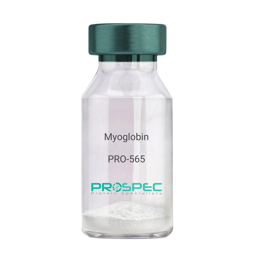
MyoglobinDescription:
Myoglobin Human
Myoglobin, MB, PVALB, MGC13548.
Product # :
PRO-565Price :
Quantity :
Shipping Method :
Shipped with Ice Packs
More Info
- description
- source
- formulation
- purity
- More Info
Description
Human Myoglobin produced in Human Cardiac Tissues having a molecular mass of 17.5kDa. Myoglobin is released from recently injured myocardial cells within a few hours of Infarction. Peak levels are reached more quickly than CK-MB or Troponin complex.
Source
Human Cardiac Tissues.
Formulation
The protein solution is in 0.05M phosphate buffer pH 7.5 containing 0.15M NaCl and 0.09% NaN3. Filtered through a 0.2µM membrane.
Purity
Greater than 96.0%.
More Info
-
Introduction
Myoglobin is a member of the globin superfamily and can be found in skeletal and cardiac muscles. It is a haemoprotein that contributs to intracellular oxygen storage and transcellular facilitated diffusion of oxygen. Myoglobin has a single-chain globular structure of 153 amino acids, containing a heme prosthetic group (iron-containing porphyrin) in the core around which the remaining apoprotein folds. Myoglobin has 8 alpha helices and a hydrophobic core. Myoglobin’s molecular weight is 16.7 kDa, and it is the primary oxygen-carrying pigment of muscle tissues. The binding of oxygen in myoglobin is different from the cooperative oxygen binding in hemoglobin, since positive collaboration is a property of multimeric/oligomeric proteins only. Instead, the binding of oxygen by myoglobin is uninfluenced by the oxygen pressure in the surrounding tissue. Myoglobin is frequently referred to as having an "instant binding tenacity" to oxygen given its hyperbolic oxygen dissociation curve. Different organisms are able to hold their breaths longer due to high concentrations of myoglobin in their muscle cells. Myoglobin is responsible for the pigments that make meat red. The color of the meat is partly determined by the charge of the iron atom in myoglobin and the oxygen attached to it. Myoglobin is found in Type I muscle, Type II A and Type II B, but it is mostly deemed that myoglobin is not found in smooth muscle. Myoglobin is discharged from damaged muscle tissue (rhabdomyolysis), which contains very high concentrations of myoglobin. Even though the released myoglobin is filtered by the kidneys, it is toxic to the renal tubular epithelium and thus may cause acute renal failure.
-
Synonyms
Myoglobin, MB, PVALB, MGC13548.
-
Physical Appearance
Sterile Filtered red solution.
-
Stability
Human Myoglobin should be stored at 2-8°C.
-
Human Virus Test
Starting material donor tested and certified negative for HIV I & II antibodies, Hepatitis B surface antigen, and Hepatitis C antibodies.
ProSpec's products are furnished for LABORATORY RESEARCH USE ONLY. The product may not be used as drugs, agricultural or pesticidal products, food additives or household chemicals.
- View Data Sheet
Name :
NQO1 Human, ActiveDescription:
NAD(P)H Dehydrogenase Quinone 1, Active 1 Human Recombinant
NAD(P)H dehydrogenase (quinone) 1, Quinone reductase 1, QR1, NAD(P)H:quinone oxidoreductase 1, DT-diaphorase, DTD, Azoreductase, Phylloquinone reductase, Menadione reductase, NQO1, DIA4, NMOR1, DHQU, NMORI.
Product # :
ENZ-1107Price :
Quantity :
Shipping Method :
Shipped with Ice Packs
More Info
- description
- source
- formulation
- purity
- biological activity
- More Info
Description
NQO1 Human Recombinant produced in E.Coli is a single, non-glycosylated, polypeptide chain containing 294 amino acids ( 1-274aa ) and having a molecular mass of 33.0 kDa. NQO1 is fused to a 20 amino acid His tag at N-terminus and purified by proprietary chromatographic techniques.
Source
Escherichia Coli.
Formulation
The NQO1 solution (1 mg/ml) contains 20mM Tris-HCl buffer (pH 8.0), 1mM DTT and 10% glycerol.
Purity
Greater than 95.0% as determined by SDS-PAGE.
Biological Activity
Specific activity is > 1,000 pmol/min/ug. One unit will convert 1 pmoles resazurin to resorufin per minute at pH 7.5 at 25°C.
More Info
-
Introduction
NQO1 belongs to the NAD(P)H dehydrogenase (quinone) family and encodes a cytoplasmic 2-electron reductase. NQO1 acts as an imperative part of cellular antioxidant defense by detoxifying quinines therefore preventing the formation of reactive oxygen species. It seems that NQO1 serves as a quinone reductase relating to conjugation reactions of hydroquinons involved in detoxification pathways in addition to biosynthetic processes such as the vitamin K-dependent gamma-carboxylation of glutamate residues in prothrombin synthesis. Altered NQO1 expression is seen in many tumors and also linked to Alzheimer’s disease. NQO1 gene mutations are linked to tardive dyskinesia which is an increased risk of hematotoxicity after exposure to benzene, and susceptibility to various forms of cancer.
-
Synonyms
NAD(P)H dehydrogenase (quinone) 1, Quinone reductase 1, QR1, NAD(P)H:quinone oxidoreductase 1, DT-diaphorase, DTD, Azoreductase, Phylloquinone reductase, Menadione reductase, NQO1, DIA4, NMOR1, DHQU, NMORI.
-
Physical Appearance
Sterile Filtered colorless solution.
-
Stability
Store at 4°C if entire vial will be used within 2-4 weeks. Store, frozen at -20°C for longer periods of time. For long term storage it is recommended to add a carrier protein (0.1% HSA or BSA).Avoid multiple freeze-thaw cycles.
-
Amino Acid Sequence
MGSSHHHHHH SSGLVPRGSH MVGRRALIVL AHSERTSFNY AMKEAAAAAL KKKGWEVVES
DLYAMNFNPI ISRKDITGKL KDPANFQYPA ESVLAYKEGH LSPDIVAEQK KLEAADLVIF
QFPLQWFGVP AILKGWFERV FIGEFAYTYA AMYDKGPFRS KKAVLSITTG GSGSMYSLQG
IHGDMNVILW PIQSGILHFC GFQVLEPQLT YSIGHTPADA RIQILEGWKK RLENIWDETP
LYFAPSSLFD LNFQAGFLMK KEVQDEEKNK KFGLSVGHHL GKSIPTDNQI KARK
ProSpec's products are furnished for LABORATORY RESEARCH USE ONLY. The product may not be used as drugs, agricultural or pesticidal products, food additives or household chemicals.
- View Data Sheet
Name :
EGF Mouse, His ActiveDescription:
Epidermal Growth Factor, His Active Mouse Recombinant
AI790464, Pro-epidermal growth factor, URG.
Product # :
CYT-1054Price :
Quantity :
Shipping Method :
Shipped with Ice Packs
More Info
- description
- source
- formulation
- purity
- biological activity
- More Info
- SDS-PAGE
Description
EGF Mouse Recombinant produced in E.Coli is a single, non-glycosylated polypeptide chain containing 77 amino acids (977-1029 a.a) and having a molecular mass of 8.6kDa.EGF is fused to a 24 amino acid His-tag at N-terminus & purified by proprietary chromatographic techniques.
Source
Escherichia Coli.
Formulation
EGF protein solution (0.25mg/ml) contains 10% glycerol, 20mM Tris-HCl (pH 8.0), 0.1M NaCl & 2mM DTT.
Purity
Greater than 95.0% as determined by SDS-PAGE.
Biological Activity
Measured in a cell proliferation assay using mouse Balb/3T3 cell. The ED50 for this effect less or equal to 1ng/ml.
SDS-PAGE
More Info
-
Introduction
Pro-Epidermal Growth Factor Isoform 1 or EGF, is a globular peptide (77aa residues) which includes three intra molecular disulfide bonds. This protein acts as a growth factor that mediates the growth and proliferation of different epithelial & epidermal cells. Among other processes that EGF is part of are inhibition of gastric secretion and wound healing. EGF is a ligand for class I tyrosine kinase receptor (c-erbB).
-
Synonyms
AI790464, Pro-epidermal growth factor, URG.
-
Physical Appearance
Sterile Filtered clear solution.
-
Stability
Store at 4°C if entire vial will be used within 2-4 weeks. Store, frozen at -20°C for longer periods of time. For long term storage it is recommended to add a carrier protein (0.1% HSA or BSA).Avoid multiple freeze-thaw cycles.
-
Amino Acid Sequence
MGSSHHHHHH SSGLVPRGSH MGSMNSYPGC PSSYDGYCLN GGVCMHIESL DSYTCNCVIG YSGDRCQTRD LRWWELR.
-
Background
Deciphering Epidermal Growth Factor Signaling: Unveiling the Potential of His-Tagged Active Mouse Recombinant
Abstract:
This research paper delves into the intricate realm of Epidermal Growth Factor (EGF) signaling, focusing on the novel application of Histidine (His)-tagged Active Mouse Recombinant EGF. By employing sophisticated methodologies encompassing protein engineering, receptor binding assays, and cellular response analyses, this study sheds light on the multifaceted molecular attributes and therapeutic prospects of this innovative variant.
Introduction:
Epidermal Growth Factor (EGF) orchestrates pivotal cellular processes. This paper delves into the intricate signaling dynamics of EGF, with a spotlight on the innovative approach of utilizing His-Tagged Active Mouse Recombinant EGF to unravel its complexities and therapeutic potential.
Protein Engineering and His-Tag Integration:
The study embarks on strategic protein engineering, introducing a Histidine (His) Tag to the Active Mouse Recombinant EGF. This His-Tag facilitates purification and subsequent analyses, enabling a comprehensive exploration of EGF signaling.
Receptor Binding Assays and Ligand Interaction:
Advanced receptor binding assays unravel the nuances of EGF's interaction with its cognate receptor, including affinities and kinetics. By employing His-Tagged EGF, the study dissects the impact of the tag on receptor binding, shedding light on its potential implications on downstream signaling.
Cellular Responses and Pathway Activation:
In vitro cellular assays unveil the complex web of signaling pathways triggered by EGF. The study employs high-throughput techniques to investigate the intricate cascades initiated by His-Tagged Active Mouse Recombinant EGF, providing insights into its potential role in cell proliferation, migration, and survival.
Structural Dynamics and Conformational Insights:
In-depth biophysical analyses, including nuclear magnetic resonance (NMR) spectroscopy, delve into the structural dynamics of His-Tagged EGF. This sheds light on potential conformational changes induced by the tag and their impact on receptor binding affinity.
Therapeutic Implications and Future Prospects:
The integration of a His Tag not only facilitates purification but also offers avenues for targeted therapies. His-Tagged EGF could serve as a platform for tailored drug delivery, enhancing the precision of interventions in various pathologies.
Challenges and Future Research Directions:
While the His Tag offers immense potential, challenges such as potential interference with receptor binding warrant scrutiny. Future research should focus on optimizing the positioning of the tag and exploring its impact on downstream signaling cascades.
Conclusion:
In a synthesis of innovative methodologies and visionary insights, the integration of a His Tag into Active Mouse Recombinant EGF emerges as a paradigm-shifting approach. This technique not only enriches our understanding of EGF signaling dynamics but also presents exciting prospects for personalized therapeutic interventions.
What is the molecular weight/Mw of EGF MOUSE, HIS ACTIVE Protein?
EGF MOUSE, HIS ACTIVE Protein has a total Mw of 8.6kDa.
What is the source or expression system of EGF MOUSE, HIS ACTIVE Protein?
Escherichia Coli.
What is the Purity of EGF MOUSE, HIS ACTIVE Protein?
EGF MOUSE, HIS ACTIVE Protein is >95% pure as determined by SDS-PAGE.
What is the Biological Activity of EGF MOUSE, HIS ACTIVE Protein?
Measured in a cell proliferation assay using mouse Balb/3T3 cell. The ED50 for this effect less or equal to 1ng/ml.
What is the amino acid sequence of EGF MOUSE, HIS ACTIVE Protein?
MGSSHHHHHH SSGLVPRGSH MGSMNSYPGC PSSYDGYCLN GGVCMHIESL DSYTCNCVIG YSGDRCQTRD LRWWELR.
What applications can EGF MOUSE, HIS ACTIVE Protein be used in?
EGF MOUSE, HIS ACTIVE Protein can probably be used in western blot, ELISA and Lateral Flow.
What is the endotoxin level for EGF MOUSE, HIS ACTIVE Protein?
The endotoxin level is minimal, EGF MOUSE, HIS ACTIVE Protein was purified using conventional chromatography techniques..
ProSpec's products are furnished for LABORATORY RESEARCH USE ONLY. The product may not be used as drugs, agricultural or pesticidal products, food additives or household chemicals.
- View Data Sheet
Name :
HAO1 Human, ActiveDescription:
Hydroxyacid Oxidase 1 Human Recombinant, Active
Hydroxyacid oxidase 1, HAOX1, Glycolate oxidase, GOX, HAO1, GOX1.
Product # :
ENZ-1094Price :
Quantity :
Shipping Method :
Shipped with Ice Packs
More Info
- description
- source
- formulation
- purity
- biological activity
- More Info
Description
HAO1 Human Recombinant produced in E.Coli is a single, non-glycosylated polypeptide chain containing 406 amino acids (1-370 a.a) and having a molecular mass of 45.0kDa. HAO1 is fused to a 36 amino acid His-tag at N-terminus & purified by proprietary chromatographic techniques.
Source
Escherichia Coli.
Formulation
HAO1 protein solution (1mg/ml) contains 20% glycerol, 20mM Tris-Hcl (pH8.0) and 0.5M NaCl.
Purity
Greater than 95% as determined by SDS-PAGE.
Biological Activity
Specific activity is > 3000 pmol/min/ug, and defined as the amount of enzyme that oxidize glyoxylate at pH 8.0 at 25C.
More Info
-
Introduction
Glycolate oxidase (HAO1) is a part of the superfamily of the alpha hydroxy acid oxidases (HAO) enzymes. HAO1 catalyses the FMN mediated oxidation of glycolate to glyoxylate and glyoxylate to oxalate by reducing oxygen to hydrogen peroxide. HAO1 is expressed mainly in the liver and pancreas and is most active on twocarbon substrates such as glycolate. HAO1 isthe main cause of hyperoxaluria, a disorder in which large deposits of calcium oxalate form kidney stones.
-
Synonyms
Hydroxyacid oxidase 1, HAOX1, Glycolate oxidase, GOX, HAO1, GOX1.
-
Physical Appearance
Sterile filtered yellowish solution.
-
Stability
Store at 4°C if entire vial will be used within 2-4 weeks. Store, frozen at -20°C for longer periods of time. For long term storage it is recommended to add a carrier protein (0.1% HSA or BSA).Avoid multiple freeze-thaw cycles.
-
Amino Acid Sequence
MRGSHHHHHH GMASMTGGQQ MGRDLYDDDD KDRWGSMLPR LICINDYEQH AKSVLPKSIY DYYRSGANDE ETLADNIAAF SRWKLYPRMLRNVAETDLST SVLGQRVSMP ICVGATAMQR MAHVDGELAT VRACQSLGTG MMLSSWATSS IEEVAEAGPE ALRWLQLYIY KDREVTKKLVRQAEKMGYKA IFVTVDTPYL GNRLDDVRNR FKLPPQLRMK NFETSTLSFS PEENFGDDSG LAAYVAKAID PSISWEDIKW LRRLTSLPIVAKGILRGDDA REAVKHGLNG ILVSNHGARQ LDGVPATIDV LPEIVEAVEG KVEVFLDGGV RKGTDVLKAL ALGAKAVFVG RPIVWGLAFQGEKGVQDVLE ILKEEFRLAM ALSGCQNVKV IDKTLVRKNP LAVSKI.
ProSpec's products are furnished for LABORATORY RESEARCH USE ONLY. The product may not be used as drugs, agricultural or pesticidal products, food additives or household chemicals.
- View Data Sheet
Name :
ANGPTL3 (PAT23D8AT) AntibodyDescription:
Mouse Anti Human Angiopoietin Like Protein 3 Clone PAT23D8AT
Angiopoietin Like 3, Angiopoietin 5, ANGPT5, ANG-5, Angiopoietin-Related Protein 3 , Angiopoietin-Like Protein 3, Angiopoietin-Like 3, Angiopoietin-5, FHBL2, ANL3.
Product # :
ANT-709Price :
Quantity :
Shipping Method :
Shipped with Ice Packs
More Info
- formulation
- More Info
Formulation
1mg/ml containing PBS, pH-7.4, 10% Glycerol and 0.02% Sodium Azide.
More Info
-
Introduction
ANGPTL3 and ANGPTL4 are angiopoietin-like proteins secreted and expressed mainly by the liver, their role being the regulation of triglyceride metabolism by inhibiting the lipolysis of triglyceride-rich lipoproteins. During different nutritional states (feeding/fasting) the levels of the circulating triglycerides are regulated by Angptl3 and Angptl4 through differential inhibition of Lipoprotein lipase (LPL) as shown by the experimental data. The molecular structure of ANGPTL3 is similar to that of the angiopoietins (vascular endothelial growth factors). Deletion mutants of human Angiopoietin 5 were used in order to demonstrate that the N-terminal domain (fragment 17-207) and not the C-terminal fibrinogen-like domain (fragment 207-460) increased the plasma triglyceride levels in mice.
-
Synonyms
Angiopoietin Like 3, Angiopoietin 5, ANGPT5, ANG-5, Angiopoietin-Related Protein 3 , Angiopoietin-Like Protein 3, Angiopoietin-Like 3, Angiopoietin-5, FHBL2, ANL3.
-
Physical Appearance
Sterile filtered colorless solution.
-
Immunogen
Anti-human ANGPTL3 mAb, is derived from hybridization of mouse F0 myeloma cells with spleen cells from BALB/c mice immunized with a recombinant human ANGPTL3 protein 243-460 amino acids purified from E. coli.
-
Ig Subclass
Mouse IgG2a heavy chain and κ light chain.
-
Clone
PAT23D8AT.
-
Applications
ANGPTL3 antibody has been tested by ELISA, Western blot analysis and ICC/IF to assure specificity and reactivity. Since application varies, however, each investigation should be titrated by the reagent to obtain optimal results.
-
Type
Mouse Anti Human Monoclonal.
-
Storage Procedures
For periods up to 1 month store at 4°C, for longer periods of time, store at -20°C. Prevent freeze thaw cycles.
-
Purification Method
ANGPTL3 antibody was purified by protein-A affinity chromatography.
ProSpec's products are furnished for LABORATORY RESEARCH USE ONLY. The product may not be used as drugs, agricultural or pesticidal products, food additives or household chemicals.
- View Data Sheet
Name :
OAT AntibodyDescription:
Ornithine aminotransferase, Mouse Anti Human
Ornithine aminotransferase mitochondrial, Ornithine delta-aminotransferase, Ornithine-oxo-acid aminotransferase, OAT, OKT, GACR, HOGA, OATASE, DKFZp781A11155.
Product # :
ANT-013Price :
Quantity :
Shipping Method :
Shipped with Ice Packs
More Info
- formulation
- More Info
Formulation
1mg/ml containing PBS, pH-7.4, & 0.1% Sodium Azide.
More Info
-
Introduction
Ornithine aminotransferase is a key enzyme in the pathway that converts arginine and ornithine into the major excitatory and inhibitory neurotransmitters glutamate and GABA. Ornithine aminotransferase (OAT) is a 49kDa nucleus-encoded protein imported into mitochondria to give the mature 48kDa OAT polypeptide. It is found in humans, animals, insects, plants and microorganisms. The OAT has a sex-differential expression in the mouse kidney. OAT plays central physiological roles in amino acid metabolism. OAT shows a large structural and mechanistic similarity to other enzymes from the subgroup III of aminotransferases that transfer an amino group from a carbon atom which doesn’t carry a carboxyl function. OAT is vital for nitrogen recycling from arginine but not for the stress-induced proline accumulation. OAT enzyme deficiency causes the autosomal recessive eye disease Gyrate Atrophy.
-
Synonyms
Ornithine aminotransferase mitochondrial, Ornithine delta-aminotransferase, Ornithine-oxo-acid aminotransferase, OAT, OKT, GACR, HOGA, OATASE, DKFZp781A11155.
-
Immunogen
Anti-human OAT mAb, is derived from hybridization of mouse F0 myeloma cells with spleen cells from BALB/c mice immunized with recombinant human OAT amino acids 33-439 purified from E. coli.
-
Ig Subclass
Mouse IgG1 heavy chain and κ light chain.
-
Applications
OAT antibody has been tested by by ELISA, Western blot and Immunofluorescence analysis to assure specificity and reactivity. Since application varies, however, each investigation should be titrated by the reagent to obtain optimal results. Recommended dilution range for Western blot analysis and Immunofluorescence is 1:250 ~ 500.
Recommended starting dilution is 1:250. -
Type
PAT23A2AT.
-
Storage Procedures
For periods up to 1 month store at 4°C, for longer periods of time, store at -20°C. Prevent freeze thaw cycles.
-
Purification Method
OAT antibody was purified from mouse ascitic fluids by protein-G affinity chromatography.
ProSpec's products are furnished for LABORATORY RESEARCH USE ONLY. The product may not be used as drugs, agricultural or pesticidal products, food additives or household chemicals.