Skip to content

High-quality research proteins.

  • 1-800-805-6531

  • Mon - Sat 8.00-18.00, Sun - Closed

  • info@prospecbio.com

  • PO Box 6591 East Brunswick NJ 08816

  • Products
    • Cytokines
    • Growth factors
    • Chemokines
    • Cd antigens
    • Neurotrophins
    • Hormones
    • Enzymes
    • Viral antigens
    • Recombinant proteins
    • Natural proteins
    • Monoclonal antibodies
    • Polyclonal antibodies
    • Thermal Packaging
  • About
    • Company
    • Founder
    • Contribution
  • Customers
    • Eastern Asia
    • Eastern Europe
    • North America
    • South America
    • South East Asia
    • Southern Asia
    • Western Europe
  • Ordering
    • Ordering Method
    • Ordering - Credit Card
    • Ordering - PO
    • Shipping Fees
    • FAQs
  • Services
    • DNA Cloning
    • Protein Expression
    • Monoclonal Antibody
    • Fermentation
    • Technology License
  • Contact us
Log in

Country/region

  • USD $ | Albania
  • USD $ | Andorra
  • USD $ | Argentina
  • USD $ | Armenia
  • USD $ | Australia
  • USD $ | Austria
  • USD $ | Azerbaijan
  • USD $ | Belarus
  • USD $ | Belgium
  • USD $ | Bolivia
  • USD $ | Brazil
  • USD $ | Bulgaria
  • USD $ | Cambodia
  • USD $ | Canada
  • USD $ | Chile
  • USD $ | China
  • USD $ | Colombia
  • USD $ | Costa Rica
  • USD $ | Croatia
  • USD $ | Cyprus
  • USD $ | Czechia
  • USD $ | Denmark
  • USD $ | Estonia
  • USD $ | Finland
  • USD $ | France
  • USD $ | Georgia
  • USD $ | Germany
  • USD $ | Greece
  • USD $ | Hong Kong SAR
  • USD $ | Hungary
  • USD $ | Iceland
  • USD $ | India
  • USD $ | Ireland
  • USD $ | Israel
  • USD $ | Italy
  • USD $ | Japan
  • USD $ | Kazakhstan
  • USD $ | Latvia
  • USD $ | Liechtenstein
  • USD $ | Lithuania
  • USD $ | Madagascar
  • USD $ | Malta
  • USD $ | Mexico
  • USD $ | Moldova
  • USD $ | Monaco
  • USD $ | Netherlands
  • USD $ | New Zealand
  • USD $ | North Macedonia
  • USD $ | Norway
  • USD $ | Panama
  • USD $ | Paraguay
  • USD $ | Peru
  • USD $ | Philippines
  • USD $ | Poland
  • USD $ | Portugal
  • USD $ | Romania
  • USD $ | Singapore
  • USD $ | Slovakia
  • USD $ | Slovenia
  • USD $ | South Africa
  • USD $ | South Korea
  • USD $ | Spain
  • USD $ | Sri Lanka
  • USD $ | Sweden
  • USD $ | Switzerland
  • USD $ | Taiwan
  • USD $ | Thailand
  • USD $ | Türkiye
  • USD $ | Ukraine
  • USD $ | United Kingdom
  • USD $ | United States
  • USD $ | Uruguay
  • USD $ | Vatican City
  • USD $ | Venezuela
  • USD $ | Vietnam
  • Facebook
  • Instagram
logoProSpec - Recombinant Protein Manufacturer for academic and R&D industry
Become a distributor
Account Log in
Wishlist
Cart Cart(0)
  • Products
    +
    • Cytokines
    • Growth factors
    • Chemokines
    • Cd antigens
    • Neurotrophins
    • Hormones
    • Enzymes
    • Viral antigens
    • Recombinant proteins
    • Natural proteins
    • Monoclonal antibodies
    • Polyclonal antibodies
    • Thermal Packaging
  • About
    +
    • Company
    • Founder
    • Contribution
  • Customers
    +
    • Eastern Asia
    • Eastern Europe
    • North America
    • South America
    • South East Asia
    • Southern Asia
    • Western Europe
  • Ordering
    +
    • Ordering Method
    • Ordering - Credit Card
    • Ordering - PO
    • Shipping Fees
    • FAQs
  • Services
    +
    • DNA Cloning
    • Protein Expression
    • Monoclonal Antibody
    • Fermentation
    • Technology License
  • Contact us

Item added to your cart

View cart
View All
  • Cytokines
  • Growth factors
  • Chemokines
  • Cd antigens
  • Neurotrophins
  • Hormones
  • Enzymes
  • Viral antigens
  • Recombinant proteins
  • Natural proteins
  • Monoclonal antibodies
  • Polyclonal antibodies
  • Thermal Packaging
  • Tumor Necrosis Factor

    Tumor Necrosis Factor

    View All
  • 4-1BB

    4-1BB

    View All
  • Adiponectin

    Adiponectin

    View All
  • AIF1

    AIF1

    View All
  • Other Cytokines

    Other Cytokines

    View All
  • AITRL

    AITRL

    View All
  • Angiopoietin

    Angiopoietin

    View All
  • Apolipoprotein

    Apolipoprotein

    View All
  • B-Cell Activating Factor

    B-Cell Activating Factor

    View All
  • Beta Defensin

    Beta Defensin

    View All
  • Bone Morphogenetic Protein

    Bone Morphogenetic Protein

    View All
  • B type Natriuretic Peptide

    B type Natriuretic Peptide

    View All
  • BST

    BST

    View All
  • Betacellulin

    Betacellulin

    View All
  • Cardiotrophin

    Cardiotrophin

    View All
  • Activin

    Activin

    View All
  • Fibroblast Growth Factor

    Fibroblast Growth Factor

    View All
  • Other Growth Factors

    Other Growth Factors

    View All
  • CSF

    CSF

    View All
  • CTGF

    CTGF

    View All
  • IGFBP

    IGFBP

    View All
  • VEGF Protein

    VEGF Protein

    View All
  • EGF

    EGF

    View All
  • Epigen

    Epigen

    View All
  • Erythropoietin

    Erythropoietin

    View All
  • Insulin-Like Growth Factor

    Insulin-Like Growth Factor

    View All
  • Growth Hormone

    Growth Hormone

    View All
  • HDGF

    HDGF

    View All
  • Hepatocyte Growth Factor

    Hepatocyte Growth Factor

    View All
  • Insulin

    Insulin

    View All
  • BCA-1/ BLC (CXCL13)

    BCA-1/ BLC (CXCL13)

    View All
  • BRAK (CXCL14)

    BRAK (CXCL14)

    View All
  • C-10 (CCL6)

    C-10 (CCL6)

    View All
  • Other Chemokines

    Other Chemokines

    View All
  • MEC (CCL28)

    MEC (CCL28)

    View All
  • LD78-beta (CCL3L1)

    LD78-beta (CCL3L1)

    View All
  • CTACK (CCL27)

    CTACK (CCL27)

    View All
  • CXCL16

    CXCL16

    View All
  • CXCL17

    CXCL17

    View All
  • Platelet Factor-4 (CXCL4)

    Platelet Factor-4 (CXCL4)

    View All
  • ENA-78 (CXCL5)

    ENA-78 (CXCL5)

    View All
  • Eotaxin (CCL11,24,26)

    Eotaxin (CCL11,24,26)

    View All
  • Exodus-2 (CCL21)

    Exodus-2 (CCL21)

    View All
  • Fractalkine (CX3CL1)

    Fractalkine (CX3CL1)

    View All
  • CXCL6

    CXCL6

    View All
  • Other CD Antigens

    Other CD Antigens

    View All
  • CD14

    CD14

    View All
  • CD164

    CD164

    View All
  • CD1

    CD1

    View All
  • CD2

    CD2

    View All
  • CD200

    CD200

    View All
  • CD207

    CD207

    View All
  • CD226

    CD226

    View All
  • CD23

    CD23

    View All
  • CD244

    CD244

    View All
  • CD247

    CD247

    View All
  • CD27

    CD27

    View All
  • CD274

    CD274

    View All
  • CD300

    CD300

    View All
  • CD33

    CD33

    View All
  • Other Neurotrophins

    Other Neurotrophins

    View All
  • Beta-NGF

    Beta-NGF

    View All
  • BDNF

    BDNF

    View All
  • CDNF

    CDNF

    View All
  • CNTF

    CNTF

    View All
  • GDNF

    GDNF

    View All
  • Glia Maturation Factor

    Glia Maturation Factor

    View All
  • MANF

    MANF

    View All
  • Midkine

    Midkine

    View All
  • Neuroglobin

    Neuroglobin

    View All
  • Neurotrophic factors

    Neurotrophic factors

    View All
  • Neuregulin

    Neuregulin

    View All
  • Neuropilin

    Neuropilin

    View All
  • Pigment Epithelium-Derived Factor

    Pigment Epithelium-Derived Factor

    View All
  • Pleiotrophin

    Pleiotrophin

    View All
  • Peptide Hormones

    Peptide Hormones

    View All
  • Other Hormones

    Other Hormones

    View All
  • HCG

    HCG

    View All
  • Endothelin

    Endothelin

    View All
  • Exendin

    Exendin

    View All
  • FSH

    FSH

    View All
  • Glucagon

    Glucagon

    View All
  • GHRP

    GHRP

    View All
  • GLP

    GLP

    View All
  • Thyrostimulin

    Thyrostimulin

    View All
  • Inhibin A

    Inhibin A

    View All
  • LHRH

    LHRH

    View All
  • Melanotan

    Melanotan

    View All
  • Procalcitonin

    Procalcitonin

    View All
  • PTH

    PTH

    View All
  • Peroxiredoxin

    Peroxiredoxin

    View All
  • Peptidase

    Peptidase

    View All
  • Synthetase

    Synthetase

    View All
  • Transferase

    Transferase

    View All
  • Hydrolase

    Hydrolase

    View All
  • Dehydrogenase

    Dehydrogenase

    View All
  • ACE2

    ACE2

    View All
  • Esterase

    Esterase

    View All
  • Protein Kinases

    Protein Kinases

    View All
  • Other Enzymes

    Other Enzymes

    View All
  • Phosphatase

    Phosphatase

    View All
  • Deaminase

    Deaminase

    View All
  • Oxygenase

    Oxygenase

    View All
  • Adenylate Kinase

    Adenylate Kinase

    View All
  • Lyase

    Lyase

    View All
  • Chagas

    Chagas

    View All
  • SARS

    SARS

    View All
  • Influenza

    Influenza

    View All
  • ASFV

    ASFV

    View All
  • Astrovirus

    Astrovirus

    View All
  • Borrelia

    Borrelia

    View All
  • Helicobacter Pylori

    Helicobacter Pylori

    View All
  • Chikungunya

    Chikungunya

    View All
  • Chlamydia

    Chlamydia

    View All
  • Cytomegalo

    Cytomegalo

    View All
  • Dengue

    Dengue

    View All
  • EBV

    EBV

    View All
  • Ebola

    Ebola

    View All
  • Feline Leukemia Virus

    Feline Leukemia Virus

    View All
  • Hepatitis A

    Hepatitis A

    View All
  • Other

    Other

    View All
  • Actin

    Actin

    View All
  • ADAM

    ADAM

    View All
  • Complement Component

    Complement Component

    View All
  • Ag85

    Ag85

    View All
  • Eukaryotic Translation Initiation Factor

    Eukaryotic Translation Initiation Factor

    View All
  • Anterior Gradient Protein

    Anterior Gradient Protein

    View All
  • Heat Shock Protein

    Heat Shock Protein

    View All
  • Alpha-2-HS-Glycoprotein

    Alpha-2-HS-Glycoprotein

    View All
  • Hemoglobin

    Hemoglobin

    View All
  • Allergy

    Allergy

    View All
  • Anaplasma

    Anaplasma

    View All
  • Angiogenin

    Angiogenin

    View All
  • Ankyrin Repeat Domain

    Ankyrin Repeat Domain

    View All
  • Annexin

    Annexin

    View All
  • Other Natural Proteins

    Other Natural Proteins

    View All
  • Aprotinin

    Aprotinin

    View All
  • Transferrin

    Transferrin

    View All
  • Avidin

    Avidin

    View All
  • Anti Coagulation Factors

    Anti Coagulation Factors

    View All
  • Natural Albumin

    Natural Albumin

    View All
  • Natural Coagulation Factors

    Natural Coagulation Factors

    View All
  • Fibronectin

    Fibronectin

    View All
  • Thrombin

    Thrombin

    View All
  • Anti Human Enzyme

    Anti Human Enzyme

    View All
  • Other Monoclonal Antibodies

    Other Monoclonal Antibodies

    View All
  • Anti Human Cytokine

    Anti Human Cytokine

    View All
  • Anti Human Heat Shock Protein

    Anti Human Heat Shock Protein

    View All
  • Anti Mouse Lymphocyte

    Anti Mouse Lymphocyte

    View All
  • Anti Human Chemokine

    Anti Human Chemokine

    View All
  • Anti Human Lymphocyte

    Anti Human Lymphocyte

    View All
  • Anti Viral Monoclonal

    Anti Viral Monoclonal

    View All
  • Anti Mouse Cytokine

    Anti Mouse Cytokine

    View All
  • Anti-GST Monoclonal

    Anti-GST Monoclonal

    View All
  • Other Polyclonal Antibodies

    Other Polyclonal Antibodies

    View All
  • Anti Viral

    Anti Viral

    View All
  • Anti-GST Monoclonal

    Anti-GST Monoclonal

    View All
Back

Search results

1000 results found for “CDNF”

Name

Description

Product #

Price

Quantity

Shipping Method

  • View Data Sheet

    Name :

    TGFB3 Human, CHO

    Description:

    Transforming Growth Factor-Beta 3 Human Recombinant, CHO

    Transforming Growth Factor-beta3, TGFB3, ARVD, FLJ16571, TGF-beta3.

    Product # :

    CYT-1259

    Price :

    Quantity :

    Shipping Method :

    Room Temp Icon

    Shipped at Room temp

    Add To Cart

    More Info

    • description
    • source
    • formulation
    • purity
    • biological activity
    • More Info

    Description

    TGFB3 Human Recombinant produced in CHO cells is a glycosylated, polypeptide homodimer chain containing 2x112 amino acids and having a total molecular mass of 25.0kDa. The TGFB3 is purified by proprietary chromatographic techniques.

    Source

    CHO cells.

    Formulation

    TGFB3 was lyophilized from a concentrated solution containing 35% (v/v) Acetonitrile and 0.1% (v/v) TFA.

    Purity

    Greater than 97.0% as determined by: (a) Analysis by HPLC. (b) Analysis by SDS-PAGE.

    Biological Activity

    The ED50 as determined by its ability to inhibit the mouse IL-4-dependent proliferation of mouse HT-2 cells is less than 0.05 ng/ml, corresponding to a specific activity of ≥ 2.0 × 10⁷ units/mg.

    More Info

    • Synonyms

      Transforming Growth Factor-beta3, TGFB3, ARVD, FLJ16571, TGF-beta3.

    • Physical Appearance

      Sterile Filtered White lyophilized (freeze-dried) powder.

    • Stability

      Lyophilized Transforming Growth Factor-Beta 3 Recombinant although stable at room temperature for 3 weeks, should be stored desiccated below -18°C. Upon reconstitution TGFB3 should be stored at 4°C between 2-7 days and for future use below -18°C. For long term storage it is recommended to add a carrier protein (0.1% HSA or BSA). Please prevent freeze-thaw cycles.

    • Solubility

      It is recommended to reconstitute the lyophilized TGFB3 in sterile 4mM HCl to a concentration of 0.1-1.0 mg/mL. Stock solutions should be apportioned into working aliquots and stored at ≤-20°C which can then be further diluted to other aqueous solutions.

    • Amino Acid Sequence

      ALDTNYCFRN LEENCCVRPL YIDFRQDLGW KWVHEPKGYY ANFCSGPCPY LRSADTTHST VLGLYNTLNP EASASPCCVP QDLEPLTILY YVGRTPKVEQ LSNMVVKSCK CS.

    • Background

      Transforming growth factor betas (TGF Betas) mediate many cell-cell interactions that occur during embryonic development. 3 TGF Betas have been identified in mammals: TGF Beta 1, TGF Beta 2 and TGF Beta 3. each are synthesized as precursor proteins that are very similar in that each is cleaved to yield a 112 amino acid polypeptide that remains associated with the latent portion of the molecule.

    ProSpec's products are furnished for LABORATORY RESEARCH USE ONLY. The product may not be used as drugs, agricultural or pesticidal products, food additives or household chemicals.

    Tgfb3 Human
  • View Data Sheet

    Name :

    IGF1 Bovine

    Description:

    IGF-1 Bovine Recombinant

    Somatomedin C, IGF-I, IGFI, IGF1, IGF-IA MGF.

    Product # :

    CYT-1261

    Price :

    Quantity :

    Shipping Method :

    Room Temp Icon

    Shipped at Room temp

    Add To Cart

    More Info

    • description
    • source
    • formulation
    • purity
    • biological activity
    • More Info

    Description

    IGF1 Bovine Recombinant produced in E.Coli is a single, non-glycosylated, polypeptide chain containing 70 amino acids and having a molecular mass of 7.6kDa. IGF- I is purified by proprietary chromatographic techniques.

    Source

    Escherichia Coli.

    Formulation

    The IGF1 Bovine was lyophilized from a 0.2µm filtered concentrated solution in PBS, pH 7.0.

    Purity

    Greater than 97.0% as determined by:
    (a) Analysis by RP-HPLC.
    (b) Analysis by SDS-PAGE.

    Biological Activity

    The biological activity was determined by the cell proliferation assay using serum free human MCF-7 cells in <2ng/ml, corresponding to a Specific Activity of >5.0 x 10⁵IU/mg.

    More Info

    • Introduction

      The somatomedins, IGFs, comprise a family of peptides that play important roles in mammalian growth and development. IGF1 mediates many of the growth-promoting effects of GH. Early studies showed that GH did not directly stimulate the incorporation of sulfate into cartilage, but rather acted through a serum factor, termed 'sulfation factor,' which later became known as 'somatomedin. Three main somatomedins have been characterized: somatomedin C (IGF1), somatomedin A (IGF2; MIM 147470), and somatomedin B.

    • Synonyms

      Somatomedin C, IGF-I, IGFI, IGF1, IGF-IA MGF.

    • Physical Appearance

      Sterile Filtered White lyophilized (freeze-dried) powder.

    • Stability

      Lyophilized IGF-1 Bovine although stable at room temperature for 3 weeks, should be stored desiccated below -18°C. Upon reconstitution bovine IGF1 should be stored at 4°C between 2-7 days and for future use below -18°C. For long term storage it is recommended to add a carrier protein (0.1% HSA or BSA). Please prevent freeze-thaw cycles.

    • Solubility

      It is recommended to reconstitute the lyophilized bovine IGF-1 in sterile 18MΩ-cm H2O at a concentration of 100µg/ml, which can then be further diluted to other aqueous solutions.

    • Amino Acid Sequence

      GPETLCGAEL VDALQFVCGD RGFYFNKPTG YGSSSRRAPQ TGIVDECCFR SCDLRRLEMY CAPLKPAKSA.

    • Background

      What is the molecular weight/Mw of IGF1 BOVINE Protein?
      IGF1 BOVINE Protein has a total Mw of 7.6kDa.

      What is the source or expression system of IGF1 BOVINE Protein?
      Escherichia Coli.

      What is the Purity of IGF1 BOVINE Protein?
      IGF1 BOVINE Protein is >97% pure as determined by SDS-PAGE.

      What is the Biological Activity of IGF1 BOVINE Protein?
      The biological activity was determined by the cell proliferation assay using serum free human MCF-7 cells in <2ng/ml, corresponding to a Specific Activity of >5.0 x 10⁵IU/mg.

      What is the amino acid sequence of IGF1 BOVINE Protein?
      GPETLCGAEL VDALQFVCGD RGFYFNKPTG YGSSSRRAPQ TGIVDECCFR SCDLRRLEMY CAPLKPAKSA.
      What applications can IGF1 BOVINE Protein be used in?
      IGF1 BOVINE Protein can probably be used in western blot, ELISA and Lateral Flow.

      What is the endotoxin level for IGF1 BOVINE Protein?
      The endotoxin level is minimal, IGF1 BOVINE Protein was purified using conventional chromatography techniques.


    ProSpec's products are furnished for LABORATORY RESEARCH USE ONLY. The product may not be used as drugs, agricultural or pesticidal products, food additives or household chemicals.

    Igf1 Bovine
  • View Data Sheet

    Name :

    PDX-1 antibody

    Description:

    Pancreas Duodenum Homeobox-1, Polyclonal Rabbit Antibody

    IPF1, IUF1, IDX-1, MODY4, PDX1, STF-1, Insulin promoter factor 1, Pancreas Duodenum Homeobox-1.

    Product # :

    ANT-162

    Price :

    Quantity :

    Shipping Method :

    Room Temp Icon

    Shipped at Room temp

    Add To Cart

    More Info

    • More Info

    More Info

    • Introduction

      PDX1 activates insulin, somatostatin, glucokinase, islet amyloid polypeptide and glucose transporter type 2 gene transcription. IPF1 is particularly involved in glucose-dependent regulation of insulin gene transcription. Pancreas Duodenum Homeobox-1 binds preferentially the DNA motif 5'-[ct]taat[tg]-3'. During development, specifies the early pancreatic epithelium, permitting its proliferation, branching and subsequent differentiation. At adult stage, it is required for maintaining the hormone-producing phenotype of the beta-cell.

    • Synonyms

      IPF1, IUF1, IDX-1, MODY4, PDX1, STF-1, Insulin promoter factor 1, Pancreas Duodenum Homeobox-1.

    • Stability

      Stable for 2 yeas at -20°C. Avoid freeze/thaw cycles.

    • Immunogen

      Recombinant PDX1 E.Coli derived.

    • Applications

      Western Blot: 1:1,000
      Immunohistochemistry: 1:500
      Optimal working dilutions must be determined by end user.
      Routinely evaluated by western blot on lysates of rat pancreatic cell line INS-1

    • Molecular Weight

      35-40 kDa.

    • Specificity

      Human and Mouse PDX-1.

    • Type

      Polyclonal Rabbit Antibody.

    ProSpec's products are furnished for LABORATORY RESEARCH USE ONLY. The product may not be used as drugs, agricultural or pesticidal products, food additives or household chemicals.

    Pdx 1 Antibody
  • View Data Sheet

    Name :

    CD72 Human

    Description:

    CD72 Human Recombinant

    B-cell differentiation antigen CD72, Lyb-2, CD72, LYB2, CD72b.

    Product # :

    PRO-486

    Price :

    Quantity :

    Shipping Method :

    Ice Icon

    Shipped with Ice Packs

    Add To Cart

    More Info

    • description
    • source
    • formulation
    • More Info

    Description

    CD72 Human Recombinant (aa 1-98) expressed in E.coli, shows a 38 kDa band on SDS-PAGE. The CD72 is purified by proprietary chromatographic techniques.

    Source

    Escherichia Coli.

    Formulation

    CD72 at 100µg/ml in 50mM Tris-HCl, pH7.5, 10mM L-glutathione (reduced).

    More Info

    • Introduction

      CD72 antigen is a member of the type II integral membrane glycoproteins which includes other related cell surface moleculaes such as the asialoglycoprotein receptors, CD23 and the Kupffer cell recpeotr. The function of CD72 is unknown but the exposure of B cells to CD72 antibodies activates a variety of signaling pathways and can induce MHC class II expression and B cell proliferation. CD72 antigen is expressed on all cells of B cell lineage with the exception of plasma cells and weakly on human tissue macrophages.

    • Synonyms

      B-cell differentiation antigen CD72, Lyb-2, CD72, LYB2, CD72b.

    • Physical Appearance

      Sterile Filtered clear solution.

    • Stability

      Store vial at -20°C to -80°C. When stored at the recommended temperature, this protein is stable for 12 months.Please prevent freeze-thaw cycles.

    • Applications

      • ELISA.
      • Inhibition Assays.
      • Western Blotting.

    ProSpec's products are furnished for LABORATORY RESEARCH USE ONLY. The product may not be used as drugs, agricultural or pesticidal products, food additives or household chemicals.

    Cd72 Human
  • View Data Sheet

    Name :

    MFGE8 Mouse

    Description:

    Milk Fat Globule-EGF Factor 8 Protein Mouse Recombinant

    Milk fat globule-EGF factor 8 protein, isoform CRA_a, Putative uncharacterized protein, Mfge8, mCG_6301.

    Product # :

    CYT-1000

    Price :

    Quantity :

    Shipping Method :

    Ice Icon

    Shipped with Ice Packs

    Add To Cart

    More Info

    • description
    • source
    • formulation
    • purity
    • More Info

    Description

    MFGE8 Mouse Recombinant produced in Sf9 Baculovirus cells is a single, glycosylated polypeptide chain containing 413 amino acids (23-426a.a.) and having a molecular mass of 46kDa (Molecular size on SDS-PAGE will appear at approximately 40-57kDa).MFGE8 is expressed with a 6 amino acid His-tag at C-Terminus and purified by proprietary chromatographic techniques.

    Source

    Sf9, Baculovirus cells.

    Formulation

    MFGE8 protein solution (0.25mg/ml) contains Phosphate Buffered Saline (pH 7.4) and 10% glycerol.

    Purity

    Greater than 90.0% as determined by SDS-PAGE.

    More Info

    • Introduction

      Milk fat globule-EGF factor 8 protein (Mfge8) is pleiotropic secreted glycoprotein which promotes mammary gland morphogenesis, angiogenesis, and tumor progression. Mfge8 has also an imperative role in tissue homeostasis and the prevention of inflammation. Mfge8 functions as a bridge between phosphatidylserine on apoptotic cells and Integrin alpha V beta 3 on phagocytes, leading to the clearance of apoptotic debris.

    • Synonyms

      Milk fat globule-EGF factor 8 protein, isoform CRA_a, Putative uncharacterized protein, Mfge8, mCG_6301.

    • Physical Appearance

      Sterile filtered colorless solution.

    • Stability

      Store at 4°C if entire vial will be used within 2-4 weeks. Store, frozen at -20°C for longer periods of time. For long term storage it is recommended to add a carrier protein (0.1% HSA or BSA).Avoid multiple freeze-thaw cycles.

    • Amino Acid Sequence

      ADLASGDFCD SSLCLNGGTC LTGQDNDIYC LCPEGFTGLV CNETERGPCS PNPCYNDAKC LVTLDTQRGD IFTEYICQCP VGYSGIHCET GCSTQLGMEG GAIADSQISA SSVYMGFMGL QRWGPELARL YRTGIVNAWT ASNYDSKPWI QVNLLRKMRV SGVMTQGASR AGRAEYLKTF KVAYSLDGRK FEFIQDESGG DKEFLGNLDN NSLKVNMFNP TLEAQYIKLY PVSCHRGCTL RFELLGCELH GCSEPLGLKN NTIPDSQMSA SSSYKTWNLR AFGWYPHLGR LDNQGKINAW TAQSNSAKEW LQVDLGTQRQ VTGIITQGAR DFGHIQYVAS YKVAHSDDGV QWTVYEEQGS SKVFQGNLDN NSHKKNIFEK PFMARYVRVL PVSWHNRITL RLELLGCHHH HHH.

    • Background

      An Insight into Milk Fat Globule-EGF Factor 8 Protein (Mouse Recombinant): Characteristics, Applications, and Future Directions

      Abstract

      The Milk Fat Globule-EGF Factor 8 (MFG-E8) protein, particularly in its mouse recombinant form, has emerged as a pivotal player in multiple physiological processes. This paper aims to elucidate its unique characteristics, delve into current methodologies employed in its study, and chart the trajectory for future research directions.

      Introduction

      Milk Fat Globule-EGF Factor 8 (MFG-E8) is a glycoprotein known for its vital role in several cellular processes, including cell signaling, apoptosis, and phagocytosis. The recombinant form of MFG-E8 derived from mouse models offers a valuable tool for researchers in deciphering its biological relevance.

      Characteristics of MFG-E8 (Mouse Recombinant)

      1. Structural Profile: The MFG-E8 protein harbors EGF-like domains, which enable its participation in numerous cellular signaling events.

      2. Expression Spectrum: While originally identified in mammary epithelial cells, its expression spectrum extends to macrophages, dendritic cells, and other tissues.

      3. Biochemical Activity: It plays a key role in facilitating the phagocytic clearance of apoptotic cells by bridging these cells to phagocytes.

      Methodologies Employed in MFG-E8 Research

      1. Production of Recombinant MFG-E8: Using bacterial expression systems, such as E. coli, mouse MFG-E8 DNA is introduced, followed by protein purification techniques like gel filtration chromatography.

      2. Assays: The phagocytosis assays employing fluorescently tagged apoptotic cells and phagocytes enable researchers to quantify MFG-E8's effectiveness in apoptotic cell clearance.

      3. Immunoblotting: Through SDS-PAGE and Western blotting, the expression and purification of MFG-E8 can be monitored and validated.

      4. Knockout Models: MFG-E8 knockout mice models help decipher its in vivo significance, especially concerning its immune-regulatory roles.

      Original Ideas & Implications

      The potential for MFG-E8, particularly in its mouse recombinant form, to serve as a therapeutic agent in autoimmune disorders remains a tantalizing prospect. Considering its role in apoptotic cell clearance, dysregulation in MFG-E8 might be implicated in the etiology of autoimmune disorders. Thus, targeting this protein therapeutically may pave the way for innovative treatments.

      Conclusions & Future Directions

      While the recombinant MFG-E8 protein has elucidated much about the biological implications of this protein, the horizon is rife with potential. Future research might focus on its therapeutic potential, particularly in autoimmunity and inflammation.

    ProSpec's products are furnished for LABORATORY RESEARCH USE ONLY. The product may not be used as drugs, agricultural or pesticidal products, food additives or household chemicals.

    Mfge8 Mouse
  • View Data Sheet

    Name :

    GZMH Human

    Description:

    Granzyme-H Human Recombinant

    Granzyme H (Cathepsin G-Like 2, Protein H-CCPX), CTSGL2, Cytotoxic T-Lymphocyte Proteinase, CCP-X, CSP-C, Cytotoxic T-Lymphocyte-Associated Serine Esterase 1, Cytotoxin Serine Protease-C, Cytotoxic Serine Protease C, Cathepsin G-Like 2, EC 3.4.21.79, EC 3.4.21, Granzyme H, EC 3.4.21, CGL-2, CTLA1, CGL2, GZMH.

    Product # :

    ENZ-896

    Price :

    Quantity :

    Shipping Method :

    Ice Icon

    Shipped with Ice Packs

    Add To Cart

    More Info

    • description
    • source
    • formulation
    • purity
    • More Info

    Description

    GZMH Human Recombinant produced in E.Coli is a single, non-glycosylated polypeptide chain containing 248 amino acids (20-246 a.a) and having a molecular mass of 27.5kDa.GZMH is fused to a 21 amino acid His-tag at N-terminus & purified by proprietary chromatographic techniques.

    Source

    Escherichia Coli.

    Formulation

    GZMH protein solution (0.25mg/ml) containing 20mM Tris-HCl (pH 8.0) and 10% glycerol.

    Purity

    Greater than 85.0% as determined by SDS-PAGE.

    More Info

    • Introduction

      Granzyme H also known as GZMH belongs to the peptidase S1 family. GZMH is an essential part for HBV eradication. The HBx protein, which is required for the replication of HBV, is cleaved at Met(79) by GZMH. Furthermore, GZMH inhibitor can abolish GZNH- as well as lymphokine-activated killer cell-mediated HBx degradation and HBV clearance. A HBx-deficient HBV is resistant to GzmH- in addition to lymphokine-activated killer cell-mediated viral clearance.

    • Synonyms

      Granzyme H (Cathepsin G-Like 2, Protein H-CCPX), CTSGL2, Cytotoxic T-Lymphocyte Proteinase, CCP-X, CSP-C, Cytotoxic T-Lymphocyte-Associated Serine Esterase 1, Cytotoxin Serine Protease-C, Cytotoxic Serine Protease C, Cathepsin G-Like 2, EC 3.4.21.79, EC 3.4.21, Granzyme H, EC 3.4.21, CGL-2, CTLA1, CGL2, GZMH.

    • Physical Appearance

      Sterile Filtered clear solution.

    • Stability

      Store at 4°C if entire vial will be used within 2-4 weeks. Store, frozen at -20°C for longer periods of time. For long term storage it is recommended to add a carrier protein (0.1% HSA or BSA).Avoid multiple freeze-thaw cycles.

    • Amino Acid Sequence

      MGSSHHHHHH SSGLVPRGSH MEIIGGHEAK PHSRPYMAFV QFLQEKSRKR CGGILVRKDF VLTAAHCQGS SINVTLGAHN IKEQERTQQF IPVKRPIPHP AYNPKNFSND IMLLQLERKA KWTTAVRPLR LPSSKAQVKP GQLCSVAGWG YVSMSTLATT LQEVLLTVQK DCQCERLFHG NYSRATEICV GDPKKTQTGF KGDSGGPLVC KDVAQGILSY GNKKGTPPGV YIKVSHFLPW IKRTMKRL

    ProSpec's products are furnished for LABORATORY RESEARCH USE ONLY. The product may not be used as drugs, agricultural or pesticidal products, food additives or household chemicals.

    Gzmh Human
  • View Data Sheet

    Name :

    CD105 (27-581) Mouse

    Description:

    Endoglin (27-581) Mouse Recombinant

    Endoglin, Cell surface MJ7/18 antigen, CD105, Eng, Edg.

    Product # :

    CYT-972

    Price :

    Quantity :

    Shipping Method :

    Ice Icon

    Shipped with Ice Packs

    Add To Cart

    More Info

    • description
    • source
    • formulation
    • purity
    • More Info

    Description

    Endoglin Mouse Recombinant produced in Sf9 Baculovirus cells is a single, glycosylated polypeptide chain containing 563 amino acids (27-581 a.a.) and having a molecular mass of 60.9kDa (Migrates at 50-70kDa on SDS-PAGE under reducing conditions).Endoglin is expressed with an 8 amino acid His tag at C-Terminus and purified by proprietary chromatographic techniques.

    Source

    Sf9, Baculovirus cells.

    Formulation

    Endoglin protein solution (0.5mg/ml) containing Phosphate Buffered Saline (pH 7.4) and 10% glycerol.

    Purity

    Greater than 95.0% as determined by SDS-PAGE.

    More Info

    • Introduction

      Endoglin is a type I membrane glycoprotein located on cell surfaces and is part of the TGF beta receptor complex.
      The Endoglin protein consists of a homodimer of 180 kDA with disulfide links. Endoglin has been found on endothelial cells, activated macrophages, fibroblasts, and smooth muscle cells. Furthermore, Endoglin has been found to be part of the TGF-beta1 receptor complex. Endoglin thus may be involved in the binding of TGF-beta1, TGF-beta3, activin-A, BMP-2, and BMP-7. Beside TGF-beta signaling endoglin may have other functions. It has been postulated that endoglin is involved in the cytoskeletal organization affecting cell morphology and migration. Endoglin has a role in the development of the cardiovascular system and in vascular remodeling. Endoglin expression is regulated during heart development . Experimental mice without the endoglin gene die due to cardiovascular abnormalities.

    • Synonyms

      Endoglin, Cell surface MJ7/18 antigen, CD105, Eng, Edg.

    • Physical Appearance

      Sterile Filtered colorless solution.

    • Stability

      Store at 4°C if entire vial will be used within 2-4 weeks. Store, frozen at -20°C for longer periods of time. For long term storage it is recommended to add a carrier protein (0.1% HSA or BSA). Avoid multiple freeze-thaw cycles.

    • Amino Acid Sequence

      ERVGCDLQPV DPTRGEVTFT TSQVSEGCVA QAANAVREVH VLFLDFPGML SHLELTLQAS KQNGTETQEV FLVLVSNKNV FVKFQAPEIP LHLAYDSSLV IFQGQPRVNI TVLPSLTSRK QILDWAATKG AITSIAALDD PQSIVLQLGQ DPKAPFLCLP EAHKDMGATL EWQPRAQTPV QSCRLEGVSG HKEAYILRIL PGSEAGPRTV TVMMELSCTS GDAILILHGP PYVSWFIDIN HSMQILTTGE YSVKIFPGSK VKGVELPDTP QGLIAEARKL NASIVTSFVE LPLVSNVSLR ASSCGGVFQT TPAPVVTTPP KDTCSPVLLM SLIQPKCGNQ VMTLALNKKH VQTLQCTITG LTFWDSSCQA EDTDDHLVLS SAYSSCGMKV TAHVVSNEVI ISFPSGSPPL RKKVQCIDMD SLSFQLGLYL SPHFLQASNT IELGQQAFVQ VSVSPLTSEV TVQLDSCHLD LGPEGDMVEL IQSRTAKGSC VTLLSPSPEG DPRFSFLLRV YMVPTPTAGT LSCNLALRPS TLSQEVYKTV SMRLNIVSPD LSGKGLEHHH HHH

    • Background

      What is the molecular weight/Mw of CD105 Protein?
      CD105 Protein has a total Mw of 60.9kDa.

      What is the source or expression system of CD105 Protein?
      Sf9, Baculovirus cells.

      What is the Purity of CD105 Protein?
      CD105 Protein is >95% pure as determined by SDS-PAGE.

      What is the Biological Activity of CD105 Protein?
      The biological functionality of CD105 Protein will be determined in the future.

      What is the amino acid sequence of CD105 Protein?
      ERVGCDLQPV DPTRGEVTFT TSQVSEGCVA QAANAVREVH VLFLDFPGML SHLELTLQAS KQNGTETQEV FLVLVSNKNV FVKFQAPEIP LHLAYDSSLV IFQGQPRVNI TVLPSLTSRK QILDWAATKG AITSIAALDD PQSIVLQLGQ DPKAPFLCLP EAHKDMGATL EWQPRAQTPV QSCRLEGVSG HKEAYILRIL PGSEAGPRTV TVMMELSCTS GDAILILHGP PYVSWFIDIN HSMQILTTGE YSVKIFPGSK VKGVELPDTP QGLIAEARKL NASIVTSFVE LPLVSNVSLR ASSCGGVFQT TPAPVVTTPP KDTCSPVLLM SLIQPKCGNQ VMTLALNKKH VQTLQCTITG LTFWDSSCQA EDTDDHLVLS SAYSSCGMKV TAHVVSNEVI ISFPSGSPPL RKKVQCIDMD SLSFQLGLYL SPHFLQASNT IELGQQAFVQ VSVSPLTSEV TVQLDSCHLD LGPEGDMVEL IQSRTAKGSC VTLLSPSPEG DPRFSFLLRV YMVPTPTAGT LSCNLALRPS TLSQEVYKTV SMRLNIVSPD LSGKGLEHHH HHH

      What applications can CD105 Protein be used in?
      CD105 Protein can probably be used in western blot, ELISA and Lateral Flow.

      What is the endotoxin level for CD105 Protein?
      The endotoxin level is minimal, CD105 Protein was purified using conventional chromatography techniques.

    ProSpec's products are furnished for LABORATORY RESEARCH USE ONLY. The product may not be used as drugs, agricultural or pesticidal products, food additives or household chemicals.

    Endoglin 27 581 Mouse
  • View Data Sheet

    Name :

    FLT1 Human

    Description:

    Vascular Endothelial Growth Factor Receptor-1 Human Recombinant

    FLT-1, FLT1, Tyrosine-protein kinase receptor FLT, Flt-1, Tyrosine-protein kinase FRT, Fms-like tyrosine kinase 1, VEGFR-1.

    Product # :

    PKA-240

    Price :

    Quantity :

    Shipping Method :

    Room Temp Icon

    Shipped at Room temp

    Add To Cart

    More Info

    • description
    • source
    • formulation
    • purity
    • biological activity
    • More Info

    Description

    Soluble FLT1 Human Recombinant produced in baculovirus is monomeric, glycosylated, polypeptide containing 687 amino acids and having a molecular mass of 96 kDa. The soluble receptor protein contains only the first 6 extracellular domains, which contain all the information necessary for binding of VEGF.The FLT1 is purified by proprietary chromatographic techniques.

    Source

    Insect Cells.

    Formulation

    FLT1 was lyophilized from a concentrated (1mg/ml) sterile solution containing 1xPBS.

    Purity

    Greater than 90.0% as determined by:
    (a) Analysis by RP-HPLC.
    (b) Analysis by SDS-PAGE.

    Biological Activity

    The activity of FLT1 was determined by its ability to inhibit the VEGF (165)-induced proliferation of HUVECs.

    More Info

    • Introduction

      Endothelial cells express three different vascular endothelial growth factor (VEGF) receptors, belonging to the family of receptor tyrosine kinases (RTKs). They are named VEGFR-1 (Flt-1), VEGFR-2 (KDR/Flk-1), VEGFR-3 (Flt-4). Their expression is almost exclusively restricted to endothelial cells, but VEGFR-1 can also be found on monocytes, dendritic cells and on trophoblast cells. The flt-1 gene was first described in 1990. The receptor contains seven immunoglobulin-like extracellular domains, a single transmembrane region and an intracellular splited tyrosine kinase domain. Compared to VEGFR-2 the Flt-1 receptor has a higher affinity for VEGF but a weaker signaling activity. VEGFR-1 thus leads not to proliferation of endothelial cells, but mediates signals for differentiation. Interestingly a naturally occuring soluble variant of VEGFR-1 (sVEGFR-1) was found in HUVE supernatants in 1996, which is generated by alternative splicing of the flt-1 mRNA. The biological functions of sVEGFR-1 still are not clear, but it seems to be an endogenous regulator of angiogenesis, binding VEGF with the same affinity as the full-length receptor.

    • Synonyms

      FLT-1, FLT1, Tyrosine-protein kinase receptor FLT, Flt-1, Tyrosine-protein kinase FRT, Fms-like tyrosine kinase 1, VEGFR-1.

    • Physical Appearance

      Sterile Filtered White lyophilized (freeze-dried) powder.

    • Stability

      Lyophilized FLT-1 although stable at room temperature for 3 weeks, should be stored desiccated below -18°C. Upon reconstitution FLT1 should be stored at 4°C between 2-7 days and for future use below -18°C. For long term storage it is recommended to add a carrier protein (0.1% HSA or BSA).Please prevent freeze-thaw cycles.

    • Solubility

      It is recommended to reconstitute the lyophilized FLT1 in sterile water not less than 100µg/ml, which can then be further diluted to other aqueous solutions.

    • Amino Acid Sequence

      SKLK DPELSLKGTQ HIMQAGQTLH LQCRGEAAHK WSLPEMVSKE SERLSITKSA CGRNGKQFCS TLTLNTAQAN HTGFYSCKYL AVPTSKKKET ESAIYIFISD TGRPFVEMYS EIPEIIHMTE GRELVIPCRV TSPNITVTLK KFPLDTLIPD GKRIIWDSRK GFIISNATYK EIGLLTCEAT VNGHLYKTNY LTHRQTNTII DVQISTPRPV KLLRGHTLVL NCTATTPLNT RVQMTWSYPD EKNKRASVRR RIDQSNSHAN IFYSVLTIDK MQNKDKGLYT CRVRSGPSFK SVNTSVHIYD KAFITVKHRK QQVLETVAGK RSYRLSMKVK AFPSPEVVWL KDGLPATEKS ARYLTRGYSL IIKDVTEEDA GNYTILLSIK QSNVFKNLTA TLIVNVKPQI YEKAVSSFPD PALYPLGSRQ ILTCTAYGIP QPTIKWFWHP CNHNHSEARC DFCSNNEESF ILDADSNMGN RIESITQRMA IIEGKNKMAS TLVVADSRIS GIYICIASNK VGTVGRNISF YITDVPNGFH VNLEKMPTEG EDLKLSCTVN KFLYRDVTWI LLRTVNNRTM HYSISKQKMA ITKEHSITLN LTIMNVSLQD SGTYACRARN VYTGEEILQK KEITIRGEHC NKKAVFSRIS KFKSTRNDCT TQSNVKH

    ProSpec's products are furnished for LABORATORY RESEARCH USE ONLY. The product may not be used as drugs, agricultural or pesticidal products, food additives or household chemicals.

    Flt1 Human
  • View Data Sheet

    Name :

    C-JUN Human (241 a.a.)

    Description:

    Jun Proto-Oncogene (1-241 a.a.) Human Recombinant

    Transcription factor AP-1, Activator protein 1, AP1, Proto-oncogene c-jun, V-jun avian sarcoma virus 17 oncogene homolog, p39, c-Jun.

    Product # :

    PKA-001

    Price :

    Quantity :

    Shipping Method :

    Ice Icon

    Shipped with Ice Packs

    Add To Cart

    More Info

    • description
    • source
    • formulation
    • purity
    • More Info

    Description

    C-JUN Human Recombinant fused with a 20 amino acid His tag at N-terminus produced in E.Coli is a single, non-glycosylated, polypeptide chain containing 261 amino acids (1-241 a.a.) and having a molecular mass of 27.3kDa. The C-JUN is purified by proprietary chromatographic techniques.

    Source

    Escherichia Coli.

    Formulation

    The C-JUN solution (0.25mg/ml) contains 20mM Tris-HCl buffer (pH 8.0), 10% glycerol, 2mM DTT and 0.1M NaCl.

    Purity

    Greater than 90.0% as determined by SDS-PAGE.

    More Info

    • Introduction

      C-JUN is a gene which, in combination with c-Fos, forms the AP-1early response transcription factor. C-JUN is activated by the JNKpathway. C-JUN is the putative transforming gene of avian sarcoma virus 17. C-JUN is a protein which is highly similar to the viral protein, and which interacts directly with specific target DNA sequences to regulate gene expression. The C-JUN gene is intronless and is mapped to 1p32-p31, a chromosomal region involved in both translocations and deletions in human malignancies.

    • Synonyms

      Transcription factor AP-1, Activator protein 1, AP1, Proto-oncogene c-jun, V-jun avian sarcoma virus 17 oncogene homolog, p39, c-Jun.

    • Physical Appearance

      Sterile Filtered colorless solution.

    • Stability

      Store at 4°C if entire vial will be used within 2-4 weeks. Store, frozen at -20°C for longer periods of time. For long term storage it is recommended to add a carrier protein (0.1% HSA or BSA).Avoid multiple freeze-thaw cycles.

    • Amino Acid Sequence

      MGSSHHHHHH SSGLVPRGSH MTAKMETTFY DDALNASFLP SESGPYGYSN PKILKQSMTL NLADPVGSLK PHLRAKNSDL LTSPDVGLLK LASPELERLI IQSSNGHITT TPTPTQFLCP KNVTDEQEGF AEGFVRALAE LHSQNTLPSV TSAAQPVNGA GMVAPAVASV AGGSGSGGFS ASLHSEPPVY ANLSNFNPGA LSSGGGAPSY GAAGLAFPAQ PQQQQQPPHH LPQQMPVQHP RLQALKEEPQ TVPEMPGETP P.

    ProSpec's products are furnished for LABORATORY RESEARCH USE ONLY. The product may not be used as drugs, agricultural or pesticidal products, food additives or household chemicals.

    Cjun Human
  • View Data Sheet

    Name :

    CALN1 Human

    Description:

    Calnueron-1 Human Recombinant

    Calcium-binding protein 8, CaBP8, Calneuron I, Calneuron-1, CALN1, CABP8.

    Product # :

    PRO-279

    Price :

    Quantity :

    Shipping Method :

    Ice Icon

    Shipped with Ice Packs

    Add To Cart

    More Info

    • description
    • source
    • formulation
    • purity
    • More Info

    Description

    CALN1 Human Recombinant fused with a 20 amino acid His tag at N-terminus produced in E.Coli is a single, non-glycosylated, polypeptide chain containing 212 amino acids (1-192 a.a.) and having a molecular mass of 24kDa. The CALN1 is purified by proprietary chromatographic techniques.

    Source

    Escherichia Coli.

    Formulation

    The CALN1 solution (0.5 mg/ml) contains 20mM Tris-HCl buffer (pH8.0), 10% glycerol, 2mM DTT, 50mM NaCl and 0.1mM PMSF.

    Purity

    Greater than 90.0% as determined by SDS-PAGE.

    More Info

    • Introduction

      CALN1 is highly similar to the calcium-binding proteins of the calmodulin family. The CALN1 protein contains two EF-hand domains and potential calcium-binding sites. Also, CALN1 negatively regulates Golgi-to-plasma membrane trafficking by interacting with PI4KB and inhibiting its activity. CALN1 has a role in the physiology of neurons and is potentially significant in memory and learning.

    • Synonyms

      Calcium-binding protein 8, CaBP8, Calneuron I, Calneuron-1, CALN1, CABP8.

    • Physical Appearance

      Sterile Filtered colorless solution.

    • Stability

      Store at 4°C if entire vial will be used within 2-4 weeks. Store, frozen at -20°C for longer periods of time. For long term storage it is recommended to add a carrier protein (0.1% HSA or BSA).Avoid multiple freeze-thaw cycles.

    • Amino Acid Sequence

      MGSSHHHHHH SSGLVPRGSH MPFHHVTAGL LYKGNYLNRS LSAGSDSEQL ANISVEELDE IREAFRVLDR DGNGFISKQE LGMAMRSLGY MPSEVELAII MQRLDMDGDG QVDFDEFMTI LGPKLVSSEG RDGFLGNTID SIFWQFDMQR ITLEELKHIL YHAFRDHLTM KDIENIIINE EESLNETSGN CQTEFEGVHS QKQNRQTCVR KS.

    ProSpec's products are furnished for LABORATORY RESEARCH USE ONLY. The product may not be used as drugs, agricultural or pesticidal products, food additives or household chemicals.

    Caln1 Human
  • View Data Sheet

    Name :

    NUCB2 Human

    Description:

    Nucleobindin-2 Human Recombinant

    Nucleobindin-2, DNA-binding protein NEFA, Gastric cancer antigen Zg4, NUCB2, NEFA, Nesfatin.

    Product # :

    PRO-492

    Price :

    Quantity :

    Shipping Method :

    Room Temp Icon

    Shipped at Room temp

    Add To Cart

    More Info

    • description
    • source
    • formulation
    • purity
    • More Info

    Description

    The Recombinant Human NUCB2 (Nesfatin) produced in E.coli has a molecular mass of 9.7kDa containing 82 amino acid residues of the human NUCB2.

    Source

    Escherichia Coli.

    Formulation

    The NUCB2 protein was lyophilized from a 0.2µm filtered concentrated solution in 1×PBS, pH 7.4.

    Purity

    Greater than 95.0% as determined by:
    (a) Analysis by RP-HPLC.
    (b) Analysis by SDS-PAGE.

    More Info

    • Introduction

      Nucleobindin-2 (also known as NUCB2 or Nesfatin) is a EF-hand calcium-binding protein. Nucleobindin-2 takes part in calcium homeostasis and is a multifunctional protein that interacts with Ca(2+) nucleic acids & various regulatory proteins in different signaling pathways. NUCB2 (Nesfatin) is localized in neuronal perikarya and dendrites of mouse brain.

    • Synonyms

      Nucleobindin-2, DNA-binding protein NEFA, Gastric cancer antigen Zg4, NUCB2, NEFA, Nesfatin.

    • Physical Appearance

      Sterile Filtered White lyophilized (freeze-dried) powder.

    • Stability

      Lyophilized NUCB2 although stable at room temperature for 3 weeks, should be stored desiccated below -18°C. Upon reconstitution NUCB2 should be stored at 4°C between 2-7 days and for future use below -18°C.For long term storage it is recommended to add a carrier protein (0.1% HSA or BSA).Please prevent freeze-thaw cycles.

    • Solubility

      It is recommended to reconstitute the lyophilized NUCB2 in sterile 18M-cm H2O not less than 100µg/ml, which can then be further diluted to other aqueous solutions.

    • Amino Acid Sequence

      VPIDIDKTKV QNIHPVESAK IEPPDTGLYY DEYLKQVIDV LETDKHFREK LQKADIEEIK SGRLSKELDL VSHHVRTKLD EL.

    ProSpec's products are furnished for LABORATORY RESEARCH USE ONLY. The product may not be used as drugs, agricultural or pesticidal products, food additives or household chemicals.

    Nucb2 Human
  • View Data Sheet

    Name :

    CD44 Antibody

    Description:

    CD44, Mouse Anti-Human

    MDU2, MDU3, MIC4, CDW44, CSPG8, HCELL, HUTCH-I, Phagocytic glycoprotein I, PGP-1, Extracellular matrix receptor-III, ECMR-III, Hermes antigen, Hyaluronate receptor, Heparan sulfate proteoglycan, Epican) CDw44.

    Product # :

    ANT-242

    Price :

    Quantity :

    Shipping Method :

    Room Temp Icon

    Shipped at Room temp

    Add To Cart

    More Info

    • formulation
    • More Info

    Formulation

    1mg/ml in PBS (after reconstitution).

    More Info

    • Introduction

      CD44 is a cell-surface glycoprotein that plays a role in cell-cell interactions, cell adhesion and migration. CD44 is a receptor for hyaluronic acid and also interacts with other ligands, such as osteopontin, collagens, and matrix metalloproteinases. CD44 participates in a wide variety of cellular functions such as lymphocyte activation, recirculation and homing, hematopoiesis, and tumor metastasis.
      CD44 and CD49d are putative activity markers and CD44 a potential novel therapeutic target in multiple sclerosis. Increased CD44 antigen is associated with relapses in non-small cell lung cancers.

    • Synonyms

      MDU2, MDU3, MIC4, CDW44, CSPG8, HCELL, HUTCH-I, Phagocytic glycoprotein I, PGP-1, Extracellular matrix receptor-III, ECMR-III, Hermes antigen, Hyaluronate receptor, Heparan sulfate proteoglycan, Epican) CDw44.

    • Solubility

      Reconstitute with of H20. Mix gently, wash the sides of the vial and wait 30-60 seconds before use.

    • Immunogen

      Purified human T-Cells.

    • Ig Subclass

      Mouse IgG1.

    • Clone

      hCD44.

    • Applications

      Staining antibody. For staining, use 10µl/1,000,000 cells.

    • Available Conjugates

      This antibody is also available conjugated to biotin and FITC. For staining with biotin or FITC-conjugated antibody use 5-10µl/106 cells.

    • Type

      Mouse Anti Human Monoclonal.

    • Storage Procedures

      Lyophilized: store at 4°C. After reconstitution, if not intended for use within a month, aliquot and store at -20°C.

    • Purification Method

      Protein-A.

    ProSpec's products are furnished for LABORATORY RESEARCH USE ONLY. The product may not be used as drugs, agricultural or pesticidal products, food additives or household chemicals.

    Cd44 Antibody
  • View Data Sheet

    Name :

    Dengue Control Antibody

    Description:

    Monoclonal Mouse Anti Dengue Control for Lateral Flow Test

    Product # :

    ANT-543

    Price :

    Quantity :

    Shipping Method :

    Ice Icon

    Shipped with Ice Packs

    Add To Cart

    More Info

    • description
    • formulation
    • purity
    • More Info

    Description

    Monoclonal Mouse Anti Dengue Control for Lateral Flow Test. Used to prepare a control line for dengue IgG/IgM rapid test, coating concentration is 0.4-0.5mg/ml, 1µl/cm.

    Formulation

    10mM phosphate buffered saline pH-7.2.

    Purity

    90% pure (12% SDS-PAGE and Coomassie staining).

    More Info

    • Physical Appearance

      Sterile Filtered clear colorless solution.

    • Stability

      Store at 4°C if entire vial will be used within 2-4 weeks. Store, frozen at -20°C for longer periods of time. For long term storage it is recommended to add a carrier protein (0.1% HSA or BSA).Avoid multiple freeze-thaw cycles.

    • Applications

      Immunoassay.

    • Purification Method

      Purified monoclonal IgG by protein A chromatography.

    ProSpec's products are furnished for LABORATORY RESEARCH USE ONLY. The product may not be used as drugs, agricultural or pesticidal products, food additives or household chemicals.

    Dengue Control Antibody
  • View Data Sheet

    Name :

    CD57 FITC Antibody

    Description:

    CD57 FITC, Mouse Anti-Human

    NK1, CD57, HNK1, LEU7, NK-1, GLCATP, GLCUATP, B3GAT1.

    Product # :

    ANT-289

    Price :

    Quantity :

    Shipping Method :

    Room Temp Icon

    Shipped at Room temp

    Add To Cart

    More Info

    • formulation
    • More Info

    Formulation

    1mg/ml in PBS (after reconstitution).

    More Info

    • Introduction

      CD57 also called B3GAT1is part of the glucuronyltransferase gene family. CD57 exhibits strict acceptor specificity, recognizing nonreducing terminal sugars and their anomeric linkages. CD57 takes part in the glucuronyl transfer reaction during the biosynthesis of the carbohydrate epitope HNK-1.

    • Synonyms

      NK1, CD57, HNK1, LEU7, NK-1, GLCATP, GLCUATP, B3GAT1.

    • Solubility

      Reconstitute with of H20. Mix gently, wash the sides of the vial and wait 30-60 seconds before use.

    • Immunogen

      Human CD57 Positive Cells.

    • Ig Subclass

      Mouse IgM.

    • Clone

      hCD57.

    • Applications

      Staining antibody. For staining, use 10µl/1,000,000 cells.

    • Available Conjugates

      This antibody is also available unconjugated and conjugated to FITC. For staining with biotin or FITC-conjugated antibody use 5-10µl/1,000,000 cells.

    • Type

      Mouse Anti Human Monoclonal.

    • Storage Procedures

      Lyophilized: store at 4°C. After reconstitution, if not intended for use within a month, aliquot and store at -20°C.

    • Purification Method

      Ion-Exchange Column.

    ProSpec's products are furnished for LABORATORY RESEARCH USE ONLY. The product may not be used as drugs, agricultural or pesticidal products, food additives or household chemicals.

    Cd57 Fitc Antibody
  • View Data Sheet

    Name :

    TGFB1 Human, HEK

    Description:

    Transforming Growth Factor-Beta 1 Human Recombinant, HEK

    Transforming growth factor beta-1, TGF-beta-1, CED, DPD1, TGFB, TGF-b 1, LAP, TGFB1.

    Product # :

    CYT-1260

    Price :

    Quantity :

    Shipping Method :

    Room Temp Icon

    Shipped at Room temp

    Add To Cart

    More Info

    • description
    • source
    • formulation
    • purity
    • biological activity
    • More Info

    Description

    TGFB1 Human Recombinant produced in 293 cells is a glycosylated homodimer polypeptide chain containing 2 x 112 amino acids and having a total molecular mass of 25.0kDa. The TGFB1 is purified by proprietary chromatographic techniques.

    Source

    HEK 293 cells.

    Formulation

    Lyophilized from a sterile filtered solution containing TFA (0.1%).

    Purity

    Greater than 98.0% as determined by: (a) Analysis by HPLC. (b) Analysis by SDS-PAGE.

    Biological Activity

    The ED50 as determined by the dose-dependent inhibition of IL-4-induced proliferation of HT-2 cells is ≤ 0.05 ng/ml, corresponding to a specific activity of ≥ 2 x 10⁷ units/mg.

    More Info

    • Synonyms

      Transforming growth factor beta-1, TGF-beta-1, CED, DPD1, TGFB, TGF-b 1, LAP, TGFB1.

    • Physical Appearance

      Sterile Filtered White lyophilized (freeze-dried) powder.

    • Stability

      Lyophilized TGFB1 although stable at room temperature for 3 weeks, should be stored desiccated below -18°C. Upon reconstitution TGFB1 Human should be stored at 4°C between 2-7 days and for future use below -18°C. For long term storage it is recommended to add a carrier protein (0.1% HSA or BSA). Please prevent freeze-thaw cycles.

    • Solubility

      It is recommended to reconstitute the lyophilized TGFB1 in sterile in 18MΩ-cm H2O at a concentration of 0.1 mg/ml, which can then be further diluted to other aqueous solutions.

    • Amino Acid Sequence

      ALDTNYCFSS TEKNCCVRQL YIDFRKDLGW KWIHEPKGYH ANFCLGPCPY IWSLDTQYSK VLALYNQHNP GASAAPCCVP QALEPLPIVY YVGRKPKVEQ LSNMIVRSCK CS.

    • Background

      Transforming growth factor betas (TGF Betas) mediate many cell-cell interactions that occur during embryonic development. 3 TGF Betas have been identified in mammals: TGF Beta 1, TGF Beta 2 and TGF Beta 3. each are synthesized as precursor proteins that are very similar in that each is cleaved to yield a 112 amino acid polypeptide that remains associated with the latent portion of the molecule.

    ProSpec's products are furnished for LABORATORY RESEARCH USE ONLY. The product may not be used as drugs, agricultural or pesticidal products, food additives or household chemicals.

    Tgf Beta 1 Hek
  • View Data Sheet

    Name :

    CSNK2A1 Human

    Description:

    Casein Kinase 2 alpha 1 Human Recombinant

    Casein kinase II subunit alpha, EC 2.7.11.1, CK II, CK2A1, CKII alpha, CSNK2A1, PKCK2, CSK21.

    Product # :

    PKA-208

    Price :

    Quantity :

    Shipping Method :

    Ice Icon

    Shipped with Ice Packs

    Add To Cart

    More Info

    • description
    • source
    • formulation
    • purity
    • More Info

    Description

    Casein Kinase 2 alpha 1 Human Recombinant is a non-glycosylated polypeptide having a molecular mass of 45.1 kDa. Casein Kinase 2 is purified by proprietary chromatographic techniques.

    Source

    Escherichia Coli.

    Formulation

    CSNK2A1 is supplied in 25mM Tris-HCl, 500mM NaCl, 1mM DTT, 500μm PMSF, 50% glycerol, pH 8.5.

    Purity

    Greater than 99% as determined by SDS-PAGE.

    More Info

    • Introduction

      The Casein kinase 2 (EC2.7.11.1) is a serine/threonine-selective protein kinasethat is a tetramer of two alpha subunits and two beta subunits. The alpha subunits have the catalytic kinase domain. Casein kinase 2 has been implicated in cell cyclecontrol, DNA repair, regulation of the circadian rhythmand other cellular processes. Casein kinase 2 activity has been reported to be activated following Wnt signaling pathwayactivation. A Pertussis toxin-sensitive G proteinand Disheveled appear to be an intermediary between Wnt-mediated activation of the Frizzled receptor and activation of casein kinase 2.
      Mice that lack casein kinase 2 alpha prime have a defect in the morphology of developing sperm.

    • Synonyms

      Casein kinase II subunit alpha, EC 2.7.11.1, CK II, CK2A1, CKII alpha, CSNK2A1, PKCK2, CSK21.

    • Physical Appearance

      Sterile Filtered clear solution.

    • Stability

      Store at 4°C if entire vial will be used within 2-4 weeks. Store, frozen at -20°C for longer periods of time. Avoid multiple freeze-thaw cycles.

    • Unit Definition

      No protease activity detectable, specific activity > 1.320 U/mg (1U = 1µmol/min at 37 degree C) using the synthetic peptide RRRDDDSDDD.

    ProSpec's products are furnished for LABORATORY RESEARCH USE ONLY. The product may not be used as drugs, agricultural or pesticidal products, food additives or household chemicals.

    Csnk2A1 Human
  • View Data Sheet

    Name :

    NPPA Human

    Description:

    Natriuretic Peptide A Human Recombinant

    Natriuretic peptides A, CDD-ANF, Cardiodilatin, CDD, Cardiodilatin-related peptide, CDP, N-terminal proatrial natriuretic peptide, ANP, PND. 

    Product # :

    CYT-1028

    Price :

    Quantity :

    Shipping Method :

    Room Temp Icon

    Shipped at Room temp

    Add To Cart

    More Info

    • description
    • source
    • formulation
    • purity
    • More Info

    Description

    NPPA Human Recombinant produced in E.Coli is a single, non-glycosylated, polypeptide chain (a.a 26-123) containing 106 amino acids including an 8 a.a N-terminal His tag. The total molecular mass is 11.7kDa (calculated).

    Source

    Escherichia Coli.

    Formulation

    NPPA filtered (0.4 µm) and lyophilized from 0.5mg/ml in 20 mM Tris buffer, 50 mM NaCl and 5% w/v trehalosa, pH 7.5.

    Purity

    Greater than 95.0% as determined by SDS-PAGE.

    More Info

    • Introduction

      Natriuretic Peptide A (NPPA) is a part of the natriuretic peptide family and is involved in cardiovascular homeostasis through regulation of natriuresis, diuresis, and vasodilation. NPPA is synthesized as a great precursor which releases a peptide from the N-terminus with similarity to vasoactive peptide, cardiodilatin, and another peptide from the C-terminus with natriuretic-diuretic activity. In female pregnancy, NPPA is promoting trophoblast invasion and spiral artery remodeling in uterus.

    • Synonyms

      Natriuretic peptides A, CDD-ANF, Cardiodilatin, CDD, Cardiodilatin-related peptide, CDP, N-terminal proatrial natriuretic peptide, ANP, PND.

    • Physical Appearance

      Filtered White lyophilized (freeze-dried) powder.

    • Stability

      Store lyophilized protein at -20°C. Aliquot the product after reconstitution to avoid repeated freezing/thawing cycles. Reconstituted protein can be stored at 4°C for a limited period of time; it does not show any change after two weeks at 4°C.

    • Solubility

      It is recommended to add deionized water to prepare a working stock solution of approximately 0.5mg/ml and let the lyophilized pellet dissolve completely. NPPA is not sterile! Please filter the product by an appropriate sterile filter before using it in the cell culture.

    • Amino Acid Sequence

      MKHHHHHHNP MYNAVSNADL MDFKNLLDHL EEKMPLEDEV VPPQVLSEPN EEAGAALSPL PEVPPWTGEV SPAQRDGGAL GRGPWDSSDR SALLKSKLRA LLTAPR.

    ProSpec's products are furnished for LABORATORY RESEARCH USE ONLY. The product may not be used as drugs, agricultural or pesticidal products, food additives or household chemicals.

    Nppa Human
  • View Data Sheet

    Name :

    BAFF Antibody

    Description:

    B-cell Activating Factor, Mouse Anti Human

    BAFF, BLYS, CD257, TALL1, THANK, ZTNF4, TALL-1, TNFSF20, TNFSF13B.

    Product # :

    ANT-367

    Price :

    Quantity :

    Shipping Method :

    Ice Icon

    Shipped with Ice Packs

    Add To Cart

    More Info

    • formulation
    • More Info

    Formulation

    1mg/ml containing PBS, pH-7.4, & 0.1% Sodium Azide.

    More Info

    • Introduction

      Binds to tnfrsf13b/taci and tnfrsf17/bcma. tnfsf13/april binds to the same 2 receptors, together, they form a 2 ligands -2 receptors pathway involved in the stimulation of b- and t-cell function and the regulation of humoral immunity. A third b-cell specific baff-receptor (baffr/br3) promotes the survival of mature b-cells and the b-cell response. B Lymphocyte Stimulator functions as a potent B-cell growth factor in costimulation assays. Administration of BAFF Human recombinant to mice disrupts splenic B-cell and T-cell zones and results in elevated levels of serum immunoglobulin.

    • Synonyms

      BAFF, BLYS, CD257, TALL1, THANK, ZTNF4, TALL-1, TNFSF20, TNFSF13B.

    • Physical Appearance

      Sterile Filtered colorless solution.

    • Immunogen

      Anti-human BAFF mAb, is derived from hybridization of mouse F0 myeloma cells with spleen cells from BALB/c mice immunized with Recombinant human BAFF amino acids 134-285 purified from E. coli.

    • Ig Subclass

      Mouse IgG3 heavy chain and κ light chain.

    • Clone

      PH4C7AT.

    • Applications

      BAFF antibody has been tested by ELISA and Western blot analysis to assure specificity and reactivity. Since application varies, however, each investigation should be titrated by the reagent to obtain optimal results. Recommended dilution range for Western blot analysis is 1:500 ~ 1,000. Recommended starting dilution is 1:500.

    • Type

      Mouse Anti Human Monoclonal.

    • Storage Procedures

      For periods up to 1 month store at 4°C, for longer periods of time, store at -20°C. Prevent freeze thaw cycles.

    • Purification Method

      BAFF antibody was purified from mouse ascitic fluids by protein-G affinity chromatography.

    ProSpec's products are furnished for LABORATORY RESEARCH USE ONLY. The product may not be used as drugs, agricultural or pesticidal products, food additives or household chemicals.

    Baff Antibody
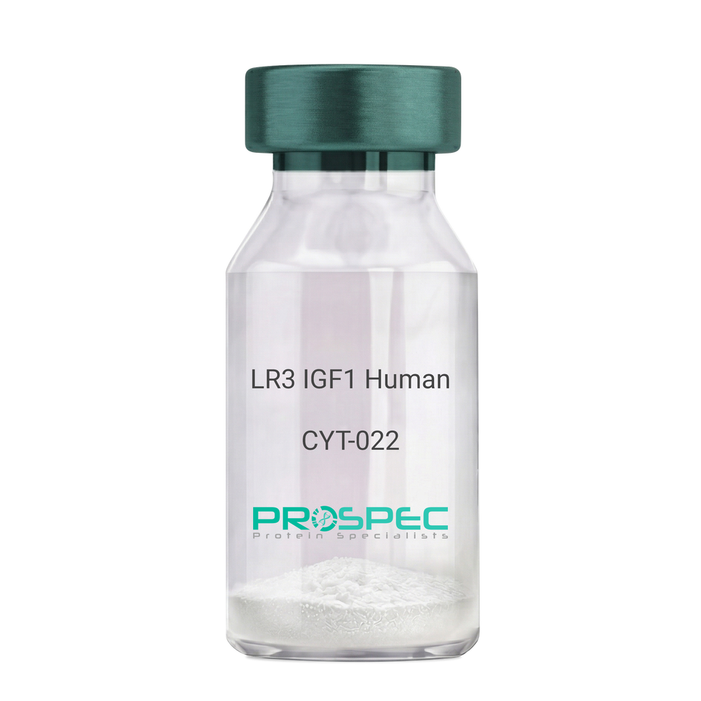
  • View Data Sheet

    Name :

    IGF1 LR3 Human

    Description:

    LR3 Insulin Like Growth Factor-1 Human Recombinant

    R3 IGF1, R3 IGF-1, R3IGF1, R3IGF-1, LONG IGF1, LONG IGF-1, LONG R3 IGF1, LONG R3IGF1, LONG R3 IGF-1, LONG R3IGF-1.

    Product # :

    CYT-022

    Price :

    Quantity :

    Shipping Method :

    Room Temp Icon

    Shipped at Room temp

    Add To Cart

    More Info

    • description
    • source
    • formulation
    • purity
    • biological activity
    • More Info
    • sds-page

    Description

    The LR3 is a long-term analog of human IGF-1, specifically designed and manufactured for mammalian cell culture to support large-scale manufacturing of recombinant biopharmaceuticals. Recombinant Human LR3 Insulin Like Growth Factor-1 produced in E.Coli is a single, non-glycosylated, polypeptide chain containing 83 amino acids and having a molecular mass of 9.1kDa.

    Source

    Escherichia Coli.

    Formulation

    Lyophilized from a 0.2µm filtered concentrated solution in 20mM PB, pH 7.2.

    Purity

    Greater than 97.0% as determined by SDS-PAGE.

    Biological Activity

    The ED50 as determined by the stimulation of protein synthesis in L6 myoblasts is less than 10ng/ml, corresponding to a specific activity of 100,000units/mg.

    sds-page

    LR3 IGF1 sds-page - Product image 1

    More Info

    • Introduction

      IGF-1 (Insulin-like growth factor-1) is a major hormonal mediator of statural growth. Under regular circumstances, GH (growth hormone) binds to its receptor in the liver, and other tissues, and stimulates the synthesis/secretion of IGF-1. In target tissues, the Type 1 IGF receptor, that is homologous to the insulin receptor, is activated by IGF-1, leading to intracellular signaling which stimulates multiple processes leading to statural growth. IGF-1 metabolic actions are partly directed at stimulating the uptake of glucose, fatty acids, and amino acids so that metabolism supports growing tissues.

    • Synonyms

      R3 IGF1, R3 IGF-1, R3IGF1, R3IGF-1, LONG IGF1, LONG IGF-1, LONG R3 IGF1, LONG R3IGF1, LONG R3 IGF-1, LONG R3IGF-1.

    • Physical Appearance

      Sterile Filtered White lyophilized (freeze-dried) powder.

    • Stability

      Lyophilized LR3 IGF1 although stable at room temperature for 3 weeks, should be stored desiccated below -18°C. Upon reconstitution the LR3 IGF1 should be stored at 4°C between 2-7 days and for future use below -18°C.For long term storage it is recommended to add a carrier protein (0.1% HSA or BSA).Please prevent freeze-thaw cycles.

    • Solubility

      It is recommended to reconstitute the lyophilized LR3 IGF1 in sterile 18M-cm H2O at a concentration of 100µg/ml, which can then be further diluted to other aqueous solutions.

    • Amino Acid Sequence

      MFPAMPLSSLFVNGPRTLCGAELVDALQFVCGDRGFYFNKPTGYGSSSRRAPQTGIV DECCFRSCDLRRLEMYCAPLKPAKSA.

    • Background

      What is the molecular weight/Mw of IGF1 LR3 HUMAN Protein?
      IGF1 LR3 HUMAN Protein has a total Mw of 9.1kDa.

      What is the source or expression system of IGF1 LR3 HUMAN Protein?
      Escherichia Coli.

      What is the Purity of IGF1 LR3 HUMAN Protein?
      IGF1 LR3 HUMAN Protein is >98% pure as determined by SDS-PAGE.

      What is the Biological Activity of IGF1 LR3 HUMAN Protein?
      The ED50 as determined by the stimulation of protein synthesis in L6 myoblasts is less than 10ng/ml, corresponding to a specific activity of 100,000units/mg.

      What is the amino acid sequence of IGF1 LR3 HUMAN Protein?
      MFPAMPLSSLFVNGPRTLCGAELVDALQFVCGDRGFYFNKPTGYGSSSRRAPQTGIV DECCFRSCDLRRLEMYCAPLKPAKSA.

      What applications can IGF1 LR3 HUMAN Protein be used in?
      IGF1 LR3 HUMAN Protein can probably be used in western blot, ELISA and Lateral Flow.

      What is the endotoxin level for IGF1 LR3 HUMAN Protein?
      The endotoxin level is minimal, IGF1 LR3 HUMAN Protein was purified using conventional chromatography techniques.


    ProSpec's products are furnished for LABORATORY RESEARCH USE ONLY. The product may not be used as drugs, agricultural or pesticidal products, food additives or household chemicals.

    Long R3 Igf1 Human
  • View Data Sheet

    Name :

    DNAL1 Antibody

    Description:

    Dynein Axonemal Light Chain 1, Mouse Anti Human

    Dynein light chain 1, axonemal, C14orf168, CILD16, DNAL1.

    Product # :

    ANT-510

    Price :

    Quantity :

    Shipping Method :

    Ice Icon

    Shipped with Ice Packs

    Add To Cart

    More Info

    • formulation
    • More Info

    Formulation

    1mg/ml containing PBS, pH-7.4, 10% Glycerol and 0.02% Sodium Azide.

    More Info

    • Introduction

      Dynein Axonemal Light Chain 1 (DNAL1) functions as a component of the outer dynein arms complex. DNAL1 acts as the molecular motor which supplies the force to move cilia in an ATP-dependent manner. DNAL1 is expressed in tissues with motile cilia or flagella and takes part in the movement of sperm flagella. Alternate splicing results in numerous transcript variants.

    • Synonyms

      Dynein light chain 1, axonemal, C14orf168, CILD16, DNAL1.

    • Physical Appearance

      Sterile filtered colorless solution.

    • Immunogen

      Anti-human DNAL1 mAb, clone PAT29E4AT, is derived from hybridization of mouse F0 myeloma cells with spleen cells from BALB/c mice immunized with a recombinant human DNAL1 protein 1-190 amino acids purified from E. coli.

    • Ig Subclass

      Mouse IgG2b heavy chain and k light chain.

    • Clone

      PAT29E4AT.

    • Applications

      The antibody has been tested by ELISA, Western blot analysis to assure specificity and reactivity. Since application varies, however, each investigation should be titrated by the reagent to obtain optimal results. Recommended starting dilution is 1:1000.

    • Type

      Mouse Anti Human Monoclonal.

    • Storage Procedures

      For periods up to 1 month store at 4°C, for longer periods of time, store at -20°C. Prevent freeze thaw cycles.

    • Purification Method

      DNAL1 antibody was purified from mouse ascitic fluids by protein-A affinity chromatography.

    ProSpec's products are furnished for LABORATORY RESEARCH USE ONLY. The product may not be used as drugs, agricultural or pesticidal products, food additives or household chemicals.

    Dnal1 Antibody
  • View Data Sheet

    Name :

    CLEC7A Human

    Description:

    C-Type Lectin Domain Family 7, Member A Human Recombinant

    BGR, Dendritic Cell-Associated C-Type Lectin-1, Dendritic Cell-Associated C-Type Lectin 1, C-Type Lectin Domain Family 7, Member A, Lectin-Like Receptor 1, CD369 Antigen, CANDF4, SCARE2, CD369, C-Type Lectin Domain Containing 7A, C-Type Lectin Domain Family 7 Member A, C-Type (Calcium Dependent, Carbohydrate-Recognition Domain) Lectin, Superfamily Member 12, C-Type Lectin Superfamily Member 12, DC-Associated C-Type Lectin 1, Beta-Glucan Receptor, Dectin-1, CLECSF12, DECTIN1.

    Product # :

    PRO-2634

    Price :

    Quantity :

    Shipping Method :

    Ice Icon

    Shipped with Ice Packs

    Add To Cart

    More Info

    • description
    • source
    • formulation
    • purity
    • More Info

    Description

    CLEC7A Human Recombinant produced in Sf9 Baculovirus cells is a single, glycosylated polypeptide chain containing 183 amino acids (71-244 a.a) and having a molecular mass of 21kDa. CLEC7A is fused to a 9 amino acid His-tag at C-terminus & purified by proprietary chromatographic techniques.

    Source

    Sf9, Baculovirus cells.

    Formulation

    The CLEC7A solution (0.5mg/1ml) contains phosphate buffered saline (pH7.4) and 10% glycerol.

    Purity

    Greater than 95.0% as determined by SDS-PAGE.

    More Info

    • Introduction

      C-type lectin domain family 7 member A 1 or CLEC7A is a protein, that in the innate immune system, acts against fungal pathogens. CLEC7A can be found in the immune system response cells such as monocytes, macrophages & neutrophils, or in dendritic and T cells. The protein is enhanced by macrophages by using GM-CSF, IL-4, or IL-13, or diminishes by dexamethasone, IL-10 and LPS.

    • Synonyms

      BGR, Dendritic Cell-Associated C-Type Lectin-1, Dendritic Cell-Associated C-Type Lectin 1, C-Type Lectin Domain Family 7, Member A, Lectin-Like Receptor 1, CD369 Antigen, CANDF4, SCARE2, CD369, C-Type Lectin Domain Containing 7A, C-Type Lectin Domain Family 7 Member A, C-Type (Calcium Dependent, Carbohydrate-Recognition Domain) Lectin, Superfamily Member 12, C-Type Lectin Superfamily Member 12, DC-Associated C-Type Lectin 1, Beta-Glucan Receptor, Dectin-1, CLECSF12, DECTIN1.

    • Physical Appearance

      Sterile Filtered clear solution.

    • Stability

      Store at 4°C if entire vial will be used within 2-4 weeks. Store, frozen at -20°C for longer periods of time. For long term storage it is recommended to add a carrier protein (0.1% HSA or BSA).Avoid multiple freeze-thaw cycles.

    • Amino Acid Sequence

      ADPRHNSGRN PEEKDNFLSR NKENHKPTES SLDEKVAPSK ASQTTGGFSQ SCLPNWIMHG KSCYLFSFSG NSWYGSKRHC SQLGAHLLKI DNSKEFEFIE SQTSSHRINA FWIGLSRNQS EGPWFWEDGS AFFPNSFQVR NTVPQESLLH NCVWIHGSEV YNQICNTSSY SICEKELHHH HHH.

    ProSpec's products are furnished for LABORATORY RESEARCH USE ONLY. The product may not be used as drugs, agricultural or pesticidal products, food additives or household chemicals.

    Clec7A Human
  • View Data Sheet

    Name :

    PDPN Antibody

    Description:

    Podoplanin, Mouse Anti Human

    Podoplanin, Glycoprotein 36, PA2.26 antigen, T1A, GP36, GP40, Gp38, OTS8, T1A2, HT1A-1, PA2.26, T1-alpha, PDPN.

    Product # :

    ANT-343

    Price :

    Quantity :

    Shipping Method :

    Ice Icon

    Shipped with Ice Packs

    Add To Cart

    More Info

    • formulation
    • More Info

    Formulation

    1mg/ml containing PBS, pH-7.4, & 0.1% Sodium Azide.

    More Info

    • Introduction

      Podoplanin is a small mucin-like type-1 transmembrane protein, typically expressed in various specialized cell types throughout the body. Podoplanin is a type-I integral membrane glycoprotein with diverse distribution in human tissues. PDPN physiological function is related to its mucin-type character. The homologous protein in other species has been described as a differentiation antigen and influenza-virus receptor.
      PDPN is expressed in lymphatic progenitor cells and afterwards during mouse development in lymphatic endothelial cells. Podoplanin is a specific marker for lymph vessel endothelial cells. Over-expression of podoplanin significantly elevates endothelial cell adhesion, migration, and tube formation. Inhibition of Podoplanin expression decreases cell adhesion in human dermal lymphatic endothelial cells. Podoplanin is used as a specific marker for lymphatic endothelium in histopathology.
      Podoplanin expression is increased in nearly all human colon, rectum, and small intestine tumors. AGGRUS may serve as a diagnostic marker that distinguishes seminomas, the majority of which over express the protein, from embryonal carcinoma in testicular germ cell tumors.

    • Synonyms

      Podoplanin, Glycoprotein 36, PA2.26 antigen, T1A, GP36, GP40, Gp38, OTS8, T1A2, HT1A-1, PA2.26, T1-alpha, PDPN.

    • Physical Appearance

      Sterile filtered colorless solution.

    • Immunogen

      Anti-human PDPN mAb is derived from hybridization of mouse FO myeloma cells with spleen cells from BALB/c mice immunized with recombinant human PDPN amino acids 1-206 purified from E. coli.

    • Ig Subclass

      Mouse IgG2b heavy chain and κ light chain.

    • Clone

      P5E2AT.

    • Applications

      PDPN antibody has been tested by ELISA and Western blot analysis to assure specificity and reactivity. Since application varies, however, each investigation should be titrated by the reagent to obtain optimal results. Recommended dilution range for Western blot analysis is 1:500 ~ 1,000. Recommended starting dilution is 1:500.

    • Type

      Mouse Anti Human Monoclonal.

    • Storage Procedures

      For periods up to 1 month store at 4°C, for longer periods of time, store at -20°C. Prevent freeze thaw cycles.

    • Purification Method

      PDPN antibody was purified from mouse ascitic fluids by protein-G affinity chromatography.

    ProSpec's products are furnished for LABORATORY RESEARCH USE ONLY. The product may not be used as drugs, agricultural or pesticidal products, food additives or household chemicals.

    Pdpn Antibody
  • View Data Sheet

    Name :

    CD44 Anti Human

    Description:

    CD44, Mouse Anti Human

    MDU2, MDU3, MIC4, CDW44, CSPG8, HCELL, HUTCH-I, Phagocytic glycoprotein I, PGP-1, Extracellular matrix receptor-III, ECMR-III, Hermes antigen, Hyaluronate receptor, Heparan sulfate proteoglycan, Epican, CDw44.

    Product # :

    ANT-453

    Price :

    Quantity :

    Shipping Method :

    Ice Icon

    Shipped with Ice Packs

    Add To Cart

    More Info

    • formulation
    • More Info

    Formulation

    1mg/ml containing PBS, pH-7.4, & 0.1% Sodium Azide.

    More Info

    • Introduction

      CD44 is a cell-surface glycoprotein that plays a role in cell-cell interactions, cell adhesion and migration. CD44 is a receptor for hyaluronic acid and also interacts with other ligands, such as osteopontin, collagens, and matrix metalloproteinases. CD44 participates in a wide variety of cellular functions such as lymphocyte activation, recirculation and homing, hematopoiesis, and tumor metastasis. CD44 and CD49d are putative activity markers and CD44 a potential novel therapeutic target in multiple sclerosis. Increased CD44 antigen is associated with relapses in non-small cell lung cancers.

    • Synonyms

      MDU2, MDU3, MIC4, CDW44, CSPG8, HCELL, HUTCH-I, Phagocytic glycoprotein I, PGP-1, Extracellular matrix receptor-III, ECMR-III, Hermes antigen, Hyaluronate receptor, Heparan sulfate proteoglycan, Epican, CDw44.

    • Immunogen

      Anti-human CD44 mAb is derived from hybridization of mouse F0 myeloma cells with spleen cells from BALB/c mice immunized with recombinant human CD44 amino acids 21-145 purified from E. coli.

    • Ig Subclass

      Mouse IgG2b heavy chain and κ light chain.

    • Clone

      P5C10AT.

    • Applications

      CD44 antibody has been tested by ELISA and Western blot analysis to assure specificity and reactivity. Since application varies, however, each investigation should be titrated by the reagent to obtain optimal results. Recommended dilution range for Western blot analysis is 1:1,000 ~ 2,000. Recommended starting dilution is 1:1,000.

    • Type

      Mouse Anti Human Monoclonal.

    • Storage Procedures

      For periods up to 1 month store at 4°C, for longer periods of time, store at -20°C. Prevent freeze thaw cycles.

    • Purification Method

      CD44 antibody was purified from mouse ascitic fluids by protein-G affinity chromatography.

    ProSpec's products are furnished for LABORATORY RESEARCH USE ONLY. The product may not be used as drugs, agricultural or pesticidal products, food additives or household chemicals.

    Anti Human Cd44
  • View Data Sheet

    Name :

    Visfatin Antibody

    Description:

    Visfatin, Mouse Anti Human

    PBEF, Pre-B cell colony-enhancing factor, Nicotinamide phosphoribosyltransferase NAmPRTase, Nampt, MGC117256, DKFZP666B131, 1110035O14Rik.

    Product # :

    ANT-368

    Price :

    Quantity :

    Shipping Method :

    Ice Icon

    Shipped with Ice Packs

    Add To Cart

    More Info

    • formulation
    • More Info

    Formulation

    1mg/ml containing PBS, pH-7.4, 10% glycerol & 0.02% Sodium Azide.

    More Info

    • Introduction

      Excess adiposity is the most important risk in the development of insulin resistance and type 2 diabetes mellitus (T2DM). Adipose tissue produces several proteins (adipocytokines) such as leptin, adiponectin, resistin, tumor necrosis factor-α, and IL-6, that modulate insulin sensitivity and appear to play an important role in the pathogenesis of insulin resistance, diabetes, dyslipidemia, inflammation, and atherosclerosis. However, the mechanisms by which fat tissue induces insulin resistance and the role of adipocytokines in the pathogenesis of T2DM have not been well established. Visfatin, also known as pre-B cell colony-enhancing factor (PBEF), is a cytokine that is highly expressed in visceral fat and was originally isolated as a secreted factor that synergizes with IL-7 and stem cell factors to promote the growth of B cell precursors. Visfatin homologs have been identified in carp, invertebrate mollusks, and bacteria, as well as in vertebrates, including humans and the mouse. It h

    • Synonyms

      PBEF, Pre-B cell colony-enhancing factor, Nicotinamide phosphoribosyltransferase NAmPRTase, Nampt, MGC117256, DKFZP666B131, 1110035O14Rik.

    • Immunogen

      Anti-human Visfatin mAb, is derived from hybridization of mouse FO myeloma cells with spleen cells from BALB/c mice immunized with Recombinant human Visfatin amino acids 1-491 purified from E. coli.

    • Ig Subclass

      Mouse IgG1heavy chain and κ light chain.

    • Clone

      P4D5AT.

    • Applications

      Visfatin antibody has been tested by ELISA and Western blot analysis to assure specificity and reactivity. Since application varies, however, each investigation should be titrated by the reagent to obtain optimal results.

    • Type

      Mouse Anti Human Monoclonal.

    • Storage Procedures

      For periods up to 1 month store at 4°C, for longer periods of time, store at -20°C. Prevent freeze thaw cycles.

    • Purification Method

      Visfatin antibody was purified from mouse ascitic fluids by protein-G affinity chromatography.

    ProSpec's products are furnished for LABORATORY RESEARCH USE ONLY. The product may not be used as drugs, agricultural or pesticidal products, food additives or household chemicals.

    Visfatin Antibody
  • 1
  • …
  • 39
  • 40
  • 41
  • 42
Your browser does not support the video tag.

Products

  • Cytokines
  • Growth Factors
  • Chemokines
  • CD Antigens
  • Neurotrophins
  • Hormones
  • Enzymes
  • Viral Antigens
  • Recombinant Proteins
  • Natural Proteins
  • Monoclonal Antibodies
  • Polyclonal Antibodies
  • Thermal Packaging

About

  • Company
  • Founder
  • Contribution
  • Contact Us
  • My Account

Ordering

  • Ordering Method
  • Ordering - Credit Card
  • Ordering PO
  • Shipping Fees
  • FAQ's

Legal

  • Terms & Conditions
  • Privacy
  • Site Map
  • info@prospecbio.com
  • Facebook
  • Instagram
  • Linkedin Profile

© 2026 ProSpec-Tany TechnoGene Ltd. All Rights Reserved.

  • Choosing a selection results in a full page refresh.
  • Opens in a new window.