Skip to content

High-quality research proteins.

  • 1-800-805-6531

  • Mon - Sat 8.00-18.00, Sun - Closed

  • info@prospecbio.com

  • PO Box 6591 East Brunswick NJ 08816

  • Products
    • Cytokines
    • Growth factors
    • Chemokines
    • Cd antigens
    • Neurotrophins
    • Hormones
    • Enzymes
    • Viral antigens
    • Recombinant proteins
    • Natural proteins
    • Monoclonal antibodies
    • Polyclonal antibodies
    • Thermal Packaging
  • About
    • Company
    • Founder
    • Contribution
  • Customers
    • Eastern Asia
    • Eastern Europe
    • North America
    • South America
    • South East Asia
    • Southern Asia
    • Western Europe
  • Ordering
    • Ordering Method
    • Ordering - Credit Card
    • Ordering - PO
    • Shipping Fees
    • FAQs
  • Services
    • DNA Cloning
    • Protein Expression
    • Monoclonal Antibody
    • Fermentation
    • Technology License
  • Contact us
Log in

Country/region

  • USD $ | Albania
  • USD $ | Andorra
  • USD $ | Argentina
  • USD $ | Armenia
  • USD $ | Australia
  • USD $ | Austria
  • USD $ | Azerbaijan
  • USD $ | Belarus
  • USD $ | Belgium
  • USD $ | Bolivia
  • USD $ | Brazil
  • USD $ | Bulgaria
  • USD $ | Cambodia
  • USD $ | Canada
  • USD $ | Chile
  • USD $ | China
  • USD $ | Colombia
  • USD $ | Costa Rica
  • USD $ | Croatia
  • USD $ | Cyprus
  • USD $ | Czechia
  • USD $ | Denmark
  • USD $ | Estonia
  • USD $ | Finland
  • USD $ | France
  • USD $ | Georgia
  • USD $ | Germany
  • USD $ | Greece
  • USD $ | Hong Kong SAR
  • USD $ | Hungary
  • USD $ | Iceland
  • USD $ | India
  • USD $ | Ireland
  • USD $ | Israel
  • USD $ | Italy
  • USD $ | Japan
  • USD $ | Kazakhstan
  • USD $ | Latvia
  • USD $ | Liechtenstein
  • USD $ | Lithuania
  • USD $ | Madagascar
  • USD $ | Malta
  • USD $ | Mexico
  • USD $ | Moldova
  • USD $ | Monaco
  • USD $ | Netherlands
  • USD $ | New Zealand
  • USD $ | North Macedonia
  • USD $ | Norway
  • USD $ | Panama
  • USD $ | Paraguay
  • USD $ | Peru
  • USD $ | Philippines
  • USD $ | Poland
  • USD $ | Portugal
  • USD $ | Romania
  • USD $ | Singapore
  • USD $ | Slovakia
  • USD $ | Slovenia
  • USD $ | South Africa
  • USD $ | South Korea
  • USD $ | Spain
  • USD $ | Sri Lanka
  • USD $ | Sweden
  • USD $ | Switzerland
  • USD $ | Taiwan
  • USD $ | Thailand
  • USD $ | Türkiye
  • USD $ | Ukraine
  • USD $ | United Kingdom
  • USD $ | United States
  • USD $ | Uruguay
  • USD $ | Vatican City
  • USD $ | Venezuela
  • USD $ | Vietnam
  • Facebook
  • Instagram
logoProSpec - Recombinant Protein Manufacturer for academic and R&D industry
Become a distributor
Account Log in
Wishlist
Cart Cart(0)
  • Products
    +
    • Cytokines
    • Growth factors
    • Chemokines
    • Cd antigens
    • Neurotrophins
    • Hormones
    • Enzymes
    • Viral antigens
    • Recombinant proteins
    • Natural proteins
    • Monoclonal antibodies
    • Polyclonal antibodies
    • Thermal Packaging
  • About
    +
    • Company
    • Founder
    • Contribution
  • Customers
    +
    • Eastern Asia
    • Eastern Europe
    • North America
    • South America
    • South East Asia
    • Southern Asia
    • Western Europe
  • Ordering
    +
    • Ordering Method
    • Ordering - Credit Card
    • Ordering - PO
    • Shipping Fees
    • FAQs
  • Services
    +
    • DNA Cloning
    • Protein Expression
    • Monoclonal Antibody
    • Fermentation
    • Technology License
  • Contact us

Item added to your cart

View cart
View All
  • Cytokines
  • Growth factors
  • Chemokines
  • Cd antigens
  • Neurotrophins
  • Hormones
  • Enzymes
  • Viral antigens
  • Recombinant proteins
  • Natural proteins
  • Monoclonal antibodies
  • Polyclonal antibodies
  • Thermal Packaging
  • Tumor Necrosis Factor

    Tumor Necrosis Factor

    View All
  • 4-1BB

    4-1BB

    View All
  • Adiponectin

    Adiponectin

    View All
  • AIF1

    AIF1

    View All
  • Other Cytokines

    Other Cytokines

    View All
  • AITRL

    AITRL

    View All
  • Angiopoietin

    Angiopoietin

    View All
  • Apolipoprotein

    Apolipoprotein

    View All
  • B-Cell Activating Factor

    B-Cell Activating Factor

    View All
  • Beta Defensin

    Beta Defensin

    View All
  • Bone Morphogenetic Protein

    Bone Morphogenetic Protein

    View All
  • B type Natriuretic Peptide

    B type Natriuretic Peptide

    View All
  • BST

    BST

    View All
  • Betacellulin

    Betacellulin

    View All
  • Cardiotrophin

    Cardiotrophin

    View All
  • Activin

    Activin

    View All
  • Fibroblast Growth Factor

    Fibroblast Growth Factor

    View All
  • Other Growth Factors

    Other Growth Factors

    View All
  • CSF

    CSF

    View All
  • CTGF

    CTGF

    View All
  • IGFBP

    IGFBP

    View All
  • VEGF Protein

    VEGF Protein

    View All
  • EGF

    EGF

    View All
  • Epigen

    Epigen

    View All
  • Erythropoietin

    Erythropoietin

    View All
  • Insulin-Like Growth Factor

    Insulin-Like Growth Factor

    View All
  • Growth Hormone

    Growth Hormone

    View All
  • HDGF

    HDGF

    View All
  • Hepatocyte Growth Factor

    Hepatocyte Growth Factor

    View All
  • Insulin

    Insulin

    View All
  • BCA-1/ BLC (CXCL13)

    BCA-1/ BLC (CXCL13)

    View All
  • BRAK (CXCL14)

    BRAK (CXCL14)

    View All
  • C-10 (CCL6)

    C-10 (CCL6)

    View All
  • Other Chemokines

    Other Chemokines

    View All
  • MEC (CCL28)

    MEC (CCL28)

    View All
  • LD78-beta (CCL3L1)

    LD78-beta (CCL3L1)

    View All
  • CTACK (CCL27)

    CTACK (CCL27)

    View All
  • CXCL16

    CXCL16

    View All
  • CXCL17

    CXCL17

    View All
  • Platelet Factor-4 (CXCL4)

    Platelet Factor-4 (CXCL4)

    View All
  • ENA-78 (CXCL5)

    ENA-78 (CXCL5)

    View All
  • Eotaxin (CCL11,24,26)

    Eotaxin (CCL11,24,26)

    View All
  • Exodus-2 (CCL21)

    Exodus-2 (CCL21)

    View All
  • Fractalkine (CX3CL1)

    Fractalkine (CX3CL1)

    View All
  • CXCL6

    CXCL6

    View All
  • Other CD Antigens

    Other CD Antigens

    View All
  • CD14

    CD14

    View All
  • CD164

    CD164

    View All
  • CD1

    CD1

    View All
  • CD2

    CD2

    View All
  • CD200

    CD200

    View All
  • CD207

    CD207

    View All
  • CD226

    CD226

    View All
  • CD23

    CD23

    View All
  • CD244

    CD244

    View All
  • CD247

    CD247

    View All
  • CD27

    CD27

    View All
  • CD274

    CD274

    View All
  • CD300

    CD300

    View All
  • CD33

    CD33

    View All
  • Other Neurotrophins

    Other Neurotrophins

    View All
  • Beta-NGF

    Beta-NGF

    View All
  • BDNF

    BDNF

    View All
  • CDNF

    CDNF

    View All
  • CNTF

    CNTF

    View All
  • GDNF

    GDNF

    View All
  • Glia Maturation Factor

    Glia Maturation Factor

    View All
  • MANF

    MANF

    View All
  • Midkine

    Midkine

    View All
  • Neuroglobin

    Neuroglobin

    View All
  • Neurotrophic factors

    Neurotrophic factors

    View All
  • Neuregulin

    Neuregulin

    View All
  • Neuropilin

    Neuropilin

    View All
  • Pigment Epithelium-Derived Factor

    Pigment Epithelium-Derived Factor

    View All
  • Pleiotrophin

    Pleiotrophin

    View All
  • Peptide Hormones

    Peptide Hormones

    View All
  • Other Hormones

    Other Hormones

    View All
  • HCG

    HCG

    View All
  • Endothelin

    Endothelin

    View All
  • Exendin

    Exendin

    View All
  • FSH

    FSH

    View All
  • Glucagon

    Glucagon

    View All
  • GHRP

    GHRP

    View All
  • GLP

    GLP

    View All
  • Thyrostimulin

    Thyrostimulin

    View All
  • Inhibin A

    Inhibin A

    View All
  • LHRH

    LHRH

    View All
  • Melanotan

    Melanotan

    View All
  • Procalcitonin

    Procalcitonin

    View All
  • PTH

    PTH

    View All
  • Peroxiredoxin

    Peroxiredoxin

    View All
  • Peptidase

    Peptidase

    View All
  • Synthetase

    Synthetase

    View All
  • Transferase

    Transferase

    View All
  • Hydrolase

    Hydrolase

    View All
  • Dehydrogenase

    Dehydrogenase

    View All
  • ACE2

    ACE2

    View All
  • Esterase

    Esterase

    View All
  • Protein Kinases

    Protein Kinases

    View All
  • Other Enzymes

    Other Enzymes

    View All
  • Phosphatase

    Phosphatase

    View All
  • Deaminase

    Deaminase

    View All
  • Oxygenase

    Oxygenase

    View All
  • Adenylate Kinase

    Adenylate Kinase

    View All
  • Lyase

    Lyase

    View All
  • Chagas

    Chagas

    View All
  • SARS

    SARS

    View All
  • Influenza

    Influenza

    View All
  • ASFV

    ASFV

    View All
  • Astrovirus

    Astrovirus

    View All
  • Borrelia

    Borrelia

    View All
  • Helicobacter Pylori

    Helicobacter Pylori

    View All
  • Chikungunya

    Chikungunya

    View All
  • Chlamydia

    Chlamydia

    View All
  • Cytomegalo

    Cytomegalo

    View All
  • Dengue

    Dengue

    View All
  • EBV

    EBV

    View All
  • Ebola

    Ebola

    View All
  • Feline Leukemia Virus

    Feline Leukemia Virus

    View All
  • Hepatitis A

    Hepatitis A

    View All
  • Other

    Other

    View All
  • Actin

    Actin

    View All
  • ADAM

    ADAM

    View All
  • Complement Component

    Complement Component

    View All
  • Ag85

    Ag85

    View All
  • Eukaryotic Translation Initiation Factor

    Eukaryotic Translation Initiation Factor

    View All
  • Anterior Gradient Protein

    Anterior Gradient Protein

    View All
  • Heat Shock Protein

    Heat Shock Protein

    View All
  • Alpha-2-HS-Glycoprotein

    Alpha-2-HS-Glycoprotein

    View All
  • Hemoglobin

    Hemoglobin

    View All
  • Allergy

    Allergy

    View All
  • Anaplasma

    Anaplasma

    View All
  • Angiogenin

    Angiogenin

    View All
  • Ankyrin Repeat Domain

    Ankyrin Repeat Domain

    View All
  • Annexin

    Annexin

    View All
  • Other Natural Proteins

    Other Natural Proteins

    View All
  • Aprotinin

    Aprotinin

    View All
  • Transferrin

    Transferrin

    View All
  • Avidin

    Avidin

    View All
  • Anti Coagulation Factors

    Anti Coagulation Factors

    View All
  • Natural Albumin

    Natural Albumin

    View All
  • Natural Coagulation Factors

    Natural Coagulation Factors

    View All
  • Fibronectin

    Fibronectin

    View All
  • Thrombin

    Thrombin

    View All
  • Anti Human Enzyme

    Anti Human Enzyme

    View All
  • Other Monoclonal Antibodies

    Other Monoclonal Antibodies

    View All
  • Anti Human Cytokine

    Anti Human Cytokine

    View All
  • Anti Human Heat Shock Protein

    Anti Human Heat Shock Protein

    View All
  • Anti Mouse Lymphocyte

    Anti Mouse Lymphocyte

    View All
  • Anti Human Chemokine

    Anti Human Chemokine

    View All
  • Anti Human Lymphocyte

    Anti Human Lymphocyte

    View All
  • Anti Viral Monoclonal

    Anti Viral Monoclonal

    View All
  • Anti Mouse Cytokine

    Anti Mouse Cytokine

    View All
  • Anti-GST Monoclonal

    Anti-GST Monoclonal

    View All
  • Other Polyclonal Antibodies

    Other Polyclonal Antibodies

    View All
  • Anti Viral

    Anti Viral

    View All
  • Anti-GST Monoclonal

    Anti-GST Monoclonal

    View All
Back

Search results

1000 results found for “epiregulin”

Name

Description

Product #

Price

Quantity

Shipping Method

  • View Data Sheet

    Name :

    NKIRAS1 Human

    Description:

    NFKB Inhibitor Interacting Ras-Like 1 Human Recombinant

    NF-kappa-B inhibitor-interacting Ras-like protein 1, I-kappa-B-interacting Ras-like protein 1, Kappa B-Ras protein 1, KappaB-Ras1, NKIRAS1, KBRAS1.

    Product # :

    PRO-1039

    Price :

    Quantity :

    Shipping Method :

    Ice Icon

    Shipped with Ice Packs

    Add To Cart

    More Info

    • description
    • source
    • formulation
    • purity
    • More Info
    • Citations

    Description

    NKIRAS1 Human Recombinant produced in E.Coli is a single, non-glycosylated polypeptide chain containing 212 amino acids (1-192 a.a) and having a molecular mass of 23.8kDa (Molecular weight on SDS-PAGE will appear higher).NKIRAS1 is fused to a 20 amino acid His-tag at N-terminus & purified by proprietary chromatographic techniques.

    Source

    Escherichia Coli.

    Formulation

    NKIRAS1 protein solution (0.25mg/ml) containing 20mM Tris-HCl buffer (pH8.0), 20% glycerol and 1mM DTT.

    Purity

    Greater than 95.0% as determined by SDS-PAGE.

    Citations

    More Info

    • Introduction

      NF-kappa-B inhibitor-interacting Ras-like protein 1 (NKIRAS1) is a member of the small GTPase superfamily. NKIRAS1 is an unusual Ras-like protein which acts as a potent regulator of NF-kappa-B activity by thwarting the degradation of NF-kappa-B inhibitor beta (NFKBIB) by most signals, describing why NFKBIB is more resistant to degradation. NKIRAS1 functions by blocking phosphorylation of NFKBIB and mediating cytoplasmic retention of p65/RELA NF-kappa-B subunit. Both GTP- and GDP-bound forms block phosphorylation of NFKBIB.

    • Synonyms

      NF-kappa-B inhibitor-interacting Ras-like protein 1, I-kappa-B-interacting Ras-like protein 1, Kappa B-Ras protein 1, KappaB-Ras1, NKIRAS1, KBRAS1.

    • Physical Appearance

      Sterile Filtered clear solution.

    • Stability

      Store at 4°C if entire vial will be used within 2-4 weeks. Store, frozen at -20°C for longer periods of time. For long term storage it is recommended to add a carrier protein (0.1% HSA or BSA).Avoid multiple freeze-thaw cycles.

    • Amino Acid Sequence

      MGSSHHHHHH SSGLVPRGSH MGKGCKVVVC GLLSVGKTAI LEQLLYGNHT IGMEDCETME DVYMASVETD RGVKEQLHLY DTRGLQEGVE LPKHYFSFAD GFVLVYSVNN LESFQRVELL KKEIDKFKDK KEVAIVVLGN KIDLSEQRQV DAEVAQQWAK SEKVRLWEVT VTDRKTLIEP FTLLASKLSQ PQSKSSFPLP GRKNKGNSNS EN.

    ProSpec's products are furnished for LABORATORY RESEARCH USE ONLY. The product may not be used as drugs, agricultural or pesticidal products, food additives or household chemicals.

    Nkiras1 Human
  • View Data Sheet

    Name :

    TGFBR1 Human, Active

    Description:

    Transforming Growth Factor Beta Receptor 1 Human Recombinant, Active

    TGFBR1, AAT5, ACVRLK4, ALK-5, ALK5, ESS1, LDS1, LDS1A, LDS2A, MSSE, SKR4, tbetaR-I, TGFR-1.

    Product # :

    PKA-135

    Price :

    Quantity :

    Shipping Method :

    Ice Icon

    Shipped with Ice Packs

    Add To Cart

    More Info

    • description
    • source
    • formulation
    • purity
    • biological activity
    • More Info
    • Citations

    Description

    TGFBR1 produced in Sf9 insect cells is a single, glycosylated polypeptide chain containing 342 amino acids (27-126a.a.) and having a molecular mass of 38kDa. TGFBR1 is expressed with 242 amino acid hIgG-His-Tag at C-Terminus and purified by proprietary chromatographic techniques.

    Source

    Sf9, Baculovirus cells.

    Formulation

    TGFBR1 protein solution (0.5mg/ml) Phosphate Buffered Saline (pH 7.4) containing 10% glycerol.

    Purity

    Greater than 90.0% as determined by SDS-PAGE.

    Biological Activity

    ≤ 2 ug/ml, measured by its binding ability in a functional ELISA with Mouse CD105. 

    Citations

    More Info

    • Synonyms

      TGFBR1, AAT5, ACVRLK4, ALK-5, ALK5, ESS1, LDS1, LDS1A, LDS2A, MSSE, SKR4, tbetaR-I, TGFR-1.

    • Physical Appearance

      Sterile filtered colorless solution.

    • Stability

      Store at 4°C if entire vial will be used within 2-4 weeks. Store, frozen at -20°C for longer periods of time. For long term storage it is recommended to add a carrier protein (0.1% HSA or BSA).Avoid multiple freeze-thaw cycles.

    • Amino Acid Sequence

      ADLLLPGATA LQCFCHLCTK DNFTCVTDGL CFVSVTETTD KVIHNSMCIA EIDLIPRDRP FVCAPSSKTG SVTTTYCCNQ DHCNKIELPT TVKSSPGLGP VELVEPKSCD KTHTCPPCPA PELLGGPSVF LFPPKPKDTL MISRTPEVTC VVVDVSHEDP EVKFNWYVDG VEVHNAKTKP REEQYNSTYR VVSVLTVLHQ DWLNGKEYKC KVSNKALPAP IEKTISKAKG QPREPQVYTL
      PPSRDELTKN QVSLTCLVKG FYPSDIAVEW ESNGQPENNY KTTPPVLDSD GSFFLYSKLT VDKSRWQQGN VFSCSVMHEA LHNHYTQKSL SLSPGKHHHH HH.

    • Background

      Transforming Growth Factor Beta Receptor 1 (TGFBR1), a transmembrane protein crucial in the TGF-β signaling pathway, holds a paramount position in regulating diverse cellular processes. Its intricate involvement in development, immune responses, tissue homeostasis, and disease has elevated TGFBR1 to a central role in biology and medicine. This research embarks on a comprehensive exploration of the TGFBR1 protein, unraveling its structural complexities, signaling mechanisms, and its far-reaching implications in various physiological and pathological contexts. By dissecting the intricacies of TGFBR1, scientists aim to decode the fundamental cellular processes it governs and explore potential therapeutic avenues in the domains of cancer, fibrosis, and immunology.

      Structural Complexity of TGFBR1:

      TGFBR1 is a serine/threonine kinase receptor with an extracellular ligand-binding domain, a transmembrane domain, and an intracellular kinase domain. Its structure allows it to interact with TGF-β ligands and initiate downstream signaling cascades. Understanding the three-dimensional architecture of TGFBR1 is pivotal for deciphering its interactions with ligands, co-receptors, and intracellular signaling partners, shedding light on the molecular intricacies of its function.

      Signaling Pathways and Physiological Functions:

      Upon ligand binding, TGFBR1 phosphorylates downstream effectors, regulating processes like cell proliferation, differentiation, apoptosis, and immune responses. TGF-β signaling mediated by TGFBR1 is vital in embryogenesis, tissue repair, and immune tolerance. Dysregulation of this pathway is implicated in numerous diseases, including cancer, fibrosis, and autoimmune disorders, underscoring the significance of TGFBR1 in maintaining cellular and tissue homeostasis.

      TGFBR1 in Cancer Biology:

      TGFBR1's dual role as a tumor suppressor and a promoter of cancer progression reflects its complexity in cancer biology. In early stages, TGFBR1 signaling suppresses cell growth and promotes apoptosis, acting as a defense against tumorigenesis. However, in advanced stages, cancer cells exploit TGFBR1 signaling to facilitate invasion, metastasis, and immune evasion. Understanding the context-dependent nature of TGFBR1's functions in cancer is pivotal for developing targeted therapies.

      Targeting TGFBR1 in Therapeutics:

      Given its critical roles in various diseases, TGFBR1 has emerged as an attractive target for therapeutic interventions. In cancer, efforts are underway to develop small molecule inhibitors and monoclonal antibodies that modulate TGFBR1 signaling, aiming to curb tumor progression. Additionally, in fibrotic disorders, targeting TGFBR1 offers hope for halting the pathological tissue remodeling characteristic of these diseases, providing potential treatments for conditions such as pulmonary fibrosis and liver cirrhosis.

      TGFBR1 Protein, with its intricate signaling mechanisms and diverse physiological roles, stands at the crossroads of fundamental cellular processes and disease pathogenesis. Its involvement in development, immune regulation, cancer, and tissue homeostasis underscores its significance in biology and medicine. As researchers delve deeper into the complexities of TGFBR1, they pave the way for innovative therapies and a deeper understanding of diseases, ultimately shaping the future of healthcare and scientific exploration. This research not only illuminates the pivotal role of TGFBR1 but also holds the promise of transformative advancements in medicine and our understanding of cellular signaling pathways.

    ProSpec's products are furnished for LABORATORY RESEARCH USE ONLY. The product may not be used as drugs, agricultural or pesticidal products, food additives or household chemicals.

    Tgfbr1 Protein
  • View Data Sheet

    Name :

    TINAGL1 Human, Sf9

    Description:

    Tubulointerstitial Nephritis Antigen Like 1 Human Recombinant, Sf9

    Tubulointerstitial nephritis antigen-like isoform 1, TINAGL1, Tubulointerstitial nephritis antigen-like, Glucocorticoid-inducible protein 5, Oxidized LDL-responsive gene 2 protein, OLRG-2, Tubulointerstitial nephritis antigen-related protein, TIN Ag-related protein, TIN-Ag-RP, GIS5, LCN7, OLRG2, TINAGL, PP6614, PSEC0088, UNQ204/PRO230.

    Product # :

    PRO-2422

    Price :

    Quantity :

    Shipping Method :

    Ice Icon

    Shipped with Ice Packs

    Add To Cart

    More Info

    • description
    • source
    • formulation
    • purity
    • More Info
    • Citations

    Description

    TINAGL1 Human Recombinant produced in Sf9 Baculovirus cells is a single, glycosylated polypeptide chain containing 455 amino acids (22-467a.a.) and having a molecular mass of 51.2kDa (Molecular size on SDS-PAGE will appear at approximately 50-70kDa). TINAGL1 is expressed with a 6 amino acid His tag at C-Terminus and purified by proprietary chromatographic techniques.

    Source

    Sf9, Baculovirus cells.

    Formulation

    TINAGL1 protein solution (0.25mg/ml) contains Phosphate Buffered Saline (pH 7.4), 30% glycerol and 1mM DTT.

    Purity

    Greater than 90.0% as determined by SDS-PAGE.

    Citations

    More Info

    • Introduction

      Tubulointerstitial Nephritis Antigen Like 1 also known as TINAGL1 is similar in sequence to tubulointerstitial nephritis antigen, a secreted glycoprotein which is identified by antibodies in several types of immune-related tubulointerstitial nephritis. Three transcript variants encoding different isoforms are known for TINAGL1.

    • Synonyms

      Tubulointerstitial nephritis antigen-like isoform 1, TINAGL1, Tubulointerstitial nephritis antigen-like, Glucocorticoid-inducible protein 5, Oxidized LDL-responsive gene 2 protein, OLRG-2, Tubulointerstitial nephritis antigen-related protein, TIN Ag-related protein, TIN-Ag-RP, GIS5, LCN7, OLRG2, TINAGL, PP6614, PSEC0088, UNQ204/PRO230.

    • Physical Appearance

      Sterile filtered colorless solution.

    • Stability

      Store at 4°C if entire vial will be used within 2-4 weeks. Store, frozen at -20°C for longer periods of time. For long term storage it is recommended to add a carrier protein (0.1% HSA or BSA).Avoid multiple freeze-thaw cycles.

    • Amino Acid Sequence

      ADLAQQGRGR RELAPGLHLR GIRDAGGRYC QEQDLCCRGR ADDCALPYLG AICYCDLFCN RTVSDCCPDF WDFCLGVPPP FPPIQGCMHG GRIYPVLGTY WDNCNRCTCQ ENRQWQCDQE PCLVDPDMIK AINQGNYGWQ AGNHSAFWGM TLDEGIRYRL GTIRPSSSVM NMHEIYTVLN PGEVLPTAFE ASEKWPNLIH EPLDQGNCAG SWAFSTAAVA SDRVSIHSLG HMTPVLSPQN LLSCDTHQQQ GCRGGRLDGA WWFLRRRGVV SDHCYPFSGR ERDEAGPAPP CMMHSRAMGR GKRQATAHCP NSYVNNNDIY QVTPVYRLGS NDKEIMKELM ENGPVQALME VHEDFFLYKG GIYSHTPVSL GRPERYRRHG THSVKITGWG EETLPDGRTL KYWTAANSWG PAWGERGHFR IVRGVNECDI ESFVLGVWGR VGMEDMGHHH HHHHH.

    ProSpec's products are furnished for LABORATORY RESEARCH USE ONLY. The product may not be used as drugs, agricultural or pesticidal products, food additives or household chemicals.

    Tinagl1 Human Sf9
  • View Data Sheet

    Name :

    PODXL Human

    Description:

    Podocalyxin-Like Human Recombinant

    Podocalyxin, Podocalyxin-like protein 1, PC, PCLP-1.

    Product # :

    PRO-2768

    Price :

    Quantity :

    Shipping Method :

    Ice Icon

    Shipped with Ice Packs

    Add To Cart

    More Info

    • description
    • source
    • formulation
    • purity
    • More Info
    • Citations

    Description

    PODXL Human Recombinant produced in HEK293 cells is a single, glycosylated polypeptide chain containing 416 amino acids (23-429 a.a.) and having a molecular mass of 43.1 kDa. PODXL is expressed with a 6 amino acid His tag at C-Terminus and purified by proprietary chromatographic techniques.

    Source

    HEK293 Cells

    Formulation

    PODXL protein solution (1mg/ml) containing Phosphate Buffered Saline (pH 7.4) and 10% glycerol.

    Purity

    Greater than 95% as determined by SDS-PAGE.

    Citations

    More Info

    • Introduction

      Podocalyxin (PODXL) is a greatly glycosylated transmembrane sialoprotein in the CD34 and endoglycan family. The PODXL protein is involved in the regulation of both adhesion and cell morphology and cancer progression. PODXL functions as an anti-adhesive molecule, which retains an open filtration pathway between neighboring foot processes in the podocyte by charge repulsion. Moreover, PODXL serves as a pro-adhesive molecule, enhancing the adherence of cells to immobilized ligands, increasing the rate of migration and cell-cell contacts in an integrin-dependent manner.

    • Synonyms

      Podocalyxin, Podocalyxin-like protein 1, PC, PCLP-1.

    • Physical Appearance

      Sterile Filtered clear solution.

    • Stability

      Store at 4°C if entire vial will be used within 2-4 weeks. Store, frozen at -20°C for longer periods of time.For long term storage it is recommended to add a carrier protein (0.1% HSA or BSA).Please avoid freeze thaw cycles.

    • Amino Acid Sequence

      DGS-SPSPSPS PSQNATQTTT DSSNKTAPTP ASSVTIMATD TAQQSTVPTS KANEILASVK ATTLGVSSDS PGTTTLAQQV SGPVNTTVAR GGGSGNPTTT IESPKSTKSA DTTTVATSTA TAKPNTTSSQ NGAEDTTNSG GKSSHSVTTD LTSTKAEHLT TPHPTSPLSP RQPTSTHPVA TPTSSGHDHL MKISSSSSTV AIPGYTFTSP GMTTTLPSSV ISQRTQQTSS QMPASSTAPS SQETVQPTSP ATALRTPTLP ETMSSSPTAA STTHRYPKTP SPTVAHESNW AKCEDLETQT QSEKQLVLNL TGNTLCAGGA SDEKLISLIC RAVKATFNPA QDKCGIRLAS VPGSQTVVVK EITIHTKLPA KDVYERLKDK WDELKEAGVS DMKLGDQGPP EEAEDRFSMP-HHHHHH.

    ProSpec's products are furnished for LABORATORY RESEARCH USE ONLY. The product may not be used as drugs, agricultural or pesticidal products, food additives or household chemicals.

    Podxl Human
  • View Data Sheet

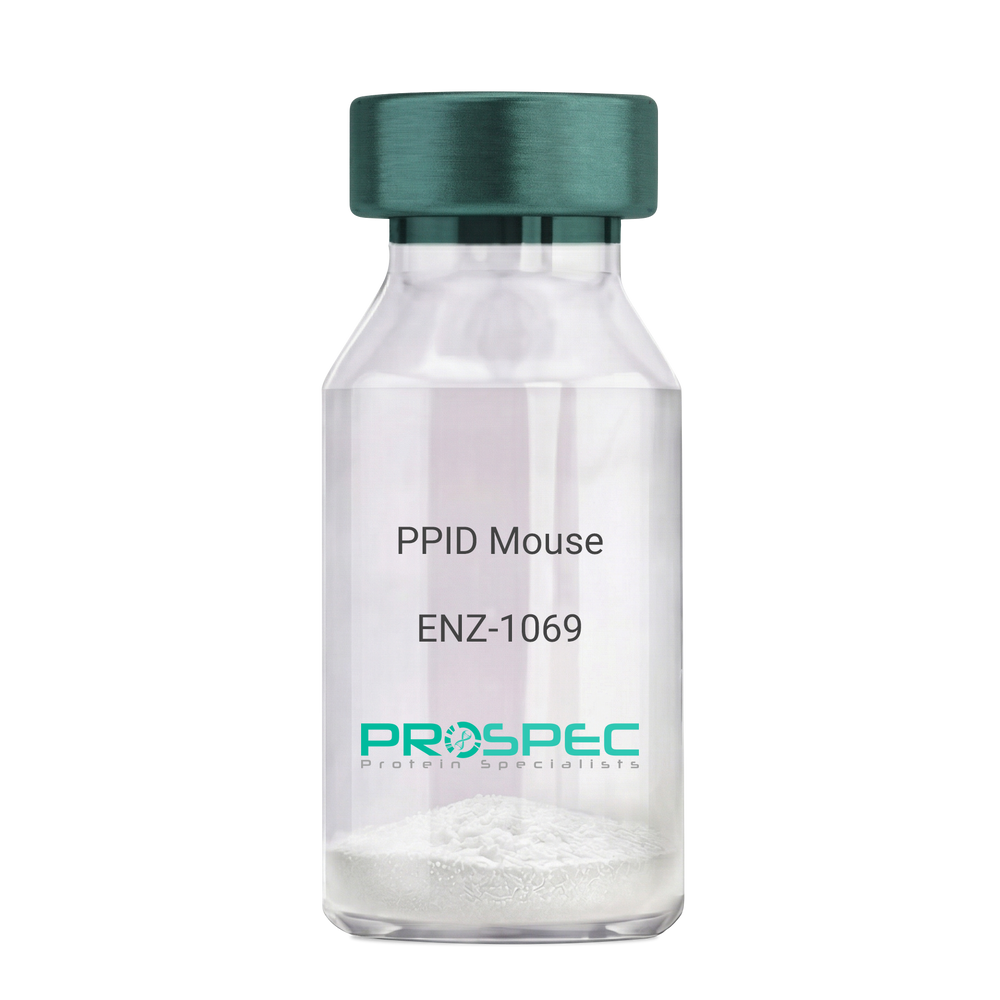
    Name :

    PPID Mouse

    Description:

    Peptidylprolyl Isomerase D Mouse Recombinant

    Peptidyl-prolyl cis-trans isomerase D, PPIase D, 40 kDa peptidyl-prolyl cis-trans isomerase, Cyclophilin-40, CYP-40, Cyclophilin-related protein, CYP40, CYPD, PPID, Peptidylprolyl Isomerase D.

    Product # :

    ENZ-1069

    Price :

    Quantity :

    Shipping Method :

    Ice Icon

    Shipped with Ice Packs

    Add To Cart

    More Info

    • description
    • source
    • formulation
    • purity
    • biological activity
    • More Info
    • Citations

    Description

    PPID Mouse Recombinant produced in E.Coli is a single, non-glycosylated polypeptide chain containing 395 amino acids (1-370a.a.) and having a molecular mass of 43.4kDa. PPID is fused to a 25 amino acid His-tag at N-terminus & purified by proprietary chromatographic techniques.

    Source

    Escherichia Coli.

    Formulation

    PPID protein solution (1mg/ml) containing 20mM Tris-Hcl buffer (pH8.0), 10% glycerol and 1mM DTT.

    Purity

    Greater than 90.0% as determined by SDS-PAGE.

    Biological Activity

    Specific activity is > 700nmol/min/mg, and is defined as the amount of enzyme that cleaves 1umole of suc-AAFP-PNA per minute at 37°C in Tris–HCl pH 8.0 using chymotrypsin.

    Citations

    More Info

    • Introduction

      Cyclophilin-D is a member of the peptidyl-prolyl cis-trans isomerase (PPIase) family. PPIases catalyze the cis-trans isomerization of proline imidic peptide bonds in oligopeptides and speeds up the protein folding. Cyclophilin-D possess PPIase activity and binds to the immunosuppressant cyclosporin-A. Cyclophilin-D is very well known that its overexpression suppresses the apoptosis in cancer cell. Cyclophilin-D suppresses apoptotic cell death by the use of mitochondrial hexokinase-2 dependent mechanism in cancer cells.

    • Synonyms

      Peptidyl-prolyl cis-trans isomerase D, PPIase D, 40 kDa peptidyl-prolyl cis-trans isomerase, Cyclophilin-40, CYP-40, Cyclophilin-related protein, CYP40, CYPD, PPID, Peptidylprolyl Isomerase D.

    • Physical Appearance

      Sterile Filtered colorless solution.

    • Stability

      Store at 4°C if entire vial will be used within 2-4 weeks. Store, frozen at -20°C for longer periods of time. For long term storage it is recommended to add a carrier protein (0.1% HSA or BSA). Please prevent freeze-thaw cycles.

    • Amino Acid Sequence

      MGSSHHHHHH SSGLVPRGSH MGSEFMSHAS PAAKPSNSKN PRVFFDVDIG GERVGRIVLE LFADIVPKTA ENFRALCTGE KGTGSTTGKP LHFKGCPFHR IIKKFMIQGG DFSNQNGTGG ESIYGEKFED ENFHYKHDRE GLLSMANAGP NTNGSQFFIT TVPTPHLDGK HVVFGQVIKG LGVARTLENV EVNGEKPAKL CVIAECGELK EGDDWGIFPK DGSGDSHPDF PEDADIDLKD VDKILLISED LKNIGNTFFK SQNWEMAIKK YAKVLRYVDS SKAVIEKADR SRLQPIALSC VLNIGACKLK MSNWQGAIDS CLEALEMDPS NTKALYRKAQ GWQGLKEYDQ ALADLKKAQE IAPGDKAIQA ELLKVKQMIK AQKDKEKAVY AKMFA.

    ProSpec's products are furnished for LABORATORY RESEARCH USE ONLY. The product may not be used as drugs, agricultural or pesticidal products, food additives or household chemicals.

    Ppid Mouse
  • View Data Sheet

    Name :

    CCL2 Human, HEK

    Description:

    Monocyte Chemotactic Protein-1/MCAF (CCL2) Human Recombinant, HEK

    CCL2, C-C motif chemokine 2, GDCF-2, HSMCR30, JE, HC11, MCAF, MCP-1, MCP1, Scya2, Sigje, SMC-CF, Immediate-early serum-responsive protein JE, Monocyte chemoattractant protein 1, Small-inducible cytokine A2.

    Product # :

    CHM-048

    Price :

    Quantity :

    Shipping Method :

    Ice Icon

    Shipped with Ice Packs

    Add To Cart

    More Info

    • description
    • source
    • formulation
    • purity
    • biological activity
    • More Info
    • Citations

    Description

    MCP1 Human HEK Recombinant produced in HEK293 Cells is a single, glycosylated polypeptide chain containing 82 amino acids (24-99a.a) and having a molecular mass of 9.5 kDa.MCP1 is expressed with a 6 amino acid His tag at C-Terminus and purified by proprietary chromatographic techniques.

    Source

    HEK293 Cells.

    Formulation

    The MCP1 solution (0.25mg/1ml) contains phosphate buffered saline (pH7.4) and 10% glycerol.

    Purity

    Greater than 90.0% as determined by SDS-PAGE.

    Biological Activity

    Measured its ability to chemoattract using THP-1 human acute monocytic leukemia cells. The ED50 range ≤ 30 ng/ml.

    Citations

    More Info

    • Introduction

      Chemokine (C-C motif) ligand 2 (CCL2) is a small cytokine belonging to the CC chemokine family that is also known as monocyte chemotactic protein-1 (MCP-1). It is found at the site of tooth eruption and bone degradation. In the bone, CCL2 is expressed by mature osteoclasts and osteoblasts and is under the control of nuclear factor ?B (NF?B).CCL2 recruits immune cells, such as monocytes, to sites of tissue injury and infection.This chemokine is produced as a protein precursor containing signal peptide of 23 amino acids and a mature peptide of 76 amino acids. It is a monomeric polypeptide, with a molecular weight of approximately 13kDa. As with many other CC chemokines, CCL2 is located on chromosome 17 in humans.The cell surface receptors that bind CCL2 are CCR2 and CCR5

    • Synonyms

      CCL2, C-C motif chemokine 2, GDCF-2, HSMCR30, JE, HC11, MCAF, MCP-1, MCP1, Scya2, Sigje, SMC-CF, Immediate-early serum-responsive protein JE, Monocyte chemoattractant protein 1, Small-inducible cytokine A2.

    • Physical Appearance

      Sterile filtered colorless solution.

    • Stability

      Store at 4°C if entire vial will be used within 2-4 weeks.Store, frozen at -20°C for longer periods of time. For long term storage it is recommended to add a carrier protein (0.1% HSA or BSA).Avoid multiple freeze-thaw cycles.

    • Amino Acid Sequence

      QPDAINAPVT CCYNFTNRKI SVQRLASYRR ITSSKCPKEA VIFKTIVAKE ICADPKQKWV QDSMDHLDKQ TQTPKTHHHH HH

    • Background

      What is the molecular weight/Mw of CCL2 HUMAN, HEK Protein?
      CCL2 HUMAN, HEK Protein has a total Mw of 9.5kDa.

      What is the source or expression system of CCL2 HUMAN, HEK Protein?
      HEK293 Cells.

      What is the Purity of CCL2 HUMAN, HEK Protein?
      CCL2 HUMAN, HEK Protein is >90% pure as determined by SDS-PAGE.

      What is the Biological Activity of CCL2 HUMAN, HEK Protein?
      Measured its ability to chemoattract using THP-1 human acute monocytic leukemia cells. The ED50 range ≤ 30 ng/ml.

      What is the amino acid sequence of CCL2 HUMAN, HEK Protein?
      QPDAINAPVT CCYNFTNRKI SVQRLASYRR ITSSKCPKEA VIFKTIVAKE ICADPKQKWV QDSMDHLDKQ TQTPKTHHHH HH

      What applications can CCL2 HUMAN, HEK Protein be used in?
      CCL2 HUMAN, HEK Protein can probably be used in western blot, ELISA and Lateral Flow.

      What is the endotoxin level for CCL2 HUMAN, HEK Protein?
      The endotoxin level is minimal, CCL2 HUMAN, HEK Protein was purified using conventional chromatography techniques.

      Who supplies recombinant CCL2 HUMAN, HEK that is in stock and available to order?
      ProSpec has the protein in stock and available for immediate delivery.

      Where can I order bulk mg quantities of CCL2 HUMAN, HEK?
      ProSpec supplies bulk mg quantities at affordable prices that meets research and academic budgets.

    ProSpec's products are furnished for LABORATORY RESEARCH USE ONLY. The product may not be used as drugs, agricultural or pesticidal products, food additives or household chemicals.

    Ccl2 Human
  • View Data Sheet

    Name :

    CREB3L2 Human

    Description:

    CAMP Responsive Element Binding Protein 3-Like 2 Human Recombinant

    CAMP Responsive Element Binding Protein 3-Like 2, BBF2H7, CAMP-Responsive Element-Binding Protein 3-Like Protein 2, BBF2 Human Homolog On Chromosome 7, Cyclic AMP-Responsive Element-Binding Protein 3-Like Protein 2, Basic Transcription Factor 2, B-ZIB Transcription Factor, FUS/BBF2H7 Protein, TCAG_1951439, Cyclic AMP-responsive element-binding protein 3-like protein 2, cAMP-responsive element-binding protein 3-like protein 2.

    Product # :

    PRO-2153

    Price :

    Quantity :

    Shipping Method :

    Ice Icon

    Shipped with Ice Packs

    Add To Cart

    More Info

    • description
    • source
    • formulation
    • purity
    • More Info
    • Citations

    Description

    CREB3L2 Human Recombinant produced in E.Coli is a single, non-glycosylated polypeptide chain containing 401 amino acids (1-378 a.a) and having a molecular mass of 44kDa. CREB3L2 is fused to a 23 amino acid His-tag at N-terminus & purified by proprietary chromatographic techniques.

    Source

    Escherichia Coli.

    Formulation

    CREB3L2 protein solution (0.25mg/ml) containing 20mM Tris-HCl buffer (pH 8.0), 0.2M NaCl, 50% glycerol and 1mM DTT.

    Purity

    Greater than 90% as determined by SDS-PAGE.

    Citations

    More Info

    • Introduction

      CAMP Responsive Element Binding Protein 3-Like 2, also known as CREB3L2 belongs to the oasis bZIP transcription factor family. Members of this family are able to dimerize however from homodimers only. CREB3L2 is a transcriptional activator. Translocations between CREB3L2 on chromosome 7 and the gene fused in sarcoma on chromosome 16 can be found in some tumors. One disease which is associated with CREB3L2 is myxofibrosarcoma.

    • Synonyms

      CAMP Responsive Element Binding Protein 3-Like 2, BBF2H7, CAMP-Responsive Element-Binding Protein 3-Like Protein 2, BBF2 Human Homolog On Chromosome 7, Cyclic AMP-Responsive Element-Binding Protein 3-Like Protein 2, Basic Transcription Factor 2, B-ZIB Transcription Factor, FUS/BBF2H7 Protein, TCAG_1951439, Cyclic AMP-responsive element-binding protein 3-like protein 2, cAMP-responsive element-binding protein 3-like protein 2.

    • Physical Appearance

      Sterile filtered colorless solution.

    • Stability

      Store at 4°C if entire vial will be used within 2-4 weeks. Store, frozen at -20°C for longer periods of time. For long term storage it is recommended to add a carrier protein (0.1% HSA or BSA).Avoid multiple freeze-thaw cycles.

    • Amino Acid Sequence

      MGSSHHHHHH SSGLVPRGSH MGSMEVLESG EQGVLQWDRK LSELSEPGDG EALMYHTHFS ELLDEFSQNV LGQLLNDPFL SEKSVSMEVE PSPTSPAPLI QAEHSYSLCE EPRAQSPFTH ITSDSFNDDE VESEKWYLST DFPSTSIKTE PITDEPPPGL VPSVTLTITA ISTPLEKEEP PLEMNTGVDS SCQTIIPKIK LEPHEVDQFL NFSPKEAPVD HLHLPPTPPS SHGSDSEGSL SPNPRLHPFS LPQTHSPSRA APRAPSALSS SPLLTAPHKL QGSGPLVLTE EEKRTLIAEG YPIPTKLPLS KSEEKALKKI RRKIKNKISA QESRRKKKEY MDSLEKKVES CSTENLELRK KVEVLENTNR TLLQQLQKLQ TLVMGKVSRT CKLAGTQTGT C.

    ProSpec's products are furnished for LABORATORY RESEARCH USE ONLY. The product may not be used as drugs, agricultural or pesticidal products, food additives or household chemicals.

    Creb3L2 Human
  • View Data Sheet

    Name :

    REN Mouse

    Description:

    Renin Mouse Recombinant

    Renin-1, Angiotensinogenase, Kidney renin, Ren1, Ren, Ren-A, Ren1c, Ren1d, Rn-1, Rnr.

    Product # :

    PRO-2251

    Price :

    Quantity :

    Shipping Method :

    Ice Icon

    Shipped with Ice Packs

    Add To Cart

    More Info

    • description
    • source
    • formulation
    • purity
    • More Info
    • Citations

    Description

    REN produced in Sf9 Baculovirus cells is a single, glycosylated polypeptide chain containing 387 amino acids (22-402 a.a.) and having a molecular mass of 42.5kDa (Migrates at 40-57kDa on SDS-PAGE under reducing conditions).REN is expressed with a 6 amino acid His tag at C-Terminus and purified by proprietary chromatographic techniques.

    Source

    Sf9, Baculovirus cells.

    Formulation

    REN protein solution (0.25mg/ml) contains Phosphate Buffered Saline (pH 7.4) and 10% glycerol.

    Purity

    Greater than 95.0% as determined by SDS-PAGE.

    Citations

    More Info

    • Introduction

      Renin is a highly specific endopeptidase which generates angiotensin I from angiotensinogen in the plasma. angiotensin I is an important regulator of blood pressure and electrolyte balance. Renin initiates a cascade of reactions that produce an elevation of blood pressure and increased sodium retention by the kidney.

    • Synonyms

      Renin-1, Angiotensinogenase, Kidney renin, Ren1, Ren, Ren-A, Ren1c, Ren1d, Rn-1, Rnr.

    • Physical Appearance

      Sterile Filtered colorless solution.

    • Stability

      Store at 4°C if entire vial will be used within 2-4 weeks. Store, frozen at -20°C for longer periods of time. For long term storage it is recommended to add a carrier protein (0.1% HSA or BSA). Avoid multiple freeze-thaw cycles.

    • Amino Acid Sequence

      LPTRTATFER IPLKKMPSVR EILEERGVDM TRLSAEWGVF TKRPSLTNLT SPVVLTNYLN TQYYGEIGIG TPPQTFKVIF DTGSANLWVP STKCSRLYLA CGIHSLYESS DSSSYMENGS DFTIHYGSGR VKGFLSQDSV TVGGITVTQT FGEVTELPLI PFMLAKFDGV LGMGFPAQAV GGVTPVFDHI LSQGVLKEEV FSVYYNRGSH LLGGEVVLGG SDPQHYQGNF HYVSISKTDS WQITMKGVSV GSSTLLCEEG CAVVVDTGSS FISAPTSSLK LIMQALGAKE KRIEEYVVNC SQVPTLPDIS FDLGGRAYTL SSTDYVLQYP NRRDKLCTLA LHAMDIPPPT GPVWVLGATF IRKFYTEFDR HNNRIGFALA RHHHHHH.

    ProSpec's products are furnished for LABORATORY RESEARCH USE ONLY. The product may not be used as drugs, agricultural or pesticidal products, food additives or household chemicals.

    Ren Mouse
  • View Data Sheet

    Name :

    AZGP1 Human

    Description:

    Alpha-2-Glycoprotein 1 Zinc-Binding Human

    Zn-alpha-2-glycoprotein, Zn-alpha-2-GP, AZGP1, ZAG, Zinc-alpha-2-glycoprotein, ZNGP1, ZA2G.

    Product # :

    PRO-1605

    Price :

    Quantity :

    Shipping Method :

    Room Temp Icon

    Shipped at Room temp

    Add To Cart

    More Info

    • description
    • source
    • formulation
    • purity
    • More Info
    • Citations

    Description

    The Human Zinc-Alpha 2 Glycoprotein produced from Human Serum has a molecular mass of 32.14kDa (calculated without glycosylation) containing 278 amino acid residues.

    Source

    Human Serum.

    Formulation

    ZA2G protein filtered (0.4µm) and lyophilized in 0.5mg/ml in 20mM TRIS and 50mM NaCl, pH 8.0.

    Purity

    Greater than 80% as determined by SDS-PAGE.

    Citations

    More Info

    • Introduction

      Zinc-alpha-2-glycoprotein (ZAG) is found in body fluids such as serum, sweat, and seminal and breast cyst fluids. It is identical in amino acid sequence to tumor-derived lipid mobilizing factor (LMF), a protein associated with the dramatic loss of adipose body stores in cancer cachexia, and has been shown to stimulate lipolysis by adipocytes in vivo and in vitro. A role for ZAG has been proposed in the regulation of body weight, and age-dependent changes in genetically influenced obesity, and also it regulates melanin production by normal and malignant melanocytes. It has also recently been classified as a novel adipokine in that it is produced by both white and brown fat adipocytes and may act in a local autocrine fashion in the reduction of adiposity in cachexia. Controlling ZAG/LMF's activity could be life-saving in the management of certain cancers and other cachexiainducing conditions, and its possible normal role in body fat store homeostasis is deserving of understanding in its own right. ZAG exhibits a class I major histocompatibility complex (MHC) fold but is a soluble protein rather than being anchored to plasma membranes and does not associate with alpha-2-microglobulin in humans. Like antigen-presenting MHC class I proteins, ZAG has an open apical groove, and X-ray crystallography of human derived ZAG revealed an unidentifiable electron density in a similar position to that occupied by antigenic peptides in classical MHC proteins and glycolipids in isoforms of CD1. This presumptive ligand is not a peptide, and the groove is too small to hold a glycolipid such as is presented by CD1 isoforms. By analogy with all other MHC class I-related proteins that have an open apical groove [some do not ], occupancy by a ligand is probably crucial to ZAG's biological function. Despite all of the structural and biochemical evidence that ZAG binds a ligand, none has so far been found by extraction from protein isolated from biological fluids. This difficulty could be because the ligand is labile, heterogeneous, or readily lost during purification procedures. Knowing more about how ZAG interacts with the compounds it has been found to bind, both natural and artificial, will inform searches for the elusive ligand(s) and its/their role in ZAG's signaling function.

    • Synonyms

      Zn-alpha-2-glycoprotein, Zn-alpha-2-GP, AZGP1, ZAG, Zinc-alpha-2-glycoprotein, ZNGP1, ZA2G.

    • Physical Appearance

      Filtered White lyophilized (freeze-dried) powder.

    • Stability

      Store lyophilized protein at -20°C. Aliquot the product after reconstitution to avoid repeated freezing/thawing cycles. Reconstituted protein can be stored at 4°C for a limited period of time; it does not show any change after two weeks at 4°C.

    • Solubility

      It is recommended to add deionized water to prepare a working stock solution of approximately 0.5 mg/ml and let the lyophilized pellet dissolve completely. Product is not sterile! Please filter the product by an appropriate sterile filter before using it in the cell culture.

    • Amino Acid Sequence

      QENQDGRYSL TYIYTGLSKH VEDVPAFQAL GSLNDLQFFR YNSKDRKSQP MGLWRQVEGM EDWKQDSQLQ KAREDIFMET LKDIVEYYND SNGSHVLQGR FGCEIENNRS SGAFWKYYYD GKDYIEFNKE IPAWVPFDPA AQITKQKWEA EPVYVQRAKA YLEEECPATL RKYLKYSKNI LDRQDPPSVV VTSHQAPGEK KKLKCLAYDF YPGKIDVHWT RAGEVQEPEL RGDVLHNGNG TYQSWVVVAV PPQDTAPYSC HVQHSSLAQP LVVPWEAS.

    • Human Virus Test

      Blood samples from each donor have been tested and found negative for HBsAg, anti-HCV, HIV Ag/Ab and syphilis.

    ProSpec's products are furnished for LABORATORY RESEARCH USE ONLY. The product may not be used as drugs, agricultural or pesticidal products, food additives or household chemicals.

    Human Azgp1
  • View Data Sheet

    Name :

    FGF 2 Human (147 a.a.)

    Description:

    Fibroblast Growth Factor Basic 147 a.a. Human Recombinant

    Prostatropin, HBGH-2, HBGF-2, FGF-2, FGF-b.

    Product # :

    CYT-557

    Price :

    Quantity :

    Shipping Method :

    Room Temp Icon

    Shipped at Room temp

    Add To Cart

    More Info

    • description
    • source
    • formulation
    • purity
    • biological activity
    • More Info
    • Citations

    Description

    Fibroblast Growth Factor-2 Human Recombinant (FGF-2) produced in E.Coli is a single, non-glycosylated, polypeptide chain containing 147 amino acids and having a molecular mass of 16.5kDa. The FGF2 is purified by proprietary chromatographic techniques.

    Source

    Escherichia Coli.

    Formulation

    The bFGF was lyophilized from a sterile filtered solution containing 20mM Tris-HCl, pH 7.6 and 150mM NaCl.

    Purity

    Greater than 98.0% as determined by SDS-PAGE.

    Biological Activity

    The ED50, Calculated by the dose- dependent proliferation of mouse BALB/c 3T3 cells is < 0.05ng/ml corresponding to a specific activity of 2.0x10⁷ units/mg.

    Citations

    More Info

    • Introduction

      FGF-basic is a member of the fibroblast growth factor (FGF) family. FGF family members possess broad mitogenic and cell survival activities, and are involved in a variety of biological processes, including embryonic development, cell growth, morphogenesis, tissue repair, tumor growth and invasion. This protein functions as a modifier of endothelial cell migration and proliferation, as well as an angiogenic factor. It acts as a mitogen for a variety of mesoderm- and neuroectoderm-derived cells in vitro, thus is thought to be involved in organogenesis. Three alternatively spliced variants encoding different isoforms have been described. The heparin-binding growth factors are angiogenic agents in vivo and are potent mitogens for a variety of cell types in vitro. There are differences in the tissue distribution and concentration of these 2 growth factors.

    • Synonyms

      Prostatropin, HBGH-2, HBGF-2, FGF-2, FGF-b.

    • Physical Appearance

      Sterile Filtered White lyophilized (freeze-dried) powder.

    • Stability

      Lyophilized basic-FGF although stable at room temperature for 3 weeks, should be stored desiccated below -18°C. Upon reconstitution FGFb should be stored at 4°C between 2-7 days and for future use below -18°C.For long term storage it is recommended to add a carrier protein (0.1% HSA or BSA).Please prevent freeze-thaw cycles.

    • Solubility

      It is recommended to reconstitute the lyophilized FGF-B in sterile 18MΩ-cm H2O not less than 100µg/ml, which can then be further diluted to other aqueous solutions.

    • Amino Acid Sequence

      MPALPEDGGS GAFPPGHFKD PKRLYCKNGG FFLRIHPDGR VDGVREKSDP HIKLQLQAEE RGVVSIKGVC ANRYLAMKED GRLLASKCVT DECFFFERLE SNNYNTYRSR KYTSWYVALK RTGQYKLGSK TGPGQKAILF LPMSAKS.

    • Background

      What is the molecular weight/Mw of FGF 2 Protein?
      FGF 2 Protein has a total Mw of 16.5kDa.

      What is the source or expression system of FGF 2 Protein?
      Escherichia Coli.

      What is the Purity of FGF 2 Protein?
      FGF 2 Protein is >98% pure as determined by SDS-PAGE.

      What is the Biological Activity of FGF 2 Protein?
      The ED50, Calculated by the dose- dependent proliferation of mouse BALB/c 3T3 cells is < 0.05ng/ml corresponding to a specific activity of 2.0x10⁷ units/mg.

      What is the amino acid sequence of FGF 2 Protein?
      MPALPEDGGS GAFPPGHFKD PKRLYCKNGG FFLRIHPDGR VDGVREKSDP HIKLQLQAEE RGVVSIKGVC ANRYLAMKED GRLLASKCVT DECFFFERLE SNNYNTYRSR KYTSWYVALK RTGQYKLGSK TGPGQKAILF LPMSAKS.

      What applications can FGF 2 Protein be used in?
      FGF 2 Protein can probably be used in western blot, ELISA and Lateral Flow.

      What is the endotoxin level for FGF 2 Protein?
      The endotoxin level is minimal, FGF 2 Protein was purified using conventional chromatography techniques.

    ProSpec's products are furnished for LABORATORY RESEARCH USE ONLY. The product may not be used as drugs, agricultural or pesticidal products, food additives or household chemicals.

    Fgf 2 147 Aa Human
  • View Data Sheet

    Name :

    PLGF2 Human, HEK

    Description:

    Placental Growth Factor-2, HEK Human Recombinant

    PIGF, PGF, PlGF-2, PLGF-2.

    Product # :

    CYT-1228

    Price :

    Quantity :

    Shipping Method :

    Ice Icon

    Shipped with Ice Packs

    Add To Cart

    More Info

    • description
    • source
    • formulation
    • purity
    • biological activity
    • More Info
    • Citations

    Description

    PLGF2 Human Recombinant produced in HEK293 cells is a single, glycosylated polypeptide chain (a.a 19-170) containing 158 amino acids and having a molecular mass of 18.1kDa. PLGF2 is fused to a 6 amino acid His tag at C-terminus and purified by proprietary chromatographic techniques.

    Source

    HEK293 cells.

    Formulation

    PLGF2 protein (0.5mg/ml) contains Phosphate-Buffered Saline (pH 7.4) and 10% glycerol.

    Purity

    Greater than 90.0% as determined by SDS-PAGE.

    Biological Activity

    Measured by its binding ability in a functional ELISA with Human VEGFR1/Flt-1.

    Citations

    More Info

    • Synonyms

      PIGF, PGF, PlGF-2, PLGF-2.

    • Physical Appearance

      Sterile Filtered colorless solution.

    • Stability

      Store at 4°C if entire vial will be used within 2-4 weeks. Store, frozen at -20°C for longer periods of time. For long term storage it is recommended to add a carrier protein (0.1% HSA or BSA). Avoid multiple freeze-thaw cycles.

    • Amino Acid Sequence

      LPAVPPQQWA LSAGNGSSEV EVVPFQEVWG RSYCRALERL VDVVSEYPSE VEHMFSPSCV SLLRCTGCCG DENLHCVPVE TANVTMQLLK IRSGDRPSYV ELTFSQHVRC ECRPLREKMK PERRRPKGRG KRRREKQRPT DCHLCGDAVP RRHHHHHH.

    • Background

      PLGF2, a homodimeric glycoprotein, exhibits a unique structural configuration essential for its interactions with VEGF receptors and other signaling molecules. The human recombinant form provides a controlled platform for studying the three-dimensional structure of PLGF2, elucidating its binding affinities and conformational dynamics. Understanding its structure is fundamental for deciphering how PLGF2 mediates angiogenic signaling and vascular remodeling.

    ProSpec's products are furnished for LABORATORY RESEARCH USE ONLY. The product may not be used as drugs, agricultural or pesticidal products, food additives or household chemicals.

    Pigf2 Human
  • View Data Sheet

    Name :

    MIF Antibody

    Description:

    Macrophage Migration Inhibitory Factor, Mouse Anti Human

    Phenylpyruvate tautomerase, Glycosylation- inhibiting factor, GIF, MMIF, MIF.

    Product # :

    ANT-311

    Price :

    Quantity :

    Shipping Method :

    Ice Icon

    Shipped with Ice Packs

    Add To Cart

    More Info

    • formulation
    • More Info
    • Citations

    Formulation

    1mg/ml containing PBS, pH-7.4, 0.02% Sodium Azide and 10% Glycerol.

    Citations

    More Info

    • Introduction

      The cytokine Macrophage migration inhibitory factor (MIF) has been identified to be secreted by the pituitary gland and the monocyte/macrophage and to play an important role in endotoxic shock. MIF has the unique property of being released from macrophages and T cells in response to physiological concentrations of glucocorticoids. The secretion of MIF is tightly regulated and decreases at high, anti-inflammatory steroid concentration.

    • Synonyms

      Phenylpyruvate tautomerase, Glycosylation- inhibiting factor, GIF, MMIF, MIF.

    • Immunogen

      Anti-human MIF mAb, is derived from hybridization of mouse SP2/0 myeloma cells with spleen cells from BALB/c mice immunized with Recombinant human MIF amino acids 1-114 purified from E. coli.

    • Ig Subclass

      Mouse IgG1 heavy chain and κ light chain.

    • Clone

      P4E4AT.

    • Applications

      MIF antibody has been tested by ELISA and Western blot analysis to assure specificity and reactivity. Since application varies, however, each investigation should be titrated by the reagent to obtain optimal results.

    • Type

      Mouse Anti Human Monoclonal.

    • Storage Procedures

      For periods up to 1 month store at 4°C, for longer periods of time, store at -20°C. Prevent freeze thaw cycles.

    • Purification Method

      MIF antibody was purified from mouse ascitic fluids by protein-G affinity chromatography.

    ProSpec's products are furnished for LABORATORY RESEARCH USE ONLY. The product may not be used as drugs, agricultural or pesticidal products, food additives or household chemicals.

    Mif Antibody
  • View Data Sheet

    Name :

    BAFF Antibody

    Description:

    B-cell Activating Factor, Mouse Anti Human

    BAFF, BLYS, CD257, TALL1, THANK, ZTNF4, TALL-1, TNFSF20, TNFSF13B.

    Product # :

    ANT-367

    Price :

    Quantity :

    Shipping Method :

    Ice Icon

    Shipped with Ice Packs

    Add To Cart

    More Info

    • formulation
    • More Info
    • Citations

    Formulation

    1mg/ml containing PBS, pH-7.4, & 0.1% Sodium Azide.

    Citations

    More Info

    • Introduction

      Binds to tnfrsf13b/taci and tnfrsf17/bcma. tnfsf13/april binds to the same 2 receptors, together, they form a 2 ligands -2 receptors pathway involved in the stimulation of b- and t-cell function and the regulation of humoral immunity. A third b-cell specific baff-receptor (baffr/br3) promotes the survival of mature b-cells and the b-cell response. B Lymphocyte Stimulator functions as a potent B-cell growth factor in costimulation assays. Administration of BAFF Human recombinant to mice disrupts splenic B-cell and T-cell zones and results in elevated levels of serum immunoglobulin.

    • Synonyms

      BAFF, BLYS, CD257, TALL1, THANK, ZTNF4, TALL-1, TNFSF20, TNFSF13B.

    • Physical Appearance

      Sterile Filtered colorless solution.

    • Immunogen

      Anti-human BAFF mAb, is derived from hybridization of mouse F0 myeloma cells with spleen cells from BALB/c mice immunized with Recombinant human BAFF amino acids 134-285 purified from E. coli.

    • Ig Subclass

      Mouse IgG3 heavy chain and κ light chain.

    • Clone

      PH4C7AT.

    • Applications

      BAFF antibody has been tested by ELISA and Western blot analysis to assure specificity and reactivity. Since application varies, however, each investigation should be titrated by the reagent to obtain optimal results. Recommended dilution range for Western blot analysis is 1:500 ~ 1,000. Recommended starting dilution is 1:500.

    • Type

      Mouse Anti Human Monoclonal.

    • Storage Procedures

      For periods up to 1 month store at 4°C, for longer periods of time, store at -20°C. Prevent freeze thaw cycles.

    • Purification Method

      BAFF antibody was purified from mouse ascitic fluids by protein-G affinity chromatography.

    ProSpec's products are furnished for LABORATORY RESEARCH USE ONLY. The product may not be used as drugs, agricultural or pesticidal products, food additives or household chemicals.

    Baff Antibody
  • View Data Sheet

    Name :

    VEGF Human, HEK

    Description:

    Vascular Endothelial Growth Factor Human Recombinant, HEK

    Vascular endothelial growth factor A, VEGF-A, Vascular permeability factor, VPF, VEGF, MGC70609.

    Product # :

    CYT-225

    Price :

    Quantity :

    Shipping Method :

    Room Temp Icon

    Shipped at Room temp

    Add To Cart

    More Info

    • description
    • source
    • formulation
    • purity
    • biological activity
    • More Info
    • Citations

    Description

    Vascular Endothelial Growth Factor Human Recombinant produced in HEK293 cells is a double, glycosylated, polypeptide chain containing 165 amino acids (27-191) and having a molecular mass of 40 kDa. The VEGF is purified by proprietary chromatographic techniques.

    Source

    HEK293 (Human Embryonic Kidney cell line).

    Formulation

    The protein was lyophilized from a 0.2µM filtered solution of 20mM PB, 150mM NaCl, pH 7.2.

    Purity

    Greater than 95.0% as determined by SDS-PAGE.

    Biological Activity

    Determined by the dose-dependent stimulation of the proliferation of human umbilical vein endothelial cells (HUVEC) using a concentration range of 4.0ng/ml, corresponding to a specific activity of 2.5 x 10⁵ Units/mg.

    Citations

    More Info

    • Introduction

      Vascular endothelial growth factor is an important signaling protein involved in both vasculogenesis and angiogenesis. As its name implies, VEGF activity has been mostly studied on cells of the vascular endothelium, although it does have effects on a number of other cell types (e.g. stimulation monocyte/ macrophagemigration, neurons, cancer cells, kidney epithelial cells ).VEGF mediates increased vascular permeability, induces angiogenesis, vasculogenesis and endothelial cell growth, promotes cell migration, and inhibits apoptosis. In vitro, VEGF has been shown to stimulate endothelial cell mitogenesisand cell migration. VEGF is also a vasodilator and increases microvascular permeability and was originally referred to as vascular permeability factor.
      Elevated levels of this protein are linked to POEMS syndrome, also known as Crow-Fukase syndrome. Mutations in this gene have been associated with proliferative and nonproliferative diabetic retinopathy.

    • Synonyms

      Vascular endothelial growth factor A, VEGF-A, Vascular permeability factor, VPF, VEGF, MGC70609.

    • Physical Appearance

      Sterile Filtered White lyophilized (freeze-dried) powder.

    • Stability

      Lyophilized Vascular Endothelial Growth Factor HEK although stable at room temperature for 3 weeks, should be stored desiccated below -18°C. Upon reconstitution VEGF HEK should be stored at 4°C between 2-7 days and for future use below -18°C. For long term storage it is recommended to add a carrier protein (0.1% HSA or BSA). Please prevent freeze-thaw cycles.

    • Solubility

      It is recommended to reconstitute the lyophilized Vascular Endothelial Growth Factor-HEK in sterile 18M-cm H2O not less than 100µg/ml, which can then be further diluted to other aqueous solutions.

    • Amino Acid Sequence

      APMAEGGGQN HHEVVKFMDV YQRSYCHPIE TLVDIFQEYP DEIEYIFKPS CVPLMRCGGC CNDEGLECVP TEESNITMQI MRIKPHQGQH IGEMSFLQHN KCECRPKKDR ARQENPCGPC SERRKHLFVQ DPQTCKCSCK NTDSRCKARQ LELNERTCRC DKPRR.

    ProSpec's products are furnished for LABORATORY RESEARCH USE ONLY. The product may not be used as drugs, agricultural or pesticidal products, food additives or household chemicals.

    Vegf Human Hek
  • View Data Sheet

    Name :

    Deslorelin

    Description:

    Deslorelin

    Product # :

    HOR-240

    Price :

    Quantity :

    Shipping Method :

    Room Temp Icon

    Shipped at Room temp

    Add To Cart

    More Info

    • description
    • formulation
    • purity
    • More Info
    • Citations

    Description

    Deslorelin is a potent LHRH/GnRH agonist has a molecular formula of C64H83N17O12, pGlu-His-Trp-Ser-Tyr-D-Trp-Leu-Arg-Pro-NHC2H5 having an Mw of 1284.4 Dalton.

    Formulation

    The Deslorelin peptide was lyophilized with no additives.

    Purity

    Greater than 99.0% as determined by RP-HPLC.

    Citations

    More Info

    • Introduction

      Deslorelin is being studied in the treatment of cancer as a way to block sex hormones made by the ovaries or testicles. It belongs to the family of drugs called gonadotropin-releasing hormone analogs. It is used for the induction of ovulation in mares. Deslorelin binds to and activates pituitary gonadotropin releasing hormone (GnRH) receptors. Continuous, prolonged administration of goserelin in males results in pituitary GnRH receptor desensitization and inhibition of pituitary secretion of follicle stimulating hormone (FSH) and luteinizing hormone (LH), leading to a significant decline in testosterone production; in females, prolonged administration results in a decrease in estradiol production.

    • Physical Appearance

      Sterile Filtered White lyophilized (freeze-dried) powder.

    • Stability

      Lyophilized Deslorelin although stable at room temperature for 3 weeks, should be stored desiccated below -18°C. Upon reconstitution Deslorelin should be stored at 4°C between 2-7 days and for future use below -18°C. For long term storage it is recommended to add a carrier protein (0.1% HSA or BSA).Please prevent freeze-thaw cycles.

    • Solubility

      It is recommended to reconstitute the lyophilized Deslorelin in sterile 18MΩ-cm H2O not less than 100µg/ml, which can then be further diluted to other aqueous solutions.

    ProSpec's products are furnished for LABORATORY RESEARCH USE ONLY. The product may not be used as drugs, agricultural or pesticidal products, food additives or household chemicals.

    Deslorelin
  • View Data Sheet

    Name :

    KRT18 Bovine

    Description:

    Cytokeratin-18 Bovine

    Keratin type I cytoskeletal 18, Cytokeratin-18, CK-18, Keratin-18, K18, KRT18,CYK18,Cell proliferation-inducing gene 46 protein.

    Product # :

    PRO-2785

    Price :

    Quantity :

    Shipping Method :

    Room Temp Icon

    Shipped at Room temp

    Add To Cart

    More Info

    • description
    • source
    • formulation
    • purity
    • More Info
    • Citations

    Description

    KRT18 Bovine having a calculated molecular mass of 45 kDa, pI-5.4.

    Source

    Bovine liver.

    Formulation

    KRT18 was lyophilized from a 1mg/ml solution containing 30mM Tris/HCI pH 8, 9M urea, 2mM EDTA, 2mM DTT and 10mM methylammonium chloride.

    Purity

    Greater than 95.0% as determined by SDS-PAGE.

    Citations

    More Info

    • Synonyms

      Keratin type I cytoskeletal 18, Cytokeratin-18, CK-18, Keratin-18, K18, KRT18,CYK18,Cell proliferation-inducing gene 46 protein.

    • Physical Appearance

      Sterile Filtered White lyophilized (freeze-dried) powder.

    • Stability

      Store the lyophilized KRT18 between 2-8°C, do not freeze. Upon reconstitution KRT18 should be stored at -20°C.Please prevent freeze-thaw cycles.

    • Solubility

      It is recommended to reconstitute the lyophilized KRT18 in sterile 18MΩ-cm H2O not less than 100µg/ml, which can then be further diluted to other aqueous solutions.

    • Background

      Keratin-18 (K18) is an intermediate filament protein that plays a vital role in maintaining the structural integrity of epithelial cells. Extensive research has been conducted on K18 in human and murine models, shedding light on its functions and implications for various epithelial tissues.

      However, the study of K18 in bovine tissues is an emerging area with potential for advancing our understanding of epithelial cell biology and its applications in veterinary medicine and biotechnology. Bovine tissues, such as the liver and gastrointestinal tract, are of particular interest due to their relevance in cattle production and food safety.

      This research aims to provide a comprehensive exploration of K18 in bovine tissues, elucidating its functions, structural significance, and potential applications.
      The primary objective of this research is to elucidate the role of K18 in bovine tissues, particularly in maintaining the structural integrity of epithelial cells.

      In vitro and ex vivo experiments, utilizing bovine epithelial cell cultures and tissue specimens, will be conducted to investigate how K18 contributes to cellular morphology, cytoskeletal organization, and tissue resilience. Understanding these mechanisms is fundamental for deciphering the complexities of epithelial cell biology in bovine species.
      The second objective is to assess the relevance of bovine K18 in veterinary medicine and cattle production. Studies involving bovine models will be conducted to evaluate the impact of K18 mutations or variations on tissue health, disease susceptibility, and meat quality. These investigations may provide valuable insights into potential applications in cattle breeding and food safety.


      The third objective is to explore the potential biotechnological applications of bovine K18. Research will investigate the use of K18-expressing bovine cells as models for studying epithelial-related diseases and for developing tissue engineering approaches for veterinary medicine and biotechnology.
      By delving into the functions and roles of K18 in bovine tissues, this research aims to expand our knowledge of epithelial cell biology, its implications for veterinary medicine, and its potential applications in biotechnology and cattle production.

    ProSpec's products are furnished for LABORATORY RESEARCH USE ONLY. The product may not be used as drugs, agricultural or pesticidal products, food additives or household chemicals.

    Keratin 18 Bovine
  • View Data Sheet

    Name :

    CTRB1 Human

    Description:

    Chymotrypsinogen-B1, Human Recombinant

    Product # :

    ENZ-1016

    Price :

    Quantity :

    Shipping Method :

    Room Temp Icon

    Shipped at Room temp

    Add To Cart

    More Info

    • description
    • source
    • formulation
    • purity
    • biological activity
    • More Info
    • Citations

    Description

    Recombinant Human CTRB1 expressed in E.coli containing 245 amino acids having a Mw of 27kDa is purified by standard chromatography techniques.

    Source

    E.coli

    Formulation

    The Human CTRB1 was lyophilized without any additives.

    Purity

    Greater than 95% as determined by HPLC.

    Biological Activity

    1100 units/mg protein.
    One unit is defined as the amount of enzyme that will hydrolyze 1.0 μmole of N-alpha-acetyl-L-tyrosine ethyl ester (ATEE) per min at pH 7.0 at 25°C.

    Citations

    More Info

    • Introduction

      Chymotrypsinogen-B1 (CTRB1) belongs to the serine protease family of enzymes and forms a main precursor of the pancreatic proteolytic enzymes. CTRB1 is located next to a related chymotrypsinogen gene. CTRB1 is a protein coding gene which encodes different isoforms which may undergo similar processing to generate the mature protein.

    • Physical Appearance

      Sterile Filtered lyophilized powder.

    • Stability

      Recombinant Human CTRB1 although stable at room temp for 1 week, should be stored desiccated below -18°C. For long term storage it is recommended to add a carrier protein (0.1% HSA or BSA).Please prevent freeze-thaw cycles.

    • Solubility

      It is recommended to reconstitute the lyophilized Human CTRB1 in 1ml 50mM HAc which can then be further diluted to other aqueous solutions.

    • Amino Acid Sequence

      CG VPAIHPVLSG LSRIVNGEDA VPGSWPWQVS LQDKTGFHFC GGSLISEDWV VTAAHCGVRT SDVVVAGEFD QGSDEENIQV LKIAKVFKNP KFSILTVNND ITLLKLATPA RFSQTVSAVC LPSADDDFPAGTLCATTGWG KTKYNANKTP DKLQQAALPL LSNAECKKSW GRRITDVMIC AGASGVSSCM GDSGGPLVCQ KDGAWTLVGI VSWGSDTCST SSPGVYARVTKLIPWVQKIL AAN.

    ProSpec's products are furnished for LABORATORY RESEARCH USE ONLY. The product may not be used as drugs, agricultural or pesticidal products, food additives or household chemicals.

    Ctrb1 Human
  • View Data Sheet

    Name :

    IRF3 Antibody

    Description:

    Interferon Regulatory Factor-3, Mouse Anti Human

    IRF-3, IRF3, Interferon Regulatory Factor 3.

    Product # :

    ANT-365

    Price :

    Quantity :

    Shipping Method :

    Ice Icon

    Shipped with Ice Packs

    Add To Cart

    More Info

    • formulation
    • More Info
    • Citations

    Formulation

    1mg/ml containing PBS, pH-7.4, & 0.1% Sodium Azide.

    Citations

    More Info

    • Introduction

      Members of the Interferon regulatory factor (IRF) family regulate gene expression critical to immune response, hemopoiesis, and proliferation. IRF-3 is a member of the IRF family, and is distinct from other family members. Its transcriptional activity is regulated solely by posttranslational modifications. It plays a crucial role in activation of innate immunity and inflammation in response to viral infection. IRF-3 mediates interferon-stimulated response element (isre) promoter activation. Functions as a molecular switch for antiviral activity. Dsrna generated during the course of an viral infection leads to IRF3 phosphorylation on the c-terminal serine/threonine cluster. This induces a conformational change, leading to its dimerization, nuclear localization and association with creb binding protein (crebbp) to form dsrna-activated factor 1 (draf1), a complex which activates the transcription of genes under the control of isre. The complex binds to the ie and prdiii regions on the ifn

    • Synonyms

      IRF-3, IRF3, Interferon Regulatory Factor 3.

    • Physical Appearance

      Sterile Filtered clear solution.

    • Immunogen

      Anti-human IRF3 mAb, is derived from hybridization of mouse SP2/0 myeloma cells with spleen cells from BALB/c mice immunized with Recombinant human IRF3 amino acids 108-166 purified from E. coli.

    • Ig Subclass

      Mouse IgG1 heavy chain and κ light chain.

    • Clone

      P3F10AT.

    • Applications

      IRF3 antibody has been tested by ELISA and Western blot analysis to assure specificity and reactivity. Since application varies, however, each investigation should be titrated by the reagent to obtain optimal results. Recommended dilution range for Western blot analysis is 1:1,000 ~ 2,000. Recommended starting dilution is 1:1,000.

    • Type

      Mouse Anti Human Monoclonal.

    • Storage Procedures

      For periods up to 1 month store at 4°C, for longer periods of time, store at -20°C. Prevent freeze thaw cycles.

    • Purification Method

      IRF3 antibody was purified from mouse ascitic fluids by protein-G affinity chromatography.

    ProSpec's products are furnished for LABORATORY RESEARCH USE ONLY. The product may not be used as drugs, agricultural or pesticidal products, food additives or household chemicals.

    Irf3 Antibody
  • View Data Sheet

    Name :

    Lanreotide

    Description:

    Lanreotide

    Product # :

    HOR-282

    Price :

    Quantity :

    Shipping Method :

    Room Temp Icon

    Shipped at Room temp

    Add To Cart

    More Info

    • description
    • formulation
    • purity
    • More Info
    • Citations

    Description

    Lanreotide is an octapeptide, an analogue of a naturally occurring hormone, somatostatin.

    Formulation

    The protein (1mg/ml) was lyophilized with 5mg manntitol and 0.04mg tween-80.

    Purity

    Greater than 99.0% as determined by:
    (a) Analysis by RP-HPLC.
    (b) Analysis by SDS-PAGE.

    Citations

    More Info

    • Introduction

      Lanreotide is a peptide inhibitor of a number of endocrine, neuroendocrine, exocrine and paracrine functions. It shows good affinity for peripheral somatostatin receptors (anterior pituitary and pancreatic). In contrast, its affinity for central receptors is much lower. This profile confers a good specificity of action at the level of growth hormone and digestive hormone secretion. Lanreotide shows a much longer duration of action than natural somatostatin. In addition, its marked selectivity for the secretion of growth hormone, compared to that of insulin, makes it a suitable candidate for the treatment of acromegaly. By inhibiting the synthesis of thyroid stimulating hormone (TSH), lanreotide also normalised thyroid function of patients with thyrotrophin secreting adenomas in 50% (8/16) of the per-protocol population treated for 6 months. There was no significant reduction in the size of the adenoma. Furthermore, the inhibitory action of lanreotide on intestinal exocrine secretion, d

    • Physical Appearance

      Sterile Filtered White lyophilized (freeze-dried) powder.

    • Stability

      Lyophilized Lanreotide although stable at room temperature for 3 weeks, should be stored desiccated below -18°C. Upon reconstitution Lanreotide should be stored at 4°C between 2-7 days and for future use below -18°C.For long term storage it is recommended to add a carrier protein (0.1% HSA or BSA).Please prevent freeze-thaw cycles.

    • Solubility

      It is recommended to reconstitute the lyophilized Lanreotide in sterile 18MΩ-cm H2O not less than 100 µg/ml, which can then be further diluted to other aqueous solutions.

    ProSpec's products are furnished for LABORATORY RESEARCH USE ONLY. The product may not be used as drugs, agricultural or pesticidal products, food additives or household chemicals.

    Lanreotide
  • View Data Sheet

    Name :

    Heparin Sodium Porcine

    Description:

    Heparin Sodium Porcine

    Heparin, Porcine Heparin, Heparin Pig.

    Product # :

    PRO-2811

    Price :

    Quantity :

    Shipping Method :

    Room Temp Icon

    Shipped at Room temp

    Add To Cart

    More Info

    • description
    • source
    • formulation
    • biological activity
    • More Info
    • Citations

    Description

    Heparin Sodium Porcine is a natural native protein.

    Source

    Porcine Mucosa.

    Formulation

    The protein (1mg/ml) was lyophilized with no additives.

    Biological Activity

    200IU/mg.

    Citations

    More Info

    • Synonyms

      Heparin, Porcine Heparin, Heparin Pig.

    • Physical Appearance

      Sterile Filtered White lyophilized (freeze-dried) powder.

    • Stability

      Lyophilized Heparin Sodium Porcine although stable at room temperature for 3 weeks, should be stored desiccated below -18°C. Upon reconstitution Heparin Sodium Porcine should be stored at 4°C between 2-7 days and for future use below -18°C. For long term storage it is recommended to add a carrier protein (0.1% HSA or BSA). Please prevent freeze-thaw cycles.

    • Solubility

      It is recommended to reconstitute the lyophilized Heparin Sodium Porcine in sterile 18MΩ-cm H2O not less than 100µg/ml, which can then be further diluted to other aqueous solutions.

    • Background

      Heparin sodium, a highly sulfated glycosaminoglycan, has long been recognized for its pivotal role as an anticoagulant. In porcine systems, the study of Heparin Sodium takes on significance not only for its therapeutic applications in veterinary medicine but also for its potential translational impact on human health. This research embarks on an exploration of Heparin Sodium in porcine contexts, unraveling its structural intricacies, pharmacological attributes, and broader implications for health and biomedical interventions. By delving into the properties of Heparin Sodium in porcine models, scientists aim to deepen our understanding of its anticoagulant mechanisms, therapeutic potential, and the potential avenues for cross-species applications in healthcare.

      Structural Insights into Porcine Heparin Sodium:

      Porcine-derived Heparin Sodium, similar to its counterparts in other species, possesses a complex and highly sulfated structure. Understanding the nuances of its structure is fundamental for deciphering how Heparin Sodium interacts with antithrombin, leading to the inhibition of clotting factors and preventing the formation of blood clots. Structural insights contribute to optimizing its therapeutic efficacy.

      Pharmacological Attributes and Anticoagulant Mechanisms:

      Heparin Sodium's pharmacological attributes are central to its anticoagulant effects. In porcine systems, Heparin Sodium acts as an indirect anticoagulant by accelerating the activity of antithrombin, a natural inhibitor of clotting factors. Elucidating the specific mechanisms by which porcine Heparin Sodium modulates coagulation pathways contributes to refining its dosage and application in veterinary medicine.

      Therapeutic Potential in Veterinary Medicine:

      Porcine Heparin Sodium holds significant therapeutic potential in veterinary medicine, where clotting disorders and thrombotic events are prevalent. Understanding its pharmacokinetics, dosage requirements, and potential interactions with other drugs is crucial for optimizing its use in porcine healthcare. Additionally, exploring its safety profile in porcine models contributes to ensuring the well-being of animals under treatment.

      Cross-Species Applications and Translational Insights:

      Beyond its veterinary applications, porcine Heparin Sodium provides insights into potential cross-species applications, particularly in translational medicine. Understanding the similarities and differences in its anticoagulant mechanisms between porcine and human systems may open avenues for therapeutic innovations with relevance to both animal and human health.

      Challenges and Future Directions:

      While porcine Heparin Sodium presents opportunities for advancements in veterinary medicine and translational research, challenges persist. Fine-tuning its applications, addressing potential variations in its anticoagulant properties across different porcine breeds, and ensuring its safety and efficacy in diverse clinical scenarios are critical considerations for translational success.

      Porcine Heparin Sodium emerges as a key player in the landscape of anticoagulation, offering a bridge between veterinary and translational medicine. Its structural insights, pharmacological attributes, and therapeutic potential in porcine systems contribute not only to veterinary care but also offer translational insights with implications for human health. As researchers continue to unravel the molecular intricacies of porcine Heparin Sodium, they not only deepen our understanding of anticoagulation in veterinary contexts but also pave the way for innovative biomedical applications, shaping the future of healthcare for both animals and humans.

    ProSpec's products are furnished for LABORATORY RESEARCH USE ONLY. The product may not be used as drugs, agricultural or pesticidal products, food additives or household chemicals.

    Heparin Sodium
  • View Data Sheet

    Name :

    HDAC2 Antibody

    Description:

    Histone Deacetylase 2, Mouse Anti Human

    Histone deacetylase 2, YAF1, HD2, YY1-associated factor 1, transcriptional regulator homolog RPD3, RPD3, EC 3.5.1.98.

    Product # :

    ANT-742

    Price :

    Quantity :

    Shipping Method :

    Ice Icon

    Shipped with Ice Packs

    Add To Cart

    More Info

    • formulation
    • More Info
    • Citations

    Formulation

    1mg/ml containing PBS, pH-7.4, 10% Glycerol and 0.02% Sodium Azide.

    Citations

    More Info

    • Introduction

      HDAC2 is a member of the histone deacetylase family that performs through the construction of large multiprotein complexes and are in charge of the deacetylation of lysine residues on the N-terminal region of the core histones. HDAC2 forms transcriptional repressor complexes by relating to a diversity of proteins, like YY1- a mammalian zinc-finger transcription factor.
      In addition, HDAC2 has a vital part in transcriptional regulation, cell cycle progression and developmental procedures.

    • Synonyms

      Histone deacetylase 2, YAF1, HD2, YY1-associated factor 1, transcriptional regulator homolog RPD3, RPD3, EC 3.5.1.98.

    • Physical Appearance

      Sterile filtered colorless solution.

    • Immunogen

      Anti-human HDAC2 mAb, is derived from hybridization of mouse F0 myeloma cells with spleen cells from BALB/c mice immunized with recombinant human HDAC2 amino acids 1-488 purified from E. coli.

    • Ig Subclass

      Mouse IgG2b heavy chain and κ light chain.

    • Clone

      PAT3B1AT.

    • Applications

      HDAC2 antibody has been tested by ELISA, Western blot analysis, ICC/IF and Flow cytometry to assure specificity and reactivity. Since application varies, however, each investigation should be titrated by the reagent to obtain optimal results.

    • Type

      Mouse Anti Human Monoclonal.

    • Storage Procedures

      For periods up to 1 month store at 4°C, for longer periods of time, store at -20°C. Prevent freeze thaw cycles.

    • Purification Method

      HDAC2 antibody was purified from mouse ascitic fluids by protein-A affinity chromatography.

    ProSpec's products are furnished for LABORATORY RESEARCH USE ONLY. The product may not be used as drugs, agricultural or pesticidal products, food additives or household chemicals.

    Hdac2 Antibody
  • View Data Sheet

    Name :

    EGFP Antibody

    Description:

    Enhanced Green Fluorescent Protein, Mouse Anti

    Green fluorescent protein, GFP.

    Product # :

    ANT-752

    Price :

    Quantity :

    Shipping Method :

    Ice Icon

    Shipped with Ice Packs

    Add To Cart

    More Info

    • formulation
    • More Info
    • Citations

    Formulation

    PBS, pH-7.4, 10% Glycerol and 0.02% Sodium Azide.

    Citations

    More Info

    • Introduction

      GFP or Green Fluorescent Protein, is a protein derivedfroma jellyfish (Aequorea Victoria) that produces bioluminescence in the green area of the seen spectrum. GFP is a helpful and very commontool for manufacturing chimeric proteins, where it acts as a fluorescent protein tag. Green Fluorescent Protein is expressed in most known cell types and is used as a noninvasive fluorescent indicator in living cells and tissues.

    • Synonyms

      Green fluorescent protein, GFP.

    • Physical Appearance

      Sterile filtered colorless solution.

    • Immunogen

      Anti EGFP mAb 1-239 amino acid is purified from E. coli.

    • Ig Subclass

      Mouse IgG2a heavy chain and κ light chain.

    • Clone

      PAT1D9AT.

    • Applications

      EGFP antibody has been tested by ELISA, Western blot analysis to assure specificity and reactivity. Since application varies, however, each investigation should be titrated by the reagent to obtain optimal results.

    • Type

      Mouse Anti Monoclonal.

    • Storage Procedures

      For periods up to 1 month store at 4°C, for longer periods of time, store at -20°C. Prevent freeze thaw cycles.

    • Purification Method

      EGFP antibody was purified from mouse ascitic fluids by protein-G affinity chromatography.

    ProSpec's products are furnished for LABORATORY RESEARCH USE ONLY. The product may not be used as drugs, agricultural or pesticidal products, food additives or household chemicals.

    Egfp Antibody
  • View Data Sheet

    Name :

    VHL PAT82B10AT Antibody

    Description:

    Von Hippel-Lindau Protein, Clone PAT82B10AT, Mouse Anti Human

    Von Hippel-Lindau disease tumor suppressor, pVHL, Protein G7, VHL, RCA1, VHL1, HRCA1. 

    Product # :

    ANT-722

    Price :

    Quantity :

    Shipping Method :

    Ice Icon

    Shipped with Ice Packs

    Add To Cart

    More Info

    • formulation
    • More Info
    • Citations

    Formulation

    1mg/ml containing PBS, pH-7.4, & 0.1% Sodium Azide.

    Citations

    More Info

    • Introduction

      Von Hippel-Lindau disease is a dominant inherited syndrome characterized by the predisposition to develop various kinds of benign and malignant tumors, including clear cell renal carcinomas, pheochromocytomas and hemangioblastomas of the central nervous system and retina. VHL syndrome is caused by germline mutation in the VHL tumor suppressor, and VHL tumors are associated with loss or mutation of the remaining wild-type allele. VHL has two domains: a roughly 100-residue NH2-terminal domain rich in b sheet (b-domain) and a smaller a-helical domain (a-domain), held together by two linkers and a polar interface. VHL protein is also involved in the degradation of hypoxia-inducible factor (HIF).

    • Synonyms

      Von Hippel-Lindau disease tumor suppressor, pVHL, Protein G7, VHL, RCA1, VHL1, HRCA1.

    • Immunogen

      Anti-human VHL mAb, is derived from hybridization of mouse F0 myeloma cells with spleen cells from BALB/c mice immunized with a recombinant human VHL protein 1-154 amino acids purified from E. coli.

    • Ig Subclass

      Mouse IgG2b heavy chain and k light chain.

    • Clone

      PAT82B10AT.

    • Applications

      VHL antibody has been tested by ELISA and Western blot analysis to assure specificity and reactivity. Since application varies, however, each investigation should be titrated by the reagent to obtain optimal results.

    • Type

      Mouse Anti Human Monoclonal.

    • Storage Procedures

      For periods up to 1 month store at 4°C, for longer periods of time, store at -20°C. Prevent freeze thaw cycles.

    • Purification Method

      VHL antibody was purified from mouse ascitic fluids by protein-G affinity chromatography.

    ProSpec's products are furnished for LABORATORY RESEARCH USE ONLY. The product may not be used as drugs, agricultural or pesticidal products, food additives or household chemicals.

    Anti Vhl
  • View Data Sheet

    Name :

    Vasopressin

    Description:

    Vasopressin

    Vasopressin-neurophysin 2-copeptin, AVP-NPII, VP, ADH, ARVP, AVRP, Argipressin, Arginine vasopressin, AVP, Vasopressin, Antidiuretic Hormone.

    Product # :

    HOR-280

    Price :

    Quantity :

    Shipping Method :

    Room Temp Icon

    Shipped at Room temp

    Add To Cart

    More Info

    • description
    • formulation
    • purity
    • More Info
    • HPLC, MS
    • Citations

    Description

    ProSpec’s 8-L-Arginine Vasopressin’s molecular weight is 1084.25 Dalton and the molecular formula is: C46H65N15O12S2.

    Formulation

    The protein (1 mg/ml) was lyophilized with no additives.

    Purity

    Greater than 98.0% as determined by by RP-HPLC.

    HPLC, MS

    vasopressin hplc - Product image 1
    vasopressin mass spec - Product image 2

    Citations

    More Info

    • Introduction

      Arginine vasopressin (AVP), also known as argipressin or antidiuretic hormone (ADH), is a human hormonethat is released when the body is low on water; it causes the kidneysto conserve water, but not salt, by concentrating the urineand reducing urine volume. It also raises blood pressure by inducing moderate vasoconstriction. It has various effects in the brain.
      A very similar substance, lysine vasopressin (LVP) or lypressin, has the same function in pigsand is often used in human therapy.
      Vasopressin is a peptide hormone. It is derived from a preprohormoneprecursor that is synthesized in the hypothalamus, from which it is liberated during transport to the posterior pituitary. Most of it is stored in the posterior partof the pituitary glandto be released into the blood stream; some of it is also released directly into the brain.
      AVP allows water reabsorption by the introduction of additional water channels in cortical and inner medullary collecting ducts.

    • Synonyms

      Vasopressin-neurophysin 2-copeptin, AVP-NPII, VP, ADH, ARVP, AVRP, Argipressin, Arginine vasopressin, AVP, Vasopressin, Antidiuretic Hormone.

    • Physical Appearance

      Sterile Filtered White lyophilized (freeze-dried) powder.

    • Stability

      Lyophilized Vasopressin although stable at room temperature for 3 weeks, should be stored desiccated below -18°C. Upon reconstitution Vasopressin should be stored at 4°C between 2-7 days and for future use below -18°C.For long term storage it is recommended to add a carrier protein (0.1% HSA or BSA).Please prevent freeze-thaw cycles.

    • Solubility

      It is recommended to reconstitute the lyophilized Vasopressin in sterile 18MΩ-cm H2O not less than 100 µg/ml, which can then be further diluted to other aqueous solutions.

    • Amino Acid Sequence

      C[Cys-Tyr-Phe-Gln-Asn-Cys]-Pro-Arg-Gly-NH2.

    • Background

      What is the molecular weight/Mw of VASOPRESSIN Protein?
      VASOPRESSIN Protein has a total Mw of 1.08kDa.


      What is the Purity of VASOPRESSIN Protein?
      VASOPRESSIN Protein is >98% pure as determined by SDS-PAGE.

      What is the Biological Activity of VASOPRESSIN Protein?
      The biological functionality of VASOPRESSIN Protein will be determined in the future.

      What is the amino acid sequence of VASOPRESSIN Protein?
      C[Cys-Tyr-Phe-Gln-Asn-Cys]-Pro-Arg-Gly-NH2.

      What applications can VASOPRESSIN Protein be used in?
      VASOPRESSIN Protein can probably be used in western blot, ELISA and Lateral Flow.

      What is the endotoxin level for VASOPRESSIN Protein?
      The endotoxin level is minimal, VASOPRESSIN Protein was purified using conventional chromatography techniques.

    ProSpec's products are furnished for LABORATORY RESEARCH USE ONLY. The product may not be used as drugs, agricultural or pesticidal products, food additives or household chemicals.

    Vasopressin
  • 1
  • …
  • 38
  • 39
  • 40
  • 41
  • 42
Your browser does not support the video tag.

Products

  • Cytokines
  • Growth Factors
  • Chemokines
  • CD Antigens
  • Neurotrophins
  • Hormones
  • Enzymes
  • Viral Antigens
  • Recombinant Proteins
  • Natural Proteins
  • Monoclonal Antibodies
  • Polyclonal Antibodies
  • Thermal Packaging

About

  • Company
  • Founder
  • Contribution
  • Contact Us
  • My Account

Ordering

  • Ordering Method
  • Ordering - Credit Card
  • Ordering PO
  • Shipping Fees
  • FAQ's

Legal

  • Terms & Conditions
  • Privacy
  • Site Map
  • info@prospecbio.com
  • Facebook
  • Instagram
  • Linkedin Profile

© 2026 ProSpec-Tany TechnoGene Ltd. All Rights Reserved.

  • Choosing a selection results in a full page refresh.
  • Opens in a new window.