Search results
1000 results found for “Ephrin”
Name
Description
Product #
Price
Quantity
Shipping Method
- View Data Sheet
Name :
Prolactin Human, PEGDescription:
Prolactin Pegylated Human Recombinant
Mammotropin, Luteotropic hormone, Luteotropin, PRL.
Product # :
CYT-1063Price :
Quantity :
Shipping Method :
Shipped at Room temp
More Info
- description
- source
- formulation
- purity
- biological activity
- More Info
Description
Prolactin Human Recombinant Pegylated produced in E.Coli is a single, non-glycosylated polypeptide chain containing 199 amino acids + an additional Ala at n-terminal. Pegylated Prolactin is mono-pegylated having a molecular mass of ~ 39 kDa, however under non-denaturing conditions it behaves as 220 kDa protein due to its increased hydrodynamic volume. The Pegylated Prolactin protein is purified by proprietary chromatographic techniques.
Source
Escherichia Coli.
Formulation
The protein was lyophilized from a concentrated (1mg/ml) solution with 0.02-0.03% NaHCO3
Purity
Greater than 99.0% as determined by:
(a) Analysis by Gel filtration.
(b) Analysis by SDS-PAGE.Biological Activity
Pegylated Human Prolactin was tested for its biological functionality in-vitro by inducing proliferation of Nb2 cells or Baf/3 cells that were stably transfected with Human Prolactin receptors, though its activity is lower than human Prolactin. However, it is anticipated that its biological activity in vivo will be higher than human Prolactin due to prolonged persistence in circulation.
More Info
-
Introduction
Prolactin is a neuroendocrine hormone synthesized primarily by the pituitary gland but also a variety of other cell types including the placenta, brain and uterus. Prolactin is secreted when eating, nursing, mating, estrogen treatment and during ovulation. Prolactin's primary role is to promote and maintain lactation but also plays a role in breast cancer development, regulation of reproductive function and immunoregulation.
-
Synonyms
Mammotropin, Luteotropic hormone, Luteotropin, PRL.
-
Physical Appearance
Sterile Filtered White lyophilized (freeze-dried) powder.
-
Stability
Lyophilized Prolactin although stable at room temperature for 3 weeks, should be stored desiccated below -18°C. Upon reconstitution Prolactin should be stored at 4°C between 2-7 days and for future use below -18°C. For long term storage it is recommended to add a carrier protein (0.1% HSA or BSA). Please prevent freeze-thaw cycles.
-
Solubility
It is recommended to reconstitute the lyophilized Prolactin in sterile 0.4% NaHCO3 pH-8-9, not less than 100µg/ml, which can then be further diluted to other aqueous solutions.
ProSpec's products are furnished for LABORATORY RESEARCH USE ONLY. The product may not be used as drugs, agricultural or pesticidal products, food additives or household chemicals.
- View Data Sheet
Name :
CTF1 RatDescription:
Cardiotrophin-1 Rat Recombinant
Cardiotrophin-1, CT-1, Ctf1.
Product # :
CYT-199Price :
Quantity :
Shipping Method :
Shipped at Room temp
More Info
- description
- source
- formulation
- purity
- biological activity
- More Info
Description
Cardiotrophin-1 Rat Recombinant produced in E.Coli is a single, non-glycosylated, polypeptide chain containing 203 amino acids and having a molecular mass of 21.4kDa.The CTF1 Rat is purified by proprietary chromatographic techniques.
Source
Escherichia Coli.
Formulation
Lyophilized from a 0.2µm filtered concentrated solution in PBS, pH 7.4.
Purity
Greater than 95.0% as determined by SDS-PAGE and HPLC analyses.
Biological Activity
The ED50 as determined by the dose-dependent proliferation of TF-1 cells was found to be < 0.5ng/ml, corresponding to a specific activity of > 2.0×10⁶ units/mg.More Info
-
Introduction
Cardiotrophin 1 (CT-1) is a 201 amino acid member of the interleukin-6 superfamily. It was identified by its ability to induce hypertrophic response in cardiac myocytes. CT-1 mRNA levels were found both in cardiac myocytes and in cardiac nonmyocytes. CT 1 was also detected in abundance in normal adult human lung and was expressed in both fetal and adult airway smooth muscle cells. CT 1 activates gp130 dependent signaling and stimulates the Janus kinase/signal transducers and activators of transcription (JAK/STAT) pathway to transduce hypertrophic and cytoprotective signals in cardiac myocytes.
CT 1 has also a neurotrophic function. CTF1 deficiency causes increased motoneuron cell death in spinal cord and brainstem nuclei of mice during a period between embryonic day 14 and the first postnatal week. Moreover, CT-1 is a hepatocyte survival factor that efficiently reduces hepatocellular damage in animal models of acute liver injury. Cardiotrophin 1 expression is augmented after hypoxic stimulation and it can protect cardiac cells when added either prior to simulated ischaemia or at the time of reoxygenation following simulated ischaemia. Cardiotrophin 1 can induce expression of the protective heat shock proteins (hsps) in cardiac cells.
Cardiotrophin-1 increased ventricular expression of ANP, brain natriuretic peptide (BNP) and angiotensinogen mRNA.
Cardiophin 1 levels were significantly elevated in patients with heart failure, patients with dilatative cardiomyopathy, moderate/severe mitral regurgitation, stable and unstable angina and after acute myocardial infarction. -
Synonyms
Cardiotrophin-1, CT-1, Ctf1.
-
Physical Appearance
Sterile Filtered White lyophilized (freeze-dried) powder.
-
Stability
Lyophilized Cardiotrophin-1 Rat although stable at room temperature for 3 weeks, should be stored desiccated below -18°C. Upon reconstitution CTF1 Rat should be stored at 4°C between 2-7 days and for future use below -18°C.Please prevent freeze-thaw cycles.
-
Solubility
It is recommended to reconstitute the lyophilized CTF1 in sterile 4mM HCl not less than 100µg/ml, which can then be further diluted to other aqueous solutions.
-
Amino Acid Sequence
MSQREGSLED HQTDSSFSFL PHLEAKIRQT HNLARLLTKY ADQLLEEYVQ QQGEPFGLPG FSPPRLPLAG LSGPAPSHAG LPVSERLRQD AAALSALPAL LDAVRRRQAE LNPRAPRLLR SLEDAARQVR ALGAAVETVL AALGAAARGP VPEPVATSAL FTSNSAAGVF SAKVLGLHVC GLYGEWVSRT EGDLGQLVPG GVA.
-
Background
What is the molecular weight/Mw of CTF1 Protein?
CTF1 Protein has a total Mw of 21.4kDa.
What is the source or expression system of CTF1 Protein?
Escherichia Coli.
What is the Purity of CTF1 Protein?
CTF1 Protein is >95% pure as determined by SDS-PAGE.
What is the Biological Activity of CTF1 Protein?
The ED50 as determined by the dose-dependent proliferation of TF-1 cells was found to be < 0.5ng/ml, corresponding to a specific activity of > 2.0×10⁶ units/mg.
What is the amino acid sequence of CTF1 Protein?
MSQREGSLED HQTDSSFSFL PHLEAKIRQT HNLARLLTKY ADQLLEEYVQ QQGEPFGLPG FSPPRLPLAG LSGPAPSHAG LPVSERLRQD AAALSALPAL LDAVRRRQAE LNPRAPRLLR SLEDAARQVR ALGAAVETVL AALGAAARGP VPEPVATSAL FTSNSAAGVF SAKVLGLHVC GLYGEWVSRT EGDLGQLVPG GVA.
What applications can CTF1 Protein be used in?
CTF1 Protein can probably be used in western blot, ELISA and Lateral Flow.
What is the endotoxin level for CTF1 Protein?
The endotoxin level is minimal, CTF1 Protein was purified using conventional chromatography techniques.
ProSpec's products are furnished for LABORATORY RESEARCH USE ONLY. The product may not be used as drugs, agricultural or pesticidal products, food additives or household chemicals.
- View Data Sheet
Name :
IL33 Mouse, HisDescription:
Interleukin-33 Mouse Recombinant, His Tag
9230117N10Rik, Il-33, Il1f11, NF-HEV, Interleukin-33.
Product # :
CYT-847Price :
Quantity :
Shipping Method :
Shipped with Ice Packs
More Info
- description
- source
- formulation
- purity
- biological activity
- More Info
Description
IL33 Mouse Recombinant produced in E.Coli is a single, non-glycosylated polypeptide chain containing 181 amino acids (109-266 a.a) and having a molecular mass of 18.1kDa.IL33 is fused to a 23 amino acid His-tag at N-terminus & purified by proprietary chromatographic techniques.
Source
Escherichia Coli.
Formulation
IL33 protein solution (0.5mg/ml) containing Phosphate buffered saline,10% glycerol and 1mM DTT.
Purity
Greater than 85.0% as determined by SDS-PAGE.
Biological Activity
Measured in a cell proliferation assay using D10.G4.1 mouse helper T cells. The ED50 for this effect is ≤ 0.1ng/ml.More Info
-
Introduction
Interleukin 33 (IL-33) is a 32kDa proinflammatory cytokine that may also regulate gene transcription in producer cells. IL-33 is structurally related to IL-1, which induces helper T cells to produce type 2 cytokines and acts through the receptor IL1RL-1 (IL1 receptor-like-1), which is known also as ST2. Binding of IL-33 to this receptor activates NF-kappa-B and MAP kinases and induces in vitro Th2 cells to produce cytokines. In vivo, IL-33 induces expression of IL-4, IL-5, IL-13 and leads to severe pathological changes in mucosal organs and in vitro, it can be divided to N-terminal fragment of 12kDa and C-terminal fragment of 18kDa by cleavage of caspase-1.
-
Synonyms
9230117N10Rik, Il-33, Il1f11, NF-HEV, Interleukin-33.
-
Physical Appearance
Sterile Filtered clear solution.
-
Stability
Store at 4°C if entire vial will be used within 2-4 weeks. Store, frozen at -20°C for longer periods of time. For long term storage it is recommended to add a carrier protein (0.1% HSA or BSA).Avoid multiple freeze-thaw cycles.
-
Amino Acid Sequence
MGSSHHHHHH SSGLVPRGSH MGSSIQGTSL LTQSPASLST YNDQSVSFVL ENGCYVINVD DSGKDQEQDQ VLLRYYESPC PASQSGDGVD GKKLMVNMSP IKDTDIWLHA NDKDYSVELQ RGDVSPPEQA FFVLHKKSSD FVSFECKNLP GTYIGVKDNQ LALVEEKDES CNNIMFKLSK I.
ProSpec's products are furnished for LABORATORY RESEARCH USE ONLY. The product may not be used as drugs, agricultural or pesticidal products, food additives or household chemicals.
- View Data Sheet
Name :
NEFL BovineDescription:
Neurofilament Light Bovine
Neurofilament light polypeptide, NF-L, NEFL, NF68, NFL, 68 kDa neurofilament protein.
Product # :
PRO-2786Price :
Quantity :
Shipping Method :
Shipped at Room temp
More Info
- description
- source
- formulation
- purity
- More Info
Description
NEFL Bovine having a calculated molecular mass of 68 kDa, pI-5.0.
Source
Bovine spinal cord.
Formulation
NEFL was lyophilized from a 1mg/ml solution containing 10mM sodium phosphate buffer pH 7.5, 6M urea, 1mM EDTA, 2mM DTT and 10mM methylammonium chloride.
Purity
Greater than 95.0% as determined by SDS-PAGE.
More Info
-
Synonyms
Neurofilament light polypeptide, NF-L, NEFL, NF68, NFL, 68 kDa neurofilament protein.
-
Physical Appearance
Filtered White lyophilized (freeze-dried) powder.
-
Stability
Store the lyophilized NEFL between 2-8°C, do not freeze. Upon reconstitution NEFL should be stored at -20°C.Please prevent freeze-thaw cycles.
-
Solubility
It is recommended to reconstitute the lyophilized NEFL in sterile 18MΩ-cm H2O not less than 100µg/ml, which can then be further diluted to other aqueous solutions.
-
Background
Neurofilament light chain (NEFL) is a critical component of the neuronal cytoskeleton, primarily found in neurons of the central and peripheral nervous systems. While extensive research has been conducted on NEFL in human and rodent models, the study of NEFL in bovine nervous tissues is an emerging area with potential for advancing our understanding of neuronal biology in larger mammals and its applications in veterinary medicine and neurobiology.
Bovine nervous tissues, such as the brain and spinal cord, are of particular interest due to their relevance in cattle health and the food industry. This research aims to provide a comprehensive exploration of NEFL in bovine nervous tissues, elucidating its functions, structural significance, and potential applications.
The primary objective of this research is to elucidate the role of NEFL in bovine nervous tissues, particularly in maintaining the structural integrity of neurons and axons. In vitro and ex vivo experiments, utilizing bovine neuronal cell cultures and tissue specimens, will be conducted to investigate how NEFL contributes to neuronal morphology, axonal transport, and neuronal resilience. Understanding these mechanisms is fundamental for deciphering the complexities of neuronal biology in bovine species.
The second objective is to assess the relevance of bovine NEFL in veterinary medicine. Studies involving bovine models will be conducted to evaluate the impact of NEFL mutations or variations on neuronal health, disease susceptibility, and neurodegenerative conditions. These investigations may provide valuable insights into potential applications in cattle health and the development of diagnostic tools for neurological disorders.
The third objective is to explore the potential applications of bovine NEFL in neurobiology and biotechnology. Research will investigate the use of bovine NEFL-expressing cells as models for studying neuronal-related diseases and for developing tissue engineering approaches for veterinary medicine and biotechnology.
By delving into the functions and roles of NEFL in bovine nervous tissues, this research aims to expand our knowledge of neuronal biology, its implications for veterinary medicine, and its potential applications in neurobiology and cattle health
ProSpec's products are furnished for LABORATORY RESEARCH USE ONLY. The product may not be used as drugs, agricultural or pesticidal products, food additives or household chemicals.
- View Data Sheet
Name :
IL1RA Mouse, His ActiveDescription:
Interleukin-1 Receptor Antagonist Mouse Recombinant, Active His Tag
IRAP, IL1F3, IL1RA, IL-1ra3, ICIL-1RA, IL1RN, IL1 inhibitor, IL-1ra, F630041P17Rik.
Product # :
CYT-1136Price :
Quantity :
Shipping Method :
Shipped with Ice Packs
More Info
- description
- source
- formulation
- purity
- biological activity
- More Info
Description
IL1RA Mouse Recombinant produced in E.Coli is a single, non-glycosylated polypeptide chain containing 177 amino acids (27-178 aa) and having a molecular mass of 20kDa.IL1RA is fused to a 25 amino acid His tag at N-terminus and purified by proprietary chromatographic techniques.
Source
Escherichia Coli.
Formulation
The IL1RA solution (1 mg/ml) contains 10% Glycerol, 20mM Tris-HCl buffer (pH 8.0) and 0.1M NaCl
Purity
Greater than 95.0% as determined by SDS-PAGE.
Biological Activity
Measured by the ability to inhibit proliferation using D10.G4.1 mouse helper T cells. ED50 for this effect is ≤ 60 ng/ml with Mouse IL-1 alpha.
More Info
-
Introduction
Interleukin-1 ra is a member of the interleukin 1 cytokine family. IL1RAinhibits the activities of interleukin 1, alpha (IL1A) and interleukin 1, beta (IL1B), and modulates a variety of interleukin 1 related immune and inflammatory responses. IL1RAand five other closely related cytokine genes form a gene cluster spanning approximately 400 kb on chromosome 2. A polymorphism of this gene is reported to be associated with increased risk of osteoporotic fractures and gastric cancer.
-
Synonyms
IRAP, IL1F3, IL1RA, IL-1ra3, ICIL-1RA, IL1RN, IL1 inhibitor, IL-1ra, F630041P17Rik.
-
Physical Appearance
Sterile Filtered colorless solution.
-
Stability
Store at 4°C if entire vial will be used within 2-4 weeks. Store, frozen at -20°C for longer periods of time. For long term storage it is recommended to add a carrier protein (0.1% HSA or BSA).Avoid multiple freeze-thaw cycles.
-
Amino Acid Sequence
MGSSHHHHHH SSGLVPRGSH MGSHMRPSGK RPCKMQAFRI WDTNQKTFYL RNNQLIAGYL
QGPNIKLEEK IDMVPIDLHS VFLGIHGGKL CLSCAKSGDD IKLQLEEVNI TDLSKNKEED
KRFTFIRSEK GPTTSFESAA CPGWFLCTTL EADRPVSLTN TPEEPLIVTK FYFQEDQ
ProSpec's products are furnished for LABORATORY RESEARCH USE ONLY. The product may not be used as drugs, agricultural or pesticidal products, food additives or household chemicals.
- View Data Sheet
Name :
TNFR2 HumanDescription:
Tumor Necrosis Factor Receptor Type 2 Human Recombinant
Tumor necrosis factor receptor superfamily member 1B, Tumor necrosis factor receptor 2, TNF-R2, Tumor necrosis factor receptor type II, TNF-RII, TNFR-II, p75, p80 TNF-alpha receptor, TNFRSF1B, TNFBR, TNFR2.
Product # :
CYT-769Price :
Quantity :
Shipping Method :
Shipped at Room temp
More Info
- description
- source
- formulation
- purity
- biological activity
- More Info
Description
TNFR2 Human produced in E.Coli is a single, non-glycosylated polypeptide chain containing 184 amino acids and having a molecular mass of 20kDa. The TNFR2 is purified by proprietary chromatographic techniques.
Source
Escherichia Coli.
Formulation
TNFR2 was Lyophilized from a 0.2µm filtered concentrated solution in PBS, pH7.4.
Purity
Greater than 97.0% as determined by SDS-PAGE.
Biological Activity
The ED50 as determined by its ability to inhibit the TNF-a mediated cytotoxicity in the L-929 cells is less than 0.2µg/ml, corresponding to a specific activity of > 5000IU/mg in the presence of 0.25ng/mL of rHuTNF-a.More Info
-
Introduction
TNFR2 belongs to the TNF-receptor superfamily. TNFR2 is receptor with high affinity for TNFSF2/TNF-alpha and approximately 5-fold lower affinity for homotrimeric TNFSF1/lymphotoxin-alpha. TNFR2 mediates the majority of the metabolic effects of TNF-alpha. In addition, knockout studies in mice propose a role for TNFR2 in protecting neurons from apoptosis by stimulating antioxidative pathways. TNFR2 expression might have a significant role in the angiogenesis, tumor cell proliferation and metastasis of Invasive micropapillary carcinoma of the breast.
There are 2 types of soluble TNF receptors: sTNFR-I and sTNFR-II, which act to neutralize the biological activities of TNF alpha and TNF beta. The levels of these soluble receptors seem to increase as a result of shedding of the extracellular domains of the membrane bound receptors. High levels of soluble TNF receptors are found in the amniotic fluid of pregnant women. TNFR2 and TNFR1 form a heterocomplex which mediates the recruitment of 2 anti-apoptotic proteins, c-IAP1 and c-IAP2, which possess E3 ubiquitin ligase activity. IAPs’ function in TNF-receptor signaling is unknown; nevertheless, c-IAP1 is believed to potentiate TNF-induced apoptosis by the ubiquitination and degradation of TNF-receptor-associated factor 2, which mediates anti-apoptotic signals. Oxidative stress promotes TNFR1 and TNFR2 self-interaction, ligand-independent and enhanced ligand-dependent TNF signaling. TNF-a, TNFR1 and TNFR2 have roles in cellular differentiation. TNFR1 and TNFR2 function in cell type-specific renal injury. -
Synonyms
Tumor necrosis factor receptor superfamily member 1B, Tumor necrosis factor receptor 2, TNF-R2, Tumor necrosis factor receptor type II, TNF-RII, TNFR-II, p75, p80 TNF-alpha receptor, TNFRSF1B, TNFBR, TNFR2.
-
Physical Appearance
Sterile Filtered White lyophilized (freeze-dried) powder.
-
Stability
Lyophilized TNFR2 although stable at room temperature for 3 weeks, should be stored desiccated below -18C. Upon reconstitution TNFR2 should be stored at 4C between 2-7 days and for future use below -18C. For long term storage it is recommended to add a carrier protein (0.1% HSA or BSA). Please prevent freeze-thaw cycles.
-
Solubility
It is recommended to reconstitute the lyophilized TNFR2 in sterile 18M-cm H2O not less than 100µg/ml, which can then be further diluted to other aqueous solutions.
-
Amino Acid Sequence
MPAQVAFTPY APEPGSTCRL REYYDQTAQM CCSKCSPGQH AKVFCTKTSD TVCDSCEDST YTQLWNWVPE CLSCGSRCSS DQVETQACTR EQNRICTCRP GWYCALSKQE GCRLCAPLRK CRPGFGVARP GTETSDVVCK PCAPGTFSNT TSSTDICRPH QICNVVAIPG NASMDAVCTS TSPT.
ProSpec's products are furnished for LABORATORY RESEARCH USE ONLY. The product may not be used as drugs, agricultural or pesticidal products, food additives or household chemicals.
- View Data Sheet
Name :
GFRA3 HumanDescription:
GDNF Family Receptor Alpha 3 Human Recombinant
GDNF Family Receptor Alpha3, GDNFR-alpha-3, GFR-alpha-3, GDNF Receptor Alpha-3, GDNFR3, GDNF Family Receptor Alpha-3, Glial Cell Line-Derived Neurotrophic Factor Receptor Alpha-3, GPI-Linked Receptor.
Product # :
CYT-399Price :
Quantity :
Shipping Method :
Shipped with Ice Packs
More Info
- description
- source
- formulation
- purity
- More Info
- sds-page
Description
GFRA3 Human Recombinant produced in E.Coli is a single, non-glycosylated polypeptide chain containing 366 amino acids (32-374a.a) and having a molecular mass of 40.7kDa.GFRA3 is fused to a 23 amino acid His-tag at N-terminus & purified by proprietary chromatographic techniques.
Source
Escherichia Coli.
Formulation
GFRA3 protein solution (1mg/ml) containing 20mM Tris-HCl buffer (pH 8.0) 0.4M urea and 10% glycerol.
Purity
Greater than 85% as determined by SDS-PAGE.
sds-page
More Info
-
Introduction
GDNF Family Receptor Alpha 3 (GFRA3) belongs to the GDNF receptor family. GFRA3 creates a signaling receptor complex with RET tyrosine kinase receptor and binds the ligand, artemin (ARTN).
-
Synonyms
GDNF Family Receptor Alpha3, GDNFR-alpha-3, GFR-alpha-3, GDNF Receptor Alpha-3, GDNFR3, GDNF Family Receptor Alpha-3, Glial Cell Line-Derived Neurotrophic Factor Receptor Alpha-3, GPI-Linked Receptor.
-
Physical Appearance
Sterile Filtered clear solution.
-
Stability
Store at 4°C if entire vial will be used within 2-4 weeks. Store, frozen at -20°C for longer periods of time.For long term storage it is recommended to add a carrier protein (0.1% HSA or BSA).Please avoid freeze thaw cycles.
-
Amino Acid Sequence
MGSSHHHHHH SSGLVPRGSH MGSDPLPTES RLMNSCLQAR RKCQADPTCS AAYHHLDSCT SSISTPLPSE EPSVPADCLE AAQQLRNSSL IGCMCHRRMK NQVACLDIYW TVHRARSLGN YELDVSPYED TVTSKPWKMN LSKLNMLKPD SDLCLKFAML CTLNDKCDRL RKAYGEACSGPHCQRHVCLR QLLTFFEKAA EPHAQGLLLC PCAPNDRGCG ERRRNTIAPN CALPPVAPNC LELRRLCFSD PLCRSRLVDF QTHCHPMDIL GTCATEQSRC LRAYLGLIGT AMTPNFVSNV NTSVALSCTC RGSGNLQEEC EMLEGFFSHN PCLTEAIAAK MRFHSQLFSQ DWPHPTFAVMAHQNEN
-
Background
What is the molecular weight/Mw of GFRA3 HUMAN Protein?
GFRA3 HUMAN Protein has a total Mw of 40.7kDa.
What is the source or expression system of GFRA3 HUMAN Protein?
Escherichia Coli.
What is the Purity of GFRA3 HUMAN Protein?
GFRA3 HUMAN Protein is >85% pure as determined by SDS-PAGE.
What is the Biological Activity of GFRA3 HUMAN Protein?
The biological functionality of GFRA3 HUMAN Protein will be determined in the future.
What is the amino acid sequence of GFRA3 HUMAN Protein?
MGSSHHHHHH SSGLVPRGSH MGSDPLPTES RLMNSCLQAR RKCQADPTCS AAYHHLDSCT SSISTPLPSE EPSVPADCLE AAQQLRNSSL IGCMCHRRMK NQVACLDIYW TVHRARSLGN YELDVSPYED TVTSKPWKMN LSKLNMLKPD SDLCLKFAML CTLNDKCDRL RKAYGEACSGPHCQRHVCLR QLLTFFEKAA EPHAQGLLLC PCAPNDRGCG ERRRNTIAPN CALPPVAPNC LELRRLCFSD PLCRSRLVDF QTHCHPMDIL GTCATEQSRC LRAYLGLIGT AMTPNFVSNV NTSVALSCTC RGSGNLQEEC EMLEGFFSHN PCLTEAIAAK MRFHSQLFSQ DWPHPTFAVMAHQNEN
What applications can GFRA3 HUMAN Protein be used in?
GFRA3 HUMAN Protein can probably be used in western blot, ELISA and Lateral Flow.
What is the endotoxin level for GFRA3 HUMAN Protein?
The endotoxin level is minimal, GFRA3 HUMAN Protein was purified using conventional chromatography techniques.
ProSpec's products are furnished for LABORATORY RESEARCH USE ONLY. The product may not be used as drugs, agricultural or pesticidal products, food additives or household chemicals.
- View Data Sheet
Name :
LiraglutideDescription:
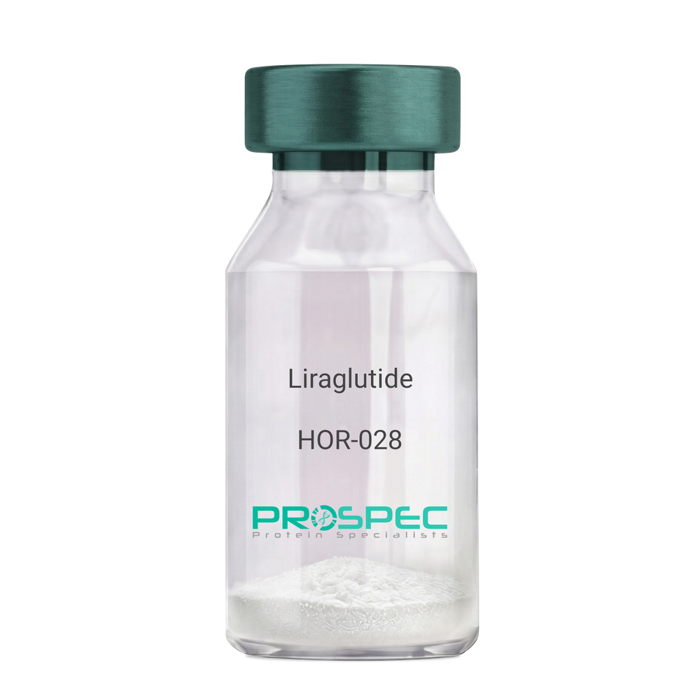
Liraglutide
Product # :
HOR-028Price :
Quantity :
Shipping Method :
Shipped at Room temp
More Info
- description
- formulation
- purity
- More Info
- HPLC, MS
Description
Liraglutide peptide is a single, non-glycosylated polypeptide chain containing 31 amino acids, having a molecular mass of 3751 Dalton and a Molecular formula of C172H265N43O51.
Formulation
The protein (1mg/ml) was lyophilized with no additives.
Purity
Greater than 97% as determined by RP-HPLC.
HPLC, MS
More Info
-
Introduction
Liraglutide is a long-acting GLP1 analogue sharing 97% a.a. homology to human GLP1. Liraglutide differs from GLP1 by the substitution of arginine for lysine at position 34. While GLP1 is degraded swiftly by DPP4 resulting in short life of insulin, Liraglutide contains a fatty acid that binds to albumin and prolongs the half-life of the structure. liraglutide causes insulin to secret in the presence of increasing glucose levels. Due toits receptor site, liraglutide inhibits glucagon secretion and delays gastric emptying.
-
Physical Appearance
Sterile Filtered White lyophilized (freeze-dried) powder.
-
Stability
Store the lyophilized Liraglutide between 2-8°C, do not freeze. Upon reconstitution Lypressin should be stored at 4°C between 2-7 days and for future use below -18°C. Please prevent freeze-thaw cycles.
-
Solubility
It is recommended to reconstitute the lyophilized Liraglutide in sterile 18MΩ-cm H2O not less than 100 µg/ml, which can then be further diluted to other aqueous solutions.
Lypressin is also soluble in 1% Acetic Acid. -
Amino Acid Sequence
H-His-Ala-Glu-Gly-Thr-Phe-Thr-Ser-Asp-Val-Ser-Ser-Tyr-Leu-Glu-Gly-Gln-Ala-Ala-Lys(γ-Glu-palmitoyl)-Glu-Phe-Ile-Ala-Trp-Leu-Val-Arg-Gly-Arg-Gly-OH
ProSpec's products are furnished for LABORATORY RESEARCH USE ONLY. The product may not be used as drugs, agricultural or pesticidal products, food additives or household chemicals.
- View Data Sheet
Name :
SEMA7A HumanDescription:
Semaphorin 7A Human Recombinant
Semaphorin-7A, CDw108, JMH blood group antigen, John-Milton-Hargen human blood group Ag, Semaphorin-K1, Sema K1, Semaphorin-L, Sema L, CD108.
Product # :
PRO-2506Price :
Quantity :
Shipping Method :
Shipped with Ice Packs
More Info
- description
- source
- formulation
- purity
- More Info
Description
SEMA7A produced in Sf9 Insect cells is a single, glycosylated polypeptide chain containing 846 amino acids (45-648 a.a.) and having a molecular mass of 95.7kDa (Molecular size on SDS-PAGE will appear at approximately 70-100kDa).SEMA7A is expressed with an 242 amino acid hIgG-His tag at C-Terminus and purified by proprietary chromatographic techniques.
Source
Sf9, Insect cells.
Formulation
SEMA7A protein solution (0.25mg/ml) contains Phosphate Buffered Saline (pH 7.4) and 10% glycerol.
Purity
Greater than 90.0% as determined by SDS-PAGE.
More Info
-
Introduction
SEMA7A (semaphorin-7A isoform 1), is a membrane-bound semaphorin which is linked with cell surfaces via a glycosylphosphatidylinositol (GPI) linkage. SEMA7A encourages formation of focal adhesion complexes, activation of the protein kinase PTK2/FAK1 and subsequent phosphorylation of MAPK3 & MAPK1. SEMA7A takes a vital part in integrin-mediated signaling and functions in regulating cell migration as well as immune responses. In addition, SEMA7A promotes the production of proinflammatory cytokines by macrophages & monocytes.
-
Synonyms
Semaphorin-7A, CDw108, JMH blood group antigen, John-Milton-Hargen human blood group Ag, Semaphorin-K1, Sema K1, Semaphorin-L, Sema L, CD108.
-
Physical Appearance
Sterile filtered colorless solution.
-
Stability
Store at 4°C if entire vial will be used within 2-4 weeks. Store, frozen at -20°C for longer periods of time. For long term storage it is recommended to add a carrier protein (0.1% HSA or BSA).Avoid multiple freeze-thaw cycles.
-
Amino Acid Sequence
ADPQGHLRSG PRIFAVWKGH VGQDRVDFGQ TEPHTVLFHE PGSSSVWVGG RGKVYLFDFP EGKNASVRTV NIGSTKGSCL DKRDCENYIT LLERRSEGLL ACGTNARHPS CWNLVNGTVV PLGEMRGYAP FSPDENSLVL FEGDEVYSTI RKQEYNGKIP RFRRIRGESE LYTSDTVMQN PQFIKATIVH QDQAYDDKIY YFFREDNPDK NPEAPLNVSR VAQLCRGDQG GESSLSVSKW NTFLKAMLVC SDAATNKNFN RLQDVFLLPD PSGQWRDTRV YGVFSNPWNY SAVCVYSLGD IDKVFRTSSL KGYHSSLPNP RPGKCLPDQQ PIPTETFQVA DRHPEVAQRV EPMGPLKTPL FHSKYHYQKV AVHRMQASHG ETFHVLYLTT DRGTIHKVVE PGEQEHSFAF NIMEIQPFRR AAAIQTMSLD AERRKLYVSS QWEVSQVPLD LCEVYGGGCH GCLMSRDPYC GWDQGRCISI YSSERSVLQS INPAEPHKEC PNPKPDKAPL QKVSLAPNSR YYLSCPMESR HATYSWRHKE NVEQSCEPGH QSPNCILFIE NLTAQQYGHY FCEAQEGSYF REAQHWQLLP EDGIMAEHLL GHACALALEP KSCDKTHTCP PCPAPELLGG PSVFLFPPKP KDTLMISRTP EVTCVVVDVS HEDPEVKFNW YVDGVEVHNA KTKPREEQYN STYRVVSVLT VLHQDWLNGK EYKCKVSNKA LPAPIEKTIS KAKGQPREPQ VYTLPPSRDE LTKNQVSLTC LVKGFYPSDI AVEWESNGQP ENNYKTTPPV LDSDGSFFLY SKLTVDKSRW QQGNVFSCSV MHEALHNHYT QKSLSLSPGK HHHHHH.
ProSpec's products are furnished for LABORATORY RESEARCH USE ONLY. The product may not be used as drugs, agricultural or pesticidal products, food additives or household chemicals.
- View Data Sheet
Name :
BMP 2 Human, HEKDescription:
Bone Morphogenetic protein-2 Human Recombinant, HEK
BMP-2, BMP2A.
Product # :
CYT-080Price :
Quantity :
Shipping Method :
Shipped at Room temp
More Info
- description
- source
- formulation
- purity
- biological activity
- More Info
Description
BMP-2 Human Recombinant produced in HEK cells is a glycosylated disulfide-linked homodimer, having a molecular weight range of 28kDa due to glycosylation. The BMP2 is purified by proprietary chromatographic techniques.
Source
HEK.
Formulation
The BMP2 was lyophilized from 0.67mg/ml in 2xPBS + 6% ethanol.
Purity
Greater than 95.0% as determined by analysis by SDS-PAGE.
Biological Activity
The specific activity as determined by the dose dependent induction of alkaline phosphatase production in the ATDC-5 cell line (Mouse chondrogenic cell line) was found to be 6.53ng/ml.
More Info
-
Introduction
BMP2 belongs to the transforming growth factor-beta (TGFB) superfamily. Bone morphogenic protein induces bone formation. BMP2 is a candidate gene for the autosomal dominant disease of fibrodysplasia (myositis) ossificans progressiva.
-
Synonyms
BMP-2, BMP2A.
-
Physical Appearance
Sterile Filtered White lyophilized (freeze-dried) powder.
-
Stability
Lyophilized BMP2 although stable at room temperature for 3 weeks, should be stored desiccated below -18°C. Upon reconstitution BMP-2 should be stored at 4°C between 2-7 days and for future use below -18°C.For long term storage it is recommended to add a carrier protein (0.1% HSA or BSA).Please prevent freeze-thaw cycles.
-
Solubility
It is recommended to reconstitute the lyophilized BMP-2 in sterile 4mM HCl containing 0.1% endotoxin-free recombinant HSA.
-
Amino Acid Sequence
QAKHKQRKRLKSSCKRHPLYVDFSDVGWNDWIVAPPGYHAFYCHGECPFPLADHLNST NHAIVQTLVNSVNSKIPKACCVPTELSAISMLYLDENEKVVLKNYQDMVVEGCGCR.
-
Background
What is the molecular weight/Mw of BMP2 Protein?
BMP2 Protein has a total Mw of 28kDa.
What is the source or expression system of BMP2 Protein?
Hek.
What is the Purity of BMP2 Protein?
BMP2 Protein is >95% pure as determined by SDS-PAGE.
What is the Biological Activity of BMP2 Protein?
The specific activity as determined by the dose dependent induction of alkaline phosphatase production in the ATDC-5 cell line (Mouse chondrogenic cell line) was found to be 6.53ng/ml.
What is the amino acid sequence of BMP2 Protein?
QAKHKQRKRLKSSCKRHPLYVDFSDVGWNDWIVAPPGYHAFYCHGECPFPLADHLNST NHAIVQTLVNSVNSKIPKACCVPTELSAISMLYLDENEKVVLKNYQDMVVEGCGCR.
What applications can BMP2 Protein be used in?
BMP2 Protein can probably be used in western blot, ELISA and Lateral Flow.
What is the endotoxin level for BMP2 Protein?
The endotoxin level is minimal, BMP2 Protein was purified using conventional chromatography techniques.
ProSpec's products are furnished for LABORATORY RESEARCH USE ONLY. The product may not be used as drugs, agricultural or pesticidal products, food additives or household chemicals.
- View Data Sheet
Name :
LBH HumanDescription:
Limb Bud And Heart Development Human Recombinant
Protein LBH, hLBH, Limb Bud And Heart Development Homolog.
Product # :
PRO-423Price :
Quantity :
Shipping Method :
Shipped with Ice Packs
More Info
- description
- source
- formulation
- purity
- More Info
Description
LBH Human Recombinant produced in E.Coli is a single, non-glycosylated polypeptide chain containing 128 amino acids (1-105 a.a) and having a molecular mass of 14.6kDa.LBH is fused to a 23 amino acid His-tag at N-terminus & purified by proprietary chromatographic techniques.
Source
Escherichia Coli.
Formulation
LBH protein solution (0.25mg/ml) containing 20mM Tris-HCl(pH 8.0), 20% glycerol, 0.15M NaCl and 1mM DTT.
Purity
Greater than 90.0% as determined by SDS-PAGE.
More Info
-
Introduction
Limb Bud And Heart Development, (LBH) belongs to the LBH family.LBH is highly expressed in the heart, and expressed at low levels in placenta, lung, skeletal muscle, kidney and liver. In addition, LBH protein is a transcriptional activator which may act in mitogen-activated protein kinase signaling pathway. Among the diseases associated with LBH are celiac disease, and rheumatoid arthritis.
-
Synonyms
Protein LBH, hLBH, Limb Bud And Heart Development Homolog.
-
Physical Appearance
Sterile Filtered clear solution.
-
Stability
Store at 4°C if entire vial will be used within 2-4 weeks. Store, frozen at -20°C for longer periods of time. For long term storage it is recommended to add a carrier protein (0.1% HSA or BSA).Avoid multiple freeze-thaw cycles.
-
Amino Acid Sequence
MGSSHHHHHH SSGLVPRGSH MGSMSIYFPI HCPDYLRSAK MTEVMMNTQP MEEIGLSPRK DGLSYQIFPD PSDFDRCCKL KDRLPSIVVE PTEGEVESGE LRWPPEEFLV QEDEQDNCEE TAKENKEQ
ProSpec's products are furnished for LABORATORY RESEARCH USE ONLY. The product may not be used as drugs, agricultural or pesticidal products, food additives or household chemicals.
- View Data Sheet
Name :
KRT18 BovineDescription:
Cytokeratin-18 Bovine
Keratin type I cytoskeletal 18, Cytokeratin-18, CK-18, Keratin-18, K18, KRT18,CYK18,Cell proliferation-inducing gene 46 protein.
Product # :
PRO-2785Price :
Quantity :
Shipping Method :
Shipped at Room temp
More Info
- description
- source
- formulation
- purity
- More Info
Description
KRT18 Bovine having a calculated molecular mass of 45 kDa, pI-5.4.
Source
Bovine liver.
Formulation
KRT18 was lyophilized from a 1mg/ml solution containing 30mM Tris/HCI pH 8, 9M urea, 2mM EDTA, 2mM DTT and 10mM methylammonium chloride.
Purity
Greater than 95.0% as determined by SDS-PAGE.
More Info
-
Synonyms
Keratin type I cytoskeletal 18, Cytokeratin-18, CK-18, Keratin-18, K18, KRT18,CYK18,Cell proliferation-inducing gene 46 protein.
-
Physical Appearance
Sterile Filtered White lyophilized (freeze-dried) powder.
-
Stability
Store the lyophilized KRT18 between 2-8°C, do not freeze. Upon reconstitution KRT18 should be stored at -20°C.Please prevent freeze-thaw cycles.
-
Solubility
It is recommended to reconstitute the lyophilized KRT18 in sterile 18MΩ-cm H2O not less than 100µg/ml, which can then be further diluted to other aqueous solutions.
-
Background
Keratin-18 (K18) is an intermediate filament protein that plays a vital role in maintaining the structural integrity of epithelial cells. Extensive research has been conducted on K18 in human and murine models, shedding light on its functions and implications for various epithelial tissues.
However, the study of K18 in bovine tissues is an emerging area with potential for advancing our understanding of epithelial cell biology and its applications in veterinary medicine and biotechnology. Bovine tissues, such as the liver and gastrointestinal tract, are of particular interest due to their relevance in cattle production and food safety.
This research aims to provide a comprehensive exploration of K18 in bovine tissues, elucidating its functions, structural significance, and potential applications.
The primary objective of this research is to elucidate the role of K18 in bovine tissues, particularly in maintaining the structural integrity of epithelial cells.In vitro and ex vivo experiments, utilizing bovine epithelial cell cultures and tissue specimens, will be conducted to investigate how K18 contributes to cellular morphology, cytoskeletal organization, and tissue resilience. Understanding these mechanisms is fundamental for deciphering the complexities of epithelial cell biology in bovine species.
The second objective is to assess the relevance of bovine K18 in veterinary medicine and cattle production. Studies involving bovine models will be conducted to evaluate the impact of K18 mutations or variations on tissue health, disease susceptibility, and meat quality. These investigations may provide valuable insights into potential applications in cattle breeding and food safety.
The third objective is to explore the potential biotechnological applications of bovine K18. Research will investigate the use of K18-expressing bovine cells as models for studying epithelial-related diseases and for developing tissue engineering approaches for veterinary medicine and biotechnology.
By delving into the functions and roles of K18 in bovine tissues, this research aims to expand our knowledge of epithelial cell biology, its implications for veterinary medicine, and its potential applications in biotechnology and cattle production.
ProSpec's products are furnished for LABORATORY RESEARCH USE ONLY. The product may not be used as drugs, agricultural or pesticidal products, food additives or household chemicals.
- View Data Sheet
Name :
SARS MERS, Sf9Description:
SARS MERS Spike Recombinant
Middle East respiratory syndrome coronavirus, Human betacoronavirus 2c EMC/2012, MERS-CoV, MERS, MERSCoV SP, Spike glycoprotein, S glycoprotein, S, Spike protein, E2, Peplomer protein
Product # :
SARS-051Price :
Quantity :
Shipping Method :
Shipped with Ice Packs
More Info
- description
- source
- formulation
- purity
- More Info
Description
SARS MERS Recombinant produced in Sf9 Baculovirus cells is a single, glycosylated polypeptide chain containing 1285 amino acids (18-1296aa) and having a molecular mass of 141.6kDa.SARS MERS is fused to a 6 amino acid His-tag at C-terminus & purified by proprietary chromatographic techniques.
Source
Sf9, Baculovirus cells.
Formulation
The SARS MERS solution (0.25mg/ml) contains 10% glycerol and Phosphate-Buffered Saline (pH 7.4).
Purity
Greater than 85.0% as determined by SDS-PAGE.
More Info
-
Introduction
Since April 2012, cases of the Middle East Respiratory Syndrome Coronavirus (MERS-CoV) have been identified in various countries. Coronaviruses are the cause of the common cold, SARS (severe acute respiratory syndrome) and other severe illnesses with high mortality rates, all are classified into coronavirus family. MERS-CoV is a new type of SARS found in the coronavirus family causing severe pneumonia with sudden and serious respiratory illness with high mortality rates as well. Since January 27th 2015, the WHO has reported 956 human cases, including 351 deaths. More cases of the new coronavirus strain are expected. Like in other coronaviruses, large surface spike glycoprotein is a central structural protein of this virus; it is located above the virion surface to bind and enter into the target cell. Spike protein has 2 domains- S1 and S2. The S1 domain is responsible for cellular tropism and interaction with target cell, while the S2 domain is responsible for membrane fusion. The C-terminal of S1 domain contains a receptor binding domain, and is also a potential target for vaccine development and an antigen for diagnosis.
-
Synonyms
Middle East respiratory syndrome coronavirus, Human betacoronavirus 2c EMC/2012, MERS-CoV, MERS, MERSCoV SP, Spike glycoprotein, S glycoprotein, S, Spike protein, E2, Peplomer protein
-
Physical Appearance
Sterile filtered colorless solution.
-
Stability
Store at 4°C if entire vial will be used within 2-4 weeks.Store, frozen at -20°C for longer periods of time.For long term storage it is recommended to add a carrier protein (0.1% HSA or BSA).Avoid multiple freeze-thaw cycles.
-
Amino Acid Sequence
YVDVGPDSVK SACIEVDIQQ TFFDKTWPRP IDVSKADGII YPQGRTYSNI TITYQGLFPY QGDHGDMYVY SAGHATGTTP QKLFVANYSQ DVKQFANGFV VRIGAAANST GTVIISPSTS ATIRKIYPAF MLGSSVGNFS DGKMGRFFNH TLVLLPDGCG TLLRAFYCIL EPRSGNHCPA GNSYTSFATY HTPATDCSDG NYNRNASLNS FKEYFNLRNC TFMYTYNITE DEILEWFGIT QTAQGVHLFS SRYVDLYGGN MFQFATLPVY DTIKYYSIIP HSIRSIQSDR KAWAAFYVYK LQPLTFLLDF SVDGYIRRAI DCGFNDLSQL HCSYESFDVE SGVYSVSSFE AKPSGSVVEQ AEGVECDFSP LLSGTPPQVY NFKRLVFTNC NYNLTKLLSL FSVNDFTCSQ ISPAAIASNC YSSLILDYFS YPLSMKSDLS VSSAGPISQF NYKQSFSNPT CLILATVPHN LTTITKPLKY SYINKCSRLL SDDRTEVPQL VNANQYSPCV SIVPSTVWED GDYYRKQLSP LEGGGWLVAS GSTVAMTEQL QMGFGITVQY GTDTNSVCPK LEFANDTKIA SQLGNCVEYS LYGVSGRGVF QNCTAVGVRQ QRFVYDAYQN LVGYYSDDGN YYCLRACVSV PVSVIYDKET KTHATLFGSV ACEHISSTMS QYSRSTRSML KRRDSTYGPL QTPVGCVLGL VNSSLFVEDC KLPLGQSLCA LPDTPSTLTP RSVRSVPGEM RLASIAFNHP IQVDQLNSSY FKLSIPTNFS FGVTQEYIQT TIQKVTVDCK QYVCNGFQKC EQLLREYGQF CSKINQALHG ANLRQDDSVR NLFASVKSSQ SSPIIPGFGG DFNLTLLEPV SISTGSRSAR SAIEDLLFDK VTIADPGYMQ GYDDCMQQGP ASARDLICAQ YVAGYKVLPP LMDVNMEAAY TSSLLGSIAG VGWTAGLSSF AAIPFAQSIF YRLNGVGITQ QVLSENQKLI ANKFNQALGA MQTGFTTTNE AFQKVQDAVN NNAQALSKLA SELSNTFGAI SASIGDIIQR LDVLEQDAQI DRLINGRLTT LNAFVAQQLV RSESAALSAQ LAKDKVNECV KAQSKRSGFC GQGTHIVSFV VNAPNGLYFM HVGYYPSNHI EVVSAYGLCD AANPTNCIAP VNGYFIKTNN TRIVDEWSYT GSSFYAPEPI TSLNTKYVAP QVTYQNISTN LPPPLLGNST GIDFQDELDE FFKNVSTSIP NFGSLTQINT TLLDLTYEML SLQQVVKALN ESYIDLKELG NYTYYNKWPH HHHHH
ProSpec's products are furnished for LABORATORY RESEARCH USE ONLY. The product may not be used as drugs, agricultural or pesticidal products, food additives or household chemicals.
- View Data Sheet
Name :
FCGRT HumanDescription:
Fc Fragment Of IgG Receptor And Transporter Human Recombinant
IgG receptor FcRn large subunit p51, FcRn, IgG Fc fragment receptor transporter alpha chain, Neonatal Fc receptor, Fcgrt, Fcrn.
Product # :
PRO-2806Price :
Quantity :
Shipping Method :
Shipped with Ice Packs
More Info
- description
- source
- formulation
- purity
- biological activity
- More Info
Description
FCGRT Human Recombinant is a glycosylated, polypeptide chain conteining 2 fragments: FCGRT (24-297aa) having a total 280 a.a and a a molecular weigh of 31.1kDa & B2M (21-119 aa) having a total 99 a.a and a a molecular weigh of 11.7kDa. FCGRT is fused to a 6 a.a his-Tag at C-terminus and is purified by proprietary chromatographic techniques.
Source
HEK293 Cells.
Formulation
The FCGRT solution (0.25mg/ml) contains 10% Glycerol and Phosphate-Buffered Saline (pH 7.4).
Purity
Greater than 95.0% as determined by SDS-PAGE.
Biological Activity
Measured by its binding ability in a functional ELISA with Human IgG1 Fc.
More Info
-
Synonyms
IgG receptor FcRn large subunit p51, FcRn, IgG Fc fragment receptor transporter alpha chain, Neonatal Fc receptor, Fcgrt, Fcrn.
-
Physical Appearance
Sterile Filtered colorless solution.
-
Stability
Store at 4°C if entire vial will be used within 2-4 weeks. Store, frozen at -20°C for longer periods of time. For long term storage it is recommended to add a carrier protein (0.1% HSA or BSA). Avoid multiple freeze-thaw cycles.
-
Amino Acid Sequence
FCGRT
AESHLSLLYH LTAVSSPAPG TPAFWVSGWL GPQQYLSYNS LRGEAEPCGA WVWENQVSWY WEKETTDLRI KEKLFLEAFK ALGGKGPYTL QGLLGCELGP DNTSVPTAKF ALNGEEFMNF DLKQGTWGGD WPEALAISQR WQQQDKAANK ELTFLLFSCP HRLREHLERG RGNLEWKEPP SMRLKARPSS PGFSVLTCSA FSFYPPELQL RFLRNGLAAG TGQGDFGPNS DGSFHASSSL TVKSGDEHHY CCIVQHAGLA QPLRVELESP AKSSHHHHHH.
B2M
IQRTPKIQVY SRHPAENGKS NFLNCYVSGF HPSDIEVDLL KNGERIEKVE HSDLSFSKDW SFYLLYYTEF TPTEKDEYAC RVNHVTLSQP KIVKWDRDM.
-
Background
Applications in Drug Delivery and Therapy:
The unique ability of FCGRT to transport immunoglobulins has spurred investigations into its potential applications in drug delivery. FCGRT Human Recombinant is being explored as a tool to improve the pharmacokinetics of therapeutic antibodies, enhancing their stability and extending their half-life in circulation. This holds significant implications for the development of antibody-based therapies for a range of diseases, including autoimmune disorders and cancer.
Modulation of Immune Responses:
Beyond its role in immunoglobulin transport, FCGRT is implicated in the modulation of immune responses. FCGRT Human Recombinant studies have unveiled its interactions with immune cells and its potential influence on the activation and regulation of the immune system. This dual role positions FCGRT as a key player not only in passive immunity but also in the dynamic orchestration of immune responses.
Challenges and Future Directions:
While the potential of FCGRT Human Recombinant in drug delivery is promising, challenges persist. Fine-tuning its applications, understanding its interactions with diverse antibodies, and optimizing strategies for therapeutic use are critical considerations for translational success. Additionally, deciphering the intricate balance between immunoglobulin transport and immune modulation is an active area of investigation.
FCGRT Human Recombinant emerges as a sentinel in the realm of immunoglobulin transport and drug delivery, offering a controlled platform for scientific exploration. Its structural insights, immunoglobulin transport functions, and therapeutic applications position it as a key player in the evolving landscape of biopharmaceuticals. As researchers continue to delve into the molecular intricacies of FCGRT, they not only enhance our understanding of immunoglobulin transport but also pave the way for transformative advancements in drug delivery and antibody-based therapies, shaping the future of precision medicine and targeted therapeutics.
ProSpec's products are furnished for LABORATORY RESEARCH USE ONLY. The product may not be used as drugs, agricultural or pesticidal products, food additives or household chemicals.
- View Data Sheet
Name :
Vimentin BovineDescription:
Bovine Vimentin
Vimentin, Vim, FLJ36605.
Product # :
PRO-525Price :
Quantity :
Shipping Method :
Shipped at Room temp
More Info
- description
- source
- formulation
- purity
- More Info
Description
Bovine Vimentin protein produced in Bovine Lens, having a molecular weight of 57 kDa.
Source
Bovine Lens.
Formulation
The protein (1mg/ml) was lyophilized from a 1mg/ml solution containing 10mM sodium phosphate pH 7.5, 6M urea, 2mM DTT, 1mM EDTA and 10mM methylammonium chloride.
Purity
Greater than 98.0% as determined by SDS-PAGE.
More Info
-
Introduction
Vimentin expression in human malignant glioma cells depends on cellular density, algorithms of drug delivery and chemo/radio treatment. Vimentin and detyrosinated microtubules provide structural support for the extensive microtentacles observed in detached tumor cells and a mechanism to promote successful metastatic spread. Primary colorectal carcinomas display aberrant expression of vimentin, and have activated Notch and TGFbeta signaling pathways. Vimentin is a strong arterial substrate for transglutaminases. Transglutaminase-mediated vimentin dimerization results in a novel unifying pathway by which vasodilatory and remodeling responses may be regulated. Ablation of vimentin expression inhibits migration and invasion of colon and breast cancer cell lines. Vimentin is the main intermediate filament protein in mesenchymal cells and is therefore of value in the differential diagnosis of undifferentiated neoplasms.
-
Synonyms
Vimentin, Vim, FLJ36605.
-
Physical Appearance
Sterile Filtered White lyophilized (freeze-dried) powder.
-
Stability
Lyophilized Bovine Vimentin although stable at room temperature for 3 weeks, should be stored desiccated below -18°C. Upon reconstitution BovineVimentin should be stored at 4°C between 2-7 days and for future use below -18°C.For long term storage it is recommended to add a carrier protein (0.1% HSA or BSA).Please prevent freeze-thaw cycles.
-
Solubility
It is recommended to reconstitute the Bovine Vimentin in sterile 18MΩ-cm H2O not less than 100µg/ml, which can then be further diluted to other aqueous solutions.
-
Reconstitution to filaments
After vimentin is dissolved in 6M urea buffer (see above), protofilaments and filament complexes are obtained by dialyzing the resulting polypeptide solution stepwise to a concentration of 4M urea and then to low salt condition (50mM NaCl, 2mM dithiothreitol, 10mM Sodium Phosphate, pH 7.4). For immunization purposes, the solution can be further dialyzed against PBS (phosphate buffered saline, e.g. Dulbecco''s PBS).- Hatzfeld M. and Franke W.W. (1985). J. Cell Biol. 101, 1826-1841.- Hatzfeld M. et al. (1987). J. Mol. Biol. 197, 237-255.
ProSpec's products are furnished for LABORATORY RESEARCH USE ONLY. The product may not be used as drugs, agricultural or pesticidal products, food additives or household chemicals.
- View Data Sheet
Name :
IFNG FelineDescription:
Interferon-gamma Feline Recombinant
Interferon gamma, IFN-gamma, IFNG.
Product # :
CYT-998Price :
Quantity :
Shipping Method :
Shipped with Ice Packs
More Info
- description
- source
- formulation
- purity
- More Info
- SDS-PAGE
Description
IFNG Feline Recombinant produced in E.Coli is a single, non-glycosylated polypeptide chain containing 167 amino acids (24-167 a.a) and having a molecular mass of 19.3kDa.IFNG is fused to a 23 amino acid His-tag at N-terminus & purified by proprietary chromatographic techniques.
Source
Escherichia Coli.
Formulation
IFNG protein solution (1mg/ml) containing PBS buffer (pH 7.4) and 10% glycerol.
Purity
Greater than 95.0% as determined by SDS-PAGE.
SDS-PAGE
More Info
-
Introduction
IFN-gamma produced by lymphocytes activated by specific antigens or mitogens.
IFN-gamma, in addition to having antiviral activity, has important immunoregulatory functions, it is a potent activator of macrophages, and has antiproliferative effects on transformed cells and it can potentiate the antiviral and antitumor effects of the type I interferons. -
Synonyms
Interferon gamma, IFN-gamma, IFNG.
-
Physical Appearance
Sterile Filtered colorless solution.
-
Stability
Store at 4°C if entire vial will be used within 2-4 weeks. Store, frozen at -20°C for longer periods of time. For long term storage it is recommended to add a carrier protein (0.1% HSA or BSA).Avoid multiple freeze-thaw cycles.
-
Amino Acid Sequence
MGSSHHHHHH SSGLVPRGSH MGSQAMFFKE IEELKGYFNA SNPDVADGGS LFVDILKNWK EESDKTIIQS QIVSFYLKMF ENLKDDDQRI QRSMDTIKED MLDKLLNTSS SKRDDFLKLI QIPVNDLQVQ RKAINELFKV MNDLSPRSNL RKRKRSQNLF RGRRASK.
-
Background
What is the molecular weight/Mw of IFNG FELINE Protein?
IFNG FELINE Protein has a total Mw of 19.3kDa.
What is the source or expression system of IFNG FELINE Protein?
Escherichia Coli.
What is the Purity of IFNG FELINE Protein?
IFNG FELINE Protein is >95% pure as determined by SDS-PAGE.
What is the Biological Activity of IFNG FELINE Protein?
The biological functionality of IFNG FELINE Protein will be determined in the future.
What is the amino acid sequence of IFNG FELINE Protein?
MGSSHHHHHH SSGLVPRGSH MGSQAMFFKE IEELKGYFNA SNPDVADGGS LFVDILKNWK EESDKTIIQS QIVSFYLKMF ENLKDDDQRI QRSMDTIKED MLDKLLNTSS SKRDDFLKLI QIPVNDLQVQ RKAINELFKV MNDLSPRSNL RKRKRSQNLF RGRRASK.
What applications can IFNG FELINE Protein be used in?
IFNG FELINE Protein can probably be used in western blot, ELISA and Lateral Flow.
What is the endotoxin level for IFNG FELINE Protein?
The endotoxin level is minimal, IFNG FELINE Protein was purified using conventional chromatography techniques.
ProSpec's products are furnished for LABORATORY RESEARCH USE ONLY. The product may not be used as drugs, agricultural or pesticidal products, food additives or household chemicals.
- View Data Sheet
Name :
IL2RA Human, sf9Description:
Interleukin-2 Receptor alpha Human Recombinant, sf9
Interleukin 2 Receptor Subunit Alpha, Interleukin 2 Receptor, Alpha, IL-2 Receptor Subunit Alpha, IL-2R Subunit Alpha, TAC Antigen, P55, Insulin-Dependent Diabetes Mellitus 10, Interleukin-2 Receptor Subunit Alpha, CD25 Antigen, IL-2-RA, IDDM10, IL2-RA, IMD41, TCGFR, CD25, IL2R.
Product # :
CYT-1020Price :
Quantity :
Shipping Method :
Shipped with Ice Packs
More Info
- description
- source
- formulation
- purity
- More Info
Description
IL2RA produced in Sf9 Baculovirus cells is a single, glycosylated polypeptide chain containing 461 amino acids (22-240 a.a.) and having a molecular mass of 52.1kDa (Molecular size on SDS-PAGE will appear at approximately 50-70kDa).IL2RA is expressed with a 239 amino acid hIgG-His tag at C-Terminus and purified by proprietary chromatographic techniques.
Source
Sf9, Baculovirus cells.
Formulation
IL2RA protein solution (0.5mg/ml) contains Phosphate Buffered Saline (pH 7.4) and 10% glycerol.
Purity
Greater than 90.0% as determined by SDS-PAGE.
More Info
-
Introduction
IL2-Ra is one of the three constituent subunits of the IL2 receptor.
IL-2Ra is released into the serum after increased cellular expression such as increased activation of B and T cells. Clinical manifestations of IL2-Ra elevation include autoimmune conditions and some leukemias and lymphomas. -
Synonyms
Interleukin 2 Receptor Subunit Alpha, Interleukin 2 Receptor, Alpha, IL-2 Receptor Subunit Alpha, IL-2R Subunit Alpha, TAC Antigen, P55, Insulin-Dependent Diabetes Mellitus 10, Interleukin-2 Receptor Subunit Alpha, CD25 Antigen, IL-2-RA, IDDM10, IL2-RA, IMD41, TCGFR, CD25, IL2R.
-
Physical Appearance
Sterile Filtered colorless solution.
-
Stability
Store at 4°C if entire vial will be used within 2-4 weeks. Store, frozen at -20°C for longer periods of time. For long term storage it is recommended to add a carrier protein (0.1% HSA or BSA). Avoid multiple freeze-thaw cycles.
-
Amino Acid Sequence
ADPELCDDDP PEIPHATFKA MAYKEGTMLN CECKRGFRRI KSGSLYMLCT GNSSHSSWDN QCQCTSSATR NTTKQVTPQP EEQKERKTTE MQSPMQPVDQ ASLPGHCREP PPWENEATER IYHFVVGQMV YYQCVQGYRA LHRGPAESVC KMTHGKTRWT QPQLICTGEM ETSQFPGEEK PQASPEGRPE SETSCLVTTT DFQIQTEMAA TMETSIFTTE YQLEPKSCDK THTCPPCPAP ELLGGPSVFL FPPKPKDTLM ISRTPEVTCV VVDVSHEDPE VKFNWYVDGV EVHNAKTKPR EEQYNSTYRV VSVLTVLHQD WLNGKEYKCK VSNKALPAPI EKTISKAKGQ PREPQVYTLP PSRDELTKNQ VSLTCLVKGF YPSDIAVEWE SNGQPENNYK TTPPVLDSDG SFFLYSKLTV DKSRWQQGNV FSCSVMHEAL HNHYTQKSLS LSPGKHHHHH H.
ProSpec's products are furnished for LABORATORY RESEARCH USE ONLY. The product may not be used as drugs, agricultural or pesticidal products, food additives or household chemicals.
- View Data Sheet
Name :
LECT2 HumanDescription:
Leukocyte Cell-Derived Chemotaxin 2 Human Recombinant
Leukocyte Cell-Derived Chemotaxin 2, Leukocyte Cell-Derived Chemotaxin-2, Chondromodulin-II, Chm-II, LECT-2, HLECT2, Chm2, LECT2.
Product # :
PRO-2037Price :
Quantity :
Shipping Method :
Shipped at Room temp
More Info
- description
- source
- formulation
- purity
- More Info
Description
LECT2 Human Recombinant produced in E.Coli is a single, non-glycosylated, polypeptide chain (Gly19-Leu151) containing 143 amino acids including a 10 aa His tag at N-terminus. The total calculated molecular mass is 16kDa.
Source
Escherichia Coli.
Formulation
LECT2 was filtered (0.4 µm) and lyophilized in 20mM Tris buffer, 50mM NaCl & pH 7.5.
Purity
Greater than 95.0% as determined by SDS-PAGE.
More Info
-
Introduction
Leukocyte Cell-Derived Chemotaxin 2 (LECT2) functions as a chemotactic factor to neutrophils. LECT2 stimulates the proliferation of chondrocytes and osteoblasts. LECT2 is strongly expressed in the liver and weakly in the testis. LECT2 is a secreted, 16kDa protein which serves as a chemotactic factor to neutrophils and stimulates the growth of chondrocytes and osteoblasts. LECT2 protein has a high sequence similarity to the chondromodulin repeat regions of the chicken myb-induced myeloid 1 protein. A polymorphism in the LECT2 gene is linked with rheumatoid arthritis.
-
Synonyms
Leukocyte Cell-Derived Chemotaxin 2, Leukocyte Cell-Derived Chemotaxin-2, Chondromodulin-II, Chm-II, LECT-2, HLECT2, Chm2, LECT2.
-
Physical Appearance
Filtered White lyophilized (freeze-dried) powder.
-
Stability
Store lyophilized protein at -20°C. Aliquot the product after reconstitution to avoid repeated freezing/thawing cycles. Reconstituted protein can be stored at 4°C for a limited period of time; it does not show any change after two weeks at 4°C.
-
Solubility
It is recommended to add deionized water to prepare a working stock solution of approximately 0.5mg/ml and let the lyophilized pellet dissolve completely. LECT2 is not sterile! Please filter the product by an appropriate sterile filter before using it in the cell culture.
-
Amino Acid Sequence
MKHHHHHHASGPWANICAGK SSNEIRTCDR HGCGQYSAQR SQRPHQGVDI LCSAGSTVYA PFTGMIVGQE KPYQNKNAIN NGVRISGRGF CVKMFYIKPI KYKGPIKKGE KLGTLLPLQK VYPGIQSHVH IENCDSSDPT AYL.
ProSpec's products are furnished for LABORATORY RESEARCH USE ONLY. The product may not be used as drugs, agricultural or pesticidal products, food additives or household chemicals.
- View Data Sheet
Name :
GPHB5 HumanDescription:
Thyrostimulin Beta Human Recombinant
Glycoprotein hormone beta-5, ZLUT1, GPHB5, GPB5.
Product # :
HOR-257Price :
Quantity :
Shipping Method :
Shipped at Room temp
More Info
- description
- source
- formulation
- purity
- More Info
Description
GPHB5 Human Recombinant produced in E.Coli is a single, non-glycosylated, polypeptide chain containing 120 amino acids and having a total molecular mass of 13.34 kDa. The Thyrostimulin contains His tag which consists of 14 additional amino acids.The amino acid sequence of the recombinant human Thyrostimulin beta subunit is 100% homologous to the amino acid sequence of the human Thyrostimulin beta subunit without signal sequence. (N-terminal 24AA).Thyrostimulin is purified by proprietary chromatographic techniques.
Source
Escherichia Coli.
Formulation
The GPHB5 filtered (0.4µm) and lyophilized from 0.5mg/ml in 0.05M Acetate buffer pH 4.
Purity
Greater than 95.0% as determined by:
(a) Analysis by RP-HPLC.
(b) Analysis by SDS-PAGE.More Info
-
Introduction
Human thyrostimulin ranks among the glycoprotein hormone family. These hormones consist of two subunits, the common alpha- and specific beta-subunits, which associate noncovalently to form a heterodimer. The alpha-subunit combines with four distinct beta-subunits giving rise to four biologically active hormones in human: FSH, LH, TSH, and CG. FSH, LH, and TSH, mainly expressed in the anterior pituitary, are essential for coordinated endocrine regulation in the hypothalamus- pituitary axis and show to activate specific G protein–coupled receptors in the thyroid (TSH receptor) and gonads (LH and FSH receptors), respectively.
The heterodimeric glycoprotein hormones have only been identified in vertebrates and are highly conserved in organisms from primitive rayfin fish (Chondrostei) to human in both primary sequences and functional characteristics.
Corticotroph-derived glycoprotein hormone (CGH), also referred to as thyrostimulin, is a noncovalent heterodimer of glycoprotein hormone alpha 2 (GPHA2) and glycoprotein hormone beta 5 (GPHB5).
Recombinant A2/B5 heterodimeric glycoproteins activates human TSH receptors, but not LH and FSH receptors, and shows high affinity to TSH receptors in a radioligand receptor assay. The heterodimer also stimulates cAMP production and thymidine incorporation by cultured thyroid cells and increases serum thyroxine levels in TSH-suppressed rats in vivo. This new heterodimeric glycoprotein hormone was named as thyrostimulin based on its thyroid-stimulating activity. The expression of thyrostimulin in the anterior pituitary known to express TSH receptors suggested a paracrine mechanism. -
Synonyms
Glycoprotein hormone beta-5, ZLUT1, GPHB5, GPB5.
-
Physical Appearance
Filtered White lyophilized (freeze-dried) powder.
-
Stability
Lyophilized Thyrostimulin although stable at room temperature for 3 weeks, should be stored desiccated below -18°C. Upon reconstitution GPHB5 should be stored at 4°C between 2-7 days and for future use below -18°C. For long term storage it is recommended to add a carrier protein (0.1% HSA or BSA).Please prevent freeze-thaw cycles.
-
Solubility
It is recommended to add 0.1M Acetate buffer pH4 to prepare a working stock solution of ~ 0.5 mg/ml and let the lyophilized pellet dissolve completely. For conversion into higher pH value, we recommend intensive dilution by relevant buffer to a concentration of 10μg/ml. In higher concentrations the solubility of this protein is limited. Product is not sterile! Please filter the product by an appropriate sterile filter before using it in the cell culture.
-
Amino Acid Sequence
MRGSHHHHHH GMASASSGNL RTFVGCAVRE FTFLAKKPGC RGLRITTDAC WGRCETWEKP ILEPPYIEAH HRVCTYNETK QVTVKLPNCA PGVDPFYTYP VAIRCDCGAC STATTECETI.
ProSpec's products are furnished for LABORATORY RESEARCH USE ONLY. The product may not be used as drugs, agricultural or pesticidal products, food additives or household chemicals.
- View Data Sheet
Name :
UPK3A HumanDescription:
Uroplakin 3A Human Recombinant
Uroplakin 3A, UPK3, UPIII, Uroplakin III, UP3A.
Product # :
PRO-988Price :
Quantity :
Shipping Method :
Shipped with Ice Packs
More Info
- description
- source
- formulation
- purity
- More Info
Description
UPK3A Human Recombinant produced in E.Coli is a single, non-glycosylated polypeptide chain containing 214 amino acids (19-207 a.a.) and having a molecular mass of 23.1kDa.UPK3A is fused to a 25 amino acid His-tag at N-terminus & purified by proprietary chromatographic techniques.
Source
E.coli.
Formulation
UPK3A protein solution (0.25mg/ml) containing 20mM Tris-HCl buffer (pH8.0), 150mM NaCl, 2mM DTT and 20% glycerol.
Purity
Greater than 90% as determined by SDS-PAGE.
More Info
-
Introduction
UPK3A is a member of the uroplakin-3 family. Uroplakins (UP) which are transmembrane proteins are important components of the urothelium that pass the lipid bilayer once (UPII, UPIIIa, and UPIIIb) or four times (UPIa and UPIb; both members of the "tetraspanin" family). All hold great luminal/extracellular domains, but only UPIIIa and UPIIIb have significant cytoplasmic shares in their C-termini. UPK3A is an extremely specific and moderately sensitive immunohistochemical marker for primary and metastatic urothelial carcinomas. Alterations in this gene are related to renal adysplasia.
-
Synonyms
Uroplakin 3A, UPK3, UPIII, Uroplakin III, UP3A.
-
Physical Appearance
Sterile Filtered colorless solution.
-
Stability
Store at 4°C if entire vial will be used within 2-4 weeks. Store, frozen at -20°C for longer periods of time. For long term storage it is recommended to add a carrier protein (0.1% HSA or BSA).Avoid multiple freeze-thaw cycles.
-
Amino Acid Sequence
MGSSHHHHHH SSGLVPRGSH MGSHMVNLQP QLASVTFATN NPTLTTVALE KPLCMFDSKE ALTGTHEVYL YVLVDSAISR NASVQDSTNT PLGSTFLQTE GGRTGPYKAV AFDLIPCSDL PSLDAIGDVS KASQILNAYL VRVGANGTCL WDPNFQGLCN PPLSAATEYR FKYVLVNMST GLVEDQTLWS DPIRTNQLTP YSTIDTWPGR RSGG.
ProSpec's products are furnished for LABORATORY RESEARCH USE ONLY. The product may not be used as drugs, agricultural or pesticidal products, food additives or household chemicals.
- View Data Sheet
Name :
MECR HumanDescription:
Mitochondrial Trans-2-Enoyl-CoA Reductase Human Recombinant
NRBF1, CGI-63, FASN2B, EC 1.3.1.38, MECR, Mitochondrial Trans-2-Enoyl-CoA Reductase.
Product # :
ENZ-533Price :
Quantity :
Shipping Method :
Shipped with Ice Packs
More Info
- description
- source
- formulation
- purity
- More Info
Description
MECR Recombinant Human produced in E.Coli is a single, non-glycosylated polypeptide chain containing 341 amino acids (54-373 a.a.) and having a molecular mass of 49.8 kDa. The MECR is fused to 21 amino acid His-Tag at N-terminus and purified by proprietary chromatographic techniques.
Source
Escherichia Coli.
Formulation
MECR Human solution containing 20mM Trsi pH-8, 0.2M NaCl, 5mM DTT & 20% glycerol.
Purity
Greater than 90.0% as determined by SDS-PAGE.
More Info
-
Introduction
MECR catalyzes the reduction of trans-2-enoyl-CoA to acyl-CoA with chain length from C6 to C16 in an NADPH dependent manner with preference to medium chain length substrate. MECR protein takes part in the mitochondrial synthesis of fatty acids.
-
Synonyms
NRBF1, CGI-63, FASN2B, EC 1.3.1.38, MECR, Mitochondrial Trans-2-Enoyl-CoA Reductase.
-
Physical Appearance
Sterile filtered colorless solution.
-
Stability
Store at 4°C if entire vial will be used within 2-4 weeks. Store, frozen at -20°C for longer periods of time. For long term storage it is recommended to add a carrier protein (0.1% HSA or BSA).Avoid multiple freeze-thaw cycles.
-
Amino Acid Sequence
MGSSHHHHHH SSGLVPRGSH MPAKVVELKN LELAAVRGSD VRVKMLAAPI NPSDINMIQG NYGLLPELPA VGGNEGVAQV VAVGSNVTGL KPGDWVIPAN AGLGTWRTEA VFSEEALIQV PSDIPLQSAA TLGVNPCTAY RMLMDFEQLQ PGDSVIQNAS NSGVGQAVIQ IAAALGLRTI NVVRDRPDIQ KLSDRLKSLG AEHVITEEEL RRPEMKNFFK DMPQPRLALN CVGGKSSTEL LRQLARGGTM VTYGGMAKQP VVASVSLLIF KDLKLRGFWL SQWKKDHSPD QFKELILTLC DLIRRGQLTA PACSQVPLQD YQSALEASMK PFISSKQILT M.
ProSpec's products are furnished for LABORATORY RESEARCH USE ONLY. The product may not be used as drugs, agricultural or pesticidal products, food additives or household chemicals.
- View Data Sheet
Name :
C3 RatDescription:
Complement C3 Rat
Complement C3, C3 and PZP-like alpha-2-macroglobulin domain-containing protein 1, C3, CPAMD1.
Product # :
PRO-2706Price :
Quantity :
Shipping Method :
Shipped with Ice Packs
More Info
- description
- source
- formulation
- purity
- More Info
Description
Rat Complement C3 produced in Rat plasma having a molecular weight of 187kDa.
Source
Rat Plasma.
Formulation
C3 solution contains phosphate buffer saline.
Purity
Greater than 95.0% as determined by SDS-PAGE.
More Info
-
Introduction
C3 is central to the activation of all 3 pathways of complement activation. Initiation of each pathway generates proteolytic enzyme complexes which binds the target surface. These enzymes cleave a peptide bond in C3 releasing the anaphylatoxin C3a and activating C3b. Most of the C3 activated during complement activation never attaches to the surface due to its thioester reaction with water forming fluid phase C3b which is rapidly inactivated by factors H and I forming iC3b. Surface-bound C3b is necessary in all 3 pathways for efficient activation of C5 and formation of C5b-9 complexes that lyse the target cell membrane.
-
Synonyms
Complement C3, C3 and PZP-like alpha-2-macroglobulin domain-containing protein 1, C3, CPAMD1.
-
Physical Appearance
Sterile filtered solution.
-
Stability
C3 Mouse is stable at 4°C if entire vial will be used within 2-4 weeks.Store, frozen below -20°C for longer periods of time.For long term storage it is recommended to add a carrier protein (0.1% HSA or BSA).Avoid multiple freeze-thaw cycles.
ProSpec's products are furnished for LABORATORY RESEARCH USE ONLY. The product may not be used as drugs, agricultural or pesticidal products, food additives or household chemicals.
- View Data Sheet
Name :
sRANKL Human, HisDescription:
Soluble RANK Ligand Human Recombinant, His Tag
Soluble Receptor Activator of NFkB Ligand, TNFSF11, TRANCE, TNF-related activation-induced cytokine, OPGL, ODF, Osteoclast differentiation factor, Tumor necrosis factor ligand superfamily member 11, Receptor activator of nuclear factor kappa B ligand, RANKL, Osteoprotegerin ligand, CD254 antigen, sRANKL, sOdf, hRANKL2.
Product # :
CYT-692Price :
Quantity :
Shipping Method :
Shipped with Ice Packs
More Info
- description
- source
- formulation
- purity
- More Info
Description
sRANKL Human Recombinant produced in E.Coli is a single, non-glycosylated polypeptide chain containing 199 amino acids (140-317 a.a) and having a molecular mass of 22.3kDa.sRANKL is fused to a 21 amino acid His-tag at N-terminus & purified by proprietary chromatographic techniques.
Source
Escherichia Coli.
Formulation
sRANKL protein solution (0.5mg/ml) containing 20mM Tris-HCl buffer (pH8.0), 20% glycerol, 0.1M NaCl and 1mM DTT.
Purity
Greater than 80.0% as determined by SDS-PAGE.
More Info
-
Introduction
RANKL binds to tnfrsf11b/opg and to tnfrsf11a/rank. Osteoclast differentiation and activation factor. Augments the ability of dendritic cells to stimulate naive t-cell proliferation. May be an important regulator of interactions between t-cells and dendritic cells and may play a role in the regulation of the t-cell-dependent immune response. sRANKL may also play an important role in enhanced bone-resorption in humoral hypercalcemia of malignancy.
-
Synonyms
Soluble Receptor Activator of NFkB Ligand, TNFSF11, TRANCE, TNF-related activation-induced cytokine, OPGL, ODF, Osteoclast differentiation factor, Tumor necrosis factor ligand superfamily member 11, Receptor activator of nuclear factor kappa B ligand, RANKL, Osteoprotegerin ligand, CD254 antigen, sRANKL, sOdf, hRANKL2.
-
Physical Appearance
Sterile Filtered colorless solution.
-
Stability
Store at 4°C if entire vial will be used within 2-4 weeks. Store, frozen at -20°C for longer periods of time. For long term storage it is recommended to add a carrier protein (0.1% HSA or BSA).Avoid multiple freeze-thaw cycles.
-
Amino Acid Sequence
MGSSHHHHHH SSGLVPRGSH MIRAEKAMVD GSWLDLAKRS KLEAQPFAHL TINATDIPSG SHKVSLSSWY HDRGWAKISN MTFSNGKLIV NQDGFYYLYA NICFRHHETS GDLATEYLQL MVYVTKTSIK IPSSHTLMKG GSTKYWSGNS EFHFYSINVG GFFKLRSGEE ISIEVSNPSL LDPDQDATYF GAFKVRDID.
ProSpec's products are furnished for LABORATORY RESEARCH USE ONLY. The product may not be used as drugs, agricultural or pesticidal products, food additives or household chemicals.
- View Data Sheet
Name :
VEGF EquineDescription:
Vascular Endothelial Growth Factor Equine Recombinant
VEGF-A, VPF, glioma-derived endothelial cell mitogen.
Product # :
CYT-1200Price :
Quantity :
Shipping Method :
Shipped at Room temp
More Info
- description
- source
- formulation
- purity
- biological activity
- More Info
Description
VEGF Equine Recombinant produced in E.Coli is a single, non-glycosylated, polypeptide chain containing 2 x 165 amino acids and having a total molecular mass of 38.6 kDa.The VEGF is purified by proprietary chromatographic techniques.
Source
Escherichia Coli.
Formulation
Lyophilized from a sterile (0.2 micron) filtered aqueous solution containing 10 mM Sodium Phosphate, pH 7.5.
Purity
Greater than 95.0% as determined by SDS-PAGE.
Biological Activity
The ED50 as determined by HUVEC Proliferation is ≤ 10 ng/mL, corresponding to a specific activity of ≥ 1.0 x 10^5 units/mg.
More Info
-
Introduction
Vascular endothelial growth factor is an important signaling protein involved in both vasculogenesis and angiogenesis. As its name implies, VEGF activity has been mostly studied on cells of the vascular endothelium, although it does have effects on a number of other cell types (e.g. stimulation monocyte/macrophage migration, neurons, cancer cells, kidney epithelial cells ).VEGF mediates increased vascular permeability, induces angiogenesis, vasculogenesis and endothelial cell growth, promotes cell migration, and inhibits apoptosis. In vitro, VEGF has been shown to stimulate endothelial cell mitogenesis and cell migration. VEGF is also a vasodilator and increases microvascular permeability and was originally referred to as vascular permeability factor. Elevated levels of this protein is linked to POEMS syndrome, also known as Crow-Fukase syndrome. Mutations in this gene have been associated with proliferative and nonproliferative diabetic retinopathy.
-
Synonyms
VEGF-A, VPF, glioma-derived endothelial cell mitogen.
-
Physical Appearance
Sterile Filtered White lyophilized (freeze-dried) powder.
-
Stability
Lyophilized VEGF although stable at room temperature for 3 weeks, should be stored desiccated below -18°C. Upon reconstitution VEGF Human should be stored at 4°C between 2-7 days and for future use below -18°C.For long term storage it is recommended to add a carrier protein (0.1% HSA or BSA).Please prevent freeze-thaw cycles.
-
Solubility
It is recommended to reconstitute the lyophilized VEGF in sterile water at a concentration of 0.1 mg/ml, which can then be further diluted to other aqueous solutions.
-
Amino Acid Sequence
MAPMAEGEHK THEVVKFMDV YQRSYCRPIE TLVDIFQEYP DEIEYIFKPS CVPLMRCGGC CNDEGLECVP TAEFNITMQI MRIKPHQSQH IGEMSFLQHS KCECRPKKDK ARQENPCGPC SERRKHLFVQ DPQTCKCSCK NTDSRCKARQ LELNERTCRC DKPRR
ProSpec's products are furnished for LABORATORY RESEARCH USE ONLY. The product may not be used as drugs, agricultural or pesticidal products, food additives or household chemicals.