Search results
1000 results found for “Cadherin”
Name
Description
Product #
Price
Quantity
Shipping Method
- View Data Sheet
Name :
GLDA E.coli, ActiveDescription:
Glycerol dehydrogenase E.coli Recombinant, Active
ECK3937, JW5556, Glycerol dehydrogenase, GDH, GLDH, b3945.
Product # :
ENZ-904Price :
Quantity :
Shipping Method :
Shipped with Ice Packs
More Info
- description
- source
- formulation
- purity
- biological activity
- More Info
- Citations
Description
GLDA E.coli Recombinant produced in E.Coli is a single, non-glycosylated polypeptide chain containing 390 amino acids (1-367 a.a) and having a molecular mass of 41.1kDa. GLDA is fused to a 23 amino acid His-tag at N-terminus & purified by proprietary chromatographic techniques.
Source
Escherichia Coli.
Formulation
GLDA protein solution (1mg/ml) containing Phosphate buffered saline (pH7.4) and 10% glycerol.
Purity
Greater than 95.0% as determined by SDS-PAGE.
Biological Activity
Specific activity: > 14 Units/ml. One unit will oxidize 1.0 umole of glycerol to dihydroxyacetone per minute at pH 8.0 at 25C.Citations
More Info
-
Introduction
Glycerol dehydrogenase (GldA) catalyzes the NAD-dependent oxidation of glycerol to dihydroxyacetone (glycerone). The GldA protein allows microorganisms to use glycerol as a source of carbon under anaerobic conditions. Furthermore, in E.coli GldA has an imperative role by regulating the intracellular level of dihydroxyacetone by catalyzing the reverse reaction, i.e. the conversion of dihydroxyacetone into glycerol. GldA possesses an extensive substrate specificity, due to its ability to oxidize 1,2-propanediol and to reduce glycolaldehyde, methylglyoxal and hydroxyacetone into ethylene glycol, lactaldehyde and 1,2-propanediol, respectively.
-
Synonyms
ECK3937, JW5556, Glycerol dehydrogenase, GDH, GLDH, b3945.
-
Physical Appearance
Sterile Filtered colorless solution.
-
Stability
Store at 4°C if entire vial will be used within 2-4 weeks. Store, frozen at -20°C for longer periods of time. For long term storage it is recommended to add a carrier protein (0.1% HSA or BSA).Avoid multiple freeze-thaw cycles.
-
Amino Acid Sequence
MGSSHHHHHH SSGLVPRGSH MGSMDRIIQS PGKYIQGADV INRLGEYLKP LAERWLVVGD KFVLGFAQST VEKSFKDAGL VVEIAPFGGE CSQNEIDRLR GIAETAQCGA ILGIGGGKTL DTAKALAHFM GVPVAIAPTI ASTDAPCSAL SVIYTDEGEF DRYLLLPNNP NMVIVDTKIV AGAPARLLAA GIGDALATWF EARACSRSGA TTMAGGKCTQ AALALAELCY NTLLEEGEKA MLAAEQHVVT PALERVIEAN TYLSGVGFES GGLAAAHAVH NGLTAIPDAH HYYHGEKVAF GTLTQLVLEN APVEEIETVA ALSHAVGLPI TLAQLDIKED VPAKMRIVAE AACAEGETIH NMPGGATPDQ VYAALLVADQ YGQRFLQEWE.
ProSpec's products are furnished for LABORATORY RESEARCH USE ONLY. The product may not be used as drugs, agricultural or pesticidal products, food additives or household chemicals.
- View Data Sheet
Name :
SF20 Mouse, HisDescription:
MYDGF Mouse Recombinant, His Tag
D17Wsu104e, Il25, Ly6elg, MYDGF, Interleukin-25, IL-25, Stromal cell-derived growth factor SF20.
Product # :
CYT-1040Price :
Quantity :
Shipping Method :
Shipped with Ice Packs
More Info
- description
- source
- formulation
- purity
- More Info
- sds-page
- Citations
Description
MYDGF Mouse Recombinant produced in E.Coli is a single, non-glycosylated polypeptide chain containing 165 amino acids (25-166 a.a) and having a molecular mass of 18.1kDa. MYDGF is fused to a 23 amino acid His-tag at N-terminus & purified by proprietary chromatographic techniques.
Source
Escherichia Coli.
Formulation
MYDGF protein solution (0.5mg/ml) containing 20mM Tris-HCl (pH8.0), 0.1M NaCl and 10% glycerol.
Purity
Greater than 95.0% as determined by SDS-PAGE.
sds-page
Citations
More Info
-
Introduction
Myeloid-derived growth factor (Mydgf) is a paracrine-acting protein and a bone marrow-derived monocyte which stimulates cardiac myocyte survival and adaptive angiogenesis for cardiac protection and repair after myocardial infarction. Mydgf induces endothelial cell proliferation through a MAPK1/3-, STAT3- and CCND1-mediated signaling lane. When comparing wild-type mice to mice with a Mydgf-deficiency, the later develop larger infarct scars and more acute contractile dysfunction.
-
Synonyms
D17Wsu104e, Il25, Ly6elg, MYDGF, Interleukin-25, IL-25, Stromal cell-derived growth factor SF20.
-
Physical Appearance
Sterile Filtered clear solution.
-
Stability
Store at 4°C if entire vial will be used within 2-4 weeks. Store, frozen at -20°C for longer periods of time. For long term storage it is recommended to add a carrier protein (0.1% HSA or BSA).Avoid multiple freeze-thaw cycles.
-
Amino Acid Sequence
MGSSHHHHHH SSGLVPRGSH MGSVSEPTTV PFDVRPGGVV HSFSQDVGPG NKFTCTFTYA SQGGTNEQWQ MSLGTSEDSQ HFTCTIWRPQ GKSYLYFTQF KAELRGAEIE YAMAYSKAAF ERESDVPLKS EEFEVTKTAV SHRPGAFKAE LSKLVIVAKA ARSEL.
ProSpec's products are furnished for LABORATORY RESEARCH USE ONLY. The product may not be used as drugs, agricultural or pesticidal products, food additives or household chemicals.
- View Data Sheet
Name :
CTGF Human (183-255)Description:
Connective Tissue Growth Factor (183-255 a.a.) Human Recombinant
CCN2, NOV2, HCS24, IGFBP8, MGC102839, CTGF, Connective Tissue Growth Factor.
Product # :
CYT-1174Price :
Quantity :
Shipping Method :
Shipped at Room temp
More Info
- description
- source
- formulation
- purity
- More Info
- Citations
Description
CTGF Human Recombinant is a single, glycosylated polypeptide chain containing 80 amino acids (183-255a.a) and having a molecular mass of 9.1kDa (calculated). CTGF is fused to a 7 a.a His tag at N-terminal.
Source
HEK293 cells.
Formulation
CTGF filtered (0.4 µm) and lyophilized from 0.5mg/ml in 20 mM Tris buffer and 50 mM NaCl, pH 7.5.
Purity
Greater than 95.0% as determined by SDS-PAGE.
Citations
More Info
-
Introduction
Connective Tissue Growth Factor is a part of the CCN family of proteins. The CCN family presently consists of six members in human also known as: Cyr61 (Cystein rich 61), CTGF, Nov (Nephroblastoma Overexpressed gene), WISP-1, 2 and 3 (Wnt-1 Induced Secreted Proteins). CCN proteins are matricellular proteins which are involved in the regulation of various cellular functions including: proliferation, differentiation, survival, adhesion and migration. They are expressed in derivatives of the three embryonic sheets and are implicated in the development of kidney, nervous system, muscle, bone marrow, cartilage and bone. During adulthood, they are implicated in wound healing, bone fracture repair, and pathologies such as: tumorigenesis,fibrosis and vascular ailments. Full length secreted CCN proteins can show an antiproliferative activity, whereas truncated isoforms are likely to stimulate proliferation and behave as oncogenes.
The full length protein consists of 4 modules: Module I shares partial identity with the N-terminal part of the Insulin-like Growth Factor Binding Proteins (IGFBPs).
Module II includes a stretch of 70amino acid residues – which shares sequence identity with the Von Willebrand Factor Type C repeat (VWC).
Module III contains sequences sharing identity with the Thrombospondin type 1 repeat (TSP1) (WSXCSXXCG), which is thought to be implicated in the binding of sulfated glycoconjugates and to be important for cell adhesion.
Module IV, also designated CT, is encoded by exon5. It is the leasts conserved one of the four domains at the level of nucleotide sequence, but it appears to be critical for several of the biological functions attributed to the CCN proteins.
Proteolysis of the secreted full-length CCN proteins that has been reported in the case of CCN2 and CCN3 might result in the production of CCN-derived peptides with high affinity for ligands that full-length CNN proteins bind only poorly. Amino-truncated CCN2 isoforms were biologically active whereas no specific biological activity has been attributed to the truncated CCN3. Although the molecular processes underlying the production of these secreted isoforms is presently unknown, it is important to note that proteolysis occur at the same amino acid residues in both CCN2 and CCN3. An elevated expression of CCN2 has also been detected by Northern blotting in human invasive mammary ductal carcinomas, dermatofibromas, pyogenic granuloma, endothelial cells of angiolipomas and angioleiomyomas, and in pancreatic tumors. A study performed with chondrosarcomas representative of various histological grades established that CCN2 expression was closely correlated with increasing levels of malignancy.
In agreement with CCN2 playing a role in brain tumor angiogenesis, immunocytochemistry studies indicated that both glioblastoma tumor cells and proliferating endothelial cells stained positive for CCN2. In astrocytomas, CCN2 expression was particularly elevated in high grade tumors, with a marked effect of CCN2 on cell proliferation. Downregulation of CCN2 expression in these cells was associated with a growth arrest at the G1/S transition while over-expression of CCN2 induced a two-fold increase of the number of cells in the G1 phase. Gene profiling analysis allowed to identify a set of about 50 genes whose expression might account for the proliferative activity of CCN2 in these cells.
CCN2 was seen in a higher proportion of mononuclear cells of patients with acute lymphoblastic leukemia. -
Synonyms
CCN2, NOV2, HCS24, IGFBP8, MGC102839, CTGF, Connective Tissue Growth Factor.
-
Physical Appearance
Filtered White lyophilized (freeze-dried) powder.
-
Stability
Store lyophilized protein at -20°C. Aliquot the product after reconstitution to avoid repeated freezing/thawing cycles. Reconstituted protein can be stored at 4°C for a limited period of time; it does not show any change after two weeks at 4°C.
-
Solubility
It is recommended to add deionized water to prepare a working stock solution of approximately 0.5mg/ml and let the lyophilized pellet dissolve completely.
-
Amino Acid Sequence
MHHHHHHRLE DTFGPDPTMI RANCLVQTTE WSACSKTCGM GISTRVTNDN ASCRLEKQSR LCMVRPCEAD LEENIKKGKK.
-
Background
What is the molecular weight/Mw of CTGF Protein?
CTGF Protein has a total Mw of 9.1kDa.
What is the source or expression system of CTGF Protein?
HEK293 cells.
What is the Purity of CTGF Protein?
CTGF Protein is >95% pure as determined by SDS-PAGE.
What is the Biological Activity of CTGF Protein?
The biological functionality of CTGF Protein will be determined in the future.
What is the amino acid sequence of CTGF Protein?
MHHHHHHRLE DTFGPDPTMI RANCLVQTTE WSACSKTCGM GISTRVTNDN ASCRLEKQSR LCMVRPCEAD LEENIKKGKK.
What applications can CTGF Protein be used in?
CTGF Protein can probably be used in western blot, ELISA and Lateral Flow.
What is the endotoxin level for CTGF Protein?
The endotoxin level is minimal, CTGF Protein was purified using conventional chromatography techniques.
ProSpec's products are furnished for LABORATORY RESEARCH USE ONLY. The product may not be used as drugs, agricultural or pesticidal products, food additives or household chemicals.
- View Data Sheet
Name :
AMBPDescription:
Alpha-1 Microglobulin Human Recombinant
Alpha-1-microglobulin/bikunin precursor, HCP, ITIL, ITI, EDC1, HI30, IATIL, ITILC, UTI, A1M, bikunin, complex-forming glycoprotein heterogeneous in charge, growth-inhibiting protein 19, inter-alpha-trypsin inhibitor light chain, protein AMBP, protein HC, trypstatin, uristatin, uronic-acid-rich protein.
Product # :
PRO-957Price :
Quantity :
Shipping Method :
Shipped with Ice Packs
More Info
- description
- source
- formulation
- purity
- More Info
- Citations
Description
AMBP Human Recombinant produced in E.coli is a single, non-glycosylated polypeptide chain containing 205 amino acids (20-203) and having a molecular mass of 23.1 kDa.AMBP is fused to a 21 amino acid His-tag at N-terminus & purified by proprietary chromatographic techniques.
Source
E.coli.
Formulation
The AMBP solution (0.5mg/ml) contains 20mM Tris-HCl buffer (pH 8.0), 1mM DTT, 0.1M NaCl and 10% glycerol.
Purity
Greater than 90% as determined by SDS-PAGE.
Citations
More Info
-
Introduction
Alpha 1-microglobulin (A1M) is a lipocalin superfamily member (kernal lipocalins). A1M is a low molecular weight protein component of plasma. A1M is distributed in plasma and extravascular compartments of all organs. Alpha-1 Microglobulin is found in mammals, birds, amphibians and fish. The primary sites of A1M synthesis are the liver and the kidney. Around the opening of the lipocalin pocket three lysyl residues are situated; those residues carry yellow-brown modification derived from the binding and degradation of heme and kynurenin (a tryptophan metabolite). A1-Microglobulin’s reductase and dehydrogenase have broad biological substrate specificity properties due to its’ free cysteine side-chain which is located in a flexible loop. Alpha-1-microglobulin is glycosylated by three separate carbohydrate chains: two complex carbohydrates which are N-linked to asparagines at residues 17 and 96, and the other simple carbohydrate which is O-linked to threonine at position 5. The carbohydrates comprise 22% of the total molecular mass of the protein. The glycosylation varies between species.
A1M exists in two forms- a free form and complexed to other macromolecules: in humans- complexed to immunoglobulin A (IgA), in rat- complexed to alpha-1-inhibitor-3. Free A1M is exceptionally heterogeneous in charge (therefore also known as protein HC), and is found tightly linked to a chromophore. The free Alpha-1-microglobulin is a monomeric protein composed of one 188 residue polypeptide and contains three cysteines, two of which (residues 75 and 173) form a conserved intra-molecular disulphide link. The chromophoric group is covalently bound to the free cysteine residue at position 34. A1M binds retinol as a major ligand, but this is probably distinct from its covalent chromophore.
Half of all human plasma A1M (approximately 0.03mg/ml) forms a 1:1 complex with about 5% of plasma immunoglobulin A. The resulting macromolecular complexes’ molecular weight is 200000, and a plasma concentration of 0.3mg/ml. The complex can exhibit both antibody activity and affect many of the biological actions of free Alpha-1-microglobulin.
Alpha-1-microglobulin was first discovered in pathological human urine.
It was suggested that A1M might be involved in tissue defense against reactive oxygen species, oxidation by heme and kynurenin. Evidence also suggests that A1M functions in the regulation of the immune system. Other functions include inhibition of stimulation of cultured lymphocytes by protein antigens; induction of cell division of lymphocytes, a mitogenic effect that can either be enhanced or inhibited by the action of other plasma components; inhibition of neutrophil granulocyte migration in vitro; and inhibition of chemotaxis. -
Synonyms
Alpha-1-microglobulin/bikunin precursor, HCP, ITIL, ITI, EDC1, HI30, IATIL, ITILC, UTI, A1M, bikunin, complex-forming glycoprotein heterogeneous in charge, growth-inhibiting protein 19, inter-alpha-trypsin inhibitor light chain, protein AMBP, protein HC, trypstatin, uristatin,
uronic-acid-rich protein. -
Physical Appearance
Sterile Filtered colorless solution.
-
Stability
Store at 4°C if entire vial will be used within 2-4 weeks. Store, frozen at -20°C for longer periods of time. For long term storage it is recommended to add a carrier protein (0.1% HSA or BSA).Avoid multiple freeze-thaw cycles.
-
Amino Acid Sequence
MGSSHHHHHH SSGLVPRGSH MGPVPTPPDN IQVQENFNIS RIYGKWYNLA IGSTCPWLKK IMDRMTVSTL VLGEGATEAE ISMTSTRWRK GVCEETSGAY EKTDTDGKFL YHKSKWNITM ESYVVHTNYD EYAIFLTKKF SRHHGPTITA KLYGRAPQLR ETLLQDFRVV AQGVGIPEDS IFTMADRGEC VPGEQEPEPI LIPRV.
ProSpec's products are furnished for LABORATORY RESEARCH USE ONLY. The product may not be used as drugs, agricultural or pesticidal products, food additives or household chemicals.
- View Data Sheet
Name :
MYLPF HumanDescription:
Myosin Light chain, Phosphorylatable, Fast Skeletal Muscle Human Recombinant
Myosin regulatory light chain 2 skeletal muscle isoform, Fast skeletal myosin light chain 2, MLC2B, MYLPF, MRLC2, MYL11, HUMMLC2B.
Product # :
PRO-243Price :
Quantity :
Shipping Method :
Shipped with Ice Packs
More Info
- description
- source
- formulation
- purity
- More Info
- Citations
Description
MYLPF produced in E.Coli is a single, non-glycosylated polypeptide chain containing 189 amino acids (1-169 a.a) and having a molecular mass of 21.2kDa.MYLPF is fused to a 20 amino acid His-tag at N-terminus & purified by proprietary chromatographic techniques.
Source
Escherichia Coli.
Formulation
MYLPF protein solution (0.25mg/ml) containing 20mM Tris-HCl buffer (pH 8.0), 1mM DTT, 10% glycerol and 100mM NaCl.
Purity
Greater than 85.0% as determined by SDS-PAGE.
Citations
More Info
-
Introduction
Myosin regulatory light chains, including MRCL3, MYLPF and MYL9, regulate contraction in smooth muscle and non-muscle cells via phosphorylation by MLCK (myosin light chain kinase). Phosphorylation of myosin regulatory light chains, catalyzed by MLCK in the presence of calcium and calmodulin, increases the actin-activated myosin ATPase activity, thus regulating the contractile activity. MYLPF is vital for fast and slow skeletal muscle development.
-
Synonyms
Myosin regulatory light chain 2 skeletal muscle isoform, Fast skeletal myosin light chain 2, MLC2B, MYLPF, MRLC2, MYL11, HUMMLC2B.
-
Physical Appearance
Sterile Filtered colorless solution.
-
Stability
Store at 4°C if entire vial will be used within 2-4 weeks. Store, frozen at -20°C for longer periods of time. For long term storage it is recommended to add a carrier protein (0.1% HSA or BSA).Avoid multiple freeze-thaw cycles.
-
Amino Acid Sequence
MGSSHHHHHH SSGLVPRGSH MAPKRAKRRT VEGGSSSVFS MFDQTQIQEF KEAFTVIDQN RDGIIDKEDL RDTFAAMGRL NVKNEELDAM MKEASGPINF TVFLTMFGEK LKGADPEDVI TGAFKVLDPE GKGTIKKKFL EELLTTQCDR FSQEEIKNMW AAFPPDVGGN VDYKNICYVI THGDAKDQE.
ProSpec's products are furnished for LABORATORY RESEARCH USE ONLY. The product may not be used as drugs, agricultural or pesticidal products, food additives or household chemicals.
- View Data Sheet
Name :
SIGLEC7 HumanDescription:
Sialic Acid Binding Ig Like Lectin 7 Human Recombinant
Sialic Acid Binding Ig Like Lectin 7, QA79 Membrane Protein, SIGLEC-7, D-Siglec, AIRM-1, CDw328, AIRM1, P75, Sialic Acid Binding Ig-Like Lectin 19, Pseudogene, Adhesion Inhibitory Receptor Molecule 1, Siglec-7, Sialic Acid Binding Ig-Like Lectin, Pseudogene 2, Sialic Acid Binding Immunoglobulin-Like Lectin 7, Adhesion Inhibitory Receptor Molecule 1, Sialic Acid Binding Ig-Like Lectin 7, Sialic Acid-Binding Ig-Like Lectin 7, CD328 Antigen, SIGLEC19P, P75/AIRM1, SIGLECP2, CD328, QA79.
Product # :
PRO-2458Price :
Quantity :
Shipping Method :
Shipped with Ice Packs
More Info
- description
- source
- formulation
- purity
- More Info
- Citations
Description
SIGLEC7 Human Recombinant produced in Sf9 Baculovirus cells is a single, glycosylated polypeptide chain containing 577 amino acids (19-353a.a.) and having a molecular mass of 64.2kDa. SIGLEC7 is expressed with a 239 amino acid hIgG-His-tag at C-Terminus and purified by proprietary chromatographic techniques.
Source
Sf9, Baculovirus cells.
Formulation
SIGLEC7 protein solution (0.5mg/ml) contains Phosphate Buffered Saline (pH 7.4) and 10% glycerol.
Purity
Greater than 90.0% as determined by SDS-PAGE.
Citations
More Info
-
Introduction
Sialic acid-binding Ig-like lectin 7 isoform 1, also known as SIGLEC7, is a newly discovered family of sialic acid-binding lectins of the immunoglobulin (Ig) superfamily. The extracellular portion has two Ig-like domains, with the amino-terminal V-set Ig domain containing amino acid residues which known to be implicated in sialic acid recognition by other Siglecs. The combination of an extracellular sialic acid binding site and an intracellular ITIM motif proposes that SIGLEC7 is involved in trans-membrane regulatory signaling reactions. Furthermore, on the cell surface SIGLEC7 exists as a monomer and is expressed on natural killer cells.
-
Synonyms
Sialic Acid Binding Ig Like Lectin 7, QA79 Membrane Protein, SIGLEC-7, D-Siglec, AIRM-1, CDw328, AIRM1, P75, Sialic Acid Binding Ig-Like Lectin 19, Pseudogene, Adhesion Inhibitory Receptor Molecule 1, Siglec-7, Sialic Acid Binding Ig-Like Lectin, Pseudogene 2, Sialic Acid Binding Immunoglobulin-Like Lectin 7, Adhesion Inhibitory Receptor Molecule 1, Sialic Acid Binding Ig-Like Lectin 7, Sialic Acid-Binding Ig-Like Lectin 7, CD328 Antigen, SIGLEC19P, P75/AIRM1, SIGLECP2, CD328, QA79.
-
Physical Appearance
Sterile filtered colorless solution.
-
Stability
Store at 4°C if entire vial will be used within 2-4 weeks. Store, frozen at -20°C for longer periods of time. For long term storage it is recommended to add a carrier protein (0.1% HSA or BSA).Avoid multiple freeze-thaw cycles.
-
Amino Acid Sequence
ADPQKSNRKD YSLTMQSSVT VQEGMCVHVR CSFSYPVDSQ TDSDPVHGYW FRAGNDISWK APVATNNPAW AVQEETRDRF HLLGDPQTKN CTLSIRDARM SDAGRYFFRM EKGNIKWNYK YDQLSVNVTA LTHRPNILIP GTLESGCFQN LTCSVPWACE QGTPPMISWM GTSVSPLHPS TTRSSVLTLI PQPQHHGTSL TCQVTLPGAG VTTNRTIQLN VSYPPQNLTV TVFQGEGTAS TALGNSSSLS VLEGQSLRLV CAVDSNPPAR LSWTWRSLTL YPSQPSNPLV LELQVHLGDE GEFTCRAQNS LGSQHVSLNL SLQQEYTGKM RPVSGVLLLE PKSCDKTHTC PPCPAPELLG GPSVFLFPPK PKDTLMISRT PEVTCVVVDV SHEDPEVKFN WYVDGVEVHN AKTKPREEQY NSTYRVVSVL TVLHQDWLNG KEYKCKVSNK ALPAPIEKTI SKAKGQPREP QVYTLPPSRD ELTKNQVSLT CLVKGFYPSD IAVEWESNGQ PENNYKTTPP VLDSDGSFFL YSKLTVDKSR WQQGNVFSCS VMHEALHNHY TQKSLSLSPG KHHHHHH.
ProSpec's products are furnished for LABORATORY RESEARCH USE ONLY. The product may not be used as drugs, agricultural or pesticidal products, food additives or household chemicals.
- View Data Sheet
Name :
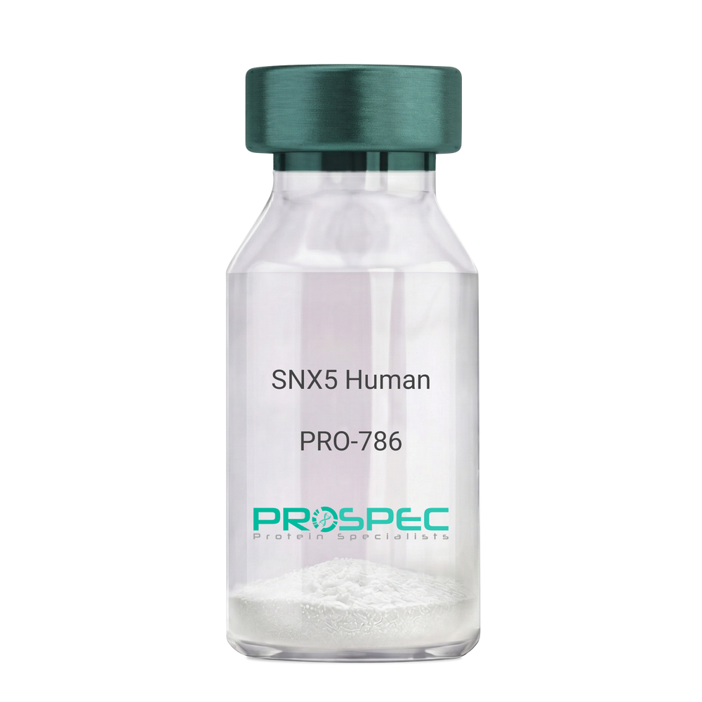
SNX5 HumanDescription:
Sorting Nexin 5 Human Recombinant
Sorting nexin-5 isoform a, Sorting nexin-5, SNX5.
Product # :
PRO-786Price :
Quantity :
Shipping Method :
Shipped with Ice Packs
More Info
- description
- source
- formulation
- purity
- More Info
- Citations
Description
SNX5 Human Recombinant produced in E.Coli is a single, non-glycosylated polypeptide chain containing 427 amino acids (1-404 a.a) and having a molecular mass of 49.2kDa.SNX5 is fused to a 23 amino acid His-tag at N-terminus & purified by proprietary chromatographic techniques.
Source
Escherichia Coli.
Formulation
SNX5 protein solution (0.25mg/ml) in phosphate buffered saline (pH7.4), 20% glycerol and 1mM DTT.
Purity
Greater than 85.0% as determined by SDS-PAGE.
Citations
More Info
-
Introduction
Sorting nexin-5 (SNX5) belongs to the sorting nexin family, whose members contains a phox (PX) domain, (which is a phosphoinositide binding domain) and are involved in intracellular trafficking. SNX5 protein is a component of the mammalian retromer complex, which facilitates cargo recovery from endosomes to the trans-Golgi network. SNX5 binds to the Fanconi anemia, complementation group A protein.
-
Synonyms
Sorting nexin-5 isoform a, Sorting nexin-5, SNX5.
-
Physical Appearance
Sterile Filtered clear solution.
-
Stability
Store at 4°C if entire vial will be used within 2-4 weeks. Store, frozen at -20°C for longer periods of time. For long term storage it is recommended to add a carrier protein (0.1% HSA or BSA).Avoid multiple freeze-thaw cycles.
-
Amino Acid Sequence
MGSSHHHHHH SSGLVPRGSH MGSMAAVPEL LQQQEEDRSK LRSVSVDLNV DPSLQIDIPD ALSERDKVKF TVHTKTTLPT FQSPEFSVTR QHEDFVWLHD TLIETTDYAG LIIPPAPTKP DFDGPREKMQ KLGEGEGSMT KEEFAKMKQE LEAEYLAVFK KTVSSHEVFL QRLSSHPVLS KDRNFHVFLE YDQDLSVRRK NTKEMFGGFF KSVVKSADEV LFTGVKEVDD FFEQEKNFLI NYYNRIKDSC VKADKMTRSH KNVADDYIHT AACLHSLALE EPTVIKKYLL KVAELFEKLR KVEGRVSSDE DLKLTELLRY YMLNIEAAKD LLYRRTKALI DYENSNKALD KARLKSKDVK LAEAHQQECC QKFEQLSESA KEELINFKRK RVAAFRKNLI EMSELEIKHA RNNVSLLQSC IDLFKNN.
ProSpec's products are furnished for LABORATORY RESEARCH USE ONLY. The product may not be used as drugs, agricultural or pesticidal products, food additives or household chemicals.
- View Data Sheet
Name :
SOST Human, HEKDescription:
Sclerostin Human Recombinant, HEK
Sclerostin, SOST, CDD, VBCH.
Product # :
PRO-2481Price :
Quantity :
Shipping Method :
Shipped at Room temp
More Info
- description
- source
- formulation
- purity
- More Info
- Citations
Description
SOST Human Recombinant produced in HEK293 cells is a single, glycosylated polypeptide chain (a.a 24-213) containing 196 amino acids including a 6 a.a C-terminal His tag. The total molecular mass is 22.4kDa (calculated).
Source
HEK293 Cells.
Formulation
SOST filtered (0.4 µm) and lyophilized from 0.5mg/ml in PBS and 5 % (w/v) trehalose, pH 7.4.
Purity
Greater than 95.0% as determined by SDS-PAGE.
Citations
More Info
-
Introduction
Sclerostin (SOST) is a secreted glycoprotein with a C-terminal cysteine knot-like (CTCK) domain and sequence similarity to the DAN (differential screening-selected gene aberrative in neuroblastoma) family of bone morphogenetic protein (BMP) antagonists. Sclerostin functions as a negative regulator of bone growth, by inhibiting bone formation. SOST is widely expressed at low levels, with highest levels in the bone, cartilage, kidney, liver, bone marrow and primary osteoblasts differentiated for 21 days. SOST gene defects cause sclerosteosis and bone dysplasia.
-
Synonyms
Sclerostin, SOST, CDD, VBCH.
-
Physical Appearance
Filtered White lyophilized (freeze-dried) powder.
-
Stability
Store lyophilized protein at -20°C. Aliquot the product after reconstitution to avoid repeated freezing/thawing cycles. Reconstituted protein can be stored at 4°C for a limited period of time; it does not show any change after one week at 4°C.
-
Solubility
It is recommended to add deionized water to prepare a working stock solution of approximately 0.5mg/ml and let the lyophilized pellet dissolve completely. SOST is not sterile! Please filter the product by an appropriate sterile filter before using it in the cell culture.
-
Amino Acid Sequence
QGWQAFKNDA TEIIPELGEY PEPPPELENN KTMNRAENGG RPPHHPFETK DVSEYSCREL HFTRYVTDGP CRSAKPVTEL VCSGQCGPAR LLPNAIGRGK WWRPSGPDFR CIPDRYRAQR VQLLCPGGEA PRARKVRLVA SCKCKRLTRF HNQSELKDFG TEAARPQKGR KPRPRARSAK ANQAELENAY HHHHHH.
ProSpec's products are furnished for LABORATORY RESEARCH USE ONLY. The product may not be used as drugs, agricultural or pesticidal products, food additives or household chemicals.
- View Data Sheet
Name :
OSM Human, 195 a.aDescription:
Oncostatin-M Human Recombinant (195 a.a.)
OSM, MGC20461, Oncostatin M.
Product # :
CYT-735Price :
Quantity :
Shipping Method :
Shipped at Room temp
More Info
- description
- source
- formulation
- purity
- biological activity
- More Info
- Citations
Description
Oncostatin-M Human Recombinant produced in E.Coli is a single, non-glycosylated polypeptide chain containing 195 amino acids and having a molecular mass of 22kDa. The OSM is purified by proprietary chromatographic techniques.
Source
Escherichia Coli.
Formulation
Lyophilized from a concentrated (1mg/ml) solution containing 1xPBS pH-7.4.
Purity
Greater than 97.0% as determined by:
(a) Analysis by RP-HPLC.
(b) Analysis by SDS-PAGE.Biological Activity
The ED50 as determined by the dose-dependent stimulation of the proliferation of human TF-1 cells is < 0.2ng/ml, corresponding to a specific activity of > 5.0x10⁶ units/mg.Citations
More Info
-
Introduction
Oncostatin M is a member of a cytokine family that includes leukemia-inhibitory factor, granulocyte colony-stimulating factor, and interleukin 6. This gene encodes a growth regulator which inhibits the proliferation of a number of tumor cell lines. It regulates cytokine production, including IL-6, G-CSF and GM-CSF from endothelial cells.
-
Synonyms
OSM, MGC20461, Oncostatin M.
-
Physical Appearance
Sterile Filtered White lyophilized (freeze-dried) powder.
-
Stability
Lyophilized Oncostatin M although stable at room temperature for 3 weeks, should be stored desiccated below -18°C. Upon reconstitution Oncostatin should be stored at 4°C between 2-7 days and for future use below -18°C.For long term storage it is recommended to add a carrier protein (0.1% HSA or BSA).Please prevent freeze-thaw cycles.
-
Solubility
It is recommended to reconstitute the lyophilized Oncostatin M in sterile 18MΩ-cm H2O not less than 100µg/ml, which can then be further diluted to other aqueous solutions.
-
Amino Acid Sequence
AAIGSCSKEY RVLLGQLQKQ TDLMQDTSRL LDPYIRIQGL DVPKLREHCR ERPGAFPSEE TLRGLGRRGF LQTLNATLGC VLHRLADLEQ RLPKAQDLER SGLNIEDLEK LQMARPNILG LRNNIYCMAQ LLDNSDTAEP TKAGRGASQP PTPTPASDAF QRKLEGCRFL HGYHRFMHSV GRVFSKWGES PNRSR.
ProSpec's products are furnished for LABORATORY RESEARCH USE ONLY. The product may not be used as drugs, agricultural or pesticidal products, food additives or household chemicals.
- View Data Sheet
Name :
IL 20 MouseDescription:
Interleukin-20 Mouse Recombinant
IL10D, ZCYTO10, IL-20, MGC96907, Four alpha helix cytokine Zcyto10.
Product # :
CYT-379Price :
Quantity :
Shipping Method :
Shipped at Room temp
More Info
- description
- source
- formulation
- purity
- biological activity
- More Info
- Citations
Description
Interleukin-20 Mouse Recombinant produced in E.Coli is a single, non-glycosylated polypeptide chain containing 152 amino acids and having a total molecular mass of 17,584 Dalton. The IL-20 is purified by proprietary chromatographic techniques.
Source
Escherichia Coli.
Formulation
Lyophilized from a concentrated (1mg/ml) solution containing no additives.
Purity
Greater than 95.0% as determined by:
(a) Analysis by RP-HPLC.
(b) Analysis by SDS-PAGE.Biological Activity
The biological activity was determined by measuring the dose dependent activation of STAT3 in human epidermal keratinocytes (HEK2a cells). Significant STAT3 activation is observed with >100ng/mL of Interleukin-20 recombinant mouse, corresponding to a specific activity of 10,000 units/mg of Interleukin-20 recombinant mouse.Citations
More Info
-
Introduction
IL20 is a cytokine structurally related to interleukin 10 (IL10). IL20 cytokine has been shown to transduce its signal through signal transducer and activator of transcription 3 (STAT3) in keratinocytes. A specific receptor for this cytokine is found to be expressed in skin and upregulated dramatically in psoriatic skin, suggesting a role for this protein in epidermal function and psoriasis.
-
Synonyms
IL10D, ZCYTO10, IL-20, MGC96907, Four alpha helix cytokine Zcyto10.
-
Physical Appearance
Sterile Filtered White lyophilized (freeze-dried) powder.
-
Stability
Lyophilized Interleukin-20 although stable at room temperature for 3 weeks, should be stored desiccated below -18°C. Upon reconstitution IL20 should be stored at 4°C between 2-7 days and for future use below -18°C.For long term storage it is recommended to add a carrier protein (0.1% HSA or BSA).Please prevent freeze-thaw cycles.
-
Solubility
It is recommended to reconstitute the lyophilized Interleukin-20 in sterile 18MΩ-cm H2O not less than 100µg/ml, which can then be further diluted to other aqueous solutions.
-
Amino Acid Sequence
The sequence of the first five N-terminal amino acids was determined and was found to be Met-Leu-Lys-Thr-Leu.
-
Protein content
Protein quantitation was carried out by two independent methods:1. UV spectroscopy at 280 nm using the absorbency value of 0.552 as the extinction coefficient for a 0.1% (1mg/ml) solution. This value is calculated by the PC GENE computer analysis program of protein sequences (IntelliGenetics). 2. Analysis by RP-HPLC, using a standard solution of IL-20 as a Reference Standard.
ProSpec's products are furnished for LABORATORY RESEARCH USE ONLY. The product may not be used as drugs, agricultural or pesticidal products, food additives or household chemicals.
- View Data Sheet
Name :
PDCD6 HumanDescription:
Programmed Cell Death 6 Human Recombinant
Programmed cell death 6, ALG-2, PEF1B, Apoptosis-linked gene 2 protein, Probable calcium-binding protein ALG-2, FLJ46208, MGC111017, MGC119050, MGC9123.
Product # :
PRO-184Price :
Quantity :
Shipping Method :
Shipped with Ice Packs
More Info
- description
- source
- formulation
- purity
- More Info
- Citations
Description
PDCD6 produced in E.Coli is a single, non-glycosylated polypeptide chain containing 211 amino acids (1-191a.a.) and having a molecular mass of 24.0 kDa. PDCD6 is fused to a 20 amino acid His-tag at N-terminus & purified by proprietary chromatographic techniques.
Source
Escherichia Coli.
Formulation
The PDCD6 protein solution (0.5mg/1ml) is formulated in 10mM sodium citrate (pH 3.5) and 40% glycerol.
Purity
Greater than 95% as determined by SDS-PAGE.
Citations
More Info
-
Introduction
PDCD6 is a calcium-binding protein and is a member of the penta-EF-hand protein family. Calcium binding is vital for homodimerization and for conformational changes essential for binding to other protein partners. PDCD6 takes part in T cell receptor-, Fas-, and glucocorticoid-induced programmed cell death. However, tests showed that in mice deficient for PDCD6, apoptosis was not blocked which suggests that PDCD6 is functionally redundant.
-
Synonyms
Programmed cell death 6, ALG-2, PEF1B, Apoptosis-linked gene 2 protein, Probable calcium-binding protein ALG-2, FLJ46208, MGC111017, MGC119050, MGC9123.
-
Physical Appearance
Sterile Filtered clear solution.
-
Stability
Store at 4°C if entire vial will be used within 2-4 weeks. Store, frozen at -20°C for longer periods of time. For long term storage it is recommended to add a carrier protein (0.1% HSA or BSA).Avoid multiple freeze-thaw cycles.
-
Amino Acid Sequence
MGSSHHHHHH SSGLVPRGSH MAAYSYRPGP GAGPGPAAGA ALPDQSFLWN VFQRVDKDRS GVISDTELQQ ALSNGTWTPF NPVTVRSIIS MFDRENKAGV NFSEFTGVWK YITDWQNVFR TYDRDNSGMI DKNELKQALS GFGYRLSDQF HDILIRKFDR QGRGQIAFDD FIQGCIVLQR LTDIFRRYDT DQDGWIQVSY EQYLSMVFSI V
ProSpec's products are furnished for LABORATORY RESEARCH USE ONLY. The product may not be used as drugs, agricultural or pesticidal products, food additives or household chemicals.
- View Data Sheet
Name :
PDGF A HumanDescription:
Platelet-Derived Growth Factor A Human Recombinant
Glioma-derived growth factor, GDGF, Osteosarcoma-derived Growth Factor, ODGF, PDGF-A, PDGF-1.
Product # :
CYT-491Price :
Quantity :
Shipping Method :
Shipped with Ice Packs
More Info
- description
- source
- formulation
- purity
- More Info
- Citations
Description
Platelet-Derived Growth Factor A Human Recombinant short chain produced in E.Coli is a non-glycosylated, polypeptide chain containing 110 amino acids fragment (87-196) and having a total Mw of 17.02kDa, with an amino-terminal hexahistidine tag. PDGF-A is purified by proprietary chromatographic techniques.
Source
Escherichia Coli.
Formulation
Platelet-derived Growth Factor A is supplied in 25mM Na-Acetate, pH-4.8, 1mM EDTA and 50% glycerol.
Purity
Greater than 95.0% as determined by SDS-PAGE.
Citations
More Info
-
Introduction
The term ‘PDGF’ refers to a family of disulphide bond-linked dimeric isoforms that act as autocrine and paracrine growth factors and are produced by a variety of cell types other than platelets.
They act as potent mitogens for almost all mesenchymally-derived cells. Aberrant expression is involved in certain cancers, fibroproliferative disorders and atherosclerosis. The protein also contributes to wound healing and neural regeneration. There are four members of the PDGF family – PDGF A, PDGF B, PDGF C and PDGF D. Two distinct types of PDGF-A exist – a short form that is soluble and a long form that is retained by the extracellular matrix. -
Synonyms
Glioma-derived growth factor, GDGF, Osteosarcoma-derived Growth Factor, ODGF, PDGF-A, PDGF-1.
-
Physical Appearance
Sterile Filtered clear solution.
-
Stability
Store at 4°C if entire vial will be used within 2-4 weeks. Store, frozen at -20°C for longer periods of time. Please avoid freeze thaw cycles.
ProSpec's products are furnished for LABORATORY RESEARCH USE ONLY. The product may not be used as drugs, agricultural or pesticidal products, food additives or household chemicals.
- View Data Sheet
Name :
PDIA3 Human, ActiveDescription:
Protein Disulfide Isomerase A3 Human Recombinant, Active
ERp57, ERp60, ERp61, GRP57, GRP58, HsT17083, P58, PI-PLC, ER60, Protein disulfide-isomerase A3, Disulfide isomerase ER-60, Endoplasmic reticulum resident protein 60, ER protein 60, 58 kDa microsomal protein, Endoplasmic reticulum resident protein 57, ER protein 57, 58 kDa glucose-regulated protein, PDIA3.
Product # :
ENZ-992Price :
Quantity :
Shipping Method :
Shipped with Ice Packs
More Info
- description
- source
- formulation
- purity
- biological activity
- More Info
- Citations
Description
PDIA3 Human Recombinant produced in E.Coli is a single, non-glycosylated polypeptide chain containing 518 amino acids (25-505 a.a.) and having a molecular wieght of 58.5 kDa. The PDIA3 is fused to 37 a.a. His-Tag at N-terminus and purified by proprietary chromatographic techniques.
Source
Escherichia Coli.
Formulation
The PDIA3 protein solution contains 20mM Tris-HCl, pH-8, 1mM DTT, 0.1M NaCl and 10% glycerol.
Purity
Greater than 95.0% as determined by SDS-PAGE.
Biological Activity
Specific activity > 20 A650/cm/min/mg. Enzymatic activity was confirmed by measuring the aggregation of insulin in the presence of DTT.Citations
More Info
-
Introduction
PDIA3 is an enzyme that belongs to the endoplasmic reticulum and interacts with lectin chaperones calreticulin and calnexin to modulate folding of newly synthesized glycoproteins. PDIA3 has protein disulfide isomerase activity. Complexes of lectins and PDIA3 mediate protein folding by promoting formation of disulfide bonds in their glycoprotein substrates. PDIA3 is expressed in the lumbar spinal cord from rats submitted to peripheral lesion during neonatal period. PDIA3 interacts with thiazide-sensitive sodium-chloride cotransporter in the kidney and is induced by glucose deprivation. PDIA3 is part of the major histocompatibility complex (MHC) class I peptide-loading complex (TAP1), which is important for formation of the final antigen conformation and export from the endoplasmic reticulum to the cell surface.
-
Synonyms
ERp57, ERp60, ERp61, GRP57, GRP58, HsT17083, P58, PI-PLC, ER60, Protein disulfide-isomerase A3, Disulfide isomerase ER-60, Endoplasmic reticulum resident protein 60, ER protein 60, 58 kDa microsomal protein, Endoplasmic reticulum resident protein 57, ER protein 57, 58 kDa glucose-regulated protein, PDIA3.
-
Physical Appearance
Sterile filtered colorless solution.
-
Stability
Store at 4°C if entire vial will be used within 2-4 weeks. Store, frozen at -20°C for longer periods of time. For long term storage it is recommended to add a carrier protein (0.1% HSA or BSA). Avoid multiple freeze-thaw cycles.
-
Amino Acid Sequence
MRGSHHHHHH GMASMTGGQQ MGRDLYDDDD KDRWGSMSDV LELTDDNFES RISDTGSAGL MLVEFFAPWC GHCKRLAPEY EAAATRLKGI VPLAKVDCTA NTNTCNKYGV SGYPTLKIFR DGEEAGAYDG PRTADGIVSH LKKQAGPASV PLRTEEEFKK FISDKDASIV GFFDDSFSEA HSEFLKAASN LRDNYRFAHT NVESLVNEYD DNGEGIILFR PSHLTNKFED KTVAYTEQKM TSGKIKKFIQ ENIFGICPHM TEDNKDLIQG KDLLIAYYDV DYEKNAKGSN YWRNRVMMVA KKFLDAGHKL NFAVASRKTF SHELSDFGLE STAGEIPVVA IRTAKGEKFV MQEEFSRDGK ALERFLQDYF DGNLKRYLKS EPIPESNDGP VKVVVAENFD EIVNNENKDV LIEFYAPWCG HCKNLEPKYK ELGEKLSKDP NIVIAKMDAT ANDVPSPYEV RGFPTIYFSP ANKKLNPKKY EGGRELSDFI SYLQREATNP PVIQEEKPKK KKKAQEDL.
ProSpec's products are furnished for LABORATORY RESEARCH USE ONLY. The product may not be used as drugs, agricultural or pesticidal products, food additives or household chemicals.
- View Data Sheet
Name :
TFF3 RatDescription:
Trefoil Factor-3 Rat Recombinant
Trefoil factor 3, Intestinal trefoil factor, rITF, Polypeptide P1.B, rP1.B, Tff3, Itf.
Product # :
CYT-781Price :
Quantity :
Shipping Method :
Shipped at Room temp
More Info
- description
- source
- formulation
- purity
- More Info
- Citations
Description
The Trefoil Factor-3 Rat was constructed as a recombinant protein with a 9 a.a C-terminal fusion of Flag-Tag (1 aa N-terminal+8 aa C-terminal). The TFF3 Rat produced in E.coli, is 7.7kDa protein containing a total of 68 amino acid residues.
Source
Escherichia Coli.
Formulation
TFF3 protein filtered (0.4µm) and lyophilized in 0.5mg/ml in 20mM TRIS and 50mM NaCl, pH 7.5.
Purity
Greater than 95.0% as determined by SDS-PAGE.
Citations
More Info
-
Introduction
Proteins of the TFF family are characterized by obtaining a minimum of 1 copy of the trefoil motif, a 40-amino acid domain that contains 3 conserved disulfides. Trefoil Factors are stable secretory proteins expressed in gastrointestinal mucosa which protect the mucosa from insults, stabilize the mucus layer and affect healing of the epithelium.TFF2 inhibits gastric acid motility & secretion. TFF2 stabilizes glycoproteins in the mucus gel through interactions with carbohydrate side chains. TFF3 induces ciliogenesis and promotes airway epithelial ciliated cell differentiation, relatively through an epidermal growth factor receptor-dependent pathway. TFF3 overexpression is crucial for progression in mouse and human hepatocellular carcinogenesis. TFF-3 is normally expressed in hepatocellular carcinoma and its expression associates with tumor grade.
-
Synonyms
Trefoil factor 3, Intestinal trefoil factor, rITF, Polypeptide P1.B, rP1.B, Tff3, Itf.
-
Physical Appearance
Filtered White lyophilized (freeze-dried) powder.
-
Stability
Store lyophilized protein at -20°C. Aliquot the product after reconstitution to avoid repeated freezing/thawing cycles. Reconstituted protein can be stored at 4°C for a limited period of time; it does not show any change after two weeks at 4°C.
-
Solubility
It is recommended to add deionized water to prepare a working stock solution of approximately 0.5 mg/ml and let the lyophilized pellet dissolve completely. Product is not sterile! Please filter the product by an appropriate sterile filter before using it in the cell culture.
-
Amino Acid Sequence
MQEFVGLSPS QCMVPANVRV DCGYPTVTSE QCNNRGCCFD SSIPNVPWCF KPLQETECT F DYKDDDDK.
ProSpec's products are furnished for LABORATORY RESEARCH USE ONLY. The product may not be used as drugs, agricultural or pesticidal products, food additives or household chemicals.
- View Data Sheet
Name :
TIMP1 RatDescription:
Tissue Inhibitor of Metalloprotease 1 Rat Recombinant
Metalloproteinase inhibitor 1, Tissue inhibitor of metalloproteinases 1, TIMP-1, TIMP1.
Product # :
ENZ-922Price :
Quantity :
Shipping Method :
Shipped with Ice Packs
More Info
- description
- source
- formulation
- purity
- More Info
- Citations
Description
TIMP1 produced in Sf9 Baculovirus cells is a single, glycosylated polypeptide chain (24-217 a.a.) and fused to a 6 aa His Tag at C-terminus containing a total of 200 amino acids and having a molecular mass of 22.3kDa.TIMP1 Ligand shows multiple bands between 18-28kDa on SDS-PAGE, reducing conditions and purified by proprietary chromatographic techniques.
Source
Sf9, Baculovirus cells.
Formulation
TIMP1 Ligand protein solution (0.5mg/ml) contains Phosphate buffered saline (pH7.4) and 10% glycerol.
Purity
Greater than 90.0% as determined by SDS-PAGE.
Citations
More Info
-
Introduction
TIMP1 is a member of the TIMP family. TIMP1 is an inducible glycoprotein produced by various cell types. The TIMP1 glycoprotein is a natural inhibitor of the matrix metalloproteinases, which a group of peptidases involved in degradation of the extracellular matrix. TIMP1 binds in a reversible mode to MMPs, with regions in the N-terminal domain binding to the MMP substrate-binding site. On top of its inhibitory function against most of the known MMPs, TIMP1 is able to promote cell proliferation in a broad range of cell types, and may also have an anti-apoptotic role. Furthermore, TIMP1 has erthyroid-potentiating activity via translocation to the nucleus and also inhibits apoptosis in B-cells.
The TIMP1 gene is situated within intron 6 of the synapsin I gene and is transcribed in the opposite direction. TIMP1 activity is dependent on the existence of disulfide bonds.
TIMP1 transcription is extremely inducible in reaction to many cytokines and hormones.
Increased TIMP1 levels are connected with squamous cell laryngeal carcinoma. TIMP1 overexpression is linked to gastric cancer. -
Synonyms
Metalloproteinase inhibitor 1, Tissue inhibitor of metalloproteinases 1, TIMP-1, TIMP1.
-
Physical Appearance
Sterile filtered colorless solution.
-
Stability
Store at 4°C if entire vial will be used within 2-4 weeks. Store, frozen at -20°C for longer periods of time. For long term storage it is recommended to add a carrier protein (0.1% HSA or BSA).Avoid multiple freeze-thaw cycles.
-
Amino Acid Sequence
CSCAPTHPQT AFCNSDLVIR AKFMGSPEII ETTLYQRYEI KMTKMLKGFD AVGNATGFRF AYTPAMESLC GYVHKSQNRS EEFLIAGRLR NGNLHITACS FLVPWHNLSP AQQKAFVKTY SAGCGVCTVF PCSAIPCKLE SDSHCLWTDQ ILMGSEKGYQ SDHFACLPRN PDLCTWQYLG VSMTRSLPLA KAEAHHHHHH
ProSpec's products are furnished for LABORATORY RESEARCH USE ONLY. The product may not be used as drugs, agricultural or pesticidal products, food additives or household chemicals.
- View Data Sheet
Name :
TNFA Mouse, Sf9Description:
Tumor Necrosis Factor-alpha Mouse Recombinant, Sf9
Tnfa, Tnfsf2, Cachectin, TNF-alpha, Tumor necrosis factor ligand superfamily member 2.
Product # :
CYT-912Price :
Quantity :
Shipping Method :
Shipped with Ice Packs
More Info
- description
- source
- formulation
- purity
- More Info
- Citations
Description
TNFA Mouse Recombinant produced in Sf9 Baculovirus cells is a single, glycosylated polypeptide chain containing 162 amino acids (80-235 a.a.) and having a molecular mass of 18kDa (Molecular size on SDS-PAGE will appear at approximately 18-28kDa).TNFA is expressed with a 6 amino acid His tag at C-Terminus and purified by proprietary chromatographic techniques.
Source
Sf9, Baculovirus cells.
Formulation
TNFA protein solution (1mg/ml) contains Phosphate buffered saline (pH7.4).
Purity
Greater than 90.0% as determined by SDS-PAGE.
Citations
More Info
-
Introduction
Tumor necrosis factor is a cytokine involved in systemic inflammation and is a member of a group of cytokines that all stimulate the acute phase reaction. TNF is mainly secreted by macrophages.
TNF causes apoptotic cell death, cellular proliferation, differentiation, inflammation, tumorigenesis and viral replication, TNF is also involved in lipid metabolism, and coagulation. TNF's primary role is in the regulation of immune cells.
Dysregulation and, in particular, overproduction of TNF have been implicated in a variety of human diseases- autoimmune diseases, insulin resistance, and cancer. -
Synonyms
Tnfa, Tnfsf2, Cachectin, TNF-alpha, Tumor necrosis factor ligand superfamily member 2.
-
Physical Appearance
Sterile filtered colorless solution.
-
Stability
Store at 4°C if entire vial will be used within 2-4 weeks. Store, frozen at -20°C for longer periods of time. For long term storage it is recommended to add a carrier protein (0.1% HSA or BSA).Avoid multiple freeze-thaw cycles.
-
Amino Acid Sequence
LRSSSQNSSD KPVAHVVANH QVEEQLEWLS QRANALLANG MDLKDNQLVV PADGLYLVYS QVLFKGQGCP DYVLLTHTVS RFAISYQEKV NLLSAVKSPC PKDTPEGAEL KPWYEPIYLG GVFQLEKGDQ LSAEVNLPKY LDFAESGQVY FGVIALHHHH HH
ProSpec's products are furnished for LABORATORY RESEARCH USE ONLY. The product may not be used as drugs, agricultural or pesticidal products, food additives or household chemicals.
- View Data Sheet
Name :
GIP HumanDescription:
Gastric Inhibitory Polypeptide Human Recombinant
Gastric inhibitory polypeptide, GIP, Incretin hormone.
Product # :
PRO-1438Price :
Quantity :
Shipping Method :
Shipped with Ice Packs
More Info
- description
- source
- formulation
- purity
- More Info
- Citations
Description
GIP Human Recombinant produced in E. coli is a single polypeptide chain containing 155 amino acids (22-153) and having a molecular mass of 17.3kDa. GIP is fused to a 23 amino acid His-tag at N-terminus & purified by proprietary chromatographic techniques.
Source
E.coli.
Formulation
The GIP solution (0.25mg/ml) contains 20mM Tris-HCl buffer (pH8.0), 50% glycerol 0.1M NaCl and 2mM DTT.
Purity
Greater than 90% as determined by SDS-PAGE.
Citations
More Info
-
Introduction
Gastric Inhibitory Polypeptide (GIP) which is a significant hormone of the enteroinsular axis has a functional profile of possible therapeutic value for type 2 diabetes. GIP is an important incretin hormone released into the circulation from endocrine K-cells of the duodenum and jejunum after ingestion of food1. GIP was evaluated for his ability to elevate cellular cAMP production. GIP promotes plasma triglyceride clearance in response to oral fat loading.
-
Synonyms
Gastric inhibitory polypeptide, GIP, Incretin hormone.
-
Physical Appearance
Sterile Filtered colorless solution.
-
Stability
Store at 4°C if entire vial will be used within 2-4 weeks. Store, frozen at -20°C for longer periods of time. For long term storage it is recommended to add a carrier protein (0.1% HSA or BSA).Avoid multiple freeze-thaw cycles.
-
Amino Acid Sequence
MGSSHHHHHH SSGLVPRGSH MGSEKKEGHF SALPSLPVGS HAKVSSPQPR GPRYAEGTFI SDYSIAMDKI HQQDFVNWLL AQKGKKNDWK HNITQREARA LELAGQANRK EEEAVEPQSS PAKNPSDEDL LRDLLIQELL ACLLDQTNLC RLRSR.
ProSpec's products are furnished for LABORATORY RESEARCH USE ONLY. The product may not be used as drugs, agricultural or pesticidal products, food additives or household chemicals.
- View Data Sheet
Name :
CENPA HumanDescription:
Centromere Protein A Human Recombinant
Histone H3-like centromeric protein A, Centromere protein A, CENP-A, Centromere autoantigen A, CENPA.
Product # :
PRO-389Price :
Quantity :
Shipping Method :
Shipped with Ice Packs
More Info
- description
- source
- formulation
- purity
- More Info
- Citations
Description
CENPA Human Recombinant produced in SF9 is a glycosylated, polypeptide chain having a molecular mass of 17,015 Dalton. CENPA is expressed with a -6x His tag and purified by proprietary chromatographic techniques.
Source
Sf9 insect cells.
Formulation
CENPA is supplied in 20mM HEPES buffer pH-8, 100mM sodium chloride, and 6M Urea.
Purity
Greater than 90% as determined by SDS-PAGE.
Citations
More Info
-
Introduction
Centromere proteins are a group of proteins which form and/or mediate the function of centromeres, the central structures of chromosomes to which spindle fibers/microtubuli attach and pull the chromosomes apart in cell division. Currently, 9 centromere proteins are known and designated CENPA to CENP-I. Most of the centromere proteins are targets of autoantibodies, the anti-centromere antibodies.
CENPA is another important centromeric autoantigen in addition to CENPB: it has a molecular weight of approx. 20 kDa and is incorporated into centromeric chromatin due to its histone-like properties. CENPA antibodies are an important marker for correct diagnosis of Scleroderma / CREST syndrome in CENPB-autoantibody negative patients. -
Synonyms
Histone H3-like centromeric protein A, Centromere protein A, CENP-A, Centromere autoantigen A, CENPA.
-
Physical Appearance
Sterile Filtered liquid formulation.
-
Stability
Store at 4°C if entire vial will be used within 2-4 weeks. Store, frozen at -20°C for longer periods of time. Avoid multiple freeze-thaw cycles.
-
Immunological Functions
1. Binds IgG-type human auto-antibodies.2. Standard ELISA test (checker-board analysis of positive/negative sera panels including CDC international reference sera).
-
coating concentration
0.15-0.4 µg/ml (depending on the type of ELISA plate and coating buffer).Suitable for biotinylation and iodination.
-
Applications
Western-Blot with monoclonal anti-hexa-His-tag antibody & Scleroderma patient sera.
ProSpec's products are furnished for LABORATORY RESEARCH USE ONLY. The product may not be used as drugs, agricultural or pesticidal products, food additives or household chemicals.
- View Data Sheet
Name :
Procalcitonin Human, HisDescription:
Procalcitonin Human Recombinant, His Tag
Procalcitonin, PCT.
Product # :
HOR-295Price :
Quantity :
Shipping Method :
Shipped with Ice Packs
More Info
- description
- source
- formulation
- purity
- More Info
- Citations
Description
Procalcitonin Human Recombinant produced in E.Coli is a single, non-glycosylated, polypeptide chain containing 114 amino acids fragment (3-116) having a molecular mass of 17.13 kDa and an amino-terminal hexahistidine tag. The PCT is purified by standard chromatographic techniques.
Source
Escherichia Coli.
Formulation
PCT is supplied in 20mM Tris-HCl pH 8.0 and 50% glycerol.
Purity
Greater than 95.0% as determined by SDS-PAGE.
Citations
More Info
-
Introduction
Procalcitonin is a peptide hormone mainly produced by the C cells of the thyroid and certain endocrine cells of the lung. Under normal expression conditions, procalcitonin is immediately cleaved into three specific fragments, an N terminal residue, calcitonin and katacalcin. Levels of unprocessed procalcitonin rise significantly after bacterial infection, trauma or shock.
-
Synonyms
Procalcitonin, PCT.
-
Physical Appearance
Sterile Filtered clear solution.
-
Stability
Store at 4°C if entire vial will be used within 2-4 weeks. Store, frozen at -20°C for longer periods of time.Please prevent freeze-thaw cycles.
-
Background
What is the molecular weight/Mw of PROCALCITONIN HUMAN, HIS Protein?
PROCALCITONIN HUMAN, HIS Protein has a total Mw of 17.13kDa.
What is the source or expression system of PROCALCITONIN HUMAN, HIS Protein?
Escherichia Coli.
What is the Purity of PROCALCITONIN HUMAN, HIS Protein?
PROCALCITONIN HUMAN, HIS Protein is >95% pure as determined by SDS-PAGE.
What is the Biological Activity of PROCALCITONIN HUMAN, HIS Protein?
The biological functionality of PROCALCITONIN HUMAN, HIS Protein will be determined in the future.
What is the amino acid sequence of PROCALCITONIN HUMAN, HIS Protein?
PROCALCITONIN HUMAN, HIS Protein is composed from 114 amino acids.
What applications can PROCALCITONIN HUMAN, HIS Protein be used in?
PROCALCITONIN HUMAN, HIS Protein can probably be used in western blot, ELISA and Lateral Flow.
What is the endotoxin level for PROCALCITONIN HUMAN, HIS Protein?
The endotoxin level is minimal, PROCALCITONIN HUMAN, HIS Protein was purified using conventional chromatography techniques.
Who supplies recombinant PROCALCITONIN HUMAN, HIS that is in stock and available to order?
ProSpec has the protein in stock and available for immediate delivery.
Where can I order bulk mg quantities of PROCALCITONIN HUMAN, HIS?
ProSpec supplies bulk mg quantities at affordable prices that meets research and academic budgets.
ProSpec's products are furnished for LABORATORY RESEARCH USE ONLY. The product may not be used as drugs, agricultural or pesticidal products, food additives or household chemicals.
- View Data Sheet
Name :
LIF Human, GSTDescription:
Leukemia Inhibitory Factor, GST tag Human Recombinant
D factor, MLPLI, HILDA, Emfilermin, Leukemia Inhibitory factor, Differentiation-stimulating factor, Melanoma-derived LPL inhibitor.
Product # :
CYT-001Price :
Quantity :
Shipping Method :
Shipped with Ice Packs
More Info
- description
- source
- formulation
- purity
- More Info
- Citations
Description
LIF produced in E.Coli is a single, non-glycosylated polypeptide chain containing 415 amino acids (23-202a.a.) and having a molecular mass of 47.2kDa.LIF is fused to a 236 amino acid His-GST tag at N-terminus & purified by proprietary chromatographic techniques.
Source
Escherichia Coli.
Formulation
The LIF GST protein solution (1mg/1ml) is formulated in 20mM Tris-HCl buffer (pH 8.0) 1mM DTT, 50mM NaCl and 20% glycerol.
Purity
Greater than 90% as determined by SDS-PAGE.
Citations
More Info
-
Introduction
Leukemia inhibitory factor, is a pleiotropic cytokine which is expressed by numerous cells including activated T lymphocytes, monocytes, mast cells and neuronal cells. LIF takes part in the induction of hematopoietic differentiation in normal and myeloid leukemia cells, induction of neuronal cell differentiation, regulator of mesenchymal to epithelial conversion during kidney development, and is a key player in immune tolerance at the maternal-fetal interface.
-
Synonyms
D factor, MLPLI, HILDA, Emfilermin, Leukemia Inhibitory factor, Differentiation-stimulating factor, Melanoma-derived LPL inhibitor.
-
Physical Appearance
Sterile Filtered clear solution.
-
Stability
Store at 4°C if entire vial will be used within 2-4 weeks. Store, frozen at -20°C for longer periods of time.Please avoid freeze thaw cycles.
-
Amino Acid Sequence
MHHHHHHMSP ILGYWKIKGL VQPTRLLLEY LEEKYEEHLY ERDEGDKWRN KKFELGLEFP NLPYYIDGDV KLTQSMAIIR YIADKHNMLG GCPKERAEIS MLEGAVLDIR YGVSRIAYSK DFETLKVDFL SKLPEMLKMF EDRLCHKTYL NGDHVTHPDF MLYDALDVVL YMDPMCLDAF PKLVCFKKRI EAIPQIDKYL KSSKYIAWPL QGWQATFGGG DHPPKSDLVP RGSHMSPLPI TPVNATCAIR HPCHNNLMNQ IRSQLAQLNG SANALFILYY TAQGEPFPNN LDKLCGPNVT DFPPFHANGT EKAKLVELYR IVVYLGTSLG NITRDQKILN PSALSLHSKL NATADILRGL LSNVLCRLCS KYHVGHVDVT YGPDTSGKDV FQKKKLGCQL LGKYKQIIAV LAQAF
ProSpec's products are furnished for LABORATORY RESEARCH USE ONLY. The product may not be used as drugs, agricultural or pesticidal products, food additives or household chemicals.
- View Data Sheet
Name :
LYVE1 HumanDescription:
Lymphatic Vessel Endothelial Hyaluronic Acid Receptor 1 Human Recombinant
Lymphatic vessel endothelial hyaluronic acid receptor 1 precursor, LYVE-1, Cell surface retention sequence-binding protein 1, CRSBP-1, Hyaluronic acid receptor, Extracellular link domain-containing protein 1.
Product # :
PKA-250Price :
Quantity :
Shipping Method :
Shipped at Room temp
More Info
- description
- source
- formulation
- purity
- More Info
- Citations
Description
Soluble LYVE1 Human Recombinant fused to a C-terminal His-tag (6xHis) produced in baculovirus is a monomeric, glycosylated, polypeptide containing 232 amino acids (Met-1 to Gly 232) and having a total molecular mass of 25 kDa but as a result of glycosilation the Mw is 40 kDa. The LYVE-1 is purified by proprietary chromatographic techniques.
Source
Insect Cells.
Formulation
LYVE1 was lyophilized from a concentrated (1 mg/ml) sterile solution containing no additives.
Purity
Greater than 95.0% as determined by(a)Analysis by RP-HPLC.
(b)Analysis by SDS-PAGE.Citations
More Info
-
Introduction
LYVE-1 has been identified as a major receptor for HA (extracellular matrix glycosaminoglycan hyaluronan) on the lymph vessel wall. The deduced amino acid sequence of LYVE-1 predicts a 322-residue type I integral membrane polypeptide 41% similar to the CD44 HA receptor with a 212-residue extracellular domain containing a single Link module the prototypic HA binding domain of the Link protein superfamily. Like CD44, the LYVE-1 molecule binds both soluble and immobilized HA. However, unlike CD44, the LYVE-1 molecule colocalizes with HA on the luminal face of the lymph vessel wall and is completely absent from blood vessels. Hence, LYVE-1 is the first lymph-specific HA receptor to be characterized and is a uniquely powerful marker for lymph vessels themselves.
-
Synonyms
Lymphatic vessel endothelial hyaluronic acid receptor 1 precursor, LYVE-1, Cell surface retention sequence-binding protein 1, CRSBP-1, Hyaluronic acid receptor, Extracellular link domain-containing protein 1.
-
Physical Appearance
Sterile Filtered White lyophilized (freeze-dried) powder.
-
Stability
Lyophilized sLYVE-1 although stable at room temperature for 3 weeks, should be stored desiccated below -18°C. Upon reconstitution sLYVE-1 should be stored at 4°C between 2-7 days and for future use below -18°C.For long term storage it is recommended to add a carrier protein (0.1% HSA or BSA).Please prevent freeze-thaw cycles.
-
Solubility
It is recommended to reconstitute the lyophilized LYVE1 in sterile water not less than 100µg/ml, which can then be further diluted to other aqueous solutions.
ProSpec's products are furnished for LABORATORY RESEARCH USE ONLY. The product may not be used as drugs, agricultural or pesticidal products, food additives or household chemicals.
- View Data Sheet
Name :
VCAM1 MouseDescription:
Vascular cell adhesion molecule 1 Mouse Recombinant
V-CAM 1, VCAM-1, CD106, Vcam1, Vcam-1, Vascular cell adhesion protein 1.
Product # :
PRO-2257Price :
Quantity :
Shipping Method :
Shipped with Ice Packs
More Info
- description
- source
- formulation
- purity
- More Info
- Citations
Description
VCAM1 Mouse Recombinant produced in Sf9 Baculovirus cells is a single, glycosylated polypeptide chain containing 682 amino acids (25-698 a.a) and having a molecular mass of 75.4kDa (Migrates at 70-100kDa on SDS-PAGE under reducing conditions). VCAM1 is fused to an 8 amino acid His-tag at C-terminus & purified by proprietary chromatographic techniques.
Source
Sf9, Baculovirus cells.
Formulation
VCAM1 protein solution (0.5mg/ml) containing Phosphate Buffered Saline (pH 7.4) and 10% glycerol.
Purity
Greater than 95.0% as determined by SDS-PAGE.
Citations
More Info
-
Introduction
VCAM1 is a member of the Ig superfamily. It is a cell surface sialoglycoprotein expressed by cytokine activated endothelium. VCAM-1 contains 6 or 7 immunoglobulin domains, and is expressed on both large and small vessels only after the endothelial cells are stimulated by cytokines. The protein has a number of functions including the regulation of leukocyte migration, leukocyte endothelial cell adhesion and signal transduction and may play a role in a number of inflammatory diseases (artherosclerosis and rheumatoid arthritis).
-
Synonyms
V-CAM 1, VCAM-1, CD106, Vcam1, Vcam-1, Vascular cell adhesion protein 1.
-
Physical Appearance
Sterile Filtered colorless solution.
-
Stability
Store at 4°C if entire vial will be used within 2-4 weeks. Store, frozen at -20°C for longer periods of time. For long term storage it is recommended to add a carrier protein (0.1% HSA or BSA).Avoid multiple freeze-thaw cycles.
-
Amino Acid Sequence
FKIEISPEYK TIAQIGDSMA LTCSTTGCES PLFSWRTQID SPLNAKVRTE GSKSVLTMEP VSFENEHSYL CTATCGSGKL ERSIHVDIYS FPKDPEIQFS GPLEVGKPVT VKCLAPDIYP VYRLEIDLFK GDQLMNRQEF SSEEMTKSLE TKSLEVTFTP VIEDIGKALV CRAKLHIDQI DSTLKERETV KELQVYISPR NTTISVHPST RLQEGGAVTM TCSSEGLPAP EIFWGRKLDN EVLQLLSGNA TLTLIAMRME DSGVYVCEGV NLIGRDKAEV ELVVQEKPFI VDISPGSQVA AQVGDSVVLT CAAIGCDSPS FSWRTQTDSP LNGVVRNEGA KSTLVLSSVG FEDEHSYLCA VTCLQRTLEK RTQVEVYSFP EDPVIKMSGP LVHGRPVTVN CTVPNVYPFD HLEIELLKGE TTLMKKYFLE EMGIKSLETK ILETTFIPTI EDTGKSLVCL ARLHSGEMES EPKQRQSVQP LYVNVAPKET TIWVSPSPIL EEGSPVNLTC SSDGIPAPKI LWSRQLNNGE LQPLSENTTL TFMSTKRDDS GIYVCEGINE AGISRKSVEL IIQVSPKDIQ LTVFPSKSVK EGDTVIISCT CGNVPETWII LKKKAKTGDM VLKSVDGSYT IRQAQLQDAG IYECESKTEV GSQLRSLTLD VKGKEHNKNY FSPELEHHHH HH.
ProSpec's products are furnished for LABORATORY RESEARCH USE ONLY. The product may not be used as drugs, agricultural or pesticidal products, food additives or household chemicals.
- View Data Sheet
Name :
CNTF Human, His ActiveDescription:
Ciliary Neurotrophic Factor Human Recombinant, His Tag Active
Ciliary neurotrophic factor, CNTF, HCNTF.
Product # :
CYT-909Price :
Quantity :
Shipping Method :
Shipped with Ice Packs
More Info
- description
- source
- formulation
- purity
- biological activity
- More Info
- Citations
Description
CNTF Human Recombinant produced in E.Coli is a single, non-glycosylated polypeptide chain containing 220 amino acids (1-200 a.a) and having a molecular mass of 25kDa. CNTF is fused to a 20 amino acid His-tag at N-terminus & purified by proprietary chromatographic techniques.
Source
Escherichia Coli.
Formulation
CNTF protein solution (1mg/ml) containing 20 mM Tris-HCl buffer (pH 8.5), 1 mM DTT,30% Glycerol and 0.2M NaCl.
Purity
Greater than 95.0% as determined by SDS-PAGE.
Biological Activity
Measured in a cell proliferation assay using TF-1 human erythroleukemic cell. The ED50 for this effects is less or equal to 1 ug/ml.Citations
More Info
-
Introduction
CNTF is a polypeptide hormone whose actions appear to be restricted to the nervous system where it promotes neurotransmitter synthesis and neurite outgrowth in certain neuronal populations. The protein is a potent survival factor for neurons and oligodendrocytes and may be relevant in reducing tissue destruction during inflammatory attacks. A mutation in this gene, which results in aberrant splicing, leads to ciliary neurotrophic factor deficiency, but this phenotype is not causally related to neurologic disease. In addition to the predominant monocistronic transcript originating from this locus, the gene is also co-transcribed with the upstream ZFP91 gene. Co-transcription from the two loci results in a transcript that contains a complete coding region for the zinc finger protein but lacks a complete coding region for ciliary neurotrophic factor.
CNTF is a survival factor for various neuronal cell types. Seems to prevent the degeneration of motor axons after axotomy. -
Synonyms
Ciliary neurotrophic factor, CNTF, HCNTF.
-
Physical Appearance
Sterile Filtered clear solution.
-
Stability
Store at 4°C if entire vial will be used within 2-4 weeks. Store, frozen at -20°C for longer periods of time. For long term storage it is recommended to add a carrier protein (0.1% HSA or BSA).Avoid multiple freeze-thaw cycles.
-
Amino Acid Sequence
MGSSHHHHHH SSGLVPRGSH MAFTEHSPLT PHRRDLCSRS IWLARKIRSD LTALTESYVK HQGLNKNINL DSADGMPVAS TDQWSELTEA ERLQENLQAY RTFHVLLARL LEDQQVHFTP TEGDFHQAIH TLLLQVAAFA YQIEELMILL EYKIPRNEAD GMPINVGDGG LFEKKLWGLK VLQELSQWTV RSIHDLRFIS SHQTGIPARG SHYIANNKKM.
-
Background
What is the molecular weight/Mw of CNTF Protein?
CNTF Protein has a total Mw of 25kDa.
What is the source or expression system of CNTF Protein?
Escherichia Coli.
What is the Purity of CNTF Protein?
CNTF Protein is >95% pure as determined by SDS-PAGE.
What is the Biological Activity of CNTF Protein?
Measured in a cell proliferation assay using TF-1 human erythroleukemic cell. The ED50 for this effects is less or equal to 1 ug/ml.
What is the amino acid sequence of CNTF Protein?
MGSSHHHHHH SSGLVPRGSH MAFTEHSPLT PHRRDLCSRS IWLARKIRSD LTALTESYVK HQGLNKNINL DSADGMPVAS TDQWSELTEA ERLQENLQAY RTFHVLLARL LEDQQVHFTP TEGDFHQAIH TLLLQVAAFA YQIEELMILL EYKIPRNEAD GMPINVGDGG LFEKKLWGLK VLQELSQWTV RSIHDLRFIS SHQTGIPARG SHYIANNKKM.
What applications can CNTF Protein be used in?
CNTF Protein can probably be used in western blot, ELISA and Lateral Flow.
What is the endotoxin level for CNTF Protein?
The endotoxin level is minimal, CNTF Protein was purified using conventional chromatography techniques.
ProSpec's products are furnished for LABORATORY RESEARCH USE ONLY. The product may not be used as drugs, agricultural or pesticidal products, food additives or household chemicals.
- View Data Sheet
Name :
RAB21 HumanDescription:
RAB21, Member RAS Oncogene Family Human Recombinant
Ras-related protein Rab-21, RAB21, KIAA0118.
Product # :
PRO-101Price :
Quantity :
Shipping Method :
Shipped with Ice Packs
More Info
- description
- source
- formulation
- purity
- More Info
- Citations
Description
RAB21 Human Recombinant fused with a 37 amino acid His tag at N-terminus produced in E.Coli is a single, non-glycosylated, polypeptide chain containing 242 amino acids (18-222 a.a.) and having a molecular mass of 27.1kDa. The RAB21 is purified by proprietary chromatographic techniques.
Source
Escherichia Coli.
Formulation
The RAB21 solution (0.5 mg/ml) contains 20mM Tris-HCl buffer (pH8.0), 10% glycerol, 2mM DTT, 200mM NaCl and 1mM PMSF.
Purity
Greater than 90.0% as determined by SDS-PAGE.
Citations
More Info
-
Introduction
Ras-related protein Rab-21 (RAB21) belongs to the Ras superfamily of small GTP-binding proteins. Rab21 is an integrin-associated protein and a positive regulator of integrin traffic. Rab21 associates with the cytoplasmic domains of alpha-integrin chains and their expression influences the endo/exocytic traffic of integrins. This function of Rab21 depends on its GTP/GDP cycle and right membrane targeting.
-
Synonyms
Ras-related protein Rab-21, RAB21, KIAA0118.
-
Physical Appearance
Sterile Filtered colorless solution.
-
Stability
Store at 4°C if entire vial will be used within 2-4 weeks. Store, frozen at -20°C for longer periods of time. For long term storage it is recommended to add a carrier protein (0.1% HSA or BSA).Avoid multiple freeze-thaw cycles.
-
Amino Acid Sequence
MRGSHHHHHH GMASMTGGQQ MGRDLYDDDD KDRWGSMYSF KVVLLGEGCV GKTSLVLRYC ENKFNDKHIT TLQASFLTKK LNIGGKRVNL AIWDTAGQER FHALGPIYYR DSNGAILVYD ITDEDSFQKV KNWVKELRKM LGNEICLCIV GNKIDLEKER HVSIQEAESY AESVGAKHYH TSAKQNKGIE ELFLDLCKRM IETAQVDERA KGNGSSQPGT ARRGVQIIDD EPQAQTSGGG CC.
ProSpec's products are furnished for LABORATORY RESEARCH USE ONLY. The product may not be used as drugs, agricultural or pesticidal products, food additives or household chemicals.