Search results
1000 results found for “Anti-GST Monoclonal”
Name
Description
Product #
Price
Quantity
Shipping Method
- View Data Sheet
Name :
HCV NS5 Genotype-2Description:
Hepatitis C Virus NS5 Genotype-2 Recombinant
Product # :
HCV-229Price :
Quantity :
Shipping Method :
Shipped with Ice Packs
More Info
- description
- formulation
- purity
- More Info
Description
The E.coli derived recombinant protein contains the HCV NS5 Genotype2 immunodominant regions.
Formulation
25mM Tris and 1mM EDTA, 1.5M urea & 50% glycerol.
Purity
HCV NS5 Genotype-2 protein is >95% pure as determined by 10% PAGE (coomassie staining).
More Info
-
Introduction
HCV is a small 50nm, enveloped, single-stranded, positive sense RNAvirus in the family Flaviviridae.
HCV has a high rate of replication with approximately one trillion particles produced each day in an infected individual. Due to lack of proofreading by the HCV RNA polymerase, the HCV has an exceptionally high mutation rate, a factor that may help it elude the host's immune response. Hepatitis C virus is classified into six genotypes(1-6) with several subtypes within each genotype. The preponderance and distribution of HCV genotypes varies globally. Genotype is clinically important in determining potential response to interferon-based therapy and the required duration of such therapy. Genotypes 1 and 4 are less responsive to interferon-based treatment than are the other genotypes (2, 3, 5 and 6). -
Physical Appearance
Sterile Filtered clear solution.
-
Stability
HCV NS5 Genotype-2 although stable at 4°C for 1 week, should be stored below -18°C. Please prevent freeze thaw cycles.
-
Applications
HCV NS5 Genotype-2 antigen is syuitable for ELISA and Western blots, excellent antigen for detection of HCV with minimal specificity problems.
-
Specificity
Immunoreactive with sera of HCV-infected individuals.
-
Purification Method
HCV NS5 Genotype-2 protein was purified by proprietary chromatographic technique.
ProSpec's products are furnished for LABORATORY RESEARCH USE ONLY. The product may not be used as drugs, agricultural or pesticidal products, food additives or household chemicals.
- View Data Sheet
Name :
Thyroglobulin HumanDescription:
Thyroglobulin Human Recombinant
Thyroglobulin, TGN, AITD3, TG.
Product # :
PRO-2803Price :
Quantity :
Shipping Method :
Shipped at Room temp
More Info
- description
- source
- formulation
- purity
- More Info
- sds-page
Description
Thyroglobulin Human produced in a mammalian cell line is a single, non-glycosylated polypeptide chain (1-2768 a.a.) and having a molecular mass of 304640 Dalton. Thyroglobulin Human is fused with GlyAlaProGly4SerHis10-tag at C-terminal and purified by proprietary chromatographic techniques.
Source
Mammalian cell line.
Formulation
Thyroglobulin was lyophilized from PBS, pH 7.4 and 5.4 % sucrose.
Purity
Greater than 95.0% as determined by SDS-PAGE.
sds-page
More Info
-
Synonyms
Thyroglobulin, TGN, AITD3, TG.
-
Physical Appearance
Sterile Filtered White lyophilized (freeze-dried) powder.
-
Stability
Lyophilized Thyroglobulin although stable at room temperature for 3 weeks, should be stored desiccated below -18°C. Upon reconstitution Thyroglobulin should be stored at 4°C between 2-7 days and for future use below -18°C. For long term storage it is recommended to add a carrier protein (0.1% HSA or BSA). Please prevent freeze-thaw cycles.
-
Solubility
It is recommended to reconstitute the lyophilized Thyroglobulin in sterile 18MΩ-cm H2O not less than 100µg/ml, which can then be further diluted to other aqueous solutions.
-
Background
Thyroglobulin, a glycoprotein primarily produced in the thyroid gland, stands at the center of thyroid hormone synthesis. Comprising a series of tyrosine residues, thyroglobulin serves as the scaffold upon which thyroid hormones are assembled. Beyond its pivotal role in thyroid physiology, thyroglobulin has garnered significant attention in the realm of thyroid disease diagnostics, offering valuable insights into thyroid function and disorders. This research delves into the intricacies of thyroglobulin human recombinant protein, exploring its biochemical properties, physiological significance, and its crucial applications in both clinical and research settings.
Structural Complexity of Thyroglobulin:
Thyroglobulin is a large, dimeric protein boasting an intricate structure composed of multiple domains. Within its structure lie tyrosine residues crucial for iodine incorporation, a process fundamental for thyroid hormone synthesis. Its size and complexity reflect the sophistication of thyroid hormone production, as thyroglobulin acts as a reservoir for thyroid hormones within the thyroid follicles.
Physiological Significance in Thyroid Function:
Thyroglobulin plays a central role in the synthesis of triiodothyronine (T3) and thyroxine (T4), the thyroid hormones essential for regulating metabolism and overall body homeostasis. During thyroid hormone synthesis, thyroglobulin is secreted into the follicular lumen, where it undergoes iodination and subsequent proteolysis, releasing T3 and T4. This process highlights the indispensable nature of thyroglobulin in thyroid hormone production, making it a key biomolecule in thyroid physiology.
ProSpec's products are furnished for LABORATORY RESEARCH USE ONLY. The product may not be used as drugs, agricultural or pesticidal products, food additives or household chemicals.
- View Data Sheet
Name :
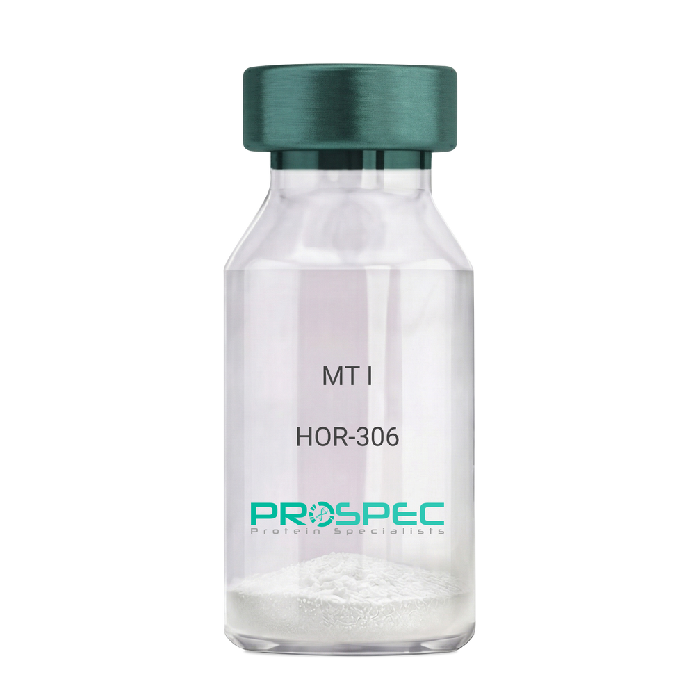
MT IDescription:
Melanotan-I
Melanotan-I, MT-I, Melanotan-1.
Product # :
HOR-306Price :
Quantity :
Shipping Method :
Shipped at Room temp
More Info
- description
- formulation
- purity
- More Info
Description
Melanotan-I has the amino acid sequence of Ser-Tyr-Ser-Nle-Glu-His-D-Phe-Arg-Trp-Gly-Lys-Pro-Val and a molecular weight of 1647.4 Dalton.
Formulation
The protein (1mg/ml) was lyophilized with no additives.
Purity
Greater than 98.0% as determined by RP-HPLC.
More Info
-
Synonyms
Melanotan-I, MT-I, Melanotan-1.
-
Physical Appearance
Sterile Filtered off-White lyophilized (freeze-dried) powder.
-
Stability
Lyophilized Melanotan-I although stable at room temperature for 3 weeks, should be stored desiccated below -18°C. Upon reconstitution MT-I should be stored at 4°C between 2-7 days and for future use below -18°C.For long term storage it is recommended to add a carrier protein (0.1% HSA or BSA).Please prevent freeze-thaw cycles.
-
Solubility
It is recommended to reconstitute the lyophilized Melanotan-I in sterile 1% acetic acid not less than 100 µg/ml, which can then be further diluted to other aqueous solutions.
ProSpec's products are furnished for LABORATORY RESEARCH USE ONLY. The product may not be used as drugs, agricultural or pesticidal products, food additives or household chemicals.
- View Data Sheet
Name :
IMPA1 AntibodyDescription:
Inositol Monophosphatase 1, Mouse Anti Human
Inositol monophosphatase 1, IMP 1, IMPase 1, Inositol-1(or 4)-monophosphatase 1, Lithium-sensitive myo-inositol monophosphatase A1, IMPA1, IMPA, IMP.
Product # :
ANT-625Price :
Quantity :
Shipping Method :
Shipped with Ice Packs
More Info
- formulation
- More Info
Formulation
1mg/ml containing PBS, pH-7.4, 10% Glycerol and 0.02% Sodium Azide.
More Info
-
Introduction
Inositol monophosphatase1 (IMPA1) is responsible for the provision of inositol essential for synthesis of phosphatidylinositol and polyphosphoinositides. IMPA1 has a central role in the phosphatidylinositol signaling pathway by catalyzing the hydrolysis of inositol monophosphates. IMPA1 has been recognized as the pharmacological target for lithium action in the brain. The IMPA1 enzyme has a magnesium-dependent phosphatase activity and is inhibited by therapeutic concentrations of lithium. Inhibition of inositol monophosphate hydroylosis and ensuing depletion of inositol for phosphatidylinositol synthesis may perhaps explain the anti-manic and anti-depressive effects of lithium administered to treat bipolar disorder.
-
Synonyms
Inositol monophosphatase 1, IMP 1, IMPase 1, Inositol-1(or 4)-monophosphatase 1, Lithium-sensitive myo-inositol monophosphatase A1, IMPA1, IMPA, IMP.
-
Physical Appearance
Sterile filtered colorless solution.
-
Immunogen
Anti-human IMPA1 mAb, is derived from hybridization of mouse F0 myeloma cells with spleen cells from BALB/c mice immunized with a recombinant human IMPA1 protein 1-277amino acids purified from E. coli.
-
Ig Subclass
Mouse IgG1 heavy chain and k light chain.
-
Clone
PAT2G5AT.
-
Applications
The antibody has been tested by ELISA, Western blot analysis, Flow cytometry and ICC/IF to assure specificity and reactivity. Since application varies, however, each investigation should be titrated by the reagent to obtain optimal results.
-
Type
Mouse Anti Human Monoclonal.
-
Storage Procedures
For periods up to 1 month store at 4°C, for longer periods of time, store at -20°C. Prevent freeze thaw cycles.
-
Purification Method
IMPA1 antibody was purified from mouse ascitic fluids by protein-A affinity chromatography.
ProSpec's products are furnished for LABORATORY RESEARCH USE ONLY. The product may not be used as drugs, agricultural or pesticidal products, food additives or household chemicals.
- View Data Sheet
Name :
FGF 1 MouseDescription:
Fibroblast Growth Factor-Acidic Mouse Recombinant
HBGF-1, ECGF-beta, FIBP, FGFIBP, FIBP-1, ECGF, ECGFA, GLIO703, FGF1, FGF-a.
Product # :
CYT-528Price :
Quantity :
Shipping Method :
Shipped at Room temp
More Info
- description
- source
- formulation
- purity
- biological activity
- More Info
Description
Fibroblast Growth Factor-acidic Mouse Recombinant (FGF-1) produced in E.Coli is a single, non-glycosylated, polypeptide chain containing 141 amino acids and having a molecular mass of 15.9kDa. The FGF acidic is purified by proprietary chromatographic techniques.
Source
Escherichia Coli.
Formulation
Lyophilized from a 0.2μm filtered concentrated solution in PBS, pH 7.0, 5% Trehalose, 0.02% Tween-80, 0.5mM DTT and 0.5mM EDTA.
Purity
Greater than 96.0% as determined by SDS-PAGE and HPLC analyses.
Biological Activity
The ED50 as determined by the dose-dependent proliferation of mouse BALB/c 3T3 cells, is less than 0.2ng/ml corresponding to a Specific Activity of 5x10⁶IU/mg.
More Info
-
Introduction
Acidic fibroblast growth factor is a member of the fibroblast growth factor (FGF) family. FGF family members possess broad mitogenic and cell survival activities, and are involved in a variety of biological processes, including embryonic development, cell growth, morphogenesis, tissue repair, tumor growth and invasion. This protein functions as a modifier of endothelial cell migration and proliferation, as well as an angiogenic factor. It acts as a mitogen for a variety of mesoderm- and neuroectoderm-derived cells in vitro, thus is thought to be involved in organogenesis. Three alternatively spliced variants encoding different isoforms have been described. The binding growth factors are angiogenic agents in vivo and are potent mitogens for a variety of cell types in vitro. There are differences in the tissue distribution and concentration of these 2 growth factors.
-
Synonyms
HBGF-1, ECGF-beta, FIBP, FGFIBP, FIBP-1, ECGF, ECGFA, GLIO703, FGF1, FGF-a.
-
Physical Appearance
Sterile Filtered White lyophilized (freeze-dried) powder.
-
Stability
Lyophilized Fibroblast Growth Factor-1 although stable at room temperature for 3 weeks, should be stored desiccated below -18°C. Upon reconstitution FGF-a should be stored at 4°C between 2-7 days and for future use below -18°C.Please prevent freeze-thaw cycles.
-
Solubility
It is recommended to reconstitute the lyophilized Fibroblast Growth Factor-acidic in sterile 18MΩ-cm H2O not less than 100µg/ml, which can then be further diluted to other aqueous solutions.
-
Amino Acid Sequence
MFNLPLGNYK KPKLLYCSNG GHFLRILPDG TVDGTRDRSD QHIQLQLSAE SAGEVYIKGT ETGQYLAMDT EGLLYGSQTP NEECLFLERL EENHYNTYTS KKHAEKNWFV GLKKNGSCKR GPRTHYGQKA ILFLPLPVSS D.
-
Background
What is the molecular weight/Mw of FGF 1 Protein?
FGF 1 Protein has a total Mw of 15.9kDa.
What is the source or expression system of FGF 1 Protein?
Escherichia Coli.
What is the Purity of FGF 1 Protein?
FGF 1 Protein is >96% pure as determined by SDS-PAGE.
What is the Biological Activity of FGF 1 Protein?
The ED50 as determined by the dose-dependent proliferation of mouse BALB/c 3T3 cells, is less than 0.2ng/ml corresponding to a Specific Activity of 5x10⁶IU/mg.
What is the amino acid sequence of FGF 1 Protein?
MFNLPLGNYK KPKLLYCSNG GHFLRILPDG TVDGTRDRSD QHIQLQLSAE SAGEVYIKGT ETGQYLAMDT EGLLYGSQTP NEECLFLERL EENHYNTYTS KKHAEKNWFV GLKKNGSCKR GPRTHYGQKA ILFLPLPVSS D.
What applications can FGF 1 Protein be used in?
FGF 1 Protein can probably be used in western blot, ELISA and Lateral Flow.
What is the endotoxin level for FGF 1 Protein?
The endotoxin level is minimal, FGF 1 Protein was purified using conventional chromatography techniques.
ProSpec's products are furnished for LABORATORY RESEARCH USE ONLY. The product may not be used as drugs, agricultural or pesticidal products, food additives or household chemicals.
- View Data Sheet
Name :
HCV NS5 Genotype-3aDescription:
Hepatitis C Virus NS5 Genotype-3a Recombinant
Product # :
HCV-232Price :
Quantity :
Shipping Method :
Shipped with Ice Packs
More Info
- description
- formulation
- purity
- More Info
Description
The E.coli derived recombinant protein contains the HCV NS5 Genotype 3a immunodominant regions, amino acids 2212-2313. The protein is fused to a GST tag at N-terminus.
Formulation
1.5M urea, 25 mM Tris-HCl, pH-8, 0.2% Triton-X & 50% Glycerol.
Purity
HCV NS5 Genotype-3a protein is >95% pure as determined by 10% PAGE (coomassie staining).
More Info
-
Introduction
HCV is a small 50nm, enveloped, single-stranded, positive sense RNAvirus in the family Flaviviridae.
HCV has a high rate of replication with approximately one trillion particles produced each day in an infected individual. Due to lack of proofreading by the HCV RNA polymerase, the HCV has an exceptionally high mutation rate, a factor that may help it elude the host's immune response. Hepatitis C virus is classified into six genotypes(1-6) with several subtypes within each genotype. The preponderance and distribution of HCV genotypes varies globally. Genotype is clinically important in determining potential response to interferon-based therapy and the required duration of such therapy. Genotypes 1 and 4 are less responsive to interferon-based treatment than are the other genotypes (2, 3, 5 and 6). -
Stability
HCV NS5 Genotype-3a although stable at 4°C for 1 week, should be stored below -18°C. Please prevent freeze thaw cycles.
-
Applications
HCV NS5 Genotype-3 antigen is suitable for ELISA and Western blots, excellent antigen for detection of HCV with minimal specificity problems.
-
Specificity
Immunoreactive with sera of HCV-infected individuals.
-
Purification Method
HCV NS5 Genotype-3a protein was purified by proprietary chromatographic technique.
ProSpec's products are furnished for LABORATORY RESEARCH USE ONLY. The product may not be used as drugs, agricultural or pesticidal products, food additives or household chemicals.
- View Data Sheet
Name :
IL8 GSTDescription:
Interleukin-8 (1-72) (CXCL8) Human Recombinant, GST Tag
Interleukin-8, IL-8, C-X-C motif chemokine 8, IL8, CXCL8
Product # :
CHM-047Price :
Quantity :
Shipping Method :
Shipped with Ice Packs
More Info
- description
- source
- formulation
- purity
- More Info
Description
Recombinant Human Interleukin-8 produced in E. coli containing 72 amino acids.Recombinant Human Interleukin-8 is fused to GST tag at its N-terminus and purified by proprietary chromatographic technique.
Source
Escherichia Coli.
Formulation
IL8 GST solution contains 25mM Tris-Base/ 25mM K2CO3.
Purity
Protein is >95% pure as determined by 10% PAGE (coomassie staining).
More Info
-
Introduction
Interleukin-8 (IL-8) is a chemokine produced by macrophages and other cell types such as epithelial cells. It is also synthesized by endothelial cells, which store IL-8 in their storage vesicles, the Weibel-Palade bodies. When first encountering an antigen, the primary cells to encounter it are the macrophages who phagocytose the particle. Upon processing, they release chemokines to signal other immune cells to come in to the site of inflammation. IL-8 is one such chemokine. It serves as a chemical signal that attracts neutrophils at the site of inflammation, and therefore is also known as Neutrophil Chemotactic Factor.
-
Synonyms
Interleukin-8, IL-8, C-X-C motif chemokine 8, IL8, CXCL8
-
Physical Appearance
Sterile Filtered clear solution.
-
Stability
Store at 4°C if entire vial will be used within 2-4 weeks.Store, frozen at -20°C for longer periods of time.For long term storage it is recommended to add a carrier protein (0.1% HSA or BSA).Avoid multiple freeze-thaw cycles.
-
Applications
Immunoassay.
ProSpec's products are furnished for LABORATORY RESEARCH USE ONLY. The product may not be used as drugs, agricultural or pesticidal products, food additives or household chemicals.
- View Data Sheet
Name :
CST3 MouseDescription:
Cystatin-C Mouse Recombinant
Post G-globulin, CST 3, CST3, Gamma-Trace, Cystatin 3, Amyloid Angiopathy and Cerebral Hemorrhage, Cystatin-C precursor, neuroendocrine basic polypeptide.
Product # :
PRO-597Price :
Quantity :
Shipping Method :
Shipped at Room temp
More Info
- description
- source
- formulation
- purity
- More Info
Description
Cystatin-C Murine Recombinant produced in E.Coli is a single, non-glycosylated, polypeptide chain containing 134 amino acids and having a molecular mass of 15kDa. The Mouse Cystatin-C is fused to His tag at N-Terminus.The Mouse Cystatin-C is purified by proprietary chromatographic techniques.
Source
Escherichia Coli.
Formulation
The sterile filtered concentrated (0.5mg/ml) protein solution was lyophilized with 20mM Tris & 50mM NaCl pH-7.5.
Purity
Greater than 95% as determined by SDS-PAGE.
More Info
-
Introduction
Cystatins are a superfamily of cysteine proteinase inhibitors found in both plants and animals. They comprise a group of proteinase inhibitors, widely distributed in tissues and body fluids, and form tight complexes with cysteine proteases such as cathepsin B, H, L and S. Cystatin C, a secreted molecule of this family, is of interest from biochemical, medicine and evolutionary points of view. Cystatin C, with molecular weight of 13260 Da, is composed of 120 amino acids, lacks carbohydrate and has two disulfide bridges located near the carboxyl terminus. Cystatin C is increased in patients with malignant diseases, and is related to the insufficiency of renal function and appears to be a better marker than creatinine. On the other hand, low levels of cystatin C involve cause the breakdown of the elastic laminae and, subsequently, the atherosclerosis and abdominal aortic aneurysm.
-
Synonyms
Post G-globulin, CST 3, CST3, Gamma-Trace, Cystatin 3, Amyloid Angiopathy and Cerebral Hemorrhage, Cystatin-C precursor, neuroendocrine basic polypeptide.
-
Physical Appearance
Sterile Filtered White lyophilized (freeze-dried) powder.
-
Stability
Store lyophilized protein at -20°C. Aliquot the product after reconstitution to avoid repeated freezing/thawing cycles. Reconstituted protein can be stored at 4°C for a limited period of time.
-
Solubility
Add deionized water to a working concentration of 0.5mg/ml and let the lyophilized pellet dissolve completely.
-
Amino Acid Sequence
MRGSHHHHHH GMASATPKQG PRMLGAPEEA DANEEGVRRA LDFAVSEYNK GSNDAYHSRA IQVVRARKQL VAGVNYFLDV EMGRTTCTKS QTNLTDCPFH DQPHLMRKAL CSFQIYSVPW KGTHSLTKFSCKNA.
ProSpec's products are furnished for LABORATORY RESEARCH USE ONLY. The product may not be used as drugs, agricultural or pesticidal products, food additives or household chemicals.
- View Data Sheet
Name :
VTCN1 Human, Sf9Description:
V-Set Domain Containing T Cell Activation Inhibitor 1 Human Recombinant, Sf9
V-set domain-containing T-cell activation inhibitor 1, B7 homolog 4, B7-H4, B7h.5, Immune costimulatory protein B7-H4, Protein B7S1, T-cell costimulatory molecule B7x, B7H4, VTCN1, B7S1, B7X, PRO1291, RP11-229A19.4.
Product # :
PRO-2439Price :
Quantity :
Shipping Method :
Shipped with Ice Packs
More Info
- description
- source
- formulation
- purity
- More Info
Description
VTCN1 Human Recombinant produced in Sf9 Baculovirus cells is a single, glycosylated polypeptide chain containing 244 amino acids (25-259a.a.) and having a molecular mass of 26.9kDa (Molecular size on SDS-PAGE will appear at approximately 28-40kDa). VTCN1 is expressed with a 9 amino acids His tag at C-Terminus and purified by proprietary chromatographic techniques.
Source
Sf9, Baculovirus cells.
Formulation
VTCN1 protein solution (0.25mg/ml) contains Phosphate Buffered Saline (pH 7.4) and 10% glycerol.
Purity
Greater than 90.0% as determined by SDS-PAGE.
More Info
-
Introduction
V-Set Domain Containing T Cell Activation Inhibitor 1, also known as VCTN1, is part of the B7 costimulatory protein family. Proteins in this family are located on the surface of antigen-presenting cells and interact with ligand bound to receptors on the surface of T cells. High level of the encoded protein has been associated with tumor progression. VCTN1 also takes part in promoting epithelial cell transformation.
-
Synonyms
V-set domain-containing T-cell activation inhibitor 1, B7 homolog 4, B7-H4, B7h.5, Immune costimulatory protein B7-H4, Protein B7S1, T-cell costimulatory molecule B7x, B7H4, VTCN1, B7S1, B7X, PRO1291, RP11-229A19.4.
-
Physical Appearance
Sterile Filtered colorless solution.
-
Stability
Store at 4°C if entire vial will be used within 2-4 weeks. Store, frozen at -20°C for longer periods of time. For long term storage it is recommended to add a carrier protein (0.1% HSA or BSA).Avoid multiple freeze-thaw cycles.
-
Amino Acid Sequence
ADPLIIGFGI SGRHSITVTT VASAGNIGED GILSCTFEPD IKLSDIVIQW LKEGVLGLVH EFKEGKDELS EQDEMFRGRT AVFADQVIVG NASLRLKNVQ LTDAGTYKCY IITSKGKGNA NLEYKTGAFS MPEVNVDYNA SSETLRCEAP RWFPQPTVVW ASQVDQGANF SEVSNTSFEL NSENVTMKVV SVLYNVTINN TYSCMIENDI AKATGDIKVT ESEIKRRSHL QLLNSKASHH HHHH.
ProSpec's products are furnished for LABORATORY RESEARCH USE ONLY. The product may not be used as drugs, agricultural or pesticidal products, food additives or household chemicals.
- View Data Sheet
Name :
LGALS4 HumanDescription:
Galectin-4 Human Recombinant
Galectin-4, Gal-4, Lactose-binding lectin 4, L-36 lactose-binding protein, L36LBP, Antigen NY-CO-27, LGALS4, lectin galactoside-binding soluble 4, GAL4.
Product # :
CYT-686Price :
Quantity :
Shipping Method :
Shipped with Ice Packs
More Info
- description
- source
- formulation
- purity
- More Info
- sds-page
Description
Galectin-4 produced in E.Coli is a single, non-glycosylated polypeptide chain containing 343 amino acids (1-323 a.a.) and having a molecular mass of 38.1kDa.Galectin-4 is fused to 20 amino acid His-Tag at N-terminus and purified by proprietary chromatographic techniques.
Source
Escherichia Coli.
Formulation
The LGALS4 protein solution contains 20mM Tris-HCl, pH-8, 1mM DTT and 10% glycerol.
Purity
Greater than 95.0% as determined by SDS-PAGE.
sds-page
More Info
-
Introduction
Galectin-4 is a member of the subfamily of galectins composed of two carbohydrate recognition domains having similar peptide chains. The galectins are a family of beta-galactoside-binding proteins having a role in modulating cell-cell and cell-matrix interactions, which inhibits chronic inflammations, GVHD, and allergic responses. LGALS4 expression is limited to small intestine, colon, and rectum, and it is underexpressed in colorectal cancer. LGALS4 binds as an endogenous ligand to glycosphingolipids having 3-O-sulfated Gal residues and bind as well to cholesterol-3-sulfate. LGALS4 takes part in cell adhesion. LGALS4 plays a role in crosslinking the lateral cell membranes of the surface-lining epithelial cells, thus supporting epithelial integrity against mechanical stress exerted by the bowel lume. LGALS4 is in charge of intestinal inflammation via selective regulation of peripheral and mucosal T-cell cell cycle, in addition to cell death by apoptosis of T-cells by a pathway independent of the activation of caspases. LGALS4 blockade decreases TNF-alpha inhibitor induced T-cell death. LGALS4 decreases pro-inflammatory cytokine secretion including IL-6 & IL-17.
-
Synonyms
Galectin-4, Gal-4, Lactose-binding lectin 4, L-36 lactose-binding protein, L36LBP, Antigen NY-CO-27, LGALS4, lectin galactoside-binding soluble 4, GAL4.
-
Physical Appearance
Sterile filtered colorless solution.
-
Stability
Store at 4°C if entire vial will be used within 2-4 weeks. Store, frozen at -20°C for longer periods of time. For long term storage it is recommended to add a carrier protein (0.1% HSA or BSA).Avoid multiple freeze-thaw cycles.
-
Amino Acid Sequence
MGSSHHHHHH SSGLVPRGSH MAYVPAPGYQ PTYNPTLPYY QPIPGGLNVG MSVYIQGVAS EHMKRFFVNF VVGQDPGSDV AFHFNPRFDG WDKVVFNTLQ GGKWGSEERK RSMPFKKGAA FELVFIVLAE HYKVVVNGNP FYEYGHRLPL QMVTHLQVDG DLQLQSINFI GGQPLRPQGP PMMPPYPGPG HCHQQLNSLP TMEGPPTFNP PVPYFGRLQG GLTARRTIII KGYVPPTGKS FAINFKVGSS GDIALHINPR MGNGTVVRNS LLNGSWGSEE KKITHNPFGP GQFFDLSIRC GLDRFKVYAN GQHLFDFAHR LSAFQRVDTL EIQGDVTLSY VQI.
-
Background
What is the molecular weight/Mw of LGALS4 HUMAN Protein?
LGALS4 HUMAN Protein has a total Mw of 38.1kDa.
What is the source or expression system of LGALS4 HUMAN Protein?
Escherichia Coli.
What is the Purity of LGALS4 HUMAN Protein?
LGALS4 HUMAN Protein is >95% pure as determined by SDS-PAGE.
What is the Biological Activity of LGALS4 HUMAN Protein?
The biological functionality of LGALS4 HUMAN Protein will be determined in the future.
What is the amino acid sequence of LGALS4 HUMAN Protein?
MGSSHHHHHH SSGLVPRGSH MAYVPAPGYQ PTYNPTLPYY QPIPGGLNVG MSVYIQGVAS EHMKRFFVNF VVGQDPGSDV AFHFNPRFDG WDKVVFNTLQ GGKWGSEERK RSMPFKKGAA FELVFIVLAE HYKVVVNGNP FYEYGHRLPL QMVTHLQVDG DLQLQSINFI GGQPLRPQGP PMMPPYPGPG HCHQQLNSLP TMEGPPTFNP PVPYFGRLQG GLTARRTIII KGYVPPTGKS FAINFKVGSS GDIALHINPR MGNGTVVRNS LLNGSWGSEE KKITHNPFGP GQFFDLSIRC GLDRFKVYAN GQHLFDFAHR LSAFQRVDTL EIQGDVTLSY VQI.
What applications can LGALS4 HUMAN Protein be used in?
LGALS4 HUMAN Protein can probably be used in western blot, ELISA and Lateral Flow.
What is the endotoxin level for LGALS4 HUMAN Protein?
The endotoxin level is minimal, LGALS4 HUMAN Protein was purified using conventional chromatography techniques.
ProSpec's products are furnished for LABORATORY RESEARCH USE ONLY. The product may not be used as drugs, agricultural or pesticidal products, food additives or household chemicals.
- View Data Sheet
Name :
FGF4 HumanDescription:
Fibroblast Growth Factor-4 Human Recombinant
HBGF4, FGF-4, FGF4, KFGF, HSTF1.
Product # :
CYT-312Price :
Quantity :
Shipping Method :
Shipped at Room temp
More Info
- description
- source
- formulation
- purity
- biological activity
- More Info
Description
FGF4 Human Recombinant produced in E.Coli is a single, non-glycosylated, polypeptide chain containing 182 amino acids and having a molecular mass of 19.8kDa. The FGF4 is purified by proprietary chromatographic techniques.
Source
Escherichia Coli.
Formulation
The FGF4 protein was lyophilized with 20mM sodium phosphate and 500mM NaCl pH-7.5.
Purity
Greater than 95.0% as determined by SDS-PAGE.
Biological Activity
The ED50 as determined by NR6R-3T3 Proliferation is 0.54ng/ml, corresponding to a specific activity of 1.8X10⁶ units/mg.
More Info
-
Introduction
FGF4 holds s comprehensive mitogenic and cell survival activities and takes part in a range of biological processes including embryonic development, cell growth, morphogenesis, tissue repair, tumor growth and invasion. FGF4 possess oncogenic transforming activity. FGF4 and FGF3, oncogenic growth factors are localized on chromosome 11. Co-amplification of both factors was found in several kinds of human tumors. FGF4 functions in bone morphogenesis and limb development through the sonic hedgehog (SHH) signaling pathway.
-
Synonyms
HBGF4, FGF-4, FGF4, KFGF, HSTF1.
-
Physical Appearance
Sterile Filtered White lyophilized (freeze-dried) powder.
-
Stability
Lyophilized FGF4 although stable at room temperature for 3 weeks, should be stored desiccated below -18°C. Upon reconstitution FGF-4 should be stored at 4°C between 2-7 days and for future use below -18°C.Please prevent freeze-thaw cycles.
-
Solubility
It is recommended to reconstitute the lyophilized FGF4 Human Recombinant sterile 18MΩ-cm H2O not less than 100µg/ml, which can then be further diluted to other aqueous solutions.
-
Amino Acid Sequence
MAPTAPNGTL EAELERRWES LVALSLARLP VAAQPKEAAV QSGAGDYLLG IKRLRRLYCN VGIGFHLQAL PDGRIGGAHA DTRDSLLELS PVERGVVSIF GVASRFFVAM SSKGKLYGSP FFTDECTFKE ILLPNNYNAY ESYKYPGMFI ALSKNGKTKK GNRVSPTMKV THFLPRL.
-
Background
What is the molecular weight/Mw of FGF4 HUMAN Protein?
FGF4 HUMAN Protein has a total Mw of 19.8kDa.
What is the source or expression system of FGF4 HUMAN Protein?
Escherichia Coli.
What is the Purity of FGF4 HUMAN Protein?
FGF4 HUMAN Protein is > 95% pure as determined by SDS-PAGE.
What is the Biological Activity of FGF4 HUMAN Protein?
The ED50 as determined by NR6R-3T3 Proliferation is 0.54ng/ml, corresponding to a specific activity of 1.8X10⁶ units/mg.
What is the amino acid sequence of FGF4 HUMAN Protein?
MAPTAPNGTL EAELERRWES LVALSLARLP VAAQPKEAAV QSGAGDYLLG IKRLRRLYCN VGIGFHLQAL PDGRIGGAHA DTRDSLLELS PVERGVVSIF GVASRFFVAM SSKGKLYGSP FFTDECTFKE ILLPNNYNAY ESYKYPGMFI ALSKNGKTKK GNRVSPTMKV THFLPRL.
What applications can FGF4 HUMAN Protein be used in?
FGF4 HUMAN Protein can probably be used in western blot, ELISA and Lateral Flow.
What is the endotoxin level for FGF4 HUMAN Protein?
The endotoxin level is minimal, FGF4 HUMAN Protein was purified using conventional chromatography techniques.
ProSpec's products are furnished for LABORATORY RESEARCH USE ONLY. The product may not be used as drugs, agricultural or pesticidal products, food additives or household chemicals.
- View Data Sheet
Name :
GM CSF RatDescription:
Granulocyte Macrophage-Colony Stimulating Factor Rat Recombinant
CSF-2, MGI-1GM, GM-CSF, Pluripoietin-alpha, Molgramostin, Sargramostim.
Product # :
CYT-395Price :
Quantity :
Shipping Method :
Shipped at Room temp
More Info
- description
- source
- formulation
- purity
- biological activity
- More Info
Description
Granulocyte Macrophage Colony Stimulating Factor Rat Recombinant produced in E.Coli is a single, non-glycosylated, polypeptide chain containing 128 amino acids and having a molecular mass of 14590.65 Dalton. GM-CSF Rat Recombinant is purified by proprietary chromatographic techniques.
Source
Escherichia Coli.
Formulation
GM-CSF Rat was lyophilized with no additives.
Purity
Greater than 96.0% as determined by:
(a)Analysis by RP-HPLC.
(b)Analysis by SDS-PAGE.Biological Activity
The ED50 as calculated by the dose-dependent stimulation of the proliferation of murine FDC-P1 cells is less than 0.01ng/ml, corresponding to a specific activity of > 1×10⁸units/mg.
More Info
-
Introduction
GMCSF is a cytokine that controls the production, differentiation, and function of granulocytes and macrophages. The active form of the protein is found extracellularly as a homodimer. This gene has been localized to a cluster of related genes at chromosome region 5q31, which is known to be associated with interstitial deletions in the 5q- syndrome and acute myelogenous leukemia. Other genes in the cluster include those encoding interleukins 4, 5, and 13.
GM-CSF stimulates the growth and differentiation of hematopoietic precursor cells from various lineages, including granulocytes, macrophages, eosinophils and erythrocytes. -
Synonyms
CSF-2, MGI-1GM, GM-CSF, Pluripoietin-alpha, Molgramostin, Sargramostim.
-
Physical Appearance
Sterile Filtered White lyophilized (freeze-dried) powder.
-
Stability
Lyophilized Granulocyte Macrophage Colony Stimulating Factor although stable at room temperature for 3 weeks, should be stored desiccated below -18°C. Upon reconstitution GMCSF should be stored at 4°C between 2-7 days and for future use below -18°C.For long term storage it is recommended to add a carrier protein (0.1% HSA or BSA).Please prevent freeze-thaw cycles.
-
Solubility
It is recommended to reconstitute the lyophilized Granulocyte Macrophage Colony Stimulating Factor in sterile 18MΩ-cm H2O not less than 100µg/ml, which can then be further diluted to other aqueous solutions.
-
Amino Acid Sequence
MAPTRSPNPV TRPWKHVDAI KEALSLLNDM RALENEKNED VDIISNEFSI
QRPTCVQTRL KLYKQGLRGN LTKLNGALTM IASHYQTNCP PTPETDCEIE
VTTFEDFIKN LKGFLFDIPF DCWKPVQK.
ProSpec's products are furnished for LABORATORY RESEARCH USE ONLY. The product may not be used as drugs, agricultural or pesticidal products, food additives or household chemicals.
- View Data Sheet
Name :
GM CSF Human, Sf9Description:
Granulocyte Macrophage-Colony Stimulating Factor Human Recombinant, Sf9
CSF-2, MGI-1GM, GMCSF, Pluripoietin-alpha, Molgramostin, Sargramostim.
Product # :
CYT-416Price :
Quantity :
Shipping Method :
Shipped at Room temp
More Info
- description
- source
- formulation
- purity
- biological activity
- More Info
Description
GM-CSF Human Recombinant produced in insect cells is a single, glycosylated, polypeptide chain containing 127 amino acids (18-144) and having a molecular mass of 14.6kDa. GM-CSF is fused to a C-terminal His -tag (6x His) and purified by proprietary chromatographic techniques.
Source
Insect Cells.
Formulation
The protein was lyophilized with PBS.
Purity
Greater than 98.0% as determined by SDS-PAGE.
Biological Activity
The ED50 as determined by the dose-dependant stimulation of the proliferation of human TF-1 cells (human erythroleukemic indicator cell line) is < 0.1 ng/ml, corresponding to a Specific Activity of 10,000,000IU/mg.More Info
-
Introduction
GMCSF is a cytokine that controls the production, differentiation, and function of granulocytes and macrophages. The active form of the protein is found extracellularly as a homodimer. This gene has been localized to a cluster of related genes at chromosome region 5q31, which is known to be associated with interstitial deletions in the 5q- syndrome and acute myelogenous leukemia. Other genes in the cluster include those encoding interleukins 4, 5, and 13.
GM-CSF stimulates the growth and differentiation of hematopoietic precursor cells from various lineages, including granulocytes, macrophages, eosinophils and erythrocytes. -
Synonyms
CSF-2, MGI-1GM, GMCSF, Pluripoietin-alpha, Molgramostin, Sargramostim.
-
Physical Appearance
Sterile Filtered White lyophilized (freeze-dried) powder.
-
Stability
Lyophilized Granulocyte Macrophage Colony Stimulating Factor although stable at room temperature for 3 weeks, should be stored desiccated below -18°C. Upon reconstitution GMCSF should be stored at 4°C between 2-7 days and for future use below -18°C.For long term storage it is recommended to add a carrier protein (0.1% HSA or BSA).Please prevent freeze-thaw cycles.
-
Solubility
It is recommended to reconstitute the lyophilized Granulocyte Macrophage Colony Stimulating Factor in sterile 18MΩ-cm H2O not less than 100µg/ml, which can then be further diluted to other aqueous solutions.
-
Amino Acid Sequence
The sequence of the first five N-terminal amino acids was determined and was found to be Ala-Pro-Ala-Arg-Ser.
-
Background
Recombinant Granulocyte-Macrophage Colony-Stimulating Factor (GMCSF) is a protein that plays a crucial role in the production and differentiation of white blood cells, including granulocytes and macrophages. It is a potent stimulator of hematopoietic stem cells, which are responsible for the production of all blood cells in the body. Recombinant GMCSF is a synthetic version of the protein that is produced using recombinant DNA technology.
Recombinant GMCSF has been extensively studied for its potential therapeutic applications in a variety of medical conditions, including cancer, autoimmune diseases, and infectious diseases. In cancer, GMCSF is used as an immunostimulatory agent to enhance the immune response against cancer cells. By stimulating the production and differentiation of white blood cells, GM-CSF can increase the number of immune cells that can recognize and attack cancer cells. This approach has been successfully used in the treatment of several types of cancer, including melanoma and leukemia.
In autoimmune diseases, recombinant GMCSF has been investigated as a potential treatment for conditions such as rheumatoid arthritis and multiple sclerosis. These diseases are characterized by an overactive immune response that attacks healthy tissues in the body. By modulating the immune response, GMCSF may be able to reduce inflammation and prevent further damage to affected tissues.
In infectious diseases, recombinant GMCSF has been studied as a potential treatment for conditions such as sepsis and HIV/AIDS. In sepsis, a severe bacterial infection, GMCSF may be able to stimulate the production of white blood cells and improve the immune response against the infection. In HIV/AIDS, GMCSF may be able to enhance the immune response against the virus and reduce the risk of opportunistic infections.
Recombinant GMCSF is typically administered by injection, either directly into the affected tissue or into the bloodstream. It is generally well-tolerated, although some patients may experience side effects such as fever, fatigue, and muscle pain.
In conclusion, recombinant GMCSF is a promising therapeutic agent with potential applications in a variety of medical conditions. Its ability to stimulate the production and differentiation of white blood cells makes it a valuable tool in the treatment of cancer, autoimmune diseases, and infectious diseases. Ongoing research is likely to uncover new uses for this protein and further refine its therapeutic potential.
ProSpec's products are furnished for LABORATORY RESEARCH USE ONLY. The product may not be used as drugs, agricultural or pesticidal products, food additives or household chemicals.
- View Data Sheet
Name :
CST3 Mouse, ActiveDescription:
Cystatin-C Mouse Recombinant, Active
Cystatin-C, Cystatin-3, Cst3, CST3
Product # :
PRO-2433Price :
Quantity :
Shipping Method :
Shipped with Ice Packs
More Info
- description
- source
- formulation
- purity
- biological activity
- More Info
Description
CST3 Mouse produced in Sf9 Insect cells is a single, glycosylated polypeptide chain containing 126 amino acids (21-140 a.a.) and having a molecular mass of 14.2kDa (Molecular size on SDS-PAGE will appear at approximately 13.5-18kDa).CST3 is expressed with a 6 amino acid His tag at C-Terminus and purified by proprietary chromatographic techniques.
Source
Sf9, Baculovirus cells.
Formulation
CST3 protein solution (1mg/ml) contains Phosphate Buffered Saline (pH 7.4) and 10% glycerol.
Purity
Greater than 95% as determined by SDS-PAGE.
Biological Activity
The IC50 value is < 1.0nM. The inhibitory function of Cystatin 3 on protease activity of papain was measured by a fluorometric assay using Z-FR-AMC at pH 7.5 at 25°C.
More Info
-
Introduction
Cystatins are a superfamily of cysteine proteinase inhibitors found in both plants and animals. They comprise a group of proteinase inhibitors, widely distributed in tissues and body fluids, and form tight complexes with cysteine proteases such as cathepsin B, H, L and S. Cystatin C, a secreted molecule of this family, is of interest from biochemical, medicine and evolutionary points of view. Cystatin C, with molecular weight of 13260 Da, is composed of 120 amino acids, lacks carbohydrate and has two disulfide bridges located near the carboxyl terminus. Cystatin C is increased in patients with malignant diseases, and is related to the insufficiency of renal function and appears to be a better marker than creatinine. On the other hand, low levels of cystatin C involve cause the breakdown of the elastic laminae and, subsequently, the atherosclerosis and abdominal aortic aneurysm.
-
Synonyms
Cystatin-C, Cystatin-3, Cst3, CST3
-
Physical Appearance
Sterile Filtered colorless solution.
-
Stability
Store at 4°C if entire vial will be used within 2-4 weeks. Store, frozen at -20°C for longer periods of time. For long term storage it is recommended to add a carrier protein (0.1% HSA or BSA). Avoid multiple freeze-thaw cycles.
-
Amino Acid Sequence
ATPKQGPRML GAPEEADANE EGVRRALDFA VSEYNKGSND AYHSRAIQVV RARKQLVAGV NYFLDVEMGR TTCTKSQTNL TDCPFHDQPH LMRKALCSFQ IYSVPWKGTH SLTKFSCKNA HHHHHH
ProSpec's products are furnished for LABORATORY RESEARCH USE ONLY. The product may not be used as drugs, agricultural or pesticidal products, food additives or household chemicals.
- View Data Sheet
Name :
CD163 HumanDescription:
CD163 Human Recombinant
CD163 Molecule, Hemoglobin Scavenger Receptor, CD163 Antigen, M130, Scavenger Receptor Cysteine-Rich Type 1 Protein M130, Macrophage-Associated Antigen, SCARI1, MM130,CD163.
Product # :
PRO-2496Price :
Quantity :
Shipping Method :
Shipped with Ice Packs
More Info
- description
- source
- formulation
- purity
- More Info
Description
CD163 produced in Sf9 Baculovirus cells is a single, glycosylated polypeptide chain containing 1015 amino acids (42-1050a.a.) and having a molecular mass of 109.8kDa. (Molecular size on SDS-PAGE will appear at approximately 100kDa). CD163 is expressed with an 6 amino acid His tag at C-Terminus and purified by proprietary chromatographic techniques.
Source
Sf9, Baculovirus cells.
Formulation
CD163 protein solution (0.5mg/ml) contains phosphate Buffered Saline (pH 7.4).
Purity
Greater than 90.0% as determined by SDS-PAGE.
More Info
-
Introduction
CD163 is an acute phase-regulated receptor which participates in the removal and endocytosis of hemoglobin/haptoglobin complexes by macrophages and thus keeps tissues from free hemoglobin-mediated oxidative damage. Furthermore, CD163 partakes in the uptake and recycling of iron, through endocytosis of hemoglobin/haptoglobin and ensuing breakdown of heme. In addition, CD163 binds hemoglobin/haptoglobin complexes in a calcium-dependent and pH-dependent way. CD163 demonstrates greater affinity for complexes of hemoglobin and multimeric haptoglobin of HP-1F phenotype than for complexes of hemoglobin and dimeric haptoglobin of HP-1S phenotype. Moreover, CD163 stimulates a cascade of intracellular signals which involves tyrosine kinase-dependent calcium recruitment, inositol triphosphate formation and secretion of IL-6 & CSF-1.
-
Synonyms
CD163 Molecule, Hemoglobin Scavenger Receptor, CD163 Antigen, M130, Scavenger Receptor Cysteine-Rich Type 1 Protein M130, Macrophage-Associated Antigen, SCARI1, MM130,CD163.
-
Physical Appearance
Sterile Filtered colorless solution.
-
Stability
Store at 4°C if entire vial will be used within 2-4 weeks. Store, frozen at -20°C for longer periods of time. For long term storage it is recommended to add a carrier protein (0.1% HSA or BSA). Avoid multiple freeze-thaw cycles.
-
Amino Acid Sequence
SSLGGTDKEL RLVDGENKCS GRVEVKVQEE WGTVCNNGWS MEAVSVICNQ LGCPTAIKAP GWANSSAGSG RIWMDHVSCR GNESALWDCK HDGWGKHSNC THQQDAGVTC SDGSNLEMRL TRGGNMCSGR IEIKFQGRWG TVCDDNFNID HASVICRQLE CGSAVSFSGS SNFGEGSGPI WFDDLICNGN ESALWNCKHQ GWGKHNCDHA EDAGVICSKG ADLSLRLVDG VTECSGRLEV RFQGEWGTIC DDGWDSYDAA VACKQLGCPT AVTAIGRVNA SKGFGHIWLD SVSCQGHEPA IWQCKHHEWG KHYCNHNEDA GVTCSDGSDL ELRLRGGGSR CAGTVEVEIQ RLLGKVCDRG WGLKEADVVC RQLGCGSALK TSYQVYSKIQ ATNTWLFLSS CNGNETSLWD CKNWQWGGLT CDHYEEAKIT CSAHREPRLV GGDIPCSGRV EVKHGDTWGS ICDSDFSLEA ASVLCRELQC GTVVSILGGA HFGEGNGQIW AEEFQCEGHE SHLSLCPVAP RPEGTCSHSR DVGVVCSRYT EIRLVNGKTP CEGRVELKTL GAWGSLCNSH WDIEDAHVLC QQLKCGVALS TPGGARFGKG NGQIWRHMFH CTGTEQHMGD CPVTALGASL CPSEQVASVI CSGNQSQTLS SCNSSSLGPT RPTIPEESAV ACIESGQLRL VNGGGRCAGR VEIYHEGSWG TICDDSWDLS DAHVVCRQLG CGEAINATGS AHFGEGTGPI WLDEMKCNGK ESRIWQCHSH GWGQQNCRHK EDAGVICSEF MSLRLTSEAS REACAGRLEV FYNGAWGTVG KSSMSETTVG VVCRQLGCAD KGKINPASLD KAMSIPMWVD NVQCPKGPDT LWQCPSSPWE KRLASPSEET WITCDNKIRL QEGPTSCSGR VEIWHGGSWG TVCDDSWDLD DAQVVCQQLG CGPALKAFKE AEFGQGTGPI WLNEVKCKGN ESSLWDCPAR RWGHSECGHK EDAAVNCTDI SVQKTPQKAT TGRSSRQSSH HHHHH
ProSpec's products are furnished for LABORATORY RESEARCH USE ONLY. The product may not be used as drugs, agricultural or pesticidal products, food additives or household chemicals.
- View Data Sheet
Name :
GALNT1 HumanDescription:
Polypeptide N-Acetylgalactosaminyltransferase 1 Human Recombinant
Polypeptide N-acetylgalactosaminyltransferase 1, GALNT1, GALNAC-T1
Product # :
enz-1098Price :
Quantity :
Shipping Method :
Shipped with Ice Packs
More Info
- description
- source
- formulation
- purity
- biological activity
- More Info
Description
GALNT1 produced in Sf9 Insect cells is a single, glycosylated polypeptide chain containing 528 amino acids (41-559a.a.) and having a molecular mass of 60.4kDa.GALNT1 is expressed with an 9 amino acid His tag at C-Terminus and purified by proprietary chromatographic techniques.
Source
Sf9, Insect cells.
Formulation
GALNT1 protein solution (1mg/ml) contains 20mM Tris-HCl buffer (pH 8.0) containing 0.1M NaCl and 10% glycerol.
Purity
Greater than 90.0% as determined by SDS-PAGE.
Biological Activity
The specific activity which is defined as the amount of enzyme that transfer 1.0 pmole of GalNAc from UDP-GalNAc to peptide EA2 per minute at pH 8.0 at 37C is > 300 pmol/min/ug.
More Info
-
Introduction
Polypeptide N-Acetylgalactosaminyltransferase 1 (Galnt1) is a part of the UDP-N-acetyl-alpha-D-galactosamine:polypeptide N-acetylgalactosaminyltransferase (GalNAc-T) family of enzymes. The initial reaction in O-linked oligosaccharide biosynthesis is catalyzed by Glant1, the transfer of an N-acetyl-D-galactosamine residue to a serine or threonine residue on the protein receptor. Moreover, Galnt1 is implicated in the glycosylation of proteins vital for bone formation for instance osteopontin and bone sialoprotein.
-
Synonyms
Polypeptide N-acetylgalactosaminyltransferase 1, GALNT1, GALNAC-T1
-
Physical Appearance
Sterile filtered colorless solution.
-
Stability
Store at 4°C if entire vial will be used within 2-4 weeks. Store, frozen at -20°C for longer periods of time. For long term storage it is recommended to add a carrier protein (0.1% HSA or BSA).Avoid multiple freeze-thaw cycles.
-
Amino Acid Sequence
ADPGLPAGDV LEPVQKPHEG PGEMGKPVVI PKEDQEKMKE MFKINQFNLM ASEMIALNRS
LPDVRLEGCK TKVYPDNLPT TSVVIVFHNE AWSTLLRTVH SVINRSPRHM IEEIVLVDDA
SERDFLKRPL ESYVKKLKVP VHVIRMEQRS GLIRARLKGA AVSKGQVITF LDAHCECTVG
WLEPLLARIK HDRRTVVCPI IDVISDDTFE YMAGSDMTYG GFNWKLNFRW YPVPQREMDR
RKGDRTLPVR TPTMAGGLFS IDRDYFQEIG TYDAGMDIWG GENLEISFRI WQCGGTLEIV
TCSHVGHVFR KATPYTFPGG TGQIINKNNR RLAEVWMDEF KNFFYIISPG VTKVDYGDIS
SRVGLRHKLQ CKPFSWYLEN IYPDSQIPRH YFSLGEIRNV ETNQCLDNMA RKENEKVGIF
NCHGMGGNQV FSYTANKEIR TDDLCLDVSK LNGPVTMLKC HHLKGNQLWE YDPVKLTLQH
VNSNQCLDKA TEEDSQVPSI RDCNGSRSQQ WLLRNVTLPE IFHHHHHH.
ProSpec's products are furnished for LABORATORY RESEARCH USE ONLY. The product may not be used as drugs, agricultural or pesticidal products, food additives or household chemicals.
- View Data Sheet
Name :
HCV NS3 Genotype-3Description:
Hepatitis C Virus NS3 Genotype-3, (1356-1459 a.a.) Recombinant
Product # :
HCV-251Price :
Quantity :
Shipping Method :
Shipped with Ice Packs
More Info
- description
- formulation
- purity
- More Info
Description
The E.coli derived recombinant protein contains the HCV NS3 immunodominant regions, amino acids 1359-1456. The protein is fused to a GST tag at N-Terminus.
Formulation
1.5M urea, 25mM Tris-HCl pH-8, 0.2% Triton-X & 50% Glycerol.
Purity
HCV NS3 Genotype-3 protein is >95% pure as determined by 10% PAGE (coomassie staining).
More Info
-
Introduction
HCV is a small 50nm, enveloped, single-stranded, positive sense RNAvirus in the family Flaviviridae.
HCV has a high rate of replication with approximately one trillion particles produced each day in an infected individual. Due to lack of proofreading by the HCV RNA polymerase, the HCV has an exceptionally high mutation rate, a factor that may help it elude the host's immune response. Hepatitis C virus is classified into six genotypes(1-6) with several subtypes within each genotype. The preponderance and distribution of HCV genotypes varies globally. Genotype is clinically important in determining potential response to interferon-based therapy and the required duration of such therapy. Genotypes 1 and 4 are less responsive to interferon-based treatment than are the other genotypes (2, 3, 5 and 6). -
Stability
HCV NS3 Genotype-3 although stable at 4°C for 1 week, should be stored below -18°C. Please prevent freeze thaw cycles.
-
Applications
HCV NS3 Genotype-3 antigen is suitable for ELISA and Western blots, excellent antigen for detection of HCV with minimal specificity problems.
-
Specificity
Immunoreactive with sera of HCV-infected individuals.
-
Purification Method
HCV NS3 Genotype-3 protein was purified by proprietary chromatographic technique.
ProSpec's products are furnished for LABORATORY RESEARCH USE ONLY. The product may not be used as drugs, agricultural or pesticidal products, food additives or household chemicals.
- View Data Sheet
Name :
HCV NS3 Genotype-5Description:
Hepatitis C Virus NS3 Genotype-5, (1192-1459 a.a.) Recombinant
Product # :
HCV-266Price :
Quantity :
Shipping Method :
Shipped with Ice Packs
More Info
- description
- formulation
- purity
- More Info
Description
The E.coli derived recombinant protein fused to a His tag contains the HCV NS3 immunodominant regions, amino acids 1192-1459.
Formulation
1.5M urea, 25mM Tris-HCl pH-8, 0.2% Triton-X & 50% Glycerol.
Purity
HCV NS3 Genotype-5 protein is >95% pure as determined by 10% PAGE (coomassie staining).
More Info
-
Introduction
HCV is a small 50nm, enveloped, single-stranded, positive sense RNAvirus in the family Flaviviridae.
HCV has a high rate of replication with approximately one trillion particles produced each day in an infected individual. Due to lack of proofreading by the HCV RNA polymerase, the HCV has an exceptionally high mutation rate, a factor that may help it elude the host's immune response. Hepatitis C virus is classified into six genotypes(1-6) with several subtypes within each genotype. The preponderance and distribution of HCV genotypes varies globally. Genotype is clinically important in determining potential response to interferon-based therapy and the required duration of such therapy. Genotypes 1 and 4 are less responsive to interferon-based treatment than are the other genotypes (2, 3, 5 and 6). -
Stability
HCV NS3 Genotype-5 although stable at 4°C for 1 week, should be stored below -18°C. Please prevent freeze thaw cycles.
-
Applications
HCV NS3 Genotype-5 antigen in ELISA and Western blots, excellent antigen for detection of HCV with minimal specificity problems.
-
Specificity
Immunoreactive with sera of HCV-infected individuals.
-
Purification Method
HCV NS3 Genotype-5 protein was purified by proprietary chromatographic technique.
ProSpec's products are furnished for LABORATORY RESEARCH USE ONLY. The product may not be used as drugs, agricultural or pesticidal products, food additives or household chemicals.
- View Data Sheet
Name :
HCV NS4 Mosaic Genotype-1Description:
Hepatitis C Virus NS4 Mosaic Genotype-1 Recombinant
Product # :
HCV-257Price :
Quantity :
Shipping Method :
Shipped with Ice Packs
More Info
- description
- formulation
- purity
- More Info
Description
The E.coli derived recombinant protein contains the HCV NS4 immunodominant regions, amino acids 1691-1710, 1712-1733 & 1921-1940, fused with a GST tag at N-terminus.
Formulation
1.5M urea, 25mM Tris-HCl, pH 8.0, 0.2% Triton-X & 50% glycerol.
Purity
Protein is >95% pure as determined by 10% PAGE (coomassie staining).
More Info
-
Introduction
HCV is a small 50nm, enveloped, single-stranded, positive sense RNAvirus in the family Flaviviridae.
HCV has a high rate of replication with approximately one trillion particles produced each day in an infected individual. Due to lack of proofreading by the HCV RNA polymerase, the HCV has an exceptionally high mutation rate, a factor that may help it elude the host's immune response. Hepatitis C virus is classified into six genotypes(1-6) with several subtypes within each genotype. The preponderance and distribution of HCV genotypes varies globally. Genotype is clinically important in determining potential response to interferon-based therapy and the required duration of such therapy. Genotypes 1 and 4 are less responsive to interferon-based treatment than are the other genotypes (2, 3, 5 and 6). -
Stability
HCV NS4 Mosaic Genotype-1 although stable at 4°C for 1 week, should be stored below -18°C. Please prevent freeze thaw cycles.
-
Applications
ELISA, Western blots and Flow-through.
-
Specificity
Immunoreactive with sera of HCV-infected individuals.
-
Purification Method
Purified by proprietary chromatographic technique.
ProSpec's products are furnished for LABORATORY RESEARCH USE ONLY. The product may not be used as drugs, agricultural or pesticidal products, food additives or household chemicals.
- View Data Sheet
Name :
HSV 2 gGDescription:
Herpes Simplex Virus-2 gG
Product # :
HSV-220Price :
Quantity :
Shipping Method :
Shipped with Ice Packs
More Info
- description
- formulation
- purity
- More Info
Description
The HSV-2 gG protein is a synthetic protein which containing the HSV-2 gG immunodominant regions.
Formulation
HSV-2 gG solution contains 25mM Tris-HCl pH8.0.
Purity
Protein is >99% pure as determined by Analytical HPLC and Mass Spec.
More Info
-
Introduction
Entry of HSV into the host cell involves interactions of several viral glycoproteins with cell surface receptors. The virus particle is covered by an envelope which, when bound to specific receptors on the cell surface, will fuse with the cell membrane and create an opening, or pore, through which the virus enters the host cell. The sequential stages of HSV entry are analagous to those of other viruses. At first, complementary receptors on the virus and cell surface bring the two membranes into proximity. In an intermediate state, the two membranes begin to merge, forming a hemifusion state. Finally, a stable entry pore is formed through which the viral envelope contents are introduced to the host cell.
-
Physical Appearance
Sterile Filtered clear solution.
-
Stability
HSV-2 gG protein although stable at 4°C for 1 week, should be stored below -18°C. Please prevent freeze thaw cycles.
-
Specificity
Immunoreactive with sera of HSV-infected individuals.
-
Purification Method
HSV-2 gG protein was purified by Prep HPLC.
ProSpec's products are furnished for LABORATORY RESEARCH USE ONLY. The product may not be used as drugs, agricultural or pesticidal products, food additives or household chemicals.
- View Data Sheet
Name :
TGFB1 (113 a.a.) HumanDescription:
Transforming Growth Factor-Beta 1 (113 a.a.) Human Recombinant
Transforming growth factor beta-1, TGF-beta-1, CED, DPD1, TGFB, TGF-b 1.
Product # :
CYT-679Price :
Quantity :
Shipping Method :
Shipped with Ice Packs
More Info
- description
- source
- formulation
- purity
- More Info
Description
TGF-b 1 Human Recombinant produced in E.Coli is a single, non-glycosylated, polypeptide chain containing 113 amino acids (279-390 a.a.) and having a total molecular mass of 12.9 kDa. TGF-b 1 (113 a.a.) is purified by proprietary chromatographic techniques.
Source
Escherichia Coli.
Formulation
The TGF-b 1 solution contains 10mM Sodium Citrate (pH3.5) and 10% glycerol.
Purity
Greater than 95.0% as determined by SDS-PAGE.
More Info
-
Introduction
Transforming growth factor betas (TGFBetas) mediate many cell-cell interactions that occur during embryonic development. Three TGFBetas have been identified in mammals. TGFBeta1, TGFBeta2 and TGFBeta3 are each synthesized as precursor proteins that are very similar in that each is cleaved to yield a 112 amino acid polypeptide that remains associated with the latent portion of the molecule.
-
Synonyms
Transforming growth factor beta-1, TGF-beta-1, CED, DPD1, TGFB, TGF-b 1.
-
Physical Appearance
Sterile Filtered colorless solution.
-
Stability
Store at 4°C if entire vial will be used within 2-4 weeks. Store, frozen at -20°C for longer periods of time. For long term storage it is recommended to add a carrier protein (0.1% HSA or BSA).Avoid multiple freeze-thaw cycles.
-
Amino Acid Sequence
MALDTNYCFS STEKNCCVRQ LYIDFRKDLG WKWIHEPKGY HANFCLGPCP YIWSLDTQYS KVLALYNQHN PGASAAPCCV PQALEPLPIVYYVGRKPKVE QLSNMIVRSC KCS.
-
Background
Title: Transforming Growth Factor-Beta 1 (113 a.a.) Human Recombinant: A Key Regulator of Cellular Processes with Therapeutic Potential
Abstract:
Transforming Growth Factor-Beta 1 (TGF-β1) is a multifunctional cytokine that plays a crucial role in various cellular processes, including cell growth, differentiation, and immune modulation. The development of TGF-β1 human recombinant proteins has provided valuable tools for studying its biological functions and therapeutic applications. This research paper explores the production process, characteristics, and potential therapeutic uses of TGF-β1 human recombinant, highlighting its importance and clinical significance.Introduction:
TGF-β1 is a pivotal cytokine involved in numerous physiological and pathological processes, such as embryonic development, tissue repair, and immune regulation. Harnessing the therapeutic potential of TGF-β1 has been facilitated by the development of TGF-β1 human recombinant proteins using recombinant DNA technology. These recombinant proteins have become valuable tools for investigating the biological functions of TGF-β1 and exploring its therapeutic applications.Production Process and Characteristics:
TGF-β1 human recombinant proteins are produced using recombinant DNA technology, allowing for the expression of the TGF-β1 gene in different host systems. The resulting recombinant proteins possess similar structural and functional characteristics to native TGF-β1. They exhibit the ability to bind to the TGF-β receptor, initiate intracellular signaling pathways, and modulate various cellular responses.Therapeutic Applications:
TGF-β1 human recombinant proteins have shown promise in a wide range of therapeutic applications. They have been investigated for their potential in tissue regeneration and wound healing, as TGF-β1 plays a crucial role in promoting cell proliferation and extracellular matrix production. Additionally, TGF-β1 has been studied in the context of fibrotic diseases, such as pulmonary fibrosis and liver fibrosis, where it is implicated in the fibrotic cascade. Furthermore, TGF-β1 has been explored as a potential target for antitumor therapies due to its involvement in tumor progression and immune evasion.Advantages and Challenges:
The use of TGF-β1 human recombinant proteins offers several advantages, including the ability to study and manipulate its biological functions in a controlled manner. Recombinant proteins also provide a consistent and reproducible source of TGF-β1, overcoming the challenges associated with sourcing native TGF-β1 from biological samples. However, challenges remain in optimizing production processes, ensuring correct protein folding, and maintaining protein stability.Conclusion:
TGF-β1 human recombinant proteins have emerged as valuable tools for studying the biological functions of TGF-β1 and exploring its therapeutic applications. The production of TGF-β1 recombinant proteins using recombinant DNA technology allows for the investigation of its diverse roles in cellular processes. The therapeutic potential of TGF-β1 human recombinant proteins extends to tissue regeneration, fibrotic diseases, and cancer research. Continued research and development efforts are essential to further optimize production processes, address challenges, and fully exploit the clinical benefits of TGF-β1 human recombinant proteins.
ProSpec's products are furnished for LABORATORY RESEARCH USE ONLY. The product may not be used as drugs, agricultural or pesticidal products, food additives or household chemicals.
- View Data Sheet
Name :
TGFB1 Human RecombinantDescription:
Transforming Growth Factor-Beta 1 Human Recombinant
Transforming growth factor beta-1, TGF-beta-1, CED, DPD1, TGFB, TGF-b 1, LAP, TGFB1.
Product # :
CYT-716Price :
Quantity :
Shipping Method :
Shipped at Room temp
More Info
- description
- source
- formulation
- purity
- biological activity
- More Info
Description
TGFB1 Human Recombinant produced in CHO cells is a glycosylated homodimeric polypeptide chain containing 2 x 112 amino acids and having a total molecular mass of 25.6kDa. The TGFB1 is purified by proprietary chromatographic techniques.
Source
CHO cells.
Formulation
Lyophilized from a sterile filtered solution containing 0.1 % trifluoroacetic acid (TFA) And trehalose (1:20 protein to Trehalose ratio).
Purity
Greater than 95.0% as determined by SDS-PAGE.
Biological Activity
The ED50 as determined by the dose-dependent inhibition of IL-4-induced proliferation of HT-2 cells is 0.142ng/ml, corresponding to a specific activity of 7.4x10⁶units/mg.
More Info
-
Introduction
Transforming growth factor betas (TGFBetas) mediate many cell-cell interactions that occur during embryonic development. Three TGFBetas have been identified in mammals. TGFBeta1, TGFBeta2 and TGFBeta3 are each synthesized as precursor proteins that are very similar in that each is cleaved to yield a 112 amino acid polypeptide that remains associated with the latent portion of the molecule.
-
Synonyms
Transforming growth factor beta-1, TGF-beta-1, CED, DPD1, TGFB, TGF-b 1, LAP, TGFB1.
-
Physical Appearance
Sterile Filtered White lyophilized (freeze-dried) powder.
-
Stability
Lyophilized TGFB1 although stable at room temperature for 3 weeks, should be stored desiccated below -18°C. Upon reconstitution TGFB1 Human should be stored at 4°C between 2-7 days and for future use below -18°C. For long term storage it is recommended to add a carrier protein (0.1% HSA or BSA). Please prevent freeze-thaw cycles.
-
Solubility
It is recommended to reconstitute the lyophilized TGFB1 in sterile 10mM HCl at a concentration of 0.1 mg/ml, which can then be further diluted to other aqueous solutions.
-
Amino Acid Sequence
ALDTNYCFSS TEKNCCVRQL YIDFRKDLGW KWIHEPKGYH ANFCLGPCPY IWSLDTQYSK VLALYNQHNP GASAAPCCVP QALEPLPIVY YVGRKPKVEQ LSNMIVRSCK CS.
-
Background
Title: Transforming Growth Factor-Beta 1 Human Recombinant: A Promising Tool for Biomedical Research
Abstract:
Transforming Growth Factor-Beta 1 (TGF-β1) is a crucial cytokine involved in diverse cellular processes. This research paper provides an in-depth analysis of human recombinant TGF-β1, focusing on its production, purification, and applications in biomedical research. The paper discusses the significance of TGF-β1 in tissue engineering, regenerative medicine, and immunology. Furthermore, it elucidates the potential therapeutic implications of recombinant TGF-β1 in various diseases and highlights ongoing research in the field. The information presented in this paper aims to enhance the understanding of TGF-β1 and its utility as a research tool in biomedical sciences.Introduction:
Transforming Growth Factor-Beta 1 (TGF-β1) is a multifunctional cytokine that regulates cellular processes such as cell growth, differentiation, and immune modulation. Human recombinant TGF-β1 is synthesized using genetic engineering techniques, enabling the production of large quantities of biologically active protein for research purposes.Production and Purification:
Recombinant TGF-β1 is typically produced in expression systems such as bacteria, yeast, or mammalian cells. The protein is then purified using various chromatographic techniques to obtain a highly pure and active form. Quality control measures ensure the biological activity and integrity of the recombinant protein.Biomedical Applications:
Human recombinant TGF-β1 has found broad applications in biomedical research. In tissue engineering and regenerative medicine, it plays a critical role in promoting cell proliferation, extracellular matrix production, and tissue repair. TGF-β1 is also involved in immune modulation, influencing immune cell differentiation and function. Recombinant TGF-β1 is a valuable tool for studying these processes and developing therapeutic interventions.Therapeutic Implications:
The dysregulation of TGF-β1 signaling is associated with various diseases, including fibrosis, cancer, and autoimmune disorders. Recombinant TGF-β1 offers potential therapeutic applications through its ability to modulate cellular responses. Ongoing research aims to develop targeted therapies that specifically regulate TGF-β1 signaling for the treatment of these conditions.Conclusion:
Human recombinant TGF-β1 holds immense potential as a research tool in biomedical sciences. Its production, purification, and applications in tissue engineering, regenerative medicine, and immunology contribute to advancing our understanding of cellular processes and disease mechanisms. With ongoing research, recombinant TGF-β1 may pave the way for novel therapeutic strategies in various medical fields.
ProSpec's products are furnished for LABORATORY RESEARCH USE ONLY. The product may not be used as drugs, agricultural or pesticidal products, food additives or household chemicals.
- View Data Sheet
Name :
PSG1 Human, Sf9Description:
Pregnancy Specific Beta-1-Glycoprotein 1 Human Recombinant, Sf9
Pregnancy Specific Beta-1-Glycoprotein 1, PSBG1, B1G1, Fetal Liver Non-Specific Cross-Reactive Antigen 1/2, Pregnancy-Specific Beta-1 Glycoprotein C/D, CD66 Antigen-Like Family Member F, PS-Beta-C/D, PS-Beta-G-1, FL-NCA-1/2, PSBG-1, PSGGA, PSG95, SP1, Pregnancy-Specific Beta-1-Glycoprotein 1, Pregnancy-Specific B-1 Glycoprotein, Pregnancy-Specific Glycoprotein 1, CD66f Antigen, DHFRP2, PSGIIA, CD66f, PBG1, PSG1.
Product # :
PRO-2452Price :
Quantity :
Shipping Method :
Shipped with Ice Packs
More Info
- description
- source
- formulation
- purity
- More Info
Description
PSG1 Human Recombinant produced in Sf9 Baculovirus cells is a single, glycosylated polypeptide chain containing 394 amino acids (35-419a.a.) and having a molecular mass of 44.6kDa (Molecular size on SDS-PAGE will appear at approximately 40-57kDa). PSG1 is expressed with a 6 amino acids His tag at C-Terminus and purified by proprietary chromatographic techniques.
Source
Sf9, Baculovirus cells.
Formulation
PSG1 protein solution (0.25mg/ml) contains Phosphate Buffered Saline (pH 7.4) and 20% glycerol.
Purity
Greater than 90% as determined by SDS-PAGE.
More Info
-
Introduction
Pregnancy Specific Beta-1-Glycoprotein 1 (PSG1) is a member of a subgroup of transcription factors which are phosphorylated upon binding to promoter sequences. PSGs belong to the carcinoembryonic antigen (CEA) family and function as early biochemical markers of syncytiotrophoblast formation. Furthermore, PSG1 is assumed to mediate placental vascular morphogenesis by enhancing VEGF-A production and endothelial tube formation. PSG1 is a major product of the syncytiotrophoblast, reaching concentrations of 100- 290 mg/l at term in the serum of pregnant women.
-
Synonyms
Pregnancy Specific Beta-1-Glycoprotein 1, PSBG1, B1G1, Fetal Liver Non-Specific Cross-Reactive Antigen 1/2, Pregnancy-Specific Beta-1 Glycoprotein C/D, CD66 Antigen-Like Family Member F, PS-Beta-C/D, PS-Beta-G-1, FL-NCA-1/2, PSBG-1, PSGGA, PSG95, SP1, Pregnancy-Specific Beta-1-Glycoprotein 1, Pregnancy-Specific B-1 Glycoprotein, Pregnancy-Specific Glycoprotein 1, CD66f Antigen, DHFRP2, PSGIIA, CD66f, PBG1, PSG1.
-
Physical Appearance
Sterile Filtered colorless solution.
-
Stability
Store at 4°C if entire vial will be used within 2-4 weeks. Store, frozen at -20°C for longer periods of time. For long term storage it is recommended to add a carrier protein (0.1% HSA or BSA).Avoid multiple freeze-thaw cycles.
-
Amino Acid Sequence
ADLQVTIEAE PTKVSEGKDV LLLVHNLPQN LTGYIWYKGQ MRDLYHYITS YVVDGEIIIY GPAYSGRETA YSNASLLIQN VTREDAGSYT LHIIKGDDGT RGVTGRFTFT LHLETPKPSI SSSNLNPRET MEAVSLTCDP ETPDASYLWW MNGQSLPMTH SLKLSETNRT LFLLGVTKYT AGPYECEIRN PVSASRSDPV TLNLLPKLPK PYITINNLNP RENKDVLNFT CEPKSENYTY IWWLNGQSLP VSPRVKRPIE NRILILPSVT RNETGPYQCE IRDRYGGIRS DPVTLNVLYG PDLPRIYPSF TYYRSGEVLY LSCSADSNPP AQYSWTINEK FQLPGQKLFI RHITTKHSGL YVCSVRNSAT GKESSKSMTV EVSGKWIPHH HHHH.
ProSpec's products are furnished for LABORATORY RESEARCH USE ONLY. The product may not be used as drugs, agricultural or pesticidal products, food additives or household chemicals.
- View Data Sheet
Name :
T.gondii MIC-3Description:
Toxoplasma Gondii MIC 3 Recombinant
Product # :
TOX-261Price :
Quantity :
Shipping Method :
Shipped with Ice Packs
More Info
- description
- source
- formulation
- purity
- More Info
Description
The E.Coli derived recombinant protein contains the MIC3 immunodominant regions, amino acids 234-306. MIC3 protein is fused to a 26Kda Glutathione transferase tag.
Source
Escherichia Coli.
Formulation
50mM Tris-HCl pH 8.0, 1mM EDTA and 8M urea.
Purity
Toxoplasma protein is >95% pure as determined by 10% PAGE (coomassie staining).
More Info
-
Introduction
The life cycle of Toxoplasma gondii has two phases. The coccidia like takes place only in members of the Felidae family which makes these animals the parasite's primary host. The asexual part of the life cycle can take place in any warm-blooded animal, like other mammals(including felines) and birds. T. gondii constructing daughter scaffolds within the mother cell. In the intermediate hosts (including felines), the parasite invades cells, forming intracellular so-called parasitophorous vacuoles containing bradyzoites, the slowly replicating form of the parasit. Vacuoles form tissue cysts mainly within the muscles and brain. Since they are within cells, the host's immune system does not detect these cysts. Resistance to antibiotics varies, but the cysts are very difficult to eradicate entirely. Within these vacuoles T. gondii propagates by a series of binary fissions until the infected cell eventually bursts and tachyzoites are released. Tachyzoites are the motile, asexually reproducing form of the parasite. Unlike the bradyzoites, the free tachyzoites are usually efficiently cleared by the host's immune response, although some manage to infect cells and form bradyzoites, thus maintaining the infection.
-
Stability
Toxoplasma protein although stable at 4°C for 1 week, should be stored below -18°C. Please prevent freeze thaw cycles.
-
Applications
Toxoplasma antigen is suitable in ELISA and Western blots, excellent antigen for detection of Toxoplasma gondii- with minimal specificity problems.
-
Specificity
Immunoreactive with sera of Toxoplasma gondii-infected individuals.
-
Purification Method
Toxoplasama protein was purified by GS-4B Sepharose-Affinity Purification.
ProSpec's products are furnished for LABORATORY RESEARCH USE ONLY. The product may not be used as drugs, agricultural or pesticidal products, food additives or household chemicals.