Search results
1000 results found for “snrp”
Name
Description
Product #
Price
Quantity
Shipping Method
- View Data Sheet
Name :
NCR3 antibodyDescription:
Mouse Anti Human Natural Cytotoxicity Triggering Receptor 3
Natural Cytotoxicity Triggering Receptor 3, LY117, 1C7, Lymphocyte Antigen 117, Activating Natural Killer Receptor P30, Natural Killer Cell P30-Related Protein, NK-p30, NKp30, CD337, MALS, Activating NK-A1 Receptor, CD337 Antigen, NKP30.
Product # :
ANT-740Price :
Quantity :
Shipping Method :
Shipped with Ice Packs
More Info
- formulation
- More Info
Formulation
1mg/ml containing PBS, pH-7.4, 10% Glycerol and 0.02% Sodium Azide.
More Info
-
Introduction
Cytotoxicity Triggering Receptor 3 also known as NCR3 is a natural cytotoxicity receptor (NCR) which assists NK cells in the lysis of tumor cells. Moreover, NCR3 interacts with CD3-zeta (CD247), a T-cell receptor. A single nucleotide polymorphism in the 5' untranslated region of NCR3 has been related with mild malaria suceptibility. Three transcript variants encoding various isoforms have been found for this gene.
-
Synonyms
Natural Cytotoxicity Triggering Receptor 3, LY117, 1C7, Lymphocyte Antigen 117, Activating Natural Killer Receptor P30, Natural Killer Cell P30-Related Protein, NK-p30, NKp30, CD337, MALS, Activating NK-A1 Receptor, CD337 Antigen, NKP30.
-
Physical Appearance
Sterile filtered colorless solution.
-
Immunogen
Anti-human NCR3 mAb, is derived from hybridization of mouse F0 myeloma cells with spleen cells from BALB/c mice immunized with a recombinant human NCR3 protein 19-138 amino acids purified from E. coli.
-
Ig Subclass
Mouse IgG1 heavy chain and k light chain.
-
Clone
PAT38D9AT.
-
Applications
NCR3 antibody has been tested by ELISA and Western blot analysis to assure specificity and reactivity. Since application varies, however, each investigation should be titrated by the reagent to obtain optimal results.
-
Type
Mouse Anti Human Monoclonal.
-
Storage Procedures
For periods up to 1 month store at 4°C, for longer periods of time, store at -20°C. Prevent freeze thaw cycles.
-
Purification Method
NCR3 antibody was purified from mouse ascitic fluids by protein-A affinity chromatography.
ProSpec's products are furnished for LABORATORY RESEARCH USE ONLY. The product may not be used as drugs, agricultural or pesticidal products, food additives or household chemicals.
- View Data Sheet
Name :
SERPINA1 Human, ActiveDescription:
Alpha-1 Antitrypsin, Active Human Recombinant
Alpha-1-antitrypsin, Alpha-1 protease inhibitor, Alpha-1-antiproteinase, SERPINA1, A1AT, PI, A1A, AAT, PI1, MGC9222, PRO2275, MGC23330.
Product # :
PRO-907Price :
Quantity :
Shipping Method :
Shipped at Room temp
More Info
- description
- source
- formulation
- purity
- biological activity
- More Info
Description
SERPINA1 Human Recombinant produced in rice is a single, non-glycosylated polypeptide chain containing 384 amino acids and having a molecular mass of 43.1 kDa.The SERPINA1 protein is purified by proprietary chromatographic techniques.
Source
Rice Grain (Oryza Sativa).
Formulation
SERPINA1 was lyophilized from a concentrated solution containing recombinant Albumin.
Purity
Greater than 95% as determined by SDS-PAGE.
Biological Activity
3~5 mg SERPINA1 will inhibit 1 mg PPE with an activity of 10.8 units per mg proteinMore Info
-
Introduction
SERPINA1 is secreted and is a serine protease inhibitor which its targets include elastase, plasmin, collagenase, leucocytic proteases, trypsin, chymotrypsin, and plasminogen activator. Defects in SERPINA1 gene can cause emphysema or liver disease. SERPINA1 is an endogenous inhibitor of serine proteases and inhibits the catalytic domain of human recombinant matriptase in vitro. Rise in SERPINA1 occurs as an acute phase response to tissue necrosis and inflammation. mutations in SERPINA1 and SLC11A1 genes change the balance between elastase produced by leukocytes during phagocytosis.
-
Synonyms
Alpha-1-antitrypsin, Alpha-1 protease inhibitor, Alpha-1-antiproteinase, SERPINA1, A1AT, PI, A1A, AAT, PI1, MGC9222, PRO2275, MGC23330.
-
Physical Appearance
Sterile Filtered White lyophilized (freeze-dried) powder.
-
Stability
Lyophilized SERPINA1 although stable at room temperature for 3 weeks, should be stored desiccated below -18°C. Upon reconstitution SERPINA1 should be stored at 4°C between 2-7 days and for future use below -18°C.For long term storage it is recommended to add a carrier protein (0.1% HSA or BSA).Please prevent freeze-thaw cycles.
-
Solubility
It is recommended to reconstitute the lyophilized SERPINA1 in sterile 18M-cm H2O not less than 100µg/ml, which can then be further diluted to other aqueous solutions.
ProSpec's products are furnished for LABORATORY RESEARCH USE ONLY. The product may not be used as drugs, agricultural or pesticidal products, food additives or household chemicals.
- View Data Sheet
Name :
NANOG AntibodyDescription:
Homeobox Protein NANOG, Mouse Anti Human
NANOG, Homeobox protein NANOG, Homeobox transcription factor Nanog, hNanog.
Product # :
ANT-422Price :
Quantity :
Shipping Method :
Shipped with Ice Packs
More Info
- formulation
- More Info
Formulation
1mg/ml containing PBS, pH-7.4, & 0.1% Sodium Azide.
More Info
-
Introduction
NANOG is a multidomain homeobox transcription factor which functions to maintain the undifferentiated state of pluripotent stem cells. NANOG expression counteracts the differentiation-promoting signals induced by the extrinsic factors LIF, Stat3 and BMP. Once NANOG expression is downregulated, cell differentiation can proceed. Proteins which regulate NANOG expression include transcription factors Oct4, SOX2, FoxD3, and Tcf3 and tumor suppressor p53.
-
Synonyms
NANOG, Homeobox protein NANOG, Homeobox transcription factor Nanog, hNanog.
-
Immunogen
Anti-human NANOG mAb, is derived from hybridization of mouse F0 myeloma cells with spleen cells from BALB/c mice immunized with recombinant human NANOG amino acids 1-154 purified from E. coli.
-
Ig Subclass
Mouse IgG2a heavy chain and κ light chain.
-
Clone
P5A10AT.
-
Applications
NANOG antibody has been tested by ELISA, Western blot and immunohistochemistry analysis to assure specificity and reactivity. Since application varies, however, each investigation should be titrated by the reagent to obtain optimal results. Recommended dilution range for Western blot is 1:500~1:1,000 and immunohistochemistry analysis is 1:50~100. Recommended starting dilution for Western blot is 1:500 and Immunohistochemistry is 1:50.
-
Type
Mouse Anti Human Monoclonal.
-
Storage Procedures
For periods up to 1 month store at 4°C, for longer periods of time, store at -20°C. Prevent freeze thaw cycles.
-
Purification Method
NANOG antibody was purified from mouse ascitic fluids by protein-G affinity chromatography.
ProSpec's products are furnished for LABORATORY RESEARCH USE ONLY. The product may not be used as drugs, agricultural or pesticidal products, food additives or household chemicals.
- View Data Sheet
Name :
PRPS1 AntibodyDescription:
Phosphoribosyl Pyrophosphate Synthetase 1, Mouse Anti Human
ARTS, CMTX5, PPRibP, PRSI, DFN2, Ribose-phosphate pyrophosphokinase 1, DFNX1, Phosphoribosyl pyrophosphate synthase I, PRS-I, PRPS1, KIAA0967.
Product # :
ANT-503Price :
Quantity :
Shipping Method :
Shipped with Ice Packs
More Info
- formulation
- More Info
Formulation
1mg/ml containing PBS, pH-7.4, 10% Glycerol and 0.02% Sodium Azide.
More Info
-
Introduction
PRPS1 catalyzes the synthesis of phosphoribosylpyrophosphate (PRPP) that is essential for nucleotide synthesis. PRPS1 catalyzes the phosphoribosylation of ribose 5-phosphate to 5-phosphoribosyl-1-pyrophosphate, which is essential for purine metabolism and nucleotide biosynthesis. Defects in PRPS1 result in phosphoribosylpyrophosphate synthetase superactivity, Charcot-Marie-Tooth disease X-linked recessive type 5 and Arts Syndrome.
-
Synonyms
ARTS, CMTX5, PPRibP, PRSI, DFN2, Ribose-phosphate pyrophosphokinase 1, DFNX1, Phosphoribosyl pyrophosphate synthase I, PRS-I, PRPS1, KIAA0967.
-
Physical Appearance
Sterile filtered colorless solution.
-
Immunogen
Anti-human PRPS1 mAb, clone PAT1E11AT, is derived from hybridization of mouse F0 myeloma cells with spleen cells from BALB/c mice immunized with a recombinant human PRPS1 protein 1-318 amino acids purified from E. coli.
-
Ig Subclass
Mouse IgG2a heavy chain and k light chain.
-
Clone
PAT1E11AT.
-
Applications
The antibody has been tested by ELISA, Western blot analysis to assure specificity and reactivity. Since application varies, however, each investigation should be titrated by the reagent to obtain optimal results. Recommended starting dilution is 1:1000.
-
Type
Mouse Anti Human Monoclonal.
-
Storage Procedures
For periods up to 1 month store at 4°C, for longer periods of time, store at -20°C. Prevent freeze thaw cycles.
-
Purification Method
PRPS1 antibody was purified from mouse ascitic fluids by protein-A affinity chromatography.
ProSpec's products are furnished for LABORATORY RESEARCH USE ONLY. The product may not be used as drugs, agricultural or pesticidal products, food additives or household chemicals.
- View Data Sheet
Name :
HBsAg adwDescription:
Hepatitis B Surface Antigen, adw Recombinant
Product # :
HBS-872Price :
Quantity :
Shipping Method :
Shipped with Ice Packs
More Info
- description
- source
- formulation
- purity
- More Info
Description
HbsAg adw produced Pichia Pastoris, having a molecular weight of approximately 24.0 kDa as shown on SDS-PAGE.
Source
Pichia Pastoris.
Formulation
Sterile Filtered solution containing 20mM Phosphate Buffer, 154mM sodium chloride, pH 7.1.
Purity
Greater than 95.0% as determined by SDS-PAGE.
More Info
-
Introduction
HBsAg is the surface antigenof the Hepatitis-B-Virus (HBV). The capsidof a virus has different surface proteins from the rest of the virus. The antigen is a protein that binds specifically on one of these surface proteins. It is commonly referred to as the Australian Antigen.
-
Physical Appearance
Sterile Filtered pale solution.
-
Stability
HBsAg Should be stored at 4°C.DO NOT FREEZE.
ProSpec's products are furnished for LABORATORY RESEARCH USE ONLY. The product may not be used as drugs, agricultural or pesticidal products, food additives or household chemicals.
- View Data Sheet
Name :
GMNN AntibodyDescription:
Geminin, Mouse Anti Human
GMNN, Geminin, DNA Replication Inhibitor, Gem, RP3-369A17.3.
Product # :
ANT-319Price :
Quantity :
Shipping Method :
Shipped with Ice Packs
More Info
- formulation
- More Info
Formulation
1mg/ml containing PBS, pH-7.4, & 0.1% Sodium Azide.
More Info
-
Introduction
Geminin is a 25 kDa nuclear protein, which inhibits DNA replication and is degraded during the mitotic phase of the cell cycle. Geminin controls replication by binding to the licensing factor Cdt1, and is involved in neural differentiation. In addition, Geminin directly interacts with Six3 and Hox homeodomain proteins during embryogenesis and inhibits their functions. Geminin can also promote DNA replication. Geminin has 2 roles in 2 different stages of the cell cycle: Geminin is a negative regulator of DNA replication during the “S phase” of the cell cycle. Inhibition of Geminin during the “S phase” (by RNAi) results in an additional round of replication of portions of the genome. During the “M phase” of the cell cycle (mitosis) Geminin stabilizes the replication factor Cdt1 promoting DNA replication during the next cell cycle. Moreover, inhibition of Geminin during mitosis (by RNAi) causes destabilization of Cdt1 protein and impairment of DNA replication during the next cell cycle. G
-
Synonyms
GMNN, Geminin, DNA Replication Inhibitor, Gem, RP3-369A17.3.
-
Immunogen
Anti-human Geminin mAb is derived from hybridization of mouse SP2/0 myeloma cells with spleen cells from BALB/c mice immunized with recombinant human Geminin amino acids 1-209 purified from E. coli.
-
Ig Subclass
Mouse IgG1 heavy chain and κ light chain.
-
Clone
P2H7AT.
-
Applications
Geminin antibody has been tested by ELISA and Western blot analysis to assure specificity and reactivity. Since application varies, however, each investigation should be titrated by the reagent to obtain optimal results. Recommended dilution range for Western blot analysis is 1:500 ~ 1,000. Recommended starting dilution is 1:500.
-
Type
Mouse Anti Human Monoclonal.
-
Storage Procedures
For periods up to 1 month store at 4°C, for longer periods of time, store at -20°C. Prevent freeze thaw cycles.
-
Purification Method
Geminin antibody was purified from mouse ascitic fluids by protein-G affinity chromatography.
ProSpec's products are furnished for LABORATORY RESEARCH USE ONLY. The product may not be used as drugs, agricultural or pesticidal products, food additives or household chemicals.
- View Data Sheet
Name :
HSV-1 gDDescription:
Herpes Simplex Virus-1 gD Recombinant
Product # :
HSV-221Price :
Quantity :
Shipping Method :
Shipped with Ice Packs
More Info
- description
- formulation
- purity
- More Info
Description
The E.Coli derived recombinant protein contains the HSV-1 gD immunodominant regions, 266-394 amino acids and fused to a GST-Tag at C-terminus.
Formulation
25mM Tris-HCl pH 8, 1mM EDTA, and 50% glycerol.
Purity
HSV-1 gD protein is >95% pure as determined by 10% PAGE (coomassie staining).
More Info
-
Introduction
Entry of HSV into the host cell involves interactions of several viral glycoproteins with cell surface receptors. The virus particle is covered by an envelope which, when bound to specific receptors on the cell surface, will fuse with the cell membrane and create an opening, or pore, through which the virus enters the host cell. The sequential stages of HSV entry are analagous to those of other viruses. At first, complementary receptors on the virus and cell surface bring the two membranes into proximity. In an intermediate state, the two membranes begin to merge, forming a hemifusion state. Finally, a stable entry pore is formed through which the viral envelope contents are introduced to the host cell.
-
Physical Appearance
Sterile Filtered clear solution.
-
Stability
HSV-1 gD protein although stable at 4°C for 1 week, should be stored below -18°C. Please prevent freeze thaw cycles.
-
Specificity
Immunoreactive with sera of HSV-infected individuals.
-
Purification Method
HSV-1 gD was purified by proprietary chromatographic technique.
ProSpec's products are furnished for LABORATORY RESEARCH USE ONLY. The product may not be used as drugs, agricultural or pesticidal products, food additives or household chemicals.
- View Data Sheet
Name :
CNTF HumanDescription:
Ciliary-Neurotrophic Factor Human Recombinant
HCNTF, CNTF, Ciliary Neurotrophic Factor.
Product # :
CYT-272Price :
Quantity :
Shipping Method :
Shipped at Room temp
More Info
- description
- source
- formulation
- purity
- biological activity
- More Info
Description
Ciliary Neurotrophic Factor Recombinant Human produced in E.Coli is a single, non-glycosylated polypeptide chain containing 199 amino acids and having a molecular mass of 22706 Dalton. The CNTF is purified by proprietary chromatographic techniques.
Source
Escherichia Coli.
Formulation
Lyophilized from a concentrated (1mg/ml) solution in water containing 5mM sodium Phosphate buffer pH=7.5 and 5mM sodium chloride.
Purity
Greater than 98.0% as determined by(a) Analysis by RP-HPLC.
(b) Analysis by SDS-PAGE.Biological Activity
The ED50 as determined by the dose-dependant stimulation of TF-1 cells is < 2 ng/ml, corresponding to a Specific Activity of 500,000IU/mg.More Info
-
Introduction
CNTF is a polypeptide hormone whose actions appear to be restricted to the nervous system where it promotes neurotransmitter synthesis and neurite outgrowth in certain neuronal populations. The protein is a potent survival factor for neurons and oligodendrocytes and may be relevant in reducing tissue destruction during inflammatory attacks. A mutation in this gene, which results in aberrant splicing, leads to ciliary neurotrophic factor deficiency, but this phenotype is not causally related to neurologic disease. In addition to the predominant monocistronic transcript originating from this locus, the gene is also co-transcribed with the upstream ZFP91 gene. Co-transcription from the two loci results in a transcript that contains a complete coding region for the zinc finger protein but lacks a complete coding region for ciliary neurotrophic factor.
CNTF is a survival factor for various neuronal cell types. Seems to prevent the degeneration of motor axons after axotomy. -
Synonyms
HCNTF, CNTF, Ciliary Neurotrophic Factor.
-
Physical Appearance
Sterile Filtered White lyophilized (freeze-dried) powder.
-
Stability
Lyophilized Ciliary Neurotrophic Factor although stable at room temperature for 3 weeks, should be stored desiccated below -18°C. Upon reconstitution CNTF should be stored at 4°C between 2-7 days and for future use below -18°C.Please prevent freeze-thaw cycles.
-
Solubility
It is recommended to reconstitute the lyophilized HCNTF in sterile 18MΩ-cm H2O not less than 100µg/ml, which can then be further diluted to other aqueous solutions.
-
Amino Acid Sequence
The sequence of the first five N-terminal amino acids was determined and was found to be Met-Ala-Phe-Thr-Glu.
-
Background
Exploring the Potential of Human Recombinant Ciliary-Neurotrophic Factor: Implications and Applications
Abstract:
Ciliary-Neurotrophic Factor (CNTF) holds remarkable promise in neurobiology and therapeutic development due to its neuroprotective and regenerative properties. This paper delves into the significance of Human Recombinant CNTF, its production methodologies, and its potential applications in treating neurodegenerative disorders. The review sheds light on the therapeutic potential of CNTF and its role in advancing neuroregeneration research.
Introduction:
CNTF, a neurotrophic cytokine, is known for its pivotal role in neuronal survival and growth. The availability of Human Recombinant CNTF allows researchers to investigate its therapeutic potential and explore avenues for developing novel treatments for neurodegenerative diseases. CNTF's ability to support neuronal health and promote regeneration makes it a promising candidate for medical interventions.
Mechanisms of Action:
CNTF interacts with specific receptor complexes, activating various downstream signaling pathways, including Janus kinase (JAK) and Signal Transducer and Activator of Transcription (STAT) pathways. These pathways contribute to cell survival, differentiation, and axonal growth, forming the foundation for CNTF's neuroprotective effects.
Production Methods:
Human Recombinant CNTF is produced by introducing the CNTF gene into suitable expression systems, often employing bacterial or mammalian cells. Ensuring proper post-translational modifications is essential for maintaining the protein's biological activity and therapeutic potential.
Therapeutic Applications:
CNTF's neuroprotective and regenerative effects offer potential therapeutic applications in neurodegenerative disorders, such as amyotrophic lateral sclerosis (ALS), retinal degeneration, and Parkinson's disease. It holds promise for preserving and restoring neuronal function, thereby improving the quality of life for affected individuals.
Challenges and Future Directions:
While Human Recombinant CNTF shows great potential, challenges include precise dosing, delivery methods, and potential side effects. Further research is needed to optimize CNTF-based therapies and assess their long-term safety and efficacy in clinical settings.
Conclusion:
Human Recombinant Ciliary-Neurotrophic Factor emerges as a critical tool in advancing our understanding of neuroprotection and neuroregeneration. Its potential in treating neurodegenerative disorders highlights the ongoing quest for innovative therapeutic approaches that harness the body's inherent ability to heal and regenerate.
What is the molecular weight/Mw of CNTF Protein?
CNTF Protein has a total Mw of 22kDa.
What is the source or expression system of CNTF Protein?
Escherichia Coli.
What is the Purity of CNTF Protein?
CNTF Protein is >98% pure as determined by SDS-PAGE.
What is the Biological Activity of CNTF Protein?
The ED50 as determined by the dose-dependant stimulation of TF-1 cells is < 2 ng/ml, corresponding to a Specific Activity of 500,000IU/mg.
What is the amino acid sequence of CNTF Protein?
CNTF Protein is composed from 199 amino acids.
What applications can CNTF Protein be used in?
CNTF Protein can probably be used in western blot, ELISA and Lateral Flow.
What is the endotoxin level for CNTF Protein?
The endotoxin level is minimal, CNTF Protein was purified using conventional chromatography techniques.
-
Protein content
CNTF quantitation was carried out by two independent methods1. UV spectroscopy at 280 nm using the absorbency value of 1.28 as the extinction coefficient for a 0.1% (1mg/ml) solution. This value is calculated by the PC GENE computer analysis program of protein sequences (IntelliGenetics). 2. Analysis by RP-HPLC, using a standard solution of CNTF Recombinant as a Reference Standard.
ProSpec's products are furnished for LABORATORY RESEARCH USE ONLY. The product may not be used as drugs, agricultural or pesticidal products, food additives or household chemicals.
- View Data Sheet
Name :
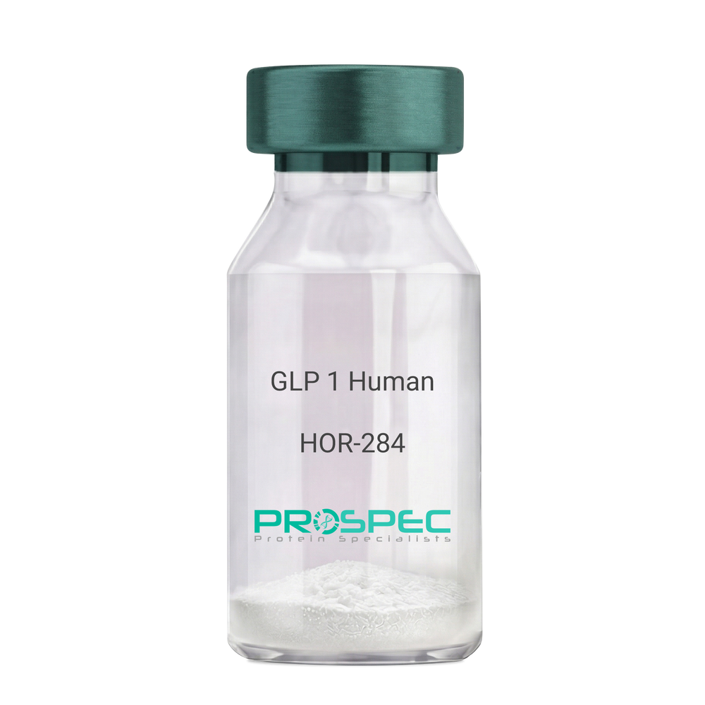
GLP 1 HumanDescription:
Human Glucagon Like Peptide-1
GLP1, Glucagon Like Peptide-1, Incretin hormone.
Product # :
HOR-284Price :
Quantity :
Shipping Method :
Shipped at Room temp
More Info
- description
- formulation
- purity
- More Info
Description
Glucagon Like Peptide-1 is a single, non-glycosylated, polypeptide chain containing 30 amino acids and having a molecular mass of 3297.7 Dalton.The GLP-1 is purified by proprietary chromatographic techniques.
Formulation
The GLP-1 peptide was lyophilized with no additives.
Purity
Greater than 96.0% as determined by RP-HPLC.
More Info
-
Introduction
Glucagon-like peptide-1 (GLP-1) is derived from the transcription product of the proglucagon gene. The major source of GLP-1 in the body is the intestinal L cell that secretes GLP-1 as a guthormone. The biologically active forms of GLP-1 are: GLP-1-(7-37) and GLP-1-(7-36)NH2.
GLP-1 secretion by L cells is dependent on the presence of nutrients in the lumen of the small intestine. The secretagogues (agents that causes or stimulates secretion) of this hormone include major nutrients like carbohydrate, proteinand lipid. Once in the circulation, GLP-1 has a half life of less than 2 minutes, due to rapid degradation by the enzyme dipeptidyl peptidase-4.
GLP-1 possesses several physiological properties that make it a subject of intensive investigation as a potential treatment of diabetes mellitus. The known physiological functions of GLP-1 include: Increases insulin secretion from the pancreas in a glucose-dependent manner, decreases glucagon secretion from the pancreas, increases beta cells mass and insulin gene expression, inhibits acid secretion and gastric emptying in the stomach, decreases food intake by increasing satiety. -
Synonyms
GLP1, Glucagon Like Peptide-1, Incretin hormone.
-
Physical Appearance
Sterile Filtered White lyophilized (freeze-dried) powder.
-
Stability
Lyophilized Glucagon Like Peptide-1 although stable at room temperature for 3 weeks, should be stored desiccated below -18°C. Upon reconstitution GLP-1 should be stored at 4°C between 2-7 days and for future use below -18°C. For long term storage it is recommended to add a carrier protein (0.1% HSA or BSA).Please prevent freeze-thaw cycles.
-
Solubility
It is recommended to reconstitute the lyophilized Glucagon Like Peptide-1 in sterile H2O at 0.5mg/ml, which can then be further diluted to other aqueous solutions.
-
Amino Acid Sequence
H-His-Ala-Glu-Gly-Thr-Phe-Thr-Ser-Asp-Val-Ser-Ser-Tyr-Leu-Glu-Gly-Gln-Ala-Ala-Lys-Glu-Phe-Ile-Ala-Trp-Leu-Val-Lys-Gly-Arg-NH2.
ProSpec's products are furnished for LABORATORY RESEARCH USE ONLY. The product may not be used as drugs, agricultural or pesticidal products, food additives or household chemicals.
- View Data Sheet
Name :
SARS Nucleocapsid PT3851Description:
Mouse Anti SARS Nucleocapsid Clone PT3851
Product # :
ANT-763Price :
Quantity :
Shipping Method :
Shipped with Ice Packs
More Info
- description
- formulation
- purity
- More Info
Description
SARS Nucleoprotein antibody clone PT3851 is specific for the nucleocapsid SARS-CoV protein and recognizes the nucleoprotein of SARS-CoV-2 by ELISA.
Formulation
1mg/ml in PBS and 0.09% NaN3.
Purity
Greater than 90% as determined by SDS-PAGE gel.
More Info
-
Introduction
SARS Coronavirus is an enveloped virus containing three outer structural proteins, namely the membrane (M), envelope (E), and spike (S) proteins. Spike (S)-glycoprotein of the virus interacts with a cellular receptor and mediates membrane fusion to allow viral entry into susceptible target cells. Accordingly, S-protein plays an important role in virus infection cycle and is the primary target of neutralizing antibodies.It has recently been shown that SARS is caused by a human coronavirus. Human coronaviruses are the major cause of upper respiratory tract illness in humans, such as the common cold. Coronaviruses are positive-stranded RNA viruses, featuring the largest viral RNA genomes known to date (27-31 kb). The first step in coronavirus infection is binding of the viral spike protein, a 139-kDa protein, to certain receptors on host cells. The spike protein is the main surface antigen of the coronavirus. The most prominent protein in the culture supernatants infected with SARS virus is a 46 kDa nucleocapsid protein. This suggests that the nucleocapsid protein is a major immunogen that may be useful for early diagnostics.
-
Physical Appearance
Sterile Filtered liquid formulation.
-
Stability
Store at 4°C, stable for 2 weeks. For long-term storage, store at -20°C.
-
Immunogen
Recombinant protein fragment 1-49 a.a. of the SARS nucleoprotein.
-
Clone
PT3851
-
Applications
ELISA.
-
Isotype
Mouse IgG2b.
-
Type
Mouse antibody Monoclonal.
-
Purification Method
Protein A affinity purified.
ProSpec's products are furnished for LABORATORY RESEARCH USE ONLY. The product may not be used as drugs, agricultural or pesticidal products, food additives or household chemicals.
- View Data Sheet
Name :
CoV-2 N (1-419), BiotinDescription:
Coronavirus 2019 Nucleocapsid (1-419 a.a.), Biotinylated Recombinant
Product # :
SARS-036Price :
Quantity :
Shipping Method :
Shipped at Room temp
More Info
- description
- source
- formulation
- purity
- More Info
Description
The HEK293 derived recombinant biotinylated protein contains the Coronavirus 2019 CoV-2 Nucleocapsid, Wuhan-Hu-1 strain, amino acids 1-419 fused to His-Avi tag at C-terminal.
Source
HEK293 Cells.
Formulation
CoV-2 Nucleocapsid protein is supplied in 50mM Tris, 0.15M NaCl & Arginine, pH7.5
Purity
Protein is >90% pure as determined SDS-PAGE.
More Info
-
Introduction
A human infecting coronavirus (viral pneumonia) called 2019 novel coronavirus, 2019-nCoV was found in the fish market at the city of Wuhan, Hubei province of China on December 2019.
The 2019-nCoV shares an 87% identity to the 2 bat-derived severe acute respiratory syndrome 2018 SARS-CoV-2 located in Zhoushan of eastern China. 2019-nCoV has an analogous receptor-BD-structure to that of 2018 SARS-CoV, even though there is a.a. diversity so thus the 2019-nCoV might bind to ACE2 receptor protein (angiotensin-converting enzyme 2) in humans.
While bats are possibly the host of 2019-nCoV, researchers suspect that animal from the ocean sold at the seafood market was an intermediate host. RSCU analysis proposes that the 2019-nCoV is a recombinant within the viral spike glycoprotein between the bat coronavirus and an unknown coronavirus.
-
Physical Appearance
Sterile Filtered White lyophilized (freeze-dried) powder.
-
Stability
Lyophilized Cov-2 Nucleocapsid protein although stable at room temperature for 3 weeks, should be stored desiccated below -18°C. Upon reconstitution CoV2 Nucleocapsid protein should be stored at 4°C between 2-7 days and for future use below -18°C.For long term storage it is recommended to add a carrier protein (0.1% HSA or BSA).Please prevent freeze-thaw cycles.
-
Purification Method
Purified by Metal-Afinity chromatographic technique.
ProSpec's products are furnished for LABORATORY RESEARCH USE ONLY. The product may not be used as drugs, agricultural or pesticidal products, food additives or household chemicals.
- View Data Sheet
Name :
IFIH1 HumanDescription:
Interferon Induced With Helicase C Domain 1 Human Recombinant
Interferon-induced helicase C domain-containing protein 1, Clinically amyopathic dermatomyositis autoantigen 140 kDa, CADM-140 autoantigen, Helicase with 2 CARD domains, Helicard, Interferon-induced with helicase C domain protein 1, Melanoma differentiation-associated protein 5, MDA-5, Murabutide down-regulated protein, RIG-I-like receptor 2, RLR-2, RNA helicase-DEAD box protein 116, IFIH1, MDA5, RH116, Hlcd, IDDM19.
Product # :
PRO-1505Price :
Quantity :
Shipping Method :
Shipped with Ice Packs
More Info
- description
- source
- formulation
- purity
- More Info
Description
IFIH1 Human Recombinant produced in SF9 is a glycosylated, polypeptide chain having a calculated molecular mass of 152,000 Dalton. IFIH1 is expressed with a -10xHis tag at N-terminus and purified by proprietary chromatographic techniques.
Source
Sf9 Insect Cells.
Formulation
IFIH1 is supplied in 20mM HEPES buffer pH-7.9, 550mM NaCl and 6M Urea.
Purity
Greater than 93.0% as determined by SDS-PAGE.
More Info
-
Introduction
IFIH1 is a DEAD box protein which is upregulated in response to treatment with beta-interferon and a protein kinase C-activating compound, mezerein. Irreversible reprogramming of melanomas can be attained by therapy with both these agents; treatment with either agent alone only achieves reversible differentiation. DEAD box proteins are implicated in several cellular processes involving alteration of RNA secondary structure such as translation initiation, nuclear and mitochondrial splicing, and ribosome and spliceosome assembly.
-
Synonyms
Interferon-induced helicase C domain-containing protein 1, Clinically amyopathic dermatomyositis autoantigen 140 kDa, CADM-140 autoantigen, Helicase with 2 CARD domains, Helicard, Interferon-induced with helicase C domain protein 1, Melanoma differentiation-associated protein 5, MDA-5, Murabutide down-regulated protein, RIG-I-like receptor 2, RLR-2, RNA helicase-DEAD box protein 116, IFIH1, MDA5, RH116, Hlcd, IDDM19.
-
Physical Appearance
Sterile Filtered clear solution.
-
Stability
Store at 4°C if entire vial will be used within 2-4 weeks. Store, frozen at -20°C for longer periods of time. Avoid multiple freeze-thaw cycles.
ProSpec's products are furnished for LABORATORY RESEARCH USE ONLY. The product may not be used as drugs, agricultural or pesticidal products, food additives or household chemicals.
- View Data Sheet
Name :
CoV OC43 HumanDescription:
Coronavirus OC43 Nucleoprotein Human Recombinant
Product # :
SARS-056Price :
Quantity :
Shipping Method :
Shipped with Ice Packs
More Info
- description
- source
- formulation
- purity
- More Info
Description
Recombinant Human Coronavirus OC43 Nucleoprotein is full length protein except the predicted signal peptide of the first 30 aa produced in E. coli and migrate at 50kDa.The CoV OC43 is fused to a 6xHis tag at its C terminal and purified by proprietary chromatographic technique.
Source
Escherichia Coli.
Formulation
CoV OC43 protein solution contains PBS and 25mM K2CO3.
Purity
Protein is >90% pure as determined by 10% PAGE (coomassie staining).
More Info
-
Introduction
OC43 is 1 of 7 known coronaviruses to infect human, including CoV-229E, CoV-NL63, CoV-HKU1, MERS-CoV, the original SARS-CoV1, and SARS-CoV-2 (Covid 19). CoV-OC43, human alpha- coronavirus 229E and NL63, beta-coronavirus and HCoV-HKU1 are responsible for the common cold. like other betacoronavirus, CoV-OC43 has an additional shorter spike protein called hemagglutinin esterase. Human coronavirus OC43 belongs to the species beta-coronavirus which are enveloped, positive-sense, singlestranded RNA virus which enters its host cell by binding to the Nacetyl-9-O-acetylneuraminic acid receptor.
-
Physical Appearance
Sterile Filtered colorless solution.
-
Stability
Store at 4°C if entire vial will be used within 2-4 weeks.Store, frozen at -20°C for longer periods of time.For long term storage it is recommended to add a carrier protein (0.1% HSA or BSA).Avoid multiple freeze-thaw cycles.
-
Amino Acid Sequence
DQFRNVQTRG RRAQPKQTST SQQPSGGNVV PYYSWFSGIT QFQKGKEFEF AEGQGVPIAP GVPATEAKGY WYRHNRRSFK TADGNQRQLL PRWYFYYLGT GPHAKDQYGT DIDGVFWVAS NQADVNTPAD IVDRDPSSDE AIPTRFPPGT VLPQGYYIEG SGRSAPNSRS TSRTSSRASS AGSRSRANSG NRAPTSGVTP DMADQIASLV LAKLGKDATK PQQVTKHTAK EVRQKILNKP RQKRSPNKQC TVQQCFGKRG PNQNFGGGEM LKLGTSDPQF PILAELAPTA GAFFFGSRLELAKVQNLSGN PDEPQKDVYE LRYNGAIRFD STLSGFETIM KVLNENLNAY QQQDGMMNMS PKPQRQRGLK NGQGENDNIS VAAPKSRVQQ NKSRELTAED ISLLKKMDEP YTEDTSEI
-
Applications
Immunoassay.
ProSpec's products are furnished for LABORATORY RESEARCH USE ONLY. The product may not be used as drugs, agricultural or pesticidal products, food additives or household chemicals.
- View Data Sheet
Name :
s100bb BovineDescription:
s100bb Bovine homodimer
Product # :
PRO-2796Price :
Quantity :
Shipping Method :
Shipped at Room temp
More Info
- description
- source
- formulation
- purity
- More Info
Description
s100bb Bovine is suitable for use as a standard in immunoassay and as an immunogen for antiserum production.
Source
Bovine brain tissue.
Formulation
s100bb was lyophilized from 5mM Tris-HCl, pH 7.5, 2mM EDTA and 5mM 2-mercaptoethanol.
Purity
Greater than 95.0% .
More Info
-
Physical Appearance
Sterile Filtered White lyophilized (freeze-dried) powder.
-
Stability
Lyophilized s100bb homodimer although stable at room temperature for 3 weeks, should be stored desiccated below -18°C. Upon reconstitution s100bb should be stored at 4°C between 2-7 days and for future use below -18°C. For long term storage it is recommended to add a carrier protein (0.1% HSA or BSA). Please prevent freeze-thaw cycles.
-
Solubility
It is recommended to reconstitute the lyophilized s100bb in sterile 18MΩ-cm H2O containing 5mM 2- mercaptoethanol not less than 100µg/ml, which can then be further diluted to other aqueous solutions.
-
Background
S100BB, a member of the S100 family of proteins, is a calcium-binding protein that plays a significant role in various cellular processes, particularly in the central nervous system (CNS). Initially identified as a brain-specific protein, S100BB has captured the attention of researchers due to its involvement in neurological signaling, brain development, and its potential implications in neurological disorders. This research delves into the enigmatic world of S100BB, aiming to uncover its diverse functions, regulatory mechanisms, and its relevance in health and disease.
Discovery and Structural Insights:
S100BB was first discovered in the brain, primarily localized in astrocytes, a type of glial cell in the CNS. Structurally, S100BB is characterized by its EF-hand motifs, allowing it to bind calcium ions, a property crucial for its function. These calcium-binding abilities enable S100BB to participate in intricate cellular signaling pathways, making it a vital player in the dynamic communication within the CNS.
Neurological Signaling and Plasticity:
Within the CNS, S100BB has been implicated in neuronal signaling and synaptic plasticity. It interacts with several target proteins, modulating their activities and influencing processes like neurotransmitter release and neuronal differentiation. Furthermore, S100BB participates in the regulation of intracellular calcium levels, a key factor in neuronal excitability and synaptic transmission. Understanding these roles is vital for unraveling the complexities of neurological function and plasticity.
ProSpec's products are furnished for LABORATORY RESEARCH USE ONLY. The product may not be used as drugs, agricultural or pesticidal products, food additives or household chemicals.
- View Data Sheet
Name :
Dengue NS1 ST3, InsectDescription:
Dengue Virus NS1 Subtype 3 Recombinant, Insect Cells
Product # :
DEN-031Price :
Quantity :
Shipping Method :
Shipped with Ice Packs
More Info
- description
- source
- formulation
- purity
- More Info
Description
Recombinant Dengue Virus NS1 Subtype 3 produced in Insect Cells is a polypeptide chain containing amino acids 775-1129 and having a molecular weight of approximately 50kDa. Dengue NS1 ST3 is purified by proprietary chromatographic technique.
Source
Insect cells.
Formulation
Dengue NS1 ST3 protein solution (1mg/ml) in 1xPBS, pH7.4, 0.1% Thimerosal, 5mM EDTA, 1µg/ml of Leupeptin, Aprotinin and Pepstatin A.
Purity
Protein is >95% pure as determined by 12.5% SDS-PAGE.
More Info
-
Introduction
Caused by one of four closely related virus serotypes of the genus Flavivirus, family Flaviviridae, each serotype is sufficiently different that there is no cross-protection and epidemics caused by multiple serotypes (hyperendemicity) can occur. In cell culture experiments and mice Morpholino antisense oligos have shown specific activity against Dengue virus.
-
Physical Appearance
Sterile filtered colorless solution.
-
Stability
Store at 4°C if entire vial will be used within 2-4 weeks. Store, frozen at -20°C for longer periods of time. For long term storage it is recommended to add a carrier protein (0.1% HSA or BSA).Avoid multiple freeze-thaw cycles.
-
Applications
Ag test control.
ProSpec's products are furnished for LABORATORY RESEARCH USE ONLY. The product may not be used as drugs, agricultural or pesticidal products, food additives or household chemicals.
- View Data Sheet
Name :
SARS Spike (306-527)Description:
SARS Spike Receptor Binding Domain(306-527 a.a.), Recombinant
Product # :
SARS-032Price :
Quantity :
Shipping Method :
Shipped at Room temp
More Info
- description
- source
- formulation
- purity
- More Info
Description
The HEK293 derived recombinant protein contains the SARS Coronavirus spike S glycoprotein Receptor Binding Domain, amino acids 306-527 fused to His tag at C-terminal.
Source
HEK293
Formulation
SARS Spike S glycoprotein RBD is lyophilized from 1x PBS pH-7.4 + 5% trehalose.
Purity
Protein is >90% pure as determined SDS-PAGE.
More Info
-
Introduction
SARS Coronavirus is an enveloped virus containing three outer structural proteins, namely the membrane (M), envelope (E), and spike (S) proteins. Spike (S)-glycoprotein of the virus interacts with a cellular receptor and mediates membrane fusion to allow viral entry into susceptible target cells. Accordingly, S-protein plays an important role in virus infection cycle and is the primary target of neutralizing antibodies.
-
Physical Appearance
Lyophilized freezed dried powder.
-
Stability
SARS Spike S1 although stable at room temperature for 3 weeks, should be stored desiccated below -18°C. Upon reconstitution SARS Spike protein should be stored at 4°C between 2-7 days and for future use below -18°C.For long term storage it is recommended to add a carrier protein (0.1% HSA or BSA).Please prevent freeze-thaw cycles.
-
Purification Method
Purified by immobilized metal affinity chromatographic technique.
ProSpec's products are furnished for LABORATORY RESEARCH USE ONLY. The product may not be used as drugs, agricultural or pesticidal products, food additives or household chemicals.
- View Data Sheet
Name :
Rantes RatDescription:
Rantes Rat Recombinant (CCL5)
Small inducible cytokine A5, CCL5, T-cell-specific RANTES protein, SIS-delta, T cell- specific protein P228, TCP228, chemokine (C-C motif) ligand 5, SISd, SCYA5, RANTES, D17S136E, MGC17164.
Product # :
CHM-352Price :
Quantity :
Shipping Method :
Shipped at Room temp
More Info
- description
- source
- formulation
- purity
- biological activity
- More Info
Description
Rantes Rat Recombinant produced in E.Coli is a single, non-glycosylated polypeptide chain containing 68 amino acids and having a molecular mass of 7876 Dalton. The Rat Rantes is purified by proprietary chromatographic techniques.
Source
Escherichia Coli.
Formulation
Lyophilized from a concentrated (1mg/ml) solution in water containing no additives.
Purity
Greater than 95.0% as determined by:
(a) Analysis by RP-HPLC.
(b) Analysis by SDS-PAGE.Biological Activity
The activity is determined by the ability to chemoattract total human lymphocytes and murine T-cells at a concentration between 1-10ng/ml corresponding to a Specific Activity of 100,000-1,000,000IU/mg.More Info
-
Introduction
Regulated upon Activation, Normal T-cell Expressed, and Secreted or RANTES is an 8 kDa protein classified as a chemotactic cytokine or chemokine. It has recently been renamed CCL5. RANTES is chemotactic for T cells, eosinophils and basophils and plays an active role in recruiting leukocytes into inflammatory sites. With the help of particular cytokines (i.e. IL-2 and IFN-?) that are released by T cells, RANTES also induces the proliferation and activation of certain natural killer (NK) cells to form CHAK (CC-Chemokine-activated killer) cells. It is also a HIV-suppressive factor released from CD8+ T cells. This chemokine has been localized to chromosome 17 in humans.
-
Synonyms
Small inducible cytokine A5, CCL5, T-cell-specific RANTES protein, SIS-delta, T cell- specific protein P228, TCP228, chemokine (C-C motif) ligand 5, SISd, SCYA5, RANTES, D17S136E, MGC17164.
-
Physical Appearance
Sterile Filtered White lyophilized (freeze-dried) powder.
-
Stability
Lyophilized Rat Rantes although stable at room temperature for 3 weeks, should be stored desiccated below -18°C. Upon reconstitution Rat CCL5 should be stored at 4°C between 2-7 days and for future use below -18°C.For long term storage it is recommended to add a carrier protein (0.1% HSA or BSA).Please prevent freeze-thaw cycles.
-
Solubility
It is recommended to reconstitute the lyophilized Rantes in sterile 18MΩ-cm H2O not less than 100µg/ml, which can then be further diluted to other aqueous solutions.
-
Amino Acid Sequence
SPYGSDTTPC CFAYLSLALP RAHVKEYFYT SSKCSNLAVV FVTRRNRQVC ANPEKKWVQE YINYLEMS.
ProSpec's products are furnished for LABORATORY RESEARCH USE ONLY. The product may not be used as drugs, agricultural or pesticidal products, food additives or household chemicals.
- View Data Sheet
Name :
Malaria Pf. MSP1Description:
Malaria Falciparum MSP1 Recombinant
Product # :
MAL-004Price :
Quantity :
Shipping Method :
Shipped with Ice Packs
More Info
- description
- formulation
- purity
- More Info
Description
Merozoite surface antigen is a protein located on the outside of the merozoite, playing an imperative role in immune reaction. About 55% cases of malaria are infected by Plasmodium falciparum (Pf). Pf MSP1 has to be used with Plasmodium vivax (Pv) together for ELISA and rapid diagnostic test, Plasmodium falciparum and vivax infection takes about 95% of Plasmodium caused infection. Recombinant Malaria Falciparum MSP1 produced in E.coli, is a 62kDa protein fused to a GST tag at N-terminus and purified by proprietary chromatographic technique.
Formulation
Sterile Filtered solution containing PBS and 25mM K2CO3.
Purity
Protein is >90% pure as determined by 10% PAGE (coomassie staining).
More Info
-
Introduction
Plasmodium falciparum is a protozoan parasite, one of the species of Plasmodium that cause malaria. Malaria parasites are members of the Apicomplexa. Apicomplexa are characterized by a set of organelles found in some stages of the parasite's life cycle. These organelles, collectively known as apical organelles because of their localization at one end of the parasite, are involved in interactions between the parasite and host. In particular, the apical organelles have been implicated in the process of host cell invasion. In the case of Plasmodium, three distinct invasive forms have been identified: sporozoite, merozoite, and ookinete.
-
Physical Appearance
Sterile Filtered clear solution.
-
Stability
Pf. MSP1 although stable at 4°C for 1 week, should be stored below -18°C. Please prevent freeze thaw cycles.
-
Applications
Rapid test and Immunoassay.
ProSpec's products are furnished for LABORATORY RESEARCH USE ONLY. The product may not be used as drugs, agricultural or pesticidal products, food additives or household chemicals.
- View Data Sheet
Name :
Dengue NS1 Paired AntibodyDescription:
Mouse Anti Dengue NS1 Paired
Product # :
ANT-531Price :
Quantity :
Shipping Method :
Shipped with Ice Packs
More Info
- description
- formulation
- purity
- More Info
Description
A paired monoclonal antibody has been developed for dengue NS1 antigen lateral flow immunoassay product. It is suggested to use 10µg/ml gold concentration (OD 1.2-1.5) to conjugate with clone 24/c, and 1.2µg/cm coating concentration for clone 2/b. The sensitivity is directly proportional to conjugated gold OD. The specimen used for the test is 80-90 µl. The rapid test developed by these 2 paired antibodies has 90% sensitivity for the samples with OD over 5 and 60% sensitivity for the sample with OD below 5 tested by ELISA. Please note that when ordering for example: 100µg antibody we ship 50µg from each of the antibodies (100µg in total).
Formulation
* Dengue NS1 Conjugation antibody (clone 24/c): (1.2mg/ml) in PBS and Sodium nitrate.
* Dengue NS1 Coating antibody (clone 2/b): (3.4mg/ml) in PBS and Sodium nitrate.Purity
Greater than 95%.
More Info
-
Introduction
Dengue NS1 is an early marker for dengue infection from the first day of fever and lasting about one week. Dengue IgM is usually detected early on the first day to 5 days of infection. Dengue NS1 could be detected from both primary and secondary infection for all four subtypes.
-
Physical Appearance
2 vials of sterile Filtered clear colorless solution.
-
Stability
Dengue NS1 Paired Antibody should be stored at 4°C.
-
Applications
Lateral flow immunoassay.
-
Assay Conditions
1. Fresh gold particle prepared within 1 to 2 days with maximal homogeneity in particle size, λ 525 to 5529, OD 1.2- 1.4. 2. Conjugation pH 8.5, conjugation time should be 6-8 hours. 3. Blocking condition: adjust OD to 8.0-8.2 for re-suspended ant-dengue NS1 conjugated colloid gold, add casein to the final 0.3-0.4%.
-
Type
Mouse antibody Monoclonal.
-
Purification Method
Purified monoclonal IgG by protein A chromatography.
ProSpec's products are furnished for LABORATORY RESEARCH USE ONLY. The product may not be used as drugs, agricultural or pesticidal products, food additives or household chemicals.
- View Data Sheet
Name :
HGFR MouseDescription:
Hepatocyte Growth Factor Receptor Mouse Recombinant
hepatocyte growth factor receptor, HGF R/c-MET, Met, AI838057, c-Met, HGF, HGFR, Par4, HGF receptor, HGF/SF receptor, Proto-oncogene c-Met, Scatter factor receptor, SF receptor, Tyrosine-protein kinase Met.
Product # :
CYT-1139Price :
Quantity :
Shipping Method :
Shipped with Ice Packs
More Info
- description
- source
- formulation
- purity
- More Info
- SDS-PAGE
Description
HGF Receptor Mouse Recombinant produced in Baculovirus is a single glycosylated polypeptide chain containing 1146 amino acids (25-931aa) and having a molecular mass of 127.8kDa. HGF is fused to a 239 amino acid hIgG-His-Tag at C-terminus and purified by proprietary chromatographic techniques.
Source
Sf9, Baculovirus cells.
Formulation
HGF Receptor protein (0.25mg/ml) contains Phosphate Buffered Saline (pH 7.4) and 10% glycerol.
Purity
Greater than 85.0% as determined by SDS-PAGE.
SDS-PAGE
More Info
-
Introduction
HGF, also known as scatter factor (SF) and Hepatocyte growth factor, is secreted from mesenchymal cells, targets and acts mainly on epithelial and endothelial cells. The protein is a paracrine cellular growth, motility and morphogenic factor. HGF can also be part of haemopoietic progenitor cells and T cells. HGF has a crucial role in embryonic organ development, mainly in myogenesis. In adults HGF takes part in organ regeneration and wound healing.
-
Synonyms
hepatocyte growth factor receptor, HGF R/c-MET, Met, AI838057, c-Met, HGF, HGFR, Par4, HGF receptor, HGF/SF receptor, Proto-oncogene c-Met, Scatter factor receptor, SF receptor, Tyrosine-protein kinase Met.
-
Physical Appearance
Sterile Filtered colorless solution.
-
Stability
Store at 4°C if entire vial will be used within 2-4 weeks. Store, frozen at -20°C for longer periods of time. For long term storage it is recommended to add a carrier protein (0.1% HSA or BSA).Avoid multiple freeze-thaw cycles.
-
Amino Acid Sequence
ECKEALVKSE MNVNMKYQLP NFTAETPIQN VVLHGHHIYL GATNYIYVLN DKDLQKVSEF KTGPVLEHPD CLPCRDCSSK ANSSGGVWKD NINMALLVDT YYDDQLISCG SVNRGTCQRH VLPPDNSADI QSEVHCMFSP EEESGQCPDC VVSALGAKVL LSEKDRFINF FVGNTINSSY PPGYSLHSIS VRRLKETQDG FKFLTDQSYI DVLPEFQDSY PIKYIHAFES NHFIYFLTVQ KETLDAQTFH TRIIRFCSVD SGLHSYMEMP LECILTEKRR KRSTREEVFN ILQAAYVSKP GANLAKQIGA SPSDDILFGV FAQSKPDSAE PVNRSAVCAF PIKYVNDFFN KIVNKNNVRC LQHFYGPNHE HCFNRTLLRN SSGCEARSDE YRTEFTTALQ RVDLFMGRLN QVLLTSISTF IKGDLTIANL GTSEGRFMQV VLSRTAHLTP HVNFLLDSHP VSPEVIVEHP SNQNGYTLVV TGKKITKIPL NGLGCGHFQS CSQCLSAPYF IQCGWCHNQC VRFDECPSGT WTQEICLPAV YKVFPTSAPL EGGTVLTICG WDFGFRKNNK FDLRKTKVLL GNESCTLTLS ESTTNTLKCT VGPAMSEHFN VSVIISNSRE TTQYSAFSYV DPVITSISPR YGPQAGGTLL TLTGKYLNSG NSRHISIGGK TCTLKSVSDS ILECYTPAQT TSDEFPVKLK IDLANRETSS FSYREDPVVY EIHPTKSFIS GGSTITGIGK TLNSVSLPKL VIDVHEVGVN YTVACQHRSN SEIICCTTPS LKQLGLQLPL KTKAFFLLDG ILSKHFDLTY VHNPVFEPFE KPVMISIGNE NVVEIKGNNI DPEAVKGEVL KVGNQSCESL HWHSGAVLCT VPSDLLKLNS ELNIEWKQAV SSTVLGKVIV QPDQNFALEP KSCDKTHTCP PCPAPELLGG PSVFLFPPKP KDTLMISRTP EVTCVVVDVS HEDPEVKFNW YVDGVEVHNA KTKPREEQYN STYRVVSVLT VLHQDWLNGK EYKCKVSNKA LPAPIEKTIS KAKGQPREPQ VYTLPPSRDE LTKNQVSLTC LVKGFYPSDI AVEWESNGQP ENNYKTTPPV LDSDGSFFLY SKLTVDKSRW QQGNVFSCSV MHEALHNHYT QKSLSLSPGK HHHHHH.
-
Background
What is the molecular weight/Mw of HGFR MOUSE Protein?
HGFR MOUSE Protein has a total Mw of 127.8kDa.
What is the source or expression system of HGFR MOUSE Protein?
Sf9, Baculovirus cells.
What is the Purity of HGFR MOUSE Protein?
HGFR MOUSE Protein is >85% pure as determined by SDS-PAGE.
What is the Biological Activity of HGFR MOUSE Protein?
The biological functionality of HGFR MOUSE Protein will be determined in the future.
What is the amino acid sequence of HGFR MOUSE Protein?
ECKEALVKSE MNVNMKYQLP NFTAETPIQN VVLHGHHIYL GATNYIYVLN DKDLQKVSEF KTGPVLEHPD CLPCRDCSSK ANSSGGVWKD NINMALLVDT YYDDQLISCG SVNRGTCQRH VLPPDNSADI QSEVHCMFSP EEESGQCPDC VVSALGAKVL LSEKDRFINF FVGNTINSSY PPGYSLHSIS VRRLKETQDG FKFLTDQSYI DVLPEFQDSY PIKYIHAFES NHFIYFLTVQ KETLDAQTFH TRIIRFCSVD SGLHSYMEMP LECILTEKRR KRSTREEVFN ILQAAYVSKP GANLAKQIGA SPSDDILFGV FAQSKPDSAE PVNRSAVCAF PIKYVNDFFN KIVNKNNVRC LQHFYGPNHE HCFNRTLLRN SSGCEARSDE YRTEFTTALQ RVDLFMGRLN QVLLTSISTF IKGDLTIANL GTSEGRFMQV VLSRTAHLTP HVNFLLDSHP VSPEVIVEHP SNQNGYTLVV TGKKITKIPL NGLGCGHFQS CSQCLSAPYF IQCGWCHNQC VRFDECPSGT WTQEICLPAV YKVFPTSAPL EGGTVLTICG WDFGFRKNNK FDLRKTKVLL GNESCTLTLS ESTTNTLKCT VGPAMSEHFN VSVIISNSRE TTQYSAFSYV DPVITSISPR YGPQAGGTLL TLTGKYLNSG NSRHISIGGK TCTLKSVSDS ILECYTPAQT TSDEFPVKLK IDLANRETSS FSYREDPVVY EIHPTKSFIS GGSTITGIGK TLNSVSLPKL VIDVHEVGVN YTVACQHRSN SEIICCTTPS LKQLGLQLPL KTKAFFLLDG ILSKHFDLTY VHNPVFEPFE KPVMISIGNE NVVEIKGNNI DPEAVKGEVL KVGNQSCESL HWHSGAVLCT VPSDLLKLNS ELNIEWKQAV SSTVLGKVIV QPDQNFALEP KSCDKTHTCP PCPAPELLGG PSVFLFPPKP KDTLMISRTP EVTCVVVDVS HEDPEVKFNW YVDGVEVHNA KTKPREEQYN STYRVVSVLT VLHQDWLNGK EYKCKVSNKA LPAPIEKTIS KAKGQPREPQ VYTLPPSRDE LTKNQVSLTC LVKGFYPSDI AVEWESNGQP ENNYKTTPPV LDSDGSFFLY SKLTVDKSRW QQGNVFSCSV MHEALHNHYT QKSLSLSPGK HHHHHH.
What applications can HGFR MOUSE Protein be used in?
HGFR MOUSE Protein can probably be used in western blot, ELISA and Lateral Flow.
What is the endotoxin level for HGFR MOUSE Protein?
The endotoxin level is minimal, HGFR MOUSE Protein was purified using conventional chromatography techniques.
ProSpec's products are furnished for LABORATORY RESEARCH USE ONLY. The product may not be used as drugs, agricultural or pesticidal products, food additives or household chemicals.
- View Data Sheet
Name :
ANP32A HumanDescription:
Acidic Nuclear Phosphoprotein 32 Family Member A Human Recombinant
I1PP2A, LANP, MAPM, PHAP1, Leucine-rich acidic nuclear protein, PP32, Mapmodulin.
Product # :
PRO-252Price :
Quantity :
Shipping Method :
Shipped with Ice Packs
More Info
- description
- source
- formulation
- purity
- More Info
Description
ANP32A Human Recombinant produced in E.Coli is a single, non-glycosylated, polypeptide chain containing 269 amino acids (1-249a.a.) and having a molecular mass of 30.7kDa. ANP32A is fused to a 20 amino acid His Tag at N-terminus and purified by proprietary chromatographic techniques.
Source
Escherichia Coli.
Formulation
ANP32A solution (0.5mg/ml) containing 20mM Tris-HCl buffer (pH8.0), 1mM DTT, 100mM NaCl, 0.1mM PMSF and 10% glycerol.
Purity
Greater than 95% as determined by SDS-PAGE.
More Info
-
Introduction
ANP32A often cooperate with MAP1B, TAF1A and Protein SET. ANP32A is involved in several cellular processes, such as proliferation, differentiation, caspase-dependent and caspase-independent apoptosis. Mapmodulin takes a part in E4F1-mediated transcriptional repression.
-
Synonyms
I1PP2A, LANP, MAPM, PHAP1, Leucine-rich acidic nuclear protein, PP32, Mapmodulin.
-
Physical Appearance
Sterile filtered colorless solution.
-
Stability
Store at 4°C if entire vial will be used within 2-4 weeks.Store, frozen at -20°C for longer periods of time. For long term storage it is recommended to add a carrier protein (0.1% HSA or BSA).Avoid multiple freeze-thaw cycles.
-
Amino Acid Sequence
MGSSHHHHHH SSGLVPRGSH MEMGRRIHLE LRNRTPSDVK ELVLDNSRSN EGKLEGLTDE FEELEFLSTI NVGLTSIANL PKLNKLKKLE LSDNRVSGGL EVLAEKCPNL THLNLSGNKI KDLSTIEPLK KLENLKSLDL FNCEVTNLND YRENVFKLLP QLTYLDGYDR DDKEAPDSDA EGYVEGLDDE EEDEDEEEYD EDAQVVEDEE DEDEEEEGEE EDVSGEEEED EEGYNDGEVD DEEDEEELGE EERGQKRKRE PEDEGEDDD.
ProSpec's products are furnished for LABORATORY RESEARCH USE ONLY. The product may not be used as drugs, agricultural or pesticidal products, food additives or household chemicals.
- View Data Sheet
Name :
SERPINF2 MouseDescription:
Serpin Peptidase Inhibitor, Clade F Member 2 Mouse Recombinant
Alpha-2-antiplasmin, Alpha-2-AP, Alpha-2-plasmin inhibitor, Alpha-2-PI, Serpin F2.
Product # :
PRO-2240Price :
Quantity :
Shipping Method :
Shipped with Ice Packs
More Info
- description
- source
- formulation
- purity
- More Info
Description
SERPINF2 produced in Sf9 Baculovirus cells is a single, glycosylated polypeptide chain containing 470 amino acids (28-491 a.a.) and having a molecular mass of 52.9kDa (Migrates at 70-100kDa on SDS-PAGE under reducing conditions). SERPINF2 is expressed with a 6 amino acid His tag at C-Terminus and purified by proprietary chromatographic techniques.
Source
Sf9, Baculovirus cells.
Formulation
SERPINF2 protein solution (1mg/ml) contains Phosphate Buffered Saline (pH 7.4) and 10% glycerol.
Purity
Greater than 90.0% as determined by SDS-PAGE.
More Info
-
Introduction
Alpha-2-antiplasmin (SERPINF2) is a serine protease inhibitor. The main targets of SERPINF2 are plasmin and trypsin; however SERPINF2 also inactivates matriptase-3/TMPRSS7 and chymotrypsin.
-
Synonyms
Alpha-2-antiplasmin, Alpha-2-AP, Alpha-2-plasmin inhibitor, Alpha-2-PI, Serpin F2.
-
Physical Appearance
Sterile Filtered colorless solution.
-
Stability
Store at 4°C if entire vial will be used within 2-4 weeks. Store, frozen at -20°C for longer periods of time. For long term storage it is recommended to add a carrier protein (0.1% HSA or BSA). Avoid multiple freeze-thaw cycles.
-
Amino Acid Sequence
VDLPGQQPVS EQAQQKLPLP ALFKLDNQDF GDHATLKRSP GHCKSVPTAE ETRRLAQAMM AFTTDLFSLV AQTSTSSNLV LSPLSVALAL SHLALGAQNQ TLHSLHRVLH MNTGSCLPHL LSHFYQNLGP GTIRLAARIY LQKGFPIKDD FLEQSERLFG AKPVKLTGKQ EEDLANINQW VKEATEGKIE DFLSELPDST VLLLLNAIHF HGFWRTKFDP SLTQKDFFHL DERFTVSVDM MHAVSYPLRW FLLEQPEIQV AHFPFKNNMS FVVVMPTYFE WNVSEVLANL TWDTLYHPSL QERPTKVWLP KLHLQQQLDL VATLSQLGLQ ELFQGPDLRG ISEQNLVVSS VQHQSTMELS EAGVEAAAAT SVAMNRMSLS SFTVNRPFLF FIMEDTIGVP LFVGSVRNPN PSALPQLQEQ RDSPDNRLIG QNDKADFHGG KTFGPDLKLA PRMEEDYPQF SSPKHHHHHH.
ProSpec's products are furnished for LABORATORY RESEARCH USE ONLY. The product may not be used as drugs, agricultural or pesticidal products, food additives or household chemicals.
- View Data Sheet
Name :
NAPSA HumanDescription:
Napsin A Aspartic Peptidase Human Recombinant
Napsin A Aspartic Peptidase, NAP1, NAPA, Kidney-Derived Aspartic Protease-Like Protein, Aspartyl Protease 4, TA01/TA02, Napsin-1, SNAPA, Asp 4, ASP4, CTB-191K22.6, EC 3.4.23.15, Pronapsin A, EC 3.4.23.5, EC 3.4.23.3, EC 3.4.23.-, EC 3.4.23, Napsin-A, KDAP, KAP,Napsin-A.
Product # :
ENZ-841Price :
Quantity :
Shipping Method :
Shipped with Ice Packs
More Info
- description
- source
- formulation
- purity
- More Info
Description
NAPSA Human Recombinant produced in E.Coli is a single, non-glycosylated polypeptide chain containing 380 amino acids (64-420 a.a) and having a molecular mass of 40.9kDa. NAPSA is fused to a 23 amino acid His-tag at N-terminus & purified by proprietary chromatographic techniques.
Source
Escherichia Coli.
Formulation
NAPSA protein solution (0.25mg/ml) containing 20mM Tris-HCl (pH8.0) and 10% glycerol.
Purity
Greater than 90.0% as determined by SDS-PAGE.
More Info
-
Introduction
Napsin A Aspartic Peptidase, also known as NAPSA is a member of the peptidase A1 family. NAPSA is involved in the processing of pneumocyte surfactant precursors. Furthermore the activation peptides of aspartic proteinases take part as inhibitors of the active site. These peptide segments/pro-parts are considered essential for correct folding, targeting, as well as control of the activation of aspartic proteinase zymogens. The pronapsin A gene is expressed mostly in lung and kidney. In addition, NAPSA translation product is expected to be a fully functional, glycosylated aspartic proteinase precursor which contains an RGD motif as well as an additional 18 residues at its C-terminus.
-
Synonyms
Napsin A Aspartic Peptidase, NAP1, NAPA, Kidney-Derived Aspartic Protease-Like Protein, Aspartyl Protease 4, TA01/TA02, Napsin-1, SNAPA, Asp 4, ASP4, CTB-191K22.6, EC 3.4.23.15, Pronapsin A, EC 3.4.23.5, EC 3.4.23.3, EC 3.4.23.-, EC 3.4.23, Napsin-A, KDAP, KAP,Napsin-A.
-
Physical Appearance
Sterile filtered colorless solution.
-
Stability
Store at 4°C if entire vial will be used within 2-4 weeks. Store, frozen at -20°C for longer periods of time. For long term storage it is recommended to add a carrier protein (0.1% HSA or BSA).Avoid multiple freeze-thaw cycles.
-
Amino Acid Sequence
MGSSHHHHHH SSGLVPRGSH MGSKPIFVPL SNYRDVQYFG EIGLGTPPQN FTVAFDTGSS NLWVPSRRCH FFSVPCWLHH RFDPKASSSF QANGTKFAIQ YGTGRVDGIL SEDKLTIGGI KGASVIFGEA LWEPSLVFAF AHFDGILGLG FPILSVEGVR PPMDVLVEQG LLDKPVFSFY LNRDPEEPDG GELVLGGSDP AHYIPPLTFV PVTVPAYWQI HMERVKVGPG LTLCAKGCAA ILDTGTSLIT GPTEEIRALH AAIGGIPLLA GEYIILCSEI PKLPAVSFLL GGVWFNLTAH DYVIQTTRNG VRLCLSGFQA LDVPPPAGPF WILGDVFLGT YVAVFDRGDM KSSARVGLAR ARTRGADLGW GETAQAQFPG.
ProSpec's products are furnished for LABORATORY RESEARCH USE ONLY. The product may not be used as drugs, agricultural or pesticidal products, food additives or household chemicals.
- View Data Sheet
Name :
SLAMF1 Human, Sf9Description:
SLAMF1 Human Recombinant, Sf9
Signaling lymphocytic activation molecule, CDw150, IPO-3, CD150, SLAMF1, SLAM.
Product # :
PRO-2393Price :
Quantity :
Shipping Method :
Shipped with Ice Packs
More Info
- description
- source
- formulation
- purity
- More Info
Description
SLAMF1 Human Recombinant produced in Sf9 Baculovirus cells is a single, glycosylated polypeptide chain containing 226 amino acids (21-237a.a.) and having a molecular mass of 25.3kDa (Molecular size on SDS-PAGE will appear at approximately 28-40kDa).SLAMF1 is expressed with a 6 amino acid His tag at C-Terminus and purified by proprietary chromatographic techniques.
Source
Sf9, Baculovirus cells.
Formulation
SLAMF1 protein solution (0.5mg/ml) contains Phosphate Buffered Saline (pH 7.4) and 10% glycerol.
Purity
Greater than 95.0% as determined by SDS-PAGE.
More Info
-
Introduction
SLAMF1 is a member of the immunoglobulin gene superfamily and is involved in T-cell stimulation. The SLAMF1 protein is constitutively expressed on peripheral blood memory T cells, T-cell clones, immature thymocytes, and a fraction of B cells, and is swiftly induced on naive T cells after activation. There are probably 2 modes of SLAM signaling: one in which the inhibitor SH2D1A serves as a negative regulator and another in which protein-tyrosine phosphatase 2C (PTPN11)-dependent signal transduction functions.
-
Synonyms
Signaling lymphocytic activation molecule, CDw150, IPO-3, CD150, SLAMF1, SLAM.
-
Physical Appearance
Sterile Filtered colorless solution.
-
Stability
Store at 4°C if entire vial will be used within 2-4 weeks. Store, frozen at -20°C for longer periods of time. For long term storage it is recommended to add a carrier protein (0.1% HSA or BSA).Avoid multiple freeze-thaw cycles.
-
Amino Acid Sequence
ADPASYGTGG RMMNCPKILR QLGSKVLLPL TYERINKSMN KSIHIVVTMA KSLENSVENK IVSLDPSEAG PPRYLGDRYK FYLENLTLGI RESRKEDEGW YLMTLEKNVS VQRFCLQLRL YEQVSTPEIK VLNKTQENGT CTLILGCTVE KGDHVAYSWS EKAGTHPLNP ANSSHLLSLT
LGPQHADNIY ICTVSNPISN NSQTFSPWPG CRTDPSETKP HHHHHH.
ProSpec's products are furnished for LABORATORY RESEARCH USE ONLY. The product may not be used as drugs, agricultural or pesticidal products, food additives or household chemicals.