Search results
1000 results found for “neuregulin”
Name
Description
Product #
Price
Quantity
Shipping Method
- View Data Sheet
Name :
HMGB1 AntibodyDescription:
High-Mobility Group Box 1, Mouse Anti Human
HMG1, HMG3, SBP-1, Amphoterin, HMGB1, High-Mobility Group Box 1.
Product # :
ANT-347Price :
Quantity :
Shipping Method :
Shipped with Ice Packs
More Info
- formulation
- More Info
Formulation
1mg/ml containing PBS, pH-7.4, & 0.1% Sodium Azide.
More Info
-
Introduction
High-mobility group box 1 protein (HMGB1), previously known as HMG-1 or amphoterin, is a member of the high mobility group box family of non-histone chromosomal proteins. Human HMGB1 is expressed as a 30 kDa, 215 amino acid (aa) single chain polypeptide containing three domains: two N-terminal globular, 70 aa positively charged DNA-binding domains (HMG boxes A and B), and a negatively charged 30 aa C-terminal region that contains only Asp and Glu.4, 5 Residues 27 - 43 and 178 - 184 contain a NLS. Posttranslational modifications of the molecule have been reported, with acetylation occurring on as many as 17 lysine residues. HMGB1 is expressed at high levels in almost all cells. It was originally discovered as a nuclear protein that could bend DNA. Such bending stabilizes nucleosome formation and regulates the expression of select genes upon recruitment by DNA binding proteins.
-
Synonyms
HMG1, HMG3, SBP-1, Amphoterin, HMGB1, High-Mobility Group Box 1.
-
Immunogen
Anti-human HMGB1 mAb is derived from hybridization of mouse F0 myeloma cells with spleen cells from BALB/c mice immunized with recombinant human HMGB1 amino acids 1-215 purified from High Five (Trichoplusia ni) insect cells.
-
Ig Subclass
Mouse IgG2b heavy chain and κ light chain.
-
Clone
PJ2E1AT.
-
Applications
HMGB1 antibody has been tested by ELISA, Western blot and immunohistochemistry analysis to assure specificity and reactivity. Since application varies, however, each investigation should be titrated by the reagent to obtain optimal results. Recommended dilution range for Western blot is 1:500 ~ 1:2,000 and immunohistochemistry analysis is 1:100~300. Recommended starting dilution for Western blot is 1:1,000 and Immunohistochemistry is 1:200.
-
Type
Mouse Anti Human Monoclonal.
-
Storage Procedures
For periods up to 1 month store at 4°C, for longer periods of time, store at -20°C. Prevent freeze thaw cycles.
-
Purification Method
HMGB1 antibody was purified from mouse ascitic fluids by protein-G affinity chromatography.
ProSpec's products are furnished for LABORATORY RESEARCH USE ONLY. The product may not be used as drugs, agricultural or pesticidal products, food additives or household chemicals.
- View Data Sheet
Name :
NKp44 AntibodyDescription:
Natural killer cell p44-related protein, Mouse Anti Human
Natural cytotoxicity triggering receptor 2, Natural killer cell p44-related protein, NK cell-activating receptor, Lymphocyte antigen 95 homolog, NK-p44, NKp44, CD336, NCR2, LY95, dJ149M18.1.
Product # :
ANT-548Price :
Quantity :
Shipping Method :
Shipped with Ice Packs
More Info
- formulation
- More Info
Formulation
1mg/ml containing PBS, pH-7.4, 10% Glycerol and 0.02% Sodium Azide.
More Info
-
Introduction
NCR2 (NKp44) belongs to the natural cytotoxicity receptor (NCR) family which is expressed on activated human NK cells. NKp44 exhibits a single extracellular Ig-like V domain and a transmembrane portion containing the charged residue (Lysine), possibly involved in the association with KARAP/DAP12 molecules. The gene coding for NKp44 maps on human chromosome 6p21.1.
-
Synonyms
Natural cytotoxicity triggering receptor 2, Natural killer cell p44-related protein, NK cell-activating receptor, Lymphocyte antigen 95 homolog, NK-p44, NKp44, CD336, NCR2, LY95, dJ149M18.1.
-
Physical Appearance
Sterile filtered colorless solution.
-
Immunogen
Anti-human NKp44 mAb, is derived from hybridization of mouse F0 myeloma cells with spleen cells from BALB/c mice immunized with a recombinant human NKp44 protein 19-130 amino acids purified from E. coli.
-
Ig Subclass
Mouse IgG2a heavy chain and k light chain.
-
Clone
PAT1G6AT.
-
Applications
NKp44 antibody has been tested by ELISA, Western blot analysis to assure specificity and reactivity. Since application varies, however, each investigation should be titrated by the reagent to obtain optimal results. Recommended starting dilution is 1:1000.
-
Type
Mouse Anti Human Monoclonal.
-
Storage Procedures
For periods up to 1 month store at 4°C, for longer periods of time, store at -20°C. Prevent freeze thaw cycles.
-
Purification Method
NKp44 antibody was purified from mouse ascitic fluids by protein-A affinity chromatography.
ProSpec's products are furnished for LABORATORY RESEARCH USE ONLY. The product may not be used as drugs, agricultural or pesticidal products, food additives or household chemicals.
- View Data Sheet
Name :
CAV1 AntibodyDescription:
Caveolin-1, Mouse Anti Human
Caveolin-1, CAV1, CAV, CGL3, BSCL3, VIP21, MSTP085.
Product # :
ANT-027Price :
Quantity :
Shipping Method :
Shipped with Ice Packs
More Info
- formulation
- More Info
Formulation
1mg/ml containing PBS, pH-7.4, & 0.1% Sodium Azide.
More Info
-
Introduction
The scaffolding protein encoded by the CAV1 gene is the key component of the caveolae plasma membranes found in most cell types. Cholesterol is vital for caveolae formation and maintenance, and caveolae structure is highly sensitive to cholesterol depletion or treatment with cholesterol binding drugs. CAV1 can tightly and specifically bind free cholesterol and artificial cholesterol containing vesicles. Caveolin 1 links integrin subunits to the tyrosine kinase FYN, which is an initiating step in coupling integrins to the Ras-ERK pathway and promoting cell cycle progression. CAV1 is a tumor suppressor gene candidate and a negative regulator of the Ras-p42/44 mitogen-activated kinase. Caveolin 1 and caveolin 2 are positioned next to each other on chromosome 7 and express co-localizing proteins which form a stable hetero-oligomeric complex cascade. Mutations in the CAV1 gene are linked to Berardinelli-Seip congenital lipodystrophy.
-
Synonyms
Caveolin-1, CAV1, CAV, CGL3, BSCL3, VIP21, MSTP085.
-
Physical Appearance
Sterile Filtered clear solution.
-
Immunogen
Anti-human CAV1 mAb, is derived from hybridization of mouse FO myeloma cells with spleen cells from BALB/c mice immunized with recombinant human CAV1 amino acids 1-104 purified from E. coli.
-
Ig Subclass
Mouse IgG2b heavy chain and k light chain.
-
Clone
PAT4C1AT.
-
Applications
CAV1 antibody has been tested by ELISA, Western blot to assure specificity and reactivity. Since application varies, however, each investigation should be titrated by the reagent to obtain optimal results. Recommended dilution range for Western blot analysis is 1:250 ~ 1000.
Recommended starting dilution is 1:500. -
Type
Mouse Anti Human Monoclonal.
-
Storage Procedures
For periods up to 1 month store at 4°C, for longer periods of time, store at -20°C. Prevent freeze thaw cycles.
-
Purification Method
CAV1 antibody was purified from mouse ascitic fluids by protein-G affinity chromatography.
ProSpec's products are furnished for LABORATORY RESEARCH USE ONLY. The product may not be used as drugs, agricultural or pesticidal products, food additives or household chemicals.
- View Data Sheet
Name :
KLRD1 AntibodyDescription:
Mouse Anti Human Killer Cell Lectin Like Receptor D1
Killer Cell Lectin Like Receptor D1, Killer Cell Lectin-Like Receptor Subfamily D, Member 1, NK Cell Receptor, CD94 Antigen, CD94, KP43, Killer Cell Lectin-Like Receptor Subfamily D Member 1, Natural Killer Cells Antigen CD94, KLRD1.
Product # :
ANT-717Price :
Quantity :
Shipping Method :
Shipped with Ice Packs
More Info
- formulation
- More Info
Formulation
1mg/ml containing PBS, pH-7.4, 10% Glycerol and 0.02% Sodium Azide.
More Info
-
Introduction
KLRD1, also known as Killer Cell Lectin Like Receptor D1, is expressed on the surface of natural killer cells in the innate immune system. KLRD1 functions as a receptor for the recognition of MHC class I HLA-E molecules by NK cells and some cytotoxic T-cells. KLRD1 can create disulfide-bonded heterodimer with NKG2 family members. CD94 & NKG2 complex interacts with (HLA)-E, Human Leukocyte Antigen on target cells on the surface of natural killer cells.
-
Synonyms
Killer Cell Lectin Like Receptor D1, Killer Cell Lectin-Like Receptor Subfamily D, Member 1, NK Cell Receptor, CD94 Antigen, CD94, KP43, Killer Cell Lectin-Like Receptor Subfamily D Member 1, Natural Killer Cells Antigen CD94, KLRD1.
-
Physical Appearance
Sterile filtered colorless solution.
-
Immunogen
Anti-human KLRD1 mAb, is derived from hybridization of mouse F0 myeloma cells with spleen cells from BALB/c mice immunized with a recombinant human KLRD1 protein 32-279 amino acids purified from E. coli.
-
Ig Subclass
Mouse IgG1 heavy chain and k light chain.
-
Clone
PAT13E3AT.
-
Applications
KLRD1 antibody has been tested by ELISA and Western blot analysis to assure specificity and reactivity. Since application varies, however, each investigation should be titrated by the reagent to obtain optimal results.
-
Type
Mouse Anti Human Monoclonal.
-
Storage Procedures
For periods up to 1 month store at 4°C, for longer periods of time, store at -20°C. Prevent freeze thaw cycles.
-
Purification Method
KLRD1 antibody was purified from mouse ascitic fluids by protein-A affinity chromatography.
ProSpec's products are furnished for LABORATORY RESEARCH USE ONLY. The product may not be used as drugs, agricultural or pesticidal products, food additives or household chemicals.
- View Data Sheet
Name :
KRT23 AntibodyDescription:
Cytokeratin 23, Mouse Anti Human
Keratin, type I cytoskeletal 23, Cytokeratin-23, CK-23, Keratin-23, K23, KRT23, CK23, HAIK1.
Product # :
ANT-081Price :
Quantity :
Shipping Method :
Shipped with Ice Packs
More Info
- formulation
- More Info
Formulation
1mg/ml containing PBS, pH-7.4, & 0.1% Sodium Azide.
More Info
-
Introduction
Cytokeratin 23 (KRT23) is a 422 amino acid intermediate filament protein. The human KRT23 gene is found on chromosome 17q21.2. Type I cytokeratins consist of acidic proteins which are organized in pairs of heterotypic keratin chains. The cytokeratin proteins have an essential role in differentiation, plus tissue specialization and function, and epithelial cells structure maintenance. Cytokeratins are portrayed to be differentiation markers in a number of epithelial cancers.
-
Synonyms
Keratin, type I cytoskeletal 23, Cytokeratin-23, CK-23, Keratin-23, K23, KRT23, CK23, HAIK1.
-
Immunogen
Anti-human KRT23 mAb, is derived from hybridization of mouse FO myeloma cells with spleen cells from BALB/c mice immunized with recombinant human KRT23 amino acids 271-422 purified from E. coli.
-
Ig Subclass
Mouse IgG1 heavy chain and k light chain.
-
Clone
PAT2F6AT.
-
Applications
KRT23 antibody has been tested by ELISA, Western blot analysis to assure specificity and reactivity. Since application varies, however, each investigation should be titrated by the reagent to obtain optimal results. Recommended dilution range for Western blot analysis is 1:500 ~ 1:3000.
-
Type
Mouse Anti Human Monoclonal.
-
Storage Procedures
For periods up to 1 month store at 4°C, for longer periods of time, store at -20°C. Prevent freeze thaw cycles.
-
Purification Method
KRT23 antibody was purified from mouse ascitic fluids by protein-G affinity chromatography.
ProSpec's products are furnished for LABORATORY RESEARCH USE ONLY. The product may not be used as drugs, agricultural or pesticidal products, food additives or household chemicals.
- View Data Sheet
Name :
GMFB AntibodyDescription:
Glia Maturation Factor Beta, Mouse Anti Human
Glia maturation factor beta, GMFB, GMF-B, GMF-beta, GMF.
Product # :
ANT-680Price :
Quantity :
Shipping Method :
Shipped with Ice Packs
More Info
- formulation
- More Info
Formulation
1mg/ml containing PBS, pH-7.4, 10% Glycerol and 0.02% Sodium Azide.
More Info
-
Introduction
Glia Maturation Factor-Beta (GMF-Beta) is a 17 kDa protein nerve gorwth factor identified as a growth and differentiation factor in the vertebrate brain.
Glia Maturation Factor-Beta stimulates differentiation of normal neurons as well as glial cells. GMFB inhibits the proliferation of the N-18 neuroblastoma line and the C6 glioma line while promoting their phenotypic expression.
GMF-beta inhances the phenotypic expression of glia & neurons thus inhibits the proliferation of their respective tumors when added to cell culture. Although astrocytes produce GMF-b and stores it inside the cells, they don’t secrete the GMF-B into the cultured medium. Cell- surface GMFb acts on the target cells at close range when cells are in direct contact. GMF-Beta is produced by thymic epithelial cells and plays an important role in T cell development in favor of CD4+ T cells.
GMF-Beta is a brain-specific protein which belongs to the actin-binding proteins (ADF) family. GMF-beta appears to play a role in the differentiation, maintenance, and regeneration of the nervous system. It also supports the progression of certain auto-immune diseases, possibly through its ability to induce the production and secretion of various pro-inflammatory cytokines. -
Synonyms
Glia maturation factor beta, GMFB, GMF-B, GMF-beta, GMF.
-
Physical Appearance
Sterile filtered colorless solution.
-
Immunogen
Anti-human GMFB mAb, is derived from hybridization of mouse F0 myeloma cells with spleen cells from BALB/c mice immunized with recombinant human GMFB amino acids 1-142 purified from E. coli.
-
Ig Subclass
Mouse IgG2a heavy chain and Kappa light chain.
-
Clone
PAT44D8AT.
-
Applications
GMFB antibody has been tested by ELISA, Western blot and ICC/IF to assure specificity and reactivity. Since application varies, however, each investigation should be titrated by the reagent to obtain optimal results.
-
Type
Mouse Anti Human Monoclonal.
-
Storage Procedures
For periods up to 1 month store at 4°C, for longer periods of time, store at -20°C. Prevent freeze thaw cycles.
-
Purification Method
GMFB antibody was purified from mouse ascitic fluids by protein-A affinity chromatography.
ProSpec's products are furnished for LABORATORY RESEARCH USE ONLY. The product may not be used as drugs, agricultural or pesticidal products, food additives or household chemicals.
- View Data Sheet
Name :
ANGPTL3 (PAT23D8AT) AntibodyDescription:
Mouse Anti Human Angiopoietin Like Protein 3 Clone PAT23D8AT
Angiopoietin Like 3, Angiopoietin 5, ANGPT5, ANG-5, Angiopoietin-Related Protein 3 , Angiopoietin-Like Protein 3, Angiopoietin-Like 3, Angiopoietin-5, FHBL2, ANL3.
Product # :
ANT-709Price :
Quantity :
Shipping Method :
Shipped with Ice Packs
More Info
- formulation
- More Info
Formulation
1mg/ml containing PBS, pH-7.4, 10% Glycerol and 0.02% Sodium Azide.
More Info
-
Introduction
ANGPTL3 and ANGPTL4 are angiopoietin-like proteins secreted and expressed mainly by the liver, their role being the regulation of triglyceride metabolism by inhibiting the lipolysis of triglyceride-rich lipoproteins. During different nutritional states (feeding/fasting) the levels of the circulating triglycerides are regulated by Angptl3 and Angptl4 through differential inhibition of Lipoprotein lipase (LPL) as shown by the experimental data. The molecular structure of ANGPTL3 is similar to that of the angiopoietins (vascular endothelial growth factors). Deletion mutants of human Angiopoietin 5 were used in order to demonstrate that the N-terminal domain (fragment 17-207) and not the C-terminal fibrinogen-like domain (fragment 207-460) increased the plasma triglyceride levels in mice.
-
Synonyms
Angiopoietin Like 3, Angiopoietin 5, ANGPT5, ANG-5, Angiopoietin-Related Protein 3 , Angiopoietin-Like Protein 3, Angiopoietin-Like 3, Angiopoietin-5, FHBL2, ANL3.
-
Physical Appearance
Sterile filtered colorless solution.
-
Immunogen
Anti-human ANGPTL3 mAb, is derived from hybridization of mouse F0 myeloma cells with spleen cells from BALB/c mice immunized with a recombinant human ANGPTL3 protein 243-460 amino acids purified from E. coli.
-
Ig Subclass
Mouse IgG2a heavy chain and κ light chain.
-
Clone
PAT23D8AT.
-
Applications
ANGPTL3 antibody has been tested by ELISA, Western blot analysis and ICC/IF to assure specificity and reactivity. Since application varies, however, each investigation should be titrated by the reagent to obtain optimal results.
-
Type
Mouse Anti Human Monoclonal.
-
Storage Procedures
For periods up to 1 month store at 4°C, for longer periods of time, store at -20°C. Prevent freeze thaw cycles.
-
Purification Method
ANGPTL3 antibody was purified by protein-A affinity chromatography.
ProSpec's products are furnished for LABORATORY RESEARCH USE ONLY. The product may not be used as drugs, agricultural or pesticidal products, food additives or household chemicals.
- View Data Sheet
Name :
KRT17 AntibodyDescription:
Cytokeratin 17, Mouse Anti Human
Keratin 17, PCHC1, 39.1, CK-17, K17, PC2, PC, cytokeratin-17, Keratin 17 Epitope S1, Keratin 17 Epitope S2, Keratin 17 Epitope S4, Keratin, Type I Cytoskeletal 1, keratin-17, Cytokeratin-17, Keratin-17.
Product # :
ANT-566Price :
Quantity :
Shipping Method :
Shipped with Ice Packs
More Info
- formulation
- More Info
Formulation
1mg/ml containing PBS, pH-7.4, 10% Glycerol and 0.02% Sodium Azide.
More Info
-
Introduction
Cytokeratin 17 also known as KRT17 is a type I cytokeratin. KRT17 is found in nail beds, hair follicles, sebaceous glands, and other epidermal appendages. Mutations in KRT17 lead to Jackson-Lawler type pachyonychia congenita and steatocystoma multiplex. KRT17 takes part in the formation and maintenance of a variety of skin appendages, particularly in determining shape and orientation of hair. KRT17 also modulates the function of TNF-alpha in the precise context of hair cycling. Moreover, KRT17 regulates protein synthesis and epithelial cell growth all through binding to the adapter protein SFN and by stimulating Akt/mTOR pathway.
-
Synonyms
Keratin 17, PCHC1, 39.1, CK-17, K17, PC2, PC, cytokeratin-17, Keratin 17 Epitope S1, Keratin 17 Epitope S2, Keratin 17 Epitope S4, Keratin, Type I Cytoskeletal 1, keratin-17, Cytokeratin-17, Keratin-17.
-
Physical Appearance
Sterile filtered colorless solution.
-
Immunogen
Anti-human KRT17 mAb, is derived from hybridization of mouse F0 myeloma cells with spleen cells from BALB/c mice immunized with a recombinant human KRT17 protein 1-432 amino acids purified from E. coli.
-
Ig Subclass
Mouse IgG2a heavy chain and k light chain.
-
Clone
PAT9F3AT.
-
Applications
KRT17 antibody has been tested by ELISA, Western blot analysis to assure specificity and reactivity. Since application varies, however, each investigation should be titrated by the reagent to obtain optimal results. Recommended starting dilution is 1:1000.
-
Type
Mouse Anti Human Monoclonal.
-
Storage Procedures
For periods up to 1 month store at 4°C, for longer periods of time, store at -20°C. Prevent freeze thaw cycles.
-
Purification Method
KRT17 antibody was purified from mouse ascitic fluids by protein-A affinity chromatography.
ProSpec's products are furnished for LABORATORY RESEARCH USE ONLY. The product may not be used as drugs, agricultural or pesticidal products, food additives or household chemicals.
- View Data Sheet
Name :
HSBP 1 HumanDescription:
Heat Shock Factor Binding Protein - 1 Human Recombinant
NPC-A-13, HSBP1, Heat shock factor-binding protein 1, Nasopharyngeal carcinoma-associated antigen 13, HSF1BP, DKFZp686D1664, DKFZp686O24200.
Product # :
HSP-001Price :
Quantity :
Shipping Method :
Shipped with Ice Packs
More Info
- description
- source
- formulation
- purity
- More Info
Description
Recombinant Human HSBP1 produced in E.Coli is a single,non-glycosylated polypeptide chain containing 76 amino acids and having a molecular mass of 8.5 kDa.
Source
Escherichia Coli.
Formulation
The HSBP1 protein (1mg/ml) solution contains 20mM Tris-HCl buffer pH-7.5, 50mM NaCl, 1mM EDTA and 20% Glycerol.
Purity
Greater than 95.0% as determined by SDS-PAGE.
More Info
-
Introduction
The heat-shock response is elicited by exposure of cells to thermal and chemical stress and through the activation of HSFs (heat shock factors) results in the elevated expression of heat-shock induced genes. Heat shock factor binding protein-1 (HSBP1), is a 76-amino-acid protein that binds to heat shock factor 1(HSF1), which is a transcription factor involved in the HS response. During HS response, HSF1 undergoes conformational transition from an inert non-DNA-binding monomer to active functional trimers. HSBP1 is nuclear-localized and interacts with the active trimeric state of HSF1 to negatively regulate HSF1 DNA-binding activity. Overexpression of HSBP1 in mammalian cells represses the transactivation activity of HSF1. When overexpressed in C.elegans HSBP1 has severe effects on survival of the animals after thermal and chemical stress consistent with a role of HSBP1 as a negative regulator of heat shock response.
-
Synonyms
NPC-A-13, HSBP1, Heat shock factor-binding protein 1, Nasopharyngeal carcinoma-associated antigen 13, HSF1BP, DKFZp686D1664, DKFZp686O24200.
-
Physical Appearance
Sterile filtered colorless solution.
-
Stability
Store at 4°C if entire vial will be used within 2-4 weeks. Store, frozen at -20°C for longer periods of time. For long term storage it is recommended to add a carrier protein (0.1% HSA or BSA).Avoid multiple freeze-thaw cycles.
-
Amino Acid Sequence
MAETDPKTVQ DLTSVVQTLL QQMQDKFQTM SDQIIGRIDD MSSRIDDLEK NIADLMTQAG VEELESENKI PATQKS.
ProSpec's products are furnished for LABORATORY RESEARCH USE ONLY. The product may not be used as drugs, agricultural or pesticidal products, food additives or household chemicals.
- View Data Sheet
Name :
VEGF Human, BaculovirusDescription:
Vascular Endothelial Growth Factor Human Recombinant, Baculovirus
Vascular Endothelial Growth Factor A, VEGF, Vascular Permeability Factor, MVCD1, VPF, Vascular Endothelial Growth Factor, VEGF-A, Vascular endothelial growth factor A.
Product # :
CYT-849Price :
Quantity :
Shipping Method :
Shipped with Ice Packs
More Info
- description
- source
- formulation
- purity
- More Info
Description
VEGF produced in Sf9 Baculovirus cells is a single, glycosylated polypeptide chain containing 171 amino acids (27-191 a.a.) and having a molecular mass of 19.9 kDa. VEGF is expressed with a 6 amino acid His tag at C-Terminus and purified by proprietary chromatographic techniques.
Source
Sf9, Baculovirus cells.
Formulation
VEGF protein solution (0.25mg/ml) contains Phosphate buffered saline (pH7.4), 30% glycerol, 1mM DTT and 0.1mM PMSF.
Purity
Greater than 85.0% as determined by SDS-PAGE.
More Info
-
Introduction
Vascular endothelial growth factor is an important signaling protein involved in both vasculogenesis and angiogenesis. As its name implies, VEGF activity has been mostly studied on cells of the vascular endothelium, although it does have effects on a number of other cell types (e.g. stimulation monocyte/ macrophagemigration, neurons, cancer cells, kidney epithelial cells ).VEGF mediates increased vascular permeability, induces angiogenesis, vasculogenesis and endothelial cell growth, promotes cell migration, and inhibits apoptosis. In vitro, VEGF has been shown to stimulate endothelial cell mitogenesisand cell migration. VEGF is also a vasodilator and increases microvascular permeability and was originally referred to as vascular permeability factor. Elevated levels of this protein are linked to POEMS syndrome, also known as Crow-Fukase syndrome. Mutations in this gene have been associated with proliferative and nonproliferative diabetic retinopathy.
-
Synonyms
Vascular Endothelial Growth Factor A, VEGF, Vascular Permeability Factor, MVCD1, VPF, Vascular Endothelial Growth Factor, VEGF-A, Vascular endothelial growth factor A.
-
Physical Appearance
Sterile filtered colorless solution.
-
Stability
Store at 4°C if entire vial will be used within 2-4 weeks. Store, frozen at -20°C for longer periods of time. For long term storage it is recommended to add a carrier protein (0.1% HSA or BSA). Avoid multiple freeze-thaw cycles.
-
Amino Acid Sequence
APMAEGGGQN HHEVVKFMDV YQRSYCHPIE TLVDIFQEYP DEIEYIFKPS CVPLMRCGGC CNDEGLECVP TEESNITMQI MRIKPHQGQH IGEMSFLQHN KCECRPKKDR ARQENPCGPC SERRKHLFVQ DPQTCKCSCK NTDSRCKARQ LELNERTCRC DKPRRHHHHH H.
ProSpec's products are furnished for LABORATORY RESEARCH USE ONLY. The product may not be used as drugs, agricultural or pesticidal products, food additives or household chemicals.
- View Data Sheet
Name :
Leptin tA RatDescription:
Leptin Antagonist Triple Mutant Rat Recombinant
Product # :
CYT-355Price :
Quantity :
Shipping Method :
Shipped at Room temp
More Info
- description
- source
- formulation
- purity
- biological activity
- More Info
Description
Leptin Antagonist Triple Mutant Rat Recombinant is a singly non-glycosilated polypeptide chain containing 146 amino and additional Ala at N-terminus acids and having a molecular mass of ~ 16 kDa, Leptin was mutated, resulting in L39A/D40A/F41A mutant. Leptin Antagonist Triple Mutant Rat Recombinant was purified by proprietary chromatographic techniques.
Source
Escherichia coli.
Formulation
The protein was lyophilized from a concentrated (0.85mg/ml) solution with 0.003mM NaHCO3.
Purity
Greater than 99.0% as determined by:
(a) Gel filtration analysis.
(b) Analysis by SDS-PAGE.Biological Activity
ProSpec’s Leptin Antagonist Triple Mutant Rat Recombinant is capable of inhibiting leptin-induced proliferation of BAF/3 cells stably transfected with the long form of human leptin receptor. It also inhibits various leptin effects in several in vitro bioassays.More Info
-
Physical Appearance
White lyophilized (freeze-dried) powder.
-
Stability
Lyophilized Leptin-Antagonist Triple Mutant Rat Recombinant although stable at room temperature for several weeks, should be stored desiccated below -18°C. Upon reconstitution at > 0.1 Leptin mutant mg/ml and up to 2 mM and filter sterilization LEP-tA mutant can be stored at 4°C or even room temperature for several weeks making it suitable for long term infusion studies using osmotic pumps. At lower concentration addition of a carrier protein (0.1% HSA or BSA) is suggested. Please prevent freeze-thaw cycles.
-
Solubility
It is recommended to reconstitute the lyophilized Leptin Antagonist Triple Mutant Rat Recombinant in sterile water or sterile 0.4% NaHCO3 adjusted to pH 8-9, not less than 100µg/ml, which can then be further diluted with other aqueous solutions.
-
Amino Acid Sequence
The sequence of the first five N-terminal amino acids was determined and was found to be Ala-Val-Pro-Ile-Gln.
-
Protein content
Protein quantization was carried out by UV spectroscopy at 280 nm using the absorbency value of 0.21 as the extinction coefficient for a 0.1% (1mg/ml) solution at pH 8.0. This value is calculated by the PC GENE computer analysis program of protein sequences (IntelliGenetics).
ProSpec's products are furnished for LABORATORY RESEARCH USE ONLY. The product may not be used as drugs, agricultural or pesticidal products, food additives or household chemicals.
- View Data Sheet
Name :
TNF a AntibodyDescription:
Tumor Necrosis Factor-alpha, Mouse-Anti Human
TNF-alpha, Tumor necrosis factor ligand superfamily member 2, TNF-a, Cachectin, DIF, TNFA, TNFSF2.
Product # :
ANT-124Price :
Quantity :
Shipping Method :
Shipped at Room temp
More Info
- formulation
- More Info
Formulation
1mg/ml in PBS (after reconstitution).
More Info
-
Introduction
Tumor necrosis factor is a cytokine involved in systemic inflammation and is a member of a group of cytokines that all stimulate the acute phase reaction. TNF is mainly secreted by macrophages.
TNF causes apoptotic cell death, cellular proliferation, differentiation, inflammation, tumorigenesisand viral replication, TNF is also involved in lipid metabolism, and coagulation. TNF's primary role is in the regulation of immune cells.
Dysregulation and, in particular, overproduction of TNF have been implicated in a variety of human diseases- autoimmune diseases, insulin resistance, and cancer. -
Synonyms
TNF-alpha, Tumor necrosis factor ligand superfamily member 2, TNF-a, Cachectin, DIF, TNFA, TNFSF2.
-
Solubility
Reconstitute with sterile H20. Mix gently, wash the sides of the vial and wait 30-60 seconds before use.
-
Immunogen
r.Human TNF-a.
-
Ig Subclass
Mouse IgG1.
-
Clone
NYRhTNFa-E2.
-
Applications
Direct ELISA, Western Blot, Immuneprecipitation, Intracellular staining.
-
Note
This antibody will bind very well to protein A in a buffer (PBS) containing high salt concentration (3M Nacl).
-
Titer
In direct ELISA, using alkaline phosphatase goat anti-mouse Ig (Jackson Laboratories) 1:10,000 dilution will yield 0.7 O.D within 10 minutes.
-
Shipping Conditions
Antibody is shipped lyophilized at ambient temperature.
-
Type
Mouse Anti Human Monoclonal.
-
Storage Procedures
In lyophilized form, for long periods, store at 4oC in a dry environment. After reconstitution, if not intended for use within a month, aliquot and store at -20oC.
-
Purification Method
ion exchange.
ProSpec's products are furnished for LABORATORY RESEARCH USE ONLY. The product may not be used as drugs, agricultural or pesticidal products, food additives or household chemicals.
- View Data Sheet
Name :
Thymosin β4Description:
Thymosin-b4 Human Recombinant
Thymosin beta-4, T beta 4, Fx , TB4X, PTMB4, TMSB4.
Product # :
HOR-003Price :
Quantity :
Shipping Method :
Shipped at Room temp
More Info
- description
- source
- formulation
- purity
- More Info
Description
Thymosin b4 Human Recombinant produced in E.Coli is a single, non-glycosylated, polypeptide chain containing 43 amino acids and having a molecular mass of 4.9kDa.The Thymosin b4 Human is purified by proprietary chromatographic techniques.
Source
Escherichia Coli.
Formulation
Lyophilized from a 0.2um filtered concentrated solution in 1xPBS, pH 7.4.
Purity
Greater than 95.0% as determined by SDS-PAGE and HPLC analyses.
More Info
-
Introduction
CB1 cannabinoid receptor-interacting protein 1 (CNRIP1) is a 164 amino acid protein and G-protein coupled receptor which is a member of the CNRIP family. The CNRIP1 interacts with the C-terminal tail of cannabinoid receptor 1. CNRIP1 is expressed in the brain tissue and, at low levels, in the testis. CNRIP1 is involved in appetite, synaptic plasticity, neuroprotection and analgesia.
-
Synonyms
Thymosin beta-4, T beta 4, Fx , TB4X, PTMB4, TMSB4.
-
Physical Appearance
Sterile Filtered White lyophilized (freeze-dried) powder.
-
Stability
Lyophilized Thymosin B4 Human although stable at room temperature for 3 weeks, should be stored desiccated below -18°C. Upon reconstitution Thymosin B4 Human should be stored at 4°C between 2-7 days and for future use below -18°C. Please prevent freeze-thaw cycles.
-
Solubility
It is recommended to reconstitute the lyophilized Thymosin B4 in sterile 18M-cm H2O not less than 100µg/ml, which can then be further diluted to other aqueous solutions.
-
Amino Acid Sequence
SDKPDMAEIE KFDKSKLKKT ETQEKNPLPS KETIEQEKQA GES
ProSpec's products are furnished for LABORATORY RESEARCH USE ONLY. The product may not be used as drugs, agricultural or pesticidal products, food additives or household chemicals.
- View Data Sheet
Name :
R-Spondin-1 HumanDescription:
R-Spondin-1 Human Recombinant
R-spondin-1, Cristin-3, mCristin-3, Roof plate-specific spondin-1, Rspo1, RSPO, R-spondin 1, R-spondin, Rspondin, CRISTIN3.
Product # :
PRO-2593Price :
Quantity :
Shipping Method :
Shipped at Room temp
More Info
- description
- source
- formulation
- purity
- biological activity
- More Info
Description
R-Spondin-1 Human Recombinant produced in CHO cells is a glycosylated monomer chain containing 243 amino acids and having a total molecular mass of 25.6kDa. RSPO1 is purified by proprietary chromatographic techniques.
Source
CHO cells.
Formulation
The protein was lyophilized from a sterile (0.2µm) filtered solution containing PBS.
Purity
Greater than 95.0% as determined by SDS-PAGE.
Biological Activity
The ED50, as calculated by the Luciferase induction in HEK-293 STF cells in the presence of Murine Wnt-3a is 47.99ng/ml corresponding to a specific activity of 2.1 x 10^4 units/mg.
More Info
-
Introduction
R-Spondin-1 (Rspo1) is a part of the Rspondin family. Rspo1 plays a role as an activator of the canonical Wnt signaling pathway by acting as a ligand for LGR4-6 receptors. Rspo1 induces the onset of crypt cell proliferation and increases intestinal epithelial healing effect. Rspo1 is negatively regulating the TGF-beta pathway.
-
Synonyms
R-spondin-1, Cristin-3, mCristin-3, Roof plate-specific spondin-1, Rspo1, RSPO, R-spondin 1, R-spondin, Rspondin, CRISTIN3.
-
Physical Appearance
Sterile Filtered White lyophilized (freeze-dried) powder.
-
Stability
Lyophilized RSPO1 although stable at room temperature for 3 weeks, should be stored desiccated below -18°C. Upon reconstitution RSPO1 should be stored at 4°C between 2-7 days and for future use below -18°C. Please prevent freeze-thaw cycles.
-
Solubility
It is recommended to reconstitute the lyophilized RSPO1in sterile 18MΩ-cm H2O not less than 100µg/ml, which can then be further diluted to other aqueous solutions.
-
Amino Acid Sequence
SRGIKGKRQR RISAEGSQAC AKGCELCSEV NGCLKCSPKL FILLERNDIR QVGVCLPSCP PGYFDARNPD MNKCIKCKIE HCEACFSHNF CTKCKEGLYL HKGRCYPACP EGSSAANGTM ECSSPAQCEM SEWSPWGPCS KKQQLCGFRR GSEERTRRVL HAPVGDHAAC SDTKETRRCT VRRVPCPEGQ KRRKGGQGRR ENANRNLARK ESKEAGAGSR RRKGQQQQQQ QGTVGPLTSA GPA.
ProSpec's products are furnished for LABORATORY RESEARCH USE ONLY. The product may not be used as drugs, agricultural or pesticidal products, food additives or household chemicals.
- View Data Sheet
Name :
Recombinant TNF-a AntibodyDescription:
Recombinant Anti Human Tumor Necrosis Factor-Alpha
Product # :
ANT-599Price :
Quantity :
Shipping Method :
Shipped with Ice Packs
More Info
- description
- source
- formulation
- purity
- biological activity
- More Info
Description
Recombinant TNF-a Antibody is a recombinant human IgG1 monoclonal antibody specific for human tumor necrosis factor (TNF). Recombinant TNF-a Antibody is produced by recombinant DNA technology in a Chinese Hamster Ovary mammalian cell expression system in a serum-free medium and has a molecular weight of approximately 148 kDa.
Source
CHO.
Formulation
The Recombinant TNF-a Antibody 53mg/ml solution contains 6.16 mg/ml of sodium chloride, 0.86 mg/ml of monobasic sodium phosphate dihydrate, 1.53 mg/ml of dibasic sodium phosphate dihydrate, 0.3 mg/ml of sodium citrate, 1.30 mg/ml of citric acidmonohydrate, 12 mg/ml of mannitol, 1mg/ml of polysorbate 80, pH-5.
Purity
Greater than 98.0% as determined by:
(a) Analysis by SEC-HPLC.
(b) Analysis by SDS-PAGE.Biological Activity
The EC50 as determined by L929 cell proliferation assay for neutralization reaction between TNF-a Antibody and TNFA, Perform a comparison of a dilution series of the Sample solution with a dilution series of the Standard solution, measured potency was found to be 1.2 X 10⁴EU/mg.More Info
-
Introduction
Tumor necrosis factor is a cytokine involved in systemic inflammation and is a member of a group of cytokines that all stimulate the acute phase reaction. TNF is mainly secreted by macrophages.
TNF causes apoptotic cell death, cellular proliferation, differentiation, inflammation, tumorigenesisand viral replication, TNF is also involved in lipid metabolism, and coagulation. TNF's primary role is in the regulation of immune cells.
Dysregulation and, in particular, overproduction of TNF have been implicated in a variety of human diseases- autoimmune diseases, insulin resistance, and cancer. -
Physical Appearance
Clear and colorless solution.
-
Stability
Recombinant TNF-a Antibody should be stored between 2-8°C and should be protected from light. DO NOT FREEZE. DO NOT SHAKE.
-
Amino Acid Sequence
LIGHT CHAIN
DIQMTQSPSSLSASVGDRVTITCRASQGIRNYLAWYQQKPGKAPKLLIYAASTLQSGVPSRFSGSGSGTDF
TLTISSLQPEDVATYYCQRYNRAPYTFGQGTKVEIKRTVAAPSVFIFPPSDEQLKSGTASVVCLLNNFYPR
EAKVQWKVDNALQSGNSQESVTEQDSKDSTYSLSSTLTLSKADYEKHKVYACEVTHQGLSSPVTKSFNRGEC
HEAVY CHAIN
EVQLVESGGGLVQPGRSLRLSCAASGFTFDDYAMHWVRQAPGKGLEWVSAITWNSGHIDYADSVEGRFTISR
DNAKNSLYLQMNSLRAEDTAVYYCAKVSYLSTASSLDYWGQGTLVTVSSASTKGPSVFPLAPSSKSTSGGTA
ALGCLVKDYFPEPVTVSWNSGALTSGVHTFPAVLQSSGLYSLSSVVTVPSSSLGTQTYICNVNHKPSNTKVD
KKVEPKSCDKTHTCPPCPAPELLGGPSVFLFPPKPKDTLMISRTPEVTCVVVDVSHEDPEVKFNWYVDGVEV
HNAKTKPREEQYNSTYRVVSVLTVLHQDWLNGKEYKCKVSNKALPAPIEKTISKAKGQPREPQVYTLPPSRD
ELTKNQVSLTCLVKGFYPSDIAVEWESNGQPENNYKTTPPVLDSDGSFFLYSKLTVDKSRWQQGNVFSCSVM
HEALHNHYTQKSLSLSPGK.
ProSpec's products are furnished for LABORATORY RESEARCH USE ONLY. The product may not be used as drugs, agricultural or pesticidal products, food additives or household chemicals.
- View Data Sheet
Name :
UMOD HumanDescription:
Uromodulin Human
Tamm-Horsfall urinary glycoprotein, THP, FJHN, HNFJ, THGP, MCKD2, ADMCKD2, UMOD, Uromodulin.
Product # :
ENZ-381Price :
Quantity :
Shipping Method :
Shipped at Room temp
More Info
- description
- source
- formulation
- purity
- More Info
Description
Uromodulin Human Native protein produced from Human Urine, is a glycosylated polypeptide chain having a total Mw of 80 kDa on SDS-PAGE.
Source
Human Urine.
Formulation
The UMOD protein was lyophilized from 0.4μm filtered solution at a concentration of 0.5mg/ml containing deionized water.
Purity
Greater than 95.0% as determined by SDS-PAGE.
More Info
-
Introduction
Uromodulin is the most abundant protein in normal urine. Its secretion in urine follows proteolytic cleavage of the ectodomain of its glycosyl phosphatidylinosital-anchored counterpart that is situated on the luminal cell surface of the loop of Henle. Uromodulin plays a role as a constitutive inhibitor of calcium crystallization in renal fluids. Secretion of uromodulin in urine provides protection against urinary tract infections caused by uropathogenic bacteria. Defects in Uromodulin expression are associated with the autosomal dominant renal disorders medullary cystic kidney disease-2 (MCKD2) and familial juvenile hyperuricemic nephropathy (FJHN). These disorders are characterized by juvenile onset of hyperuricemia, gout, and progressive renal failure. While several transcript variants may exist for this gene, the full-length natures of only two have been described to date. UMOD is involved in regulating the circulating activity of cytokines as it binds to il-1, il-2 and tnf with high affinity.
-
Synonyms
Tamm-Horsfall urinary glycoprotein, THP, FJHN, HNFJ, THGP, MCKD2, ADMCKD2, UMOD, Uromodulin.
-
Physical Appearance
Filtered White lyophilized (freeze-dried) powder.
-
Stability
Lyophilized UMOD although stable at room temperature for 3 weeks, should be stored at 2-8°C. DO NOT FREEZE!
-
Solubility
Add deionized water to prepare a working stock solution of approximately 0.5mg/mL and let the lyophilized pellet dissolve completely. Product is not sterile! Please filter the product by an appropriate sterile filter before using it in the cell culture.
ProSpec's products are furnished for LABORATORY RESEARCH USE ONLY. The product may not be used as drugs, agricultural or pesticidal products, food additives or household chemicals.
- View Data Sheet
Name :
LRPAP1 AntibodyDescription:
Low Density Lipoprotein Receptor-Related Protein Associated Protein 1, Mouse Anti Human
Low density lipoprotein-related protein-associated protein 1 (alpha-2-macroglobulin receptor-associated protein 1), A2MRAP, HBP44, RAP, A2RAP, Alpha-2-MRAP, LRPAP1.
Product # :
ANT-547Price :
Quantity :
Shipping Method :
Shipped with Ice Packs
More Info
- formulation
- More Info
Formulation
1mg/ml containing PBS, pH-7.4, 10% Glycerol and 0.02% Sodium Azide.
More Info
-
Introduction
LRPAP1 is a member of the alpha-2-MRAP family. LRPAP1 cooperates with LRP1/alpha-2-macroglobulin receptor and glycoprotein 330. LRPAP1, located on the cell surface, creates a complex with the alpha 2 macroglobulin receptor light and heavy chains and has part in the pathogenesis of membrane glomerular nephritis.
-
Synonyms
Low density lipoprotein-related protein-associated protein 1 (alpha-2-macroglobulin receptor-associated protein 1), A2MRAP, HBP44, RAP, A2RAP, Alpha-2-MRAP, LRPAP1.
-
Physical Appearance
Sterile filtered colorless solution.
-
Immunogen
Anti-human LRPAP1 mAb, is derived from hybridization of mouse F0 myeloma cells with spleen cells from BALB/c mice immunized with a recombinant human LRPAP1 protein 35-357 amino acids purified from E. coli.
-
Ig Subclass
Mouse IgG1 heavy chain and k light chain.
-
Clone
PAT8G8AT.
-
Applications
LRPAP1 antibody has been tested by ELISA, Western blot analysis to assure specificity and reactivity. Since application varies, however, each investigation should be titrated by the reagent to obtain optimal results. Recommended starting dilution is 1:500.
-
Type
Mouse Anti Human Monoclonal.
-
Storage Procedures
For periods up to 1 month store at 4°C, for longer periods of time, store at -20°C. Prevent freeze thaw cycles.
-
Purification Method
LRPAP1 antibody was purified from mouse ascitic fluids by protein-A affinity chromatography.
ProSpec's products are furnished for LABORATORY RESEARCH USE ONLY. The product may not be used as drugs, agricultural or pesticidal products, food additives or household chemicals.
- View Data Sheet
Name :
MIF Human, ActiveDescription:
Macrophage Migration Inhibitory Factor Human Recombinant (Active)
Phenylpyruvate tautomerase, Glycosylation-inhibiting factor, GIF, MMIF, MIF.
Product # :
CYT-596Price :
Quantity :
Shipping Method :
Shipped at Room temp
More Info
- description
- source
- formulation
- purity
- biological activity
- More Info
Description
MIF human Recombinant was cloned into an E.coli expression vector and was purified to apparent homogeneity by using conventional column chromatography techniques.Macrophage Inducing Factor Human Recombinant is a single, non-glycosylated, polypeptide chain containing 115 amino acids and having a molecular mass of 12.5 kDa.
Source
Escherichia Coli.
Formulation
MIF-Protein was lyophilized from 10mM sodium phosphate buffer pH-7.5.
Purity
Greater than 97.0% as determined by:
(a) Analysis by RP-HPLC.
(b) Analysis by SDS-PAGE.Biological Activity
Human PBMCs were cultured with 0 to 1000ng/ml Human MIF. Production of IL-8 was measured via ELISA after 24 hours. The ED50 which was found to be 88-132ng/ml.More Info
-
Introduction
The cytokine Macrophage migration inhibitory factor (MIF) has been identified to be secreted by the pituitary gland and the monocyte/macrophage and to play an important role in endotoxic shock. MIF has the unique property of being released from macrophages and T cells in response to physiological concentrations of glucocorticoids. The secretion of MIF is tightly regulated and decreases at high, anti-inflammatory steroid concentration.
-
Synonyms
Phenylpyruvate tautomerase, Glycosylation-inhibiting factor, GIF, MMIF, MIF.
-
Physical Appearance
Sterile Filtered White lyophilized (freeze-dried) powder.
-
Stability
Lyophilized MIF-protein although stable at room temperature for 3 weeks, should be stored desiccated below -18°C. Upon reconstitution MIF-protein should be stored at 4°C between 2-7 days and for future use below -18°C.For long term storage it is recommended to add a carrier protein (0.1% HSA or BSA).Please prevent freeze-thaw cycles.
-
Solubility
It is recommended to reconstitute the lyophilized MIF-Protein in sterile 18MΩ-cm H2O at a concentration between 0.1mg-1mg per 1ml.
-
Amino Acid Sequence
MPMFIVNTNVPRASVPDGFLSELTQQLAQATGKPPQYIAVHVVPDQLM
AFGGSSEPCALCSLHSIGKIGGAQNRSYSKLLCGLLAERLRISPDRVY
INYYDMNAANVGWNNSTFA.
ProSpec's products are furnished for LABORATORY RESEARCH USE ONLY. The product may not be used as drugs, agricultural or pesticidal products, food additives or household chemicals.
- View Data Sheet
Name :
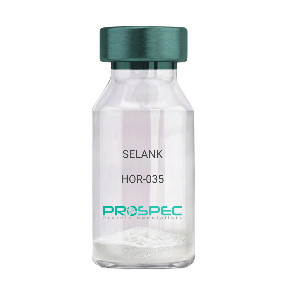
SELANKDescription:
SELANK
Product # :
HOR-035Price :
Quantity :
Shipping Method :
Shipped at Room temp
More Info
- description
- formulation
- purity
- More Info
Description
SELANK Synthetic is a single, non-glycosylated polypeptide chain containing 7 amino acids, having a molecular mass of 751.89 Dalton and a Molecular formula of C33H57N11O9.
Formulation
The protein was lyophilized with no additives.
Purity
Greater than 97.0% as determined by analysis by RP-HPLC.
More Info
-
Physical Appearance
Sterile Filtered White lyophilized (freeze-dried) powder.
-
Stability
Lyophilized SELANK although stable at room temperature for 3 weeks, should be stored desiccated below -18°C. Upon reconstitution SELANK should be stored at 4°C between 2-7 days and for future use below -18°C. For long term storage it is recommended to add a carrier protein (0.1% HSA or BSA). Please prevent freeze-thaw cycles.
-
Solubility
It is recommended to reconstitute the lyophilized SELANK in sterile 18MΩ-cm H2O not less than 100 µg/ml, which can then be further diluted to other aqueous solutions.
-
Amino Acid Sequence
H-Thr-Lys-Pro-Arg-Pro-Gly-Pro-OH.
-
Background
Selank, also known as TP-7, is a synthetic peptide that has garnered significant attention in the scientific community due to its potential anxiolytic, neuroprotective, and nootropic effects. This heptapeptide, derived from the naturally occurring human tetrapeptide Tuftsin, has been shown to possess a wide range of biological activities, including enhancing memory, reducing anxiety, and potentially mitigating the effects of neurodegenerative diseases.
Selank's primary mechanism of action involves its interaction with the GABAergic system, the body's main inhibitory neurotransmitter system. By modulating the activity of GABA receptors, Selank can exert a calming effect on the brain, reducing anxiety and enhancing cognitive function. Studies by Semenova et al. (2014) have demonstrated that Selank can enhance memory and learning in rats, suggesting potential applications in cognitive enhancement and the treatment of cognitive disorders.
In addition to its anxiolytic and nootropic effects, Selank has been shown to possess neuroprotective properties. Research by Ershov et al. (2016) found that Selank could protect neurons from oxidative stress, suggesting potential applications in the treatment of neurodegenerative diseases such as Alzheimer's and Parkinson's disease.
Given its anxiolytic, nootropic, and neuroprotective effects, Selank has been proposed as a potential therapeutic agent for a variety of conditions, including anxiety disorders, cognitive disorders, and neurodegenerative diseases. For instance, a study by Medvedev et al. (2017) found that Selank could improve symptoms in patients with generalized anxiety disorder, indicating its potential as a therapeutic agent in the treatment of anxiety disorders.
While research on Selank is promising, it is important to note that most studies have been conducted in animals or in vitro. More research is needed to fully understand the potential effects and applications of Selank in humans. However, the existing body of research suggests that Selank could be a promising tool in the treatment of anxiety disorders, cognitive disorders, and neurodegenerative diseases.
ProSpec's products are furnished for LABORATORY RESEARCH USE ONLY. The product may not be used as drugs, agricultural or pesticidal products, food additives or household chemicals.
- View Data Sheet
Name :
ISG15 HumanDescription:
ISG15 Human Recombinant
ISG15, G1P2, IFI15, UCRP, ISG15 Ubiquitin-like modifier.
Product # :
PRO-611Price :
Quantity :
Shipping Method :
Shipped with Ice Packs
More Info
- description
- source
- formulation
- purity
- More Info
Description
ISG15 Human Recombinant fused to N-terminal Calmudulin Tag (151 a.a.) produced in E.Coli is a single, non-glycosylated polypeptide chain containing a total of 308 amino acids (157 a.a. without tag) and having a molecular mass of 34 kDa.
Source
Escherichia Coli.
Formulation
The ISG15 solution contains 20mM Tris-HCl pH-8, 100mM NaCl and 1mM DTT.
Purity
Greater than 98.0% as determined by SDS-PAGE.
More Info
-
Introduction
ISG15 ubiquitin-like Modifier is conjugated to the intracellular target proteins after IFN-alpha or IFN-beta stimulation. ISG15 enzymatic pathway is partially distinct from that of ubiquitin, differing in substrate specificity and interaction with ligating enzymes. ISG15 conjugation pathway uses a dedicated E1 enzyme, but seems to converge with the ub conjugation pathway at the level of a specific E2 enzyme. ISG15 protein targets include STAT1, SERPINA 3G/SPI2A, JAK1, MAPK3/ERK1, PLCG1, EIF2AK2/PKR, MX1/MXA, and RIG-1. Deconjugated by USP18/UBP43 shows specific chemotactic activity towards neutrophils and activates them to induce release of eosinophil chemotactic factors. ISG15 serves as a trans-acting binding factor directing the association of ligated target proteins to intermediate filaments. ISG15 plays a role in autocrine, paracrine and endocrine mechanisms, as in cell-to-cell signaling, possibly partly by inducing ifn-gamma secretion by monocytes and macrophages. ISG15 displays antiviral activity during viral infections. ISG15 in response to IFN-TAU secreted by the conceptus, ligates to and regulate proteins involved in the release of prostaglandin F2-alpha and thus prevent lysis of the corpus luteum and maintain the pregnancy. ISG15 shares several common properties with other ubiquitin-like molecules, but its activity is strongly regulated by precise signaling pathways that have a role in innate immunity.
-
Synonyms
ISG15, G1P2, IFI15, UCRP, ISG15 Ubiquitin-like modifier.
-
Physical Appearance
Sterile Filtered colorless solution.
-
Stability
Store at 4°C if entire vial will be used within 2-4 weeks. Store, frozen at -20°C for longer periods of time. For long term storage it is recommended to add a carrier protein (0.1% HSA or BSA).Avoid multiple freeze-thaw cycles.
-
Amino Acid Sequence
MADQLTEEQI AEFKEAFSLF DKDGDGTITT KELGTVMRSL GQNPTEAELQ DMINEVDADG NGTIDFPEFL TMMARKMKDT DSEEEIREAF RVFDKDGNGY ISAAELRHVM TNLGEKLTDE EVDEMIREAD IDGDGQVNYE EFVQMMTAKG SMGWDLTVKM LAGNEFQVSL SSSMSVSELK AQITQKIGVH AFQQRLAVHP SGVALQDRVP LASQGLGPGS TVLLVVDKCD EPLNILVRNN KGRSSTYEVR LTQTVAHLKQ QVSGLEGVQD DLFWLTFEGK PLEDQLPLGE YGLKPLSTVF MNLRLRGG.
ProSpec's products are furnished for LABORATORY RESEARCH USE ONLY. The product may not be used as drugs, agricultural or pesticidal products, food additives or household chemicals.
- View Data Sheet
Name :
LanreotideDescription:
Lanreotide
Product # :
HOR-282Price :
Quantity :
Shipping Method :
Shipped at Room temp
More Info
- description
- formulation
- purity
- More Info
Description
Lanreotide is an octapeptide, an analogue of a naturally occurring hormone, somatostatin.
Formulation
The protein (1mg/ml) was lyophilized with 5mg manntitol and 0.04mg tween-80.
Purity
Greater than 99.0% as determined by:
(a) Analysis by RP-HPLC.
(b) Analysis by SDS-PAGE.More Info
-
Introduction
Lanreotide is a peptide inhibitor of a number of endocrine, neuroendocrine, exocrine and paracrine functions. It shows good affinity for peripheral somatostatin receptors (anterior pituitary and pancreatic). In contrast, its affinity for central receptors is much lower. This profile confers a good specificity of action at the level of growth hormone and digestive hormone secretion. Lanreotide shows a much longer duration of action than natural somatostatin. In addition, its marked selectivity for the secretion of growth hormone, compared to that of insulin, makes it a suitable candidate for the treatment of acromegaly. By inhibiting the synthesis of thyroid stimulating hormone (TSH), lanreotide also normalised thyroid function of patients with thyrotrophin secreting adenomas in 50% (8/16) of the per-protocol population treated for 6 months. There was no significant reduction in the size of the adenoma. Furthermore, the inhibitory action of lanreotide on intestinal exocrine secretion, d
-
Physical Appearance
Sterile Filtered White lyophilized (freeze-dried) powder.
-
Stability
Lyophilized Lanreotide although stable at room temperature for 3 weeks, should be stored desiccated below -18°C. Upon reconstitution Lanreotide should be stored at 4°C between 2-7 days and for future use below -18°C.For long term storage it is recommended to add a carrier protein (0.1% HSA or BSA).Please prevent freeze-thaw cycles.
-
Solubility
It is recommended to reconstitute the lyophilized Lanreotide in sterile 18MΩ-cm H2O not less than 100 µg/ml, which can then be further diluted to other aqueous solutions.
ProSpec's products are furnished for LABORATORY RESEARCH USE ONLY. The product may not be used as drugs, agricultural or pesticidal products, food additives or household chemicals.
- View Data Sheet
Name :
NECTIN3 HumanDescription:
Nectin Cell Adhesion Molecule 3 Human Recombinant
Nectin-3, CDw113, Nectin cell adhesion molecule 3, Poliovirus receptor-related protein 3, CD113, PVRL3, PRR3, NECTIN-3, PVRR3, PPR3.
Product # :
PRO-2504Price :
Quantity :
Shipping Method :
Shipped with Ice Packs
More Info
- description
- source
- formulation
- purity
- More Info
Description
NECTIN3 produced in Sf9 Insect cells is a single, glycosylated polypeptide chain containing 355 amino acids (58-404 a.a.) and having a molecular mass of 39.1kDa (Molecular size on SDS-PAGE will appear at approximately 40-57kDa).NECTIN3 is expressed with an 8 amino acid His tag at C-Terminus and purified by proprietary chromatographic techniques.
Source
Sf9, Insect cells.
Formulation
NECTIN3 protein solution (0.25mg/ml) contains Phosphate Buffered Saline (pH 7.4), 20% glycerol and 1mM DTT.
Purity
Greater than 90.0% as determined by SDS-PAGE.
More Info
-
Introduction
NECTIN3, also known as Nectin Cell Adhesion Molecule 3, is part of the nectin family. NECTIN3 is intended to initiate cell-cell adhesion to stimulate cell attachment and to allow subsequent formation of JAM-and cadherin-based intercellular junctions. NECTIN3 induces endocytosis-mediated down-regulation of PVR from the cell surface, which results in reduction of cell movement & proliferation. Following Nectin-3 activity adds strength to the junction through trans-interaction with various molecules.
-
Synonyms
Nectin-3, CDw113, Nectin cell adhesion molecule 3, Poliovirus receptor-related protein 3, CD113, PVRL3, PRR3, NECTIN-3, PVRR3, PPR3.
-
Physical Appearance
Sterile filtered colorless solution.
-
Stability
Store at 4°C if entire vial will be used within 2-4 weeks. Store, frozen at -20°C for longer periods of time. For long term storage it is recommended to add a carrier protein (0.1% HSA or BSA).Avoid multiple freeze-thaw cycles.
-
Amino Acid Sequence
GPIIVEPHVT AVWGKNVSLK CLIEVNETIT QISWEKIHGK SSQTVAVHHP QYGFSVQGEY QGRVLFKNYS LNDATITLHN IGFSDSGKYI CKAVTFPLGN AQSSTTVTVL VEPTVSLIKG PDSLIDGGNE TVAAICIAAT GKPVAHIDWE GDLGEMESTT TSFPNETATI ISQYKLFPTR FARGRRITCV VKHPALEKDI RYSFILDIQY APEVSVTGYD GNWFVGRKGV NLKCNADANP PPFKSVWSRL DGQWPDGLLA SDNTLHFVHP LTFNYSGVYI CKVTNSLGQR SDQKVIYISD PPTTTTLQPT IQWHPSTADI EDLATEPKKL PFPLSTLATI KDDTIATLEH HHHHH.
ProSpec's products are furnished for LABORATORY RESEARCH USE ONLY. The product may not be used as drugs, agricultural or pesticidal products, food additives or household chemicals.
- View Data Sheet
Name :
NKp46 MouseDescription:
Natural Cytotoxicity Receptor NKp46 Mouse Recombinant
Natural cytotoxicity triggering receptor 1, Activating receptor 1, mAR-1, Lymphocyte antigen 94, Natural killer cell p46-related protein, NK-p46, NKp46, mNKp46, CD335, Ncr1, Ly94.
Product # :
PRO-2365Price :
Quantity :
Shipping Method :
Shipped with Ice Packs
More Info
- description
- source
- formulation
- purity
- More Info
Description
NKp46 Mouse Recombinant produced in Sf9 Baculovirus cells is a single, glycosylated polypeptide chain containing 481 amino acids (17-255 a.a) and having a molecular mass of 54.4kDa (Molecular size on SDS-PAGE will appear at approximately 50-70kDa).NKp46 is fused to a 239 amino acid hIgG-His Tag at C-terminus & purified by proprietary chromatographic techniques.
Source
Sf9, Baculovirus cells.
Formulation
NKp46 protein solution (0.25mg/ml) contains Phosphate Buffered Saline (pH 7.4) and 10% glycerol.
Purity
Greater than 85.0% as determined by SDS-PAGE.
More Info
-
Introduction
A natural cytotoxicity receptor (NCR) NKp46 has been shown to represent a novel NK cell-specific molecule involved in human NK cell activation. The natural cytotoxicity receptors (NCRs) are a recently characterized family of Ig-like activation receptors that appear to be major triggering receptors in tumor cell recognition. The three known NCRs include NKp46 and NKp30, which are expressed on circulating NKcells, and NKp44, which is expressed only on activating NK cells. NKp46 has been implicated in NK cell-mediated lysis of several autologous tumor cells, pathogen-infected cell lines and mononuclear phagocytes infected with an intracellular bacterium. The Lysis of tumor cells by NK-cells involves recognition by NKp46 of heparan sulfate moieties of membrane heparan sulfate proteoglycans. Furthermore, NKp46 is a surface receptor involved in NK-cell cell death by apoptosis. NKp46 has two extracellular Ig-like domains followed by a ~40 residue stalk region, a type I transmembrane domain, and a short cytoplasmic tail. The extracellular Ig-like domain of NKp46 (22-255aa) is purified by FPLC gel-filtration chromatography, after refolding of the isolated inclusion bodies in a redox buffer. In addition, engagement of the antigen with the monoclonal antibody stimulates intracellular calcium levels and the synthesis of cytokines. CD59 is an NKp46 coreceptor (by physical association) together they activate cytotoxicity of human NK-cells, their engagement results in tyrosine phosphorylation of CD3-zeta chains associated with NKp46. Reduced cell surface expression of NKp46 and other NK-cell receptors is linked to the impaired NK-cell cytolytic function in viremic HIV-1 infection.
-
Synonyms
Natural cytotoxicity triggering receptor 1, Activating receptor 1, mAR-1, Lymphocyte antigen 94, Natural killer cell p46-related protein, NK-p46, NKp46, mNKp46, CD335, Ncr1, Ly94.
-
Physical Appearance
Sterile Filtered colorless solution.
-
Stability
Store at 4°C if entire vial will be used within 2-4 weeks. Store, frozen at -20°C for longer periods of time. For long term storage it is recommended to add a carrier protein (0.1% HSA or BSA). Avoid multiple freeze-thaw cycles.
-
Amino Acid Sequence
ADLQRINTEK ETLPKPIIWA KPSIMVTNGN SVNIWCQGAQ SASEYQLYFE GSFFALERPK PSRSMNKVRF FISQMTSHTA GIYTCFYQSG ELWSKSSNPL KLVVTGLYDT PNLWVYPRPE VTLGENVTFF CQLKTATSKF FLLKERGSNH IQNKYGNIQA EFPMGPVTRA HRGTYRCFGSYNDYAWSFPS EPVTLLITGG VENSSLAPTD PTSSLDYWEF DLSTNESGLQ KDSAFWDHTT QNLEPKSCDK THTCPPCPAP ELLGGPSVFL FPPKPKDTLM ISRTPEVTCV VVDVSHEDPE VKFNWYVDGV EVHNAKTKPR EEQYNSTYRV VSVLTVLHQD WLNGKEYKCK VSNKALPAPIEKTISKAKGQ PREPQVYTLP PSRDELTKNQ VSLTCLVKGF YPSDIAVEWE SNGQPENNYK TTPPVLDSDG SFFLYSKLTV DKSRWQQGNV FSCSVMHEAL HNHYTQKSLS LSPGKHHHHH H
ProSpec's products are furnished for LABORATORY RESEARCH USE ONLY. The product may not be used as drugs, agricultural or pesticidal products, food additives or household chemicals.
- View Data Sheet
Name :
SYT4 HumanDescription:
Synaptotagmin IV Human Recombinant
HsT1192, KIAA1342, Synaptotagmin-4, Synaptotagmin IV, SytIV, SYT4.
Product # :
PRO-1375Price :
Quantity :
Shipping Method :
Shipped with Ice Packs
More Info
- description
- source
- formulation
- purity
- More Info
Description
SYT4 Human Recombinant produced in E.Coli is a single, non-glycosylated polypeptide chain containing 409 amino acids (38-425a.a) and having a molecular mass of 46.1kDa. SYT4 is fused to a 21 amino acid His-tag at N-terminus & purified by proprietary chromatographic techniques.
Source
E.coli.
Formulation
SYT4 protein solution (0.25mg/ml) containing 20mM Tris-HCl buffer (pH 8.0), 0.1M NaCl, 20% glycerol and 1mM DTT.
Purity
Greater than 90% as determined by SDS-PAGE.
More Info
-
Introduction
Synaptotagmin-4 (SYT4) is a part of the synaptotagmin proteins family. Synaptotagmins are a large gene family of synaptic vesicle type III integral membrane proteins that operate as regulators of both exocytosis and endocytosis and are involved in neurotransmitter secretion from small secretory vesicles. SYT4 is expressed in neuronal tissues, and has the highest mRNA levels in the hippocampus. The proximity of the SYT4 gene to markers of several psychiatric disorders implies an involvement of SYT4 in human disease such as pheochromocytoma, and parkinson's disease.
-
Synonyms
HsT1192, KIAA1342, Synaptotagmin-4, Synaptotagmin IV, SytIV, SYT4.
-
Physical Appearance
Sterile Filtered colorless solution.
-
Stability
Store at 4°C if entire vial will be used within 2-4 weeks. Store, frozen at -20°C for longer periods of time. For long term storage it is recommended to add a carrier protein (0.1% HSA or BSA).Avoid multiple freeze-thaw cycles.
-
Amino Acid Sequence
MGSSHHHHHH SSGLVPRGSH MCQRKSSKSN KTPPYKFVHV LKGVDIYPEN LNSKKKFGAD DKNEVKNKPA VPKNSLHLDL EKRDLNGNFP KTNLKPGSPS DLENATPKLF LEGEKESVSP ESLKSSTSLT SEEKQEKLGT LFFSLEYNFE RKAFVVNIKE ARGLPAMDEQ SMTSDPYIKM TILPEKKHKV KTRVLRKTLD PAFDETFTFY GIPYTQIQEL ALHFTILSFD RFSRDDIIGE VLIPLSGIEL SEGKMLMNRE IIKRNVRKSS GRGELLISLC YQSTTNTLTV VVLKARHLPK SDVSGLSDPY VKVNLYHAKK RISKKKTHVK KCTPNAVFNE LFVFDIPCEG LEDISVEFLV LDSERGSRNE VIGQLVLGAA AEGTGGEHWK EICDYPRRQI AKWHVLCDG.
ProSpec's products are furnished for LABORATORY RESEARCH USE ONLY. The product may not be used as drugs, agricultural or pesticidal products, food additives or household chemicals.