Search results
1000 results found for “Secretagogin”
Name
Description
Product #
Price
Quantity
Shipping Method
- View Data Sheet
Name :
GHRP4Description:
Growth Hormone Releasing Peptide-4
GHRP-4, GHRP4.
Product # :
HOR-022Price :
Quantity :
Shipping Method :
Shipped at Room temp
More Info
- description
- formulation
- purity
- More Info
Description
Growth Hormone Releasing Peptide-4 Synthetic is a single, non-glycosylated polypeptide chain containing 4 amino acids, having a molecular mass of 607.73 Dalton and a Molecular formula of C34H37N7O4.
Formulation
The protein was lyophilized with no additives.
Purity
Greater than 97.0% as determined by analysis by RP-HPLC.
More Info
-
Introduction
GH-releasing peptides (GHRPs) are synthetic peptides that like GHRH effect on pituitary somatotrophs to stimulate GH release. The GHRP4 is one of several synthetic met-enkephalin analogs that owns unnatural D-amino acids. They were developed for their growth hormone (GH) releasing activity and called GH secretatogues. They lack opioid activity but are potent stimulators of GH release. These secretatogues are distinct from the growth hormone releasing hormone (GHRH or GHRF) as they share no sequence relation and derive their function through action at a completely different receptor, the ghrelin receptor.
-
Synonyms
GHRP-4, GHRP4.
-
Physical Appearance
Sterile Filtered White lyophilized (freeze-dried) powder.
-
Stability
Lyophilized Growth Hormone Releasing Peptide-4 although stable at room temperature for 3 weeks, should be stored desiccated below -18°C. Upon reconstitution GHRP4 should be stored at 4°C between 2-7 days and for future use below -18°C. For long term storage it is recommended to add a carrier protein (0.1% HSA or BSA). Please prevent freeze-thaw cycles.
-
Solubility
It is recommended to reconstitute the lyophilized GHRP4 in sterile 18MΩ-cm H2O not less than 100 µg/ml, which can then be further diluted to other aqueous solutions.
-
Amino Acid Sequence
(D-Trp)-Ala-Trp-(D-Phe)-NH2.
ProSpec's products are furnished for LABORATORY RESEARCH USE ONLY. The product may not be used as drugs, agricultural or pesticidal products, food additives or household chemicals.
- View Data Sheet
Name :
RSG1 HumanDescription:
REM2 and RAB-Like Small GTPase 1 Human Recombinant
REM2- and Rab-like small GTPase 1, RSG1, C1orf89, RP4-733M16.4.
Product # :
PRO-1334Price :
Quantity :
Shipping Method :
Shipped with Ice Packs
More Info
- description
- source
- formulation
- purity
- More Info
Description
RSG1 Human Recombinant produced in E.Coli is a single, non-glycosylated polypeptide chain containing 281 amino acids (1-258 a.a) and having a molecular mass of 30.9kDa.RSG1 is fused to a 23 amino acid His-tag at N-terminus & purified by proprietary chromatographic techniques.
Source
Escherichia Coli.
Formulation
RSG1 protein solution (0.25mg/ml) containing 20mM Tris-HCl buffer (pH 8.5), 0.2M NaCl, 50% glycerol and 2mM DTT.
Purity
Greater than 85.0% as determined by SDS-PAGE.
More Info
-
Introduction
REM2 and RAB-Like Small GTPase 1 (RSG1) plays a part in targeted membrane trafficking most likely at the level of vesicle fusion with membranes. The RSG1 protein is involved in cilium biogenesis by regulating the transportation of cargo proteins to the basal body and to the apical tips of cilia. RSG1 is a potential effector of the planar cell polarity signaling pathway. RSG1 is also involved in exocytosis in secretory cells.
-
Synonyms
REM2- and Rab-like small GTPase 1, RSG1, C1orf89, RP4-733M16.4.
-
Physical Appearance
Sterile Filtered colorless solution.
-
Stability
Store at 4°C if entire vial will be used within 2-4 weeks. Store, frozen at -20°C for longer periods of time. For long term storage it is recommended to add a carrier protein (0.1% HSA or BSA).Avoid multiple freeze-thaw cycles.
-
Amino Acid Sequence
MGSSHHHHHH SSGLVPRGSH MGSMARPPVP GSVVVPNWHE SAEGKEYLAC ILRKNRRRVF GLLERPVLLP PVSIDTASYK IFVSGKSGVG KTALVAKLAG LEVPVVHHET TGIQTTVVFW PAKLQASSRV VMFRFEFWDC GESALKKFDH MLLACMENTD AFLFLFSFTD RASFEDLPGQ LARIAGEAPG VVRMVIGSKF DQYMHTDVPE RDLTAFRQAW ELPLLRVKSV PGRRLADGRT LDGRAGLADV AHILNGLAEQ LWHQDQVAAG LLPNPPESAP E.
ProSpec's products are furnished for LABORATORY RESEARCH USE ONLY. The product may not be used as drugs, agricultural or pesticidal products, food additives or household chemicals.
- View Data Sheet
Name :
ANKRD1 HumanDescription:
Ankyrin Repeat Domain 1 Human Recombinant
Ankyrin Repeat Domain 1 (Cardiac Muscle), Cardiac Ankyrin Repeat Protein, Cytokine-Inducible Gene C-193 Protein, Ankyrin Repeat Domain-Containing Protein 1, Cytokine-Inducible Nuclear Protein, Liver Ankyrin Repeat Domain 1, BA320F15.2, CARP, ALRP, CVARP, MCARP, HA1A2, C193.
Product # :
PRO-1219Price :
Quantity :
Shipping Method :
Shipped with Ice Packs
More Info
- description
- source
- formulation
- purity
- More Info
Description
ANKRD1 Human Recombinant produced in E. coli is a single polypeptide chain containing 342 amino acids (1-319) and having a molecular mass of 38.6 kDa.ANKRD1 is fused to a 23 amino acid His-tag at N-terminus & purified by proprietary chromatographic techniques.
Source
E.coli.
Formulation
The ANKRD1 solution contains 20mM Tris-HCl buffer (pH 8.5), 0.2M NaCl, 5mM DTT and 50% glycerol.
Purity
Greater than 85% as determined by SDS-PAGE.
More Info
-
Introduction
ANKRD1 takes part in endothelial cell activation and performs as a nuclear transcription factor which negatively controls the expression of cardiac genes.
-
Synonyms
Ankyrin Repeat Domain 1 (Cardiac Muscle), Cardiac Ankyrin Repeat Protein, Cytokine-Inducible Gene C-193 Protein, Ankyrin Repeat Domain-Containing Protein 1, Cytokine-Inducible Nuclear Protein, Liver Ankyrin Repeat Domain 1, BA320F15.2, CARP, ALRP, CVARP, MCARP, HA1A2, C193.
-
Physical Appearance
Sterile Filtered colorless solution.
-
Stability
Store at 4°C if entire vial will be used within 2-4 weeks. Store, frozen at -20°C for longer periods of time. For long term storage it is recommended to add a carrier protein (0.1% HSA or BSA).Avoid multiple freeze-thaw cycles.
-
Amino Acid Sequence
MGSSHHHHHH SSGLVPRGSH MGS MMVLKVE ELVTGKKNGN GEAGEFLPED FRDGEYEAAV TLEKQEDLKT LLAHPVTLGE QQWKSEKQRE AELKKKKLEQ RSKLENLEDL EIIIQLKKRK KYRKTKVPVV KEPEPEIITE PVDVPTFLKA ALENKLPVVE KFLSDKNNPD VCDEYKRTAL HRACLEGHLA IVEKLMEAGA QIEFRDMLES TAIHWASRGG NLDVLKLLLN KGAKISARDK LLSTALHVAV RTGHYECAEH LIACEADLNA KDREGDTPLH DAVRLNRYKM IRLLIMYGAD LNIKNCAGKT PMDLVLHWQN GTKAIFDSLR ENSYKTSRIA TF
ProSpec's products are furnished for LABORATORY RESEARCH USE ONLY. The product may not be used as drugs, agricultural or pesticidal products, food additives or household chemicals.
- View Data Sheet
Name :
SERTAD1 HumanDescription:
SERTA Domain Containing 1 Human Recombinant
SERTA domain-containing protein 1, CDK4-binding protein p34SEI1, SEI-1, Transcriptional regulator interacting with the PHD-bromodomain 1, TRIP-Br1, SERTAD1, SEI1.
Product # :
PRO-1157Price :
Quantity :
Shipping Method :
Shipped with Ice Packs
More Info
- description
- source
- formulation
- purity
- More Info
Description
SERTAD1 Human Recombinant produced in E.Coli is a single, non-glycosylated polypeptide chain containing 260 amino acids (1-236 a.a) and having a molecular mass of 27.3kDa (Molecular weight on SDS-PAGE will appear higher).SERTAD1 is fused to a 24 amino acid His-tag at N-terminus & purified by proprietary chromatographic techniques.
Source
Escherichia Coli.
Formulation
SERTAD1 protein solution (0.5mg/ml) containing 20mM Tris-HCl buffer (pH8.0), 10% glycerol and 1mM DTT.
Purity
Greater than 85% as determined by SDS-PAGE.
More Info
-
Introduction
SERTA domain-containing protein (SERTAD1) functions with E2F-responsive promoters to integrate signals provided by PHD- and/or bromodomain-containing transcription factors. SERTAD1 stimulates E2F-1/DP-1 transcriptional activity. SERTAD1 reduces the activity of cyclin D1/CDK4 resistant to the inhibitory effects of p16(INK4a). In addition, SERTAD1 interacts with the PHD-bromodomain of TIF1, TRIM28/TIF1B and p300/CBP. Furthermore, SERTAD1 binds to DP1 and interacts with CDK4.
-
Synonyms
SERTA domain-containing protein 1, CDK4-binding protein p34SEI1, SEI-1, Transcriptional regulator interacting with the PHD-bromodomain 1, TRIP-Br1, SERTAD1, SEI1.
-
Physical Appearance
Sterile filtered colorless solution.
-
Stability
Store at 4°C if entire vial will be used within 2-4 weeks. Store, frozen at -20°C for longer periods of time. For long term storage it is recommended to add a carrier protein (0.1% HSA or BSA).Avoid multiple freeze-thaw cycles.
-
Amino Acid Sequence
MGSSHHHHHH SSGLVPRGSH MGSHMLSKGL KRKREEEEEK EPLAVDSWWL DPGHTAVAQA PPAVASSSLF DLSVLKLHHS LQQSEPDLRH LVLVVNTLRR IQASMAPAAA LPPVPSPPAA PSVADNLLAS SDAALSASMA SLLEDLSHIE GLSQAPQPLA DEGPPGRSIG GAAPSLGALD LLGPATGCLL DDGLEGLFED IDTSMYDNEL WAPASEGLKP GPEDGPGKEE APELDEAELD YLMDVLVGTQ ALERPPGPGR.
ProSpec's products are furnished for LABORATORY RESEARCH USE ONLY. The product may not be used as drugs, agricultural or pesticidal products, food additives or household chemicals.
- View Data Sheet
Name :
SNRPA1 HumanDescription:
Small Nuclear Ribonucleoprotein Polypeptide A1 Human Recombinant
U2 small nuclear ribonucleoprotein A', U2 snRNP A', SNRPA1, Lea1.
Product # :
PRO-1015Price :
Quantity :
Shipping Method :
Shipped with Ice Packs
More Info
- description
- source
- formulation
- purity
- More Info
Description
SNRPA1 Human Recombinant produced in E.Coli is a single, non-glycosylated polypeptide chain containing 275 amino acids (1-255 a.a.) and having a molecular mass of 30.5kDa. SNRPA1 is fused to a 20 amino acid His-tag at N-terminus & purified by proprietary chromatographic techniques.
Source
Escherichia Coli.
Formulation
SNRPA1 protein solution (0.5mg/ml) containing 20mM Tris-HCl buffer (pH8.0), 30% glycerol, 0.2M NaCl,2mM DTT and 0.1mM PMSF.
Purity
Greater than 80% as determined by SDS-PAGE.
More Info
-
Introduction
U2 small nuclear ribonucleoprotein A (SNRPA1), a 255 amino acid protein, is a component of the U2 snRNP which forms a complex with U2 snRNP B (U2B). U2 snRNP A and U2 snRNP B form together a complex which binds to the U2 snRNA hairpin IV. The configuration of the U2 snRNP A/U2 snRNP B dimer and the delicate variations of a few key residues regulate the snRNP-RNA-binding specificity.
-
Synonyms
U2 small nuclear ribonucleoprotein A', U2 snRNP A', SNRPA1, Lea1.
-
Physical Appearance
Sterile Filtered clear solution.
-
Stability
Store at 4°C if entire vial will be used within 2-4 weeks. Store, frozen at -20°C for longer periods of time. For long term storage it is recommended to add a carrier protein (0.1% HSA or BSA).Avoid multiple freeze-thaw cycles.
-
Amino Acid Sequence
MGSSHHHHHH SSGLVPRGSH MVKLTAELIE QAAQYTNAVR DRELDLRGYK IPVIENLGAT LDQFDAIDFS DNEIRKLDGF PLLRRLKTLL VNNNRICRIG EGLDQALPCL TELILTNNSL VELGDLDPLA SLKSLTYLSI LRNPVTNKKH YRLYVIYKVP QVRVLDFQKV KLKERQEAEK MFKGKRGAQL AKDIARRSKT FNPGAGLPTD KKKGGPSPGD VEAIKNAIAN ASTLAEVERL KGLLQSGQIP GRERRSGPTD DGEEEMEEDT VTNGS.
ProSpec's products are furnished for LABORATORY RESEARCH USE ONLY. The product may not be used as drugs, agricultural or pesticidal products, food additives or household chemicals.
- View Data Sheet
Name :
ESM1 Human, SF9Description:
Endothelial Cell-Specific Molecule 1 Human Recombinant, Sf9
Endothelial Cell Specific Molecule 1, Endothelial Cell-Specific Molecule 1, ESM-1, Endocan.
Product # :
PRO-2588Price :
Quantity :
Shipping Method :
Shipped with Ice Packs
More Info
- description
- source
- formulation
- purity
- More Info
Description
ESM1 produced in Sf9 Baculovirus cells is a single, glycosylated polypeptide chain containing 174 amino acids (20-184a.a.) and having a molecular mass of 19.2kDa. (Molecular size on SDS-PAGE will appear at approximately 18-28kDa). ESM1 is expressed with a 9 amino acid His tag at C-Terminus and purified by proprietary chromatographic techniques.
Source
Sf9, Baculovirus cells.
Formulation
ESM1 protein solution (0.25mg/ml) contains Phosphate Buffered Saline (pH 7.4) and 10% glycerol.
Purity
Greater than 95.0% as determined by SDS-PAGE.
More Info
-
Introduction
ESM1, also knows as, Endothelial cell-specific molecule 1 is a protein encoded by a gene called ESM1 in humans. ESM1 is a secreted protein that is highly expressed in human kidney and lung tissues (in the endothelial cells). There are suggestions regarding the protein’s involvement in endothelium-dependent pathological diseases, due to his regulation that is performed by cytokines.
-
Synonyms
Endothelial Cell Specific Molecule 1, Endothelial Cell-Specific Molecule 1, ESM-1, Endocan.
-
Physical Appearance
Sterile Filtered clear solution.
-
Stability
Store at 4°C if entire vial will be used within 2-4 weeks. Store, frozen at -20°C for longer periods of time. For long term storage it is recommended to add a carrier protein (0.1% HSA or BSA).Avoid multiple freeze-thaw cycles.
-
Amino Acid Sequence
ADLWSNNYAV DCPQHCDSSE CKSSPRCKRT VLDDCGCCRV CAAGRGETCY RTVSGMDGMK CGPGLRCQPS NGEDPFGEEF GICKDCPYGT FGMDCRETCN CQSGICDRGT GKCLKFPFFQ YSVTKSSNRF VSLTEHDMAS GDGNIVREEV VKENAAGSPV MRKWLNPRHH HHHH.
ProSpec's products are furnished for LABORATORY RESEARCH USE ONLY. The product may not be used as drugs, agricultural or pesticidal products, food additives or household chemicals.
- View Data Sheet
Name :
Fibronectin RecombinantDescription:
Fibronectin Human Recombinant
Product # :
PRO-2621Price :
Quantity :
Shipping Method :
Shipped at Room temp
More Info
- description
- source
- formulation
- purity
- biological activity
- More Info
Description
Fibronectin Human Recombinant produced in E.Coli is a single, non-glycosylated polypeptide chain containing 574 amino acids and having a molecular mass of 62.6kDa. The Fibronectin is purified by proprietary chromatographic techniques.
Source
Escherichia Coli.
Formulation
Lyophilized from a 0.2μm filtered concentrated solution in 20 mM Tris-HCl, pH 8.0, 150 mM NaCl, with 5 % Trehalose and 0.02 % Tween-20.
Purity
Greater than 95.0% as determined by:
(a) Analysis by RP-HPLC.
(b) Analysis by SDS-PAGE.Biological Activity
Was measured by its ability to support cell attachment and spreading when used as a substratum for cell culture. The recommended concentration in this application for this effect is typically 1-5 μg/cm2. Fibronectin can also be added to the media to support cell spreading at a concentration of 0.5-50 μg/ml. Optimal concentrations will need to be determined for individual user applications.
More Info
-
Introduction
Fibronectin takes part in several cellular processes, including tissue repair, embryogenesis, blood clotting, and cell migration/adhesion.Plasma fibronectin level is elevated in severe coronary artery disease. Increased plasma fibronectin levels are related with venous thromboembolism (VTE) particularly in males, and extend the probable association between biomarkers and risk factors for arterial atherothrombosis and VTE. Fibronectin consists in 2 main forms: 1) as an insoluble glycoprotein dimer that serves as a linker in the etracellular matrix and 2) as a soluble disulphide linked dimer found in the plasma. The plasma form is produced by hepatocytes, and the ECM form is synthesized by fibroblasts, chondrocytes, endothelial cells, macrophages, as well as certain epithelial cells. Fibronectin alos takes part as a general cell adhesion molecule by anchoring cells to collagen or proteoglycan substrates. Fibronectin organizes cellular interaction with the ECM by binding to different components of the extracellular matrix and to membrane-bound Fibronectin receptors on cell surfaces.
-
Physical Appearance
Sterile Filtered White lyophilized (freeze-dried) powder.
-
Stability
Lyophilized Fibronectin although stable at room temperature for 3 weeks, should be stored desiccated below -18°C. Upon reconstitution Fibronectin should be stored at 4°C between 2-7 days and for future use below -18°C. Please prevent freeze-thaw cycles.
-
Solubility
It is recommended to reconstitute the lyophilized Fibronectin in sterile 18MΩ-cm H2O not less than 100µg/ml, which can then be further diluted to other aqueous solutions.
ProSpec's products are furnished for LABORATORY RESEARCH USE ONLY. The product may not be used as drugs, agricultural or pesticidal products, food additives or household chemicals.
- View Data Sheet
Name :
VTN Human, Sf9Description:
Vitronectin Human Recombinant, Sf9
VN, S-protein, Serum-spreading factor, V75, VTN.
Product # :
pro-2594Price :
Quantity :
Shipping Method :
Shipped with Ice Packs
More Info
- description
- source
- formulation
- purity
- biological activity
- More Info
Description
VTN produced in Sf9 Insect cells is a single, glycosylated polypeptide chain containing 468 amino acids (20-478a.a.) and having a molecular mass of 53.3kDa.VTN is expressed with an 6 amino acid His tag at C-Terminus and purified by proprietary chromatographic techniques.
Source
Sf9, Insect cells.
Formulation
VTN protein solution (0.25mg/ml) contains Phosphate Buffered Saline (pH 7.4) and 10% glycerol.
Purity
Greater than 90.0% as determined by SDS-PAGE.
Biological Activity
Measured by the ability of the immobilized protein to support the adhesion of B16-F10 mouse melanoma cells. The ED50 is 5ug/ml when cells are added to VTN coated plates.
More Info
-
Introduction
Vitronectin (VTN) which is a part of the pexin family is a cell adhesion and spreading factor found in serum and tissues. VTN interacts with glycosaminoglycans and proteoglycans. VTN inhibits the membrane-damaging effect of the terminal cytolytic complement pathway and binds to numerous serpin serine protease inhibitors. Scientists have been noticed an over expression of VTN, integrins and plasminogen in migrating cells during wound healing.
-
Synonyms
VN, S-protein, Serum-spreading factor, V75, VTN.
-
Physical Appearance
Sterile Filtered colorless solution.
-
Stability
Store at 4°C if entire vial will be used within 2-4 weeks. Store, frozen at -20°C for longer periods of time. For long term storage it is recommended to add a carrier protein (0.1% HSA or BSA).Avoid multiple freeze-thaw cycles.
-
Amino Acid Sequence
ADPDQESCKG RCTEGFNVDK KCQCDELCSY YQSCCTDYTA ECKPQVTRGD VFTMPEDEYT
VYDDGEEKNN ATVHEQVGGP SLTSDLQAQS KGNPEQTPVL KPEEEAPAPE VGASKPEGID
SRPETLHPGR PQPPAEEELC SGKPFDAFTD LKNGSLFAFR GQYCYELDEK AVRPGYPKLI
RDVWGIEGPI DAAFTRINCQ GKTYLFKGSQ YWRFEDGVLD PDYPRNISDG FDGIPDNVDA
ALALPAHSYS GRERVYFFKG KQYWEYQFQH QPSQEECEGS SLSAVFEHFA MMQRDSWEDI
FELLFWGRTS AGTRQPQFIS RDWHGVPGQV DAAMAGRIYI SGMAPRPSLA KKQRFRHRNR
KGYRSQRGHS RGRNQNSRRP SRATWLSLFS SEESNLGANN YDDYRMDWLV PATCEPIQSV
FFFSGDKYYR VNLRTRRVDT VDPPYPRSIA QYWLGCPAPG HLHHHHHH.
ProSpec's products are furnished for LABORATORY RESEARCH USE ONLY. The product may not be used as drugs, agricultural or pesticidal products, food additives or household chemicals.
- View Data Sheet
Name :
Streptavidin-NCDescription:
Streptavidin-NC Recombinant
Product # :
PRO-338Price :
Quantity :
Shipping Method :
Shipped with Ice Packs
More Info
- description
- source
- formulation
- purity
- More Info
Description
Recombinant Streptavidin-NC produced in E.Coli is a single, non-glycosylated, polypeptide chain having a molecular mass of 24kDa. Streptavidin-NC is engineered to bind to nitrocellulose.
Source
Escherichia Coli.
Formulation
The sterile solution contains 10mM K2HPO4-KH2PO4, pH 7.3.
Purity
Greater than 93.0% as determined by SDS-PAGE.
More Info
-
Introduction
Streptavidin is a tetrameric protein secreted by Streptomyces avidinii which binds firmly to biotin. Streptavidin is wilyde used in molecular biology through its unique high affinity for the vitamin biotin. The dissociation constant (Kd) of the biotin-streptavidin complex is about ~10-15 mol/L. The strong affinity recognition of biotin and biotinylated molecules has made streptavidin one of the most important components in diagnostics and laboratory kits. The streptavidin/biotin system has one of the biggest free energies of association of yet observed for noncovalent binding of a protein and small ligand in aqueous solution (K_assoc = 10**14). The complexes are also extremely stable over a wide range of temperature and pH.
-
Physical Appearance
Sterile Liquid formulation.
-
Stability
Streptavidin-NC although stable at 4°C for 3 weeks, should be stored below -18°C. Please prevent freeze thaw cycles.
ProSpec's products are furnished for LABORATORY RESEARCH USE ONLY. The product may not be used as drugs, agricultural or pesticidal products, food additives or household chemicals.
- View Data Sheet
Name :
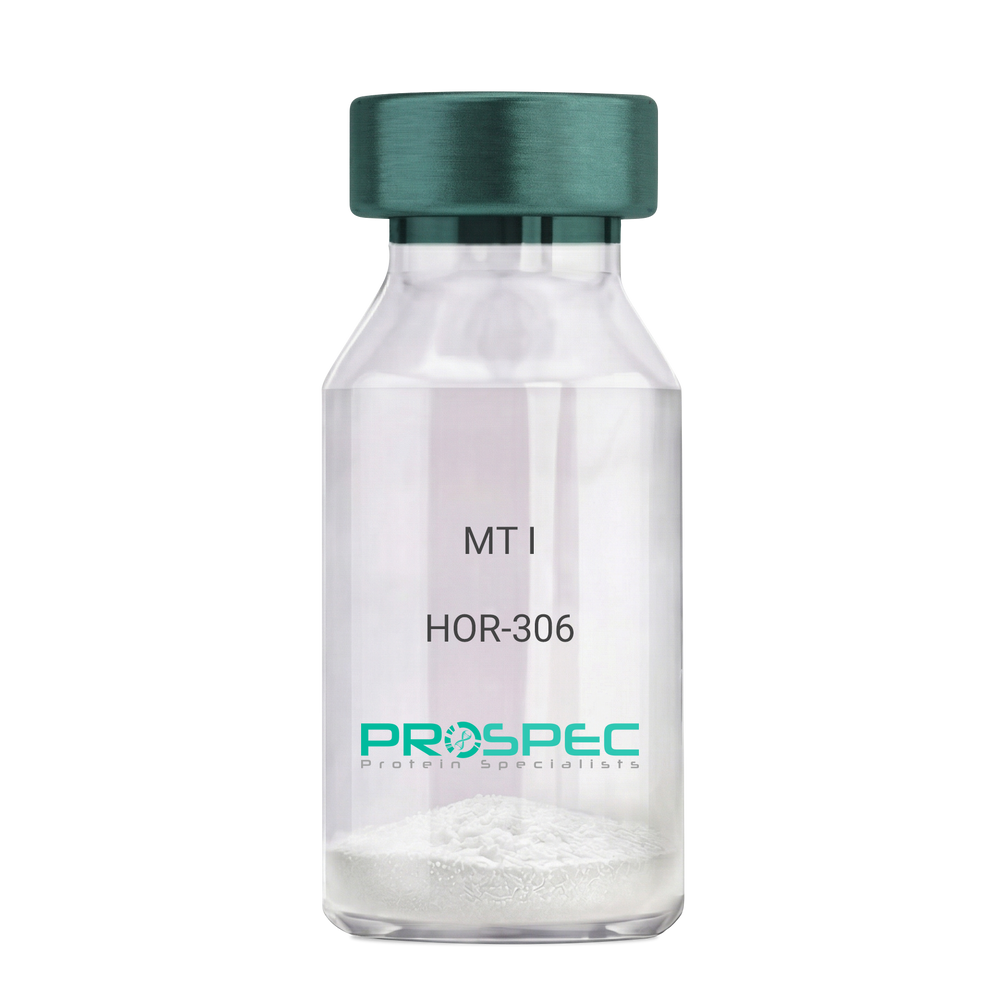
MT IDescription:
Melanotan-I
Melanotan-I, MT-I, Melanotan-1.
Product # :
HOR-306Price :
Quantity :
Shipping Method :
Shipped at Room temp
More Info
- description
- formulation
- purity
- More Info
Description
Melanotan-I has the amino acid sequence of Ser-Tyr-Ser-Nle-Glu-His-D-Phe-Arg-Trp-Gly-Lys-Pro-Val and a molecular weight of 1647.4 Dalton.
Formulation
The protein (1mg/ml) was lyophilized with no additives.
Purity
Greater than 98.0% as determined by RP-HPLC.
More Info
-
Synonyms
Melanotan-I, MT-I, Melanotan-1.
-
Physical Appearance
Sterile Filtered off-White lyophilized (freeze-dried) powder.
-
Stability
Lyophilized Melanotan-I although stable at room temperature for 3 weeks, should be stored desiccated below -18°C. Upon reconstitution MT-I should be stored at 4°C between 2-7 days and for future use below -18°C.For long term storage it is recommended to add a carrier protein (0.1% HSA or BSA).Please prevent freeze-thaw cycles.
-
Solubility
It is recommended to reconstitute the lyophilized Melanotan-I in sterile 1% acetic acid not less than 100 µg/ml, which can then be further diluted to other aqueous solutions.
ProSpec's products are furnished for LABORATORY RESEARCH USE ONLY. The product may not be used as drugs, agricultural or pesticidal products, food additives or household chemicals.
- View Data Sheet
Name :
SEPSECS MouseDescription:
Selenocysteinyl-tRNA(Sec) synthase Mouse Recombinant
AA986712, D5Ertd135e, SecS, SLA, SLA/LP autoantigen, SLA-p35, Selenocysteinyl-tRNA(Sec) synthase, Selenocysteine synthase, Liver-pancreas antigen, Soluble liver antigen, Sec synthase, UGA suppressor tRNA-associated protein, SepSecS.
Product # :
ENZ-1081Price :
Quantity :
Shipping Method :
Shipped with Ice Packs
More Info
- description
- source
- formulation
- purity
- More Info
Description
SEPSECS produced in E.Coli is a single, non-glycosylated polypeptide chain containing 527 amino acids (1-504 a.a.) and having a molecular mass of 57.7kDa.SEPSECS is fused to a 23 amino acid His-tag at N-terminus & purified by proprietary chromatographic techniques.
Source
Escherichia Coli.
Formulation
The SEPSECS protein solution (0.25 mg/ml) is formulated in 20mM Tris-HCl buffer (pH7.5) 1mM DTT, 0.2M NaCl and 50% glycerol.
Purity
Greater than 90% as determined by SDS-PAGE.
More Info
-
Introduction
SEPSECS catalyzes the last step of sec synthesis by converting O-phosphoseryl-tRNA(sec) to selenocysteinyl-tRNA(sec) using selenophosphate as the selenium donor. Furthermore, SEPSECS protein is considered a specific marker of autoimmune hepatitis.
-
Synonyms
AA986712, D5Ertd135e, SecS, SLA, SLA/LP autoantigen, SLA-p35, Selenocysteinyl-tRNA(Sec) synthase, Selenocysteine synthase, Liver-pancreas antigen, Soluble liver antigen, Sec synthase, UGA suppressor tRNA-associated protein, SepSecS.
-
Physical Appearance
Sterile Filtered clear solution.
-
Stability
Store at 4°C if entire vial will be used within 2-4 weeks. Store, frozen at -20°C for longer periods of time. For long term storage it is recommended to add a carrier protein (0.1% HSA or BSA).Avoid multiple freeze-thaw cycles
-
Amino Acid Sequence
MGSSHHHHHH SSGLVPRGSH MGSMNPESFA AGERRVSPAY VRQGCEARRA HEHLIRLLLE QGKCPEDGWD ESTLELFLHE LAVMDSNNFL GNCGVGEREG RVASALVARR HYRFIHGIGR SGDISAVQPK AAGSSLLNKI TNSLVLNVIK LAGVHSVASC FVVPMATGMS LTLCFLTLRH
KRPKAKYIIW PRIDQKSCFK SMVTAGFEPV VIENVLEGDE LRTDLKAVEA KIQELGPEHI LCLHSTTACF APRVPDRLEE LAVICANYDI PHVVNNAYGL QSSKCMHLIQ QGARVGRIDA FVQSLDKNFM VPVGGAIIAG FNEPFIQDIS KMYPGRASAS PSLDVLITLL SLGCSGYRKL
LKERKEMFVY LSTQLKKLAE AHNERLLQTP HNPISLAMTL KTIDGHHDKA VTQLGSMLFT RQVSGARAVP LGNVQTVSGH TFRGFMSHAD NYPCAYLNAA AAIGMKMQDV DLFIKRLDKC LNIVRKEQTR ASVVSGADRN KAEDADIEEM ALKLDDVLGD VGQGPAL.
ProSpec's products are furnished for LABORATORY RESEARCH USE ONLY. The product may not be used as drugs, agricultural or pesticidal products, food additives or household chemicals.
- View Data Sheet
Name :
Leptin Mouse, PEGDescription:
Pegylated Mouse Leptin Recombinant
OB Protein, Obesity Protein, OBS, Obesity factor.
Product # :
CYT-591Price :
Quantity :
Shipping Method :
Shipped at Room temp
More Info
- description
- source
- formulation
- purity
- biological activity
- More Info
Description
Leptin Mono-Pegylated Mouse Recombinant produced in E.Coli is a single, non-glycosylated, polypeptide chain containing 146 amino acids and an additional Ala at N-terminus. Pegylated Mouse Leptin contains PEG 20 kDa at its N-terminus and having a molecular mass of 35.6 kDa as determined by mass spectrometry. Since its enlarged hydrodymanic volume Pegylated Leptin runs on SDS-PAGE as A 48 kDa protein and in gel-filtration on Superdex 200 as over 100 kDa protein. Pegylated Mouse Leptin half-life in circulation after SC injection was over 20 hours. Mouse Leptin was purified by proprietary chromatographic techniques according to Salomon et al (2006) Protein Expression and Purification 47, 128–136 and then pegylated.
Source
Escherichia Coli.
Formulation
The mouse Leptin was lyophilized from a concentrated (0.65mg/ml) solution containing 0.003mM NaHCO3.
Purity
Greater than 99.0% as determined by:
(a) Analysis by Gel-Filtration.
(b) Analysis by SDS-PAGE.Biological Activity
Pegylated mouse Leptin is capable of stimulatng proliferation of BAF/3 cells stably transfected with the long form of human leptin receptor. Pegylated mouse Leptin in vitro activity is only slightly lower than the non-pegylated antagonist but in vivo it has profound weight reducing effect (as compared to the non-pegylated leptin), resulting mainly from reduced food intake.More Info
-
Introduction
A 16-kDa peptide hormone secreted from white adipocytes and implicated in the regulation of food intake and energy balance. Leptin provides the key afferent signal from fat cells in the feedback system that controls body fat stores.
-
Synonyms
OB Protein, Obesity Protein, OBS, Obesity factor.
-
Physical Appearance
Sterile Filtered White lyophilized (freeze-dried) powder.
-
Stability
Lyophilized Leptin although stable at room temperature for 3 weeks, should be stored desiccated below -18°C. Upon reconstitution Leptin should be stored at 4°C between 2-7 days and for future use below -18°C.For long term storage it is recommended to add a carrier protein (0.1% HSA or BSA).Please prevent freeze-thaw cycles.
-
Solubility
It is recommended to reconstitute the lyophilized Leptin in sterile 0.4% NaHCO3 adjusted to pH-8.5 not less than 100µg/ml, which can then be further diluted to other aqueous solutions.
ProSpec's products are furnished for LABORATORY RESEARCH USE ONLY. The product may not be used as drugs, agricultural or pesticidal products, food additives or household chemicals.
- View Data Sheet
Name :
M CSF MouseDescription:
Macrophage-Colony Stimulating Factor Mouse Recombinant
CSF-1, Lanimostim, MCSF, M-CSF.
Product # :
CYT-439Price :
Quantity :
Shipping Method :
Shipped at Room temp
More Info
- description
- source
- formulation
- purity
- biological activity
- More Info
Description
Macrophage Colony Stimulating Factor Mouse Recombinant produced in E.coli is a disulfide linked homodimer, non-glycosylated, polypeptide chain containing 2 x 156 amino acids and having a total molecular mass of 36.4 KD.MCSF is purified by proprietary chromatographic techniques.
Source
Escherichia Coli.
Formulation
The protein was lyophilized from a sterile (0.2µm) filtered solution containing 10mM sodium phosphate, 50mM sodium chloride, pH 7.5.
Purity
Greater than 95.0% as determined by SDS-PAGE.
Biological Activity
The ED50, as calculated by the dose-dependant stimulation of the proliferation of murine M-NFS-60 indicator cells is 1.33ng/ml corresponding to a specific activity of 7.5x10⁵ units/mg.
More Info
-
Introduction
Granulocyte/Macrophage Colony-Stimulating Factors are cytokines that act in hematopoiesis by controlling the production, differentiation, and function of 2 related white cell populations of the blood, the granulocytes and the monocytes-macrophages. CSF-1 induces cells of the monocyte/macrophage lineage. It plays a role in immunological defenses, bone metabolism, lipoproteins clearance, fertility and pregnancy.
-
Synonyms
CSF-1, Lanimostim, MCSF, M-CSF.
-
Physical Appearance
Sterile Filtered White lyophilized (freeze-dried) powder.
-
Stability
Lyophilized M-CSF although stable at room temperature for 3 weeks, should be stored desiccated below -18°C. Upon reconstitution MCSF should be stored at 4°C between 2-7 days and for future use below -18°C.Please prevent freeze-thaw cycles.
-
Solubility
It is recommended to reconstitute the lyophilized M-CSF in sterile 18MΩ-cm H2O not less than 100µg/ml, which can then be further diluted to other aqueous solutions.
-
Amino Acid Sequence
MKEVSEHCSH MIGNGHLKVL QQLIDSQMET SCQIAFEFVD QEQLDDPVCY LKKAFFLVQD IIDETMRFKD NTPNANATER LQELSNNLNS CFTKDYEEQN KACVRTFHET PLQLLEKIKN FFNETKNLLE KDWNIFTKNC NNSFAKCSSR DVVTKP.
-
Background
Macrophage-Colony Stimulating Factor Mouse Recombinant: An In-Depth Analysis
Abstract:
Macrophage-Colony Stimulating Factor (M-CSF) is a crucial cytokine involved in the regulation of macrophage biology, including their differentiation, survival, and function. This research paper provides an in-depth analysis of M-CSF Mouse Recombinant, focusing on its structure, signaling pathways, and diverse functions in the context of human research. Additionally, the paper explores the therapeutic potential of M-CSF modulation in various diseases.
Introduction:
M-CSF plays a vital role in the development and maintenance of macrophages, key immune cells involved in innate immunity and tissue homeostasis. This paper aims to provide a comprehensive analysis of M-CSF Mouse Recombinant, highlighting its importance in human macrophage biology and its potential therapeutic applications.
Structure and Function of M-CSF:
M-CSF is a homodimeric protein that binds to its receptor, CSF-1R, leading to the activation of downstream signaling pathways. It regulates the proliferation, survival, and activation of macrophages, influencing immune responses and tissue remodeling processes.
Signaling Pathways:
Upon binding to CSF-1R, M-CSF triggers various intracellular signaling pathways, including the MAPK pathway, PI3K/Akt pathway, and JAK/STAT pathway. These pathways regulate gene expression and mediate cellular responses, impacting macrophage functions.
Role in Macrophage Development and Function:
M-CSF is essential for the differentiation and maturation of macrophages from hematopoietic progenitor cells. It promotes the survival, proliferation, and activation of macrophages, enhancing their phagocytic activity, cytokine production, and antigen presentation capabilities.
Therapeutic Potential:
Given its crucial role in macrophage biology, M-CSF modulation has emerged as a potential therapeutic strategy. M-CSF inhibitors and CSF-1R antagonists have shown promise in the treatment of inflammatory and autoimmune diseases, as well as certain cancers. Targeting M-CSF signaling can modulate immune responses and affect disease progression.
Clinical Applications and Future Directions:
The therapeutic potential of M-CSF modulation is being explored in various clinical settings. Clinical trials investigating M-CSF inhibitors as monotherapy or combination therapy are underway in diseases such as rheumatoid arthritis and cancer. Future research should focus on understanding the intricate mechanisms of M-CSF signaling, optimizing therapeutic strategies, and developing personalized treatment approaches.
ProSpec's products are furnished for LABORATORY RESEARCH USE ONLY. The product may not be used as drugs, agricultural or pesticidal products, food additives or household chemicals.
- View Data Sheet
Name :
S.Typhi OMPDescription:
Salmonella Typhi Outer Membrane Protein Recombinant
Product # :
STY-002Price :
Quantity :
Shipping Method :
Shipped with Ice Packs
More Info
- description
- source
- formulation
- purity
- More Info
Description
Recombinant Salmonella Typhi Outer Membrane Protein produced in E.coli contains 315 amino acids, and fused to a 6 His Tag at C-terminus, migrating as a 33kDa band on SDS-PAGE.S. typhi outer membrane protein is a central pathogen in S. typhi infection, and is directly exposed to the outside to interact with the human immune system.
Source
Escherichia Coli.
Formulation
Sterile Filtered solution containing 10mM Tris-HCl, 1mM EDTA and 50mM arginine.
Purity
Protein is >95% pure as determined by 12% PAGE (coomassie staining).
More Info
-
Introduction
Salmonella Typhi is a pathogen causing typhoid fever, affecting over 17 million people with approximately 600,000 deaths annually worldwide. If untreated, typhoid fever cases result in mortality rates ranging from 12-30%.
-
Physical Appearance
Sterile Filtered solution.
-
Stability
S.Typhi OMP although stable at 4°C for 1 week, should be stored below -18°C. Please prevent freeze thaw cycles.
ProSpec's products are furnished for LABORATORY RESEARCH USE ONLY. The product may not be used as drugs, agricultural or pesticidal products, food additives or household chemicals.
- View Data Sheet
Name :
Adiponectin Human (72-244)Description:
Adiponectin (72-244) Human Recombinant
Acrp30, AdipoQ, GBP-28, APM-1, ACDC.
Product # :
CYT-1231Price :
Quantity :
Shipping Method :
Shipped at Room temp
More Info
- description
- source
- formulation
- purity
- More Info
Description
The Adiponectin Human is created as a recombinant protein with a 4kda N-terminal fusion of His Tag. The Adiponectin His-Tagged Fusion Protein, produced in E. coli, is a 24kDa protein containing 173 amino acid residues of the Acrp30 Human, 72-244 amino acids.
Source
Escherichia Coli.
Formulation
Each mg was lyophilized with 1xPBS, 0.4% SDS and 4mM DTT.
Purity
Greater than 90% as determined by SDS-PAGE.
More Info
-
Synonyms
Acrp30, AdipoQ, GBP-28, APM-1, ACDC.
-
Physical Appearance
Filtered White lyophilized (freeze-dried) powder.
-
Stability
Store lyophilized Adiponectin at -20°C. Aliquot the product after reconstitution to avoid repeated freezing/thawing cycles. Reconstituted protein can be stored at 4°C for a limited period of time; it does not show any change after two weeks at 4°C.
-
Solubility
It is recommended to add deionized water to prepare a working stock solution of approximately 0.5mg/ml and let the lyophilized pellet dissolve completely. Product is not sterile! Please filter the product by an appropriate sterile filter before using it on cell culture.
-
Amino Acid Sequence
IGPKGDI GETGVPGAEG PRGFPGIQGR KGEPGEGAYV YRSAFSVGLE TYVTIPNMPI RFTKIFYNQQ NHYDGSTGKF HCNIPGLYYF AYHITVYMKD VKVSLFKKDK AMLFTYDQYQ ENNVDQASGS VLLHLEVGDQ VWLQVYGEGE RNGLYADNDN DSTFTGFLLY HDTN
-
Background
Adiponectin is a protein produced and secreted by adipose tissue. Adiponectin takes part in regulating glucose levels as well as fatty acid breakdown.
Adiponectin ‘s Functions:
Anti-Inflammatory Effects - Adiponectin has anti-inflammatory properties that helps mitigate chronic inflammation.
Regulation of Glucose and Lipid Metabolism - Adiponectin Enhances insulin sensitivity, helping in regulation of blood sugar levels and also promotes fatty acid oxidation, which helps reduce fat accumulation.
Cardiovascular Health - It may influence vascular health and is associated with a lower risk of cardiovascular diseases.
Levels and Health Implications:
Normal Levels - usually, higher levels of adiponectin are associated with a lower risk of metabolic syndrome, cardiovascular diseases and type 2 diabetes.
Low Levels - Reduced adiponectin levels are often linked with obesity, insulin resistance, and other metabolic disorders.
Factors Influencing on the Adiponectin Levels:
Weight - High body fat (especially visceral fat) can lower adiponectin levels.
Diet and Exercise - Regular physical activity and a healthy diet can increase adiponectin levels.
Genetics - Genetic factors might also be an influence on an individual adiponectin level.
Adiponectin is an important component in metabolic health, therefore continuing the research of its functions and regulation keeps advance our understanding of its role in diseases like diabetes and cardiovascular conditions.
What is the molecular weight / Mw of ADIPONECTIN Protein?
ADIPONECTIN Protein has a total Mw of 24kDa.What is the source or expression system of ADIPONECTIN Protein?
Escherichia Coli.
What is the Purity of ADIPONECTIN Protein?
ADIPONECTINProtein is >90% pure as determined by SDS-PAGE.
What is the Biological Activity of ADIPONECTIN Protein?
The biological functionality of ADIPONECTIN Protein will be determined in the future.
What is the amino acid sequence of ADIPONECTIN Protein?
IGPKGDI GETGVPGAEG PRGFPGIQGR KGEPGEGAYV YRSAFSVGLE TYVTIPNMPI RFTKIFYNQQ NHYDGSTGKF HCNIPGLYYF AYHITVYMKD VKVSLFKKDK AMLFTYDQYQ ENNVDQASGS VLLHLEVGDQ VWLQVYGEGE RNGLYADNDN DSTFTGFLLY HDTN.
What applications can ADIPONECTIN Protein be used in ?
ADIPONECTIN Protein can probably be used in western blot, ELISA and Lateral Flow.
What is the endotoxin level for ADIPONECTIN Protein?
The endotoxin level is minimal, ADIPONECTIN Protein was purified using conventional chromatography techniques.
ProSpec's products are furnished for LABORATORY RESEARCH USE ONLY. The product may not be used as drugs, agricultural or pesticidal products, food additives or household chemicals.
- View Data Sheet
Name :
HERV-KDescription:
Endogenous retrovirus K Envelope Human Recombinant
Product # :
HER-001Price :
Quantity :
Shipping Method :
Shipped with Ice Packs
More Info
- description
- source
- formulation
- purity
- More Info
Description
The E.Coli derived HERV-K recombinant truncated protein is fused to a Six histidine tag at C-terminus and has a MW of 51.5kDa (pI 9.06).
Source
Escherichia Coli.
Formulation
10mM Tris-HCl, pH 7.2.
Purity
Protein is >90% pure as determined by SDS PAGE.
More Info
-
Introduction
HERVK is related with several tumors such as passenger virus, and anti-Retroviral therapy against amyotrophic lateral sclerosis/ALS has exhibited to decrease the symptoms of amyotrophic lateral sclerosis/ALS. High counts of viral sequences exist in the human genome but stay silent. Though, in pathological conditions, these viruses are produced. Human Endogenous Retrovirus-K, is produced in neurons of a subpopulation of patients with amyotrophic lateral sclerosis/ALS, a progressive neurodegenerative disease. The envelope protein of this HERV results in degeneration of neurons, and transgenic animals producing this protein advance an ALS-like syndrome triggered by nucleolar dysfunction in motor neurons. Reactivation of the HERVK is controlled by the transcription factor TDP-43. Therefore, therapeutic tactics against this virus alter the course of the disease.
-
Physical Appearance
Sterile Filtered clear solution.
-
Stability
HERV-K although stable at 4°C for 1 week, should be stored below -18°C. Please prevent freeze thaw cycles.
-
Amino Acid Sequence
MWTVPSFTND SYQVYNVFST NSFQLLTVKR TPHEAWRVPL TTKTNKTKGL PDCPKKPTNG PFIVTSILWD NCNAPKAVVL QTLAMGIVID WAPKGHYWQD CSSKNTLCSE FIYSLDYIEH GWQSYTMRQR VSPYPFKWMD TGIAPPRPKI IHPFFTPEHP ELWKLAAALS GIKIWNTTYQ LLRTKTKTPT FNITLISEWV IPIRSCVKPP YMLLVGNIIM MPDAQTIECH NCKLFTCIDA TFNPTTSILL VRAREGVWIP VSLHRPWESS PSIHIVNEVL KDILKRTKRF IFTLIAVLAG LLAVTATAAT AGVAIRSSVQ TAHYVEACQK NSSRLWNSQA QIDQKLANQI NDLRQSVTWL GDRVMNLQHR MQLQCDWNTS DYCITPYAYN QDQHSWENVS RHLKAWDDNL TLDISQLKEQIFEASQAHLS TVPGSHIFEG ITKQLPDFNP FKWLKPVRGS LLLLALLILV CLCCLLLVCRCL.
-
Applications
Detection of Human Endogenious Retrovirus K in individuals is untested.
ProSpec's products are furnished for LABORATORY RESEARCH USE ONLY. The product may not be used as drugs, agricultural or pesticidal products, food additives or household chemicals.
- View Data Sheet
Name :
Chlamydia PGP-3DDescription:
Chlamidia Trachomatis PGP-3D Recombinant
Product # :
CHT-007Price :
Quantity :
Shipping Method :
Shipped with Ice Packs
More Info
- description
- source
- formulation
- purity
- More Info
Description
The E.coli derived recombinant protein contains Chlamydia Trachomatis PGP-3D full length protein epitope. The protein is fused to a 6xHis-Tag at C-terminus.
Source
Escherichia Coli.
Formulation
10mM Tris-HCl, pH 8, 1.5M Urea and 50% Glycerol.
Purity
Protein is >90% pure as determined by SDS-PAGE.
More Info
-
Introduction
Chlamydia is a common term for infection with any bacterium belonging to the phylum Chlamydiae. This term derives from the name of the bacterial genusChlamydiain the family Chlamydiaceae, order Chlamydiales, class and phylum Chlamydiae. There are two genera in Chlamydiaceae: Chlamydia and Chlamydophila. The genus Chlamydia includes three species: C. trachomatis, C. muridarum, and C. suis.
-
Stability
Chlamydia PGP-3D although stable at 4°C for 1 week, should be stored below -18°C. Please prevent freeze thaw cycles.
-
Applications
Chlamydia PGP-3D protein is suitable for use in ELISA. Each laboratory should determine an optimum working titer for use in its particular application. Other applications have not been tested but use in such assays should not necessarily be excluded.
-
Specificity
Immunoreactive with sera of Chlamydia Trachomatis infected individuals.
-
Purification Method
Chlamydia PGP-3D protein was purified by proprietary chromatographic technique.
ProSpec's products are furnished for LABORATORY RESEARCH USE ONLY. The product may not be used as drugs, agricultural or pesticidal products, food additives or household chemicals.
- View Data Sheet
Name :
HSV-2 gD (525-578)Description:
Herpes Simplex Virus-2 gD (525-578 a.a.) Recombinant
Product # :
HSV-229Price :
Quantity :
Shipping Method :
Shipped with Ice Packs
More Info
- description
- source
- formulation
- purity
- More Info
Description
The E.Coli derived recombinant protein contains the HSV-2 gD (525-578) immunodominant region. HSV2 gD is fused to a six histidine his tag at c-terminus and purified by proprietary chromatographic techniques.
Source
Escherichia Coli.
Formulation
(1mg/ml) in 1x PBS
Purity
Protein is >95% pure as determined by SDS-PAGE
More Info
-
Introduction
Receptors across the cell membrane interact with some viral glycoproteins therefor enabling HSV to enter the host cell. The HSV enter through pores created by the binding of particular receptors across the cell membrane with the virus's coating envelope, following by a fusion of the HSV and the host cell. HSV enters the host cell through the same mechanism and stages as other viruses do. Initially, matching receptors across the virus's envelope and host cell's membrane interacts and bring the two together. During the transitional stage, begins fusion between the host cell and virus (hemifusion state). The closing stage happens when a steady pore was made; through these pores the virus's particles enter the cell.
-
Physical Appearance
Sterile Filtered clear solution.
-
Stability
HSV-2 gD although stable at 4°C for 1 week, should be stored below -18°C. Please prevent freeze thaw cycles.
-
Applications
ELISA & WB.
ProSpec's products are furnished for LABORATORY RESEARCH USE ONLY. The product may not be used as drugs, agricultural or pesticidal products, food additives or household chemicals.
- View Data Sheet
Name :
HIV-1 gag p17, p24Description:
HIV-1 gag p17, p24 Recombinant
Product # :
HIV-121Price :
Quantity :
Shipping Method :
Shipped with Ice Packs
More Info
- description
- source
- formulation
- purity
- More Info
Description
HIV-1 p17, p24 is a non-glycosylated polypeptide chain, containing sequence of HIV-1 immunodominant regions p17-p24. The protein is fused to a GST tag at N-terminus.
Source
Escherichia Coli.
Formulation
1.5M urea, 25mM Tris-HCl pH 8.0, 0.2% Tween-20 & 50% Glycerol.
Purity
Greater than 95.0% as determined by SDS-PAGE.
More Info
-
Introduction
Human immunodeficiency virus (HIV) is a retrovirusthat can lead to a condition in which the immune systembegins to fail, leading to opportunistic infections. HIV primarily infects vital cells in the humanimmune systemsuch as helper T cells(specifically CD4+ T cells), macrophagesand dendritic cells. HIV infection leads to low levels of CD4+ T cells through three main mechanisms: firstly, direct viral killing of infected cells; secondly, increased rates of apoptosisin infected cells; and thirdly, killing of infected CD4+ T cells by CD8 cytotoxic lymphocytesthat recognize infected cells. When CD4+ T cell numbers decline below a critical level, cell-mediated immunityis lost, and the body becomes progressively more susceptible to opportunistic infections. HIV was classified as a member of the genus Lentivirus, part of the family of Retroviridae. Lentiviruses have many common morphologies and biological properties. Many species are infected by lentiviruses, which are characteristically responsible for long-duration illnesses with a long incubation period. Lentiviruses are transmitted as single-stranded, positive-sense, enveloped RNA viruses. Upon entry of the target cell, the viral RNA genomeis converted to double-stranded DNAby a virally encoded reverse transcriptasethat is present in the virus particle. This viral DNA is then integrated into the cellular DNA by a virally encoded integraseso that the genome can be transcribed. Once the virus has infected the cell, two pathways are possible: either the virus becomes latentand the infected cell continues to function, or the virus becomes active and replicates, and a large number of virus particles are liberated that can then infect other cells.
-
Physical Appearance
Sterile filtered colorless clear solution.
-
Stability
HIV-1 gag p17, p24 although stable at 4°C for 1 week, should be stored below -18°C. Please prevent freeze thaw cycles.
-
Applications
HIV-1 p17, p24 antigen is suitable for ELISA and Western blots, excellent antigen for early detection of HIV seroconvertors with minimal specificity problems.
-
Specificity
Immunoreactive with all sera of HIV-1 infected individuals.
ProSpec's products are furnished for LABORATORY RESEARCH USE ONLY. The product may not be used as drugs, agricultural or pesticidal products, food additives or household chemicals.
- View Data Sheet
Name :
Lymphotactin RatDescription:
Lymphotactin (XCL1) Rat Recombinant
XCL1, Cytokine SCM-1, ATAC, Lymphotaxin, SCM-1-alpha, Small inducible cytokine C1, XC chemokine ligand 1, LTN, LPTN, SCM1, SCM-1, SCYC1, SCM-1a.
Product # :
CHM-038Price :
Quantity :
Shipping Method :
Shipped at Room temp
More Info
- description
- source
- formulation
- purity
- biological activity
- More Info
Description
Lymphotactin (XCL1) Rat Recombinant produced in E.Coli is a non-glycosylated polypeptide chain containing 93 amino acids and having a molecular mass of approximately 10.0kDa.Lymphotactin is purified by proprietary chromatographic techniques.
Source
Escherichia Coli.
Formulation
Lyophilized from a 0.2μm filtered concentrated solution in PBS, pH 7.4.
Purity
Greater than 97.0% as determined by:
(a) Analysis by RP-HPLC.
(b) Analysis by SDS-PAGE.Biological Activity
The ED50 as determined by a chemotaxis bioassay using human XCR1 transfected murine BaF3 cells < 100 ng/ml, corresponding to a specific activity of > 1.0 × 10⁴ IU/mg.
More Info
-
Introduction
XCL1 is a small cytokine belongs to the XC chemokine family that is also known as lymphotactin. XCL1 is found in high levels in spleen, thymus, intestine and peripheral blood leukocytes, and at lower levels in lung, prostate gland and ovary. Cellular sources for XCL1 include activated thymic and peripheral blood CD8+ T cells. This chemokine attracts T cells. In humans, XCL1 is closely related to XCL2, whose gene is found at the same locus on chromosome 1. XCL1 induces it chemotactic function by binding to a chemokine receptor called XCR1.
-
Synonyms
XCL1, Cytokine SCM-1, ATAC, Lymphotaxin, SCM-1-alpha, Small inducible cytokine C1, XC chemokine ligand 1, LTN, LPTN, SCM1, SCM-1, SCYC1, SCM-1a.
-
Physical Appearance
Sterile Filtered White lyophilized (freeze-dried) powder.
-
Stability
Lyophilized XCL1 although stable at room temperature for 3 weeks, should be stored desiccated below -18°C. Upon reconstitution Lymphotactin should be stored at 4°C between 2-7 days and for future use below -18°C.Please prevent freeze-thaw cycles.
-
Solubility
It is recommended to reconstitute the lyophilized Lymphotactin in sterile 18MΩ-cm H2O not less than 100µg/ml, which can then be further diluted to other aqueous solutions.
-
Amino Acid Sequence
VGTEVLQESI CVSLRTQRLP VQKIKTYTIK EGAMRAVIFV TKRGLRICAD PQAKWVKTAI KTVDGRASAS KSKAETIPTQ AQRSASTAVT LTG.
ProSpec's products are furnished for LABORATORY RESEARCH USE ONLY. The product may not be used as drugs, agricultural or pesticidal products, food additives or household chemicals.
- View Data Sheet
Name :
p38a/SAPK2 HumanDescription:
p38a/SAPK2 Human Recombinant
Mitogen-activated protein kinase 14, EC 2.7.11.24, Mitogen-activated protein kinase p38 alpha, MAP kinase p38 alpha, Cytokine suppressive anti-inflammatory drug-binding protein, CSAID-binding protein, CSBP, MAX-interacting protein 2, MAP kinase MXI2, SAPK2A, RK, p38, EXIP, Mxi2, CSBP1, CSBP2, CSPB1, PRKM14, PRKM15, p38ALPHA.
Product # :
PKA-217Price :
Quantity :
Shipping Method :
Shipped with Ice Packs
More Info
- description
- source
- formulation
- purity
- More Info
Description
p38/SAPK2 is a non-glycosilated polypeptide produced by phosphorylation of the purified p38 alpha with MKK6 having a molecular mass of 42.7 kDa.
Source
Escherichia Coli.
Formulation
p38/SAPK2 is supplied in 25mM Tris-HCl, 150mM NaCl, 1mM DTT, 50% glycerol, pH 8.5.
Purity
Greater than 95% as determined by SDS-PAGE.
More Info
-
Introduction
p38a/SAPK2 is a member of the MAP kinase family. MAP kinases act as an integration point for multiple biochemical signals, and are involved in a wide variety of cellular processes such as proliferation, differentiation, transcription regulation and development. This kinase is activated by various environmental stresses and proinflammatory cytokines. The activation requires its phosphorylation by MAP kinase kinases (MKKs), or its autophosphorylation triggered by the interaction of MAP3K7IP1/TAB1 protein with this kinase. The substrates of this kinase include transcription regulator ATF2, MEF2C, and MAX, cell cycle regulator CDC25B, and tumor suppressor p53, which suggest the roles of this kinase in stress related transcription and cell cycle regulation, as well as in genotoxic stress response. Four alternatively spliced transcript variants of this gene encoding distinct isoforms have been reported.
-
Synonyms
Mitogen-activated protein kinase 14, EC 2.7.11.24, Mitogen-activated protein kinase p38 alpha, MAP kinase p38 alpha, Cytokine suppressive anti-inflammatory drug-binding protein, CSAID-binding protein, CSBP, MAX-interacting protein 2, MAP kinase MXI2, SAPK2A, RK, p38, EXIP, Mxi2, CSBP1, CSBP2, CSPB1, PRKM14, PRKM15, p38ALPHA.
-
Physical Appearance
Sterile Filtered clear solution.
-
Stability
Store at 4°C if entire vial will be used within 2-4 weeks. Store, frozen at -20°C for longer periods of time. Avoid multiple freeze-thaw cycles.
-
Unit Definition
No protease activity detectable, specific activity is 2.357.900 U*/mg (*1 U = 1 pmol/min transferred to myelin basic protein at 30°C).
ProSpec's products are furnished for LABORATORY RESEARCH USE ONLY. The product may not be used as drugs, agricultural or pesticidal products, food additives or household chemicals.
- View Data Sheet
Name :
Dengue Envelope-4 45kDaDescription:
Dengue Virus Subtype 4 Envelope 45kDa Recombinant
Product # :
DEN-024Price :
Quantity :
Shipping Method :
Shipped with Ice Packs
More Info
- description
- source
- formulation
- purity
- More Info
Description
Recombinant Dengue Virus Subtype 4 Envelope 45kDa (a.a 43-413) is produced in E.coli. This protein is fused to a 6xHis tag at C-terminus.
Source
E.coli.
Formulation
PBS and 25mM arginine.
Purity
Protein is >95% pure as determined by 12% PAGE (coomassie staining).
More Info
-
Introduction
Dengue fever is affected by 1 of 4 closely linked virus serotypes of the genus Flavivirus, family Flaviviridae. One might have dengue fever infected by the different serotype virus after the primary infection. Detection of particular antibodies to dengue viruses is used for the diagnosis in clinic. Recently, lateral flow rapid test products have become a most suitable and known method in the clinical diagnosis. Though, there is difficulty for manufacturers to have the dengue antigens with a whole coverage for Dengue IgG & IgM recognition for all 4 serotype infections as well as with colloid gold binding ability. 8 dengue antigens have been established for the lateral flow products which are well defined for their coverage of dengue IgG & IgM recognition. The researcher can select the products below based on their own specific application.
-
Stability
Dengue Envelope-4 45kDa although stable at 4°C for 1 week, should be stored below -18°C. Please prevent freeze thaw cycles.
-
Purification Method
Purified by proprietary chromatographic technique.
ProSpec's products are furnished for LABORATORY RESEARCH USE ONLY. The product may not be used as drugs, agricultural or pesticidal products, food additives or household chemicals.
- View Data Sheet
Name :
UCN3 HumanDescription:
Urocortin 3 Human Recombinant
Stresscopin, urocortin-iii.
Product # :
HOR-056Price :
Quantity :
Shipping Method :
Shipped at Room temp
More Info
- description
- source
- formulation
- purity
- More Info
Description
The UCN3Human is created as a recombinant protein with a 4kda N-terminal fusion of His Tag. The UCN3His-Tagged Fusion Protein, produced in E. coli, is a 20kDa protein containing 161 amino acid residues of the UCN3Human, 1-161 amino acids.
Source
Escherichia Coli.
Formulation
Each mg was lyophilized with 1xPBS, 0.4% SDS and 4mM DTT.
Purity
Greater than 90% as determined by SDS-PAGE.
More Info
-
Synonyms
Stresscopin, urocortin-iii.
-
Physical Appearance
Filtered White lyophilized (freeze-dried) powder.
-
Stability
Store lyophilized UCN3at -20°C. Aliquot the product after reconstitution to avoid repeated freezing/thawing cycles. Reconstituted protein can be stored at 4°C for a limited period of time; it does not show any change after two weeks at 4°C.
-
Solubility
It is recommended to add deionized water to prepare a working stock solution of approximately 0.5mg/ml and let the lyophilized pellet dissolve completely. Product is not sterile! Please filter the product by an appropriate sterile filter before using it on cell culture.
-
Background
Urocortin 3 also known as UCN3 is a part of the corticotropin-releasing factor (CRF) family of peptides, which are involved in metabolism, stress response and neuroendocrine regulation. UCN3 exerts its biological effects through interactions with CRFR2. UCN3 takes a main part in mediating few important biological processes, specially those associated with energy metabolism, stress response and the regulation of the hypothalamic-pituitary-adrenal (HPA) axis. UCN3 expressed mainly in the brain in areas such as the hypothalamus, as well as in peripheral tissues, including the heart and adipose tissue.
What is the molecular weight/Mw of UCN3 HUMAN Protein?
UCN3 HUMAN Protein has a total Mw of 20kDa.
What is the source or expression system of UCN3 HUMAN Protein?
Escherichia Coli.
What is the Purity of UCN3 HUMAN Protein?
UCN3 HUMAN Protein is >90% pure as determined by SDS-PAGE.
What is the Biological Activity of UCN3 HUMAN Protein?
The biological functionality of UCN3 HUMAN Protein will be determined in the future.
What applications can UCN3 HUMAN Protein be used in?
UCN3 HUMAN Protein can probably be used in western blot, ELISA and Lateral Flow.
What is the endotoxin level for UCN3 HUMAN Protein?
The endotoxin level is minimal, UCN3 HUMAN Protein was purified using conventional chromatography techniques.
ProSpec's products are furnished for LABORATORY RESEARCH USE ONLY. The product may not be used as drugs, agricultural or pesticidal products, food additives or household chemicals.
- View Data Sheet
Name :
IL 18 Human, HisDescription:
Interleukin-18 Human Recombinant, His Tag
Interferon-gamma-inducing factor, IGIF, IL-1g, IL-18, IL1F4, MGC12320, IFN-gamma-inducing factor, Interleukin-1 gamma, IL-1 gamma, Iboctadekin, IL18.
Product # :
CYT-663Price :
Quantity :
Shipping Method :
Shipped with Ice Packs
More Info
- description
- source
- formulation
- purity
- More Info
Description
Interleukin-18 Human Recombinant produced in E.Coli is a single, non-glycosylated, Polypeptide chain containing 157 amino acids fragment (37-193) having a molecular weight of 20kDa and fused with a 4.5kDa amino-terminal hexahistidine tag. The IL-18 His is purified by proprietary chromatographic techniques.
Source
Escherichia Coli.
Formulation
Interleukin -18 His-Tag protein is supplied in 20mM Tris-HCl pH-8 and 50% glycerol.
Purity
Greater than 95.0% as determined by SDS-PAGE.
More Info
-
Introduction
IL-18 is a proinflammatory cytokine. This cytokine can induce the IFN-gamma production of T cells. The combination of this cytokine and IL12 has been shown to inhibit IL4 dependent IgE and IgG1 production, and enhance IgG2a production of B cells. IL-18 binding protein (IL18BP) can specifically interact with this cytokine, and thus negatively regulate its biological activity.
-
Synonyms
Interferon-gamma-inducing factor, IGIF, IL-1g, IL-18, IL1F4, MGC12320, IFN-gamma-inducing factor, Interleukin-1 gamma, IL-1 gamma, Iboctadekin, IL18.
-
Physical Appearance
Sterile Filtered clear solution.
-
Stability
Store at 4°C if entire vial will be used within 2-4 weeks. Store, frozen at -20°C for longer periods of time. Please avoid freeze thaw cycles.
ProSpec's products are furnished for LABORATORY RESEARCH USE ONLY. The product may not be used as drugs, agricultural or pesticidal products, food additives or household chemicals.