Search results
1000 results found for “Resistin”
Name
Description
Product #
Price
Quantity
Shipping Method
- View Data Sheet
Name :
MMP 9 RabbitDescription:
Matrix Metalloproteinase-9 Rabbit Recombinant
Matrix metalloproteinase-9, MMP-9, 92 kDa type IV collagenase, 92 kDa gelatinase, Gelatinase B, GELB, MMP9, CLG4B.
Product # :
ENZ-121Price :
Quantity :
Shipping Method :
Shipped with Ice Packs
More Info
- description
- source
- formulation
- purity
- More Info
- Citations
Description
MMP-9 Rabbit Recombinant is a full length secreted protein (688 amino acids - a.a. 20-707). The MMP-9 is expressed in insect cells and fused to a 30 aa C-terminal Myc-His tag, having a total MW of 79.94kDa. Purified MMP9 protein appears at 95kDa on SDS-PAGE gel due to protein modification.
Source
Baculovirus system, insect cells.
Formulation
The MMP-9 solution (0.3mg/ml) contains 50mM Tris, 150mM NaCl, 10% Glycerol, pH 7.5.
Purity
Greater than 85.0% as determined by SDS-PAGE.
Citations
More Info
-
Introduction
Matrix metalloproteinases are a family of zinc and calcium-dependent endopeptidases that break down extracellular matrix proteins. The MMP9 is secreted as a 92kDa zymogen. Cleavage of ProMMP-9 results in the active enzyme, having a molecular weight of approximately 82kDa. MMP9 is composed of the following domains: a gelatin-binding domain consisting of three fibronectin type II units, a catalytic domain containing the zinc-binding site, a proline-rich type V collagen-homologous domain and a hemopexin-like domain. MMP9 is produced by the several cell types: monocytes, macrophages, neutrophils, keratinocytes, fibroblasts, osteoclasts and endothelial cells. MMP9 is involved in inflammatory responses, tissue remodeling, wound healing, tumor growth and metastasis. MMP9 may also play an important part in local proteolysis of the extracellular matrix and in leukocyte migration, as well as in bone osteoclastic resorption. MMP9 cleaves type IV and type V collagens into large C-terminal three qu
-
Synonyms
Matrix metalloproteinase-9, MMP-9, 92 kDa type IV collagenase, 92 kDa gelatinase, Gelatinase B, GELB, MMP9, CLG4B.
-
Physical Appearance
Sterile Filtered clear solution.
-
Stability
Store at 4°C if entire vial will be used within 2-4 weeks. Store, frozen at -20°C for longer periods of time. For long term storage it is recommended to add a carrier protein (0.1% HSA or BSA).Avoid multiple freeze-thaw cycles.
-
Amino Acid Sequence
APRRRQPTLVVFPGELRTRLTDRQLAEEYLFRYGYTRVASMHGDSQSLRLPLLLLQK
HLSLPETGELDNATLEAMRAPRCGVPDVGKFQTFEGDLKWHHHNITYWIQNYSEDLP
RDVIDDAFARAFALWSAVTPLTFTRVYSRDADIVIQFGVAEHGDGYPFDGKDGLLAHA
FPPGPGIQGDAHFDDEELWSLGKGVVVPTYFGNADGAPCHFPFTFEGRSYTACTTD
GRSDGMAWCSTTADYDTDRRFGFCPSERLYTQDGNADGKPCEFPFIFQGRTYSACT
TDGRSDGHRWCATTASYDKDKLYGFCPTRADSTVVGGNSAGELCVFPFVFLGKEYS
SCTSEGRRDGRLWCATTSNFDSDKKWGFCPDKGYSLFLVAAHEFGHALGLDHSSVP
ERLMYPMYRYLEGSPLHEDDVRGIQHLYGPNPNPQPPATTTPEPQPTAPPTACPTWP
ATVRPSEHPTTSPTGAPSAGPTGPPTASPSAAPTASLDPAEDVCNVNVFDAIAEIGNK
LHVFKDGRYWRFSEGSGRRPQGPFLIADTWPALPAKLDSAFEEPLTKKLFFFSGRQV
WVYTGASVLGPRRLDKLGLGPEVPHVTGALPRAGGKVLLFGAQRFWRFDVKTQTVD
SRSGAPVDQMFPGVPLNTHDVFQYREKAYFCQDRFFWRVSTRNEVNLVDQVGYVS
FDILHCPEDENLYFQGLEEQKLISEEDLNSAVDHHHHHH.
ProSpec's products are furnished for LABORATORY RESEARCH USE ONLY. The product may not be used as drugs, agricultural or pesticidal products, food additives or household chemicals.
- View Data Sheet
Name :
LGALS3 Mouse, ActiveDescription:
Galectin-3 Mouse Recombinant, BioActive
Lectin galactose binding soluble 3, Lectin, galactose binding, soluble 3, CBP35, GAL3, GALBP, GALIG, LGALS2, MAC2.
Product # :
CYT-1151Price :
Quantity :
Shipping Method :
Shipped with Ice Packs
More Info
- description
- source
- formulation
- purity
- biological activity
- More Info
- sds-page
- Citations
Description
LGALS3 Mouse Recombinant produced in E.Coli is a single, non-glycosylated polypeptide chain containing 287 amino acids ( 1-264 a.a) and having a molecular mass of 29.8kDa.LGALS3 is fused to a 23 amino acid His-tag at N-terminus & purified by proprietary chromatographic techniques.
Source
E.coli.
Formulation
LGALS3 protein (0.5mg/ml) contains 20mM Tris-HCl buffer (pH 8.0), 0.15M NaCl, 50% glycerol,1mM DTT and 2mM EDTA.
Purity
Greater than 95% as determined by SDS-PAGE.
Biological Activity
Measured by its ability to agglutinate human red blood cells. The ED50 for this effect is ≥ 25ug/ml.
sds-page
Citations
More Info
-
Introduction
Galectin 3, or LGALS3, is a protein which belongs to the animal lectins family, that binds betagalactoside residues selectively. LGALS3 is originated and leaves cells through ectocytosis. The protein is capable of inhibition apoptosis and the development of cancer. Galectin 3 is found in epithelial tissues in organisms, it can be located in dendritic cells, Kupffer cells, macrophages etc. LGALS3 levels elevated when inflammation is generating, cell proliferation, trans-activation by viral proteins and cell differentiation.
-
Synonyms
Lectin galactose binding soluble 3, Lectin, galactose binding, soluble 3, CBP35, GAL3, GALBP, GALIG, LGALS2, MAC2.
-
Physical Appearance
Sterile Filtered colorless solution.
-
Stability
Store at 4°C if entire vial will be used within 2-4 weeks. Store, frozen at -20°C for longer periods of time. For long term storage it is recommended to add a carrier protein (0.1% HSA or BSA).Avoid multiple freeze-thaw cycles.
-
Amino Acid Sequence
MGSSHHHHHH SSGLVPRGSH MGSMADSFSL NDALAGSGNP NPQGYPGAWG NQPGAGGYPG AAYPGAYPGQ APPGAYPGQA PPGAYPGQAP PSAYPGPTAP GAYPGPTAPG AYPGSTAPGA FPGQPGAPGA YPSAPGGYPA AGPYGVPAGP LTVPYDLPLP GGVMPRMLIT IMGTVKPNAN RIVLDFRRGN DVAFHFNPRF NENNRRVIVC NTKQDNNWGK EERQSAFPFE SGKPFKIQVL VEADHFKVAV NDAHLLQYNH RMKNLREISQ LGISGDITLT SANHAMI.
-
Background
What is the molecular weight/Mw of LGALS3 MOUSE Protein?
LGALS3 MOUSE Protein has a total Mw of 29.8kDa.
What is the source or expression system of LGALS3 MOUSE Protein?
Escherichia Coli.
What is the Purity of LGALS3 MOUSE Protein?
LGALS3 MOUSE Protein is >95% pure as determined by SDS-PAGE.
What is the Biological Activity of LGALS3 MOUSE Protein?
Measured by its ability to agglutinate human red blood cells. The ED50 for this effect is ≥ 25ug/ml.
What is the amino acid sequence of LGALS3 MOUSE Protein?
MGSSHHHHHH SSGLVPRGSH MGSMADSFSL NDALAGSGNP NPQGYPGAWG NQPGAGGYPG AAYPGAYPGQ APPGAYPGQA PPGAYPGQAP PSAYPGPTAP GAYPGPTAPG AYPGSTAPGA FPGQPGAPGA YPSAPGGYPA AGPYGVPAGP LTVPYDLPLP GGVMPRMLIT IMGTVKPNAN RIVLDFRRGN DVAFHFNPRF NENNRRVIVC NTKQDNNWGK EERQSAFPFE SGKPFKIQVL VEADHFKVAV NDAHLLQYNH RMKNLREISQ LGISGDITLT SANHAMI.
What applications can LGALS3 MOUSE Protein be used in?
LGALS3 MOUSE Protein can probably be used in western blot, ELISA and Lateral Flow.
ProSpec's products are furnished for LABORATORY RESEARCH USE ONLY. The product may not be used as drugs, agricultural or pesticidal products, food additives or household chemicals.
- View Data Sheet
Description:
Prolactin Antagonist Ovine Recombinant, Mutant
Mammotropin, Luteotropic hormone, Luteotropin, PRL, Prolactin.
Product # :
CYT-705Price :
Quantity :
Shipping Method :
Shipped at Room temp
More Info
- description
- source
- formulation
- purity
- biological activity
- More Info
- Citations
Description
Prolactin Ovine Antagonist Recombinant produced in E.Coli is a single, non-glycosylated polypeptide chain containing 199 amino acids and an additional Ala at N-terminus and having a molecular mass of 23kDa. The mutant R129G is DES 9 amino acids truncated form from its N-terminus which has higher inhibitory activity. Ovine Prolactin Antagonist is purified by proprietary chromatographic techniques.
Source
Escherichia Coli.
Formulation
Ovine Prolactin was lyophilized from a concentrated (1mg/ml) solution with 0.02%-0.03% NaHCO3.
Purity
Greater than 99.0% as determined by Gel Filtration & SDS-PAGE.
Biological Activity
Ovine Prolactin Antagonist mutant form is devoid of agonistic activity and capable of inhibiting biological activity of oPRL or other lactogenic hormones as evidenced by proliferation assay of Nb2 or other cells. The truncated form is more potent inhibitor.Citations
More Info
-
Introduction
Prolactin is a lactogenic hormone secreted by the adenohypophysis .Besides its major action on lactation, in some species prolactin exerts effects on reproduction, maternal behavior, fat metabolism, immunomodulation and osmoregulation.Prolactin has been shown also to have cytokine-like activities and to have important immunoregulatory activities. It contributes to the development of lymphoid tissues and the maintenance of physiological immune function and also modulates a variety of T-cell immune responses. Prolactin has been reported to activate cellular proliferation in nonreproductive tissue, such as liver, spleen, and thymus. It induces significant proliferation in aortic smooth muscle cells and also enhances proliferation of these cells induced by PDGF . Prolactin also appears to be directly mitogenic for pancreatic beta cells. Prolactin is also mitogenic for cultured astrocytes.
-
Synonyms
Mammotropin, Luteotropic hormone, Luteotropin, PRL, Prolactin.
-
Physical Appearance
Sterile Filtered White lyophilized (freeze-dried) powder.
-
Stability
Lyophilized Ovine Prolactin Antagonist although stable at room temperature for 3 weeks, should be stored desiccated below -18°C. Upon reconstitution Ovine Prolactin Antagonist should be stored at 4°C between 2-7 days and for future use below -18°C. For long term storage it is recommended to add a carrier protein (0.1% HSA or BSA). Please prevent freeze-thaw cycles.
-
Solubility
It is recommended to reconstitute the lyophilized Ovine Prolactin Antagonist in sterile 18MΩ-cm H2O not less than 100µg/ml, which can then be further diluted to other aqueous solutions.
-
Amino Acid Sequence
The sequence of the first five N-terminal amino acids was determined and was found to be Ala-Thr-Pro-Val-Cys-Pro.
ProSpec's products are furnished for LABORATORY RESEARCH USE ONLY. The product may not be used as drugs, agricultural or pesticidal products, food additives or household chemicals.
- View Data Sheet
Name :
SSTDescription:
Somatostatin
Growth hormone release-inhibiting factor, SST, SMS, SMST, GHIH.
Product # :
HOR-299Price :
Quantity :
Shipping Method :
Shipped at Room temp
More Info
- description
- formulation
- purity
- More Info
- Citations
Description
Somatostatin Synthetic is a single, non-glycosylated polypeptide chain containing 14 amino acids, having a molecular mass of 1637.9 Dalton and a Molecular formula of C76H104N18O19S2. The CAS# is 38916-34-6.
Formulation
The protein was lyophilized with no additives.
Purity
Greater than 98.0% as determined by:
(a) Analysis by RP-HPLC.
(b) Analysis by SDS-PAGE.Citations
More Info
-
Introduction
Somatostatin (also known as growth hormone inhibiting hormone (GHIH) or somatotropin release-inhibiting hormone (SRIF) is a peptide hormone that regulates the endocrine systemand affects neurotransmission and cell proliferation via interaction with G-protein-coupled somatostatin receptors and inhibition of the release of numerous secondary hormones. Somatostatin has two active forms produced by alternative cleavage of a single preproprotein: one of 14 amino acids, the other of 28 amino acids.
-
Synonyms
Growth hormone release-inhibiting factor, SST, SMS, SMST, GHIH.
-
Physical Appearance
Sterile Filtered White lyophilized (freeze-dried) powder.
-
Stability
Lyophilized SST although stable at room temperature for 3 weeks, should be stored desiccated below -18°C. Upon reconstitution Somatostatin should be stored at 4°C between 2-7 days and for future use below -18°C.For long term storage it is recommended to add a carrier protein (0.1% HSA or BSA).Please prevent freeze-thaw cycles.
-
Solubility
It is recommended to reconstitute the lyophilized Somatostatin in sterile 18MΩ-cm H2O not less than 100 µg/ml, which can then be further diluted to other aqueous solutions.
-
Amino Acid Sequence
Ala-Gly-Cys-Lys-Asn-Phe- Phe-Trp-Lys-Thr-Phe-Thr-Ser-Cys-OH.
ProSpec's products are furnished for LABORATORY RESEARCH USE ONLY. The product may not be used as drugs, agricultural or pesticidal products, food additives or household chemicals.
- View Data Sheet
Name :
Axin1 AntibodyDescription:
Axin-1, Mouse Anti Human
Axin-1, Axis inhibition protein 1, hAxin, AXIN1, AXIN, MGC52315.
Product # :
ANT-021Price :
Quantity :
Shipping Method :
Shipped with Ice Packs
More Info
- formulation
- More Info
- Citations
Formulation
1mg/ml containing PBS, pH-7.4, 10% Glycerol and 0.02% Sodium Azide.
Citations
More Info
-
Introduction
AXIN1 is a cytoplasmic protein, which contains a regulation of G-protein signaling (RGS) domain and a dishevelled and axin (DIX) domain. Axin1 is has been characterized as a tumor suppressor. In addition, Axin1 has an essential role in the formation of the embryonic neural axis. It has also been reported that Axin is found in mitotic spindles and centrosomes. AXIN1 interacts with adenomatosis polyposis coli, catenin beta-1, glycogen synthase kinase 3 beta, protein phosphate 2, and itself. AXIN1 functions as a negative regulator of the WNT1 signaling pathway and is able to induce apoptosis. Axin can be identified in a mouse mutant strain, created by random integration of a transgene. Mutations in the AXIN1 gene are linked to hepatocellular carcinoma, hepatoblastomas, ovarian endometriod adenocarcinomas, and medullablastomas.
-
Synonyms
Axin-1, Axis inhibition protein 1, hAxin, AXIN1, AXIN, MGC52315.
-
Immunogen
Anti-human Axin1 mAb, is derived from hybridization of mouse F0 myeloma cells with spleen cells from BALB/c mice immunized with recombinant human Axin1 amino acids 546-752 purified from E. coli.
-
Ig Subclass
Mouse IgG2b heavy chain and κ light chain.
-
Clone
PAT1A4AT.
-
Applications
Axin1 antibody has been tested by ELISA, Western blot, Immunofluorescence, and Immunoprecipitation analyses to assure specificity and reactivity. Since applications vary, however, each investigation should be titrated by a reagent to obtain optimal results. Recommended dilution range for Western blot analysis and Immunofluorescence is 1:250 ~ 500.
Recommended starting dilution is 1:500. -
Type
Mouse Anti Human Monoclonal.
-
Storage Procedures
For periods up to 1 month store at 4°C, for longer periods of time, store at -20°C. Prevent freeze thaw cycles.
-
Purification Method
Axin1 antibody was purified from mouse ascitic fluids by protein-G affinity chromatography.
ProSpec's products are furnished for LABORATORY RESEARCH USE ONLY. The product may not be used as drugs, agricultural or pesticidal products, food additives or household chemicals.
- View Data Sheet
Name :
BuserelinDescription:
Buserelin
Product # :
HOR-255Price :
Quantity :
Shipping Method :
Shipped at Room temp
More Info
- description
- formulation
- purity
- More Info
- Citations
Description
Buserelin contains 9 amino acids Glu-His-Trp-Ser-Tyr-D-Ser(tBu)-Leu-Arg-Pro-NHEt and having a molecular weight of 1239.44 Dalton.
Formulation
The Buserelin peptide was lyophilized with no additives.
Purity
Greater than 98.0% as determined by:
(a) Analysis by RP-HPLC.
(b) Analysis by SDS-PAGE.Citations
More Info
-
Introduction
Buserelin belongs to the group of gonadotrophin releasing hormone (gonadorelin) analogues (LHRH agonist). It acts on the pituitary gland which controls the amount of many different types of hormones (chemical messengers). It alters the amount of hormones, particularly the oestrogens androgens. This alteration of hormone levels can be exploited to treat cancers of the prostate gland, which are stimulated to grow by testosterone. Buserelin lowers the levels of testosterone, which starves the tumour of testosterone and causes it to shrink.
-
Physical Appearance
Sterile Filtered White lyophilized (freeze-dried) powder.
-
Stability
Lyophilized Buserelin although stable at room temperature for 3 weeks, should be stored desiccated below -18°C. Upon reconstitution Buserelin should be stored at 4°C between 2-7 days and for future use below -18°C. For long term storage it is recommended to add a carrier protein (0.1% HSA or BSA).Please prevent freeze-thaw cycles.
-
Solubility
It is recommended to reconstitute the lyophilized Buserelin in sterile 18MΩ-cm H2O not less than 100 µg/ml, which can then be further diluted to other aqueous solutions.
ProSpec's products are furnished for LABORATORY RESEARCH USE ONLY. The product may not be used as drugs, agricultural or pesticidal products, food additives or household chemicals.
- View Data Sheet
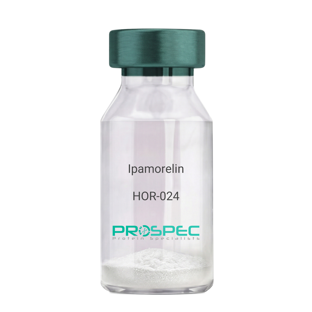
Name :
IpamorelinDescription:
Ipamorelin
Ipamorelin
Product # :
HOR-024Price :
Quantity :
Shipping Method :
Shipped at Room temp
More Info
- description
- formulation
- purity
- More Info
- Citations
Description
Ipamorelin Synthetic is a single, non-glycosylated polypeptide chain containing 4 amino acids, having a molecular mass of 711.85 Dalton and a Molecular formula of C38H49N9O5.
Formulation
The protein was lyophilized with no additives.
Purity
Greater than 97.0% as determined by analysis by RP-HPLC.
Citations
More Info
-
Introduction
Ipamorelin is a peptide selective agonist of the ghrelin/growth hormone secretagogue receptor and a growth hormone secretagogue. Ipamorelin is a pentapeptide that was derived from GHRP1. Ipamorelin significantly increases plasma growth hormone levels in both animals and humans. Like pralmorelin and GHRP-6, ipamorelin does not affect prolactin, FSH, LH or TSH levels. However, unlike GHRP2 and GHRP6, but as growth hormone-releasing hormone (GHRH), ipamorelin does not stimulate the secretion of adrenocorticotropic hormone (ACTH) or cortisol, and is highly selective for inducing the secretion only of GH.
-
Synonyms
Ipamorelin
-
Physical Appearance
Sterile Filtered White lyophilized (freeze-dried) powder.
-
Stability
Lyophilized Ipamorelin although stable at room temperature for 3 weeks, should be stored desiccated below -18°C. Upon reconstitution Ipamorelin should be stored at 4°C between 2-7 days and for future use below -18°C. For long term storage it is recommended to add a carrier protein (0.1% HSA or BSA). Please prevent freeze-thaw cycles.
-
Solubility
It is recommended to reconstitute the lyophilized Ipamorelin in sterile 18MΩ-cm H2O not less than 100 µg/ml, which can then be further diluted to other aqueous solutions.
-
Amino Acid Sequence
Aib-His-D-2-Nal-D-Phe-Lys-NH2.
ProSpec's products are furnished for LABORATORY RESEARCH USE ONLY. The product may not be used as drugs, agricultural or pesticidal products, food additives or household chemicals.
- View Data Sheet
Name :
CSTB AntibodyDescription:
Cystatin B, Mouse Anti Human
Cystatin-B, Stefin-B, Liver thiol proteinase inhibitor, CPI-B, CSTB, CST6, EPM1, PME, STFB.
Product # :
ANT-339Price :
Quantity :
Shipping Method :
Shipped with Ice Packs
More Info
- formulation
- More Info
- Citations
Formulation
1mg/ml containing PBS, pH-7.4, & 0.1% Sodium Azide.
Citations
More Info
-
Introduction
Type 1 cystatins are also called stefins which function as intracellular thiol protease inhibitors. Cystatin-B protein is able to form a dimer stabilized by noncovalent forces, inhibiting papain and cathepsins l, h and b. CSTB protein protects proteases leakage from lysosomes. Mutations in Stefin-B gene cause primary defects in patients with progressive myoclonic epilepsy (EPM1), a degenerative disease of the central nervous system. CSTB is overexpressed & elevated in the serum of HCC patients. Cystatin-B in vivo has a polymeric structure which is sensitive to the redox environment. Cystatin-B inhibits bone resorption by down-regulating intracellular cathepsin K activity despite increased osteoclast survival. Protein and mRNA levels of stefin B are significantly lower in atypical benign meningiomas. Stefins-A & Stefin-B which belong to the type-1 Cystatins, are up-regulated in lung tumours and thus able to counteract harmful tumour-associated proteolytic activity. Human stefin-A & Stef
-
Synonyms
Cystatin-B, Stefin-B, Liver thiol proteinase inhibitor, CPI-B, CSTB, CST6, EPM1, PME, STFB.
-
Immunogen
Anti-human CSTB mAb is derived from hybridization of mouse F0 myeloma cells with spleen cells from BALB/c mice immunized with recombinant human CSTB amino acids 1-98 purified from E. coli.
-
Ig Subclass
Mouse IgG2b heavy chain and κ light chain.
-
Clone
P2F1AT.
-
Applications
CSTB antibody has been tested by ELISA and Western blot analysis to assure specificity and reactivity. Since application varies, however, each investigation should be titrated by the reagent to obtain optimal results. Recommended dilution range for Western blot analysis is 1:1,000~ 2,000. Recommended starting dilution is 1:1,000.
-
Type
Mouse Anti Human Monoclonal.
-
Storage Procedures
For periods up to 1 month store at 4°C, for longer periods of time, store at -20°C. Prevent freeze thaw cycles.
-
Purification Method
CSTB antibody was purified from mouse ascitic fluids by protein-G affinity chromatography.
ProSpec's products are furnished for LABORATORY RESEARCH USE ONLY. The product may not be used as drugs, agricultural or pesticidal products, food additives or household chemicals.
- View Data Sheet
Name :
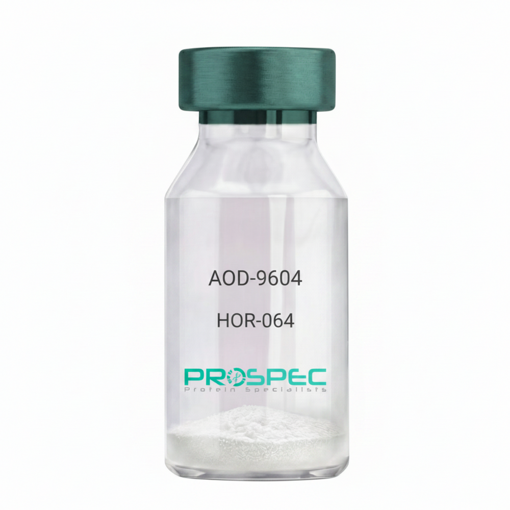
AOD-9604Description:
AOD-9604
Product # :
HOR-064Price :
Quantity :
Shipping Method :
Shipped at Room temp
More Info
- description
- formulation
- purity
- More Info
- Citations
Description
AOD-9604 is a synthetic single, non-glycosylated polypeptide chain containing 16 amino acids, having a molecular mass of 1815 Dalton and a Molecular formula of C78H123N23O23 S2.
Formulation
The protein was lyophilized with no additives.
Purity
Greater than 97.0% as determined by analysis by RP-HPLC.
Citations
More Info
-
Physical Appearance
Sterile Filtered White lyophilized (freeze-dried) powder.
-
Stability
Lyophilized AOD-9604 although stable at room temperature for 3 weeks, should be stored desiccated below -18°C. Upon reconstitution AOD-9604 should be stored at 4°C between 2-7 days and for future use below -18°C.
For long term storage it is recommended to add a carrier protein (0.1% HSA or BSA).
Please prevent freeze-thaw cycles.
-
Solubility
It is recommended to reconstitute the lyophilized AOD-9604 in sterile 18MΩ-cm H2O not less than 100 µg/ml, which can then be further diluted to other aqueous solutions.
-
Amino Acid Sequence
H-Tyr-Leu-Arg-Ile-Val-Gln-Cys-Arg-SerVal-Glu-Gly-Ser-Cys-Gly-Phe-OH (Disulfide bond).
-
Background
AOD-9604 is a synthetic analog of the C-terminal fragment (177–191) AOD-9604 stimulates lipolysis (fat breakdown) without the unwanted growth-promoting or insulin-antagonizing effects of full-length hGH. AOD-9604 is a potent lipolytic tool in animal models and researchers focus on its regenerative properties in joint health and its ability to modulate beta3 adrenergic receptors without the systemic risks of growth hormone.
ProSpec's products are furnished for LABORATORY RESEARCH USE ONLY. The product may not be used as drugs, agricultural or pesticidal products, food additives or household chemicals.
- View Data Sheet
Name :
TREM2 AntibodyDescription:
Triggering receptor expressed on myeloid cells 2, Mouse Anti Human
Triggering receptor expressed on myeloid cells 2, Triggering receptor expressed on monocytes 2, TREM-2, TREM2, Trem2a, Trem2b, Trem2c.
Product # :
ANT-429Price :
Quantity :
Shipping Method :
Shipped with Ice Packs
More Info
- formulation
- More Info
- Citations
Formulation
1mg/ml containing PBS, pH-7.4, & 0.1% Sodium Azide.
Citations
More Info
-
Introduction
TREM2 is a membrane protein that forms a receptor signaling complex with TYROBP. TREM2 may be involved in chronic inflammation by triggering the production of constitutive inflammatory cytokines. Defects in the TREM2 gene are a source of polycystic lipomembranous osteodysplasia with sclerosing leukoencephalopathy (PLOSL).
-
Synonyms
Triggering receptor expressed on myeloid cells 2, Triggering receptor expressed on monocytes 2, TREM-2, TREM2, Trem2a, Trem2b, Trem2c.
-
Immunogen
Anti-human TREM2 mAb, is derived from hybridization of mouse F0 myeloma cells with spleen cells from BALB/c mice immunized with recombinant human TREM2 amino acids 19-161 purified from E. coli.
-
Ig Subclass
Mouse IgG3 heavy chain and κ light chain.
-
Clone
P2B5AT.
-
Applications
TREM2 antibody has been tested by ELISA and Western blot analysis to assure specificity and reactivity. Since application varies, however, each investigation should be titrated by the reagent to obtain optimal results. Recommended dilution range for Western blot analysis is 1:500 ~ 1,000.Recommended starting dilution is 1:500.
-
Type
Mouse Anti Human Monoclonal.
-
Storage Procedures
For periods up to 1 month store at 4°C, for longer periods of time, store at -20°C. Prevent freeze thaw cycles.
-
Purification Method
TREM2 antibody was purified from mouse ascitic fluids by protein-G affinity chromatography.
ProSpec's products are furnished for LABORATORY RESEARCH USE ONLY. The product may not be used as drugs, agricultural or pesticidal products, food additives or household chemicals.
- View Data Sheet
Name :
LysostaphinDescription:
Lysostaphin Recombinant
Lysostaphin, EC 3.4.24.75, Glycyl-glycine endopeptidase.
Product # :
ENZ-269Price :
Quantity :
Shipping Method :
Shipped at Room temp
More Info
- description
- source
- formulation
- purity
- biological activity
- More Info
- Citations
Description
Lysostaphin Recombinant produced in E.Coli is a non-glycosylated polypeptide chain having a molecular mass of 26.92 kDa.
Source
Escherichia Coli.
Formulation
The protein was lyophilized without any additives.
Purity
98% as determined by RP-HPLC.
Biological Activity
Assessed by the decrease in turbidity of a suspension of heat-killed Staphylococcus aureus at pH-8, 30°C. Lysostaphin is a zinc enzyme therefore EDTA is an inhibitory factor.
Citations
More Info
-
Introduction
Lysostaphin, an endopeptidase specific for the cell wall peptidoglycan of staphylococci, is an extremely potent anti-staphylococcal agent. Lysostaphin is used as a research and diagnostic tool. Because it lyses staphylococci efficiently, it is widely used when preparing staphylococcal DNA or other cellular components for genetic and biochemical studies and for the preparation of protoplasts for transformation. Preparation and analysis of bacterial DNA has become a powerful tool used by clinical and other microbiologists in epidemiological studies aimed at tracing sources of infection or bacterial contamination.
-
Synonyms
Lysostaphin, EC 3.4.24.75, Glycyl-glycine endopeptidase.
-
Physical Appearance
Sterile Filtered lyophilized powder.
-
Stability
Lyophilized Lysostaphin although stable at room temperature for 2 weeks, should be stored desiccated below -18°C. Upon reconstitution Lysostaphin can be stored at 4°C up to 3 weeks and for future use below -18°C.Please prevent freeze-thaw cycles.
-
Solubility
It is recommended to reconstitute the lyophilized Lysostaphin in 20mM sodium acetate, pH 4.5 for optimal stability, which can then be further diluted.
-
Protein content
Protein quantitation was carried out by two independent methods 1. UV spectroscopy at 280 nm using the absorbency value of 2.02 as the extinction coefficient for a 0.1% (1mg/ml) solution. This value is calculated by the PC GENE computer analysis program of protein sequences (IntelliGenetics). 2. Analysis RP-HPLC, using a calibrated solution of Lysostaphin as a Reference standard.
-
Specific Activity
Determined to be 3,540 units/mg.
ProSpec's products are furnished for LABORATORY RESEARCH USE ONLY. The product may not be used as drugs, agricultural or pesticidal products, food additives or household chemicals.
- View Data Sheet
Name :
ST13 PAT5C6AT AntibodyDescription:
HSP70 Interacting Protein Clone PAT5C6AT, Mouse Anti Human
AAG2, SNC6, HSPABP, FAM10A1, FAM10A4, HSPABP1, ST-13, Hsc70-interacting protein, Suppression of tumorigenicity protein 13, Putative tumor suppressor ST13, Protein FAM10A1, Progesterone receptor-associated p48 protein, Renal carcinoma antigen NY-REN-33, ST13, HIP, HOP, P48, PRO0786, FLJ27260, MGC129952.
Product # :
ANT-536Price :
Quantity :
Shipping Method :
Shipped with Ice Packs
More Info
- formulation
- More Info
- Citations
Formulation
1mg/ml containing PBS, pH-7.4, 10% Glycerol and 0.02% Sodium Azide.
Citations
More Info
-
Introduction
ST13 is an adaptor protein (co-chaperone) that mediates the association of HSP70 & HSP90 and appears in early receptor complexes. ST13 plays a role in the assembly process of glucocorticoid receptor, which requires the assistance of multiple molecular chaperones. The expression of ST13 is downregulated in colorectal carcinoma tissue signifying that is candidate tumor suppressor gene. Through common binding to both Hsp70 and Hsp90, ST13 functions as an adaptor that can integrate Hsp70 and Hsp90 interactions. The expression of ST13 decreases in colorectal cancer tissue compared with that in adjacent normal tissue. ST13 is mostly expressed in colorectal epithelia and adenocarcinoma cells. ST13 functions to promote the efficiency of glucocorticoid receptor maturation in cells. The expression levels of the ST13 gene were significantly decreased in primary tumors compared with adjacent mucosa.
-
Synonyms
AAG2, SNC6, HSPABP, FAM10A1, FAM10A4, HSPABP1, ST-13, Hsc70-interacting protein, Suppression of tumorigenicity protein 13, Putative tumor suppressor ST13, Protein FAM10A1, Progesterone receptor-associated p48 protein, Renal carcinoma antigen NY-REN-33, ST13, HIP, HOP, P48, PRO0786, FLJ27260, MGC129952.
-
Physical Appearance
Sterile filtered colorless solution.
-
Immunogen
Anti-human ST13 mAb, is derived from hybridization of mouse F0 myeloma cells with spleen cells from BALB/c mice immunized with a recombinant human ST13 protein 1-369 amino acids purified from E. coli.
-
Ig Subclass
Mouse IgG2a heavy chain and k light chain.
-
Clone
PAT5C6AT.
-
Applications
ST13 antibody has been tested by ELISA, Western blot analysis to assure specificity and reactivity. Since application varies, however, each investigation should be titrated by the reagent to obtain optimal results. Recommended starting dilution is 1:1000.
-
Type
Mouse Anti Human Monoclonal.
-
Storage Procedures
For periods up to 1 month store at 4°C, for longer periods of time, store at -20°C. Prevent freeze thaw cycles.
-
Purification Method
ST13 antibody was purified from mouse ascitic fluids by protein-A affinity chromatography.
ProSpec's products are furnished for LABORATORY RESEARCH USE ONLY. The product may not be used as drugs, agricultural or pesticidal products, food additives or household chemicals.
- View Data Sheet
Name :
Streptavidin AntibodyDescription:
Streptavidin, Mouse Antibody
Product # :
ANT-345Price :
Quantity :
Shipping Method :
Shipped with Ice Packs
More Info
- formulation
- More Info
- Citations
Formulation
1mg/ml containing PBS, pH-7.4, 0.02% Sodium Azide and 10% glycerol.
Citations
More Info
-
Introduction
Streptavidin is a tetrameric protein secreted by Streptomyces avidinii which binds firmly to biotin. Streptavidin is wilyde used in molecular biology through its unique high affinity for the vitamin biotin. The dissociation constant (Kd) of the biotin-streptavidin complex is about ~10-15 mol/L. The strong affinity recognition of biotin and biotinylated molecules has made streptavidin one of the most important components in diagnostics and laboratory kits. The streptavidin/biotin system has one of the biggest free energies of association of yet observed for noncovalent binding of a protein and small ligand in aqueous solution (K_assoc = 10**14). The complexes are also extremely stable over a wide range of temperature and pH.
-
Physical Appearance
Sterile Filtered clear solution.
-
Immunogen
Anti Streptavidin mAb is derived from hybridization of mouse F0 myeloma cells with spleen cells from BALB/c mice immunized with recombinant Streptavidin amino acids 25-183 purified from E. coli.
-
Ig Subclass
Mouse IgG2b heavy chain and κ light chain.
-
Clone
P1C2AT.
-
Applications
Streptavidin antibody has been tested by ELISA and Western blot analysis to assure specificity and reactivity. Since application varies, however, each investigation should be titrated by the reagent to obtain optimal results.
-
Type
Mouse Antibody Monoclonal.
-
Storage Procedures
For periods up to 1 month store at 4°C, for longer periods of time, store at -20°C. Prevent freeze thaw cycles.
-
Purification Method
Streptavidin antibody was purified from mouse ascitic fluids by protein-G affinity chromatography.
ProSpec's products are furnished for LABORATORY RESEARCH USE ONLY. The product may not be used as drugs, agricultural or pesticidal products, food additives or household chemicals.
- View Data Sheet
Name :
HIV-1 p24, BiotinDescription:
HIV-1 p24 Recombinant, Biotin Labeled
Product # :
HIV-106Price :
Quantity :
Shipping Method :
Shipped with Ice Packs
More Info
- description
- source
- formulation
- purity
- More Info
- Citations
Description
The E.coli derived 39 kDa biotin labeled recombinant protein is a non-glycosylated polypeptide chain, containing the HIV-1 p24 immunodominant regions, amino acids 77-436.The HIV-1 p24 Biotinylated is fused to beta-galactosidase (114 kDa) at the N-terminus.
Source
Escherichia Coli.
Formulation
8M Urea, 20mM Tris-HCl pH 8.0, 10mM b-mercaptoethanol.
Purity
Greater than 95.0% as determined by SDS-PAGE.
Citations
More Info
-
Introduction
Human immunodeficiency virus (HIV) is a retrovirusthat can lead to a condition in which the immune systembegins to fail, leading to opportunistic infections. HIV primarily infects vital cells in the humanimmune systemsuch as helper T cells(specifically CD4+ T cells), macrophagesand dendritic cells. HIV infection leads to low levels of CD4+ T cells through three main mechanisms: firstly, direct viral killing of infected cells; secondly, increased rates of apoptosisin infected cells; and thirdly, killing of infected CD4+ T cells by CD8 cytotoxic lymphocytesthat recognize infected cells. When CD4+ T cell numbers decline below a critical level, cell-mediated immunityis lost, and the body becomes progressively more susceptible to opportunistic infections. HIV was classified as a member of the genus Lentivirus, part of the family of Retroviridae. Lentiviruses have many common morphologies and biological properties. Many species are infected by lentiviruses, which are characteristically responsible for long-duration illnesses with a long incubation period. Lentiviruses are transmitted as single-stranded, positive-sense, enveloped RNA viruses. Upon entry of the target cell, the viral RNA genomeis converted to double-stranded DNAby a virally encoded reverse transcriptasethat is present in the virus particle. This viral DNA is then integrated into the cellular DNA by a virally encoded integraseso that the genome can be transcribed. Once the virus has infected the cell, two pathways are possible: either the virus becomes latentand the infected cell continues to function, or the virus becomes active and replicates, and a large number of virus particles are liberated that can then infect other cells.
-
Physical Appearance
Sterile filtered colorless clear solution.
-
Stability
HIV-1 p24 although stable at 4°C for 1 week, should be stored below -18°C. Please prevent freeze thaw cycles.
-
Applications
HIV-1 p24 antigen is suitable for ELISA and Western blots, excellent antigen for early detection of HIV seroconvertors with minimal specificity problems.
-
Specificity
Immunoreactive with all sera of HIV-1 infected individuals.
ProSpec's products are furnished for LABORATORY RESEARCH USE ONLY. The product may not be used as drugs, agricultural or pesticidal products, food additives or household chemicals.
- View Data Sheet
Name :
REXO1 HumanDescription:
RNA Exonuclease 1 Human Recombinant
RNA exonuclease 1 homolog, Elongin-A-binding protein 1, EloA-BP1, Transcription elongation factor B polypeptide 3-binding protein 1, REXO1, ELOABP1, KIAA1138, TCEB3BP1.
Product # :
ENZ-767Price :
Quantity :
Shipping Method :
Shipped with Ice Packs
More Info
- description
- source
- formulation
- purity
- More Info
- Citations
Description
REXO1 Human Recombinant produced in E.coli is a single, non-glycosylated polypeptide chain containing 198 amino acids (1060-1221a.a) and having a molecular mass of 22.3kDa. REXO1 is fused to a 36 amino acid His-tag at N-terminus & purified by proprietary chromatographic techniques.
Source
Escherichia Coli.
Formulation
The REXO1 solution (0.25mg/ml) contains 20mM Tris-HCl buffer (pH 8.0), 0.15M NaCl, 10% glycerol and 1mM DTT.
Purity
Greater than 90% as determined by SDS-PAGE.
Citations
More Info
-
Introduction
REXO1 is an exonuclease domain-containing protein which binds to Elongin. The Elongin complex stimulates the rate of transcription elongation by RNA polymerase II by suppressing the transient pausing of the polymerase at many sites along the DNA template. REXO1 is composed of 1221 amino acids and its mRNA is ubiquitously expressed. REXO1 is a putative stem cell marker and is highly expressed in embryonic and adult stem cells.
-
Synonyms
RNA exonuclease 1 homolog, Elongin-A-binding protein 1, EloA-BP1, Transcription elongation factor B polypeptide 3-binding protein 1, REXO1, ELOABP1, KIAA1138, TCEB3BP1.
-
Physical Appearance
Sterile Filtered colorless solution.
-
Stability
Store at 4°C if entire vial will be used within 2-4 weeks. Store, frozen at -20°C for longer periods of time. For long term storage it is recommended to add a carrier protein (0.1% HSA or BSA). Avoid multiple freeze-thaw cycles.
-
Amino Acid Sequence
MRGSHHHHHH GMASMTGGQQ MGRDLYDDDD KDRWGSIYAL DCEMSYTTYG LELTRVTVVD TDVHVVYDTF VKPDNEIVDY NTRFSGVTEA DLADTSVTLR DVQAVLLSMF SADTILIGHS LESDLLALKV IHSTVVDTSV LFPHRLGLPY KRSLRNLMAD YLRQIIQDNV DGHSSSEDAG ACMHLVIWKV REDAKTKR.
ProSpec's products are furnished for LABORATORY RESEARCH USE ONLY. The product may not be used as drugs, agricultural or pesticidal products, food additives or household chemicals.
- View Data Sheet
Name :
VEGF HumanDescription:
Vascular Endothelial Growth Factor Human Recombinant
Vascular endothelial growth factor A, VEGF-A, Vascular permeability factor, VPF, VEGF, MGC70609.
Product # :
CYT-241Price :
Quantity :
Shipping Method :
Shipped at Room temp
More Info
- description
- source
- formulation
- purity
- biological activity
- More Info
- Citations
Description
Vascular Endothelial Growth Factor Human Recombinant produced in E.Coli is a double, non-glycosylated, polypeptide chain containing 165 amino acids and having a molecular mass of 38.2kDa.The VEGF is purified by proprietary chromatographic techniques.
Source
Escherichia Coli.
Formulation
The VEGF protein was lyophilized from a concentrated (1mg/ml) solution with no additives.
Purity
Greater than 95.0% as determined by:
(a) Analysis by RP-HPLC.
(b) Analysis by SDS-PAGE.Biological Activity
Determined by the dose-dependent stimulation of the proliferation of human umbilical vein endothelial cells (HUVEC) using a concentration range of 3.7-5.6 ng/ml, corresponding to a Specific Activity of 178,570-270,270IU/mg.Citations
More Info
-
Introduction
Vascular endothelial growth factor (VEGF) is an important signaling protein involved in vessel formation As its name implies, VEGF activity has been mostly studied on cells of the vascular endothelium, although it does have effects on a number of other cell types (e.g. stimulation monocyte/macrophage migration, neurons, cancer cells, kidney epithelial cells ). VEGF mediates increased vascular permeability, induces vasculogenesis and endothelial cell production, promotes cell migration, and inhibits apoptosis. In vitro, VEGF has been shown to stimulate endothelial cell mitogenesis and cell migration. VEGF is also a vasodilator and increases microvascular permeability and was originally referred to as vascular permeability factor. VEGF is located in normal cartilage though only osteoarthritic cartilage expresses the VEGF receptors, NP1, VEGFR1 and VEGFR2. The VEGF level in the culture media from OA chondrocytes was more than 3 folds higher than in media from normal chondrocytes
-
Synonyms
Vascular endothelial growth factor A, VEGF-A, Vascular permeability factor, VPF, VEGF, MGC70609.
-
Physical Appearance
Sterile Filtered White lyophilized (freeze-dried) powder.
-
Stability
Lyophilized VEGF although stable at room temperature for 3 weeks, should be stored desiccated below -18°C. Upon reconstitution VEGF should be stored at 4°C between 2-7 days and for future use below -18°C.For long term storage it is recommended to add a carrier protein (0.1% HSA or BSA).Please prevent freeze-thaw cycles.
-
Solubility
It is recommended to reconstitute the lyophilized VEGF in sterile 18M-cm H2O not less than 100µg/ml, which can then be further diluted to other aqueous solutions.
-
Amino Acid Sequence
APMAEGGGQN HHEVVKFMDV YQRSYCHPIE TLVDIFQEYP DEIEYIFKPS CVPLMRCGGC CNDEGLECVP TEESNITMQI MRIKPHQGQH IGEMSFLQHN KCECRPKKDR ARQENPCGPC SERRKHLFVQ DPQTCKCSCK NTDSRCKARQ LELNERTCRC DKPRR
-
Protein content
VEGF protein quantitation was carried out by two independent methods:1. UV spectroscopy at 280 nm using the absorbency value of 0.2875 as the extinction coefficient for a 0.1% (1mg/ml) solution. This value is calculated by the PC GENE computer analysis program of protein sequences (IntelliGenetics). 2. Analysis by RP-HPLC, using a calibrated solution of VEGF as a Reference Standard.
ProSpec's products are furnished for LABORATORY RESEARCH USE ONLY. The product may not be used as drugs, agricultural or pesticidal products, food additives or household chemicals.
- View Data Sheet
Name :
MALD3Description:
Non-Specific Lipid-Transfer Protein Mal d 3 Recombinant
Non-specific lipid-transfer protein, LTP, MALD3.
Product # :
ALR-023Price :
Quantity :
Shipping Method :
Shipped with Ice Packs
More Info
- description
- source
- formulation
- purity
- More Info
- Citations
Description
Recombinant Non-Specific Lipid-Transfer Protein Mal d 3 produced in SF9 is a glycosylated, polypeptide chain having a calculated molecular mass of 12kDa.MALD3 is expressed with a 6xHis tag and purified by proprietary chromatographic techniques.
Source
Sf9 insect cells.
Formulation
MALD3 is supplied in 20mM HEPES buffer pH-8.0, 200mM NaCl and 20% glycerol.
Purity
Greater than 80.0% as determined by SDS-PAGE.
Citations
More Info
-
Introduction
MALD3 (a non-specific lipid transfer protein) is a major apple allergen. LTPs are found mainly in aerial plant organs such as leaves, seeds, flowers, and fruit. MALD3 is a heat-stable protein which is characterized by having resistance to pepsin hydrolysis.
-
Synonyms
Non-specific lipid-transfer protein, LTP, MALD3.
-
Physical Appearance
Sterile Filtered colorless solution.
-
Stability
Store at 4°C if entire vial will be used within 2-4 weeks.Store, frozen at -20°C for longer periods of time.Avoid multiple freeze-thaw cycles.
-
Immunological Functions
1. Binds IgE-type human antibodies.2. Immunodot test with positive/negative samples
-
Applications
Tested by LAL (Limulus Amoebocyte Lysate) chromogenic endotoxin assay.
ProSpec's products are furnished for LABORATORY RESEARCH USE ONLY. The product may not be used as drugs, agricultural or pesticidal products, food additives or household chemicals.
- View Data Sheet
Name :
HIV-2 gp32, BiotinDescription:
HIV-2 gp32 Recombinant, Biotin Labeled
Product # :
HIV-135Price :
Quantity :
Shipping Method :
Shipped with Ice Packs
More Info
- description
- source
- formulation
- purity
- More Info
- Citations
Description
HIV-2 gp32 Biotin Labeled recombinant- contains the full-length sequence of HIV-2 envelope immunodominant regions gp32 having a Mw of 32kDa and fused to a beta-galactosidase at N-terminus.
Source
Escherichia Coli.
Formulation
0.01M Na2CO3, 10mM EDTA, 14mM beta-ME and 0.02% Sarcosyl.
Purity
Greater than 95.0% as determined by SDS-PAGE.
Citations
More Info
-
Introduction
HIV-1 and HIV-2 appear to package their RNA differently. HIV-1 binds to any appropriate RNA whereas HIV-2 preferentially binds to mRNA which creates the Gag protein itself. This means that HIV-1 is better able to mutate. HIV-2 is transmitted in the same ways as HIV-1: Through exposure to bodily fluids such as blood, semen, tears and vaginal fluids.
Immunodeficiency develops more slowly with HIV-2.
HIV-2 is less infectious in the early stages of the virus than with HIV-1.
The infectiousness of HIV-2 increases as the virus progresses.
Major differences include reduced pathogenicity of HIV-2 relative to HIV-1, enhanced immune control of HIV-2 infection and often some degree of CD4-independence. Despite considerable sequence and phenotypic differences between HIV-1 and 2 envelopes, structurally they are quite similar. Both membrane-anchored proteins eventually form the 6-helix bundles from the N-terminal and C-terminal regions of the ectodomain, which is common to many viral and cellular fusion proteins and which seems to drive fusion.
HIV-1 gp41 helical regions can form more stable 6-helix bundles than HIV-2 gp41 helical regions however HIV-2 fusion occurs at a lower threshold temperature (25°C), does not require Ca2+ in the medium, is insensitive to treatment of target cells with cytochalasin B, and is not affected by target membrane glycosphingolipid composition. -
Physical Appearance
Sterile filtered colorless clear solution.
-
Stability
HIV-2 gp-32 although stable at room temperature for 3 weeks, should be stored at 4°C.
ProSpec's products are furnished for LABORATORY RESEARCH USE ONLY. The product may not be used as drugs, agricultural or pesticidal products, food additives or household chemicals.
- View Data Sheet
Name :
TerlipressinDescription:
Terlipressin
Product # :
HOR-287Price :
Quantity :
Shipping Method :
Shipped at Room temp
More Info
- description
- formulation
- purity
- More Info
- Citations
Description
Terlipressin contains 12 amino acids Gly-Gly-Gly-c[Cys-Tyr-Phe-Gln-Asn-Cys]-Pro-Lys-Gly-NH2 and having a molecular weight of 1227.37 Dalton.
Formulation
The protein (1 mg/ml) was lyophilized with no additives.
Purity
Greater than 98.0% as determined by:
(a) Analysis by RP-HPLC.
(b) Analysis by SDS-PAGE.Citations
More Info
-
Introduction
Terlipressin is similar to a naturally occurring hormone present in the body, known as antidiuretic hormone (ADH) or vasopressin. ADH has two main effects in the body. Firstly, it causes narrowing of blood vessels (vasoconstriction), thereby limiting blood flow to a particular area of the body. It also acts on receptors in the kidney to retain water in the body, which helps to prevent excessive loss of water in the urine. Terlipressin is commonly used to stop bleeding of varices in the food pipe (oesophagus). Varices are fragile distended veins that can occur in various parts of the body such as the oesophagus. This is caused by an increase in blood pressure in certain diseases such as severe liver disease. These fragile varices can rupture and lead to life threatening bleeding. Terlipressin is therefore given to narrow blood vessels, and so restricting blood flow to the varices and stopping the bleeding.
-
Physical Appearance
Sterile Filtered White lyophilized (freeze-dried) powder.
-
Stability
Lyophilized Terlipressin although stable at room temperature for 3 weeks, should be stored desiccated below -18°C. Upon reconstitution Terlipressin should be stored at 4°C between 2-7 days and for future use below -18°C.For long term storage it is recommended to add a carrier protein (0.1% HSA or BSA).Please prevent freeze-thaw cycles.
-
Solubility
It is recommended to reconstitute the lyophilized Terlipressin18MΩ-cm H2O not less than 100 µg/ml, which can then be further diluted to other aqueous solutions.
ProSpec's products are furnished for LABORATORY RESEARCH USE ONLY. The product may not be used as drugs, agricultural or pesticidal products, food additives or household chemicals.
- View Data Sheet
Name :
DIRAS1 HumanDescription:
DIRAS family, GTP-binding RAS-like 1 Human Recombinant
GTP-binding protein Di-Ras1, Distinct subgroup of the Ras family member 1, Ras-related inhibitor of cell growth, Rig, Small GTP-binding tumor suppressor 1, DIRAS1, GBTS1, Di-Ras1, FLJ42681.
Product # :
PRO-256Price :
Quantity :
Shipping Method :
Shipped with Ice Packs
More Info
- description
- source
- formulation
- purity
- More Info
- Citations
Description
DIRAS1 Human Recombinant produced in E.Coli is a single, non-glycosylated, polypeptide chain containing 215 amino acids (1-195a.a.) and having a molecular mass of 24.1 kDa. DIRAS1 protein is fused to a 20 amino acid His tag at N-terminus and is purified by standard chromatography.
Source
Escherichia Coli.
Formulation
DIRAS1 Human solution (1mg/ml) containing 20mM Tris-HCl buffer(pH 8.0) containing 20% glycerol, 1mM DTT, 0.1M NaCl and 1mM EDTA.
Purity
Greater than 95% as determined by SDS-PAGE.
Citations
More Info
-
Introduction
DIRAS1 is a member of a distinct branch of the functionally diverse Ras superfamily of monomeric GTPases. DIRAS1 demonstrates low GTPase activity and exists mainly in the GTP-bound form. Ras proteins operate as binary molecular switches that control intracellular signaling networks. Ras-regulated signal pathways regulate such processes as actin cytoskeletal integrity, proliferation, differentiation, cell adhesion, apoptosis, and cell migration. Ras and ras-related proteins are frequently deregulated in cancers, leading to increased invasion and metastasis, and decreased apoptosis.
-
Synonyms
GTP-binding protein Di-Ras1, Distinct subgroup of the Ras family member 1, Ras-related inhibitor of cell growth, Rig, Small GTP-binding tumor suppressor 1, DIRAS1, GBTS1, Di-Ras1, FLJ42681.
-
Physical Appearance
Sterile filtered colorless solution.
-
Stability
Store at 4°C if entire vial will be used within 2-4 weeks. Store, frozen at -20°C for longer periods of time. For long term storage it is recommended to add a carrier protein (0.1% HSA or BSA).Avoid multiple freeze-thaw cycles.
-
Amino Acid Sequence
MGSSHHHHHH SSGLVPRGSH MPEQSNDYRV VVFGAGGVGK SSLVLRFVKG TFRDTYIPTI EDTYRQVISC DKSVCTLQIT DTTGSHQFPA MQRLSISKGH AFILVFSVTS KQSLEELGPI YKLIVQIKGS VEDIPVMLVG KCDETQREV DTREAQAVAQ EWKCAFMETS AKMNYNVKEL FQELLTLETR RNMSLNIDGK RSGKQKRTDR KGKC.
ProSpec's products are furnished for LABORATORY RESEARCH USE ONLY. The product may not be used as drugs, agricultural or pesticidal products, food additives or household chemicals.
- View Data Sheet
Name :
CRP RatDescription:
C-Reactive Protein Rat Recombinant
Ptx1, C-reactive protein, Pentraxin 1, C-Reactive Protein Pentraxin-Related.
Product # :
PRO-1421Price :
Quantity :
Shipping Method :
Shipped with Ice Packs
More Info
- description
- source
- formulation
- purity
- More Info
- Citations
Description
CRP produced in Sf9 Insect cells is a single, glycosylated polypeptide chain containing 217 amino acids (20-230 a.a.) and having a molecular mass of 24.1kDa. (Molecular size on SDS-PAGE will appear at approximately 28-40 kDa).CRP is expressed with a 6 amino acid His tag at C-Terminus and purified by proprietary chromatographic techniques.
Source
Sf9, Insect cells.
Formulation
CRP protein solution (0.5mg/ml) contains Phosphate Buffered Saline (pH 7.4) and 10% glycerol.
Purity
Greater than 95.0% as determined by SDS-PAGE.
Citations
More Info
-
Introduction
CRP is an acute phase protein that correlates with inflammatory disease and is synthesized by hepatocytes during the acute phase response by certain cytokines (IL-1 and TNF Alpha and Beta). CRP levels increase dramatically (up to 1,000 fold) and serve as a useful marker of inflammation in such conditions as bacterial infection, rheumatoid arthritis, viral infections, transplantation rejection, meningitis, myocardial infarction, septicemia, osteomyelitis and others. CRP is also highly correlated to Serum Amyloid A levels.
-
Synonyms
Ptx1, C-reactive protein, Pentraxin 1, C-Reactive Protein Pentraxin-Related.
-
Physical Appearance
Sterile filtered colorless solution.
-
Stability
Store at 4°C if entire vial will be used within 2-4 weeks.Store, frozen at -20°C for longer periods of time. For long term storage it is recommended to add a carrier protein (0.1% HSA or BSA).Avoid multiple freeze-thaw cycles.
-
Amino Acid Sequence
HEDMSKQAFV FPGVSATAYV SLEAESKKPL EAFTVCLYAH ADVSRSFSIF SYATKTSFNE ILLFWTRGQG FSIAVGGPEI LFSASEIPEV PTHICATWES ATGIVELWLD GKPRVRKSLQ KGYIVGTNAS IILGQEQDSY GGGFDANQSL VGDIGDVNMW DFVLSPEQIN AVYVGRVFSP NVLNWRALKY ETHGDVFIKP QLWPLTDCCE SHHHHHH.
ProSpec's products are furnished for LABORATORY RESEARCH USE ONLY. The product may not be used as drugs, agricultural or pesticidal products, food additives or household chemicals.
- View Data Sheet
Name :
Betacellulin His HumanDescription:
Betacellulin Human Recombinant, His Tag
Betacellulin, Probetacellulin.
Product # :
CYT-077Price :
Quantity :
Shipping Method :
Shipped with Ice Packs
More Info
- description
- source
- formulation
- purity
- More Info
- sds-page
- Citations
Description
BTC Human Recombinant produced in E.coli is a single, non-glycosylated polypeptide chain containing 101 amino acids (32-111) and having a molecular mass of 11.3 kDa.BTC is fused to a 21 amino acid His-tag at N-terminus & purified by proprietary chromatographic techniques.
Source
E.coli.
Formulation
The BTC solution (0.25mg/ml) contains 20mM Tris-HCl buffer (pH 8.0), 5mM DTT, 0.2M NaCl and 20% glycerol.
Purity
Greater than 90% as determined by SDS-PAGE.
sds-page
Citations
More Info
-
Introduction
Btc is a potent mitogen for retinal pigment epithelial cells and vascular smooth muscle cells. The effects of betacellulin are probably mediated by the egf receptor and other related receptors.
-
Synonyms
Betacellulin, Probetacellulin.
-
Physical Appearance
Sterile Filtered colorless solution.
-
Stability
Store at 4°C if entire vial will be used within 2-4 weeks. Store, frozen at -20°C for longer periods of time. For long term storage it is recommended to add a carrier protein (0.1% HSA or BSA).Avoid multiple freeze-thaw cycles.
-
Amino Acid Sequence
MGSSHHHHHH SSGLVPRGSH MDGNSTRSPE TNGLLCGDPE ENCAATTTQS KRKGHFSRCP KQYKHYCIKG RCRFVVAEQT PSCVCDEGYI GARCERVDLF Y
-
Background
What is the molecular weight/Mw of BETACELLULIN Protein?
BETACELLULIN Protein has a total Mw of 11.3kDa.
What is the source or expression system of BETACELLULIN Protein?
Escherichia Coli.
What is the Purity of BETACELLULIN Protein?
BETACELLULIN Protein is >90% pure as determined by SDS-PAGE.
What is the Biological Activity of BETACELLULIN Protein?
The biological functionality of BETACELLULIN Protein will be determined in the future.
What is the amino acid sequence of BETACELLULIN Protein?
MGSSHHHHHH SSGLVPRGSH MDGNSTRSPE TNGLLCGDPE ENCAATTTQS KRKGHFSRCP KQYKHYCIKG RCRFVVAEQT PSCVCDEGYI GARCERVDLF Y
What applications can BETACELLULIN Protein be used in?
BETACELLULIN Protein can probably be used in western blot, ELISA and Lateral Flow.
What is the endotoxin level for BETACELLULIN Protein?
The endotoxin level is minimal, BETACELLULIN Protein was purified using conventional chromatography techniques.
ProSpec's products are furnished for LABORATORY RESEARCH USE ONLY. The product may not be used as drugs, agricultural or pesticidal products, food additives or household chemicals.
- View Data Sheet
Name :
RDH12 HumanDescription:
Retinol Dehydrogenase 12 Human Recombinant
Retinol dehydrogenase 12 (all-trans/9-cis/11-cis), LCA3, LCA13, SDR7C2, All-trans and 9-cis retinol dehydrogenase, short chain dehydrogenase/reductase family 7C, member 2, FLJ30273, EC 1.1.1.100.
Product # :
ENZ-233Price :
Quantity :
Shipping Method :
Shipped with Ice Packs
More Info
- description
- source
- formulation
- purity
- More Info
- Citations
Description
RDH12 Human Recombinant produced in E.coli is a single, non-glycosylated polypeptide chain containing 303 amino acids (39-316) and having a molecular mass of 33.5kDa.RDH12 is fused to a 25 amino acid His-tag at N-terminus & purified by proprietary chromatographic techniques.
Source
E.coli.
Formulation
The RDH12 solution (0.25mg/ml) contains 20mM Tris-HCl buffer (pH 8.0), 200mM NaCl, 2mM DTT and 40% glycerol.
Purity
Greater than 90% as determined by SDS-PAGE.
Citations
More Info
-
Introduction
RDH12 is a member of the short-chain Dehydrogenases / Reductases (SDR) family. RDH12 is found generally in brain, stomach, eye, skeletal muscle and kidney. RDH12 is a NADPH-dependent retinal reductase whose main activity is toward 9-cis and all-trans-retinol. In addition, RDH12 takes part in the metabolism of short-chain aldehydes but does not display steroid dehydrogenase activity.
-
Synonyms
Retinol dehydrogenase 12 (all-trans/9-cis/11-cis), LCA3, LCA13, SDR7C2, All-trans and 9-cis retinol dehydrogenase, short chain dehydrogenase/reductase family 7C, member 2, FLJ30273, EC 1.1.1.100.
-
Physical Appearance
Sterile Filtered colorless solution.
-
Stability
Store at 4°C if entire vial will be used within 2-4 weeks. Store, frozen at -20°C for longer periods of time. For long term storage it is recommended to add a carrier protein (0.1% HSA or BSA).Avoid multiple freeze-thaw cycles.
-
Amino Acid Sequence
MGSSHHHHHH SSGLVPRGSH MGSHMGKVVV ITGANTGIGK ETARELASRG ARVYIACRDV LKGESAASEI RVDTKNSQVL VRKLDLSDTK SIRAFAEGFL AEEKQLHILI NNAGVMMCPY SKTADGFETH LGVNHLGHFL LTYLLLERLK VSAPARVVNV SSVAHHIGKI PFHDLQSEKR YSRGFAYCHS KLANVLFTRE LAKRLQGTGV TTYAVHPGVV RSELVRHSSL LCLLWRLFSP FVKTAREGAQ TSLHCALAEG LEPLSGKYFS DCKRTWVSPR ARNNKTAERL WNVSCELLGI RWE
ProSpec's products are furnished for LABORATORY RESEARCH USE ONLY. The product may not be used as drugs, agricultural or pesticidal products, food additives or household chemicals.
- View Data Sheet
Description:
Mouse Anti SARS Nucleocapsid Biotinylated
Product # :
ANT-764Price :
Quantity :
Shipping Method :
Shipped with Ice Packs
More Info
- description
- formulation
- purity
- More Info
- Citations
Description
SARS Nucleoprotein Biotinylated antibody clone PT3851 is specific for the nucleocapsid SARS-CoV protein and recognizes the nucleoprotein of SARS-CoV-2 by ELISA.
Formulation
1mg/ml in DPBS, 1% BSA and 0.1% Proclin 950.
Purity
Greater than 90% as determined by SDS-PAGE gel.
Citations
More Info
-
Introduction
SARS Coronavirus is an enveloped virus containing three outer structural proteins, namely the membrane (M), envelope (E), and spike (S) proteins. Spike (S)-glycoprotein of the virus interacts with a cellular receptor and mediates membrane fusion to allow viral entry into susceptible target cells. Accordingly, S-protein plays an important role in virus infection cycle and is the primary target of neutralizing antibodies.It has recently been shown that SARS is caused by a human coronavirus. Human coronaviruses are the major cause of upper respiratory tract illness in humans, such as the common cold. Coronaviruses are positive-stranded RNA viruses, featuring the largest viral RNA genomes known to date (27-31 kb). The first step in coronavirus infection is binding of the viral spike protein, a 139-kDa protein, to certain receptors on host cells. The spike protein is the main surface antigen of the coronavirus. The most prominent protein in the culture supernatants infected with SARS virus is a 46 kDa nucleocapsid protein. This suggests that the nucleocapsid protein is a major immunogen that may be useful for early diagnostics.
-
Physical Appearance
Sterile Filtered liquid formulation.
-
Stability
Store at 4°C, stable for 2 weeks. For long-term storage, store at -20°C.
-
Immunogen
Recombinant protein fragment 1-49 a.a. of the SARS nucleoprotein.
-
Clone
PT3851
-
Applications
ELISA.
-
Isotype
Mouse IgG2b.
-
Type
Mouse antibody Monoclonal.
-
Purification Method
Protein A affinity purified.
ProSpec's products are furnished for LABORATORY RESEARCH USE ONLY. The product may not be used as drugs, agricultural or pesticidal products, food additives or household chemicals.