Search results
1000 results found for “Mortality Factor”
Name
Description
Product #
Price
Quantity
Shipping Method
- View Data Sheet
Name :
IL 7 MouseDescription:
Interleukin-7 Mouse Recombinant
Lymphopoietin 1 (LP-1), pre-B cell factor, IL-7.
Product # :
CYT-372Price :
Quantity :
Shipping Method :
Shipped at Room temp
More Info
- description
- source
- formulation
- purity
- biological activity
- More Info
Description
Interleukin-7 Mouse Recombinant produced in E.Coli is a single, non-glycosylated polypeptide chain containing 129 amino acids and having a molecular mass of 14.9kDa.
Source
Escherichia Coli.
Formulation
Filtered (0.2µm) and lyophilized from a concentrated (1mg/ml) solution in 1×PBS, pH7.4 and 2% trehalose.
Purity
Greater than 96.0% as determined by:
(a) Analysis by RP-HPLC.
(b) Analysis by SDS-PAGE.Biological Activity
The ED50was determined by the dose-dependent stimulation of the proliferation of murine 2E8 cells is less than 0.2 ng/ml, corresponding to a specific activity of >5.0×10⁶ IU/mg.More Info
-
Introduction
Interleukin-7 is a potent lymphoid cell growth factor produced primarily by stromal cells. IL-7 has been shown to support the proliferation and differentiation of pre B- and early T cells as well as displaying a biological effect on cells of NK and myeloid lineages.
-
Synonyms
Lymphopoietin 1 (LP-1), pre-B cell factor, IL-7.
-
Physical Appearance
Sterile Filtered White lyophilized (freeze-dried) powder.
-
Stability
Lyophilized Interleukin-7 although stable at room temperature for 3 weeks, should be stored desiccated below -18°C. Upon reconstitution IL7 should be stored at 4°C between 2-7 days and for future use below -18°C.For long term storage it is recommended to add a carrier protein (0.1% HSA or BSA).Please prevent freeze-thaw cycles.
-
Solubility
It is recommended to reconstitute the lyophilized Interleukin 7 in sterile 18MΩ-cm H2O not less than 100µg/ml, which can then be further diluted to other aqueous solutions.
-
Amino Acid Sequence
ECHIKDKEGK AYESVLMISI DELDKMTGTD SNCPNNEPNF FRKHVCDDTK EAAFLNRAAR KLKQFLKMNI SEEFNVHLLT VSQGTQTLVN CTSKEEKNVK EQKKNDACFL KRLLREIKTC WNKILKGSI.
ProSpec's products are furnished for LABORATORY RESEARCH USE ONLY. The product may not be used as drugs, agricultural or pesticidal products, food additives or household chemicals.
- View Data Sheet
Name :
EIF2S1 AntibodyDescription:
Eukaryotic Translation Initiation Factor 2 Subunit 1 Alpha, Mouse Anti Human
Eukaryotic translation initiation factor 2 subunit 1, Eukaryotic translation initiation factor 2 subunit alpha, eIF-2-alpha, EIF-2alpha, EIF-2A, EIF2, EIF-2, EIF2A, EIF-2A.
Product # :
ANT-059Price :
Quantity :
Shipping Method :
Shipped with Ice Packs
More Info
- formulation
- More Info
Formulation
1mg/ml containing PBS, pH-7.4, & 0.1% Sodium Azide.
More Info
-
Introduction
EIF2S1 participates in the premature steps of protein synthesis by forming a ternary complex with GTP and initiator tRNA which binds to a 40S ribosomal subunit, followed by mRNA binding to create a 43S pre-initiation complex. Junction of the 60S ribosomal subunit to form the 80S initiation complex is preceded by hydrolysis of the GTP bound to eIF-2 and release of an eIF-2-GDP binary complex. In order for eIF-2 to recycle and catalyze another round of initiation, the GDP bound to eIF-2 should exchange with GTP by way of a reaction catalyzed by eIF-2B.
-
Synonyms
Eukaryotic translation initiation factor 2 subunit 1, Eukaryotic translation initiation factor 2 subunit alpha, eIF-2-alpha, EIF-2alpha, EIF-2A, EIF2, EIF-2, EIF2A, EIF-2A.
-
Immunogen
Anti-human EIF2S1 mAb, is derived from hybridization of mouse FO myeloma cells with spleen cells from BALB/c mice immunized with recombinant human EIF2S1 amino acids 1-315 purified from E. coli.
-
Ig Subclass
Mouse IgG2b heavy chain and kappa light chain.
-
Clone
PEIF2S1AT.
-
Applications
EIF2S1 antibody has been tested by ELISA and Western blot analysis to assure specificity and reactivity. Since application varies, however, each investigation should be titrated by the reagent to obtain optimal results.
Recommended dilution range for Western blot analysis is 1:1,000. -
Type
Mouse Anti Human Monoclonal.
-
Storage Procedures
For periods up to 1 month store at 4°C, for longer periods of time, store at -20°C. Prevent freeze thaw cycles.
-
Purification Method
EIF2S1 antibody was purified from mouse ascitic fluids by protein-G affinity chromatography.
ProSpec's products are furnished for LABORATORY RESEARCH USE ONLY. The product may not be used as drugs, agricultural or pesticidal products, food additives or household chemicals.
- View Data Sheet
Name :
Activin-A RatDescription:
Activin-A Rat Recombinant
Inhba, Inhibin beta A, FSH releasing protein.
Product # :
CYT-147Price :
Quantity :
Shipping Method :
Shipped at Room temp
More Info
- description
- source
- formulation
- purity
- biological activity
- More Info
Description
Active form Activin-A Rat Recombinant produced in e.coli is a homodimeric, non-glycosylated, polypeptide chain containing 2 x 117 amino acids and having a molecular weight of 26.2kDa.The Active form Activin-A is purified by standard chromatographic techniques.
Source
E.Coli.
Formulation
Rat Activin-A was lyophilized from a concentrated 1mg/ml protein solution containing 0.02% TFA.
Purity
Greater than 95% as obsereved by SDS-PAGE.
Biological Activity
Biological activity is assessed by the ability to induce cytoxicity of MPC-11 cells and was found to be 8.8ng/mlMore Info
-
Introduction
Activins are homodimers or heterodimers of the different β subunit isoforms, part of the TGFβ family. Mature Activin A has two 116 amino acids residues βA subunits (βA-βA). Activin displays an extensive variety of biological activities, including mesoderm induction, neural cell differentiation, bone remodelling, haematopoiesis, and reproductive physiology. Activins takes part in the production and regulation of hormones such as FSH, LH, GnRH and ACTH. Cells that are identified to express Activin A include fibroblasts, endothelial cells, hepatocytes, vascular smooth muscle cells, macrophages, keratinocytes, osteoclasts, bone marrow monocytes, prostatic epithelium, neurons, chondrocytes, osteoblasts, Leydig cells, Sertoli cells, and ovarian granulosa cells.
-
Synonyms
Inhba, Inhibin beta A, FSH releasing protein.
-
Physical Appearance
Lyophilized freeze dried powder.
-
Stability
Lyophilized Activin-A although stable at room temperature for 3 weeks, should be stored desiccated below -18°C. Upon reconstitution Activin-A should be stored at 4°C between 2-7 days and for future use below -18°C.For long term storage it is recommended to add a carrier protein (0.1% HSA or BSA).Please prevent freeze-thaw cycles.
-
Solubility
Rat INHBA protein should be reconstituted in distilled pyrogen free water to a concentration of 100ug /ml which can then be further diluted to other aqueous solutions.
-
Amino Acid Sequence
MGLECDGKVN ICCKKQFFVS FKDIGWNDWI IAPSGYHANY CEGECPSHIA GTSGSSLSFH STVINHYRMR GHSPFANLKS CCVPTKLRPM SMLYYDDGQN IIKKDIQNMI VEECGCS.
-
Background
What is the molecular weight / Mw of Activin A Protein?
Activin A Protein has a total Mw of 26.2 kDa.
What is the source or expression system of Activin A Protein?
Ecoli
What is the Purity of Activin A Protein?
Activin A Protein is >95% pure as determined by SDS-PAGE.
What is the Biological Activity of Activin A Protein?
Biological activity is assessed by the ability to induce cytoxicity of MPC-11 cells and was found to be 8.8ng/ml corresponding to a specific activity of 110,000units/mg.
What is the endotoxin level for Activin A Protein?
The endotoxin level is minimal, ACTIVIN A Protein was purified using conventional chromatography techniques.
What is the amino acid sequence of ACTIVIN A Protein?
MGLECDGKVN ICCKKQFFVS FKDIGWNDWI IAPSGYHANY CEGECPSHIA GTSGSSLSFH STVINHYRMR GHSPFANLKS CCVPTKLRPM SMLYYDDGQN IIKKDIQNMI VEECGCS.
What applications can ACTIVIN A Protein be used in?
ACTIVIN A Protein can probably be used in western blot, ELISA and Lateral Flow.
ProSpec's products are furnished for LABORATORY RESEARCH USE ONLY. The product may not be used as drugs, agricultural or pesticidal products, food additives or household chemicals.
- View Data Sheet
Name :
IL 12 MouseDescription:
Interleukin-12 Mouse Recombinant
NKSF, CTL maturation factor (TCMF), Cytotoxic lymphocyte maturation factor (CLMF), TSF, Edodekin-alpha, IL-12.
Product # :
CYT-144Price :
Quantity :
Shipping Method :
Shipped at Room temp
More Info
- description
- source
- formulation
- purity
- biological activity
- More Info
Description
Interleukin-12 Mouse Recombinant produced in HEK 293 cells is a glycosylated disulfide linked heterodimeric polypeptide containing 506 amino acids and having a molecular weight of 75 kDa comprised of disulfide-bonded 35 kDa (p35) and 40 kDa (p40) subunits.The IL-12 is purified by proprietary chromatographic techniques.
Source
HEK-293 Cells.
Formulation
The protein was lyophilized from 0.5xPBS, pH 7.5.
Purity
Greater than 95% as determined by SDS-PAGE.
Biological Activity
IL-12 Mouse has full biological activity when compared to standards. The ED50 is determined by the dose-dependent induces secretion of SEAP in HEK-Blue IL-12 cells is 2.302 ng/ml corresponding to a specific activity of 4.3x10⁵ units/mg.
More Info
-
Introduction
IL-12 is a heterodimeric cytokine that stimulates the production of IFNgamma from T-cells and natural killer cells, and also induces differentiation of Th1 helper cells. It is an initiator of cell-mediated immunity.
-
Synonyms
NKSF, CTL maturation factor (TCMF), Cytotoxic lymphocyte maturation factor (CLMF), TSF, Edodekin-alpha, IL-12.
-
Physical Appearance
Sterile Filtered White lyophilized (freeze-dried) powder.
-
Stability
Lyophilized mouse IL-12 although stable at room temperature for 3 weeks, should be stored below -18°C. Upon reconstitution IL12 should be stored at 4°C between 2-7 days and for future use below -18°C.For long term storage it is recommended to add a carrier protein (0.1% HSA or BSA).Please prevent freeze-thaw cycles.
-
Solubility
It is recommended to reconstitute the lyophilized murine IL12 in sterile distilled pyrogen free water at 100µg/ml, which can then be further diluted to other aqueous solutions.
-
Amino Acid Sequence
p35 Subunit: RVIPVSGPAR CLSQSRNLLK TTDDMVKTAR EKLKHYSCTA EDIDHEDITR DQTSTLKTCL PLELHKNESC LATRETSSTT RGSCLPPQKT SLMMTLCLGS IYEDLKMYQT EFQAINAALQ NHNHQQIILD KGMLVAIDEL MQSLNHNGET LRQKPPVGEA DPYRVKMKLC ILLHAFSTRV VTINRVMGYL SSA.
p40 Subunit: MWELEKDVYV VEVDWTPDAP GETVNLTCDT PEEDDITWTS DQRHGVIGSG KTLTITVKEF LDAGQYTCHK GGETLSHSHL LLHKKENGIW STEILKNFKN KTFLKCEAPN YSGRFTCSWL VQRNMDLKFN IKSSSSSPDS RAVTCGMASL SAEKVTLDQR DYEKYSVSCQ EDVTCPTAEE TLPIELALEA RQQNKYENYS TSFFIRDIIK PDPPKNLQMK PLKNSQVEVS
WEYPDSWSTP HSYFSLKFFV RIQRKKEKMK ETEEGCNQKG AFLVEKTSTE VQCKGGNVCV QAQDRYYNSS CSKWACVPCR VRS.
ProSpec's products are furnished for LABORATORY RESEARCH USE ONLY. The product may not be used as drugs, agricultural or pesticidal products, food additives or household chemicals.
- View Data Sheet
Name :
DAP HumanDescription:
Death-Associated Protein Human Recombinant
Death-associated protein 1, DAP-1, DAP, DAP1, Death-Associated Protein.
Product # :
PRO-2055Price :
Quantity :
Shipping Method :
Shipped with Ice Packs
More Info
- description
- source
- formulation
- purity
- More Info
Description
DAP Human Recombinant produced in E.Coli is a single, non-glycosylated polypeptide chain containing 125 amino acids (1-102 a.a.) and having a molecular mass of 13.6kDa.DAP is fused to a 23 amino acid His-tag at N-terminus & purified by proprietary chromatographic techniques.
Source
Escherichia Coli.
Formulation
DAP protein solution (0.25mg/ml) containing 20mM Tris-HCl buffer (pH8.0), 0.15M NaCl, 10% glycerol and 1mM DTT.
Purity
Greater than 80% as determined by SDS-PAGE.
More Info
-
Introduction
Death-Associated Protein (DAP) is a proline-rich protein. DAP, which is a negative regulator of autophagy, takes part in the mediation of IFN-gamma-induced cell death.
-
Synonyms
Death-associated protein 1, DAP-1, DAP, DAP1, Death-Associated Protein.
-
Physical Appearance
Sterile Filtered clear solution.
-
Stability
Store at 4°C if entire vial will be used within 2-4 weeks. Store, frozen at -20°C for longer periods of time.For long term storage it is recommended to add a carrier protein (0.1% HSA or BSA).Please avoid freeze thaw cycles.
-
Amino Acid Sequence
MGSSHHHHHH SSGLVPRGSH MGSMSSPPEG KLETKAGHPP AVKAGGMRIV QKHPHTGDTK EEKDKDDQEW ESPSPPKPTV FISGVIARGD KDFPPAAAQV AHQKPHASMD KHPSPRTQHI QQPRK.
ProSpec's products are furnished for LABORATORY RESEARCH USE ONLY. The product may not be used as drugs, agricultural or pesticidal products, food additives or household chemicals.
- View Data Sheet
Description:
Leptin Super Antagonist Human Recombinant
Product # :
CYT-1238Price :
Quantity :
Shipping Method :
Shipped at Room temp
More Info
- description
- source
- formulation
- purity
- biological activity
- More Info
Description
Super Leptin Antagonist Human Recombinant is a single polypeptide chain containing 146 amino acids. Super Human Leptin Antagonist was mutated, resulting in D23L/L39A/D40A/F41A super human leptin antagonist that was purified by proprietary chromatographic techniques.
Source
Escherichia coli.
Formulation
The protein was lyophilized from a concentrated (1mg/ml) solution with 0.003mM NaHCO3.
Purity
Greater than 98.0% as determined by:
(a) Gel filtration analysis.
(b) Analysis by SDS-PAGE.
Biological Activity
ProSpec’s super human leptin antagonist is capable of inhibiting leptin-induced proliferation of BAF/3 cells stably transfected with the long form of human leptin.
More Info
-
Physical Appearance
White lyophilized (freeze-dried) powder.
-
Stability
Lyophilized Super Leptin Antagonist although stable at room temperature for several weeks, should be stored desiccated below -18°C. Upon reconstitution of super human leptin antagonist at > 0.1 mg/ml and up to 2 mg/ml and filter sterilization super human leptin antagonist can be stored at 4C or even room temperature for several weeks making it suitable for long term infusion studies using osmotic pumps. At lower concentration addition of a carrier protein (0.1% HSA or BSA) is suggested. Please prevent freeze-thaw cycles.
-
Solubility
It is recommended to reconstitute the lyophilized Super Leptin Antagonist in sterile 0.4% NaHCO3 adjusted to pH 8-9, not less than 100µg/ml, which can then be further diluted to other aqueous solutions.
-
Amino Acid Sequence
The sequence of the first five N-terminal amino acids was determined and was found to be Ala-Val-Pro-Ile-Gln.
-
Background
Leptin is a hormone which takes part in regulating body weight, metabolism and reproductive function. Leptin is a~16 kDa protein which is encoded by the obese gene. leptin is expressed predominantly by adipocytes, which fits with the idea that body weight is sensed as the total mass of fat in the body. Smaller amounts of leptin are also secreted by cellsin the epithelium of the stomach and in the placenta. Leptin receptors are highly expressed in areas of the hypothalamus which regulates body weight, as well as in T lymphocytes and vascular endothelial cells.
ProSpec's products are furnished for LABORATORY RESEARCH USE ONLY. The product may not be used as drugs, agricultural or pesticidal products, food additives or household chemicals.
- View Data Sheet
Name :
LGALS7 MouseDescription:
Galectin-7 Mouse Recombinant
Galectin-7, Gal-7, HKL-14, PI7, p53-induced gene 1 protein, LGALS7, PIG1, LGALS7B, GAL7, LGALS7A.
Product # :
CYT-184Price :
Quantity :
Shipping Method :
Shipped with Ice Packs
More Info
- description
- source
- formulation
- purity
- More Info
- SDS-PAGE
Description
LGALS7 mouse Recombinant produced E. coli is a single polypeptide chain containing 159 amino acids (1-136) and having a molecular mass of 17.6kDa.LGALS7 is fused to a 23 amino acid His-tag at N-terminus & purified by proprietary chromatographic techniques.
Source
Escherichia Coli.
Formulation
The LGALS7 solution (1mg/ml) contains 20mM Tris-HCl buffer (pH 8.0), 0.1M NaCl, 20% glycerol and 2mM DTT.
Purity
Greater than 95.0% as determined by SDS-PAGE.
SDS-PAGE
More Info
-
Introduction
Galectins are a family of animal lectins with an affinity for beta-galactosides. This family has at least 14 identified members. Galectins share similarities in the CRD (the carbohydrate recognition domain). Galectins are synthesized as cytosolic proteins. Though localized principally in the cytoplasm and lacking a classical signal peptide, galectins can also be stimulated to secretion by non-classical pathways or alternatively targeted to the nucleus. Galectins are involved in modulating cell-cell and cell-matrix interactions. Human Galectin-7 belongs to the prototypical Galectins containing a single CRD, which is initially identified in human epidermis as a monomer. The Galectin-7 expression is induced by tumor suppressor protein p53 and associated with apoptosis. Galectin-7 is a pro-apoptotic protein which functions intracellularlly upstream of JNK activation and mitochondrial cytochrome c release. The correlation of Galectin-7 with the UV-induced apoptosis of keratinocytes presents a critical mechanism in the maintenance of epidermal homeostasis. Human Galectin-7 is localized in both nucleus and cytoplasm.
-
Synonyms
Galectin-7, Gal-7, HKL-14, PI7, p53-induced gene 1 protein, LGALS7, PIG1, LGALS7B, GAL7, LGALS7A.
-
Physical Appearance
Sterile Filtered colorless solution.
-
Stability
Store at 4°C if entire vial will be used within 2-4 weeks. Store, frozen at -20°C for longer periods of time. For long term storage it is recommended to add a carrier protein (0.1% HSA or BSA).Avoid multiple freeze-thaw cycles.
-
Amino Acid Sequence
MGSSHHHHHH SSGLVPRGSH MGSMSATQHK TSLPQGVRVG TVMRIRGMVP DQAGRFHVNL LCGEEQGADA ALHFNPRLDT SEVVFNTKEQ GKWGREERGT GIPFERGQPF EVLLIATEEG FKAVVGDDEY LHFHHRMPPA RVRLVEVGGD VQLHSVKIF.
-
Background
What is the molecular weight/Mw of LGALS7 MOUSE Protein?
LGALS7 MOUSE Protein has a total Mw of 17.6kDa.
What is the source or expression system of LGALS7 MOUSE Protein?
Escherichia Coli.
What is the Purity of LGALS7 MOUSE Protein?
LGALS7 MOUSE Protein is >95% pure as determined by SDS-PAGE.
What is the Biological Activity of LGALS7 MOUSE Protein?
The biological functionality of LGALS7 MOUSE Protein will be determined in the future.
What is the amino acid sequence of LGALS7 MOUSE Protein?
MGSSHHHHHH SSGLVPRGSH MGSMSATQHK TSLPQGVRVG TVMRIRGMVP DQAGRFHVNL LCGEEQGADA ALHFNPRLDT SEVVFNTKEQ GKWGREERGT GIPFERGQPF EVLLIATEEG FKAVVGDDEY LHFHHRMPPA RVRLVEVGGD VQLHSVKIF.
What applications can LGALS7 MOUSE Protein be used in?
LGALS7 MOUSE Protein can probably be used in western blot, ELISA and Lateral Flow.
What is the endotoxin level for LGALS7 MOUSE Protein?
The endotoxin level is minimal, LGALS7 MOUSE Protein was purified using conventional chromatography techniques.
ProSpec's products are furnished for LABORATORY RESEARCH USE ONLY. The product may not be used as drugs, agricultural or pesticidal products, food additives or household chemicals.
- View Data Sheet
Name :
C-JUN HumanDescription:
Jun Proto-Oncogene Human Recombinant
Transcription factor AP-1, Activator protein 1, AP1, Proto-oncogene c-jun, V-jun avian sarcoma virus 17 oncogene homolog, p39, c-Jun.
Product # :
PKA-323Price :
Quantity :
Shipping Method :
Shipped at Room temp
More Info
- description
- source
- formulation
- purity
- biological activity
- More Info
Description
C-JUN amino acids 1-81 produced in E.coli, is a non-glycosylated, polypeptide chain having a molecular mass of 52 kDa.C-JUN is a maltose binding protein (MBP) fusion protein with an amino-terminal polyhistidine tag and purified by proprietary chromatographic techniques.
Source
Escherichia Coli.
Formulation
C-JUN is supplied as lyophilized powder containing no additives.
Purity
Greater than 95% as determined by SDS-PAGE.
Biological Activity
C-Jun is phosphorylatable in vitro, using either recombinant active JNK1 or JNK2, or with JNK immunoprecipitated from stimulated cells. This phosphorylation can be monitored by Western blot analysis using an antibody directed to c-Jun [pS73], in conjunction with chemiluminescence detection methods. Optimization of the cell stimulation protocol, cell lysis procedure, and reaction conditions may be required for each specific application.More Info
-
Introduction
C-JUN is a gene which, in combination with c-Fos, forms the AP-1early response transcription factor. C-JUN is activated by the JNKpathway. C-JUN is the putative transforming gene of avian sarcoma virus 17. C-JUN is a protein which is highly similar to the viral protein, and which interacts directly with specific target DNA sequences to regulate gene expression. The C-JUN gene is intronless and is mapped to 1p32-p31, a chromosomal region involved in both translocations and deletions in human malignancies.
-
Synonyms
Transcription factor AP-1, Activator protein 1, AP1, Proto-oncogene c-jun, V-jun avian sarcoma virus 17 oncogene homolog, p39, c-Jun.
-
Physical Appearance
Sterile Filtered White lyophilized (freeze-dried) powder.
-
Stability
Store at 4°C if entire vial will be used within 1-2 weeks. Store, frozen at -20°C for longer periods of time. Avoid multiple freeze-thaw cycles.
-
Solubility
It is recommended to centrifuge the vial prior to opening in order to bring the contents to the bottom. The reconstitution of the lyophilized c-Jun is recommended in 40mM Tris, pH 7.5, to a concentration of 0.2-1.0 mg/ml.
-
Note
Kinase activity may vary depending on the substrate and reaction conditions.
ProSpec's products are furnished for LABORATORY RESEARCH USE ONLY. The product may not be used as drugs, agricultural or pesticidal products, food additives or household chemicals.
- View Data Sheet
Name :
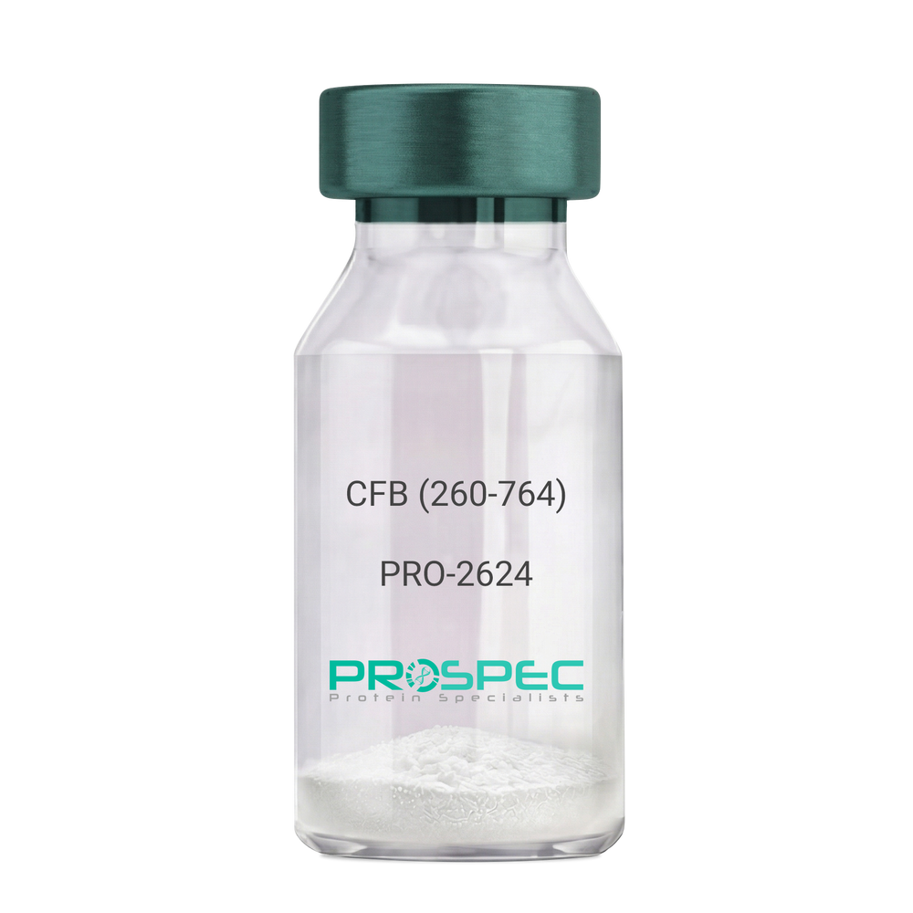
CFB (260-764) Human, Sf9Description:
Complement Factor B (260-764 a.a.) Human Recombinant, Sf9
CFB, C3/C5 convertase, Glycine-rich beta glycoprotein, GBG, PBF2, Properdin factor B, BF, BFD, AHUS4, ARMD14, CFAB, CFBD, FB, FBI12, GBG, H2-Bf.
Product # :
PRO-2624Price :
Quantity :
Shipping Method :
Shipped with Ice Packs
More Info
- description
- source
- formulation
- purity
- More Info
Description
CFB Human Recombinant produced in Sf9 Baculovirus cells is a single, glycosylated polypeptide chain containing 514 amino acids (260-764 a.a) and having a molecular mass of 58.1kDa.CFB is fused to an 9 amino acid His-tag at C-terminus & purified by proprietary chromatographic techniques.
Source
Sf9, Baculovirus cells.
Formulation
CFB protein solution (0.5mg/ml) containing Phosphate Buffered Saline (pH 7.4) and 20% glycerol.
Purity
Greater than 90.0% as determined by SDS-PAGE.
More Info
-
Introduction
Factor B is a protein, build from a single chain that circulates in the blood. Complement factor D cleaves and activates factor B, thus creating a catalytic subunit Bb and noncatalytic chain Ba. Subunit Bb is a serine protease that can bind to C3b and forms alternative pathway C3 convertase. Subunit Ba acts as an inhibitor for proliferation of lymphocytes, when Bb is part of the lymphocytes’ proliferation. The gene that codes for this protein is located on chromosome six.
-
Synonyms
CFB, C3/C5 convertase, Glycine-rich beta glycoprotein, GBG, PBF2, Properdin factor B, BF, BFD, AHUS4, ARMD14, CFAB, CFBD, FB, FBI12, GBG, H2-Bf.
-
Physical Appearance
Sterile Filtered colorless solution.
-
Stability
Store at 4°C if entire vial will be used within 2-4 weeks. Store, frozen at -20°C for longer periods of time. For long term storage it is recommended to add a carrier protein (0.1% HSA or BSA).Avoid multiple freeze-thaw cycles.
-
Amino Acid Sequence
ADPKIVLDPS GSMNIYLVLD GSDSIGASNF TGAKKCLVNL IEKVASYGVK PRYGLVTYAT
YPKIWVKVSE ADSSNADWVT KQLNEINYED HKLKSGTNTK KALQAVYSMM SWPDDVPPEG
WNRTRHVIIL MTDGLHNMGG DPITVIDEIR DLLYIGKDRK NPREDYLDVY VFGVGPLVNQ
VNINALASKK DNEQHVFKVK DMENLEDVFY QMIDESQSLS LCGMVWEHRK GTDYHKQPWQ
AKISVIRPSK GHESCMGAVV SEYFVLTAAH CFTVDDKEHS IKVSVGGEKR DLEIEVVLFH
PNYNINGKKE AGIPEFYDYD VALIKLKNKL KYGQTIRPIC LPCTEGTTRA LRLPPTTTCQ
QQKEELLPAQ DIKALFVSEE EKKLTRKEVY IKNGDKKGSC ERDAQYAPGY DKVKDISEVV
TPRFLCTGGV SPYADPNTCR GDSGGPLIVH KRSRFIQVGV ISWGVVDVCK NQKRQKQVPA HARDFHINLF QVLPWLKEKL QDEDLGFLHH HHHH
ProSpec's products are furnished for LABORATORY RESEARCH USE ONLY. The product may not be used as drugs, agricultural or pesticidal products, food additives or household chemicals.
- View Data Sheet
Name :
Leptin HorseDescription:
Leptin Horse Recombinant
OB Protein, Obesity Protein, OBS, Obesity factor.
Product # :
CYT-504Price :
Quantity :
Shipping Method :
Shipped at Room temp
More Info
- description
- source
- formulation
- purity
- biological activity
- More Info
Description
Leptin Horse Recombinant produced in E.Coli is a single, non-glycosylated, polypeptide chain containing 146 amino acids and having a molecular mass of 16 kDa.The Leptin is purified by proprietary chromatographic techniques.
Source
Escherichia Coli.
Formulation
The protein was lyophilized from a concentrated (1mg/ml) solution with 0.0045mM NaHCO3.
Purity
Greater than 95.0% as determined by:
(a) Analysis by SEC-HPLC.
(b) Analysis by SDS-PAGE.Biological Activity
Biological active as evidenced by inducing proliferation of BAF/3 cells stably transfected with the long form of human leptin receptor.More Info
-
Introduction
A 16-kDa peptide hormone secreted from white adipocytes and implicated in the regulation of food intake and energy balance. Leptin provides the key afferent signal from fat cells in the feedback system that controls body fat stores.
-
Synonyms
OB Protein, Obesity Protein, OBS, Obesity factor.
-
Physical Appearance
Sterile Filtered White lyophilized (freeze-dried) powder.
-
Stability
Lyophilized Leptin although stable at room temperature for 3 weeks, should be stored desiccated below -18°C. Upon reconstitution Leptin should be stored at 4°C between 2-7 days and for future use below -18°C.For long term storage it is recommended to add a carrier protein (0.1% HSA or BSA).Please prevent freeze-thaw cycles.
-
Solubility
It is recommended to reconstitute the lyophilized Leptin in sterile 18MΩ-cm H2O not less than 100µg/ml, which can then be further diluted to other aqueous solutions.
-
Amino Acid Sequence
The sequence of the first five N-terminal amino acids was determined and was found to be Ala-Val-Pro-Ile-Gln.
-
Protein content
Protein quantitation was carried out by two independent methods1. UV spectroscopy at 280 nm using the absorbency value of 0.1 as the extinction coefficient for a 0.1% (1mg/ml) solution. This value is calculated by the PC GENE computer analysis program of protein sequences (IntelliGenetics). 2. Analysis by RP-HPLC,using calibrated solution of Leptin Horse as a Reference Standard.
ProSpec's products are furnished for LABORATORY RESEARCH USE ONLY. The product may not be used as drugs, agricultural or pesticidal products, food additives or household chemicals.
- View Data Sheet
Name :
AHSP HumanDescription:
Alpha Hemoglobin Stabilizing Protein Human Recombinant
Alpha-hemoglobin-stabilizing protein, Erythroid-associated factor, Erythroid differentiation-related factor, AHSP, EDRF, ERAF.
Product # :
PRO-720Price :
Quantity :
Shipping Method :
Shipped with Ice Packs
More Info
- description
- source
- formulation
- purity
- More Info
Description
AHSP Human Recombinant produced in E.Coli is a single, non-glycosylated, polypeptide chain containing 102 amino acids (1-102 a.a.) and having a molecular mass of 11.8kDa.The AHSP is purified by proprietary chromatographic techniques.
Source
Escherichia Coli.
Formulation
The AHSP protein solution (1mg/ml) contains 20mM Tris-HCl buffer (pH8.0) and 10% glycerol.
Purity
Greater than 95.0% as determined by SDS-PAGE.
More Info
-
Introduction
Alpha-hemoglobin stabilizing protein (AHSP) is an erythroid-specific protein that acts as a chaperone to prevent the aggregation of A-hemoglobin during normal erythroid cell development. AHSP specifically protects free A-hemoglobin from precipitation in live cells and in solution. AHSP is expected to modulate pathological states of alpha-hemoglobin excess such as beta-thalassemia. Furthermore, AHSP promotes alpha globin chain stability in human erythropoiesis. In addition, the AHSP stabilizes the alpha-Hb chain, thus avoiding its precipitation and its ability to generate ROS, which is implicated in cell death. AHSP is expressed in blood and bone marrow. AHSP subunit is a monomer, it forms a heterodimer with free alpha-hemoglobin. On the other hand, AHSP does not bind beta-hemoglobin nor alpha2beta2 hemoglobin A. AHSP is downregulated in TSEs (transmissible spongiform encephalopathies).
-
Synonyms
Alpha-hemoglobin-stabilizing protein, Erythroid-associated factor, Erythroid differentiation-related factor, AHSP, EDRF, ERAF.
-
Physical Appearance
Sterile Filtered colorless solution.
-
Stability
Store at 4°C if entire vial will be used within 2-4 weeks.Store, frozen at -20°C for longer periods of time. For long term storage it is recommended to add a carrier protein (0.1% HSA or BSA).Avoid multiple freeze-thaw cycles.
-
Amino Acid Sequence
MALLKANKDL ISAGLKEFSV LLNQQVFNDP LVSEEDMVTV VEDWMNFYIN YYRQQVTGEP QERDKALQEL RQELNTLANP FLAKYRDFLK SHELPSHPPP SS.
ProSpec's products are furnished for LABORATORY RESEARCH USE ONLY. The product may not be used as drugs, agricultural or pesticidal products, food additives or household chemicals.
- View Data Sheet
Name :
RABIF HumanDescription:
RAB Interacting Factor Human Recombinant
MSS4, RASGFR3, RASGRF3, Rab-interacting factor.
Product # :
PRO-300Price :
Quantity :
Shipping Method :
Shipped with Ice Packs
More Info
- description
- source
- formulation
- purity
- More Info
Description
RABIF Human Recombinant produced in E.Coli is a single, non-glycosylated, polypeptide chain containing 143 amino acids (1-123 a.a) and having a molecular mass of 16kDa. RABIF is fused to a 20 amino acid His Tag at N-Terminus and purified by proprietary chromatographic techniques.
Source
Escherichia Coli.
Formulation
The RABIF protein solution (0.5mg/ml) contains 20mM Tris-HCl pH-8, 1mM DTT, 0.1M NaCl and 20% glycerol.
Purity
Greater than 85% as determined by SDS-PAGE.
More Info
-
Introduction
RABIF is a guanine-nucleotide-releasing protein is involved in vesicular transport. RABIF stimulates GTP-GDP exchange in Sec4 and Rab and binds to a subset of genetically related Rab proteins.
-
Synonyms
MSS4, RASGFR3, RASGRF3, Rab-interacting factor.
-
Physical Appearance
Sterile Filtered clear colorless solution.
-
Stability
Store at 4°C if entire vial will be used within 2-4 weeks. Store, frozen at -20°C for longer periods of time. For long term storage it is recommended to add a carrier protein (0.1% HSA or BSA).Avoid multiple freeze-thaw cycles.
-
Amino Acid Sequence
MGSSHHHHHH SSGLVPRGSH MEPAEQPSELVSAEGRNRKA VLCQRCGSRV LQPGTALFSR RQLFLPSMRK KPALSDGSNP DGDLLQEHWL VEDMFIFENV GFTKDVGNIK FLVCADCEIG PIGWHCLDDK NSFYVALERV SHE.
ProSpec's products are furnished for LABORATORY RESEARCH USE ONLY. The product may not be used as drugs, agricultural or pesticidal products, food additives or household chemicals.
- View Data Sheet
Name :
Resistin Human (64-110)Description:
Resistin (64-110) Human Recombinant
Cysteine-rich secreted protein FIZZ3, Adipose tissue-specific secretory factor, ADSF.
Product # :
CYT-1232Price :
Quantity :
Shipping Method :
Shipped at Room temp
More Info
- description
- source
- formulation
- purity
- More Info
Description
The Resistin Human is created as a recombinant protein with a 4kda N-terminal fusion of His Tag. The Resistin His-Tagged Fusion Protein, produced in E. coli, is a 12kDa protein containing 47 amino acid residues of the Resistin Human, 64-110 amino acids.
Source
Escherichia Coli.
Formulation
Each mg was lyophilized with 1xPBS, 0.4% SDS and 4mM DTT.
Purity
Greater than 80% as determined by SDS-PAGE.
More Info
-
Synonyms
Cysteine-rich secreted protein FIZZ3, Adipose tissue-specific secretory factor, ADSF.
-
Physical Appearance
Filtered White lyophilized (freeze-dried) powder.
-
Stability
Store lyophilized Resistin at -20°C. Aliquot the product after reconstitution to avoid repeated freezing/thawing cycles. Reconstituted protein can be stored at 4°C for a limited period of time; it does not show any change after two weeks at 4°C.
-
Solubility
It is recommended to add deionized water to prepare a working stock solution of approximately 0.5mg/ml and let the lyophilized pellet dissolve completely. Product is not sterile! Please filter the product by an appropriate sterile filter before using it on cell culture.
-
Background
Human resistin is an adipokine primarily secreted by adipose tissue, mainly in response to obesity and inflammatory conditions.
Resistin Function
Resistin takes part in insulin resistance, which can be the cause of the development of type 2 diabetes. Resistin can also affect glucose metabolism and insulin signalling.
Regulation
Levels of resistin are influenced by factors such as inflammation, obesity and certain hormones. It tends to increase in conditions associated with obesity and metabolic syndrome.
Clinical Relevance
Elevated levels of resistin have been associated with obesity-related conditions, cardiovascular diseases, and metabolic disorders. Resisting is considered as a potential biomarker for these conditions.
Resistin Mechanism
Resistin promotes insulin resistance through different pathways such as the modulation of inflammatory processes and the inhibition of insulin signaling in target tissues like liver and muscle.
Research
Ongoing studies are exploring resistin’s role in metabolic regulation, the exact mechanisms of action of resistin and its potential as a therapeutic target for treating metabolic diseases.
Overall, resistin is a critical factor in metabolic health, mainly in the context of diabetes and obesity.
ProSpec's products are furnished for LABORATORY RESEARCH USE ONLY. The product may not be used as drugs, agricultural or pesticidal products, food additives or household chemicals.
- View Data Sheet
Name :
CCN1 HumanDescription:
Cysteine-Rich Angiogenic Inducer 61 Human Recombinant
CYR61, Protein CYR61, Cysteine-rich angiogenic inducer 61, IGF-binding protein 10, IGFBP-10, IBP-10, Protein GIG1, CCN family member 1, CCN1, GIG1, IGFBP10.
Product # :
CYT-164Price :
Quantity :
Shipping Method :
Shipped at Room temp
More Info
- description
- source
- formulation
- purity
- biological activity
- More Info
Description
CYR61 Human Recombinant produced in E.Coli is a single, non-glycosylated, polypeptide chain containing 357 amino acids and having a molecular mass of 39.5kDa.The CYR61 is purified by proprietary chromatographic techniques.
Source
Escherichia Coli.
Formulation
Lyophilized from a 0.2m filtered concentrated solution in PBS, pH 7.4.
Purity
Greater than 95.0% as determined by SDS-PAGE.
Biological Activity
The ED50 was determined by the proliferation of mouse 3T3 cells is < 2.0 ug/ml, corresponding to a specific activity of > 500 units/mg.More Info
-
Introduction
CYR61 is a growth factor-inducible, immediate-early gene that has multifaceted activities in various cancers. CYR61 is a secreted, cysteine-rich, binding protein which is encoded by a growth factor-inducible immediate-early gene. Acting as an extracellular, matrix-associated signaling molecule, CYR61 promotes the adhesion of endothelial cells through interaction with integrin and enhances growth factor-induced DNA synthesis in the same cell type.
-
Synonyms
CYR61, Protein CYR61, Cysteine-rich angiogenic inducer 61, IGF-binding protein 10, IGFBP-10, IBP-10, Protein GIG1, CCN family member 1, CCN1, GIG1, IGFBP10.
-
Physical Appearance
Sterile Filtered White lyophilized (freeze-dried) powder.
-
Stability
Lyophilized CYR61 Human although stable at room temperature for 3 weeks, should be stored desiccated below -18°C. Upon reconstitution CYR61 should be stored at 4°C between 2-7 days and for future use below -18°C.Please prevent freeze-thaw cycles.
-
Solubility
It is recommended to reconstitute the lyophilized CYR61 in sterile 18M-cm H2O not less than 100µg/ml, which can then be further diluted to other aqueous solutions.
-
Amino Acid Sequence
TCPAACHCPL EAPKCAPGVG LVRDGCGCCK VCAKQLNEDC SKTQPCDHTK GLECNFGASS TALKGICRAQ SEGRPCEYNS RIYQNGESFQ PNCKHQCTCI DGAVGCIPLC PQELSLPNLG CPNPRLVKVT GQCCEEWVCD EDSIKDPMED QDGLLGKELG FDASEVELTR NNELIAVGKG SSLKRLPVFG MEPRILYNPL QGQKCIVQTT SWSQCSKTCG TGISTRVTND NPECRLVKET RICEVRPCGQ PVYSSLKKGK KCSKTKKSPE PVRFTYAGCL SVKKYRPKYC GSCVDGRCCT
PQLTRTVKMR FRCEDGETFS KNVMMIQSCK CNYNCPHANE AAFPFYRLFN DIHKFRD -
Background
Title: Cysteine-Rich Angiogenic Inducer 61 Human Recombinant: A Potential Regulator of Angiogenesis
Abstract:
Cysteine-rich angiogenic inducer 61 (CYR61) is an important extracellular matrix-associated protein that plays a significant role in angiogenesis and cell adhesion. This research paper provides a comprehensive analysis of human recombinant CYR61, focusing on its production, characterization, and potential applications in regulating angiogenesis. The paper discusses the significance of CYR61 in physiological and pathological angiogenesis, including wound healing, tumor development, and cardiovascular diseases. Furthermore, it explores ongoing research and clinical trials investigating the therapeutic potential of recombinant CYR61 in various pathological conditions. The information presented in this paper aims to enhance our understanding of human recombinant CYR61 and its utility as a research tool and a potential regulator of angiogenesis.Introduction:
Cysteine-rich angiogenic inducer 61 (CYR61) is an extracellular matrix-associated protein that plays a crucial role in angiogenesis, the formation of new blood vessels from pre-existing ones. Human recombinant CYR61, produced through genetic engineering techniques, provides researchers with a valuable tool to study its biological functions and explore its therapeutic potential.Production and Characterization:
Recombinant CYR61 is typically generated using expression systems such as mammalian cells or bacteria. The protein is then purified and characterized to ensure its structural integrity and functional activity. Quality control measures are implemented to confirm the specificity and bioactivity of the recombinant CYR61.Role in Angiogenesis:
CYR61 is involved in various aspects of angiogenesis, including endothelial cell proliferation, migration, and tube formation. It interacts with integrins and other cell surface receptors to modulate signaling pathways involved in angiogenic processes. Recombinant CYR61 serves as a valuable tool for studying the mechanisms underlying angiogenesis and exploring its potential as a therapeutic target.Therapeutic Implications:
The dysregulation of angiogenesis is associated with several pathological conditions, including cancer, cardiovascular diseases, and chronic wounds. Recombinant CYR61 has shown promise as a potential regulator of angiogenesis and a therapeutic agent. It can be used to promote or inhibit angiogenesis, depending on the specific context. Ongoing research and clinical trials are investigating the therapeutic applications of recombinant CYR61 in various diseases, including cancer and ischemic disorders.Conclusion:
Human recombinant CYR61 is a valuable research tool and a potential regulator of angiogenesis. Its production, characterization, and applications in modulating angiogenic processes contribute to our understanding of angiogenesis and the development of novel therapeutic interventions. Continued research and clinical trials exploring the therapeutic potential of recombinant CYR61 offer promising prospects for improving outcomes in cancer, cardiovascular diseases, and wound healing.What is the molecular weight/Mw of CCN1 Protein?
CCN1 Protein has a total Mw of 39.5kDa.
What is the source or expression system of CCN1 Protein?
Escherichia Coli.
What is the Purity of CCN1 Protein?
CCN1 Protein is >95% pure as determined by SDS-PAGE.
What is the Biological Activity of CCN1 Protein?
The ED50 was determined by the proliferation of mouse 3T3 cells is < 2.0 ug/ml, corresponding to a specific activity of > 500 units/mg.
What is the amino acid sequence of CCN1 Protein?
TCPAACHCPL EAPKCAPGVG LVRDGCGCCK VCAKQLNEDC SKTQPCDHTK GLECNFGASS TALKGICRAQ SEGRPCEYNS RIYQNGESFQ PNCKHQCTCI DGAVGCIPLC PQELSLPNLG CPNPRLVKVT GQCCEEWVCD EDSIKDPMED QDGLLGKELG FDASEVELTR NNELIAVGKG SSLKRLPVFG MEPRILYNPL QGQKCIVQTT SWSQCSKTCG TGISTRVTND NPECRLVKET RICEVRPCGQ PVYSSLKKGK KCSKTKKSPE PVRFTYAGCL SVKKYRPKYC GSCVDGRCCT
PQLTRTVKMR FRCEDGETFS KNVMMIQSCK CNYNCPHANE AAFPFYRLFN DIHKFRD
What applications can CCN1 Protein be used in?
CCN1 Protein can probably be used in western blot, ELISA and Lateral Flow.
What is the endotoxin level for CCN1 Protein?
The endotoxin level is minimal, CCN1 Protein was purified using conventional chromatography techniques.
ProSpec's products are furnished for LABORATORY RESEARCH USE ONLY. The product may not be used as drugs, agricultural or pesticidal products, food additives or household chemicals.
- View Data Sheet
Name :
NDUFS5 HumanDescription:
Histidine NADH Dehydrogenase Fe-S Protein 5 Human Recombinant
CI-15k, CI15K, NADH dehydrogenase [ubiquinone] iron-sulfur protein 5, Complex I-15 kDa, NADH-ubiquinone oxidoreductase 15 kDa subunit, CI-15 kDa, NDUFS5.
Product # :
ENZ-771Price :
Quantity :
Shipping Method :
Shipped with Ice Packs
More Info
- description
- source
- formulation
- purity
- More Info
Description
NDUFS5 Human Recombinant produced in E.coli is a single, non-glycosylated polypeptide chain containing 129 amino acids (1-106a.a) and having a molecular mass of 14.9kDa. NDUFS5 is fused to a 23 amino acid His-tag at N-terminus & purified by proprietary chromatographic techniques.
Source
Escherichia Coli.
Formulation
The NDUFS5 solution (1mg/ml) contains 20mM Tris-HCl buffer (pH8.0), 10% glycerol and 0.4M Urea.
Purity
Greater than 85% as determined by SDS-PAGE.
More Info
-
Introduction
Histidine NADH Dehydrogenase Fe-S Protein 5 (NDUFS5) belongs to the NADH dehydrogenase (ubiquinone) iron-sulfur protein family. NDUFS5 is a subunit of the mitochondrial membrane respiratory chain NADH dehydrogenase (Complex I), which doesn’t take part in catalysis. Complex I is transferring the electrons from NADH to the respiratory chain.
-
Synonyms
CI-15k, CI15K, NADH dehydrogenase [ubiquinone] iron-sulfur protein 5, Complex I-15 kDa, NADH-ubiquinone oxidoreductase 15 kDa subunit, CI-15 kDa, NDUFS5.
-
Physical Appearance
Sterile Filtered colorless solution.
-
Stability
Store at 4°C if entire vial will be used within 2-4 weeks. Store, frozen at -20°C for longer periods of time. For long term storage it is recommended to add a carrier protein (0.1% HSA or BSA). Avoid multiple freeze-thaw cycles.
-
Amino Acid Sequence
MGSSHHHHHH SSGLVPRGSH MGSMPFLDIQ KRFGLNIDRW LTIQSGEQPY KMAGRCHAFE KEWIECAHGI GYTRAEKECK IEYDDFVECL LRQKTMRRAG TIRKQRDKLI KEGKYTPPPH HIGKGEPRP.
ProSpec's products are furnished for LABORATORY RESEARCH USE ONLY. The product may not be used as drugs, agricultural or pesticidal products, food additives or household chemicals.
- View Data Sheet
Name :
PSMF1 HumanDescription:
Proteasome Inhibitor Subunit 1 Human Recombinant
Proteasome inhibitor PI31 subunit, hPI31, PSMF1, PI31.
Product # :
ENZ-224Price :
Quantity :
Shipping Method :
Shipped with Ice Packs
More Info
- description
- source
- formulation
- purity
- More Info
Description
PSMF1 Human Recombinant produced in E.coli is a single, non-glycosylated polypeptide chain containing 291 amino acids (1-271) and having a molecular mass of 31.9kDa.PSMF1 is fused to a 20 amino acid His-tag at N-terminus & purified by proprietary chromatographic techniques.
Source
Escherichia Coli.
Formulation
The PSMF1 solution (0.5mg/ml) contains 20mM Tris-HCl buffer (pH8.0), 20% glycerol, 1mM DTT and 0.1mM PMSF.
Purity
Greater than 85.0% as determined by SDS-PAGE.
More Info
-
Introduction
PSMF1 is a protein which hinders the activation of the proteasome by the 11S and 19S regulators. The 26S proteasome is a multicatalytic proteinase complex with an extremely ordered structure composed of 2 complexes, a 20S core and a 19S regulator. The 20S core is comprised of 4 rings of 28 non-identical subunits; 2 rings are composed of 7 alpha subunits and 2 rings are composed of 7 beta subunits.
-
Synonyms
Proteasome inhibitor PI31 subunit, hPI31, PSMF1, PI31.
-
Physical Appearance
Sterile Filtered colorless solution.
-
Stability
Store at 4°C if entire vial will be used within 2-4 weeks. Store, frozen at -20°C for longer periods of time. For long term storage it is recommended to add a carrier protein (0.1% HSA or BSA).Avoid multiple freeze-thaw cycles.
-
Amino Acid Sequence
MGSSHHHHHH SSGLVPRGSH MAGLEVLFAS AAPAITCRQD ALVCFLHWEV VTHGYCGLGV GDQPGPNDKK SELLPAGWNN NKDLYVLRYE YKDGSRKLLV KAITVESSMI LNVLEYGSQQ VADLTLNLDD YIDAEHLGDF HRTYKNSEEL RSRIVSGIIT PIHEQWEKAN VSSPHREFPP ATAREVDPLR IPPHHPHTSR QPPWCDPLGP FVVGGEDLDP FGPRRGGMIV DPLRSGFPRA LIDPSSGLPN RLPPGAVPPG ARFDPFGPIG TSPPGPNPDH LPPPGYDDMY L.
ProSpec's products are furnished for LABORATORY RESEARCH USE ONLY. The product may not be used as drugs, agricultural or pesticidal products, food additives or household chemicals.
- View Data Sheet
Name :
GroES HumanDescription:
GroES (HSP10) Human Recombinant
CPN10, GROES, HSP10, HSPE1, Chaperonin-10, 10 kDa heat shock protein mitochondrial, 10 kDa chaperonin, Early-pregnancy factor, EPF.
Product # :
HSP-015Price :
Quantity :
Shipping Method :
Shipped with Ice Packs
More Info
- description
- source
- formulation
- purity
- More Info
Description
Recombinant GroES produced in E.Coli is a single, non-glycosylated polypeptide chain containing 102 amino acids and having a molecular mass of 10 kDa.
Source
Escherichia Coli.
Formulation
The GroES protein contains 20mM Tris buffer pH-8 & 50mM NaCl.
Purity
Greater than 95.0% as determined by SDS-PAGE.
More Info
-
Introduction
HSP10 is part of the molecular chaperons, that are crucial for thir efficient folding of proteins in normal as well as stress conditions. GroES function is to bind to HSP60 in the presence of ATP, thus causing a change in the HSP60 conformation & enclosing the protein substrate within the complex. ATP hydrolysis by chaperonin-60 which destabilizes the HSP10-HSP60 complex, thereby allowing it to dissociate and secrete the substrate protein.
GroES having the NCBI accession number of NP_002148 was purified by using conventional chromatography techniques. -
Synonyms
CPN10, GROES, HSP10, HSPE1, Chaperonin-10, 10 kDa heat shock protein mitochondrial, 10 kDa chaperonin, Early-pregnancy factor, EPF.
-
Physical Appearance
Sterile filtered colorless solution.
-
Stability
Store at 4°C if entire vial will be used within 2-4 weeks. Store, frozen at -20°C for longer periods of time. For long term storage it is recommended to add a carrier protein (0.1% HSA or BSA). Avoid multiple freeze-thaw cycles.
-
Amino Acid Sequence
MAGQAFRKFL PLFDRVLVER SAAETVTKGG IMLPEKSQGK VLQATVVAVG SGSKGKGGEI QPVSVKVGDK VLLPEYGGTK VVLDDKDYFL FRDGDILGKY VD.
ProSpec's products are furnished for LABORATORY RESEARCH USE ONLY. The product may not be used as drugs, agricultural or pesticidal products, food additives or household chemicals.
- View Data Sheet
Name :
IL 16 MouseDescription:
Interleukin-16 Mouse Recombinant
LCF, Lymphocyte Chemoattractant Factor, prIL-16, KIAA4048, mKIAA4048, Il16, IL-16, Interleukin-16.
Product # :
CYT-559Price :
Quantity :
Shipping Method :
Shipped at Room temp
More Info
- description
- source
- formulation
- purity
- More Info
Description
Interleukin-16 Mouse Recombinant produced in E.Coli is a single, non-glycosylated polypeptide chain containing 127 amino acids and having a molecular mass of 13.2 kDa. The Mouse IL-16 is purified by proprietary chromatographic techniques.
Source
Escherichia Coli.
Formulation
Murine IL-16 was lyophilized from 1mg/ml solution after extensive dialysis against 10mM sodium phosphate buffer, pH-7.5.
Purity
Greater than 90.0% as determined by(a) Analysis by RP-HPLC.
(b) Analysis SDS-PAGE.More Info
-
Introduction
IL-16 is a pleiotropic cytokine that functions as a chemoattractant, a modulator of T cell activation, and an inhibitor of HIV replication. The signaling process of IL-16 is mediated by CD4. The product of this gene undergoes proteolytic processing, which is found to yield two functional proteins. IL-16 functions exclusively attributed to the secreted C-terminal peptide, while the N-terminal product may play a role in cell cycle control. Caspase 3 is reported to be involved in the proteolytic processing of this protein. Two transcript variants encoding different isoforms have been found for this gene.
IL-16 stimulates a migratory response in cd4+ lymphocytes, monocytes, and eosinophils. Also induces t-lymphocyte expression of interleukin 2 receptor. ligand for cd4. -
Synonyms
LCF, Lymphocyte Chemoattractant Factor, prIL-16, KIAA4048, mKIAA4048, Il16, IL-16, Interleukin-16.
-
Physical Appearance
Sterile Filtered White lyophilized (freeze-dried) powder.
-
Stability
Lyophilized Mouse IL-16 although stable at room temperature for 3 weeks, should be stored desiccated below -18°C. Upon reconstitution mouse IL16 should be stored at 4°C between 2-7 days and for future use below -18°C.For long term storage it is recommended to add a carrier protein (0.1% HSA or BSA).Please prevent freeze-thaw cycles.
-
Solubility
It is recommended to reconstitute the lyophilized Murine IL16 in sterile 18MΩ-cm H2O not less than 100µg/ml, which can then be further diluted to other aqueous solutions.
-
Amino Acid Sequence
MHDLNSSTDS AASASAASDI SVESKEATVC TVTLEKTSAG LGFSLEGGKG SLHGDKPLTI NRIFKGDRTG EMVQPGDEIL QLAGTAVQGL TRFEAWNVIK ALPDGPVTIV IRRTSLQCKQ TTASADS.
ProSpec's products are furnished for LABORATORY RESEARCH USE ONLY. The product may not be used as drugs, agricultural or pesticidal products, food additives or household chemicals.
- View Data Sheet
Name :
IL 32A Human, HisDescription:
Interleukin-32 alpha Human Recombinant , His Tag
NK4, TAIF, TAIFa, TAIFb, TAIFc, TAIFd, IL-32beta, IL-32alpha, IL-32delta, IL-32gamma, Interleukin-32, IL-32, Natural killer cells protein 4, Tumor necrosis factor alpha-inducing factor, IL-32a, IL32a, IL32, Interleukin-32 alpha.
Product # :
CYT-648Price :
Quantity :
Shipping Method :
Shipped with Ice Packs
More Info
- description
- source
- formulation
- purity
- More Info
Description
Interleukin-32 human recombinant produced in E.Coli is a single, non-glycosylated, polypeptide chain containing 168 amino acids (1-131 a.a.) and having a molecular mass of 19.1 kDa. The IL-32 is fused to a 37 amino acid His Tag at N-terminus and purified by conventional chromatography.
Source
Escherichia Coli.
Formulation
The IL-32 His Tag protein solution contains 20mM Tris-HCl, pH-8, 1mM DTT and 10% glycerol.
Purity
Greater than 95.0% as determined by SDS-PAGE.
More Info
-
Introduction
IL-32 is part of the cytokine family and contains a tyrosine sulfation site, 3 potential N-myristoylation sites, multiple putative phosphorylation sites, and an RGD cell-attachment sequence. IL-32 expression is elevated after the activation of T-cells by mitogens or the activation of NK cells by IL-2. IL-32 induces the production of TNF-a from macrophage cells. IL-32 pro-inflammatory pathway is activated in response to influenza A virus infection. Dysregulation of IL-32 in myelodysplastic syndrome and chronic myelomonocytic leukemia modulates apoptosis and impairs NK function.
Induction of TNF, IL-1beta, and IL-6 by IL-32 is intervened by p38-MAPK. IL-32 induced monocyte-to-macrophage differentiation is mediated through nonapoptotic, caspase-3-dependent mechanisms. IL32 plays an important role in the pathogenesis of rheumatoid arthritis. IL-32 is involved in activation-induced cell death in T cells, through its intracellular actions. IL-32 is a cell-associated proinflammatory cytokine, which is particularly stimulated by mycobacteria through a caspase-1- and IL-18-dependent production of interferon gamma.
IL-32 is associated with TNF-a, IL-1beta, and IL-18. IL32 is involved in human rheumatoid arthritis and is a novel target in autoimmune diseases. -
Synonyms
NK4, TAIF, TAIFa, TAIFb, TAIFc, TAIFd, IL-32beta, IL-32alpha, IL-32delta, IL-32gamma, Interleukin-32, IL-32, Natural killer cells protein 4, Tumor necrosis factor alpha-inducing factor, IL-32a, IL32a, IL32, Interleukin-32 alpha.
-
Physical Appearance
Sterile filtered colorless solution.
-
Stability
Store at 4°C if entire vial will be used within 2-4 weeks. Store, frozen at -20°C for longer periods of time. For long term storage it is recommended to add a carrier protein (0.1% HSA or BSA).Avoid multiple freeze-thaw cycles.
-
Amino Acid Sequence
MRGSHHHHHH GMASMTGGQQ MGRDLYDDDD KDRWGSHMCF PKVLSDDMKK LKARMHQAIE RFYDKMQNAE SGRGQVMSSL AELEDDFKEGYLETVAAYYE EQHPELTPLL EKERDGLRCR GNRSPVPDVE DPATEEPGES FCDKSYGAPR GDKEELTPQK CSEPQSSK.
ProSpec's products are furnished for LABORATORY RESEARCH USE ONLY. The product may not be used as drugs, agricultural or pesticidal products, food additives or household chemicals.
- View Data Sheet
Name :
IL 5 RatDescription:
Interleukin-5 Rat Recombinant
EDF, BCDFII, TRF, T-cell replacing factor, Eosinophil differentiation factor, B cell differentiation factor I, IL-5.
Product # :
CYT-387Price :
Quantity :
Shipping Method :
Shipped at Room temp
More Info
- description
- source
- formulation
- purity
- biological activity
- More Info
Description
Interleukin-5 Rat Recombinant produced in E.Coli is a dimeric, non-glycosylated polypeptide chain containing 113 amino acids and having a molecular mass of 13074 Dalton.The IL-5 is purified by proprietary chromatographic techniques.
Source
Escherichia Coli.
Formulation
Lyophilized from a concentrated (1mg/ml) solution in water containing no additives.
Purity
Greater than 95.0% as determined by:
(a) Analysis by RP-HPLC.
(b) Analysis by SDS-PAGE.Biological Activity
The ED50 range=0.3-1.0 ng/ml as determined by the dose-dependant stimulation of the proliferation of BCL-1 cells.More Info
-
Introduction
The protein encoded by this gene is a cytokine that acts as a growth and differentiation factor for both B cells and eosinophils. This cytokine is a main regulator of eosinopoiesis, eosinophil maturation and activation. The elevated production of this cytokine is reported to be related to asthma or hypereosinophilic syndromes. The receptor of this cytokine is a heterodimer, whose beta subunit is shared with the receptors for interleukine 3 (IL3) and colony stimulating factor 2 (CSF2/GM-CSF). This gene, together with those for interleukin 4 (IL4), interleukin 13 (IL13), and CSF2, form a cytokine gene cluster on chromosome 5. This cytokine, IL4, and IL13 are found to be regulated coordinately by long-range regulatory elements spread over 120 kilobases on chromosome 5q31.
-
Synonyms
EDF, BCDFII, TRF, T-cell replacing factor, Eosinophil differentiation factor, B cell differentiation factor I, IL-5.
-
Physical Appearance
Sterile Filtered White lyophilized (freeze-dried) powder.
-
Stability
Lyophilized Interleukin-5 Rat although stable at room temperature for 3 weeks, should be stored desiccated below -18°C. Upon reconstitution IL5 Rat should be stored at 4°C between 2-7 days and for future use below -18°C.Please avoid freeze thaw cycles.
-
Solubility
It is recommended to reconstitute the lyophilized Interleikin-5 Rat in sterile 18MΩ-cm H2O not less than 100µg/ml, which can then be further diluted to other aqueous solutions.
-
Amino Acid Sequence
The sequence of the first five N-terminal amino acids was determined and was found to be Met-Glu-Ile-Pro-Met.
ProSpec's products are furnished for LABORATORY RESEARCH USE ONLY. The product may not be used as drugs, agricultural or pesticidal products, food additives or household chemicals.
- View Data Sheet
Name :
TYMP HumanDescription:
Thymidine Phosphorylase Human Recombinant
Thymidine phosphorylase, Gliostatin, Platelet-derived endothelial cell growth factor, PD-ECGF, TdRPase, TYMP, ECGF1, TP, MNGIE, MEDPS1, MTDPS1, PDECGF, hPD-ECGF.
Product # :
ENZ-005Price :
Quantity :
Shipping Method :
Shipped with Ice Packs
More Info
- description
- source
- formulation
- purity
- More Info
Description
TYMP Human Recombinant fused with a 21 amino acid His tag at N-terminus produced in E.Coli is a single, non-glycosylated, polypeptide chain containing 493 amino acids (11-482 a.a.) and having a molecular mass of 51.3kDa. The TYMP is purified by proprietary chromatographic techniques.
Source
Escherichia Coli.
Formulation
The TYMP solution (1mg/ml) contains 20mM Tris-HCl buffer (pH 8.0), 1mM DTT and 10% glycerol.
Purity
Greater than 90.0% as determined by SDS-PAGE.
More Info
-
Introduction
Thymidine phosphorylase precursor (TYMP) is a platelet-derived endothelial cell growth factor that catalyzes the formation of thymine and 2-deoxy-D-ribose-1-phosphate from thymidine and orthophosphate. TYMP is an angiogenic inducer that potently stimulates the growth of endothelial cells and induces chemotaxis. TYMP has a highly restricted target cell specificity acting only on endothelial cells. An increased expression of TYMP is found in a broad array of different solid tumors and inflammatory diseases and is frequently associated with poor prognosis. Mutations in the TYMP gene are linked to mitochondrial neurogastrointestinal encephalomyopathy.
-
Synonyms
Thymidine phosphorylase, Gliostatin, Platelet-derived endothelial cell growth factor, PD-ECGF, TdRPase, TYMP, ECGF1, TP, MNGIE, MEDPS1, MTDPS1, PDECGF, hPD-ECGF.
-
Physical Appearance
Sterile Filtered colorless solution.
-
Stability
Store at 4°C if entire vial will be used within 2-4 weeks. Store, frozen at -20°C for longer periods of time. For long term storage it is recommended to add a carrier protein (0.1% HSA or BSA).Avoid multiple freeze-thaw cycles.
-
Amino Acid Sequence
MGSSHHHHHH SSGLVPRGSH MAPPAPGDFS GEGSQGLPDP SPEPKQLPEL IRMKRDGGRL SEADIRGFVA AVVNGSAQGA QIGAMLMAIR LRGMDLEETS VLTQALAQSG QQLEWPEAWR QQLVDKHSTG GVGDKVSLVL APALAACGCK VPMISGRGLG HTGGTLDKLE SIPGFNVIQS PEQMQVLLDQ AGCCIVGQSE QLVPADGILY AARDVTATVD SLPLITASIL SKKLVEGLSA LVVDVKFGGA AVFPNQEQAR ELAKTLVGVG ASLGLRVAAA LTAMDKPLGR CVGHALEVEE ALLCMDGAGP PDLRDLVTTL GGALLWLSGH AGTQAQGAAR VAAALDDGSA LGRFERMLAA QGVDPGLARA LCSGSPAERR QLLPRAREQE ELLAPADGTV ELVRALPLAL VLHELGAGRS RAGEPLRLGV GAELLVDVGQ RLRRGTPWLR VHRDGPALSG PQSRALQEAL VLSDRAPFAA PSPFAELVLP PQQ.
ProSpec's products are furnished for LABORATORY RESEARCH USE ONLY. The product may not be used as drugs, agricultural or pesticidal products, food additives or household chemicals.
- View Data Sheet
Name :
IL 3 Human, Sf9Description:
Interleukin-3 Human Recombinant, Sf9
MCGF (Mast cell growth factor), Multi-CSF, HCGF, P-cell stimulation factor, IL-3, MGC79398, MGC79399.
Product # :
CYT-417Price :
Quantity :
Shipping Method :
Shipped at Room temp
More Info
- description
- source
- formulation
- purity
- biological activity
- More Info
Description
Interleukin-3 Human Recombinant produced in insect cells is a single, glycosylated polypeptide chain containing 133 amino acids and having a molecular mass of 15000 Dalton. The IL-3 CSF is fused to a C-terminal His-tag (6x His) and purified by proprietary chromatographic techniques.
Source
Insect Cells.
Formulation
Lyophilized from a concentrated (1mg/ml) solution in water containing 0.5X PBS pH-7.4.
Purity
Greater than 95.0% as determined by(a) Analysis by RP-HPLC.
(b) Analysis by SDS-PAGE.Biological Activity
The ED50 as determined by the dose-dependant stimulation of TF-1 cells is < 0.1 ng/ml, corresponding to a Specific Activity of 10,000,000IU/mg.More Info
-
Introduction
IL3 is a potent growth promoting cytokine. This cytokine is capable of supporting the proliferation of a broad range of hematopoietic cell types. It is involved in a variety of cell activities such as cell growth, differentiation and apoptosis. This cytokine has been shown to also possess neurotrophic activity, and it may be associated with neurologic disorders.
-
Synonyms
MCGF (Mast cell growth factor), Multi-CSF, HCGF, P-cell stimulation factor, IL-3, MGC79398, MGC79399.
-
Physical Appearance
Sterile Filtered White lyophilized (freeze-dried) powder.
-
Stability
Lyophilized Interleukin-3 although stable at room temperature for 3 weeks, should be stored desiccated below -18°C. Upon reconstitution IL3 should be stored at 4°C between 2-7 days and for future use below -18°C.For long term storage it is recommended to add a carrier protein (0.1% HSA or BSA).Please prevent freeze-thaw cycles.
-
Solubility
It is recommended to reconstitute the lyophilized Interleukin-3 in sterile 18MΩ-cm H2O not less than 100µg/ml, which can then be further diluted to other aqueous solutions.
-
Amino Acid Sequence
The sequence of the first five N-terminal amino acids was determined and was found to be Ala-Pro-Ala-Pro-Thr.
ProSpec's products are furnished for LABORATORY RESEARCH USE ONLY. The product may not be used as drugs, agricultural or pesticidal products, food additives or household chemicals.
- View Data Sheet
Name :
MAFK HumanDescription:
V-maf Musculoaponeurotic Fibrosarcoma Oncogene K Human Recombinant
NFE2U, P18, Erythroid transcription factor NF-E2 p18 subunit, MAFK, Transcription factor MafK.
Product # :
PRO-025Price :
Quantity :
Shipping Method :
Shipped with Ice Packs
More Info
- description
- source
- formulation
- purity
- More Info
Description
MAFK Human Recombinant produced in E.Coli is a single, non-glycosylated polypeptide chain containing 176 amino acids (1-156 a.a.) and having a molecular mass of 19.7kDa.MAFK is fused to a 20 amino acid His-tag at N-terminus & purified by proprietary chromatographic techniques.
Source
Escherichia Coli.
Formulation
MAFK protein solution (0.5mg/ml) contains 20mM Tris-HCl buffer (pH 8.0), 0.4M Urea and 10% glycerol.
Purity
Greater than 85.0% as determined by SDS-PAGE.
More Info
-
Introduction
MAFK is a member of the bZIP family and Maf subfamily. Members from Maf family of basic region/leucine zipper (bZIP) transcription factors influence transcription in a positive or negative way, depending on their protein associate and the context of the target promoter. Small Mafs act as transcriptional repressors when they dimerize among themselves. Yet, they seem to function as transcriptional activators by dimerizing with other basic-zipper proteins and employing them to specific DNA-binding sites.
-
Synonyms
NFE2U, P18, Erythroid transcription factor NF-E2 p18 subunit, MAFK, Transcription factor MafK.
-
Physical Appearance
Sterile Filtered clear solution.
-
Stability
Store at 4°C if entire vial will be used within 2-4 weeks. Store, frozen at -20°C for longer periods of time. For long term storage it is recommended to add a carrier protein (0.1% HSA or BSA).Avoid multiple freeze-thaw cycles.
-
Amino Acid Sequence
MGSSHHHHHH SSGLVPRGSH MTTNPKPNKA LKVKKEAGEN APVLSDDELV SMSVRELNQH LRGLTKEEVT RLKQRRRTLK NRGYAASCRI KRVTQKEELE RQRVELQQEV EKLARENSSM RLELDALRSK YEALQTFART VARGPVAPSK VATTSVITIV KSTELSSTSV PFSAAS.
ProSpec's products are furnished for LABORATORY RESEARCH USE ONLY. The product may not be used as drugs, agricultural or pesticidal products, food additives or household chemicals.
- View Data Sheet
Name :
FGFR1 Human, (22-285)Description:
Fibroblast Growth Factor Receptor-1 Human Recombinant, (22-285 a.a.)
FGFR-1, bFGF-R, C-FGR, CD331, fms-related tyrosine kinase 2, Pfeiffer syndrome, CEK, FLG, FLT2, KAL2, BFGFR, FGFBR, HBGFR, FGFR1/FGFR1OP2 FUSION GENE, FGFR1/ZNF198 FUSION GENE, FLG FGFR1/BCR FUSION GENE, FLG protein, FMS-LIKE GENE, N-sam tyrosine kinase, basic fibroblast growth factor receptor 1.
Product # :
PKA-114Price :
Quantity :
Shipping Method :
Shipped with Ice Packs
More Info
- description
- source
- formulation
- purity
- More Info
Description
FGFR1 Human Recombinant produced in Sf9 Baculovirus cells is a single, glycosylated polypeptide chain containing 272 amino acids (22-285) and having a molecular mass of 30.4kDa. (Molecular size on SDS-PAGE will appear at approximately 40-57kDa). FGFR1 is fused to a 8 amino acid His-tag at C-terminus & purified by proprietary chromatographic techniques.
Source
Sf9, Baculovirus cells.
Formulation
The FGFR1 solution (0.25mg/1ml) contains phosphate buffered Saline (pH7.4), and 10% glycerol.
Purity
Greater than 90.0% as determined by SDS-PAGE.
More Info
-
Introduction
Fibroblast Growth Factors (FGFs) comprise a family of at least 18 structurally related proteins that are involved in a multitude of physiological and pathological cellular processes, including cell growth, differentiation, angiogenesis, wound healing and tumorigenesis. The biological activities of the FGFs are mediated by a family of type I transmembrane tyrosine kinases which undergo dimerization and autophosphorylation after ligand binding. Multiple forms of FGFR-1 to -3 are generated by alternative splicing of the mRNAs. A frequent splicing event involving FGFR-1 and -2 results in receptors containing all three Ig domains, referred to as the alpha isoform, or only IgII and IgIII, referred to as the ? isoform. Only the alpha isoform has been identified for FGFR-3 and FGFR-4. Additional splicing events for FGFR-1 to -3, involving the C-terminal half of the IgIII domain encoded by two mutually exclusive alternative exons, generate FGF receptors with alternative IgIII domains (IIIb and IIIc). An IIIa isoform which is a secreted FGF binding protein containing only the N-terminal half of the IgIII domain plus some intron sequences has also been reported for FGFR-1. Mutations in FGFR-1 to -3 have been found in patients with birth defects involving craniosynostosis.
-
Synonyms
FGFR-1, bFGF-R, C-FGR, CD331, fms-related tyrosine kinase 2, Pfeiffer syndrome, CEK, FLG, FLT2, KAL2, BFGFR, FGFBR, HBGFR, FGFR1/FGFR1OP2 FUSION GENE, FGFR1/ZNF198 FUSION GENE, FLG FGFR1/BCR FUSION GENE, FLG protein, FMS-LIKE GENE, N-sam tyrosine kinase, basic fibroblast growth factor receptor 1.
-
Physical Appearance
Sterile Filtered colorless solution.
-
Stability
Store at 4°C if entire vial will be used within 2-4 weeks.Store, frozen at -20°C for longer periods of time. For long term storage it is recommended to add a carrier protein (0.1% HSA or BSA).Avoid multiple freeze-thaw cycles.
-
Amino Acid Sequence
RPSPTLPEQD ALPSSEDDDD DDDSSSEEKE TDNTKPNPVA PYWTSPEKME KKLHAVPAAK TVKFKCPSSG TPNPTLRWLK NGKEFKPDHRIGGYKVRYAT WSIIMDSVVP SDKGNYTCIV ENEYGSINHT YQLDVVERSP HRPILQAGLP ANKTVALGSN VEFMCKVYSD PQPHIQWLKH IEVNGSKIGP DNLPYVQILK TAGVNTTDKE MEVLHLRNVS FEDAGEYTCL AGNSIGLSHH SAWLTVLEAL EERPAVMTSP LYLELEHHHH HH.
ProSpec's products are furnished for LABORATORY RESEARCH USE ONLY. The product may not be used as drugs, agricultural or pesticidal products, food additives or household chemicals.