Search results
1000 results found for “Microfibrillar Associated Protein”
Name
Description
Product #
Price
Quantity
Shipping Method
- View Data Sheet
Name :
sRAGE MouseDescription:
Advanced Glycosylation End Product-Specific Receptor Mouse Recombinant
Advanced glycosylation end product-specific receptor, Receptor for advanced glycosylation end products, AGER, SRAGE, RAGE, MGC22357.
Product # :
PRO-2781Price :
Quantity :
Shipping Method :
Shipped at Room temp
More Info
- description
- source
- formulation
- purity
- More Info
Description
sRAGE Mouse Recombinant produced in HEK cells is a single, glycosylated, polypeptide chain containing 317 amino acids (Gly23–Asp333) and having a molecular mass of 34.0kDa. sRAGE Mouse is fused to a 6 a.a his tag at C-Terminus and is purified by proprietary chromatographic techniques.
Source
HEK 293.
Formulation
The filtered (0.4µm) concentrated protein solution was lyophilized with PBS, PH 7.4.
Purity
Greater than 95% as determined by SDS-PAGE.
More Info
-
Synonyms
Advanced glycosylation end product-specific receptor, Receptor for advanced glycosylation end products, AGER, SRAGE, RAGE, MGC22357.
-
Physical Appearance
Sterile Filtered White lyophilized (freeze-dried) powder.
-
Stability
Store lyophilized protein at -20°C. Aliquot the product after reconstitution to avoid repeated freezing/thawing cycles. Reconstituted protein can be stored at 4°C for a limited period of time.
-
Solubility
It is recommended to add deionized water to prepare a working stock solution of approximately 0.5mg/ml and let the lyophilized pellet dissolve completely.
-
Amino Acid Sequence
GQNITARIGE PLVLSCKGAP KKPPQQLEWK LNTGRTEAWK VLSPQGGPWD SVARILPNGS LLLPATGIVD EGTFRCRATN RRGKEVKSNY RVRVYQIPGK PEIVDPASEL TASVPNKVGT CVSEGSYPAG TLSWHLDGKL LIPDGKETLV KEETRRHPET GLFTLRSELT VIPTQGGTHP TFSCSFSLGL PRRRPLNTAP IQLRVREPGP PEGIQLLVEP EGGIVAPGGT VTLTCAISAQ PPPQVHWIKD GAPLPLAPSP VLLLPEVGHE DEGTYSCVAT HPSHGPQESP PVSIRVTETG DEGPAEGEGL DHHHHHH.
-
Background
The soluble receptor for advanced glycation end products, sRAGE, is a multifunctional protein known for its involvement in diverse physiological processes, including inflammation, aging, and chronic diseases. Research using mouse models has been instrumental in unraveling the complexities of sRAGE biology and its implications for health and disease. This study aims to provide a comprehensive exploration of sRAGE in mouse physiology, shedding light on its various functions and potential applications in understanding aging and disease mechanisms.
The primary objective of this research is to elucidate the impact of sRAGE in mouse models on aging processes. In vivo experiments utilizing genetically modified mice with altered sRAGE expression will be conducted to investigate how sRAGE influences the aging process, including effects on tissue homeostasis, oxidative stress, and longevity. Understanding these mechanisms is fundamental for deciphering the role of sRAGE in age-related diseases.
The second objective is to assess the clinical relevance of sRAGE in mouse models of chronic diseases. Mouse models of diseases such as diabetes, Alzheimer's disease, and cancer will be employed to explore how sRAGE modulation affects disease progression, inflammation, and tissue damage. These investigations may provide valuable insights into potential therapeutic strategies targeting sRAGE in various chronic diseases.
The third objective is to explore the broader implications of sRAGE in mouse physiology, including its effects on immunity, tissue repair, and metabolic regulation. Research will investigate its roles in immune cell function, wound healing, and glucose homeostasis. Understanding the multifaceted properties of sRAGE in mouse models may open new avenues for therapeutic interventions in various health and disease contexts.
By delving into the diverse functions of sRAGE in mouse physiology, this research aims to expand our knowledge of its physiological roles and clinical applications. The findings may contribute to the development of targeted interventions for age-related diseases and chronic conditions influenced by sRAGE.
ProSpec's products are furnished for LABORATORY RESEARCH USE ONLY. The product may not be used as drugs, agricultural or pesticidal products, food additives or household chemicals.
- View Data Sheet
Name :
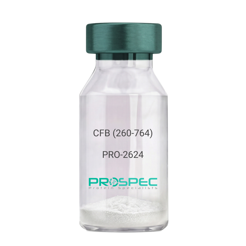
CFB (260-764) Human, Sf9Description:
Complement Factor B (260-764 a.a.) Human Recombinant, Sf9
CFB, C3/C5 convertase, Glycine-rich beta glycoprotein, GBG, PBF2, Properdin factor B, BF, BFD, AHUS4, ARMD14, CFAB, CFBD, FB, FBI12, GBG, H2-Bf.
Product # :
PRO-2624Price :
Quantity :
Shipping Method :
Shipped with Ice Packs
More Info
- description
- source
- formulation
- purity
- More Info
Description
CFB Human Recombinant produced in Sf9 Baculovirus cells is a single, glycosylated polypeptide chain containing 514 amino acids (260-764 a.a) and having a molecular mass of 58.1kDa.CFB is fused to an 9 amino acid His-tag at C-terminus & purified by proprietary chromatographic techniques.
Source
Sf9, Baculovirus cells.
Formulation
CFB protein solution (0.5mg/ml) containing Phosphate Buffered Saline (pH 7.4) and 20% glycerol.
Purity
Greater than 90.0% as determined by SDS-PAGE.
More Info
-
Introduction
Factor B is a protein, build from a single chain that circulates in the blood. Complement factor D cleaves and activates factor B, thus creating a catalytic subunit Bb and noncatalytic chain Ba. Subunit Bb is a serine protease that can bind to C3b and forms alternative pathway C3 convertase. Subunit Ba acts as an inhibitor for proliferation of lymphocytes, when Bb is part of the lymphocytes’ proliferation. The gene that codes for this protein is located on chromosome six.
-
Synonyms
CFB, C3/C5 convertase, Glycine-rich beta glycoprotein, GBG, PBF2, Properdin factor B, BF, BFD, AHUS4, ARMD14, CFAB, CFBD, FB, FBI12, GBG, H2-Bf.
-
Physical Appearance
Sterile Filtered colorless solution.
-
Stability
Store at 4°C if entire vial will be used within 2-4 weeks. Store, frozen at -20°C for longer periods of time. For long term storage it is recommended to add a carrier protein (0.1% HSA or BSA).Avoid multiple freeze-thaw cycles.
-
Amino Acid Sequence
ADPKIVLDPS GSMNIYLVLD GSDSIGASNF TGAKKCLVNL IEKVASYGVK PRYGLVTYAT
YPKIWVKVSE ADSSNADWVT KQLNEINYED HKLKSGTNTK KALQAVYSMM SWPDDVPPEG
WNRTRHVIIL MTDGLHNMGG DPITVIDEIR DLLYIGKDRK NPREDYLDVY VFGVGPLVNQ
VNINALASKK DNEQHVFKVK DMENLEDVFY QMIDESQSLS LCGMVWEHRK GTDYHKQPWQ
AKISVIRPSK GHESCMGAVV SEYFVLTAAH CFTVDDKEHS IKVSVGGEKR DLEIEVVLFH
PNYNINGKKE AGIPEFYDYD VALIKLKNKL KYGQTIRPIC LPCTEGTTRA LRLPPTTTCQ
QQKEELLPAQ DIKALFVSEE EKKLTRKEVY IKNGDKKGSC ERDAQYAPGY DKVKDISEVV
TPRFLCTGGV SPYADPNTCR GDSGGPLIVH KRSRFIQVGV ISWGVVDVCK NQKRQKQVPA HARDFHINLF QVLPWLKEKL QDEDLGFLHH HHHH
ProSpec's products are furnished for LABORATORY RESEARCH USE ONLY. The product may not be used as drugs, agricultural or pesticidal products, food additives or household chemicals.
- View Data Sheet
Name :
MYL2 HumanDescription:
Myosin Light Chain 2 Human Recombinant
MLC2, CMH10, MYL-2, Myosin regulatory light chain 2, MYOSIN LIGHT CHAIN REGULATORY VENTRICULAR, DKFZp779C0562, MYL2, MLC-2v, MLC-2.
Product # :
PRO-524Price :
Quantity :
Shipping Method :
Shipped with Ice Packs
More Info
- description
- source
- formulation
- purity
- More Info
Description
MYL2 Human Recombinant produced in E.Coli is a single, non-glycosylated, polypeptide chain containing 186 amino acids (1-166) and having a molecular mass of 20.9 kDa. MYL2 is fused to a 20 amino acid His Tag fused at N-terminus and purified by proprietary chromatographic techniques.
Source
Escherichia Coli.
Formulation
MYL2 solution containing 20mM Tris pH-8, 40% glycerol and 5mM CaCl2.
Purity
Greater than 95.0% as determined by SDS-PAGE.
More Info
-
Introduction
MYL2 regulatory light chain is associated with cardiac myosin beta (or slow) heavy chain. Ca+ activates the phosphorylation of regulatory light chain that in turn activates contraction. Mutations in this MYL2 gene are related with mid-left ventricular chamber type hypertrophic cardiomyopathy. MYL2 is an essential protein that plays a role in the regulation of myosin ATPase activity in smooth muscle. MYL2 phosphorylation is regulated by ROCK and MLC kinase and is involved in platelet biogenesis by controlling proplatelet formation and fragmentation.
-
Synonyms
MLC2, CMH10, MYL-2, Myosin regulatory light chain 2, MYOSIN LIGHT CHAIN REGULATORY VENTRICULAR, DKFZp779C0562, MYL2, MLC-2v, MLC-2.
-
Physical Appearance
Sterile filtered colorless solution.
-
Stability
Store at 4°C if entire vial will be used within 2-4 weeks. Store, frozen at -20°C for longer periods of time. For long term storage it is recommended to add a carrier protein (0.1% HSA or BSA).Avoid multiple freeze-thaw cycles.
-
Amino Acid Sequence
MGSSHHHHHH SSGLVPRGSH MAPKKAKKRA GGANSNVFSM FEQTQIQEFK EAFTIMDQNR DGFIDKNDLR DTFAALGRVN VKNEEIDEMI KEAPGPINFT VFLTMFGEKL KGADPEETIL NAFKVFDPEG KGVLKADYVR EMLTTQAERF SKEEVDQMFA AFPPDVTGNL DYKNLVHIIT HGEEKD.
ProSpec's products are furnished for LABORATORY RESEARCH USE ONLY. The product may not be used as drugs, agricultural or pesticidal products, food additives or household chemicals.
- View Data Sheet
Name :
MEMO1 AntibodyDescription:
Mediator of Cell Motility 1, Mouse Anti Human
Protein MEMO1, C21orf19-like protein, Hepatitis C virus NS5A-transactivated protein 7, HCV NS5A-transactivated protein 7, Mediator of ErbB2-driven cell motility 1, Mediator of cell motility 1, Memo-1, MEMO1, C2orf4, MEMO, NS5ATP7, CGI-27.
Product # :
ANT-090Price :
Quantity :
Shipping Method :
Shipped with Ice Packs
More Info
- formulation
- More Info
Formulation
1mg/ml containing PBS, pH-7.4, & 0.1% Sodium Azide.
More Info
-
Introduction
Mediator of ErbB2-driven cell motility 1 (MEMO1) is a member of the UPF0103 family. MEMO1 regulates cell migration by transmitting extracellular chemotactic signals to the microtubule cytoskeleton. Furthermore, MEMO1 controls the localization of APC and CLASP2 to the cell membrane, using the regulation of GSK3B activity. MEMO1 is essential for breast carcinoma cell migration, suggesting a key role in tumorigenesis. MEMO1 is also a mediator of ERBB2 signaling.
-
Synonyms
Protein MEMO1, C21orf19-like protein, Hepatitis C virus NS5A-transactivated protein 7, HCV NS5A-transactivated protein 7, Mediator of ErbB2-driven cell motility 1, Mediator of cell motility 1, Memo-1, MEMO1, C2orf4, MEMO, NS5ATP7, CGI-27.
-
Immunogen
Anti-human MEMO1 mAb, is derived from hybridization of mouse FO myeloma cells with spleen cells from BALB/c mice immunized with recombinant human MEMO1 amino acids 1-297 purified from E. coli.
-
Ig Subclass
Mouse IgG1 heavy chain and kappa light chain.
-
Clone
P1E9AT.
-
Type
Mouse Anti Human Monoclonal.
-
Storage Procedures
For periods up to 1 month store at 4°C, for longer periods of time, store at -20°C. Prevent freeze thaw cycles.
-
Purification Method
MEMO1 antibody was purified from mouse ascitic fluids by protein-G affinity chromatography.
ProSpec's products are furnished for LABORATORY RESEARCH USE ONLY. The product may not be used as drugs, agricultural or pesticidal products, food additives or household chemicals.
- View Data Sheet
Name :
Tissue Factor HumanDescription:
Coagulation Factor III Human Recombinant
Tissue factor, Coagulation factor III, Thromboplastin, CD142, TF, F3, TFA.
Product # :
PRO-312Price :
Quantity :
Shipping Method :
Shipped with Ice Packs
More Info
- description
- source
- formulation
- purity
- More Info
- sds-page
Description
Tissue factor Human Recombinant produced in E.Coli is single, a non-glycosylated, Polypeptide chain containing 219 amino acids of the extracellular domain (33-251) having a molecular mass of 29.39 kDa and fused with a 4.5kDa amino-terminal hexahistidine tag. The Tissue factor is purified by proprietary chromatographic techniques.
Source
Escherichia Coli.
Formulation
Tissue factor protein is supplied in 20mM Tris-HCl pH8.0, 1mM EDTA and 50% glycerol.
Purity
Greater than 95.0% as determined by SDS-PAGE.
sds-page
More Info
-
Introduction
Tissue factor is well-known as the main cellular initiator of blood coagulation. The Tissue factor gene encodes coagulation factor III which is a cell surface glycoprotein that enables cells to initiate the blood coagulation cascades, and functions as the high-affinity receptor for the coagulation factor VII. Following vessel injury, the Tissue Factor and Factor VIIa complex activates the coagulation protease cascade, which leads to fibrin deposition and activation of platelets. The ensuing complex presents a catalytic event, which is responsible for initiation of the coagulation protease cascades by specific limited proteolysis. Therefore, Tissue factor has a role in normal hemostasis by initiating the cell-surface assembly and propagation of the coagulation protease cascade. Tissue Factor can also be stimulated by the inflammatory mediators interleukin 1 and TNF, as well as by endotoxin, to appear on monocytes and vascular endothelial cells as a component of cellular immune response.
Tissue factor is the only one in the coagulation pathway for which a congenital deficiency has not been described. Certain levels of Tissue Factor are essential for the maintained viability and growth of endothelium and Tissue Factor-expressing tumor cells. Additionally, abnormal Tissue Factor expression inside the vasculature initiates life threatening thrombosis in various diseases, for example sepsis, atherosclerosis, and cancer. Alternative spliced Tissue Factor expression advances tumor growth, and is linked to increased tumor cell proliferation and angiogenesis in pancreatic cancer. -
Synonyms
Tissue factor, Coagulation factor III, Thromboplastin, CD142, TF, F3, TFA.
-
Physical Appearance
Sterile Filtered clear solution.
-
Stability
Store at 4°C if entire vial will be used within 2-4 weeks. Store, frozen at -20°C for longer periods of time. Please avoid freeze thaw cycles.
ProSpec's products are furnished for LABORATORY RESEARCH USE ONLY. The product may not be used as drugs, agricultural or pesticidal products, food additives or household chemicals.
- View Data Sheet
Name :
BCAM HumanDescription:
Basal Cell Adhesion Molecule (CD239) Human Recombinant
Basal cell adhesion molecule isoform 1, BCAM, AU, CD239, LU, MSK19, Auberger B antigen, B-CAM cell surface glycoprotein, F8/G253 antigen, Lutheran antigen, Lutheran blood group glycoprotein, CD_antigen: CD239, LU, MSK19.
Product # :
PRO-2429Price :
Quantity :
Shipping Method :
Shipped with Ice Packs
More Info
- description
- source
- formulation
- purity
- More Info
Description
BCAM produced in Sf9 Baculovirus cells is a single, glycosylated polypeptide chain containing 755 amino acids (32-547a.a.) and having a molecular mass of 83.2kDa. (Molecular size on SDS-PAGE will appear at approximately 70-100kDa). BCAM is expressed with a 239 amino acid hIgG-His tag at C-Terminus and purified by proprietary chromatographic techniques.
Source
Sf9, Baculovirus cells.
Formulation
BCAM protein solution (0.5mg/ml) containing Phosphate Buffered Saline (pH 7.4), and 10% glycerol.
Purity
Greater than 90% as determined by SDS-PAGE.
More Info
-
Introduction
Basal Cell Adhesion Molecule (BCAM) which is a product of alternate splicing of the Lutheran blood group molecule is a part of the immunoglobulin superfamily. BCAM contains five extracellular immunoglobulin domains, a single transmembrane domain, and a short C-terminal cytoplasmic tail. BCAM protein is upregulated following malignant transformation of some cell types in vivo and in vitro. Furthermore, BCAM interacts with integrin in sickle red cells, and participates in vasoocclusive episodes.
-
Synonyms
Basal cell adhesion molecule isoform 1, BCAM, AU, CD239, LU, MSK19, Auberger B antigen, B-CAM cell surface glycoprotein, F8/G253 antigen, Lutheran antigen, Lutheran blood group glycoprotein, CD_antigen: CD239, LU, MSK19.
-
Physical Appearance
Sterile Filtered colorless solution.
-
Stability
Store at 4°C if entire vial will be used within 2-4 weeks. Store, frozen at -20°C for longer periods of time. For long term storage it is recommended to add a carrier protein (0.1% HSA or BSA).Avoid multiple freeze-thaw cycles.
-
Amino Acid Sequence
EVRLSVPPLV EVMRGKSVIL DCTPTGTHDH YMLEWFLTDR SGARPRLASA EMQGSELQVT MHDTRGRSPP YQLDSQGRLV LAEAQVGDER DYVCVVRAGA AGTAEATARL NVFAKPEATE VSPNKGTLSV MEDSAQEIAT CNSRNGNPAP KITWYRNGQR LEVPVEMNPE GYMTSRTVRE ASGLLSLTST LYLRLRKDDR DASFHCAAHY SLPEGRHGRL DSPTFHLTLH YPTEHVQFWV GSPSTPAGWV REGDTVQLLC RGDGSPSPEY TLFRLQDEQE EVLNVNLEGN LTLEGVTRGQ SGTYGCRVED YDAADDVQLS KTLELRVAYL DPLELSEGKV LSLPLNSSAV VNCSVHGLPT PALRWTKDST PLGDGPMLSL SSITFDSNGT YVCEASLPTV PVLSRTQNFT LLVQGSPELK TAEIEPKADG SWREGDEVTL ICSARGHPDP KLSWSQLGGS PAEPIPGRQG WVSSSLTLKV TSALSRDGIS CEASNPHGNK RHVFHFGTVS PQTSQAVEPK SCDKTHTCPP CPAPELLGGP SVFLFPPKPK DTLMISRTPE VTCVVVDVSH EDPEVKFNWY VDGVEVHNAK TKPREEQYNS TYRVVSVLTV LHQDWLNGKE YKCKVSNKAL PAPIEKTISK AKGQPREPQV YTLPPSRDEL TKNQVSLTCL VKGFYPSDIA VEWESNGQPE NNYKTTPPVL DSDGSFFLYS KLTVDKSRWQ QGNVFSCSVM HEALHNHYTQ KSLSLSPGKH HHHHH.
ProSpec's products are furnished for LABORATORY RESEARCH USE ONLY. The product may not be used as drugs, agricultural or pesticidal products, food additives or household chemicals.
- View Data Sheet
Name :
SERPIND1 HumanDescription:
Serpin Peptidase Inhibitor, Clade D Member 1 Human Recombinant
Serpin Family D Member 1, Cysteine Proteinase Inhibitor Clade D Member 1, Serpin Peptidase Inhibitor Clade D Member 1, Protease Inhibitor Leuserpin-2, Serpin D1, HCF2, HLS2, Leuserpin 2, D22S673, THPH10, HC-II, HCII, HC2, LS2.
Product # :
PRO-2050Price :
Quantity :
Shipping Method :
Shipped with Ice Packs
More Info
- description
- source
- formulation
- purity
- More Info
Description
SERPIND1 Human Recombinant produced in E.coli is a single, non-glycosylated polypeptide chain containing 465 amino acids (58-499) and having a molecular mass of 53.3kDa.SERPIND1 is fused to a 23 amino acid His-Tag at N-terminus and purified by proprietary chromatographic techniques.
Source
Escherichia Coli.
Formulation
The SERPIND1 solution (1mg/ml) contains 20mM Tris-HCl buffer (pH 8.0), 10% glycerol and 0.4M Urea.
Purity
Greater than 85% as determined by SDS-PAGE.
More Info
-
Introduction
Serpin Peptidase Inhibitor, Clade D Member 1 (SERPIND1) is a serine proteinase inhibitor which rapidly inhibits thrombin in the presence of dermatan sulfate. SERPIND1 gene is a member of the serpin gene superfamily. SERPIND1 protein contains 5 exons and 4 introns. SERPIND1 shares homology with antithrombin III and other members of the alpha 1-antitrypsin superfamily. SERPIND1 gene mutations are linked with cofactor II deficiency. SERPIND1 protein is activated by dermatan sulfate, and glycosaminoglycans. Allelic variations in the SERPIND1 gene are linked with cofactor II deficiency.
-
Synonyms
Serpin Family D Member 1, Cysteine Proteinase Inhibitor Clade D Member 1, Serpin Peptidase Inhibitor Clade D Member 1, Protease Inhibitor Leuserpin-2, Serpin D1, HCF2, HLS2, Leuserpin 2, D22S673, THPH10, HC-II, HCII, HC2, LS2.
-
Physical Appearance
Sterile Filtered colorless solution.
-
Stability
Store at 4°C if entire vial will be used within 2-4 weeks. Store, frozen at -20°C for longer periods of time. For long term storage it is recommended to add a carrier protein (0.1% HSA or BSA).Avoid multiple freeze-thaw cycles.
-
Amino Acid Sequence
MGSSHHHHHH SSGLVPRGSH MGSDFHKENT VTNDWIPEGE EDDDYLDLEK IFSEDDDYID IVDSLSVSPT DSDVSAGNIL QLFHGKSRIQ RLNILNAKFA FNLYRVLKDQ VNTFDNIFIA PVGISTAMGM ISLGLKGETH EQVHSILHFK DFVNASSKYE ITTIHNLFRK LTHRLFRRNF GYTLRSVNDL YIQKQFPILL DFKTKVREYY FAEAQIADFS DPAFISKTNN HIMKLTKGLI KDALENIDPA TQMMILNCIY FKGSWVNKFP VEMTHNHNFR LNEREVVKVS MMQTKGNFLA ANDQELDCDI LQLEYVGGIS MLIVVPHKMS GMKTLEAQLT PRVVERWQKS MTNRTREVLL PKFKLEKNYN LVESLKLMGI RMLFDKNGNM AGISDQRIAI DLFKHQGTIT VNEEGTQATT VTTVGFMPLS TQVRFTVDRP FLFLIYEHRT SCLLFMGRVA NPSRS.
ProSpec's products are furnished for LABORATORY RESEARCH USE ONLY. The product may not be used as drugs, agricultural or pesticidal products, food additives or household chemicals.
- View Data Sheet
Name :
PF4V1 HumanDescription:
Platelet Factor 4 Variant 1 Human Recombinant
PF4V1, Platelet Factor 4 Variant 1, CXCL4L1, CXCL4V1, PF4alt, PF4var1, SCYB4V1, PF4-ALT, PF4A, C-X-C Motif Chemokine 4, Platelet Factor 4 Variant, Platelet Factor 4, Variant 1 (PF4-Like) , C-X-C Motif Chemokine 4 Variant.
Product # :
CHM-027Price :
Quantity :
Shipping Method :
Shipped with Ice Packs
More Info
- description
- source
- formulation
- purity
- More Info
Description
PF4V1 Human Recombinant produced in E.Coli is a single, non-glycosylated polypeptide chain containing 97 amino acids (31-104 a.a.) and having a molecular mass of 10.6kDa.PF4V1 is fused to a 23 amino acid His-tag at N-terminus & purified by proprietary chromatographic techniques.
Source
Escherichia Coli.
Formulation
PF4V1 protein solution (0.25mg/ml) contains 20mM Tris-HCl buffer (pH 8.0), 0.2M NaCl, 50% glycerol and 1mM DTT.
Purity
Greater than 85.0% as determined by SDS-PAGE.
More Info
-
Introduction
Platelet Factor 4 Variant 1 (PF4V1) is a member of the intercrine alpha (chemokine CxC) family. PF4V1 is an inhibitor of angiogenesis and inhibitor of endothelial cell chemotaxis (in vitro).
-
Synonyms
PF4V1, Platelet Factor 4 Variant 1, CXCL4L1, CXCL4V1, PF4alt, PF4var1, SCYB4V1, PF4-ALT, PF4A, C-X-C Motif Chemokine 4, Platelet Factor 4 Variant, Platelet Factor 4, Variant 1 (PF4-Like) , C-X-C Motif Chemokine 4 Variant.
-
Physical Appearance
Sterile Filtered colorless clear solution.
-
Stability
Store at 4°C if entire vial will be used within 2-4 weeks. Store, frozen at -20°C for longer periods of time. For long term storage it is recommended to add a carrier protein (0.1% HSA or BSA).Please avoid freeze thaw cycles.
-
Amino Acid Sequence
MGSSHHHHHH SSGLVPRGSH MGSFARAEAE EDGDLQCLCV KTTSQVRPRH ITSLEVIKAG PHCPTAQLIA TLKNGRKICL DLQALLYKKI IKEHLES.
ProSpec's products are furnished for LABORATORY RESEARCH USE ONLY. The product may not be used as drugs, agricultural or pesticidal products, food additives or household chemicals.
- View Data Sheet
Name :
sRAGE HumanDescription:
Advanced Glycosylation End Product-Specific Receptor Human Recombinant
Advanced glycosylation end product-specific receptor, Receptor for advanced glycosylation end products, AGER, SRAGE, RAGE, MGC22357.
Product # :
PRO-600Price :
Quantity :
Shipping Method :
Shipped at Room temp
More Info
- description
- source
- formulation
- purity
- More Info
Description
sRAGE Human Recombinant produced in E.Coli is a single, non-glycosylated, polypeptide chain containing 339 amino acids and having a molecular mass of 36.5 kDa. The Human sRAGE is fused to a 14 a.a. His tag at N-Terminus.The Human sRAGE is purified by proprietary chromatographic techniques.
Source
Escherichia Coli.
Formulation
The sterile filtered (0.4 µm) concentrated (0.5mg/ml) protein solution was lyophilized with 30mM acetate buffer pH-4.
Purity
Greater than 95% as determined by SDS-PAGE.
More Info
-
Introduction
sRAGE is a member of the immunoglobulin superfamily of cell surface molecules. sRAGE is a receptor for various molecules, including the amyloidogenic form of serum amyloid A, amyloid-beta protein, members of the S100/calgranulin superfamily and advanced glycation end products. sRAGE lies within the major histocompatibility complex (MHC) class III region on chromosome 6. Alternative splicing results in two transcript variants encoding different isoforms. sRAGE mediates interactions of nonenzymatic glycosylated proteins which accumulate in vascular tissue during aging & at an increasing rate in diabetes. sRAGE is a receptor for amyloid beta peptide.
-
Synonyms
Advanced glycosylation end product-specific receptor, Receptor for advanced glycosylation end products, AGER, SRAGE, RAGE, MGC22357.
-
Physical Appearance
Sterile Filtered White lyophilized (freeze-dried) powder.
-
Stability
Store lyophilized protein at -20°C. Aliquot the product after reconstitution to avoid repeated freezing/thawing cycles. Reconstituted protein can be stored at 4°C for a limited period of time.
-
Solubility
Add 0.1M Acetate buffer pH4 to a working concentration of 0.5mg/ml and let the lyophilized pellet dissolve completely.
-
Amino Acid Sequence
MRGSHHHHHH GMASAQNITA RIGEPLVLKC KGAPKKPPQR LEWKLNTGRT EAWKVLSPQG GGPWDSVARV LPNGSLFLPAV GIQDEGIFRCQ AMNRNGKETKS NYRVRVYQIP GKPEIVDSASE LTAGVPNKVG TCVSEGSYPA GTLSWHLDGKPL VPNEKGVSVK EQTRRHPETG LFTLQSELMV TPARGGDPRP TFSCSFSPGL PRHRALRTAP IQPRVWEPVPL EEVQLVVEPE GGAVAPGGTV TLTCEVPAQP SPQIHWMKDGVP LPLPPSPVLI LPEIGPQDQG TYSCVATHSS HGPQESRAVS ISIIEPGEEG PTAGEGFDKV REAEDSPQHM
ProSpec's products are furnished for LABORATORY RESEARCH USE ONLY. The product may not be used as drugs, agricultural or pesticidal products, food additives or household chemicals.
- View Data Sheet
Name :
Troponin C-I HumanDescription:
Cardiac Troponin C-I Complex Human Recombinant
Troponin I cardiac muscle, Cardiac troponin I, TNNI3, TNNC1, CMH7, RCM1, cTnI, CMD2A, MGC116817, Troponin C slow skeletal and cardiac muscles, TN-C, TNNC1, TNNC, TNC, CMD1Z.
Product # :
PRO-345Price :
Quantity :
Shipping Method :
Shipped with Ice Packs
More Info
- description
- source
- formulation
- purity
- More Info
Description
Recombinant Human Cardiac Troponin C-I complex produced in E.Coli is a single, non-glycosylated, polypeptide chain having a molecular mass of approximately 50kDa. The Molar ratio on cTnC to cTnI is 1:1. The Cardiac Troponin C-I is purified by proprietary chromatographic techniques.
Source
Escherichia Coli.
Formulation
Human Cardiac Troponin C-I complex solution contains 500 mM Nacl, 20mM Tris-HCl and 60 mM B-mercaptoethanol, pH 7.5.
Purity
Greater than 90.0% as determined by Analysis by SDS-PAGE.
More Info
-
Introduction
Troponin Complex is a heteromeric protein playing an important role in the regulation of skeletal and cardiac muscle contraction. It consists of three subunits, Troponin I, Troponin T and TroponinC. Each subunit is responsible for part of Troponin Complex function. E.g. Troponin I inhibits ATP-ase activity of acto-myosin.Troponin T and Troponin Iare presented in cardiac muscles in different forms than in skeletal muscles. Purified subunits of rcTnI, rcTnC and rcTnT are recomplexed in vitro under appropriate conditions.
-
Synonyms
Troponin I cardiac muscle, Cardiac troponin I, TNNI3, TNNC1, CMH7, RCM1, cTnI, CMD2A, MGC116817, Troponin C slow skeletal and cardiac muscles, TN-C, TNNC1, TNNC, TNC, CMD1Z.
-
Physical Appearance
Sterile Filtered colourless liquid formualtion.
-
Stability
Store at 4°C if entire vial will be used within 2-4 weeks. Store, frozen at -20°C for longer periods of time. Avoid multiple freeze-thaw cycles.
ProSpec's products are furnished for LABORATORY RESEARCH USE ONLY. The product may not be used as drugs, agricultural or pesticidal products, food additives or household chemicals.
- View Data Sheet
Name :
SFTPB HumanDescription:
Surfactant Protein B Human Recombinant
Pulmonary surfactant-associated protein B, SP-B, 18 kDa pulmonary-surfactant protein, 6 kDa protein, Pulmonary surfactant-associated proteolipid SPL(Phe), SFTPB, SFTP3.
Product # :
PRO-2585Price :
Quantity :
Shipping Method :
Shipped at Room temp
More Info
- description
- source
- formulation
- purity
- More Info
Description
SFTPB Human Recombinant is a single, glycosylated polypeptide chain containing 363 amino acids (25-381a.a) and having a molecular mass of 40.4kDa (calculated). SFPTB is fused to a 6 a.a on C-terminal.
Source
HEK293 Cells.
Formulation
SFTPB filtered (0.4 µm) and lyophilized from 0.5mg/ml solution in PBS and 5% trehalose (w/v), pH 7.4.
Purity
Greater than 95.0% as determined by SDS-PAGE.
More Info
-
Introduction
SFTPB is an amphipathic surfactant protein critical for lung function and homeostasis after birth. Pulmonary surfactant is a surface-active lipoprotein complex composed of 90% lipids and 10% proteins which consist of plasma proteins and apolipoproteins SPA, SPB, SPC and SPD. Pulmonary surfactant-associated proteins support alveolar stability by lowering the surface tension at the air-liquid interface in the peripheral air spaces. The SPB improves the rate of spreading and increases the stability of surfactant monolayers in vitro. SPB increases the collapse pressure of palmitic acid to approximately 70 millinewtons per meter.
-
Synonyms
Pulmonary surfactant-associated protein B, SP-B, 18 kDa pulmonary-surfactant protein, 6 kDa protein, Pulmonary surfactant-associated proteolipid SPL(Phe), SFTPB, SFTP3.
-
Physical Appearance
Filtered White lyophilized (freeze-dried) powder.
-
Stability
Store lyophilized protein at -20°C. Aliquot the product after reconstitution to avoid repeated freezing/thawing cycles. Reconstituted protein can be stored at 4°C for a limited period of time; it does not show any change after two weeks at 4°C.
-
Solubility
It is recommended to add deionized water to prepare a working stock solution of approximately 0.5mg/ml and let the lyophilized pellet dissolve completely.
-
Amino Acid Sequence
WTTSSLACAQ GPEFWCQSLE QALQCRALGH CLQEVWGHVG ADDLCQECED IVHILNKMAK EAIFQDTMRK FLEQECNVLP LKLLMPQCNQ VLDDYFPLVI DYFQNQTDSN GICMHLGLCK SRQPEPEQEP GMSDPLPKPL RDPLPDPLLD KLVLPVLPGA LQARPGPHTQ DLSEQQFPIP LPYCWLCRAL IKRIQAMIPK GALAVAVAQV CRVVPLVAGG ICQCLAERYS VILLDTLLGR MLPQLVCRLV LRCSMDDSAG PRSPTGEWLPR DSECHLCMSV TTQAGNSSEQ AIPQAMLQAC VGSWLDREKC KQFVEQHTPQ LLTLVPRGWD AHTTCQALGV CGTMSSPLQC IHSPDLHHHHHH.
ProSpec's products are furnished for LABORATORY RESEARCH USE ONLY. The product may not be used as drugs, agricultural or pesticidal products, food additives or household chemicals.
- View Data Sheet
Name :
Myoglobin HumanDescription:
Myoglobin Human Recombinant
Myoglobin, MB, PVALB, MGC13548.
Product # :
PRO-336Price :
Quantity :
Shipping Method :
Shipped with Ice Packs
More Info
- description
- source
- formulation
- purity
- More Info
Description
Myoglobin Human Recombinant produced in E.Coli is a non-glycosylated polypeptide chain having a molecular mass of 17.67 kDa. The Myoglobin is purified by proprietary chromatographic techniques.
Source
Escherichia Coli.
Formulation
The sterile solution contains phosphate-buffered saline (pH 7.4) and 0.05% NaN3.
Purity
Greater than 95.0% as determined by SDS-PAGE.
More Info
-
Introduction
Myoglobin is a member of the globin superfamily and can be found in skeletal and cardiac muscles. It is a haemoprotein that contributs to intracellular oxygen storage and transcellular facilitated diffusion of oxygen. Myoglobin has a single-chain globular structure of 153 amino acids, containing a heme prosthetic group (iron-containing porphyrin) in the core around which the remaining apoprotein folds. Myoglobin has 8 alpha helices and a hydrophobic core. Myoglobin’s molecular weight is 16.7 kDa, and it is the primary oxygen-carrying pigment of muscle tissues. The binding of oxygen in myoglobin is different from the cooperative oxygen binding in hemoglobin, since positive collaboration is a property of multimeric/oligomeric proteins only. Instead, the binding of oxygen by myoglobin is uninfluenced by the oxygen pressure in the surrounding tissue. Myoglobin is frequently referred to as having an "instant binding tenacity" to oxygen given its hyperbolic oxygen dissociation curve. Different organisms are able to hold their breaths longer due to high concentrations of myoglobin in their muscle cells. Myoglobin is responsible for the pigments that make meat red. The color of the meat is partly determined by the charge of the iron atom in myoglobin and the oxygen attached to it. Myoglobin is found in Type I muscle, Type II A and Type II B, but it is mostly deemed that myoglobin is not found in smooth muscle. Myoglobin is discharged from damaged muscle tissue (rhabdomyolysis), which contains very high concentrations of myoglobin. Even though the released myoglobin is filtered by the kidneys, it is toxic to the renal tubular epithelium and thus may cause acute renal failure.
-
Synonyms
Myoglobin, MB, PVALB, MGC13548.
-
Physical Appearance
Sterile Filtered brownish solution.
-
Stability
Myoglobin should be stored at 4°C. For long term storage it is recommended to add a carrier protein (0.1% HSA or BSA).Please do not freeze.
ProSpec's products are furnished for LABORATORY RESEARCH USE ONLY. The product may not be used as drugs, agricultural or pesticidal products, food additives or household chemicals.
- View Data Sheet
Name :
PTPRN HumanDescription:
Protein Tyrosine Phosphatase Receptor Type N Human Recombinant
Receptor-type tyrosine-protein phosphatase-like N, R-PTP-N, Islet cell antigen 512, ICA 512, Islet cell autoantigen 3, PTP IA-2, PTPRN, ICA3, ICA512.
Product # :
ENZ-1162Price :
Quantity :
Shipping Method :
Shipped with Ice Packs
More Info
- description
- source
- formulation
- purity
- More Info
Description
Recombinant Human Protein Tyrosine Phosphatase Receptor Type N produced in SF9 is a glycosylated, polypeptide chain having a calculated molecular mass of 46kDa. PTPRN is expressed with a 6xHis tag and purified by proprietary chromatographic techniques.
Source
Sf9 insect cells.
Formulation
PTPRN is supplied in 50mM Sodium phosphate (pH 8.0) and 20% Glycerol.
Purity
Greater than 95.0% as determined by SDS-PAGE.
More Info
-
Introduction
Protein Tyrosine Phosphatase Receptor Type N (PTPRN) is a catalytically inactive protein and a major target of autoimmune response in diabetes mellitus. The long C-terminal intracellular tail covers the majority of autoantibody epitopes. PTPRN is expressed in neural, neuroendocrine and pancreatic islet cells.
-
Synonyms
Receptor-type tyrosine-protein phosphatase-like N, R-PTP-N, Islet cell antigen 512, ICA 512, Islet cell autoantigen 3, PTP IA-2, PTPRN, ICA3, ICA512.
-
Physical Appearance
Sterile Filtered colorless solution.
-
Stability
Store at 4°C if entire vial will be used within 2-4 weeks. Store, frozen at -20°C for longer periods of time. Avoid multiple freeze-thaw cycles.
ProSpec's products are furnished for LABORATORY RESEARCH USE ONLY. The product may not be used as drugs, agricultural or pesticidal products, food additives or household chemicals.
- View Data Sheet
Name :
SPG21 HumanDescription:
Spastic Paraplegia 21 Human Recombinant
MAST, ACP33, GL010, BM-019, MASPARDIN, SPG21, Spastic paraplegia 21 autosomal recessive Mast syndrome protein, Acid cluster protein 33.
Product # :
PRO-677Price :
Quantity :
Shipping Method :
Shipped with Ice Packs
More Info
- description
- source
- formulation
- purity
- More Info
Description
SPG21 produced in E.Coli is a single, non-glycosylated polypeptide chain containing 328 amino acids (1-308 a.a.) and having a molecular mass of 94.4 kDa.SPG21 is expressed with a 20 amino acid His tag at N-Terminus and purified by proprietary chromatographic techniques.
Source
Escherichia Coli.
Formulation
The SPG21 protein solution contains 20mM Tris-HCl, pH-8.
Purity
Greater than 95.0% as determined by SDS-PAGE.
More Info
-
Introduction
SPG21 binds to the hydrophobic C-terminal amino acids of CD4 which take part in suppression of CD-4 dependant T cell activation. The interaction with CD4 is mediated by the noncatalytic alpha/beta hydrolase fold domain. SPG21 adapts the stimulatory activity of CD4. SPG21 is broadly expressed in diverse tissues including heart, brain, placenta, lung, liver, skeletal muscle, kidney and pancreas. Mutations in SPG21 cause Mast syndrome, an autosomal-recessive complicated form of hereditary spastic paraplegia characterized by dementia, thin corpus callosum and white matter abnormalities.
-
Synonyms
MAST, ACP33, GL010, BM-019, MASPARDIN, SPG21, Spastic paraplegia 21 autosomal recessive Mast syndrome protein, Acid cluster protein 33.
-
Physical Appearance
Sterile filtered colorless solution.
-
Stability
Store at 4°C if entire vial will be used within 2-4 weeks. Store, frozen at -20°C for longer periods of time. For long term storage it is recommended to add a carrier protein (0.1% HSA or BSA). Avoid multiple freeze-thaw cycles.
-
Amino Acid Sequence
MGSSHHHHHH SSGLVPRGSH MGEIKVSPDY NWFRGTVPLK KIIVDDDDSK IWSLYDAGPR SIRCPLIFLP PVSGTADVFF RQILALTGWG YRVIALQYPV YWDHLEFCDG FRKLLDHLQL DKVHLFGASL GGFLAQKFAE YTHKSPRVHS LILCNSFSDT SIFNQTWTAN SFWLMPAFML KKIVLGNFSS GPVDPMMADA IDFMVDRLES LGQSELASRL TLNCQNSYVE PHKIRDIPVT IMDVFDQSAL STEAKEEMYK LYPNARRAHL KTGGNFPYLC RSAEVNLYVQ IHLLQFHGTK YAAIDPSMVS AEELEVQKGS LGISQEEQ.
ProSpec's products are furnished for LABORATORY RESEARCH USE ONLY. The product may not be used as drugs, agricultural or pesticidal products, food additives or household chemicals.
- View Data Sheet
Name :
OLR1 Human, Sf9Description:
Oxidized Low Density Lipoprotein Receptor 1 Human Recombinant, Sf9
Oxidized Low Density Lipoprotein Receptor 1, C-Type Lectin Domain Family 8 Member A, Lectin-Type Oxidized LDL Receptor 1, CLEC8A, HLOX-1, LOX1, Oxidised Low Density Lipoprotein (Lectin-Like) Receptor 1, Oxidized Low Density Lipoprotein (Lectin-Like) Receptor 1, Oxidized Low-Density Lipoprotein Receptor 1, Soluble Form, Oxidized Low-Density Lipoprotein Receptor 1, Scavenger Receptor Class E, Member 1, Lectin-Like Oxidized LDL Receptor 1, Lectin-Like OxLDL Receptor 1, Ox LDL Receptor 1, Ox-LDL Receptor 1, SCARE1, LOXIN, SLOX1, LOX-1, Oxidized low-density lipoprotein receptor 1, Ox-LDL receptor 1, C-type lectin domain family 8 member A, LOX-1, Lectin-like oxLDL receptor 1, hLOX-1, Lectin-type oxidized LDL receptor 1.
Product # :
PRO-2373Price :
Quantity :
Shipping Method :
Shipped with Ice Packs
More Info
- description
- source
- formulation
- purity
- More Info
Description
OLR1 Human Recombinant produced in Sf9 Baculovirus cells is a single, glycosylated polypeptide chain containing 225 amino acids (58-273a.a) and having a molecular mass of 25.8kDa (Molecular size on SDS-PAGE will appear at approximately 28-40kDa). OLR1 is fused to a 6 amino acid His-tag at C-terminus & purified by proprietary chromatographic techniques.
Source
Sf9, Baculovirus cells.
Formulation
OLR1 protein solution (1mg/ml) containing Phosphate Buffered Saline (pH 7.4) and 10% glycerol.
Purity
Greater than 95% as determined by SDS-PAGE.
More Info
-
Introduction
OLR1 is a type II membrane protein which belongs to the C-type lectin family and performs as a cell-surface receptor for Ox-LDL. Ox-LDL has a part in early ather-osclerosis, which includes the transformation of monocyte-derived macro-phages to foam cells in atherosclerotic lesions. In addition, OLR1 protein triggers the activation of the NF?B signal transduction pathway.
-
Synonyms
Oxidized Low Density Lipoprotein Receptor 1, C-Type Lectin Domain Family 8 Member A, Lectin-Type Oxidized LDL Receptor 1, CLEC8A, HLOX-1, LOX1, Oxidised Low Density Lipoprotein (Lectin-Like) Receptor 1, Oxidized Low Density Lipoprotein (Lectin-Like) Receptor 1, Oxidized Low-Density Lipoprotein Receptor 1, Soluble Form, Oxidized Low-Density Lipoprotein Receptor 1, Scavenger Receptor Class E, Member 1, Lectin-Like Oxidized LDL Receptor 1, Lectin-Like OxLDL Receptor 1, Ox LDL Receptor 1, Ox-LDL Receptor 1, SCARE1, LOXIN, SLOX1, LOX-1, Oxidized low-density lipoprotein receptor 1, Ox-LDL receptor 1, C-type lectin domain family 8 member A, LOX-1, Lectin-like oxLDL receptor 1, hLOX-1, Lectin-type oxidized LDL receptor 1.
-
Physical Appearance
Sterile Filtered colorless solution.
-
Stability
Store at 4°C if entire vial will be used within 2-4 weeks. Store, frozen at -20°C for longer periods of time. For long term storage it is recommended to add a carrier protein (0.1% HSA or BSA).Avoid multiple freeze-thaw cycles.
-
Amino Acid Sequence
ADPMQLSQVS DLLTQEQANL THQKKKLEGQ ISARQQAEEA SQESENELKE MIETLARKLN EKSKEQMELH HQNLNLQETL KRVANCSAPC PQDWIWHGEN CYLFSSGSFN WEKSQEKCLS LDAKLLKINS TADLDFIQQA ISYSSFPFWM GLSRRNPSYP WLWEDGSPLM PHLFRVRGAV SQTYPSGTCA YIQRGAVYAE NCILAAFSIC QKKANLRAQH HHHHH.
ProSpec's products are furnished for LABORATORY RESEARCH USE ONLY. The product may not be used as drugs, agricultural or pesticidal products, food additives or household chemicals.
- View Data Sheet
Name :
SNCA A30P/A53T HumanDescription:
Alpha Synuclein A30P/A53T Human Recombinant
Alpha-synuclein, Non-A beta component of AD amyloid, Non-A4 component of amyloid precursor, NACP, PD1, PARK1, PARK4, MGC110988, a-Synuclein, SNCA.
Product # :
PRO-160Price :
Quantity :
Shipping Method :
Shipped with Ice Packs
More Info
- description
- source
- formulation
- purity
- More Info
Description
A-Synuclein A30P/A53T Human Recombinant which is a Parkinson’s disease-related double mutant, produced in E.Coli is a single, non-glycosylated polypeptide chain of 140 amino acids having a molecular mass of 14.5kDa (molecular size on SDS-PAGE will appear higher). The Recombinant Human a-Synuclein A30P/A53T is purified by proprietary chromatographic techniques.
Source
Escherichia Coli.
Formulation
The SNCA A30P/A53T protein solution (1mg/ml) contains 20mM Tris-HCl buffer pH 7.5 and 100mM NaCl.
Purity
Greater than 95.0% as determined by SDS-PAGE.
More Info
-
Introduction
a-Synuclein (amino acids 1-140), an acidic neuronal protein of 140 amino acids, is extremely heat-resistant and is natively unfolded with an extended structure primarily composed of random coils. a-synuclein has been suggested to be implicated in the pathogenesis of Parkinson’s disease and related neurodegenerative disorders, and more recently, to be an important regulatory component of vesicular transport in neuronal cells. Moreover, recent studies have shown that a-synuclein has chaperone activity and that this activity is lost upon removing its C-terminal acidic tail (amino acids 96-140).
-
Synonyms
Alpha-synuclein, Non-A beta component of AD amyloid, Non-A4 component of amyloid precursor, NACP, PD1, PARK1, PARK4, MGC110988, a-Synuclein, SNCA.
-
Physical Appearance
Sterile filtered colorless solution.
-
Stability
Store at 4°C if entire vial will be used within 2-4 weeks. Store, frozen at -20°C for longer periods of time. For long term storage it is recommended to add a carrier protein (0.1% HSA or BSA). Avoid multiple freeze-thaw cycles.
-
Amino Acid Sequence
MDVFMKGLSK AKEGVVAAAE KTKQGVAEAP GKTKEGVLYV GSKTKEGVVH GVTTVAEKTK EQVTNVGGAV VTGVTAVAQK TVEGAGSIAA ATGFVKKDQL GKNEEGAPQE GILEDMPVDP DNEAYEMPSE EGYQDYEPEA.
ProSpec's products are furnished for LABORATORY RESEARCH USE ONLY. The product may not be used as drugs, agricultural or pesticidal products, food additives or household chemicals.
- View Data Sheet
Name :
Protein Cys-A/GDescription:
Protein Cys-A/G Recombinant
Product # :
PRO-1929Price :
Quantity :
Shipping Method :
Shipped at Room temp
More Info
- description
- source
- formulation
- purity
- More Info
Description
Protein-A/G Recombinant produced in E.Coli is a single non-glycosylated polypeptide chain fused with a Cys at N-terminus. Protein-A/G is comprised of 5 IgG-binding regions of protein A (E-D-A-B-C) and 2 of protein G (C1-C3) containing 430 amino acids in total and having a molecular mass of 47.8kDa. Cell wall binding region, cell membrane binding region and albumin binding region have been eliminated from the recombinant Protein A/G to guarantee the maximum specific IgG binding.
Source
Escherichia Coli.
Formulation
Protein-A/G was lyophilized without any additives.
Purity
Greater than 96.0% as determined by:
(a) Analysis by RP-HPLC.
(b) Analysis by SDS-PAGE.More Info
-
Introduction
The recombinant Protein A/G is a genetically engineered protein comprised of 7 IgG-binding domains EDABC-C1C3, corresponding to the Protein A and G domains which are included in the recombinant sequence. The Protein A part is from Staphylococcus aureus segments E, D, A, B and C. The Protein G part is from Streptococcus segments C1 and C3. The recombinant Protein A/G has a broader binding capacity than either Protein A or Protein G alone. The recombinant Protein A/G is ideal for purification of monoclonal or polyclonal IgG antibodies. Protein A/G binds to various human, mouse and rat IgG subclasses such as the human IgG1, IgG2, IgG3, IgG4; mouse IgG2a, IgG2b, IgG3 and rat IgG2a, IgG2c. In addition, Protein A/G binds to total IgG from cow, goat, sheep, horse, rabbit, guinea pig, pig, dog and cat.
-
Physical Appearance
Sterile Filtered White lyophilized (freeze-dried) powder.
-
Stability
Lyophilized Protein-A/G although stable at room temperature for 3 weeks, should be stored desiccated below -18°C. Upon reconstitution Protein-A/G should be stored at 4°C between 2-7 days and for future use below -18°C. For long term storage it is recommended to add a carrier protein (0.1% HSA or BSA). Please prevent freeze-thaw cycles.
-
Solubility
It is recommended to reconstitute the lyophilized Protein-A/G in sterile 18M-cm H2O not less than 0.1mg/ml, which can then be further diluted to other aqueous solutions.
-
Amino Acid Sequence
CNAAQHDEAQ QNAFYQVLNM PNLNADQRNG FIQSLKDDPS QSANVLGEAQ KLNDSQAPKA DAQQNNFNKD QQSAFYEILN MPNLNEAQRN GFIQSLKDDP SQSTNVLGEA KKLNESQAPK ADNNFNKEQQ NAFYEILNMP NLNEEQRNGF IQSLKDDPSQ SANLLSEAKK LNESQAPKAD NKFNKEQQNA FYEILHLPNL NEEQRNGFIQ SLKDDPSQSA NLLAEAKKLN DAQAPKADNK FNKEQQNAFY EILHLPNLTE EQRNGFIQSL KDDPSVSKEI LAEAKKLNDA QAPKEEDSLE GSGSGTYKLI LNGKTLKGET TTEAVDAATA EKVFKQYAND NGVDGEWTYD DATKTFTVTE KPEVIDASEL TPAVTTYKLV INGKTLKGET TTKAVDAETA EKAFKQYAND NGVDGVWTYD DATKTFTVTE.
ProSpec's products are furnished for LABORATORY RESEARCH USE ONLY. The product may not be used as drugs, agricultural or pesticidal products, food additives or household chemicals.
- View Data Sheet
Name :
CDH11 HumanDescription:
Cadherin 11 Human Recombinant
CAD11, CDHOB, OB, OSF-4, Cadherin 11, Osteoblast cadherin, OB-cadherin.
Product # :
PRO-1368Price :
Quantity :
Shipping Method :
Shipped with Ice Packs
More Info
- description
- source
- formulation
- purity
- More Info
Description
CDH11 Human Recombinant produced in E.Coli is a single, non-glycosylated polypeptide chain containing 601 amino acids (54-617 a.a) and having a molecular mass of 66.2kDa. CDH11 is fused to a 37 amino acid His-tag at N-terminus & purified by proprietary chromatographic techniques.
Source
E.coli.
Formulation
CDH11 protein solution (1mg/ml) containing 20mM Tris-HCl buffer (pH8.0), 10% glycerol and 0.1M NaCl.
Purity
Greater than 85% as determined by SDS-PAGE.
More Info
-
Introduction
Cadherin 11 (CDH11) belongs to the cadherin superfamily (integral membrane proteins) which mediate calcium-dependent cell-cell adhesion. CDH11 is defined based on lacking a HAV cell adhesion recognition sequence specific to type I cadherins. CDH11 protein’s expression in osteoblastic cell lines, and its upregulation during differentiation, suggests a specific function in bone development and maintenance. CDH11 contributes to the sorting of heterogeneous cell types.
-
Synonyms
CAD11, CDHOB, OB, OSF-4, Cadherin 11, Osteoblast cadherin, OB-cadherin.
-
Physical Appearance
Sterile Filtered colorless solution.
-
Stability
Store at 4°C if entire vial will be used within 2-4 weeks. Store, frozen at -20°C for longer periods of time. For long term storage it is recommended to add a carrier protein (0.1% HSA or BSA).Avoid multiple freeze-thaw cycles.
-
Amino Acid Sequence
MRGSHHHHHH GMASMTGGQQ MGRDLYDDDD KDRWGSMGWV WNQFFVIEEY TGPDPVLVGR LHSDIDSGDG NIKYILSGEG AGTIFVIDDK SGNIHATKTL DREERAQYTL MAQAVDRDTN RPLEPPSEFI VKVQDINDNP PEFLHETYHA NVPERSNVGT SVIQVTASDA DDPTYGNSAK LVYSILEGQP YFSVEAQTGI IRTALPNMDR EAKEEYHVVI QAKDMGGHMG GLSGTTKVMI TLTDVNDNPP KFPQSVYQMS VSEAAVPGEE VGRVKAKDPD IGENGLVTYN IVDGDGMESF EITTDYETQE GVIKLKKPVD FETKRAYSLK VEAANVHIDP KFISNGPFKD TVTVKIAVED ADEPPMFLAP SYIHEVQENA AAGTVVGRVH AKDPDAANSP IRYSIDRHTD LDRFFTINPE DGFIKTTKPL DREETAWLNI TVFAAEIHNR HQEAKVPVAI RVLDVNDNAP KFAAPYEGFI CESDQTKPLS NQPIVTISAD DKDDTANGPR FIFSLPPEII HNPNFTVRDN RDNTAGVYAR RGGFSRQKQD LYLLPIVISD GGIPPMSSTN TLTIKVCGCD VNGALLSCNA EAYILNAGLS T.
ProSpec's products are furnished for LABORATORY RESEARCH USE ONLY. The product may not be used as drugs, agricultural or pesticidal products, food additives or household chemicals.
- View Data Sheet
Name :
SMAD4 HumanDescription:
Mothers Against Decapentaplegic Homolog 4 Human Recombinant
JIP, DPC4, MADH4, SMAD-4, DPC-4, MADH-4, Mothers against decapentaplegic homolog 4, Mothers against DPP homolog 4, SMAD 4, hSMAD4, Deletion target in pancreatic carcinoma 4, SMAD4, SMAD family member 4.
Product # :
PRO-459Price :
Quantity :
Shipping Method :
Shipped with Ice Packs
More Info
- description
- source
- formulation
- purity
- More Info
Description
SMAD4 Human Recombinant produced in E.Coli is a single, non-glycosylated polypeptide chain containing 572 amino acids (1-552) and having a molecular mass of 62.6 kDa. SMAD4 is fused to a 20 amino acid His-Tag at N-Terminus and purified by standard chromatography techniques.
Source
Escherichia Coli.
Formulation
The SMAD4 protein solution contains 20mM Tris-HCl pH-8, and 20% glycerol.
Purity
Greater than 90.0% as determined by SDS-PAGE.
More Info
-
Introduction
SMAD4 is part of the SMAD family of proteins that mediate signal transduction by the TGF-beta/activin/BMP-2/4 cytokine superfamily from receptor Ser/Thr protein kinases at the cell surface to the nucleus. SMAD4 promotes the binding of the SMAD2/SMAD4/FAST-1 complex to DNA and provides the function of activation required for SMAD1 or SMAD2 to stimulate transcription acts as a tumor suppressor. SMAD4 is a target molecule for functional inactivation in cervical cancer. SMAD4 is an important biomarker for malignant transformation atakes part in inducing apoptosis by modulating Bcl-2/Bax balance.
-
Synonyms
JIP, DPC4, MADH4, SMAD-4, DPC-4, MADH-4, Mothers against decapentaplegic homolog 4, Mothers against DPP homolog 4, SMAD 4, hSMAD4, Deletion target in pancreatic carcinoma 4, SMAD4, SMAD family member 4.
-
Physical Appearance
Sterile Filtered colorless solution.
-
Stability
Store at 4°C if entire vial will be used within 2-4 weeks. Store, frozen at -20°C for longer periods of time. For long term storage it is recommended to add a carrier protein (0.1% HSA or BSA).Avoid multiple freeze-thaw cycles.
-
Amino Acid Sequence
MGSSHHHHHH SSGLVPRGSH MDNMSITNTP TSNDACLSIV HSLMCHRQGG ESETFAKRAI ESLVKKLKEK KDELDSLITA ITTNGAHPSK CVTIQRTLDG RLQVAGRKGF PHVIYARLWR WPDLHKNELK HVKYCQYAFD LKCDSVCVNP YHYERVVSPG IDLSGLTLQS NAPSSMMVKD EYVHDFEGQP SLSTEGHSIQ TIQHPPSNRA STETYSTPAL LAPSESNATS TANFPNIPVA STSQPASILG GSHSEGLLQI ASGPQPGQQQ NGFTGQPATY HHNSTTTWTG SRTAPYTPNL PHHQNGHLQH HPPMPPHPGH YWPVHNELAF QPPISNHPAP EYWCSIAYFE MDVQVGETFK VPSSCPIVTV DGYVDPSGGD RFCLGQLSNV HRTEAIERAR LHIGKGVQLE CKGEGDVWVR CLSDHAVFVQ SYYLDREAGR APGDAVHKIY PSAYIKVFDL RQCHRQMQQQ AATAQAAAAA QAAAVAGNIP GPGSVGGIAP AISLSAAAGI GVDDLRRLCI LRMSFVKGWG PDYPRQSIKE TPCWIEIHLH RALQLLDEVL HTMPIADPQP LD.
ProSpec's products are furnished for LABORATORY RESEARCH USE ONLY. The product may not be used as drugs, agricultural or pesticidal products, food additives or household chemicals.
- View Data Sheet
Name :
RB1 HumanDescription:
Retinoblastoma Associated Protein Human Recombinant
RB, OSRC, RB-1, RB1, p105-Rb, OSTEOSARCOMA, RETINOBLASTOMA-RELATED,PP110, Retinoblastoma-associated protein.
Product # :
PRO-584Price :
Quantity :
Shipping Method :
Shipped at Room temp
More Info
- description
- source
- formulation
- purity
- More Info
Description
Retinoblastoma Human Recombinant fused with 6X His tag produced in E.Coli is a single, non-glycosylated, polypeptide chain containing 146 amino acids and having a molecular mass of 16.5 kDa.The Retinoblastoma is purified by proprietary chromatographic techniques.
Source
Escherichia Coli.
Formulation
The RB1 was lyophilized from 1xPBS pH-7.4.
Purity
Greater than 95.0% as determined by:
(a) Analysis by RP-HPLC.
(b) Analysis by SDS-PAGE.More Info
-
Introduction
Retinoblastoma (RB) is an embryonic malignant neoplasm of retinal origin. It almost always presents in early childhood and is often bilateral. Spontaneous regression ('cure') occurs in some cases. Retinoblastoma acts as a regulator of other genes and forms a complex with adenovirus e1a and with sv40 large t antigen. Retinoblastoma acts as a tumor suppressor and modulats functionally certain cellular proteins with which t and e1a compete for pocket binding. Retinoblastoma is potent inhibitor of e2f-mediated trans-activation, recruits and targets histone methyltransferase suv39h1 leading to epigenetic transcriptional repression, inhibits the intrinsic kinase activity of taf1.
-
Synonyms
RB, OSRC, RB-1, RB1, p105-Rb, OSTEOSARCOMA, RETINOBLASTOMA-RELATED,PP110, Retinoblastoma-associated protein.
-
Physical Appearance
Sterile Filtered White lyophilized (freeze-dried) powder.
-
Stability
Lyophilized Retinoblastoma although stable at room temperature for 3 weeks, should be stored desiccated below -18°C. Upon reconstitution Retinoblastoma should be stored at 4°C between 2-7 days and for future use below -18°C.For long term storage it is recommended to add a carrier protein (0.1% HSA or BSA).Please prevent freeze-thaw cycles.
-
Solubility
It is recommended to reconstitute the lyophilized Retinoblastoma in sterile 18MΩ-cm H2O not less than 100 µg/ml, which can then be further diluted to other aqueous solutions.
-
Amino Acid Sequence
MASFPSSPLRIPGGNIYISPLKSPYKISEGLPTPTKMTPRSRILVSIGESFG
TSEKFQKINQMVCNSDRVLKRSAEGSNPPKPLKKLRFDIEGSDEADGSK
HLPGESKFQQKLAEMTSTRTRMQKQKMNDSMDTSNKEEKHHHHHH.
ProSpec's products are furnished for LABORATORY RESEARCH USE ONLY. The product may not be used as drugs, agricultural or pesticidal products, food additives or household chemicals.
- View Data Sheet
Name :
PDRG1 HumanDescription:
p53 and DNA-Damage Regulated 1 Human Recombinant
p53 and DNA damage-regulated protein 1, PDRG1, C20orf126, PDRG.
Product # :
PRO-007Price :
Quantity :
Shipping Method :
Shipped with Ice Packs
More Info
- description
- source
- formulation
- purity
- More Info
Description
PDRG1 Human Recombinant fused with a 20 amino acid His tag at N-terminus produced in E.Coli is a single, non-glycosylated, polypeptide chain containing 153 amino acids (1-133 a.a.) and having a molecular mass of 17.6kDa. The PDRG1 is purified by proprietary chromatographic techniques.
Source
Escherichia Coli.
Formulation
The PDRG1 solution (0.5mg/ml) contains 20mM Tris-HCl buffer (pH8.0), 0.2M NaCl, 5mM DTT and 20% glycerol.
Purity
Greater than 90.0% as determined by SDS-PAGE.
More Info
-
Introduction
PDRG1 is a 133 amino acid protein that localizes to the cytoplasm and belongs to the prefoldin subunit beta family. PDRG1 is expressed primarily in normal testicular tissue, it is functionally induced by ultraviolet light and is thought to have a role in chaperone-mediated protein folding, possibly having a role in cellular degradation. PDRG1 is linked to Creutzfeldt-Jakob disease, amyotrophic lateral sclerosis, spinal muscular atrophy, ring chromosome 20 epilepsy syndrome and Alagille syndrome.
-
Synonyms
p53 and DNA damage-regulated protein 1, PDRG1, C20orf126, PDRG.
-
Physical Appearance
Sterile Filtered colorless solution.
-
Stability
Store at 4°C if entire vial will be used within 2-4 weeks. Store, frozen at -20°C for longer periods of time. For long term storage it is recommended to add a carrier protein (0.1% HSA or BSA).Avoid multiple freeze-thaw cycles.
-
Amino Acid Sequence
MGSSHHHHHH SSGLVPRGSH MLSPEAERVL RYLVEVEELA EEVLADKRQI VDLDTKRNQN REGLRALQKD LSLSEDVMVC FGNMFIKMPH PETKEMIEKD QDHLDKEIEK LRKQLKVKVN RLFEAQGKPE LKGFNLNPLN QDELKALKVI LKG.
ProSpec's products are furnished for LABORATORY RESEARCH USE ONLY. The product may not be used as drugs, agricultural or pesticidal products, food additives or household chemicals.
- View Data Sheet
Name :
RALA MouseDescription:
V-ral Simian Leukemia Viral Oncogene Homolog A Mouse Recombinant
Ras-related protein Ral-A, Rala, Ral, Ral-a, RALA.
Product # :
PRO-2497Price :
Quantity :
Shipping Method :
Shipped with Ice Packs
More Info
- description
- source
- formulation
- purity
- More Info
Description
RALA Mouse Recombinant produced in E.Coli is a single, non-glycosylated polypeptide chain containing 227 amino acids (1-203 a.a) and having a molecular mass of 25.7kDa. RALA is fused to a 24 amino acid His-tag at N-terminus & purified by proprietary chromatographic techniques.
Source
Escherichia Coli.
Formulation
RALA protein solution (0.5mg/ml) 20mM Tris-Hcl buffer (pH8.5) containing 30% glycerol, 0.1M NaCl and 0.1mM PMSF.
Purity
Greater than 90.0% as determined by SDS-PAGE.
More Info
-
Introduction
Ras-related protein Ral-A (RALA) is a member of the small GTPase superfamily, Ras family of proteins. RALA mediates a distinct downstream signaling pathway from Ras which facilitates cellular transformation. Furthermore, RALA is thought to be involved in various signaling cascades, including regulation of the cytoskeleton, vesicle trafficking, and endocytosis.
-
Synonyms
Ras-related protein Ral-A, Rala, Ral, Ral-a, RALA.
-
Physical Appearance
Sterile Filtered clear solution.
-
Stability
Store at 4°C if entire vial will be used within 2-4 weeks. Store, frozen at -20°C for longer periods of time. For long term storage it is recommended to add a carrier protein (0.1% HSA or BSA).Avoid multiple freeze-thaw cycles.
-
Amino Acid Sequence
MGSSHHHHHH SSGLVPRGSH MGSHMAANKP KGQNSLALHK VIMVGSGGVG KSALTLQFMY DEFVEDYEPT KADSYRKKVV LDGEEVQIDI LDTAGQEDYA AIRDNYFRSG EGFLCVFSIT EMESFAATAD FREQILRVKE DENVPFLLVG NKSDLEDKRQ VSVEEAKNRA DQWNVNYVET SAKTRANVDK VFFDLMREIR ARKMEDSKEK NGKKKRKSLA KRIRERC
ProSpec's products are furnished for LABORATORY RESEARCH USE ONLY. The product may not be used as drugs, agricultural or pesticidal products, food additives or household chemicals.
- View Data Sheet
Name :
Avidin ProteinDescription:
Avidin
Avidin, AVD, AVID.
Product # :
PRO-500Price :
Quantity :
Shipping Method :
Shipped at Room temp
More Info
- description
- source
- biological activity
- More Info
Description
Avidin is a glycosylated polypeptide chain having a molecular mass of 68kDa and containing 4 subunits each with a binding site for biotin. The Avidin is purified by affinity chromatographic techniques.The purification procedure ensures minimal contamination by other proteins or DNA.The resulting high activity and purity of the product gives very low non-specific binding (NSB).
Source
Hen's egg white.
Biological Activity
15.0 units/mg protein, 1 unit binds 1µg biotin.
More Info
-
Introduction
Avidin is a tetrameric protein of 4 identical subunits (homotetramer) each of which can bind to biotin with a high degree of affinity and specificity. Avidin molecular weight in its tetrameric form is estimated to be between 66-69 kDa. Avidin is produced in the oviducts of birds, reptiles and amphibians and is subsequently deposited in the whites of their eggs. In the chicken egg white, avidin makes up roughly 0.05% of total protein (approximately 1.8 mg per egg). 10% of Avidin’s molecular weight is ascribed to carbohydrate content which is composed of four to five mannose and three N-acetylglucosamine residues. Avidin has at least three distinctive oligosaccharide structural type which are similar in structure and composition. The dissociation constant (KD) of avidin is approximately 10-15M, making it one of the strongest known non-covalent bonds.
-
Synonyms
Avidin, AVD, AVID.
-
Physical Appearance
Sterile Filtered white lyophilized powder.
-
Stability
Lyophilized Avidin although stable at room temperature for 3 weeks, should be stored desiccated below -18°C. Upon reconstitution Avidin should be stored at 4°C between 2-7 days and for future use below -18°C.For long term storage it is recommended to add a carrier protein (0.1% HSA or BSA).Please prevent freeze-thaw cycles.
-
Solubility
It is recommended to reconstitute the lyophilized Avidin in sterile 18MΩ-cm H2O not less than 100µg/ml or more than 10mg/ml solutions.
ProSpec's products are furnished for LABORATORY RESEARCH USE ONLY. The product may not be used as drugs, agricultural or pesticidal products, food additives or household chemicals.
- View Data Sheet
Name :
RAGE Human, Sf9Description:
Advanced Glycosylation End Product-Specific Receptor Human Recombinant, Sf9
Advanced Glycosylation End-Product Specific Receptor, Advanced Glycosylation End Product-Specific Receptor, RAGE, Receptor for Advanced Glycation End-Products Variant 20, Receptor for Advanced Glycosylation End Products, Receptor for Advanced Glycation End-Products, RAGE Isoform NtRAGE-Delta, RAGE Isoform SRAGE-Delta, SCARJ1.
Product # :
PRO-2645Price :
Quantity :
Shipping Method :
Shipped with Ice Packs
More Info
- description
- source
- formulation
- purity
- biological activity
- More Info
Description
RAGE Human Recombinant produced in Sf9 Baculovirus cells is a single, glycosylated polypeptide chain containing 561 amino acids (24-342 a.a) and having a molecular mass of 61.2kDa. RAGE is fused to a 242 amino acid hIgG-His-tag at C-terminus & purified by proprietary chromatographic techniques.
Source
Sf9, Baculovirus cells.
Formulation
The RAGE solution (0.25mg/1ml) contains Phosphate-Buffered Saline (pH 7.4) and 10% glycerol.
Purity
Greater than 90.0% as determined by SDS-PAGE.
Biological Activity
Measured by the ability of the binding activity in a functional ELISA. The ED50 range ≤15ug/ml.
More Info
-
Introduction
Advanced Glycosylation End Product-Specific Receptor or RAGE is an immunoglobulin, part of the immunoglobulin transmembrane proteins family. RAGE moderates interactions between advanced glycosylation end products, also known as AGE. These signaling has a crucial part in the regulation of TNFalpha,oxidative stress, endothelial dysfunction in type 2 diabetesproduction and expression. RAGE helps the translocation of ABPP (amyloid-beta peptide) through the membrane of cells to the intracellular area in the cortical neurons.
-
Synonyms
Advanced Glycosylation End-Product Specific Receptor, Advanced Glycosylation End Product-Specific Receptor, RAGE, Receptor for Advanced Glycation End-Products Variant 20, Receptor for Advanced Glycosylation End Products, Receptor for Advanced Glycation End-Products, RAGE Isoform NtRAGE-Delta, RAGE Isoform SRAGE-Delta, SCARJ1.
-
Physical Appearance
Sterile Filtered colorless solution.
-
Stability
Store at 4°C if entire vial will be used within 2-4 weeks. Store, frozen at -20°C for longer periods of time. For long term storage it is recommended to add a carrier protein (0.1% HSA or BSA).Avoid multiple freeze-thaw cycles.
-
Amino Acid Sequence
ADLQNITARI GEPLVLKCKG APKKPPQRLE WKLNTGRTEA WKVLSPQGGG PWDSVARVLP NGSLFLPAVG IQDEGIFRCQ AMNRNGKETK SNYRVRVYQI PGKPEIVDSA SELTAGVPNK VGTCVSEGSY PAGTLSWHLD GKPLVPNEKG VSVKEQTRRH PETGLFTLQS ELMVTPARGG DPRPTFSCSF SPGLPRHRAL RTAPIQPRVW EPVPLEEVQL VVEPEGGAVA PGGTVTLTCE VPAQPSPQIH WMKDGVPLPL PPSPVLILPE IGPQDQGTYS CVATHSSHGP QESRAVSISI IEPGEEGPTA GSVGGSGLGT LALEPKSCDK THTCPPCPAP ELLGGPSVFL FPPKPKDTLM ISRTPEVTCV VVDVSHEDPE VKFNWYVDGV EVHNAKTKPR EEQYNSTYRV VSVLTVLHQD WLNGKEYKCK VSNKALPAPI EKTISKAKGQ PREPQVYTLP PSRDELTKNQ VSLTCLVKGF YPSDIAVEWE SNGQPENNYK TTPPVLDSDG SFFLYSKLTV DKSRWQQGNV FSCSVMHEA HNHYTQKSLS LSPGKHHHHH H.
ProSpec's products are furnished for LABORATORY RESEARCH USE ONLY. The product may not be used as drugs, agricultural or pesticidal products, food additives or household chemicals.