Search results
1000 results found for “Hypoxia-Inducible Factor”
Name
Description
Product #
Price
Quantity
Shipping Method
- View Data Sheet
Name :
ANGPT1 HumanDescription:
Angiopoietin-1 Human Recombinant
Angiopoietin 1, KIAA0003, ANG-1, AGP1, AGPT.
Product # :
CYT-074Price :
Quantity :
Shipping Method :
Shipped with Ice Packs
More Info
- description
- source
- formulation
- purity
- More Info
- sds-page
- Citations
Description
ANGPT1 Human Recombinant produced in E.coli is a single, non-glycosylated polypeptide chain containing 480 amino acids (20-498) and having a molecular mass of 55.6 kDa.The ANGPT1 is purified by proprietary chromatographic techniques.
Source
E.coli.
Formulation
The ANGPT1 solution (1mg/ml) contains 20mM Tris-HCl buffer (pH 8.0), 1M Urea and 5% glycerol.
Purity
Greater than 85% as determined by SDS-PAGE.
sds-page
Citations
More Info
-
Introduction
ANGPT1 is an angiogenic factor which intervenes in blood vessel maturation and takes part in endothelial development. ANGPT1 is a secreted ligand for Tie-2, a cell surface receptor tyrosine kinase expressed in endothelial and hemopoietic cells. The glycosylated ANGPT1 protein has a coiled-coil region in the amino terminus and a fibrinogen-like domain at the carboxy terminus.
-
Synonyms
Angiopoietin 1, KIAA0003, ANG-1, AGP1, AGPT.
-
Physical Appearance
Sterile Filtered colorless solution.
-
Stability
Store at 4°C if entire vial will be used within 2-4 weeks. Store, frozen at -20°C for longer periods of time. For long term storage it is recommended to add a carrier protein (0.1% HSA or BSA).Avoid multiple freeze-thaw cycles.
-
Amino Acid Sequence
MSNQRRSPEN SGRRYNRIQH GQCAYTFILP EHDGNCREST TDQYNTNALQ RDAPHVEPDF SSQKLQHLEH VMENYTQWLQ KLENYIVENM KSEMAQIQQN AVQNHTATML EIGTSLLSQT AEQTRKLTDV ETQVLNQTSR LEIQLLENSL STYKLEKQLL QQTNEILKIH EKNSLLEHKI LEMEGKHKEE LDTLKEEKEN LQGLVTRQTY IIQELEKQLN RATTNNSVLQ KQQLELMDTV HNLVNLCTKE GVLLKGGKRE EEKPFRDCAD VYQAGFNKSG IYTIYINNMP EPKKVFCNMD VNGGGWTVIQ HREDGSLDFQ RGWKEYKMGF GNPSGEYWLG NEFIFAITSQ RQYMLRIELM DWEGNRAYSQ YDRFHIGNEK QNYRLYLKGH TGTAGKQSSL ILHGADFSTK DADNDNCMCK CALMLTGGWW FDACGPSNLN GMFYTAGQNH GKLNGIKWHY FKGPSYSLRS TTMMIRPLDF
-
Background
This factor was found in the conditioned medium of the human neuroepithelioma cell line SHEP1 and the mouse myoblast cell line C2C12ras. The cDNA encoding a protein of 498 amino acids was isolated by using a secretion-trap expression cloning procedure exploiting the presence of a signal sequence present in growth factors that are secreted by producer cells (Davis et al, 1996). Murine and human factors show 97 % identity at the amino acid level. For a related factor see: CDT6. The human gene has been mapped to chromosome 8q22.3-q23 (Cheung et al, 1998).
The expression of angiopoietin-1 mRNA is downregulated by PDGF, EGF, IL1-beta, and TGF-beta (Enholm et al, 1997).
Angiopoietin-1 is a ligand for the receptor-like tyrosine kinase designated TIE-2 (Davis et al, 1996). Binding of Angiopoietin-1 to its receptor induces tyrosine phosphorylation of the cytoplasmic receptor domain. A naturally occuring antagonist of Angiopoietin-1 binding to TIE-2 is Angiopoietin-2. Angiopoietin-1 also binds to the TIE-1 receptor and this interaction appears to be critical for the development of the right-hand side venous system. It is dispensable for the left-hand side venous system, suggesting that right-hand and left-hand side vascular networks are established early before asymmetrical features of the network become morphologically discernible Loughna and Sato, 2001).
Angiopoietin-1 does not directly promote the growth of cultured endothelial cells. Angiopoietin-1 is chemotactic for endothelial cells (Witzenbichler et al, 1998). Excess soluble TIE-2 receptors abolish the chemotactic response of endothelial cells toward angiopoietin-1. Angiopoietin-1 has been shown to counteract cell death by apoptosis in cultured endothelial cells (Holash et al, 1999). Angiopoietin-1 also acts as an apoptosis survival factor for endothelial cells and this effect is augmented by the presence of VEGF (Kwak et al, 1999).
Angiopoietin-2 dose-dependently blocks directed migration toward Angiopoietin-1. Carlson et al (2001) have shown that Ang-1 binds rather selectively to vitronectin and that Ang-1 can directly support adhesion of human umbilical vein endothelial cells and fibroblasts in a process mediated by integrins.
The physiologic roles of Angiopoietin-1 and its receptor are limited to angiogenic processes that occur subsequent to the earlier vasculogenic and angiogenic actions of the VEGF family and their receptors. However, it is unlike most of the known angiogenesis factors such as VEGF and other classical endothelial cell growth factors in that addition of the factor to cultures of endothelial cells does not directly promote cell growth even though the TIE-2 receptor becomes activated. Angiopoietin-1 also appears to be incapable of inducing the formation of tubules by endothelial cells.
Angiopoietins can potentiate the effects of other angiogenic cytokines. An investigation of the impact of angiopoietins on neovascularization in vivo in the cornea micropocket assay of neovascularization demonstrates that neither Angiopoietin-1 nor Angiopoietin-2 alone promote neovascularization. Holash et al (1999) have shown that a subset of tumors initially grows by coopting existing host vessels. Regression of these vessels via a process that involves disruption of interactions between endothelial cells and smooth muscle cells as well as cell death by apoptosis of endothelial cells first causes loss of tumour cells before angiogenesis begins at the tumor margin and the tumor is rescued under the influence of VEGF, Angiopoietin-1, and probably other angiogenic stimuli.
The embryonic expression pattern of Angiopoietin-1 suggests that it plays a particularly important role in the developing heart. Angiopoietin-1 is expressed highly in the myocardial wall surrounding the endocardium expressing TIE-2. Expression of Angiopoietin-1 becomes much more widespread later in development.
ANGPT1 Protein has a total Mw of 55.6kDa.
What is the source or expression system of ANGPT1 Protein?
Escherichia Coli.
What is the Purity of ANGPT1 Protein?
ANGPT1 Protein is >85% pure as determined by SDS-PAGE.
What is the Biological Activity of ANGPT1 Protein?
The biological functionality of ANGPT1 Protein will be determined in the future.
What is the amino acid sequence of ANGPT1 Protein?
MSNQRRSPEN SGRRYNRIQH GQCAYTFILP EHDGNCREST TDQYNTNALQ RDAPHVEPDF SSQKLQHLEH VMENYTQWLQ KLENYIVENM KSEMAQIQQN AVQNHTATML EIGTSLLSQT AEQTRKLTDV ETQVLNQTSR LEIQLLENSL STYKLEKQLL QQTNEILKIH EKNSLLEHKI LEMEGKHKEE LDTLKEEKEN LQGLVTRQTY IIQELEKQLN RATTNNSVLQ KQQLELMDTV HNLVNLCTKE GVLLKGGKRE EEKPFRDCAD VYQAGFNKSG IYTIYINNMP EPKKVFCNMD VNGGGWTVIQ HREDGSLDFQ RGWKEYKMGF GNPSGEYWLG NEFIFAITSQ RQYMLRIELM DWEGNRAYSQ YDRFHIGNEK QNYRLYLKGH TGTAGKQSSL ILHGADFSTK DADNDNCMCK CALMLTGGWW FDACGPSNLN GMFYTAGQNH GKLNGIKWHY FKGPSYSLRS TTMMIRPLDF
What applications can ANGPT1 Protein be used in?
ANGPT1 Protein can probably be used in western blot, ELISA and Lateral Flow.
What is the endotoxin level for ANGPT1 Protein?
The endotoxin level is minimal, ANGPT1 Protein was purified using conventional chromatography techniques.
ProSpec's products are furnished for LABORATORY RESEARCH USE ONLY. The product may not be used as drugs, agricultural or pesticidal products, food additives or household chemicals.
- View Data Sheet
Name :
HPSE HumanDescription:
Heparanase-1 Human Recombinant
Heparanase, HPA, HSE1, HPA1, HPR1, HPSE1, heparanase-1, EC 3.2.1.166, Endo-glucoronidase, HEP, Heparanase-1, Hpa1.
Product # :
ENZ-778Price :
Quantity :
Shipping Method :
Shipped with Ice Packs
More Info
- description
- source
- formulation
- purity
- More Info
- Citations
Description
HPSE Human Recombinant produced in E.Coli is a single, non-glycosylated polypeptide chain containing 531 amino acids (36-543 a.a) and having a molecular mass of 60kDa.HPSE is fused to a 23 amino acid His-tag at N-terminus.
Source
Escherichia Coli.
Formulation
HPSE protein solution (1mg/ml) containing 20mM Tris-HCl (pH 8.0), 10% glycerol and 0.4M Urea.
Purity
Greater than 85% as determined by SDS-PAGE.
Citations
More Info
-
Introduction
HPSE also known as Heparanase-1 is an enzyme which cleaves heparan sulfate proteoglycans to allow cell movement through changing the extracellular matrix. In fact, the Heparan sulfate proteoglycans are major components of the basement membrane and extracellular matrix. In addition, this cleavage can release bioactive molecules from the extracellular matrix. HPSE is significant for the overall degradation of proteins in lysosomes.
-
Synonyms
Heparanase, HPA, HSE1, HPA1, HPR1, HPSE1, heparanase-1, EC 3.2.1.166, Endo-glucoronidase, HEP, Heparanase-1, Hpa1.
-
Physical Appearance
Sterile Filtered clear solution.
-
Stability
Store at 4°C if entire vial will be used within 2-4 weeks. Store, frozen at -20°C for longer periods of time.For long term storage it is recommended to add a carrier protein (0.1% HSA or BSA).Please avoid freeze thaw cycles.
-
Amino Acid Sequence
MGSSHHHHHH SSGLVPRGSH MGSQDVVDLD FFTQEPLHLV SPSFLSVTID ANLATDPRFL ILLGSPKLRT LARGLSPAYL RFGGTKTDFL IFDPKKESTF EERSYWQSQV NQDICKYGSI PPDVEEKLRL EWPYQEQLLL REHYQKKFKN STYSRSSVDV LYTFANCSGL DLIFGLNALL RTADLQWNSS NAQLLLDYCS SKGYNISWEL GNEPNSFLKK ADIFINGSQL GEDFIQLHKL LRKSTFKNAK LYGPDVGQPR RKTAKMLKSF LKAGGEVIDS VTWHHYYLNG RTATKEDFLN PDVLDIFISS VQKVFQVVES TRPGKKVWLG ETSSAYGGGA PLLSDTFAAG FMWLDKLGLS ARMGIEVVMR QVFFGAGNYH LVDENFDPLP DYWLSLLFKK LVGTKVLMAS VQGSKRRKLR VYLHCTNTDN PRYKEGDLTL YAINLHNVTK YLRLPYPFSN KQVDKYLLRP LGPHGLLSKS VQLNGLTLKM VDDQTLPPLM EKPLRPGSSL GLPAFSYSFF VIRNAKVAAC I.
ProSpec's products are furnished for LABORATORY RESEARCH USE ONLY. The product may not be used as drugs, agricultural or pesticidal products, food additives or household chemicals.
- View Data Sheet
Name :
HTATIP2 HumanDescription:
HIV-1 Tat Interactive Protein 2 Human Recombinant
TIP30, CC3, SDR44U1, HTATIP2, EC=1.1.1.-, HIV-1 TAT-interactive protein 2, FLJ26963.
Product # :
ENZ-546Price :
Quantity :
Shipping Method :
Shipped with Ice Packs
More Info
- description
- source
- formulation
- purity
- More Info
- Citations
Description
HTATIP2 Recombinant Human produced in E.Coli is a single, non-glycosylated polypeptide chain containing 262 amino acids (1-242 a.a.) and having a molecular mass of 29.3 kDa. The HTATIP2 is fused to a 20 amino acid His-Tag at N-terminus and purified by proprietary chromatographic techniques.
Source
Escherichia Coli.
Formulation
HTATIP2 protein solution (1mg/ml) containing 20mM Tris-HCl pH-8, 1mM DTT & 10% glycerol.
Purity
Greater than 95.0% as determined by SDS-PAGE.
Citations
More Info
-
Introduction
HTATIP2 is part of the short-chain dehydrogenases/reductases (SDR) family which acts as a tumor suppressor in metabolic suppression, inhibition of angiogenesis and induces the expression of apoptosis related genes Bad and Siva. HTATIP2 cooperates with the activation domain of HIV-1 TAT and enhances its transcription by phosphorylating RNA polymerase II (Pol II). Defects in HTATIP2 are related with hepatocellular carcinomas and apoptotic resistant tumor cells, implicating a probable use for HTATIP2 in antitumor therapy.
-
Synonyms
TIP30, CC3, SDR44U1, HTATIP2, EC=1.1.1.-, HIV-1 TAT-interactive protein 2, FLJ26963.
-
Physical Appearance
Sterile filtered colorless solution.
-
Stability
Store at 4°C if entire vial will be used within 2-4 weeks. Store, frozen at -20°C for longer periods of time. For long term storage it is recommended to add a carrier protein (0.1% HSA or BSA).Avoid multiple freeze-thaw cycles.
-
Amino Acid Sequence
MGSSHHHHHH SSGLVPRGSH MAETEALSKL REDFRMQNKS VFILGASGET GRVLLKEILE QGLFSKVTLI GRRKLTFDEE AYKNVNQEVV DFEKLDDYAS AFQGHDVGFC CLGTTRGKAG AEGFVRVDRD YVLKSAELAK AGGCKHFNLL SSKGADKSSN FLYLQVKGEV EAKVEELKFD RYSVFRPGVL LCDRQESRPG EWLVRKFFGS LPDSWARGHS VPVVTVVRAM LNNVVRPRDK QMELLENKAI HDLGKAHGSL KP.
ProSpec's products are furnished for LABORATORY RESEARCH USE ONLY. The product may not be used as drugs, agricultural or pesticidal products, food additives or household chemicals.
- View Data Sheet
Name :
OSGEP HumanDescription:
O-Sialoglycoprotein Endopeptidase Human Recombinant
O-Sialoglycoprotein Endopeptidase, T(6)A37 Threonylcarbamoyladenosine Biosynthesis Protein OSGEP, TRNA Threonylcarbamoyladenosine Biosynthesis Protein OSGEP, N6-L-Threonylcarbamoyladenine Synthase, T(6)A Synthase, HOSGEP, GCPL1, Probable TRNA Threonylcarbamoyladenosine Biosynthesis Protein OSGEP, Probable TRNA N6-Adenosine Threonylcarbamoyltransferase , Probable O-Sialoglycoprotein Endopeptidase, EC 3.4.24.57, EC 2.3.1.234, FLJ20411, OSGEP1, PRSMG1, KAE1, Probable tRNA N6-adenosine threonylcarbamoyltransferase.
Product # :
ENZ-821Price :
Quantity :
Shipping Method :
Shipped with Ice Packs
More Info
- description
- source
- formulation
- purity
- More Info
- Citations
Description
OSGEP Human Recombinant produced in E.Coli is a single, non-glycosylated polypeptide chain containing 358 amino acids (1-335 a.a) and having a molecular mass of 38.8kDa. OSGEP is fused to a 23 amino acid His-tag at N-terminus & purified by proprietary chromatographic techniques.
Source
Escherichia Coli.
Formulation
OSGEP protein solution (0.5mg/ml) containing 20mM Tris-HCl buffer (pH8.0), 10% glycerol and 0.4M Urea.
Purity
Greater than 80.0% as determined by SDS-PAGE.
Citations
More Info
-
Introduction
O-Sialoglycoprotein Endopeptidase, also known as OSGEP is a member of the KAE1 / TsaD family. OSGEP is essential for the formation of threonylcarbamoyl group on adenosine at position 37 (t6A37) in tRNAs which read codons beginning with adenine. OSGEP take a direct catalytic part in the above reaction, however other proteins of the complex are required to fulfill this activity.
-
Synonyms
O-Sialoglycoprotein Endopeptidase, T(6)A37 Threonylcarbamoyladenosine Biosynthesis Protein OSGEP, TRNA Threonylcarbamoyladenosine Biosynthesis Protein OSGEP, N6-L-Threonylcarbamoyladenine Synthase, T(6)A Synthase, HOSGEP, GCPL1, Probable TRNA Threonylcarbamoyladenosine Biosynthesis Protein OSGEP, Probable TRNA N6-Adenosine Threonylcarbamoyltransferase , Probable O-Sialoglycoprotein Endopeptidase, EC 3.4.24.57, EC 2.3.1.234, FLJ20411, OSGEP1, PRSMG1, KAE1, Probable tRNA N6-adenosine threonylcarbamoyltransferase.
-
Physical Appearance
Sterile filtered colorless solution.
-
Stability
Store at 4°C if entire vial will be used within 2-4 weeks. Store, frozen at -20°C for longer periods of time. For long term storage it is recommended to add a carrier protein (0.1% HSA or BSA).Avoid multiple freeze-thaw cycles.
-
Amino Acid Sequence
MGSSHHHHHH SSGLVPRGSH MGSMPAVLGF EGSANKIGVG VVRDGKVLAN PRRTYVTPPG TGFLPGDTAR HHRAVILDLL QEALTESGLT SQDIDCIAYT KGPGMGAPLV SVAVVARTVA QLWNKPLVGV NHCIGHIEMG RLITGATSPT VLYVSGGNTQ VIAYSEHRYR IFGETIDIAV GNCLDRFARV LKISNDPSPG YNIEQMAKRG KKLVELPYTV KGMDVSFSGI LSFIEDVAHR MLATGECTPE DLCFSLQETV FAMLVEITER AMAHCGSQEA LIVGGVGCNV RLQEMMATMC QERGARLFAT DERFCIDNGA MIAQAGWEMF RAGHRTPLSD SGVTQRYRTD EVEVTWRD.
ProSpec's products are furnished for LABORATORY RESEARCH USE ONLY. The product may not be used as drugs, agricultural or pesticidal products, food additives or household chemicals.
- View Data Sheet
Name :
PHB2 HumanDescription:
Prohibitin 2 Human Recombinant
BAP, Bap37, BCAP37, p22, PNAS-141, REA, Prohibitin-2, B-cell receptor-associated protein BAP37, D-prohibitin, Repressor of estrogen receptor activity, PHB2.
Product # :
PRO-1533Price :
Quantity :
Shipping Method :
Shipped with Ice Packs
More Info
- description
- source
- formulation
- purity
- More Info
- Citations
Description
PHB2 Human Recombinant produced in E. coli is a single polypeptide chain containing 322 amino acids (1-299) and having a molecular mass of 35.7kDa. PHB2 is fused to a 23 amino acid His-tag at N-terminus & purified by proprietary chromatographic techniques.
Source
Escherichia Coli.
Formulation
The PHB2 solution (1mg/ml) contains 20mM Tris-HCl buffer (pH 8.0), 0.4M UREA and 10% glycerol.
Purity
Greater than 85% as determined by SDS-PAGE.
Citations
More Info
-
Introduction
Prohibitin 2, also known as PHB2, mediates transcriptional repression by nuclear hormone receptors via recruitment of histone deacetylases by similarity. PHB2 functions as an estrogen receptor (ER)-selective coregulator which potentiates the inhibitory activities of antiestrogens and represses the activity of estrogens. PHB2 which is involved in regulating mitochondrial respiration activity and in aging, competes with NCOA1 for modulation of ER transcriptional activity.
-
Synonyms
BAP, Bap37, BCAP37, p22, PNAS-141, REA, Prohibitin-2, B-cell receptor-associated protein BAP37, D-prohibitin, Repressor of estrogen receptor activity, PHB2.
-
Physical Appearance
Sterile Filtered clear solution.
-
Stability
Store at 4°C if entire vial will be used within 2-4 weeks. Store frozen at -20°C for longer periods of time. For long term storage it is recommended to add a carrier protein (0.1% HSA or BSA).Avoid multiple freeze-thaw cycles.
-
Amino Acid Sequence
MGSSHHHHHH SSGLVPRGSH MGSMAQNLKD LAGRLPAGPR GMGTALKLLL GAGAVAYGVR ESVFTVEGGH RAIFFNRIGG VQQDTILAEG LHFRIPWFQY PIIYDIRARP RKISSPTGSK DLQMVNISLR VLSRPNAQEL PSMYQRLGLD YEERVLPSIV NEVLKSVVAK FNASQLITQR AQVSLLIRRE LTERAKDFSL ILDDVAITEL SFSREYTAAV EAKQVAQQEA QRAQFLVEKA KQEQRQKIVQ AEGEAEAAKM LGEALSKNPG YIKLRKIRAA QNISKTIATS QNRIYLTADN LVLNLQDESF TRGSDSLIKG KK.
ProSpec's products are furnished for LABORATORY RESEARCH USE ONLY. The product may not be used as drugs, agricultural or pesticidal products, food additives or household chemicals.
- View Data Sheet
Name :
Eptifibatide HumanDescription:
Eptifibatide Human
Product # :
PRO-358Price :
Quantity :
Shipping Method :
Shipped at Room temp
More Info
- description
- formulation
- purity
- More Info
- Citations
Description
Synthetic Eptifibatide is a 6 amino acid peptide having a MW of 832.5 Dalton the following sequence Mpr-Har-Gly-Asp-Trp-Pro-Cys-NH2.
Formulation
Eptifibatide was lyophilized with no additives.
Purity
Greater than 98.0% as determined by:
(a) Analysis by RP-HPLC.
(b) Analysis by SDS-PAGE.Citations
More Info
-
Introduction
Eptifibatide binds to the platelet receptor glycoprotein (GP) IIb/IIIa of human platelets and inhibits platelet aggregation. Eptifibatide is a cyclic peptide derived from a protein discovered in the venom of the southeastern pygmy rattlesnake (Sistrurus miliarus barbouri). Eptifibatide belongs to the arginin-glycin-aspartat-mimetics and reversibly binds to platelets.
-
Physical Appearance
Sterile Filtered White lyophilized (freeze-dried) powder.
-
Stability
Lyophilized Eptifibatide although stable at room temperature for 3 weeks, should be stored desiccated below -18°C. Upon reconstitution Eptifibatide should be stored at 4°C between 2-7 days and for future use below -18°C.For long term storage it is recommended to add a carrier protein (0.1% HSA or BSA).Please prevent freeze-thaw cycles.
-
Solubility
It is recommended to reconstitute the lyophilized Eptifibatide in sterile 18MΩ-cm H2O not less than 100µg/ml, which can then be further diluted to other aqueous solutions.
ProSpec's products are furnished for LABORATORY RESEARCH USE ONLY. The product may not be used as drugs, agricultural or pesticidal products, food additives or household chemicals.
- View Data Sheet
Name :
EGF Mouse, His ActiveDescription:
Epidermal Growth Factor, His Active Mouse Recombinant
AI790464, Pro-epidermal growth factor, URG.
Product # :
CYT-1054Price :
Quantity :
Shipping Method :
Shipped with Ice Packs
More Info
- description
- source
- formulation
- purity
- biological activity
- More Info
- SDS-PAGE
- Citations
Description
EGF Mouse Recombinant produced in E.Coli is a single, non-glycosylated polypeptide chain containing 77 amino acids (977-1029 a.a) and having a molecular mass of 8.6kDa.EGF is fused to a 24 amino acid His-tag at N-terminus & purified by proprietary chromatographic techniques.
Source
Escherichia Coli.
Formulation
EGF protein solution (0.25mg/ml) contains 10% glycerol, 20mM Tris-HCl (pH 8.0), 0.1M NaCl & 2mM DTT.
Purity
Greater than 95.0% as determined by SDS-PAGE.
Biological Activity
Measured in a cell proliferation assay using mouse Balb/3T3 cell. The ED50 for this effect less or equal to 1ng/ml.
SDS-PAGE
Citations
More Info
-
Introduction
Pro-Epidermal Growth Factor Isoform 1 or EGF, is a globular peptide (77aa residues) which includes three intra molecular disulfide bonds. This protein acts as a growth factor that mediates the growth and proliferation of different epithelial & epidermal cells. Among other processes that EGF is part of are inhibition of gastric secretion and wound healing. EGF is a ligand for class I tyrosine kinase receptor (c-erbB).
-
Synonyms
AI790464, Pro-epidermal growth factor, URG.
-
Physical Appearance
Sterile Filtered clear solution.
-
Stability
Store at 4°C if entire vial will be used within 2-4 weeks. Store, frozen at -20°C for longer periods of time. For long term storage it is recommended to add a carrier protein (0.1% HSA or BSA).Avoid multiple freeze-thaw cycles.
-
Amino Acid Sequence
MGSSHHHHHH SSGLVPRGSH MGSMNSYPGC PSSYDGYCLN GGVCMHIESL DSYTCNCVIG YSGDRCQTRD LRWWELR.
-
Background
Deciphering Epidermal Growth Factor Signaling: Unveiling the Potential of His-Tagged Active Mouse Recombinant
Abstract:
This research paper delves into the intricate realm of Epidermal Growth Factor (EGF) signaling, focusing on the novel application of Histidine (His)-tagged Active Mouse Recombinant EGF. By employing sophisticated methodologies encompassing protein engineering, receptor binding assays, and cellular response analyses, this study sheds light on the multifaceted molecular attributes and therapeutic prospects of this innovative variant.
Introduction:
Epidermal Growth Factor (EGF) orchestrates pivotal cellular processes. This paper delves into the intricate signaling dynamics of EGF, with a spotlight on the innovative approach of utilizing His-Tagged Active Mouse Recombinant EGF to unravel its complexities and therapeutic potential.
Protein Engineering and His-Tag Integration:
The study embarks on strategic protein engineering, introducing a Histidine (His) Tag to the Active Mouse Recombinant EGF. This His-Tag facilitates purification and subsequent analyses, enabling a comprehensive exploration of EGF signaling.
Receptor Binding Assays and Ligand Interaction:
Advanced receptor binding assays unravel the nuances of EGF's interaction with its cognate receptor, including affinities and kinetics. By employing His-Tagged EGF, the study dissects the impact of the tag on receptor binding, shedding light on its potential implications on downstream signaling.
Cellular Responses and Pathway Activation:
In vitro cellular assays unveil the complex web of signaling pathways triggered by EGF. The study employs high-throughput techniques to investigate the intricate cascades initiated by His-Tagged Active Mouse Recombinant EGF, providing insights into its potential role in cell proliferation, migration, and survival.
Structural Dynamics and Conformational Insights:
In-depth biophysical analyses, including nuclear magnetic resonance (NMR) spectroscopy, delve into the structural dynamics of His-Tagged EGF. This sheds light on potential conformational changes induced by the tag and their impact on receptor binding affinity.
Therapeutic Implications and Future Prospects:
The integration of a His Tag not only facilitates purification but also offers avenues for targeted therapies. His-Tagged EGF could serve as a platform for tailored drug delivery, enhancing the precision of interventions in various pathologies.
Challenges and Future Research Directions:
While the His Tag offers immense potential, challenges such as potential interference with receptor binding warrant scrutiny. Future research should focus on optimizing the positioning of the tag and exploring its impact on downstream signaling cascades.
Conclusion:
In a synthesis of innovative methodologies and visionary insights, the integration of a His Tag into Active Mouse Recombinant EGF emerges as a paradigm-shifting approach. This technique not only enriches our understanding of EGF signaling dynamics but also presents exciting prospects for personalized therapeutic interventions.
What is the molecular weight/Mw of EGF MOUSE, HIS ACTIVE Protein?
EGF MOUSE, HIS ACTIVE Protein has a total Mw of 8.6kDa.
What is the source or expression system of EGF MOUSE, HIS ACTIVE Protein?
Escherichia Coli.
What is the Purity of EGF MOUSE, HIS ACTIVE Protein?
EGF MOUSE, HIS ACTIVE Protein is >95% pure as determined by SDS-PAGE.
What is the Biological Activity of EGF MOUSE, HIS ACTIVE Protein?
Measured in a cell proliferation assay using mouse Balb/3T3 cell. The ED50 for this effect less or equal to 1ng/ml.
What is the amino acid sequence of EGF MOUSE, HIS ACTIVE Protein?
MGSSHHHHHH SSGLVPRGSH MGSMNSYPGC PSSYDGYCLN GGVCMHIESL DSYTCNCVIG YSGDRCQTRD LRWWELR.
What applications can EGF MOUSE, HIS ACTIVE Protein be used in?
EGF MOUSE, HIS ACTIVE Protein can probably be used in western blot, ELISA and Lateral Flow.
What is the endotoxin level for EGF MOUSE, HIS ACTIVE Protein?
The endotoxin level is minimal, EGF MOUSE, HIS ACTIVE Protein was purified using conventional chromatography techniques..
ProSpec's products are furnished for LABORATORY RESEARCH USE ONLY. The product may not be used as drugs, agricultural or pesticidal products, food additives or household chemicals.
- View Data Sheet
Name :
S100A8 Human, His-MycDescription:
S100 Calcium Binding Protein A8, His-Myc Tag Human Recombinant
S100 Calcium Binding Protein A8, S100 Calcium-Binding Protein A8 (Calgranulin A), Migration Inhibitory Factor-Related Protein 8, Leukocyte L1 Complex Light Chain , Urinary Stone Protein Band A, Calprotectin L1L Subunit, Cystic Fibrosis Antigen, Calgranulin-A, MRP8, CAGA, CFAG, P8, S100 Calcium Binding Protein A8 (Calgranulin A), S100 Calcium-Binding Protein A8, Calgranulin A, 60B8AG, CP-10, MA38, MRP-8, CGLA, L1Ag, MIF, NIF.
Product # :
PRO-2329Price :
Quantity :
Shipping Method :
Shipped with Ice Packs
More Info
- description
- source
- formulation
- purity
- More Info
- Citations
Description
S100A8 Human Recombinant produced in E.coli is a single, non-glycosylated polypeptide chain containing 127 amino acids (1-93) and having a molecular mass of 14.5kDa. S100A8 is fused to a 24 aa His-tag at N-terminus and to a 10 aa Myc-tag at C-terminus.
Source
Escherichia Coli.
Formulation
The S100A8 solution (1mg/ml) contains 20mM Tris-HCl (pH 8.0), 10% glycerol and 0.1M NaCl.
Purity
Greater than 90.0% as determined by SDS-PAGE.
Citations
More Info
-
Introduction
S100A8 is a part of the S100 family of proteins containing 2 EF-hand calcium-binding motifs. S100 proteins are localized in the cytoplasm and/or nucleus of a broad range of cells, and participate in the regulation of cellular processes such as cell cycle progression and differentiation. S100A8 plays a role in the inhibition of casein kinase and as a cytokine. S100A8 altered expression is related with cystic fibrosis disease. S100A8 is a calcium-binding protein that has antimicrobial activity against bacteria and fungi.S100A8 is crucial for resistance towards invasion by pathogenic bacteria. S100A8 up-regulates transcription of genes that are under the control of NF-kappa-B. S100A8 plays a role in the development of endotoxic shock in response to bacterial lipopolysaccharide. S100A8 endorses tubulin polymerization and promotes phagocyte migration and infiltration of granulocytes at sites of wounding. S100A8 takes part as a pro-inflammatory mediator in acute and chronic inflammation and up-regulates the release of IL8 and cell-surface expression of ICAM1.
-
Synonyms
S100 Calcium Binding Protein A8, S100 Calcium-Binding Protein A8 (Calgranulin A), Migration Inhibitory Factor-Related Protein 8, Leukocyte L1 Complex Light Chain , Urinary Stone Protein Band A, Calprotectin L1L Subunit, Cystic Fibrosis Antigen, Calgranulin-A, MRP8, CAGA, CFAG, P8, S100 Calcium Binding Protein A8 (Calgranulin A), S100 Calcium-Binding Protein A8, Calgranulin A, 60B8AG, CP-10, MA38, MRP-8, CGLA, L1Ag, MIF, NIF.
-
Physical Appearance
Sterile Filtered colorless solution.
-
Stability
Store at 4°C if entire vial will be used within 2-4 weeks. Store, frozen at -20°C for longer periods of time. For long term storage it is recommended to add a carrier protein (0.1% HSA or BSA).Avoid multiple freeze-thaw cycles.
-
Amino Acid Sequence
MGSSHHHHHH SSGLVPRGSH MGSHMLTELE KALNSIIDVY HKYSLIKGNF HAVYRDDLKK LLETECPQYI RKKGADVWFK ELDINTDGAV NFQEFLILVI KMGVAAHKKS HEESHKEEQK LISEEDL.
ProSpec's products are furnished for LABORATORY RESEARCH USE ONLY. The product may not be used as drugs, agricultural or pesticidal products, food additives or household chemicals.
- View Data Sheet
Name :
Noggin MouseDescription:
Noggin Mouse Recombinant
Noggin, SYM1, SYNS1, NOG.
Product # :
CYT-600Price :
Quantity :
Shipping Method :
Shipped at Room temp
More Info
- description
- source
- formulation
- purity
- biological activity
- More Info
- Citations
Description
Noggin Mouse Recombinant produced in E.Coli is a non-glycosylated, disulfide-linked protein consisting of two 206 amino acid polypeptide chains, having a total molecular mass of approximately 46.4 kDa (each chain 23.2 kDa).
Source
Escherichia Coli.
Formulation
Lyophilized from a 0.2μm filtered solution in 30% acetonitrile, 0.1% TFA.
Purity
Greater than 95.0% as determined by SDS-PAGE.
Biological Activity
The ED50 as determined by inhibiting BMP-4-induced alkaline phosphatase production of murine ATDC5 cells is less than 2ng/ml, corresponding to a specific activity of > 5.0 × 10⁵ IU/mg in the presence of 5ng/ml BMP-4.Citations
More Info
-
Introduction
The secreted polypeptide noggin, encoded by the NOG gene, binds and inactivates members of the transforming growth factor-beta (TGF-beta) superfamily signaling proteins, such as bone morphogenetic protein-4 (BMP4). By diffusing through extracellular matrices more efficiently than members of the TGF-beta superfamily, noggin may have a principal role in creating morphogenic gradients. Noggin appears to have pleiotropic effect, both early in development as well as in later stages. It was originally isolated from Xenopus based on its ability to restore normal dorsal-ventral body axis in embryos that had been artificially ventralized by UV treatment. The results of the mouse knockout of noggin suggest that it is involved in numerous developmental processes, such as neural tube fusion and joint formation. Recently, several dominant human NOG mutations in unrelated families with proximal symphalangism (SYM1) and multiple synostoses syndrome (SYNS1) were identified; both SYM1 and SYNS1 have multiple joint fusion as their principal feature, and map to the same region (17q22) as NOG. All NOG mutations altered evolutionarily conserved amino acid residues. The amino acid sequence of human noggin is highly homologous to that of Xenopus, rat and mouse.
-
Synonyms
Noggin, SYM1, SYNS1, NOG.
-
Physical Appearance
Sterile Filtered White lyophilized (freeze-dried) powder.
-
Stability
Lyophilized Mouse Noggin although stable at room temperature for 3 weeks, should be stored desiccated below -18°C. Upon reconstitution Mouse Noggin should be stored at 4°C between 2-7 days and for future use below -18°C.For long term storage it is recommended to add a carrier protein (0.1% HSA or BSA).Please prevent freeze-thaw cycles.
-
Solubility
It is recommended to be briefly centrifuged prior to opening to bring the contents to the bottom. Reconstitute in 10mM HAc to a concentration of 0.1-1.0 mg/ml. Further dilutions should be made in appropriate buffered solutions.
-
Amino Acid Sequence
MQHYLHIRPAPSDNLPLVDLIEHPDPIFDPKEKDLNETLLRSLLGGHYD
PGFMATSPPEDRPGGGGGPAGGAEDLAELDQLLRQRPSGAMPSEIKG
LEFSEGLAQGKKQRLSKKLRRKLQMWLWSQTFCPVLYAWNDLGSRF
WPRYVKVGSCFSKRSCSVPEGMVCKPSKSVHLTVLRWRCQRRGQR
CGWIPIQYPIISECKCSC.
ProSpec's products are furnished for LABORATORY RESEARCH USE ONLY. The product may not be used as drugs, agricultural or pesticidal products, food additives or household chemicals.
- View Data Sheet
Name :
IL3 RatDescription:
Interleukin-3 Rat Recombinant
MCGF (Mast cell growth factor), Multi-CSF, HCGF, P-cell stimulation factor, IL-3, MGC79398, MGC79399.
Product # :
CYT-383Price :
Quantity :
Shipping Method :
Shipped at Room temp
More Info
- description
- source
- formulation
- purity
- biological activity
- More Info
- Citations
Description
Interleukin-3 Rat Recombinant produced in E.Coli is a single, non-glycosylated polypeptide chain containing 144 amino acids and having a molecular mass of 16.3kDa. The IL-3 is purified by proprietary chromatographic techniques.
Source
Escherichia Coli.
Formulation
Interleukin-3 Rat was lyophilized from a 0.2µm filtered concentrated solution in PBS, pH 7.4.
Purity
Greater than 97.0% as determined by:
(a) Analysis by RP-HPLC.
(b) Analysis by SDS-PAGE.Biological Activity
The ED50 as determined by the dose-dependent stimulation of the proliferation of thymidine uptake by murine MC-9 cells is < 10 ng/ml, corresponding to a specific activity of >1.0 x 10⁵ units/mg.Citations
More Info
-
Introduction
Interleukin-3 is a pleiotropic cytokine produced primarily by activated T cells.
IL-3 is thought to function via specific cell surface receptors to stimulate the proliferation, differentiation and survival of haematopoietic cell lines. IL-3 has also been shown to affect the functional activity of a variety of other cell types including mast cells, eosinophils, megakaryocytes and basophils. -
Synonyms
MCGF (Mast cell growth factor), Multi-CSF, HCGF, P-cell stimulation factor, IL-3, MGC79398, MGC79399.
-
Physical Appearance
Sterile Filtered White lyophilized (freeze-dried) powder.
-
Stability
Lyophilized Interleukin-3 although stable at room temperature for 3 weeks, should be stored desiccated below -18°C. Upon reconstitution IL3 should be stored at 4°C between 2-7 days and for future use below -18°C.For long term storage it is recommended to add a carrier protein (0.1% HSA or BSA).Please prevent freeze-thaw cycles.
-
Solubility
It is recommended to reconstitute the lyophilized Interleukin 3 in sterile 18MΩ-cm H2O not less than 100µg/ml, which can then be further diluted to other aqueous solutions.
-
Amino Acid Sequence
MISDRGSDAH HLLRTLDCRT IALEILVKLP YPQVSGLNNS DDKANLRNST LRRVNLDEFL KSQEEFDSQD TTDIKSKLQK LKCCIPAAAS DSVLPGVYNK DLDDFKKKLR FYVIHLKDLQ PVSVSRPPQP TSSSDNFRPM TVEC.
-
Background
What is the molecular weight/Mw of IL 3 RAT Protein?
IL 3 RAT Protein has a total Mw of 16.3kDa.
What is the source or expression system of IL 3 RAT Protein?
Escherichia Coli.
What is the Purity of IL 3 RAT Protein?
IL 3 RAT Protein is >97% pure as determined by SDS-PAGE.
What is the Biological Activity of IL 3 RAT Protein?
The ED50 as determined by the dose-dependent stimulation of the proliferation of thymidine uptake by murine MC-9 cells is < 10 ng/ml, corresponding to a specific activity of >1.0 x 10⁵ units/mg.
What is the amino acid sequence of IL 3 RAT Protein?
MISDRGSDAH HLLRTLDCRT IALEILVKLP YPQVSGLNNS DDKANLRNST LRRVNLDEFL KSQEEFDSQD TTDIKSKLQK LKCCIPAAAS DSVLPGVYNK DLDDFKKKLR FYVIHLKDLQ PVSVSRPPQP TSSSDNFRPM TVEC.
What applications can IL 3 RAT Protein be used in?
IL 3 RAT Protein can probably be used in western blot, ELISA and Lateral Flow.
What is the endotoxin level for IL 3 RAT Protein?
The endotoxin level is minimal, IL 3 RAT Protein was purified using conventional chromatography techniques.
Who supplies recombinant IL 3 RAT that is in stock and available to order?
ProSpec has the protein in stock and available for immediate delivery.
Where can I order bulk mg quantities of IL 3 RAT?
ProSpec supplies bulk mg quantities at affordable prices that meets research and academic budgets.
ProSpec's products are furnished for LABORATORY RESEARCH USE ONLY. The product may not be used as drugs, agricultural or pesticidal products, food additives or household chemicals.
- View Data Sheet
Name :
CoV-2 S1, Sf9Description:
Coronavirus 2019 Spike Glycoprotein-S1 Sf9, Recombinant
Product # :
SARS-019Price :
Quantity :
Shipping Method :
Shipped with Ice Packs
More Info
- description
- source
- formulation
- purity
- More Info
- Citations
Description
The Sf9 derived recombinant protein contains the Coronavirus 2019 CoV-2 Spike Glycoprotein S1, Wuhan-Hu-1 strain, amino acids 1-674 fused to His tag at C-terminal.
Source
Sf9, Baculovirus Cells.
Formulation
CoV-2 S1 protein solution is supplied in DPBS.
Purity
Protein is >85% pure as determined SDS-PAGE.
Citations
More Info
-
Introduction
A human infecting coronavirus (viral pneumonia) called 2019 novel coronavirus, 2019-nCoV was found in the fish market at the city of Wuhan, Hubei province of China on December 2019.
The 2019-nCoV shares an 87% identity to the 2 bat-derived severe acute respiratory syndrome 2018 SARS-CoV-2 located in Zhoushan of eastern China. 2019-nCoV has an analogous receptor-BD-structure to that of 2018 SARS-CoV, even though there is a.a. diversity so thus the 2019-nCoV might bind to ACE2 receptor protein (angiotensin-converting enzyme 2) in humans.
While bats are possibly the host of 2019-nCoV, researchers suspect that animal from the ocean sold at the seafood market was an intermediate host. RSCU analysis proposes that the 2019-nCoV is a recombinant within the viral spike glycoprotein between the bat coronavirus and an unknown coronavirus.
-
Physical Appearance
Sterile Filtered clear solution.
-
Stability
CoV-2 S1 Glycoprotein is shipped on ice packs. Upon arrival, Store at -20°C.
-
Purification Method
Purified by Metal-Afinity chromatographic technique.
ProSpec's products are furnished for LABORATORY RESEARCH USE ONLY. The product may not be used as drugs, agricultural or pesticidal products, food additives or household chemicals.
- View Data Sheet
Name :
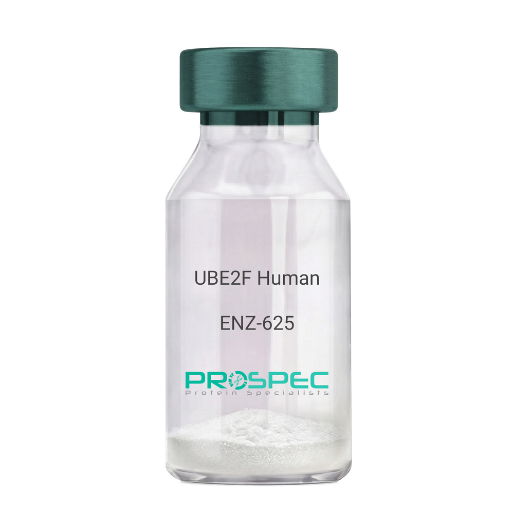
UBE2F HumanDescription:
Ubiquitin-Conjugating Enzyme E2F Human Recombinant
Ubiquitin-conjugating enzyme E2F (putative), NEDD8 carrier protein UBE2F, NEDD8 protein ligase UBE2F, NEDD8-conjugating enzyme 2, NEDD8-conjugating enzyme UBE2F, NCE2, EC 6.3.2.-.
Product # :
ENZ-625Price :
Quantity :
Shipping Method :
Shipped with Ice Packs
More Info
- description
- source
- formulation
- purity
- More Info
- Citations
Description
UBE2F Human Recombinant produced in E. coli is a single polypeptide chain containing 209 amino acids (1-185) and having a molecular mass of 23.7 kDa.UBE2F is fused to a 24 amino acid His-tag at N-terminus & purified by proprietary chromatographic techniques.
Source
E.coli.
Formulation
The UBE2F solution (1mg/1ml) contains 20mM Tris-HCl buffer (pH 8.0), 200mM NaCl, 1mM DTT and 30% glycerol.
Purity
Greater than 95% as determined by SDS-PAGE.
Citations
More Info
-
Introduction
Ubiquitin-conjugating enzyme E2 F (UBE2F) is a member of the ubiquitin-conjugating enzyme family and UBE2F subfamily. Ubiquitin-conjugating enzymes or sometimes as ubiquitin-carrier enzymes, run the second step in the ubiquitination reaction which targets a protein for degradation using the proteasome. The UBE2F receives the ubiquitin-like protein NEDD8 from the UBA3-NAE1 E1 complex and catalyzes its covalent attachment to other proteins.
RBX2-UBE2F complex neddylates specific target proteins, for example CUL5 as can be seen from the specific interaction with the E3 ubiquitin ligase RBX2, but not RBX1. -
Synonyms
Ubiquitin-conjugating enzyme E2F (putative), NEDD8 carrier protein UBE2F, NEDD8 protein ligase UBE2F, NEDD8-conjugating enzyme 2, NEDD8-conjugating enzyme UBE2F, NCE2, EC 6.3.2.-.
-
Physical Appearance
Sterile Filtered colorless solution.
-
Stability
Store at 4°C if entire vial will be used within 2-4 weeks. Store, frozen at -20°C for longer periods of time. For long term storage it is recommended to add a carrier protein (0.1% HSA or BSA).Avoid multiple freeze-thaw cycles.
-
Amino Acid Sequence
MGSSHHHHHH SSGLVPRGSH MGSHMLTLAS KLKRDDGLKG SRTAATASDS TRRVSVRDKL LVKEVAELEA NLPCTCKVHF PDPNKLHCFQ LTVTPDEGYY QGGKFQFETE VPDAYNMVPP KVKCLTKIWH PNITETGEIC LSLLREHSID GTGWAPTRTL KDVVWGLNSL FTDLLNFDDP LNIEAAEHHL RDKEDFRNKV DDYIKRYAR
ProSpec's products are furnished for LABORATORY RESEARCH USE ONLY. The product may not be used as drugs, agricultural or pesticidal products, food additives or household chemicals.
- View Data Sheet
Name :
sRANKL MouseDescription:
RANK Ligand Soluble Mouse Recombinant
Soluble Receptor Activator of NFkB Ligand, TNFSF11, TRANCE, TNF-related activation-induced cytokine, OPGL, ODF, Osteoclast differentiation factor, Tumor necrosis factor ligand superfamily member 11, Receptor activator of nuclear factor kappa B ligand, RANKL, Osteoprotegerin ligand, CD254 antigen, sRANKL, sOdf.
Product # :
CYT-320Price :
Quantity :
Shipping Method :
Shipped at Room temp
More Info
- description
- source
- formulation
- purity
- biological activity
- More Info
- Citations
Description
sRANKL Mouse Recombinant produced in E.coli is single, non-glycosylated, polypeptide chain containing 174 amino acids ( 143-316 a.a.) and having a total molecular mass of 19.9kDa. CD254 is purified by proprietary chromatographic techniques.
Source
Escherichia Coli.
Formulation
The protein (1mg/ml) was lyophilized with 10mM Na2PO4, pH 7.5 & 50mM NaCl.
Purity
Greater than 95.0% as determined by SDS-PAGE.
Biological Activity
Measured by its ability to induce osteoclast formation on murine RAW264.7 cells using a concentration of 50ng/ml shown in “Corning® Osteo Assay Surface 24 Well Plates with Transwell® Permeable Supports- A Useful Tool for Co-Culture Studies” by Rebecca M. Wood and Mark Rothenber, corresponding to a specific activity of 20,000Units/mg.
Citations
More Info
-
Introduction
RANKL binds to tnfrsf11b/opg and to tnfrsf11a/rank. Osteoclast differentiation and activation factor. augments the ability of dendritic cells to stimulate naive t-cell proliferation. May be an important regulator of interactions between t-cells and dendritic cells and may play a role in the regulation of the t-cell-dependent immune response. sRANKL may also play an important role in enhanced bone-resorption in humoral hypercalcemia of malignancy.
-
Synonyms
Soluble Receptor Activator of NFkB Ligand, TNFSF11, TRANCE, TNF-related activation-induced cytokine, OPGL, ODF, Osteoclast differentiation factor, Tumor necrosis factor ligand superfamily member 11, Receptor activator of nuclear factor kappa B ligand, RANKL, Osteoprotegerin ligand, CD254 antigen, sRANKL, sOdf.
-
Physical Appearance
Sterile Filtered White lyophilized (freeze-dried) powder.
-
Stability
Lyophilized TNFSF11 although stable at room temperature for 3 weeks, should be stored desiccated below -18°C. Upon reconstitution sRANKL should be stored at 4°C between 2-7 days and for future use below -18°C. For long term storage it is recommended to add a carrier protein (0.1% HSA or BSA). Please prevent freeze-thaw cycles.
-
Solubility
It is recommended to reconstitute the lyophilized sRANKL in sterile 18MΩ-cm H2O not less than 100µg/ml, which can then be further diluted to other aqueous solutions.
-
Amino Acid Sequence
PAMMEGSWLD VAQRGKPEAQ PFAHLTINAA SIPSGSHKVT LSSWYHDRGW AKISNMTLSN GKLRVNQDGF YYLYANICFR HHETSGSVPT DYLQLMVYVV KTSIKIPSSH NLMKGGSTKN WSGNSEFHFY SINVGGFFKL RAGEEISIQV SNPSLLDPDQ DATYFGAFKV QDID.
ProSpec's products are furnished for LABORATORY RESEARCH USE ONLY. The product may not be used as drugs, agricultural or pesticidal products, food additives or household chemicals.
- View Data Sheet
Name :
POR Human, ActiveDescription:
P450 Oxidoreductase Human Recombinant, Active
P450 (Cytochrome) Oxidoreductase, EC 1.6.2.4, CYPOR, P450R, CPR, NADPH-Dependent Cytochrome P450 Reductase, NADPH--Cytochrome P450 Reductase, NADPH--cytochrome P450 reductase.
Product # :
ENZ-1176Price :
Quantity :
Shipping Method :
Shipped with Ice Packs
More Info
- description
- source
- formulation
- purity
- biological activity
- More Info
- Citations
Description
POR produced in Sf9 Baculovirus cells is a single, glycosylated polypeptide chain containing 686 amino acids (1-680a.a.) and having a molecular mass of 77.9kDa. POR is expressed with a 6 amino acid His tag at C-Terminus and purified by proprietary chromatographic techniques.
Source
Sf9, Baculovirus cells.
Formulation
POR protein solution (0.25mg/ml) contains Phosphate buffer saline (pH 7.4) and 10% glycerol.
Purity
Greater than 95.0% as determined by SDS-PAGE.
Biological Activity
Specific activity is > 4,000 pmol/min/mg. Defined by the amount of enzyme that reduction of 1 pmole cytochrome-C by NADPH/min. at pH-8 25C.
Citations
More Info
-
Introduction
P450 Oxidoreductase, also known as POR is a flavoprotein which contributes electrons to all microsomal P450 enzymes. POR is localized to the endoplasmic reticulum, where it is also capable of transfering electrons to heme oxygenase as well as cytochrome b5. POR is structurally related to two separate flavoprotein families; first one is ferredoxin nucleotide reductase and the second flavodoxin.
-
Synonyms
P450 (Cytochrome) Oxidoreductase, EC 1.6.2.4, CYPOR, P450R, CPR, NADPH-Dependent Cytochrome P450 Reductase, NADPH--Cytochrome P450 Reductase, NADPH--cytochrome P450 reductase.
-
Physical Appearance
Sterile filtered colorless solution.
-
Stability
Store at 4°C if entire vial will be used within 2-4 weeks. Store, frozen at -20°C for longer periods of time. For long term storage it is recommended to add a carrier protein (0.1% HSA or BSA).Avoid multiple freeze-thaw cycles.
-
Amino Acid Sequence
MINMGDSHVD TSSTVSEAVA EEVSLFSMTD MILFSLIVGL LTYWFLFRKK KEEVPEFTKI QTLTSSVRES SFVEKMKKTG RNIIVFYGSQ TGTAEEFANR LSKDAHRYGM RGMSADPEEY DLADLSSLPE IDNALVVFCM ATYGEGDPTD NAQDFYDWLQ ETDVDLSGVK FAVFGLGNKT YEHFNAMGKY VDKRLEQLGA QRIFELGLGD DDGNLEEDFI TWREQFWLAV CEHFGVEATG EESSIRQYEL VVHTDIDAAK VYMGEMGRLK SYENQKPPFD AKNPFLAAVT TNRKLNQGTE RHLMHLELDI SDSKIRYESG DHVAVYPAND SALVNQLGKI LGADLDVVMS LNNLDEESNK KHPFPCPTSY RTALTYYLDI TNPPRTNVLY ELAQYASEPS EQELLRKMAS SSGEGKELYL SWVVEARRHI LAILQDCPSL RPPIDHLCEL LPRLQARYYS IASSSKVHPN SVHICAVVVE YETKAGRINK GVATNWLRAK EPVGENGGRA LVPMFVRKSQ FRLPFKATTP VIMVGPGTGV APFIGFIQER AWLRQQGKEV GETLLYYGCR RSDEDYLYRE ELAQFHRDGA LTQLNVAFSR EQSHKVYVQH LLKQDREHLW KLIEGGAHIY VCGDARNMAR DVQNTFYDIV AELGAMEHAQ AVDYIKKLMT KGRYSLDVWS HHHHHH.
ProSpec's products are furnished for LABORATORY RESEARCH USE ONLY. The product may not be used as drugs, agricultural or pesticidal products, food additives or household chemicals.
- View Data Sheet
Name :
SPP1 Human, ActiveDescription:
Osteopontin Human Recombinant, BioActive
OPN, SPP-1, BNSP, BSPI, ETA-1, Bone sialoprotein 1, BSP I.Early T lymphocyte activation 1, ETA 1, ETA1, MGC110940, Nephropontin, Secreted phosphoprotein 1, SPP 1, SPP1, urinary stone protein, uropontin.
Product # :
CYT-1166Price :
Quantity :
Shipping Method :
Shipped with Ice Packs
More Info
- description
- source
- formulation
- purity
- biological activity
- More Info
- Citations
Description
SPP1 Human Recombinant produced in E.Coli is a single, non-glycosylated polypeptide chain containing 321 amino acids (17-314 a.a.) and having a molecular mass of 36.2kDa. SPP1 is fused to a 23 amino acid His tag at N-Terminus and purified by proprietary chromatographic techniques.
Source
Escherichia Coli.
Formulation
The SPP1 solution (1mg/ml) contains 20mM Tris-HCl buffer (pH 7.5), 1mM DTT, 10% glycerol and 2mM EDTA.
Purity
Greater than 90.0% as determined by SDS-PAGE.
Biological Activity
Measured by the ability of the immobilized protein to support the adhesion of HEK293 human embryonic kidney cells. When cells are added to OPN coated plates 10ug/ml. This effect is more to 40%.
Citations
More Info
-
Introduction
Osteopontin is a glycoprotein that was primarilyfound in osteoblasts and takes part in bone remodeling, immune functions in fibroblasts, macrophages, & lymphocytes during inflammation and wound healing. SPP1 highlybinds to hydroxyapatite. SPP1 forms an integral part of the mineralized matrix. SPP1 is vital to cell-matrix interaction.
Secreted Phosphoprotein-1 protects against cardiac ischemia-reperfusion injury through late preconditioning. Expression of Ostepontin and CD44 in hepatocellular carcinoma is linked to advanced tumor stage &leads to prognosis information. SPP1 is the most over-expressed gene in intrahepatic cholangiocarcinoma. Secreted Phosphoprotein-1 overexpression is related with interstitial lung diseases. -
Synonyms
OPN, SPP-1, BNSP, BSPI, ETA-1, Bone sialoprotein 1, BSP I.Early T lymphocyte activation 1, ETA 1, ETA1, MGC110940, Nephropontin, Secreted phosphoprotein 1, SPP 1, SPP1, urinary stone protein, uropontin.
-
Physical Appearance
Sterile Filtered clear colorless solution.
-
Stability
Store at 4°C if entire vial will be used within 2-4 weeks. Store, frozen at -20°C for longer periods of time. For long term storage it is recommended to add a carrier protein (0.1% HSA or BSA).Avoid multiple freeze-thaw cycles.
-
Amino Acid Sequence
MGSSHHHHHH SSGLVPRGSH RSMIPVKQAD SGSSEEKQLY NKYPDAVATW LNPDPSQKQN LLAPQNAVSS EETNDFKQET LPSKSNESHD HMDDMDDEDD DDHVDSQDSI DSNDSDDVDD TDDSHQSDES HHSDESDELV TDFPTDLPAT EVFTPVVPTV DTYDGRGDSV VYGLRSKSKK FRRPDIQYPD ATDEDITSHM ESEELNGAYK AIPVAQDLNA PSDWDSRGKD SYETSQLDDQ SAETHSHKQS RLYKRKANDE SNEHSDVIDS QELSKVSREF HSHEFHSHED MLVVDPKSKE EDKHLKFRIS HELDSASSEV N.
ProSpec's products are furnished for LABORATORY RESEARCH USE ONLY. The product may not be used as drugs, agricultural or pesticidal products, food additives or household chemicals.
- View Data Sheet
Name :
FGF5 HumanDescription:
Fibroblast Growth Factor-5 Human Recombinant
Fibroblast Growth Factor 5, Heparin-Binding Growth Factor 5, Smag-82, HBGF-5, TCMGLY, FGF-5, FGF5.
Product # :
CYT-957Price :
Quantity :
Shipping Method :
Shipped at Room temp
More Info
- description
- source
- formulation
- purity
- More Info
- Citations
Description
FGF5 Human Recombinant produced in E.coli is a single, non-glycosylated polypeptide chain having containing 252 amino acids and having a molecular mass of 27.7kDa.The FGF-5 is purified by proprietary chromatographic techniques.
Source
Escherichia Coli.
Formulation
FGF-5 protein was lyophilized from a 0.2µm filtered solution in 10mM sodium phosphate and 100mM sodium chloride pH 7.5.
Purity
Greater than 95.0% as determined by SDS-PAGE.
Citations
More Info
-
Introduction
Fibroblast Growth Factor-5 (FGF5) belongs to the FGF family of mitogenic peptides. In vitro, rhFGF5 is a mitogen for Balb/3T3 fibroblasts and bovine heart endothelial cells. FGF5 is also a major muscle-derived survival factor for cultured spinal motoneurons. In vivo, FGF5 is assumed to play central roles in both embryology and neurobiology. Developmentally, FGF5 mRNA is originally found in the embryoblast followed by the lateral somatic mesoderm, where it may play a part in angiogenesis, as well as the myotomes cranial to the tail region, where it may delay terminal myoblast differentiation during cell migration.
-
Synonyms
Fibroblast Growth Factor 5, Heparin-Binding Growth Factor 5, Smag-82, HBGF-5, TCMGLY, FGF-5, FGF5.
-
Physical Appearance
Sterile Filtered White lyophilized (freeze-dried) powder.
-
Stability
Lyophilized FGF5 although stable at room temperature for 3 weeks, should be stored desiccated below -18°C. Upon reconstitution FGF-5 should be stored at 4°C between 2-7 days and for future use below -18°C. For long term storage it is recommended to add a carrier protein (0.1% HSA or BSA). Please prevent freeze-thaw cycles.
-
Solubility
It is recommended to reconstitute the lyophilized FGF5 in sterile 18M-cm H2O not less than 100µg/ml, which can then be further diluted to other aqueous solutions.
-
Amino Acid Sequence
MAWAHGEKRL APKGQPGPAA TDRNPRGSSS RQSSSSAMSS SSASSSPAAS LGSQGSGLEQ SSFQWSPSGR RTGSLYCRVG IGFHLQIYPD GKVNGSHEAN MLSVLEIFAV SQGIVGIRGV FSNKFLAMSK KGKLHASAKF TDDCKFRERF QENSYNTYAS AIHRTEKTGR EWYVALNKRG KAKRGCSPRV KPQHISTHFL PRFKQSEQPE LSFTVTVPEK KKPPSPIKSK IPLSAPRKNT NSVKYRLKFR FG.
-
Background
What is the molecular weight/Mw of FGF5 HUMAN Protein?
FGF5 HUMAN Protein has a total Mw of 27.7kDa.
What is the source or expression system of FGF5 HUMAN Protein?
Escherichia Coli.
What is the Purity of FGF5 HUMAN Protein?
FGF5 HUMAN Protein is >95% pure as determined by SDS-PAGE.
What is the Biological Activity of FGF5 HUMAN Protein?
The biological functionality of FGF5 HUMAN Protein will be determined in the future.
What is the amino acid sequence of FGF5 HUMAN Protein?
MAWAHGEKRL APKGQPGPAA TDRNPRGSSS RQSSSSAMSS SSASSSPAAS LGSQGSGLEQ SSFQWSPSGR RTGSLYCRVG IGFHLQIYPD GKVNGSHEAN MLSVLEIFAV SQGIVGIRGV FSNKFLAMSK KGKLHASAKF TDDCKFRERF QENSYNTYAS AIHRTEKTGR EWYVALNKRG KAKRGCSPRV KPQHISTHFL PRFKQSEQPE LSFTVTVPEK KKPPSPIKSK IPLSAPRKNT NSVKYRLKFR FG.
What applications can FGF5 HUMAN Protein be used in?
FGF5 HUMAN Protein can probably be used in western blot, ELISA and Lateral Flow.
What is the endotoxin level for FGF5 HUMAN Protein?
The endotoxin level is minimal, FGF5 HUMAN Protein was purified using conventional chromatography techniques.
ProSpec's products are furnished for LABORATORY RESEARCH USE ONLY. The product may not be used as drugs, agricultural or pesticidal products, food additives or household chemicals.
- View Data Sheet
Name :
G CSF MouseDescription:
Granulocyte-Colony Stimulating Factor Mouse Recombinant
CSF3, MGI-1G, GM-CSF beta, Pluripoietin, G-CSF, GCSF.
Product # :
CYT-410Price :
Quantity :
Shipping Method :
Shipped at Room temp
More Info
- description
- source
- formulation
- purity
- biological activity
- More Info
- Citations
Description
Granulocyte Colony Stimulating Factor Mouse Recombinant produced in E.coli is a single, non-glycosylated, polypeptide chain containing 178 amino acids and having a molecular mass of approximately 18.9kDa. G-CSF is purified by proprietary chromatographic techniques.
Source
Escherichia Coli.
Formulation
G-CSF Lyophilized from 10mM NaCitrate, pH 4.0 and 150mM NaCl.
Purity
Greater than 98.0% as determined by:
(a) Analysis by RP-HPLC.
(b) Analysis by SDS-PAGE.Biological Activity
The ED50 as determined by a cell proliferation assay using murine NFS-60 cells is < 0.05 ng/ml, corresponding to a Specific Activity of 2 x 10⁷IU/mg.Citations
More Info
-
Introduction
Granulocyte Colony Stimulating Factor is a growth factor and/or cytokine produced by the endothelium, macrophages and a number of other immune cells. GCSF stimulates the bone marrow to produce granulocytes and also to stimulate the survival, proliferation, differentiation and function of neutrophil granulocyte progenator cells and mature neutrophils.
-
Synonyms
CSF3, MGI-1G, GM-CSF beta, Pluripoietin, G-CSF, GCSF.
-
Physical Appearance
Sterile Filtered White lyophilized (freeze-dried) powder.
-
Stability
Lyophilized Granulocyte Colony Stimulating Factor although stable at room temperature for 3 weeks, should be stored desiccated below -18°C. Upon reconstitution GCSF should be stored at 4°C between 2-7 days and for future use below -18°C.For long term storage it is recommended to add a carrier protein (0.1% HSA or BSA).Please prevent freeze-thaw cycles.
-
Solubility
It is recommended to reconstitute the lyophilized Granulocyte Colony Stimulating Factor in sterile 18MΩ-cm H2O not less than 100µg/ml, which can then be further diluted to other aqueous solutions.
-
Amino Acid Sequence
VPLVTVSAL PPSLPLPRSF LLKSLEQVRK IQASGSVLLE QLCATYKLCH PEELVLLGHS LGIPKASLSG CSSQALQQTQ CLSQLHSGLC LYQGLLQALS GISPALAPTL DLLQLDVANF ATTIWQQMEN LGVAPTVQPT QSAMPAFTSA FQRRAGGVLA ISYLQGFLET ARLALHHLA.
-
Background
What is the molecular weight/Mw of G CSF MOUSE Protein?
G CSF MOUSE Protein has a total Mw of 18.9kDa.
What is the source or expression system of G CSF MOUSE Protein?
Escherichia Coli.
What is the Purity of G CSF MOUSE Protein?
G CSF MOUSE Protein is >98% pure as determined by SDS-PAGE.
What is the Biological Activity of G CSF MOUSE Protein?
The ED50 as determined by a cell proliferation assay using murine NFS-60 cells is < 0.05 ng/ml, corresponding to a Specific Activity of 2 x 10⁷IU/mg.
What is the amino acid sequence of G CSF MOUSE Protein?
VPLVTVSAL PPSLPLPRSF LLKSLEQVRK IQASGSVLLE QLCATYKLCH PEELVLLGHS LGIPKASLSG CSSQALQQTQ CLSQLHSGLC LYQGLLQALS GISPALAPTL DLLQLDVANF ATTIWQQMEN LGVAPTVQPT QSAMPAFTSA FQRRAGGVLA ISYLQGFLET ARLALHHLA.
What applications can G CSF MOUSE Protein be used in?
G CSF MOUSE Protein can probably be used in western blot, ELISA and Lateral Flow.
What is the endotoxin level for G CSF MOUSE Protein?
The endotoxin level is minimal, G CSF MOUSE Protein was purified using conventional chromatography techniques.
ProSpec's products are furnished for LABORATORY RESEARCH USE ONLY. The product may not be used as drugs, agricultural or pesticidal products, food additives or household chemicals.
- View Data Sheet
Name :
EBI3 MacaqueDescription:
Epstein Barr Virus Induced 3 Macaque Recombinant
IL-27B, IL27B, IL 27-B, EBI-3, Interleukin-27 beta, IL-27 subunit beta, Epstein-Barr virus-induced gene 3 protein homolog, EBI3.
Product # :
CYT-1036Price :
Quantity :
Shipping Method :
Shipped at Room temp
More Info
- description
- source
- formulation
- purity
- More Info
- Citations
Description
EBI3 Macaque Recombinant produced in E.Coli is a single, non-glycosylated polypeptide chain containing 210 amino acids and having a molecular mass of 23.4kDa. The EBI3 is purified by proprietary chromatographic techniques.
Source
Escherichia Coli.
Formulation
Lyophilized from a sterile filtered solution containing 0.1 % trifluoroacetic acid (TFA).
Purity
Greater than 95.0% as determined by SDS-PAGE.
Citations
More Info
-
Introduction
EBI3 has an induced expression in B lymphocytes in reaction to Epstein-Barr virus infection. EBI3 encodes a secreted glycoprotein belonging to the hematopoietin receptor family, and heterodimerizes with a 28 kDa protein to form iIL-27. EBI3 drives rapid clonal expansion of naive cd4(+) t-cells. EBI3 strongly synergizes with IL-12 to activate IFN-gamma production of naive cd4(+) t-cells. EBI3 mediates its biologic effects through the cytokine receptor wsx-1/tccr.
-
Synonyms
IL-27B, IL27B, IL 27-B, EBI-3, Interleukin-27 beta, IL-27 subunit beta, Epstein-Barr virus-induced gene 3 protein homolog, EBI3.
-
Physical Appearance
Sterile Filtered White lyophilized (freeze-dried) powder.
-
Stability
Lyophilized EBI3 although stable at room temperature for 3 weeks, should be stored desiccated below -18°C. Upon reconstitution EBI3 Macaque should be stored at 4°C between 2-7 days and for future use below -18°C. For long term storage it is recommended to add a carrier protein (0.1% HSA or BSA). Please prevent freeze-thaw cycles.
-
Solubility
It is recommended to reconstitute the lyophilized EBI3 in sterile 18MΩ-cm H2O at a concentration of 100µg/ml, which can then be further diluted to other aqueous solutions.
-
Amino Acid Sequence
MRKGPPAALT LPRVQCRAPR YPIAVDCSWT LPPAPNSTSP VSFIATYRFG MAARGHSWPC LQQTPASTSC TIADVRLFSM APYVLNVTAV HPWGSSSSFV PFIAEHIIKP DPPEGVRLSP LAERQLQVQW EPPRSWPFPE IFSLKYWIRY KRQGAARFHQ VGPIEATSFI LRAVRPRARY CVQVAAQDLT DYGELSDWSL PATTPMSPGK.
-
Background
What is the molecular weight/Mw of EBI3 Protein?
EBI3 Protein has a total Mw of 23.4kDa.
What is the source or expression system of EBI3 Protein?
Escherichia Coli.
What is the Purity of EBI3 Protein?
EBI3 Protein is >90% pure as determined by SDS-PAGE.
What is the Biological Activity of EBI3 Protein?
The biological functionality of EBI3 Protein will be determined in the future.
What is the amino acid sequence of EBI3 Protein?
MRKGPPAALT LPRVQCRAPR YPIAVDCSWT LPPAPNSTSP VSFIATYRFG MAARGHSWPC LQQTPASTSC TIADVRLFSM APYVLNVTAV HPWGSSSSFV PFIAEHIIKP DPPEGVRLSP LAERQLQVQW EPPRSWPFPE IFSLKYWIRY KRQGAARFHQ VGPIEATSFI LRAVRPRARY CVQVAAQDLT DYGELSDWSL PATTPMSPGK.
What applications can EBI3 Protein be used in?
EBI3 Protein can probably be used in western blot, ELISA and Lateral Flow.
What is the endotoxin level for EBI3 Protein?
The endotoxin level is minimal, EBI3 Protein was purified using conventional chromatography techniques.
ProSpec's products are furnished for LABORATORY RESEARCH USE ONLY. The product may not be used as drugs, agricultural or pesticidal products, food additives or household chemicals.
- View Data Sheet
Name :
SUOX HumanDescription:
Sulfite Oxidase Human Recombinant
Sulfite Oxidase, EC 1.8.3.1, Sulfite oxidase, mitochondrial.
Product # :
ENZ-887Price :
Quantity :
Shipping Method :
Shipped with Ice Packs
More Info
- description
- source
- formulation
- purity
- More Info
- Citations
Description
SUOX Human Recombinant produced in E.Coli is a single, non-glycosylated polypeptide chain containing 489 amino acids (80-545 a.a) and having a molecular mass of 53.9kDa. SUOX is fused to a 23 amino acid His-tag at N-terminus & purified by proprietary chromatographic techniques.
Source
E.coli.
Formulation
SUOX protein solution (1mg/ml) containing Phosphate buffered saline (pH7.4), 30% glycerol and 1mM DTT.
Purity
Greater than 90% as determined by SDS-PAGE.
Citations
More Info
-
Introduction
Sulfite oxidase, also known as SUOX is a homodimeric protein localized to the intermembrane space of mitochondria. Each subunit includes a heme domain as well as a molybdopterin-binding domain. The SUOX enzyme catalyzes the oxidation of sulfite to sulfate, the last reaction in the oxidative degradation of the sulfur amino acids cysteine and methionine. In addition, the deficiency of SUOX results in neurological abnormalities which are often fatal at an early age.
-
Synonyms
Sulfite Oxidase, EC 1.8.3.1, Sulfite oxidase, mitochondrial.
-
Physical Appearance
Sterile Filtered colorless solution.
-
Stability
Store at 4°C if entire vial will be used within 2-4 weeks. Store, frozen at -20°C for longer periods of time. For long term storage it is recommended to add a carrier protein (0.1% HSA or BSA).Avoid multiple freeze-thaw cycles.
-
Amino Acid Sequence
MGSSHHHHHH SSGLVPRGSH MGSESTHIYT KEEVSSHTSP ETGIWVTLGS EVFDVTEFVD LHPGGPSKLM LAAGGPLEPF WALYAVHNQS HVRELLAQYK IGELNPEDKV APTVETSDPY ADDPVRHPAL KVNSQRPFNA EPPPELLTEN YITPNPIFFT RNHLPVPNLD PDTYRLHVVG APGGQSLSLS LDDLHNFPRY EITVTLQCAG NRRSEMTQVK EVKGLEWRTG AISTARWAGA RLCDVLAQAG HQLCETEAHV CFEGLDSDPT GTAYGASIPL ARAMDPEAEV LLAYEMNGQP LPRDHGFPVR VVVPGVVGAR HVKWLGRVSV QPEESYSHWQ RRDYKGFSPS VDWETVDFDS APSIQELPVQ SAITEPRDGE TVESGEVTIK GYAWSGGGRA VIRVDVSLDG GLTWQVAKLD GEEQRPRKAW AWRLWQLKAP VPAGQKELNI VCKAVDDGYN VQPDTVAPIW NLRGVLSNAW HRVHVYVSP.
ProSpec's products are furnished for LABORATORY RESEARCH USE ONLY. The product may not be used as drugs, agricultural or pesticidal products, food additives or household chemicals.
- View Data Sheet
Name :
NDUFV2 HumanDescription:
NADH Dehydrogenase Flavoprotein 2 Human Recombinant
CI-24k, NADH dehydrogenase [ubiquinone] flavoprotein 2, mitochondrial, NADH-ubiquinone oxidoreductase 24 kDa subunit, NDUFV2.
Product # :
ENZ-743Price :
Quantity :
Shipping Method :
Shipped with Ice Packs
More Info
- description
- source
- formulation
- purity
- More Info
- Citations
Description
NDUFV2 Human Recombinant produced in E.Coli is a single, non-glycosylated polypeptide chain containing 240 amino acids (33-249 a.a) and having a molecular mass of 26.1kDa.NDUFV2 is fused to a 23 amino acid His-tag at N-terminus & purified by proprietary chromatographic techniques.
Source
Escherichia Coli.
Formulation
NDUFV2 protein solution (1mg/ml) containing 20mM Tris-HCl buffer (pH 8.0), 0.15M NaCl, 10% glycerol and 1mM DTT.
Purity
Greater than 90.0% as determined by SDS-PAGE.
Citations
More Info
-
Introduction
NADH Dehydrogenase Flavoprotein 2 (NDUFV2) is a 24 kDa Core subunit of the mitochondrial membrane respiratory chain NADH dehydrogenase (Complex I) which is a part of the minimal assembly required for catalysisthe. NDUFV2 is aslo involved in electron transfer. Mutations in NDUFV2 are implicated in Parkinson's disease, bipolar disorder, schizophrenia, and have been discovered in one case of early onset hypertrophic cardiomyopathy and encephalopathy.
-
Synonyms
CI-24k, NADH dehydrogenase [ubiquinone] flavoprotein 2, mitochondrial, NADH-ubiquinone oxidoreductase 24 kDa subunit, NDUFV2.
-
Physical Appearance
Sterile Filtered colorless solution.
-
Stability
Store at 4°C if entire vial will be used within 2-4 weeks. Store, frozen at -20°C for longer periods of time. For long term storage it is recommended to add a carrier protein (0.1% HSA or BSA).Avoid multiple freeze-thaw cycles.
-
Amino Acid Sequence
MGSSHHHHHH SSGLVPRGSH MGSGAGGALF VHRDTPENNP DTPFDFTPEN YKRIEAIVKN YPEGHKAAAV LPVLDLAQRQ NGWLPISAMN KVAEVLQVPP MRVYEVATFY TMYNRKPVGK YHIQVCTTTP CMLRNSDSIL EAIQKKLGIK VGETTPDKLF TLIEVECLGA CVNAPMVQIN DNYYEDLTAK DIEEIIDELK AGKIPKPGPR SGRFSCEPAG GLTSLTEPPK GPGFGVQAGL.
ProSpec's products are furnished for LABORATORY RESEARCH USE ONLY. The product may not be used as drugs, agricultural or pesticidal products, food additives or household chemicals.
- View Data Sheet
Name :
Epigen HumanDescription:
Epigen Human Recombinant
EPG, Epigen, PRO9904, ALGV3072, FLJ75542, EPGN, Epithelial mitogen.
Product # :
CYT-601Price :
Quantity :
Shipping Method :
Shipped at Room temp
More Info
- description
- source
- formulation
- purity
- biological activity
- More Info
- Citations
Description
Epigen Recombinant Human produced in E.Coli is a single, non-glycosylated, polypeptide chain containing 72 amino acids and having a molecular mass of 7.9 kDa. Epigen is purified by proprietary chromatographic techniques.
Source
Escherichia Coli.
Formulation
EPGN was lyophilized from 20mM PBS buffer pH-7.4 .
Purity
Greater than 98.0% as determined by:
(a) Analysis by RP-HPLC.
(b) Analysis by SDS-PAGE.Biological Activity
Determined by the dose-dependent stimulation of the proliferation of murine Balb/3T3 cells. The expected ED50 for this effect is less than 300 ng/ml, corresponding to a specific activity of > 3.3 ×10³ IU/mg.
Citations
More Info
-
Introduction
EPGN is an EGF-related polypeptide growth factor that signals through the ErbB receptor-1. EPGN is produced in numerous tissues, including the testis, liver, heart and in certain tumor cells. EPGN is mitogenic for fibroblasts and epithelial cells. Human EPGN is originally synthesized as a glycosylated 14.7 kDa transmembrane precursor protein, which is processed by proteolytic cleavage to produce a mature soluble sequence.
-
Synonyms
EPG, Epigen, PRO9904, ALGV3072, FLJ75542, EPGN, Epithelial mitogen.
-
Physical Appearance
Sterile Filtered White lyophilized (freeze-dried) powder.
-
Stability
Lyophilized Epigen although stable at room temperature for 3 weeks, should be stored desiccated below -18°C. Upon reconstitution EPGN should be stored at 4°C between 2-7 days and for future use below -18°C.For long term storage it is recommended to add a carrier protein (0.1% HSA or BSA).Please prevent freeze-thaw cycles.
-
Solubility
It is recommended to reconstitute the lyophilized Epigen in sterile 18MΩ-cm H2O not less than 100µg/ml, which can then be further diluted to other aqueous solutions.
-
Amino Acid Sequence
AVTVTPPITA QQADNIEGPI ALKFSHLCLE DHNSYCINGA CAFHHELEKA ICRCFTGYTG ERCEHLTLTS YA
-
Background
What is the molecular weight/Mw of EPIGEN Protein?
EPIGEN Protein has a total Mw of 7.9kDa.
What is the source or expression system of EPIGEN Protein?
Escherichia Coli.
What is the Purity of EPIGEN Protein?
EPIGEN Protein is >98% pure as determined by SDS-PAGE.
What is the Biological Activity of EPIGEN Protein?
Determined by the dose-dependent stimulation of the proliferation of murine Balb/3T3 cells. The expected ED50 for this effect is less than 300 ng/ml, corresponding to a specific activity of > 3.3 ×10³ IU/mg.
What is the amino acid sequence of EPIGEN Protein?
AVTVTPPITA QQADNIEGPI ALKFSHLCLE DHNSYCINGA CAFHHELEKA ICRCFTGYTG ERCEHLTLTS YA
What applications can EPIGEN Protein be used in?
EPIGEN Protein can probably be used in western blot, ELISA and Lateral Flow.
What is the endotoxin level for EPIGEN Protein?
The endotoxin level is minimal, EPIGEN Protein was purified using conventional chromatography techniques.
ProSpec's products are furnished for LABORATORY RESEARCH USE ONLY. The product may not be used as drugs, agricultural or pesticidal products, food additives or household chemicals.
- View Data Sheet
Name :
FHIT Human, GSTDescription:
Fragile Histidine Triad Human Recombinant, GST Tag
Bis(5'-adenosyl)-triphosphatase, EC 3.6.1.29, Diadenosine 5',5'''-P1,P3-triphosphate hydrolase, Dinucleosidetriphosphatase, AP3A hydrolase, AP3Aase, Fragile histidine triad protein, FHIT, FRA3B.
Product # :
PRO-297Price :
Quantity :
Shipping Method :
Shipped with Ice Packs
More Info
- description
- source
- formulation
- More Info
- Citations
Description
FHIT Human Recombinant full length protein expressed in E.coli, shows a 43 kDa band on SDS-PAGE. The FHIT is fused to GST-Tag and purified by proprietary chromatographic techniques.
Source
Escherichia Coli.
Formulation
FHIT in 50mM Tris-Acetate, pH7.5, 1mM EDTA and 20% Glycerol.
Citations
More Info
-
Introduction
FHIT is a member of the histidine triad gene family. FHIT gene encodes a diadenosine 5',5'''-P1,P3-triphosphate hydrolase involved in purine metabolism. FHIT gene includes the common fragile site FRA3B on chromosome 3, where carcinogen-induced damage can lead to translocations and abnormal transcripts of this gene. The FHIT protein is a tumor suppressor with reduced or no expression in numerous types of cancer. FHIT may also act as a tumor suppressor in normal cells. Alterations and deletions of the FHIT (Fragile Histidine Triad) gene are strongly linked to the genesis and establishment of human tumors of the lung, cervix, breast, colon, stomach, and pancreas. The expression of FHIT is more often lost in cancers of individuals with familial mutations causing deficiency in DNA repair genes such as BRCA1, BRCA2 and MSH2. In vitro FHIT functions as a hydrolase that cleaves diadenosine triphosphate (Ap3A) to ADP and AMP. The FHIT -Ap3A enzyme-substrate complex seems to be the tumor suppressor signal. The restoration of FHIT expression in FHIT -deficient cancer cells leads to apoptosis, involving the intrinsic caspase pathway, in cancer-derived cells and in tumor xenografts.
-
Synonyms
Bis(5'-adenosyl)-triphosphatase, EC 3.6.1.29, Diadenosine 5',5'''-P1,P3-triphosphate hydrolase, Dinucleosidetriphosphatase, AP3A hydrolase, AP3Aase, Fragile histidine triad protein, FHIT, FRA3B.
-
Physical Appearance
Sterile Filtered clear solution.
-
Stability
Store vial at -20°C to -80°C. When stored at the recommended temperature, this protein is stable for 12 months.Please prevent freeze-thaw cycles.
-
Applications
• ELISA
• Inhibition Assays
• Western Blotting.
ProSpec's products are furnished for LABORATORY RESEARCH USE ONLY. The product may not be used as drugs, agricultural or pesticidal products, food additives or household chemicals.
- View Data Sheet
Name :
LGALS7 HumanDescription:
Galectin-7 Human Recombinant
Galectin-7, Gal-7, HKL-14, PI7, p53-induced gene 1 protein, LGALS7, PIG1, LGALS7B, GAL7, LGALS7A.
Product # :
CYT-016Price :
Quantity :
Shipping Method :
Shipped at Room temp
More Info
- description
- source
- formulation
- purity
- More Info
- Citations
Description
Galectin-7 Human Recombinant produced in E.Coli is a single, non-glycosylated, Polypeptide chain containing 136 amino acids and having a molecular mass of 15kDa.The LGALS7 is purified by proprietary chromatographic techniques.
Source
Escherichia Coli.
Formulation
LGALS7 was lyophilized from a concentrated (1mg/ml) solution in 20mM Tris, 150mM NaCl, 1mM EDTA and 5% Trehalose, pH 8.
Purity
Greater than 95.0% as determined by SDS-PAGE.
Citations
More Info
-
Introduction
Galectins are a family of animal lectins with an affinity for beta-galactosides. This family has at least 14 identified members. Galectins share similarities in the CRD (the carbohydrate recognition domain). Galectins are synthesized as cytosolic proteins. Though localized principally in the cytoplasm and lacking a classical signal peptide, galectins can also be stimulated to secretion by non-classical pathways or alternatively targeted to the nucleus. Galectins are involved in modulating cell-cell and cell-matrix interactions. Human Galectin-7 belongs to the prototypical Galectins containing a single CRD, which is initially identified in human epidermis as a monomer. The Galectin-7 expression is induced by tumor suppressor protein p53 and associated with apoptosis. Galectin-7 is a pro-apoptotic protein which functions intracellularlly upstream of JNK activation and mitochondrial cytochrome c release. The correlation of Galectin-7 with the UV-induced apoptosis of keratinocytes presents a critical mechanism in the maintenance of epidermal homeostasis. Human Galectin-7 is localized in both nucleus and cytoplasm.
-
Synonyms
Galectin-7, Gal-7, HKL-14, PI7, p53-induced gene 1 protein, LGALS7, PIG1, LGALS7B, GAL7, LGALS7A.
-
Physical Appearance
Sterile Filtered White lyophilized (freeze-dried) powder.
-
Stability
Lyophilized LGALS7 although stable at room temperature for 3 weeks, should be stored desiccated below -18°C. Upon reconstitution Galectin-7 should be stored at 4°C between 2-7 days and for future use below -18°C.Please prevent freeze-thaw cycles.
-
Solubility
It is recommended to reconstitute the lyophilized Galectin-7 in sterile distilled H2O not less than 100µg/ml, which can then be further diluted to other aqueous solutions.
-
Amino Acid Sequence
MSNVPHKSSLPEGIRPGTVLRIRGLVPPNASRFHVNLLCGEEQGSDAALHFNP
RLDTSEVVFNSKEQGSWGREERGPGVPFQRGQPFEVLIIASDDGFKAVVGDAQ
YHHFRHRLPLARVRLVEVGGDVQLDSVRIF -
Background
What is the molecular weight/Mw of LGALS7 HUMAN Protein?
LGALS7 HUMAN Protein has a total Mw of 15kDa.
What is the source or expression system of LGALS7 HUMAN Protein?
Escherichia Coli.
What is the Purity of LGALS7 HUMAN Protein?
LGALS7 HUMAN Protein is >95% pure as determined by SDS-PAGE.
What is the Biological Activity of LGALS7 HUMAN Protein?
The biological functionality of LGALS7 HUMAN Protein will be determined in the future.
What is the amino acid sequence of LGALS7 HUMAN Protein?
MSNVPHKSSLPEGIRPGTVLRIRGLVPPNASRFHVNLLCGEEQGSDAALHFNP
RLDTSEVVFNSKEQGSWGREERGPGVPFQRGQPFEVLIIASDDGFKAVVGDAQ
YHHFRHRLPLARVRLVEVGGDVQLDSVRIF
What applications can LGALS7 HUMAN Protein be used in?
LGALS7 HUMAN Protein can probably be used in western blot, ELISA and Lateral Flow.
What is the endotoxin level for LGALS7 HUMAN Protein?
The endotoxin level is minimal, LGALS7 HUMAN Protein was purified using conventional chromatography techniques.
ProSpec's products are furnished for LABORATORY RESEARCH USE ONLY. The product may not be used as drugs, agricultural or pesticidal products, food additives or household chemicals.
- View Data Sheet
Name :
KLF12 HumanDescription:
Kruppel-Like Factor 12 Human Recombinant
AP-2rep, AP2REP, HSPC122, Krueppel-like factor 12, Transcriptional repressor AP-2rep.
Product # :
PRO-2090Price :
Quantity :
Shipping Method :
Shipped with Ice Packs
More Info
- description
- source
- formulation
- purity
- More Info
- Citations
Description
KLF12 Human Recombinant produced in E.Coli is a single, non-glycosylated polypeptide chain containing 425 amino acids (1-402 a.a) and having a molecular mass of 46.6kDa.KLF12 is fused to a 23 amino acid His-tag at N-terminus & purified by proprietary chromatographic techniques.
Source
Escherichia Coli.
Formulation
KLF12 protein solution (0.5mg/ml) contains 20mM Tris-HCl buffer, (pH8.0) ,10% glycerol and 0.4M Urea.
Purity
Greater than 85.0% as determined by SDS-PAGE.
Citations
More Info
-
Introduction
Krueppel-like factor 12 (KLF12) belongs to the Kruppel-like zinc finger protein family and can repress expression of the AP-2 alpha gene by binding to a particular site in the AP-2 alpha gene promoter. AP-2 alpha (Activator protein-2 alpha) is a developmentally-regulated transcription factor and vital regulator of gene expression during vertebrate development and carcinogenesis. KLF12 gene repression entails binding with a corepressor, CtBP1.
-
Synonyms
AP-2rep, AP2REP, HSPC122, Krueppel-like factor 12, Transcriptional repressor AP-2rep.
-
Physical Appearance
Sterile Filtered clear solution.
-
Stability
Store at 4°C if entire vial will be used within 2-4 weeks. Store, frozen at -20°C for longer periods of time. For long term storage it is recommended to add a carrier protein (0.1% HSA or BSA).Avoid multiple freeze-thaw cycles.
-
Amino Acid Sequence
MGSSHHHHHH SSGLVPRGSH MGSMNIHMKR KTIKNINTFE NRMLMLDGMP AVRVKTELLE SEQGSPNVHN YPDMEAVPLL LNNVKGEPPE DSLSVDHFQT QTEPVDLSIN KARTSPTAVS SSPVSMTASA SSPSSTSTSS SSSSRLASSP TVITSVSSAS SSSTVLTPGP LVASASGVGG QQFLHIIHPV PPSSPMNLQS NKLSHVHRIP VVVQSVPVVY TAVRSPGNVN NTIVVPLLED GRGHGKAQMD PRGLSPRQSK SDSDDDDLPN VTLDSVNETG STALSIARAV QEVHPSPVSR VRGNRMNNQK FPCSISPFSI ESTRRQRRSE SPDSRKRRIH RCDFEGCNKV YTKSSHLKAH RRTHTGEKPY KCTWEGCTWK FARSDELTRH YRKHTGVKPF KCADCDRSFS RSDHLALHRR RHMLV.
ProSpec's products are furnished for LABORATORY RESEARCH USE ONLY. The product may not be used as drugs, agricultural or pesticidal products, food additives or household chemicals.